Review

Near-Infrared-II Fluorescence Imaging of Tumors with Organic Small-Molecule Fluorophores

by
Mao Guo
1,
Xiaomu Hu
2,* and
Wei Du
1,*
1
New Cornerstone Science Laboratory, MOE Key Laboratory for Analytical Science of Food Safety and Biology, College of Chemistry, Fuzhou University, Fuzhou 350108, China
2
Department of Pharmacy, 900TH Hospital of Joint Logistic Support Force of PLA, Fuzhou 350025, China
*
Authors to whom correspondence should be addressed.
Sensors 2025, 25(22), 7080; https://doi.org/10.3390/s25227080
Submission received: 15 October 2025 / Revised: 14 November 2025 / Accepted: 18 November 2025 / Published: 20 November 2025
(This article belongs to the Special Issue Fluorescence Sensors for Biological and Medical Applications)

Abstract

Over the past decade, near-infrared-II (NIR-II, 1000–1700 nm) fluorescence imaging has become a focal point in tumor imaging due to its advantages of low light scattering, weak biological autofluorescence, extraordinary penetration depth, high signal-to-background ratio, and micron-level high resolution. To date, a large number of NIR-II materials have been developed for tumor imaging. Among them, NIR-II organic small-molecule fluorophores have emerged as research hotspots owing to their distinctive advantages, such as superior optical properties, excellent controllability, favorable biocompatibility, and tunable pharmacokinetics. In this review, we summarize the latest progress in lNIR-II fluorescent probes based on organic small-molecule fluorophores for tumor imaging, focusing on their structural features, design principles of NIR-II fluorescent probes, and applications in tumor imaging. Finally, we will discuss the challenges, future prospects, and development directions of organic small-molecule fluorophores for NIR-II fluorescence imaging of tumors.

1. Introduction

Cancer remains a major public health burden characterized by high incidence, mortality, and pronounced inter- and intratumoral heterogeneity. Tumor initiation, progression, and metastasis are tightly regulated by the tumor microenvironment (TME) [1,2,3], a dynamic ecosystem comprising malignant cells, vasculature, immune cells, cancer-associated fibroblasts, diverse signaling molecules, and the extracellular matrix (ECM) [3,4]. The TME exhibits hallmark pathophysiological features, including aberrant vasculature, redox dysregulation (e.g., elevated reactive oxygen species, ROS), hypoxia, acidosis, hyperactivation of specific enzymes (e.g., matrix metalloproteinases), and abnormal expression of molecular biomarkers. These dysregulated entities—such as receptor proteins (e.g., epidermal growth factor receptor (EGFR), human epidermal growth factor receptor 2 (HER2)), tumor-associated antigens (e.g., PSA, CEA), ROS, metabolites, and nucleic acid alterations—constitute tumor-specific biomarkers and therapeutic targets, forming the foundation for molecular imaging and targeted therapy [5,6,7]. Collectively, they drive proliferation, invasion, metastasis, and therapy resistance, while complicating early detection and precise intervention [2,8]. Many cancers are curable when detected early and treated effectively. Thus, technologies for early detection, accurate localization, and real-time visualization are essential to improve diagnosis, guide surgery, assess treatment response, and monitor prognosis [9,10,11,12].
Clinically, commonly used imaging modalities include computed tomography (CT), magnetic resonance imaging (MRI), positron emission tomography (PET), and ultrasonography [13,14,15,16,17]. Each contributes uniquely to tumor diagnosis, staging, and treatment assessment: CT affords high spatial resolution, MRI provides excellent soft-tissue contrast, PET reports metabolic activity, and ultrasound offers real-time, user-friendly operation [15,18,19,20]. Nonetheless, these modalities have notable limitations: limited spatial resolution (e.g., PET), ionizing radiation exposure (e.g., CT and PET), high costs (e.g., MRI and PET), and challenges in achieving long-term, highly sensitive real-time imaging [17,19,20,21,22]. Against this backdrop, optical imaging—particularly fluorescence imaging—has gained prominence in basic and translational oncology owing to its high sensitivity, high spatiotemporal resolution, absence of ionizing radiation, and capability for multiwavelength real-time imaging [23,24,25]. Fluorescence imaging allows precise tumor localization and margin delineation, and with, specific molecular probes, reveals TME physiology and molecular signatures, thereby aiding early diagnosis, intraoperative navigation, and therapy monitoring [26,27,28,29].
Within fluorescence imaging, near-infrared-I (NIR-I, 700–900 nm) imaging has achieved meaningful advances in preclinical research and selected clinical applications [30,31,32,33]. However, strong tissue scattering and autofluorescence within this spectral window constrain penetration depth (<1 cm), diminish the signal-to-noise ratio (SNR), and limit spatial resolution, thereby hindering deep-tissue imaging [34,35]. By contrast, fluorescence imaging in the second near-infrared window (NIR-II, 1000–1700 nm) has rapidly emerged as a highly promising in vivo modality due to markedly reduced tissue scattering, minimal autofluorescence, and lower photon attenuation [36,37,38,39]. Compared with NIR-I, NIR-II offers deeper tissue penetration (up to several centimeters) and higher spatiotemporal resolution, enabling high-contrast, micrometer-scale biological imaging [40,41,42,43,44]. However, it is crucial to note that the benefits across the broad NIR-II spectrum are not uniform. Light scattering in biological tissues decreases monotonically with increasing wavelength from 400 to 1700 nm, whereas water absorption exhibits a distinct peak between 1350 and 1600 nm (Figure 1a,b) [45]. Due to the trade-off between reduced scattering and elevated water absorption, the 1000–1350 nm range is particularly favorable for deep-tissue imaging, offering lower scattering and relatively low water absorption, compared to other NIR-II regions.
A broad spectrum of emitters has been developed for NIR-II imaging, including carbon nanotubes [46,47], quantum dots [48,49,50], lanthanide-doped nanoparticles [51,52,53], and a variety of organic materials such as conjugated polymer nanoparticles (CPNs) and small-molecule fluorophores [54,55,56,57]. The potential long-term biotoxicity and metabolic retention of inorganic nanomaterials, however, substantially impede their clinical translation. In contrast, CPNs and small-molecule fluorophores offer obvious advantages, such as biocompatibility and tunable optical properties through chemical structure modulation. However, CPNs often suffer from slow metabolism and poor water solubility when used for in vivo imaging. In terms of small-molecule fluorophores, they exhibit faster and more predictable clearance than inorganic nanomaterials and CPNs, making them among the most promising NIR-II imaging agents for clinical use [58,59,60,61,62]. A notable clinical milestone was reported in 2020, when Tian et al. successfully performed NIR-II fluorescence-guided surgery in 23 patients with primary and metastatic liver cancer using indocyanine green (ICG), marking a critical step toward clinical translation of organic NIR-II fluorescent probes [63].
This review systematically summarizes recent advances in NIR-II fluorescent probes based on organic small-molecule fluorophores for tumor imaging, with emphasis on molecular structural features, rational design strategies, and applications in tumor diagnosis, real-time intraoperative navigation, and therapy-response monitoring. We conclude by discussing key challenges and future directions in this rapidly evolving field.

2. NIR-II Organic Small-Molecule Fluorophores

Substantial progress has been made in the molecular design of small-molecule fluorophores with emission in the NIR-II region. Current efforts predominantly focus on four structural archetypes: donor–acceptor–donor (D-A-D) frameworks, cyanine derivatives, boron-dipyrromethene (BODIPY) derivatives, and xanthene dyes [64,65]. These architectures afford tunable photophysical properties and strong potential for biomedical applications. Recent strategies focus on rational structural optimization to improve optical performance, achieving higher fluorescence quantum yield (QY), greater photostability, and better aqueous solubility. Such functionally refined probes are increasingly used for high-contrast in vivo tumor imaging and pathophysiological visualization [66,67]. Some representative NIR-II organic small-molecule fluorophores and their photophysical properties have been listed and summarized in Table 1.

2.1. Donor–Acceptor–Donor NIR-II Small-Molecule Fluorophores

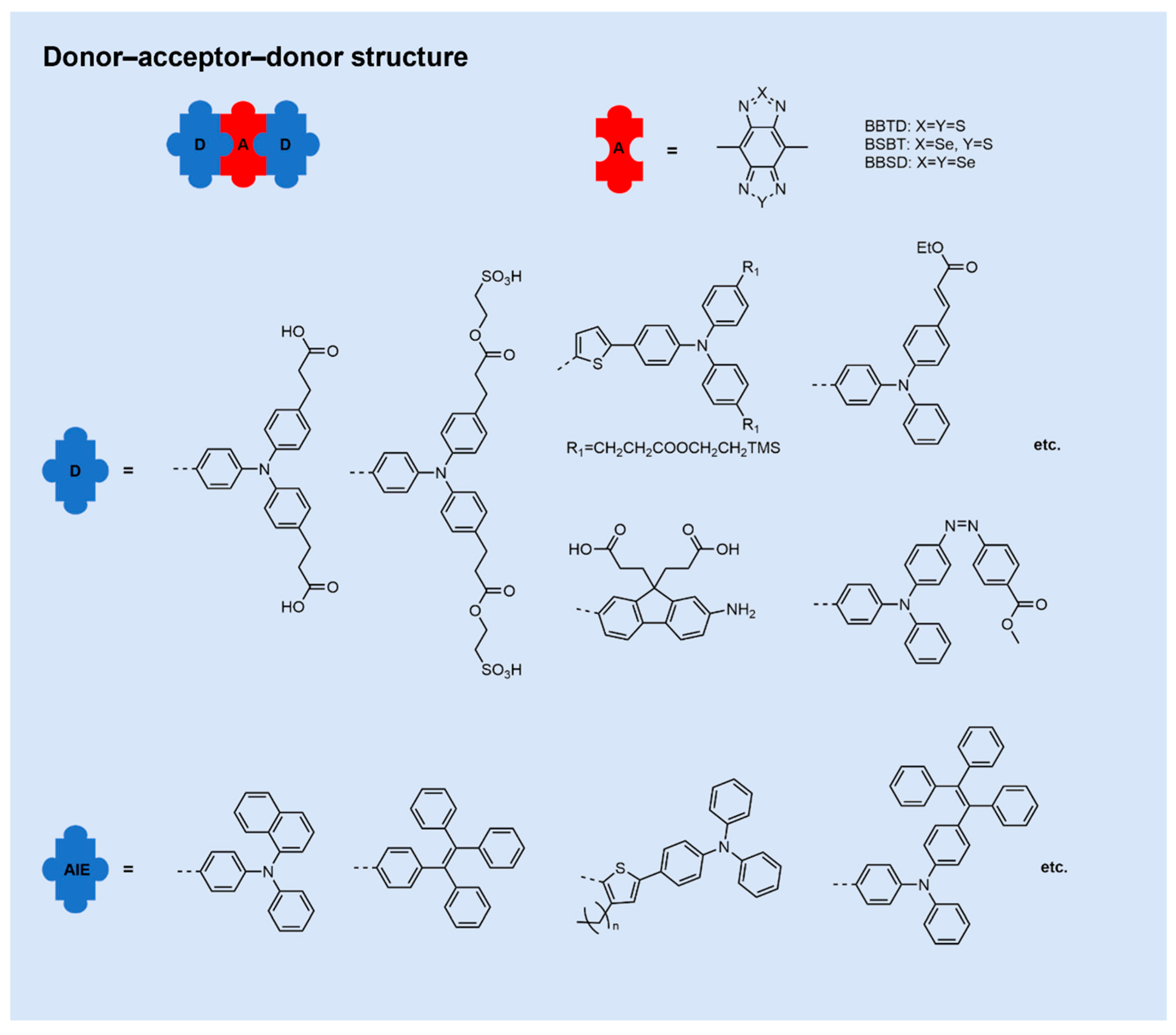
D-A-D fluorophores generally comprise an electron-accepting (A) core symmetrically connected to two electron-donating units via π-conjugated linkers (Figure 2). Among reported scaffolds, benzobisthiadiazole (BBTD) is a prototypical acceptor due to its strong electron-withdrawing character, narrow bandgap, high molar extinction coefficient, long-wavelength emission, structural tunability, and pronounced intramolecular charge-transfer (ICT) features, collectively conferring excellent photostability [65,76,77]. Alternative acceptors recently explored include [1,2,5]thiadiazolo[3,4-f]benzotriazole (TBZ) [78], thienoisoindigo (TIIG) [79], 6,7-bis(4-hexyloxyphenyl)-4,9-di(thiophen-2-yl)[1,2,5]thiadiazolo[3,4-g]quinoxaline (TTQ) [79,80], [1,2,5]thiadiazolo[3,4-g]quinoxaline (TQ), thienothiadiazole (TTD) [81,82,83], and 6,7-dithienyl [1,2,5]thiadiazolo[3,4-g]quinoxaline (TQT) [84]. Donor units commonly incorporate phenyl-based motifs such as triphenylamine (TPA), tetraphenylethylene (TPE), alkylfluorenes, or alkylbenzenes, while thiophene derivatives frequently serve as π-bridges [85].
Strategic molecular engineering tunes the electron-donating/withdrawing strengths of donor and acceptor components, thereby modulating the highest occupied molecular orbital–lowest unoccupied molecular orbital (HOMO–LUMO) gap and precisely adjusting NIR-II absorption/emission profiles [86,87,88]. Common approaches to enhance fluorescence quantum yield include: (1) Incorporation of steric-shielding units to suppress nonradiative decay [89,90]; (2) inhibition of twisted intramolecular charge-transfer (TICT) states [91]; (3) confinement within hydrophobic microenvironments to reduce solvent-induced quenching [92]; (4) construction of aggregation-induced emission (AIE)-active scaffolds [93,94]. Owing to their well-defined structures, tunable photophlysics, favorable metabolism, and excellent biocompatibility, D-A-D small molecules have emerged as pivotal candidates for organic NIR-II probes, driving intense research interest.

2.2. NIR-II Cyanine Fluorophores

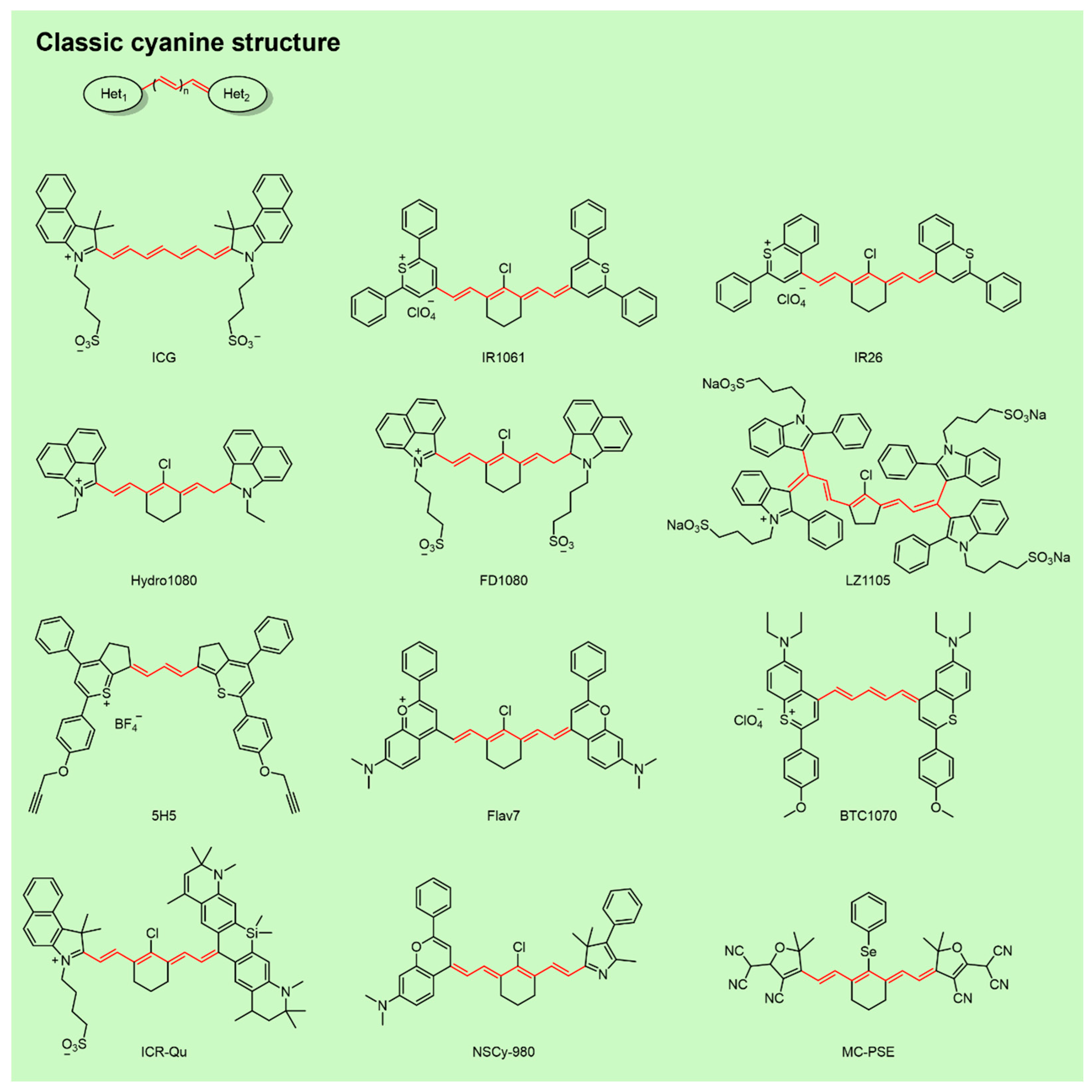
Cyanine fluorophores feature a central polymethine chain [(CH)ₙ] conjugated to terminal heterocycles containing nitrogen, sulfur, or oxygen (Figure 3) [95]. Typically, one positively charged heterocycle acts as an electron acceptor, while the opposing heterocycle serves as an electron donor, producing an intrinsic push–pull effect that extends π-conjugation. NIR-II emission commonly arises from ICT facilitated by excited-state structural distortion: upon excitation, elongation of central C–C bonds increase rotational freedom, perturbs backbone symmetry, and activates long-wavelength emission [85,96].
Bathochromic shifts in absorption/emission can be achieved by polymethine chain elongation, donor–acceptor modulation (e.g., sulfur-for-oxygen substitution), or J-aggregation, thereby improving penetration depth and spatial resolution in tissues [71,97,98]. Photostability and quantum yield can be enhanced by installing electron-withdrawing groups or steric hindrance along the polymethine bridge, and by forming protein complexes [70,74,99]. To overcome hydrophobicity and improve biocompatibility, two main strategies are employed: (1) Nanoprecipitation with amphiphilic polymers to encapsulate the dye, and (2) molecular engineering with hydrophilic substituents such as sulfonates or Poly(ethylene glycol) (PEG) chains [95,100]. With high molar extinction coefficients, tunable emissions, synthetic versatility, and modularity, cyanine fluorophores hold substantial promise for biomedical imaging and have been widely applied for in vivo tumor visualization.

2.3. NIR-II BODIPY Fluorophores

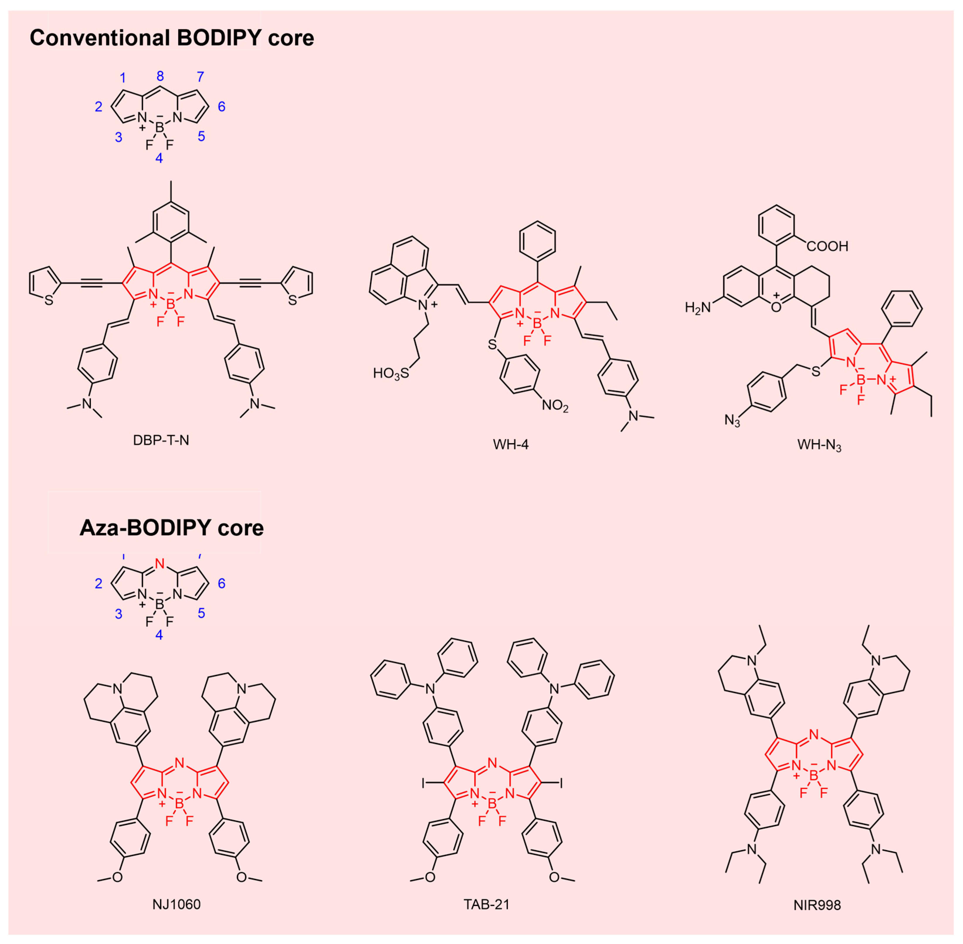
BODIPY dyes are distinguished by high fluorescence quantum yields, narrow emission bandwidths, robust photostability, synthetic versatility, minimal autofluorescence, low cytotoxicity, and favorable biocompatibility (Figure 4) [101,102,103]. The rigid, planar, highly conjugated core consists of two pyrrole rings chelated to a BF2 unit, enabling extensive site-specific modification. Based on core variation, BODIPY derivatives are broadly categorized as conventional BODIPYs or aza-BODIPYs [66].
Aza BODIPYs, formed by isoelectronic substitution of the meso carbon of BODIPYs with nitrogen, exhibit reduced HOMO–LUMO gaps and bathochromic extending into the NIR-II region [85,104], broadening their utility in deep tissue imaging and phototherapy. Hydrophilic functionalization at the 2,6-positions, combined with quaternary ammonium groups on boron, markedly improves aqueous solubility, obviating the need for extrinsic encapsulation or PEGylation [100,105,106]. The electron-deficient aza-BODIPY scaffold also supports D-A-D construction via donor installation at the 3,5- or 1,7-positions, promoting strong ICT and enabling NIR-II emission [85,107]. The inherent synthetic flexibility of the BODIPY core facilitates incorporation of functional moieties for multimodal theranostics, significantly advancing tumor-targeted precision imaging.

2.4. NIR-II Xanthene Fluorophores

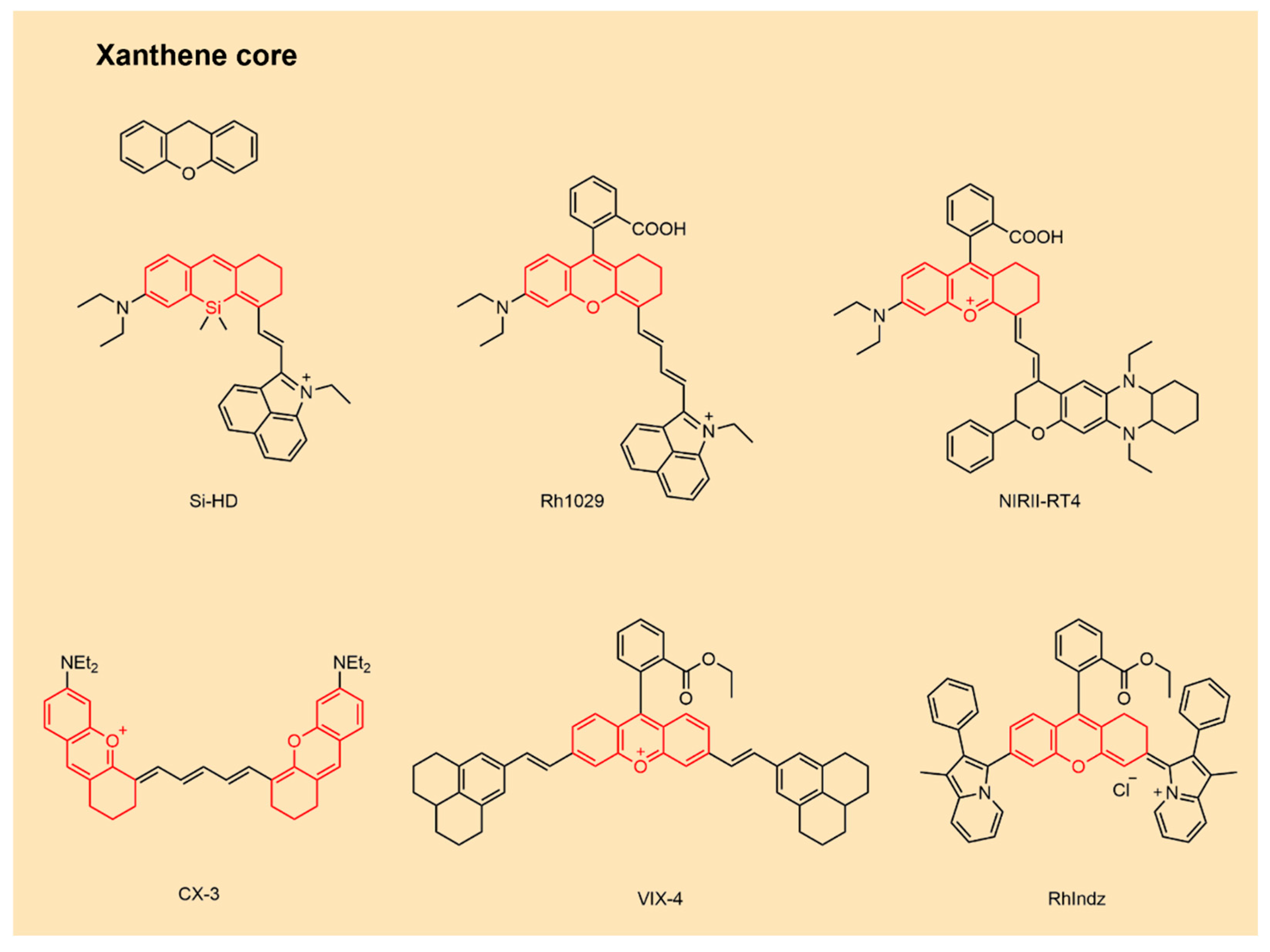
Xanthene dyes possess a tricyclic core in which two benzene rings are bridged by an oxygen atom and a methylene group, forming a six-membered heterocycle (Figure 5). This architecture affords high molar extinction coefficients, superior photostability, low cytotoxicity, high fluorescence quantum yields, facile functionalization, and tunable emission. Representative derivatives include rhodamine, fluoresceine, and related analogs [60,66,108].
Extensive efforts have targeted bathochromic shifts through π-extension strategies that expand conjugation within the xanthene scaffold. Covalent fusion of naphthalene rings (fused polycyclic aromatics) or installation of styryl groups (rigid π-bridges) effectively extends π-systems and red-shifts absorption/emission [66,75,109]. Heteroatom substitution of the central oxygen with silicon or sulfur narrows the HOMO–LUMO gap, yielding further red-shifts. Increasing molecular rigidity via planarization suppresses rotation about nitrogen centers and mitigates TICT. Incorporation of julolidine or tetrahydroquinoline (THQ) substituents enhances quantum yield and can produce large Stokes shifts by strengthening ICT [28,108].

3. Design Strategy of NIR-II Fluorescent Probes Based on Organic Small-Molecule Fluorophores for Tumor Imaging

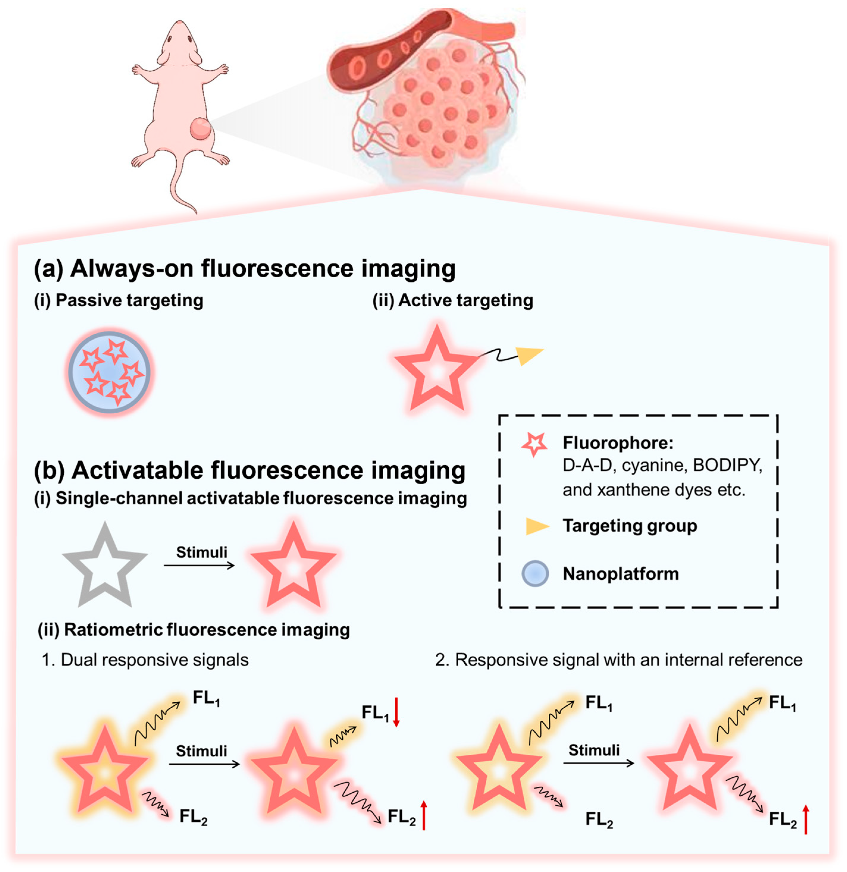
Based on the signal generation mechanism, there are two major strategies for designing NIR-II fluorescent probes for tumor imaging using organic small-molecule fluorophores (Figure 6): always-on fluorescence imaging strategy and activatable fluorescence imaging. “Turn-off” probes (for example, scaffolds quenched until biomarker interaction) are not considered here due to their susceptibility to environmental perturbations (e.g., temperature- or oxygen-induced false quenching) and their typically low SNRs [110,111]. This section focuses on probes that either exhibit intrinsically stable emission or undergo tumor-specific activation, thereby enabling reliable in vivo imaging contrast with deep-tissue penetration.

3.1. Always-On Fluorescence Imaging

Always-on fluorescence imaging strategy employs probes that maintain fluorescent throughout systemic circulation. This strategy uses tumor-targeting moieties to selectively accumulate signaling agents in tumors, creating concentration differences between malignant and healthy tissues and enabling lesion visualization through differential signal intensity. Its key advantages—no need for external activation, real-time monitoring, and predictable signal output—support intraoperative navigation. However, its imaging performance is tightly coupled to delivery efficiency, typically achieved via active or passive targeting [112,113,114]. Typically, always-on fluorescence imaging of tumors can be achieved either by passive targeting strategy or active targeting strategy (Figure 6a).
For passive targeting, hydrophobic dyes are always encapsulated within nanocarriers (e.g., DSPE-PEG micelles, DOPC liposomes, or human serum albumin (HSA)) to exploit the enhanced permeability and retention (EPR) effect in solid tumors. Rapid angiogenesis produces leaky vasculature that permits macromolecules (approximately 100–800 nm in diameter) to extravasate and accumulate in tumor tissue; concurrently, impaired lymphatic drainage promotes retention [115]. Thus, the size of passive targeting probes is always tuned to the nanometric range (typically 20–200 nm) to promote selective accumulation in tumor tissue. This strategy offers broad applicability and prolonged circulation, but may suffer from modest tumor accumulation and nonspecific uptake by the liver and spleen [116].
For active targeting, fluorophores are covalently conjugated to tumor-targeting moieties (antibodies, peptides, or small-molecule ligands). High-affinity ligand–receptor interactions or antigen–antibody recognition facilitates efficient fluorophore binding to the membrane of tumor cells [117,118,119,120,121]. Continued optimization of hydrophilicity, biocompatibility, and size will further enhance targeted accumulation and penetration–retention in vivo [114]. For example, flexible PEG linkers can help alleviate steric hindrance and minimize self-quenching, thereby improving specificity and accelerating tumor enrichment [112,122,123].

3.2. Activatable Fluorescence Imaging

Unlike always-on fluorescence imaging, activatable fluorescence imaging strategy reduces dependence on absolute probe concentration and enable more precise lesion identification [114,124,125]. A canonical activatable probe comprises three elements: a fluorophore, a linker, and a recognition moiety [126]. Commonly used recognition moieties include protease-cleavable peptide linkers (e.g., for MMP-2/9 or caspase-3) paired with quenchers (e.g., BHQ-3), pH-sensitive tertiary amines, and units responsive to redox species like ROS, reactive nitrogen species (RNS), and reactive sulfur species (RSS) [127,128,129]. Activatable fluorescent probes remain in the fluorescence “off” state until triggered by tumor-associated stimuli such as enzymes, ROS, or acidic pH, at which point they switch to an emissive (“on”) state. Activatable fluorescent probes offer ultralow background interference and microenvironment-responsive specificity, thereby improving sensitivity and selectivity. NIR-II activatable probes support noninvasive, real-time monitoring of tumor biomarkers and microenvironmental cues and are commonly implemented as single-channel or ratiometric systems (Figure 6b).
Single-channel probes recognize tumors through an on–off switching mechanism within a single emission channel upon response to specific tumor-associated stimuli [127,128]. Despite their utility, single-channel probes can be influenced by probe distribution and tissue-dependent attenuation, complicating quantitative analysis [130]. By contrast, ratiometric activatable probes provide dual-signal self-calibration to minimize variability related to instruments and environmental factors, thereby enabling more accurate semi-quantitative and quantitative measurements [114,131]. Generally, there are two different types of a ratiometric activatable fluorescence imaging strategies (Figure 6b). (1) Dual responsive signals, where a probe generates two interconnected outputs—one enhanced and one reduced upon responding to the target stimulus, enabling reversible ratiometric detection through wavelength-intensity ratios [131,132]. (2) Responsive signal with an internal reference, in which a target-sensitive channel is combined with a stable reference channel to allow real-time normalization and reliable ratiometric measurement [133].

4. Advances in NIR-II Fluorescent Probes Based on Organic Small-Molecule Fluorophores for Tumor Imaging

The development of new NIR-II organic small-molecule fluorophores and advanced molecular design strategies has established NIR-II fluorescent probes with deeper tissue penetration and higher spatiotemporal resolution, conferring distinct advantages for tumor imaging. This section highlights representative studies of NIR-II fluorescent probes based on organic small-molecule fluorophores, aiming to inform and guide the design and application of next-generation fluorescent probes for tumor imaging.

4.1. Always-On NIR-II Fluorescent Probes

The unique structural and metabolic features of tumors (e.g., aberrant angiogenesis and impaired lymphatic drainage) provide a biological basis for targeted probe design. Recent reported always-on NIR-II fluorescent probes have adopted either a passive targeting strategy or an active targeting strategy to improve targeting ability toward malignant tissues [134,135].

4.1.1. Passive Targeting Probes

NIR-II organic small-molecule fluorophores typically exhibit limited aqueous solubility and poor tumor-targeting capability. To confer passive tumor-targeting capability to these fluorophores, chemical modification and nanocarrier encapsulation represent the two primary strategies.
Chemical modification can directly optimize the physicochemical properties of NIR-II organic small-molecule fluorophores (e.g., solubility, hydrodynamic size, and surface charge), thereby prolonging circulation, reducing carrier dependence, and enhancing EPR effect utilization. PEGylation is a well-established approach to amplify the EPR effect by extending blood circulation and improving vascular permeability [136,137]. The hydrophilic PEG layer reduces opsonization and protein adsorption, thereby minimizing liver and spleen clearance. It also masks surface charge, weakens electrostatic interactions with healthy endothelium, and adjusts hydrodynamic diameter to promote extravasation.
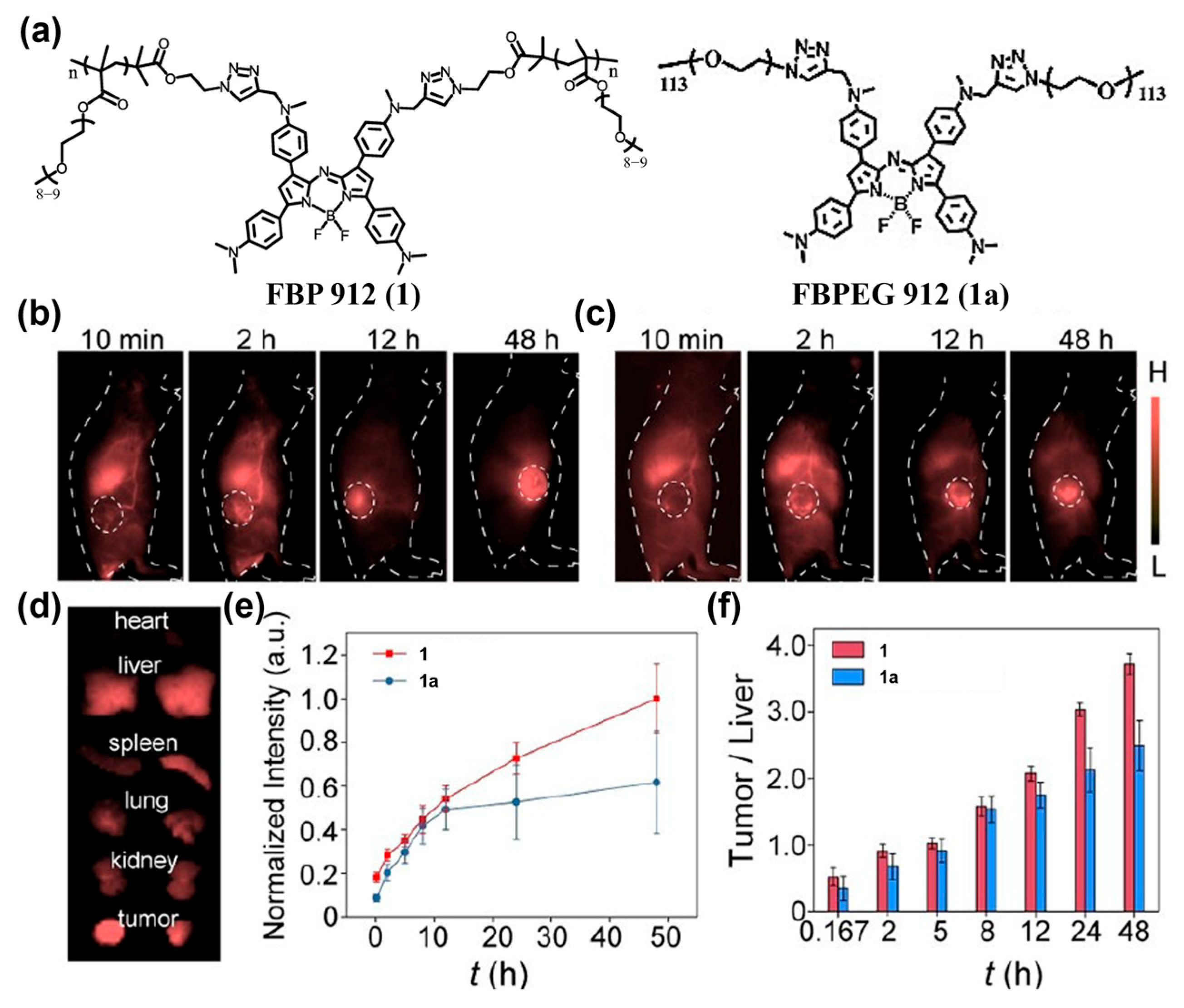
Zhang and co-workers introduced PEG chains onto an aza-BODIPY core to construct a brush-like macromolecular probe, FBP 912 (1) (Figure 7a) [138]. This polymer brush exhibits a prolonged circulation half-life (t1/2 ≈ 6.1 h); its brush architecture effectively suppresses aqueous-phase quenching, yielding substantially higher NIR-II brightness than that of a conventionally PEGylated, renally clearable control fluorophore 1a (t1/2 < 2 h). After tumor-bearing mice were intravenously (i.v.) injected with 1 and subjected to NIR-II fluorescence imaging, the tumor-to-background ratio (TBR) of 1 reached 8.2 ± 0.5 at 24 h, exceeding previous renally clearable probes and showing effective passive targeting imaging of subcutaneous tumors (Figure 7b–f).
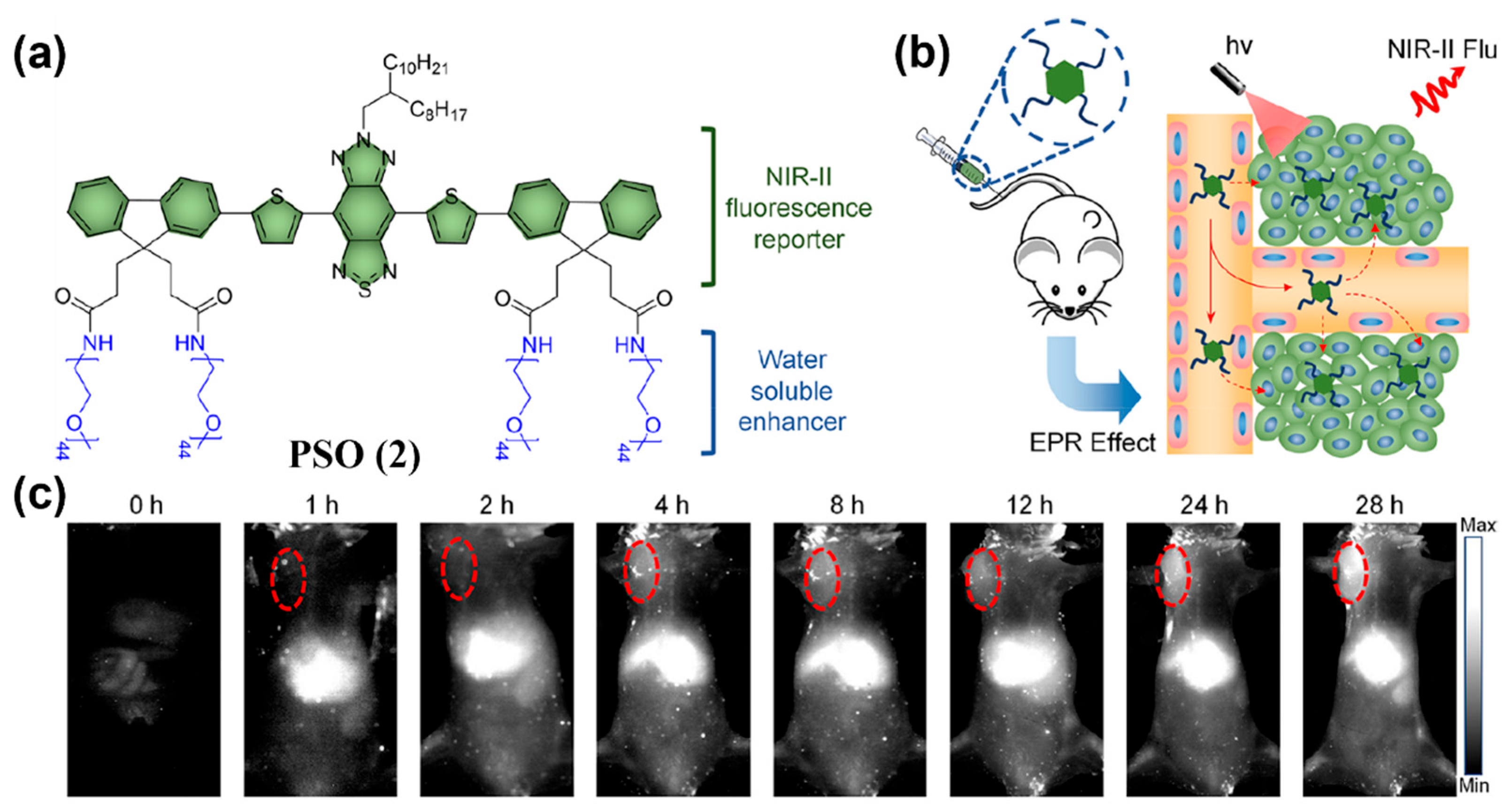
PEGylation of other NIR-II fluorophores can also endow them with passive tumor-targeting capability. Fan and colleagues appended four hydrophilic PEG chains to a selenium-oxanthene (SO)-based NIR-II emissive scaffold and obtained a water-soluble probe, PSO (2) (Figure 8a) [139]. Under passive targeting, 2 enabled clear tumor imaging as early as 4 h post-dosing (with a 5.4-fold enhancement in signal relative to surrounding tissue) and maintained strong signals at 28 h, indicating excellent NIR-II performance (Figure 8b,c). In addition, 2 exhibited favorable renal clearance (Figure 8c).
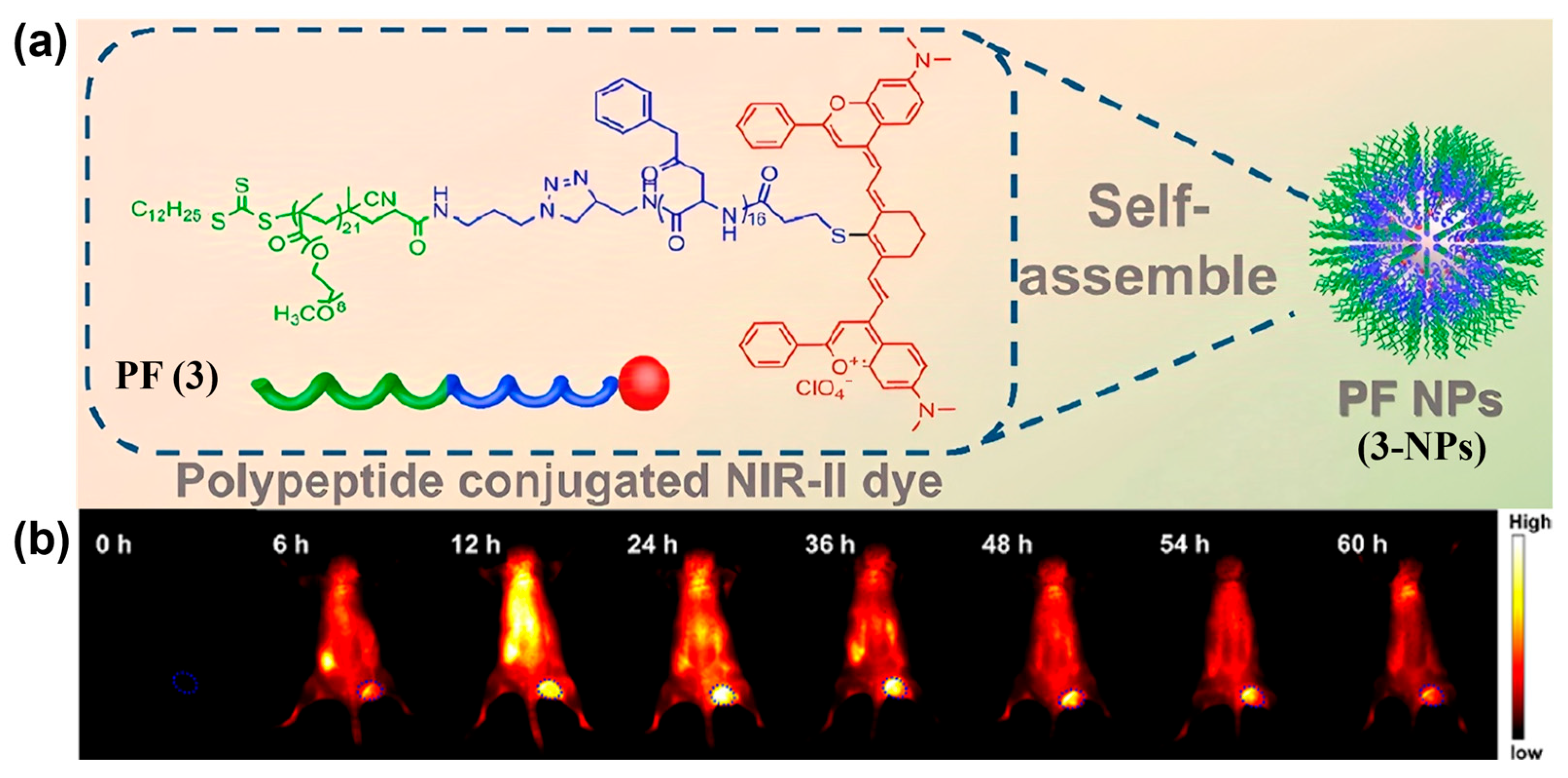
Furthermore, covalently attaching hydrophilic groups to hydrophobic dyes creates amphiphilic structures that self-assemble into stable nanoaggregates in water; precise size control within the EPR window further promotes passive targeting [140,141]. For example, Li et al. modified the small-molecule NIR-II fluorophore Flav7 with an amphiphilic peptide and constructed probe PF (3), which self-assembled into uniform micelles (i.e., 3-NPs) in water with a particle size of about 90 nm and a surface potential of −12.5 mV (Figure 9a) [142]. 3-NPs accumulated at tumor sites via the combined effects of EPR and nanomaterial-induced endothelial leakiness (NanoEL). As shown in Figure 9b, within 24 h of i.v. administration of 3-NPs, tumor fluorescence intensity and TBR continuously increased, indicating the excellent tumor-targeted imaging performance of 3-NPs.
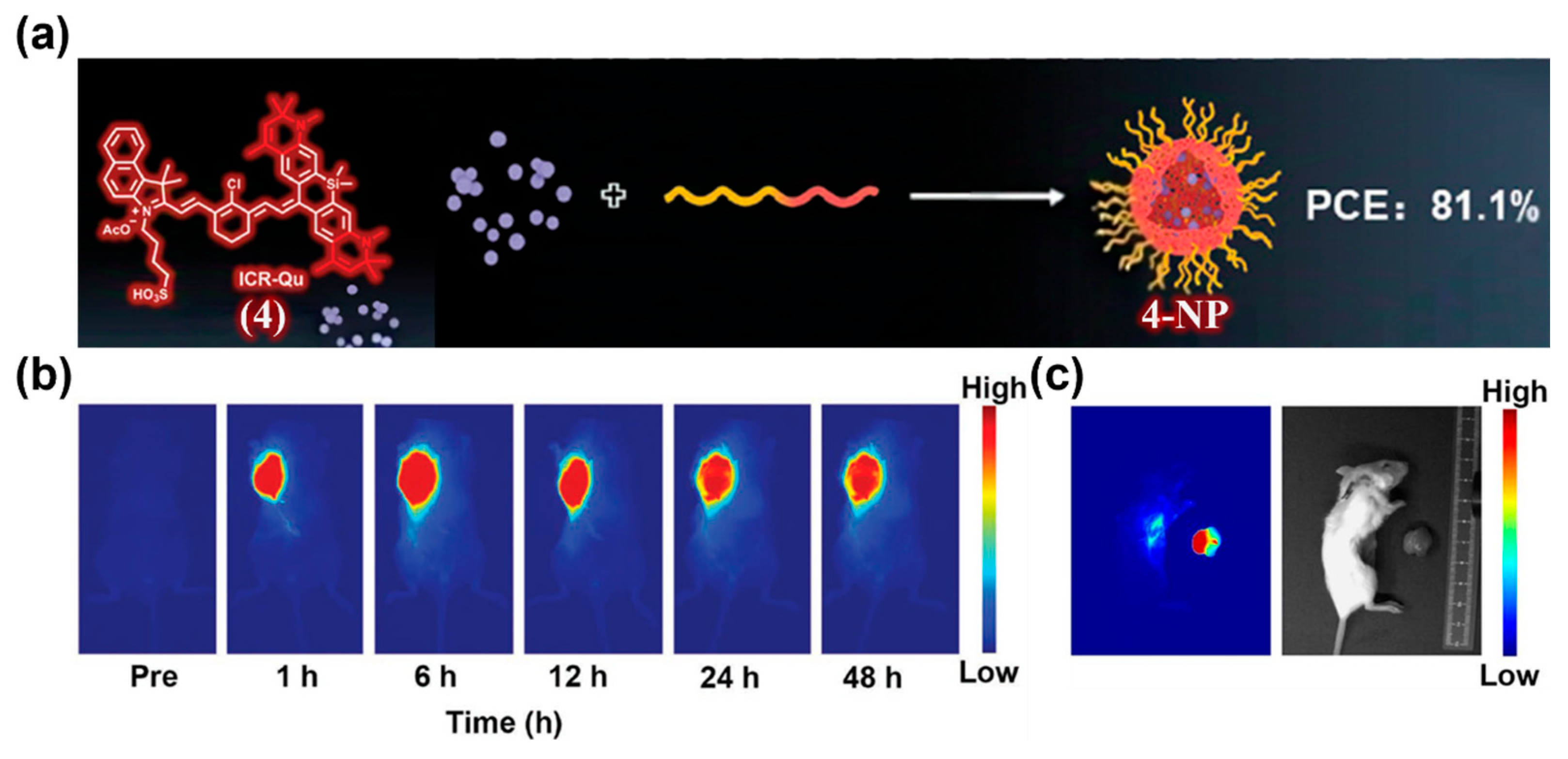
Besides PEGylation and self-assembly, nanocarrier encapsulation is also a widely used strategy to endow small-molecule NIR-II fluorophores with the EPR effect [143,144,145]. Common carriers include phospholipids (liposomes), polymeric systems (e.g., PEG-PLGA, PLA micelles), and albumin. Encapsulation protects dyes from rapid degradation and clearance, improves in vivo stability, and, owing to the nanoscopic size, significantly prolongs blood circulation to achieve efficient passive enrichment at tumors or other lesions, thereby enhancing imaging and therapeutic outcomes. For example, Li and co-workers reported an ICG-like NIR-II fluorophore, ICR-Qu (4), and encapsulate it with DSPE-PEG2000 to form 4-NP (Figure 10a) [146]. 4-NP possessed high photostability and photothermal conversion efficiency (PCE, 81.1%). After intratumoral administration of 4-NP into 4T1 tumor-bearing BALB/c mice, strong NIR-II fluorescence signals were observed in the tumor region, from 1 to 6 h post-injection, with significant fluorescence persisting at 48 h (Figure 10b). Fluorescence imaging-guided tumor resection enabled clear delineation of excised tumors and residual lesions, indicating the high sensitivity of 4-NP for tumor imaging and intraoperative navigation (Figure 10c).
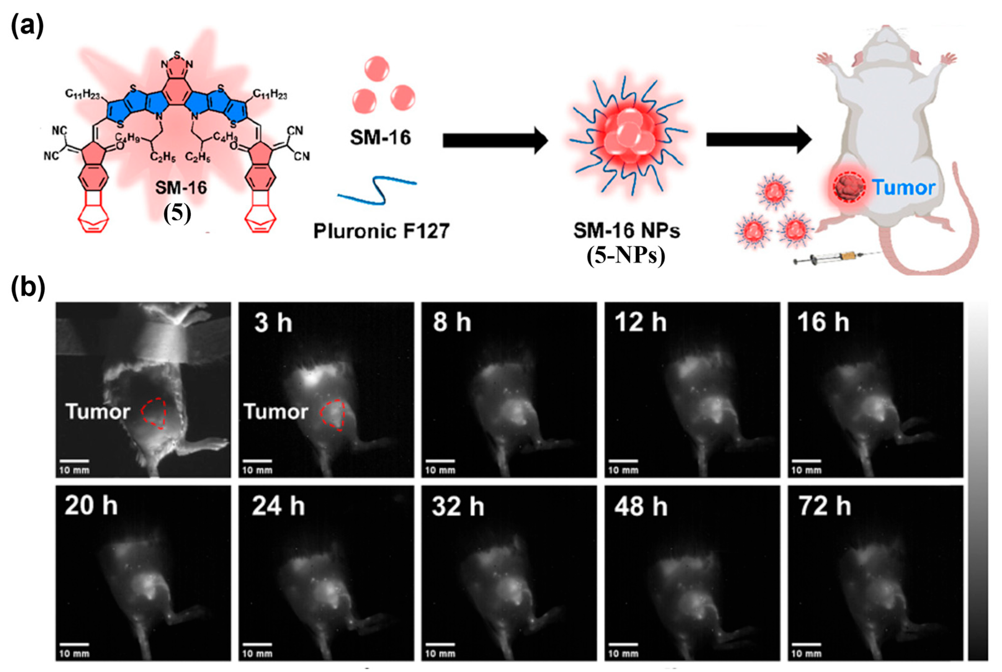
Pluronic F127 (PEO100-PPO65-PEO100) is also a commonly used polymer for the encapsulation of small-molecule NIR-II fluorophores. It is a prototypical triblock copolymer whose hydrophobic PPO forms the micellar core and hydrophilic PEO forms the corona [147]. Wang et al. designed an A–D–A′–D–A dye, SM-16 (5), and formulated it with Pluronic F127 to obtain SM 16 NPs (5-NPs) as a new NIR-II fluorescent probe (Figure 11a) [148]. Following i.v. injection into 4T1-tumor-bearing mice, 5-NPs produced tumor-localized fluorescence substantially stronger than surrounding tissues under EPR guidance, with peak contrast at approximately 16 h post-dosing (Figure 11b).
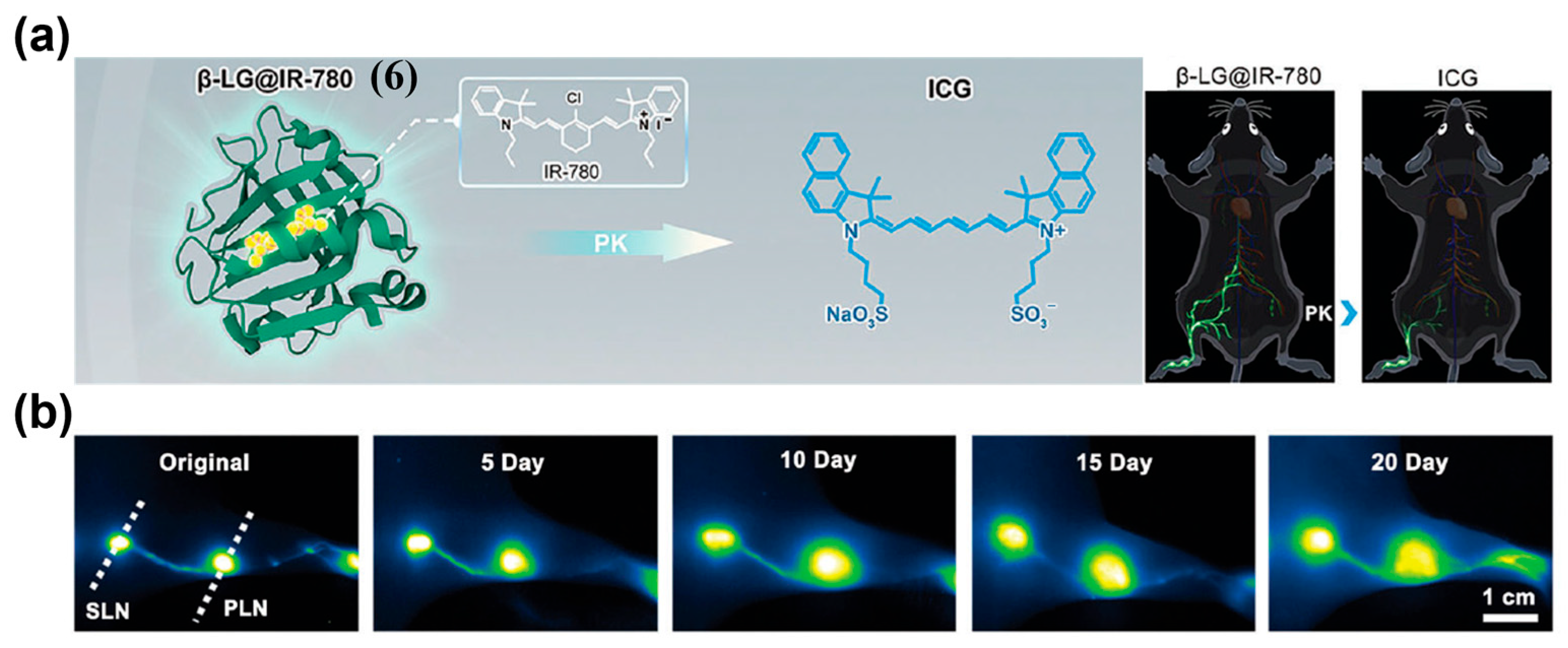
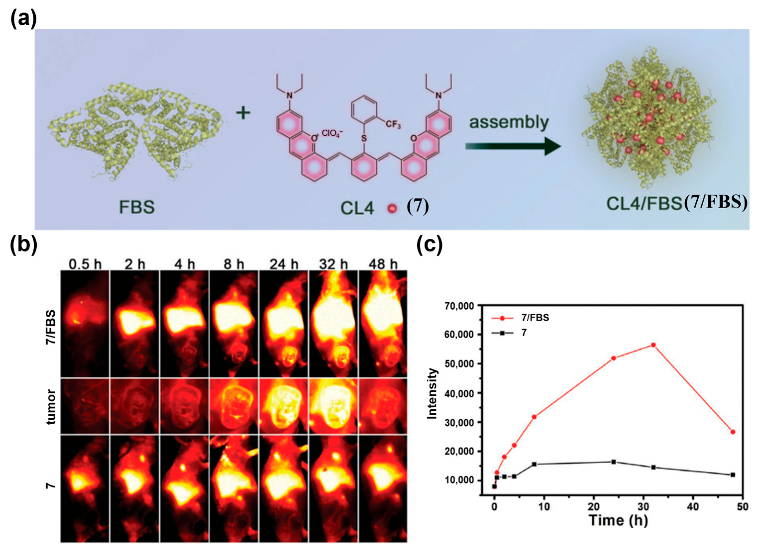
In addition, passive tumor targeting can also be achieved through protein-dye complexes formed with albumin or components of fetal bovine serum (FBS) via hydrophobic interactions or covalent bonds [149,150]. Zhu et al. assembled β-lactoglobulin (β-LG, 18.4 kDa) with the cyanine dye IR-780 to construct a protein@dye probe, β-LG@IR-780 (6) (Figure 12a) [151]. This probe exhibited excellent NIR-II brightness, photostability, and favorable biosafety. When 6 was applied in a murine lymphatic metastasis model, NIR-II lymphangiography detected pronounced lymph node enlargement by day 5 post-tumor inoculation, and monitored its gradual deterioration over time (Figure 12b). The inoculated side showed clear lymphatic dilation and faster lymph node visualization than the contralateral side, enabling early detection of tumor-induced lymph node enlargement. Zhang and co-workers combined FBS with the NIR-II J-aggregate dye CL4 (7) to form the CL4/FBS (7/FBS) complex (Figure 13a) [152]. This complex exhibited maximal fluorescence enhancement at 1235 nm, uniform particle size, suitable zeta potential, and excellent photo- and pH stability, all of which are conducive to EPR-mediated tumor NIR-II fluorescence imaging in vivo (Figure 13b,c).

4.1.2. Active Targeting Probes

Compared with passive targeting probes, NIR-II fluorescent probes based on active-targeting strategies generally exhibit superior tumor accumulation and imaging contrast [121]. The targeting ligands used for tumor NIR-II imaging primarily include four classes: peptide-based motifs, antibodies and their fragments, small-molecule ligands and inhibitors, and biomimetic targeting motifs. Representative advances and key performance metrics are summarized below.
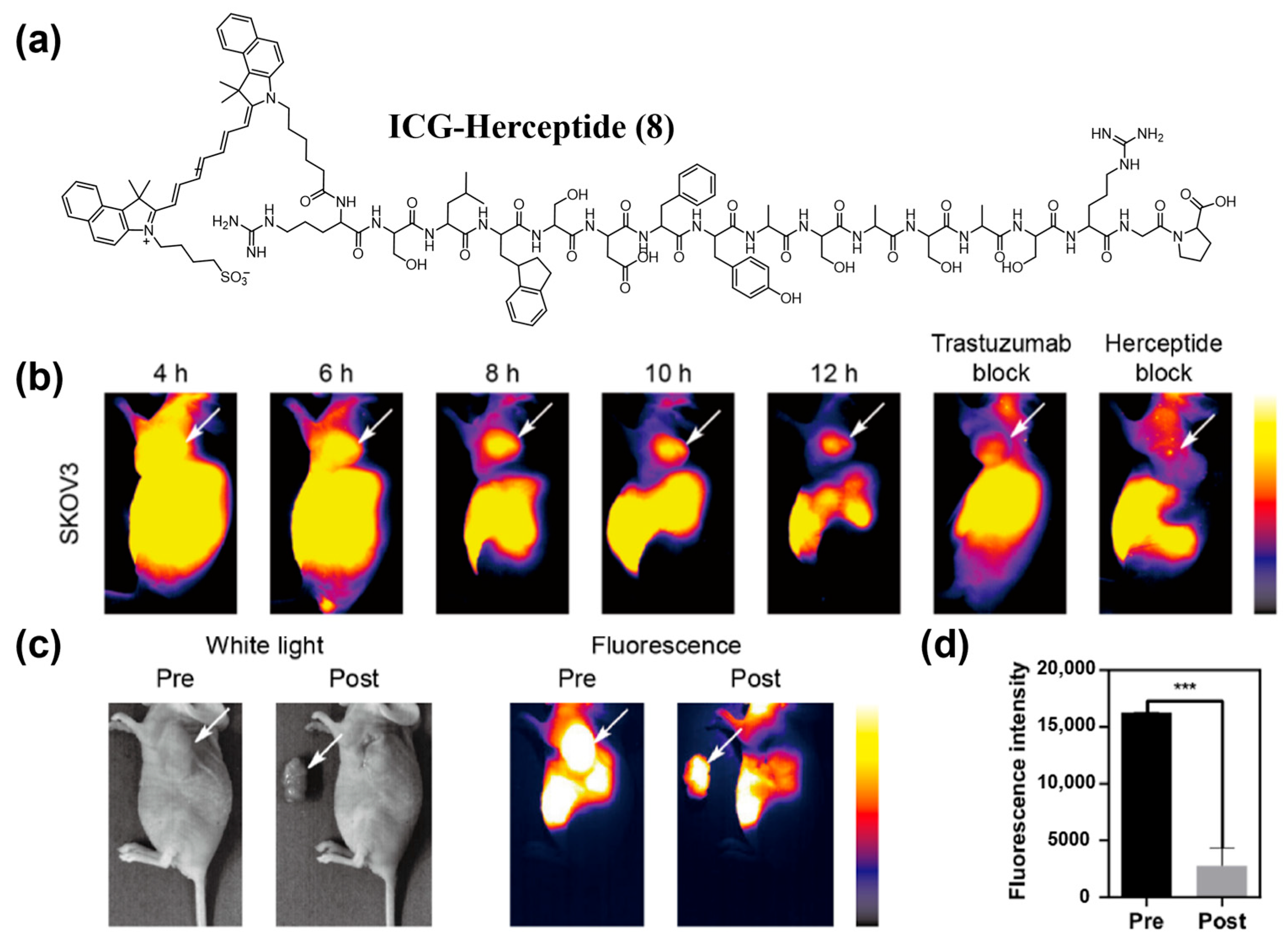
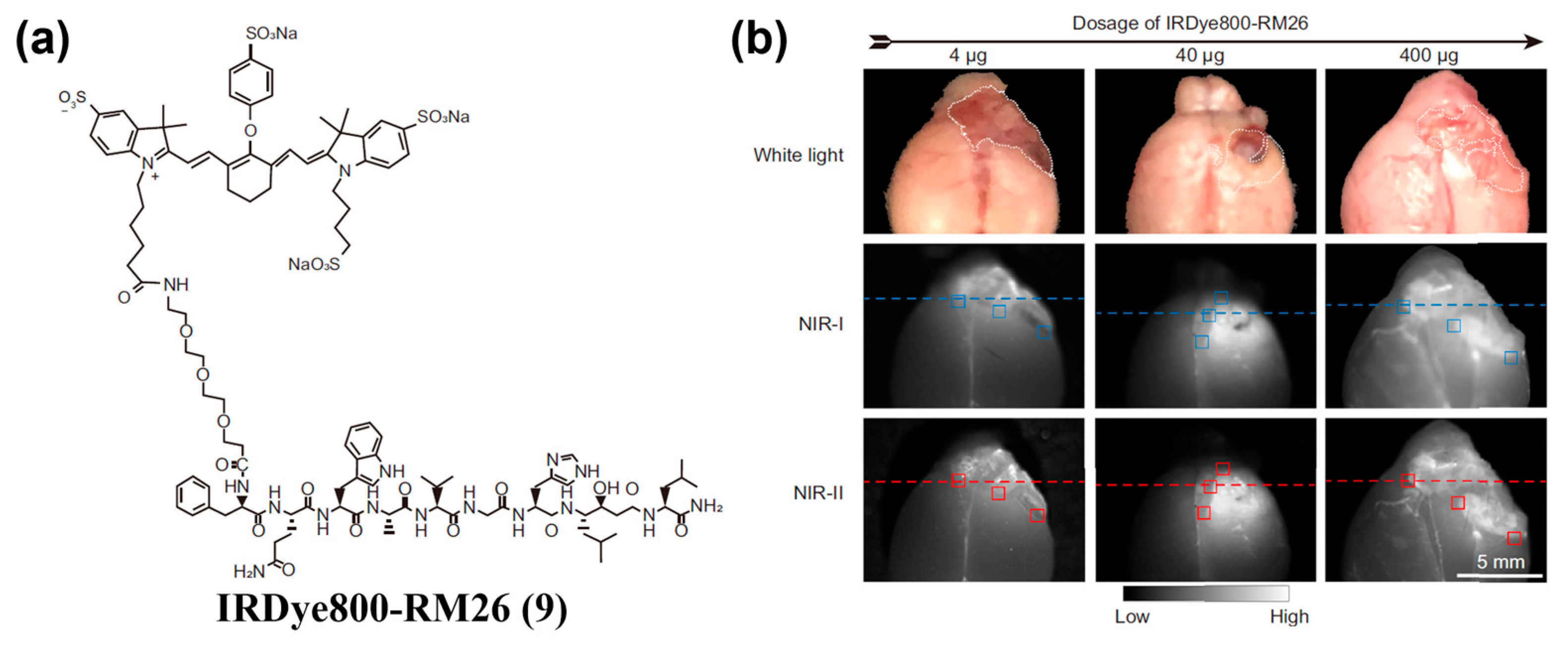
Peptide ligands achieve specificity by recognizing receptors or adhesion molecules overexpressed on tumor cell surfaces (e.g., integrin αvβ3). To date, various tumor-targeting peptides have been used to modify NIR-II organic small-molecule fluorophores to endow fluorescent probes with active targeting ability. For example, Cao et al. identified a novel peptide, Herceptide, that binds specifically to HER2 [153]. They conjugated Herceptide with ICG to yielded ICG–Herceptide (8), enabling NIR-II fluorescence imaging of HER2-positive tumors with a peak tumor-to-normal tissue (T/N) ratio of 7.3 and high-contrast intraoperative guidance in SKOV3 subcutaneous xenografts (Figure 14a–c). Wang and colleagues developed the optical probe IRDye800–RM26 (9) targeting the gastrin-releasing peptide receptor (GRPR) for NIR-II fluorescence imaging and intraoperative guidance in malignant brain tumors (Figure 15a,b) [154].
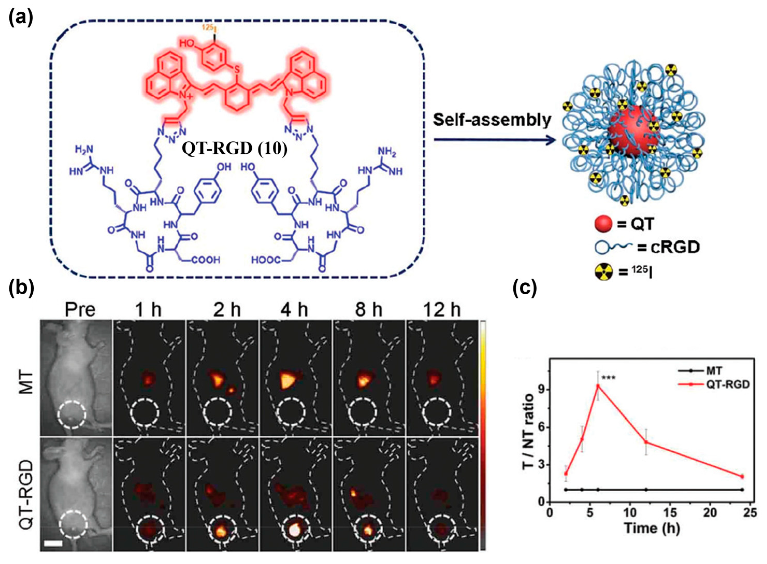
To further enhance tumor specificity, researchers often combine passive and active targeting by integrating nanotechnologies with active targeting warhead. The obtained nanoparticles thus integrate ligand-mediated recognition with EPR-driven enrichment [78]. RGD peptide is a commonly used hydrophilic tumor-targeting warhead that binds to integrin αvβ3 with high affinity and promotes receptor-mediated endocytosis. Conjugating RGD with a hydrophobic NIR-II organic small-molecule fluorophore always yields amphiphilic molecules that tend to self-assemble into nanostructures with both passive and active tumor-targeting abilities. Zhao et al. used a NIR-II-emissive heptamethine cyanine scaffold and dual-site conjugated cRGD to obtain QT–RGD (10), which self-assembled into nanoparticles (Figure 16a) [155]. In a 4T1 mouse model, tumor fluorescence peaked at approximately 4 h after injection of 10, with a high signal-to-background ratio (SBR) of 8.2, whereas the control probe (i.e., MT) without RGD modification showed only weak signals (Figure 16b,c).
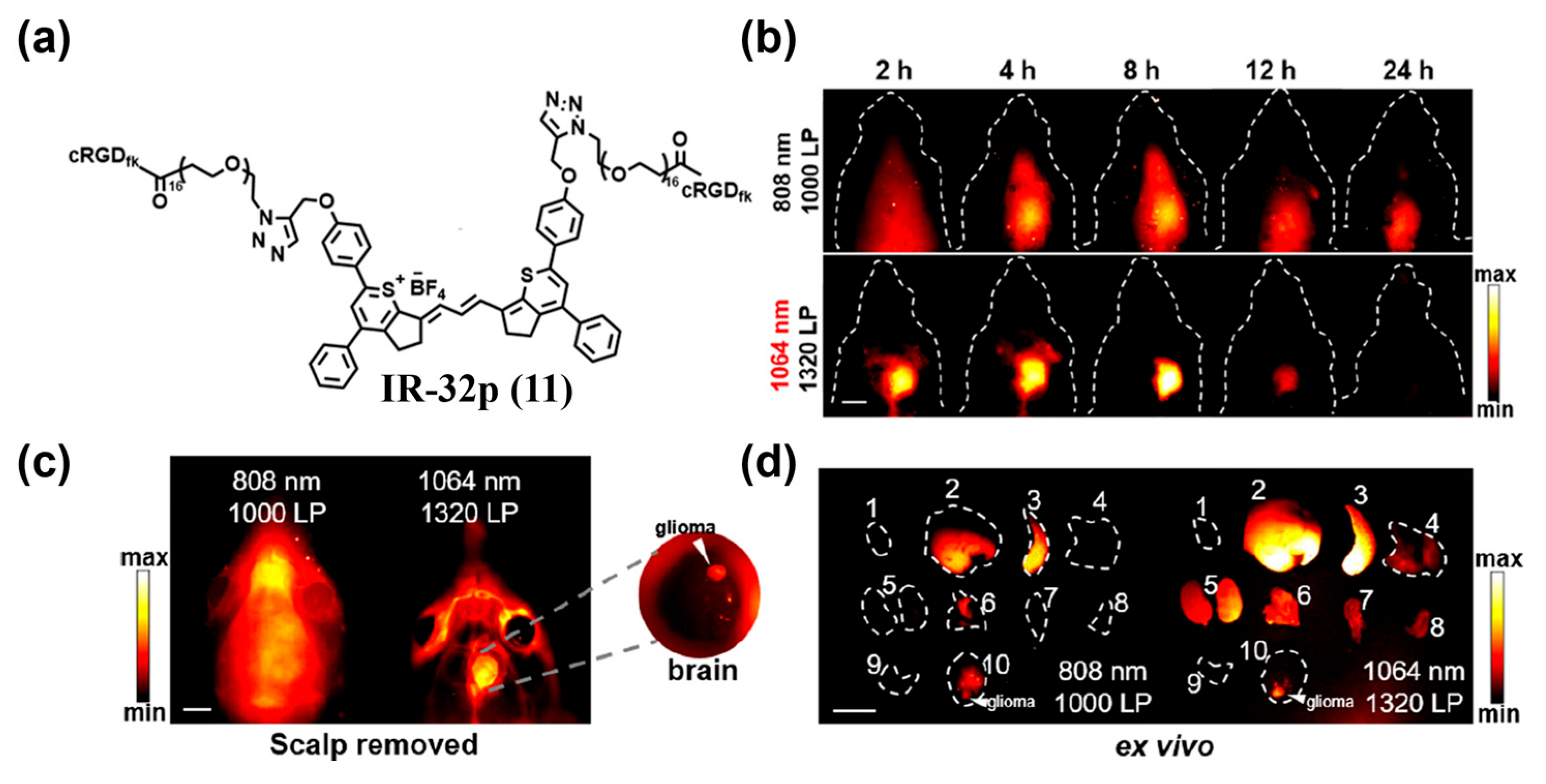
However, dye self-assembly often reduces fluorescence intensity due to the aggregation-caused quenching (ACQ) effect. To suppress self-assembly of RGD-modified fluorophores, incorporating a more hydrophilic linker, such as PEG, is an effective approach. Lyu et al. developed the small-molecule NIR-IIa probe IR–32p (11) targeting αvβ3 integrin by connecting an IR-dye and RGD with a PEG linker (Figure 17a) [156]. After i.v. injection of 11 into an orthotopic glioma model for NIR-II fluorescence imaging, peak T/N ratios were achieved at 8 h, reaching about 4.1 and 6.9 under 808 nm and 1064 nm excitation, respectively (Figure 17b). Notably, after scalp removal, T/N increased to ~9.4 under 1064 nm excitation (Figure 17c). Ex vivo imaging further corroborated αvβ3 specificity of 111 (Figure 17d).
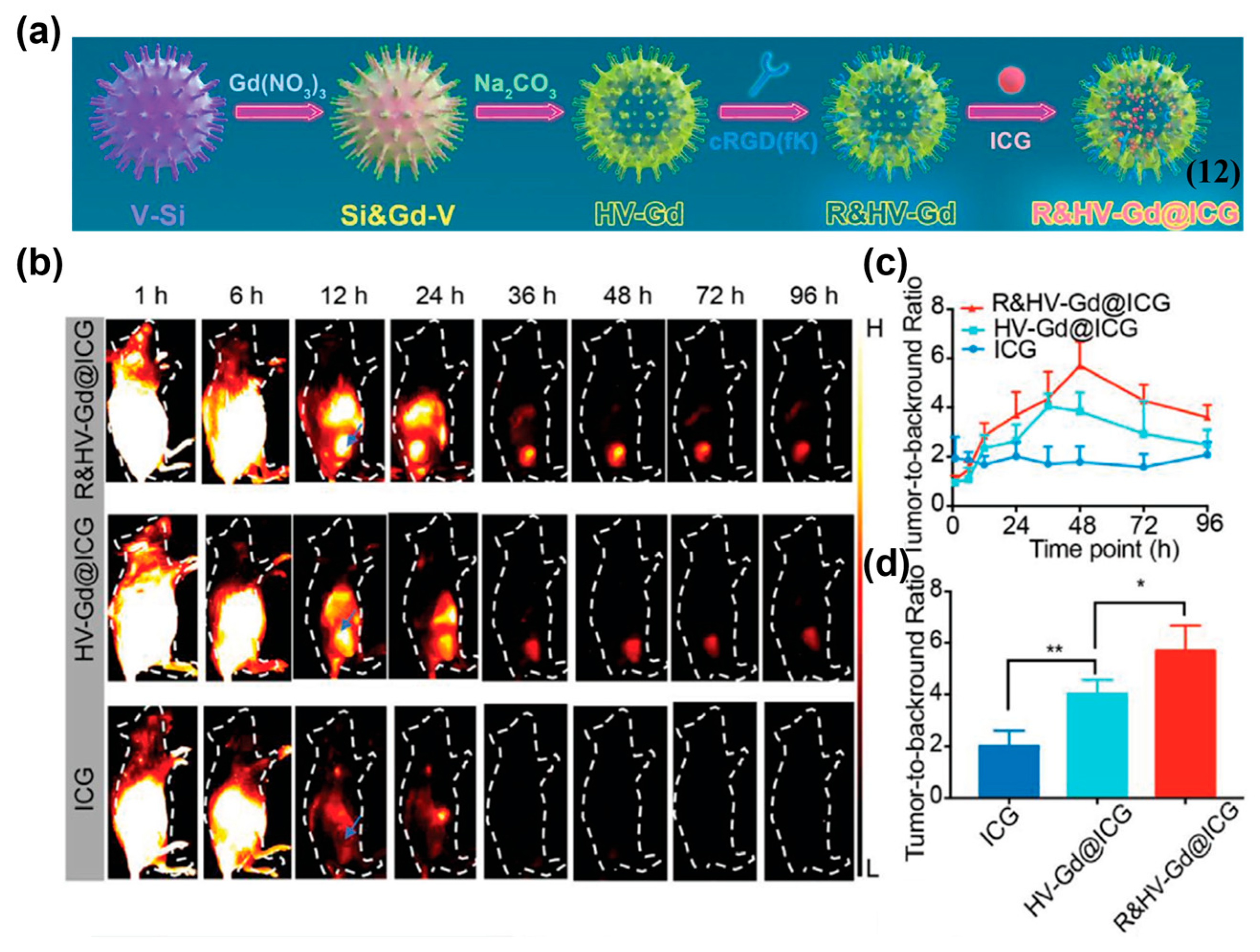
Besides self-assembly, combined passive and active targeting can also be achieved by loading NIR-II organic small-molecule fluorophores with nanocarriers and further modified with active targeting warhead. Yang et al. prepared a biodegradable nanoprobe, R&HV–Gd@ICG (12), by loading ICG into a hollow virus-like gadolinium nanoparticle and modifying the nanoparticle surface with cyclic RGD pentapeptide (cRGDfK), enabling enhanced in vivo imaging of breast cancer (Figure 18a) [157]. In 4T1-tumor-bearing mice, compared with free ICG and the unmodified control probe (HV-Gd@ICG), 12 achieved the highest NIR-II fluorescence intensity, as well as the highest TBR of 5.7 ± 1.0 at 48 h, indicating its excellent tumor-targeted imaging ability (Figure 18b–d).
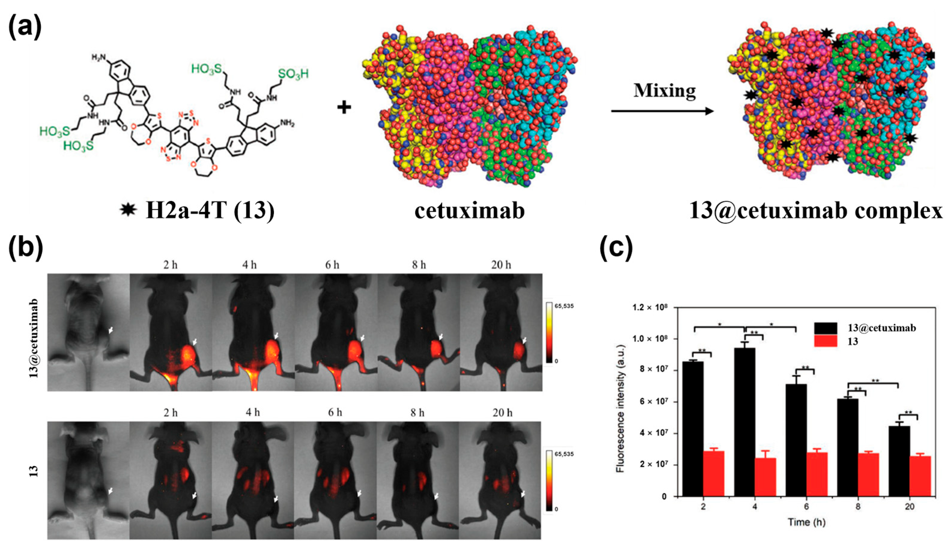
Antibody-based targeting is also a widely used active targeting strategy. It exploits the high-affinity binding of antibodies to tumor-specific antigens (e.g., bevacizumab against VEGF [158], cetuximab against EGFR [159]) to enable tumor-targeted imaging. To date, several ICG-antibody conjugates for tumor-targeted NIR-II fluorescence imaging have been reported [160,161,162]. Notably, the recombinant monoclonal antibody cetuximab (Erbitux®), a Food and Drug Administration (FDA)-approved drug, has been reported for use in NIR-II fluorescence imaging [163]. Zeng et al. synthesized the water-soluble small-molecule NIR-II dye H2a–4T (13) and complexed it with cetuximab to form 13@Cetuximab (Figure 19a) [69]. In an HCT116 colorectal cancer model, the complex clearly delineated tumors between 2–20 h, with a T/N of 5.51 ± 0.12 at 4 h (Figure 19b,c). In comparison, the free dye (13) produced substantially weaker tumor signals at all time points (Figure 19b,c). These results demonstrated the efficient active targeting performance of 13@Cetuximab to EGFR-overexpressing tumors.
Small-molecule ligand-based tumor targeting enables directed probe accumulation at tumor sites via metabolism- or receptor-mediated mechanisms [164]. For instance, folic acid (FA) targets folate receptors overexpressed in various tumors (e.g., ovarian and breast cancers) [165]. Shi et al. used a FA-ICG conjugate for in vivo NIR-II/NIR-I fluorescence imaging of orthotopic U87MG human GBM tumors [166]. Interestingly, the NIR-II fluorescence signal offered superior contrast at all time points compared to the NIR-I fluorescence signal.
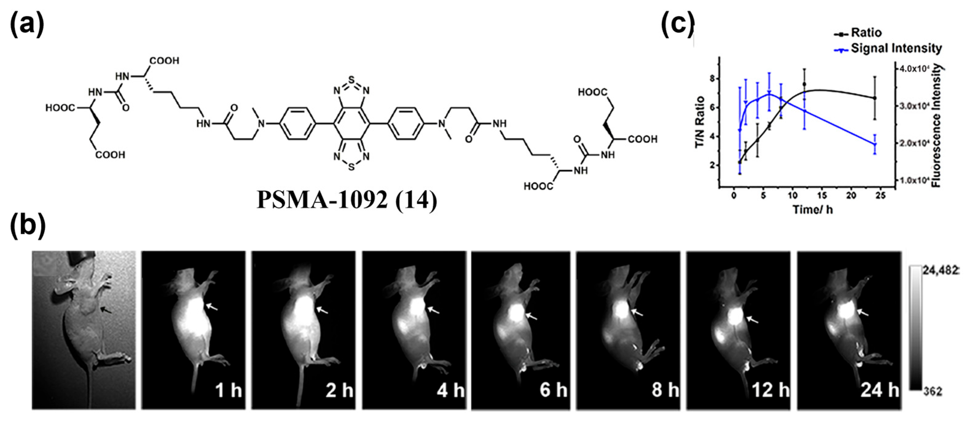
Besides receptor-mediated mechanisms, small-molecule ligand-antigen interaction-based targeting is also an effective tumor-targeting strategy, such as the inhibition effect between (S)-5-amino-1-carboxypentyl)carbamoyl)-L-glutamic acid (Glu-urea-Lys) and prostate-specific membrane antigen (PSMA). Cui et al. designed PSMA–1092 (14), which contained two Glu–urea–Lys motifs with an inhibition constant (Ki) of 80 pM (Figure 20a) [167]. After tail vein injection of 14 into LNCaP tumor-bearing mice, tumor margins were clearly defined at 2–4 h by NIR-II fluorescence imaging, with peak tumor uptake occurring at 6 h and a maximum T/N of 7.62 ± 1.05 achieved at 12 h (Figure 20b,c). These imaging results clearly demonstrated the targeting capability of compound 14 for tumors.
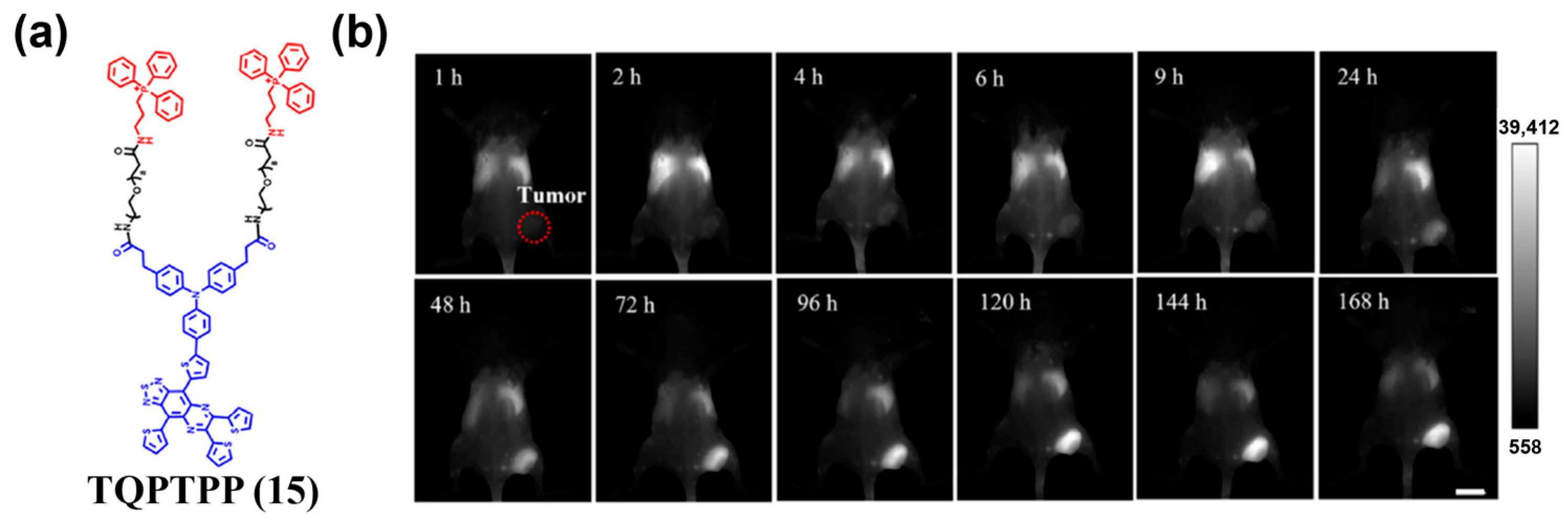
Small molecule–organelle interaction can also be utilized for tumor targeting. Li et al. introduced the lipophilic cation triphenylphosphine (TPP) into a donor–acceptor (D–A) NIR-II organic small-molecule fluorophore and constructed the mitochondria-targeted probe TQPTPP (15) (Figure 21a) [168]. When applying the probe for in vivo NIR-II fluorescence imaging of 4T1 tumors, the probe exhibited prolonged tumor retention, with maximal signal observed on day 5 and an SBR of about 9 on day 6, providing an effective tool for subcellular-level active targeting in NIR-II fluorescence imaging (Figure 21b).
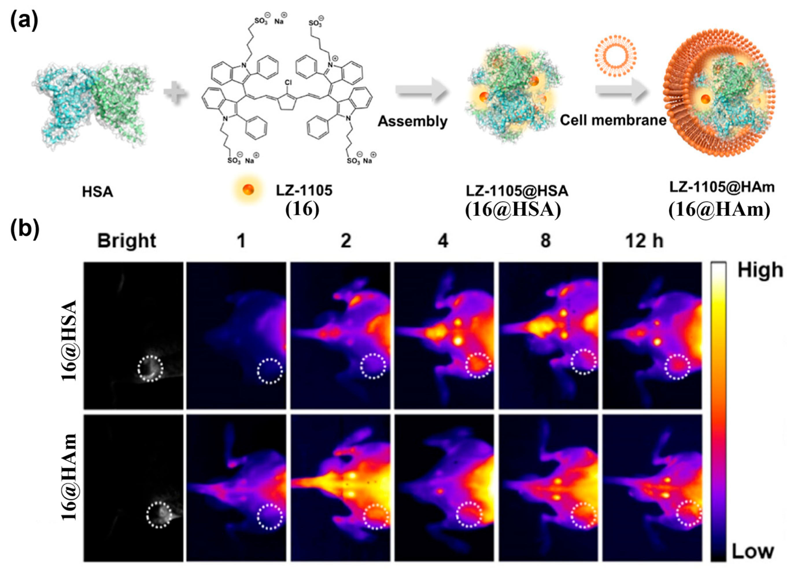
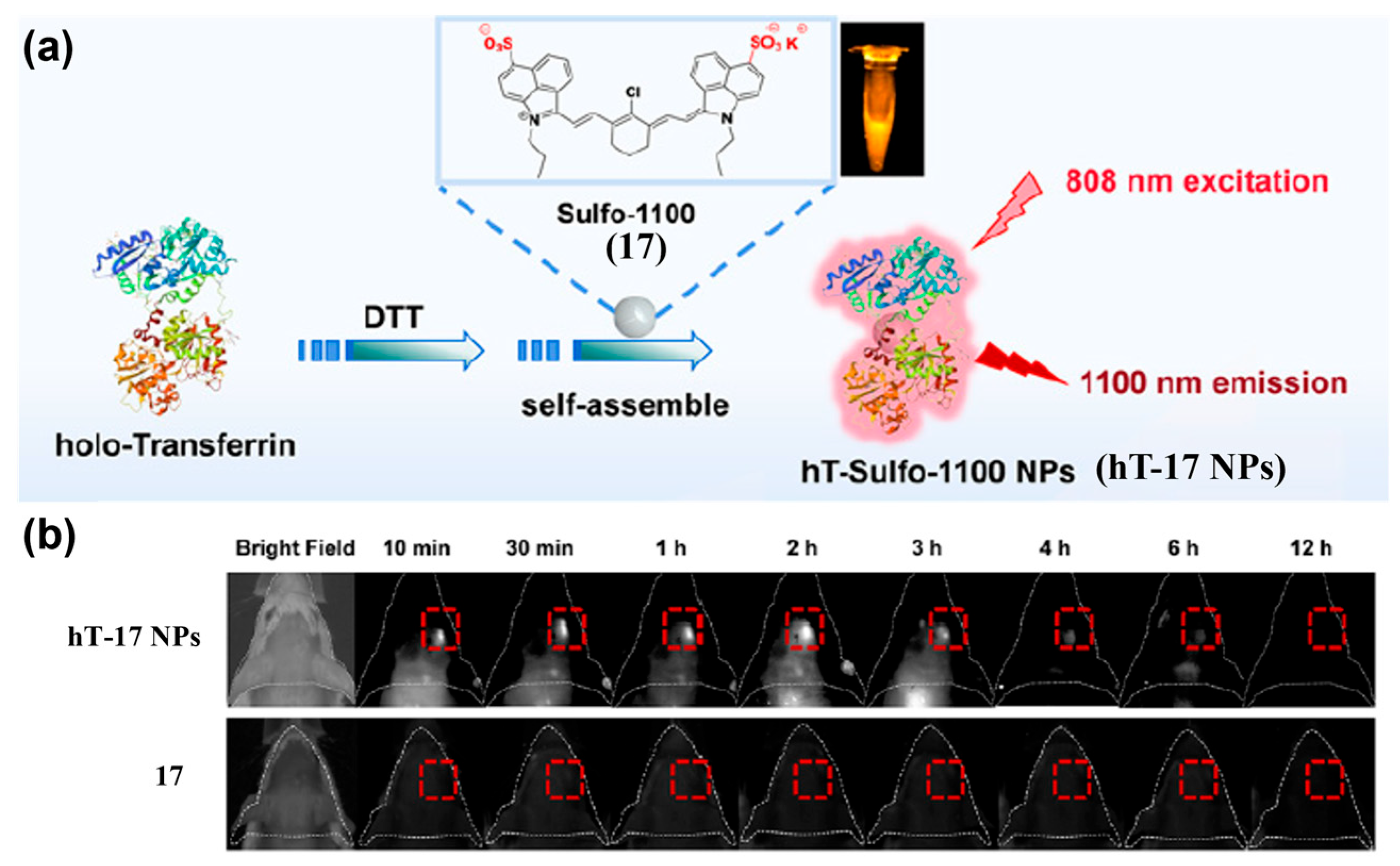
By cloaking probes with endogenous biological membranes (e.g., red blood cells, platelets, macrophages, stem cells, or tumor cell membranes) or natural structures, biomimetic modifications can impart tumor tropism while reducing immunogenicity and improving biocompatibility [169,170,171]. Jiang et al. developed LZ-1105@HAm (16@HAm), an albumin–NIR-II cyanine dye assembly coated with A549 tumor cell membranes (Figure 22a) [172]. Homologous recognition by A549-derived membranes markedly enhanced tumor accumulation and NIR-II fluorescence signals in the tumor, reaching maximal uptake at 2 h post-injection, with fluorescence intensity approximately 4.7-fold higher than that of the uncoated control (i.e., 16@HSA) (Figure 22b). Wei et al. self-assembled the NIR cyanine dye Sulfo-1100 (17) with holo-transferrin to form hT-Sulfo-1100 nanoparticles (hT-17 NPs) (Figure 23a) [173]. This platform crossed the blood–brain barrier and targeted glioma tumor, yielding strong NIR-II fluorescence signals at orthotopic gliomas as early as 10 min post-i.v. injection into an orthotopic glioma model (Figure 23b). In comparison, 17 alone showed no effective tumor signal in the orthotopic glioma model, demonstrating excellent deep-tissue and transcranial imaging capabilities of hT-17 NPs.
Active-targeting NIR-II fluorescent probes based on organic small-molecule fluorophores—spanning peptides, antibodies, small molecules, and biomimetic designs—enable precise recognition of tumor-associated receptors and microenvironments, with marked advances in in vivo imaging contrast, spatiotemporal resolution, and intraoperative guidance. The broadly applicable RGD–αvβ3 axis, in particular, serves as a key design module that enhances imaging quality and therapeutic gains across multiple solid tumors. Looking ahead, integrating multi-receptor co-targeting, controllable release, and clinically translatable formulation engineering is expected to accelerate the trajectory from animal validation to clinical application.

4.2. Activatable NIR-II Fluorescent Probes

Recent advances in small-molecule fluorophore-based NIR-II probes aim to improve detection accuracy, biocompatibility, stability, photobleaching resistance, deep-tissue imaging, and clinical translation. Activatable small-molecule NIR-II probes leverage robust photophysical switching mechanisms to translate TME hallmarks into high-contrast optical readouts. By leveraging TME-specific biochemical cues, activatable probes suppress background fluorescence and reduce nonspecific signals, thereby enabling precise, real-time tumor visualization [174,175,176].
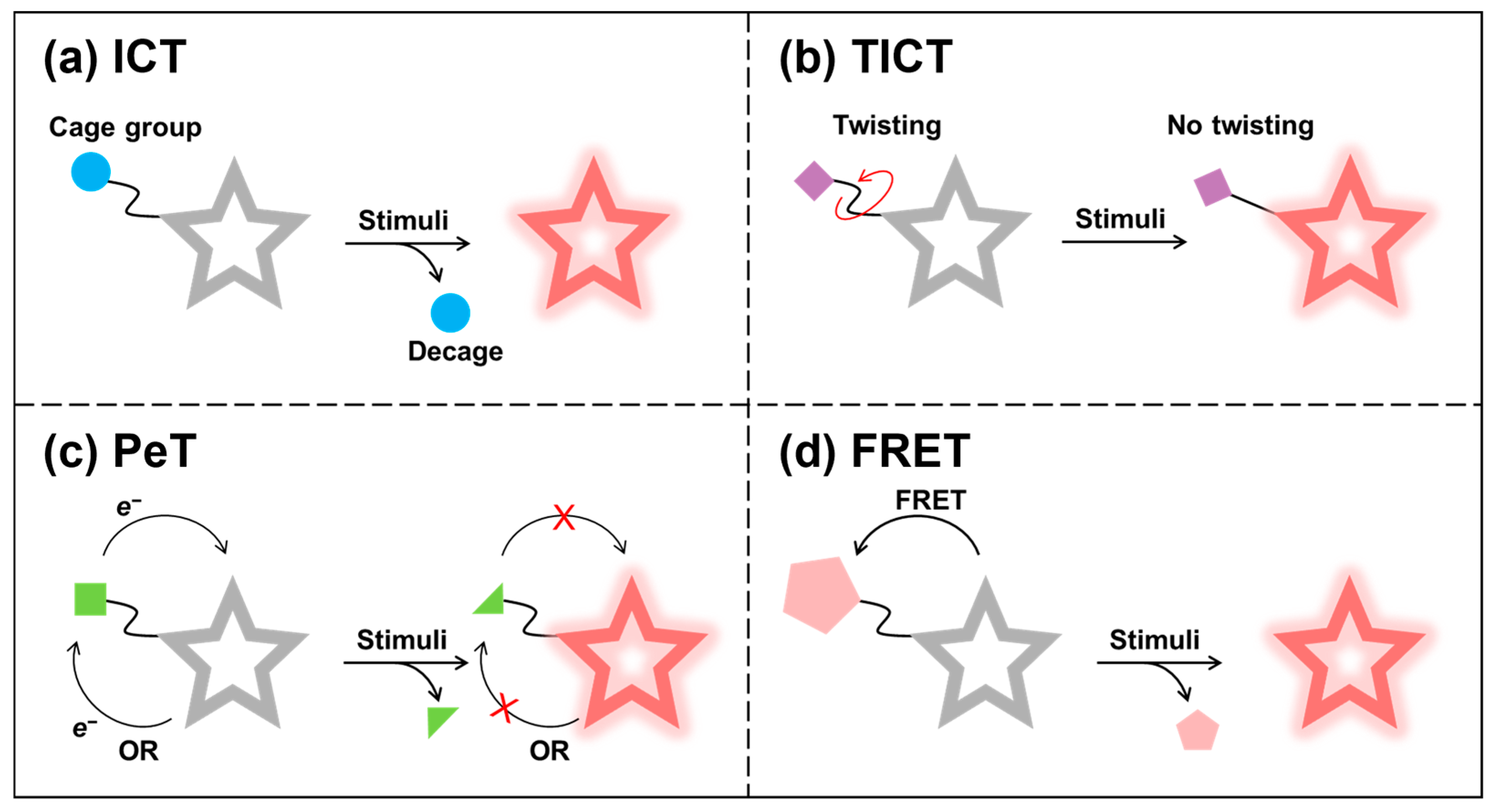
Activatable NIR-II fluorescent probes are typically created by attaching a stimulus-responsive group to a NIR-II organic small-molecule fluorophore, quenching fluorescence through mechanisms such as ICT, TICT, photo-induced electron transfer (PeT), or fluorescence resonance energy transfer (FRET) (Figure 24a–d). Target engagement in the TME induces changes in the probe’s fluorescence properties—typically fluorescence intensity, emission wavelength, or lifetime—thus reporting the presence and dynamics of tumor-associated analytes.
ICT-based activatable probes typically use the recognition unit as a caging group directly linked to the fluorophore, modulating donor–acceptor “push–pull” electron flow. Target-induced cleavage or transformation restores ICT, causing spectral shifts and increased fluorescence [177,178]. For TICT-based activatable probes, donor and acceptor groups adopt a twisted geometry to form a charge-transfer state that typically relaxes via red-shifted emission or non-radiative decay, quenching fluorescence [179,180]. Activation that restricts molecular motion (for example, higher viscosity) suppresses TICT and turns on emission. When the frontier orbitals of the receptor are located between the HOMO and the LUMO of the fluorophore, PeT effect will occur to quench fluorescence [181,182,183]. Target-induced structural or electronic changes disrupt PeT, thereby restoring fluorescence. In FRET-based activatable probes, non-radiative energy transfer from a donor to an acceptor (fluorophore or quencher) suppresses donor emission [184,185,186]. Target recognition that disrupts donor–acceptor coupling activates the fluorescent signal.
Leveraging these mechanisms, diverse activatable NIR-II fluorescent probes based on NIR-II organic small-molecule fluorophores have been rationally designed for tumor imaging. By readout mode, they can be broadly categorized into single-channel probes and ratiometric (self-calibrating) probes.

4.2.1. Single-Channel Probes

Single-channel probes are programmed to respond to overexpressed tumor biomarkers and, upon activation, produce an intensified fluorescence signal suitable for tracking and delineating tumors. They have been widely applied to image ROS, reactive RNS, biothiols, pH, viscosity, and enzymes in real time [125]. Dysregulated redox homeostasis, distinctive TME features, and aberrant enzyme expression serve as orthogonal “keys” to unlock fluorescence, enabling high-contrast, spatiotemporally resolved tumor visualization in vivo.
Redox-Activated Probes
Redox balance is fundamental to cellular integrity and is mediated by reactive species networks—ROS (for example, O2·, H2O2), RNS (for example, NO, ONOO), and RSS (for example, H2S, glutathione (GSH))—whose dysregulation is implicated in cancer pathogenesis and progression [187,188,189].
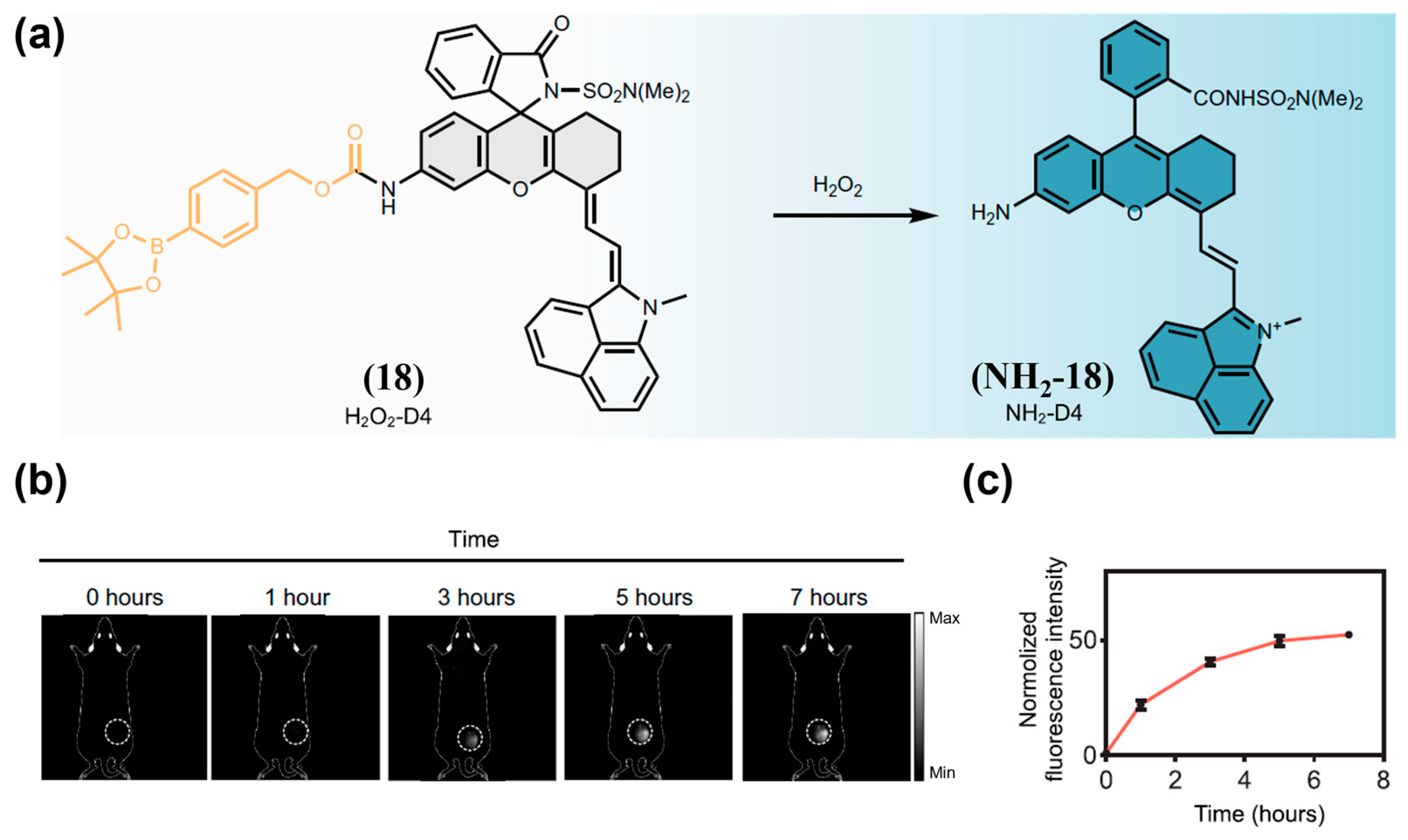
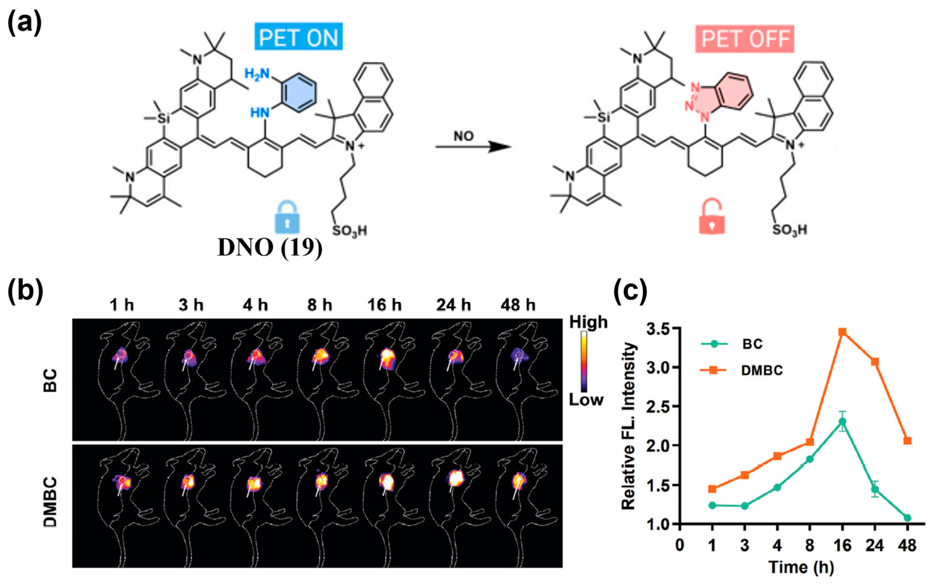
ROS and RNS are always used as the targeted biomarker for tumor imaging. For instance, Li and colleagues reported H2O2-D4 (18), a NIR-II fluorescent probe featuring a highly specific turn-on response to H2O2 (Figure 25a) [190]. Real-time NIR-II fluorescence imaging of a 4T1 tumor xenograft model using 18 revealed a pronounced tumor-localized signal increase, reaching approximately 50-fold above background at 7 h post-administration, enabling clear tumor delineation (Figure 25b,c). An NO-responsive NIR-II fluorescence/photoacoustic probe (DNPS) was developed by encapsulating DNO (19) into DSPE-PEG2000 micelles [191]. Upon NO exposure, donor nitrosation converts 19 into a benzotriazole, suppressing PeT effect and activating NIR-II fluorescence (Figure 26a). DNPS was then successfully applied to differentiate NO expression between diabetic and non-diabetic breast cancer models, exhibiting a 2.3-fold stronger and more persistent signal in diabetic tumors (Figure 26b,c).
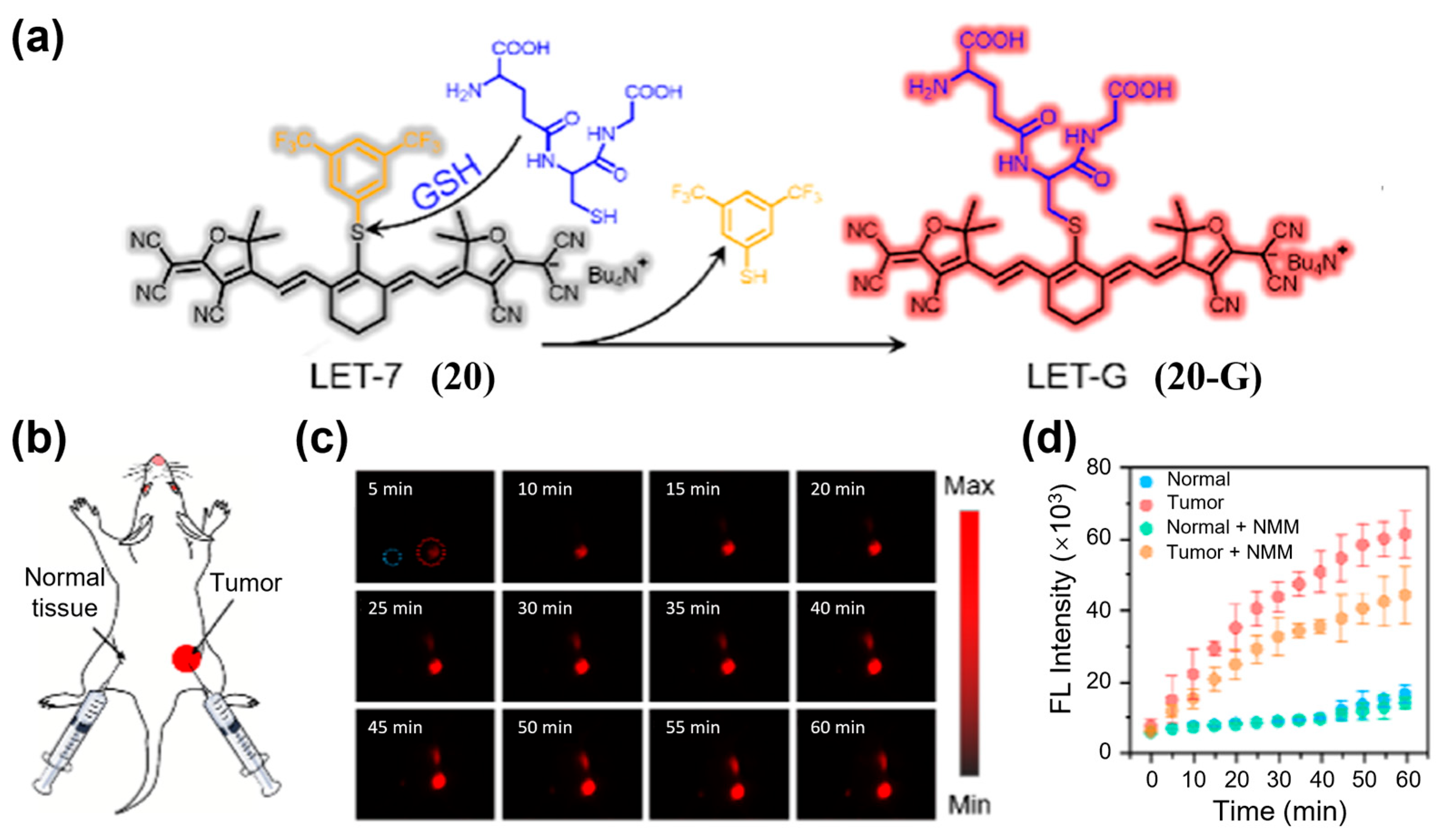
Another type of biomarker reflecting tumor redox balance is reactive sulfur species, which can also serve as a stimulus for activated NIR-II fluorescence imaging of tumors. Lin et al. developed LET-7 (20) (Figure 27a), the first GSH-activatable NIR-II fluorescent probe for selective in vivo GSH visualization [192]. After injection of 20 into 4T1 tumor-bearing mice, NIR-II fluorescence signals in the tumor region rapidly and selectively increased within 60 min, clearly delineating the lesion while normal tissues remained dark (Figure 27b–d).
Enzyme-Activated Probes
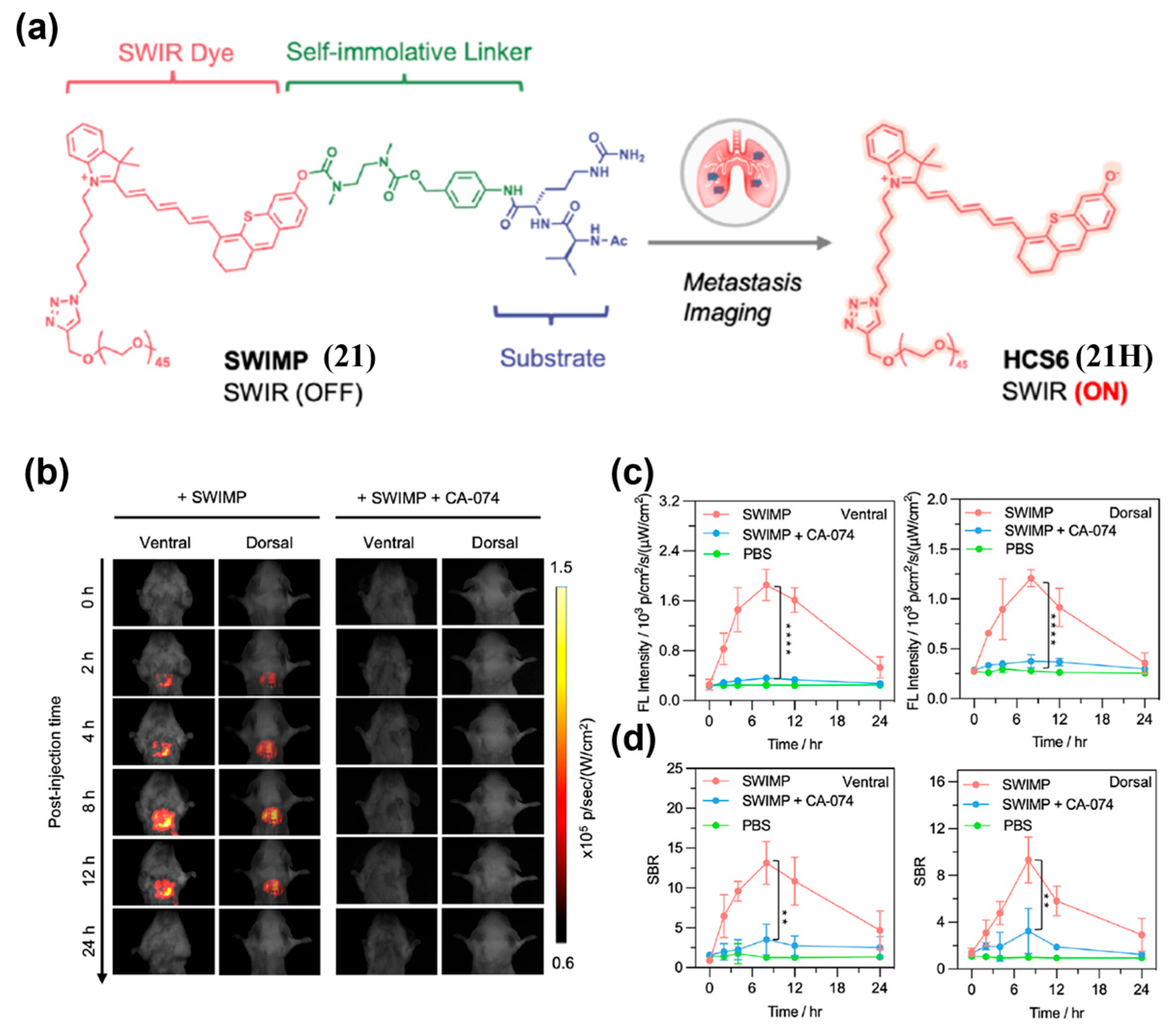
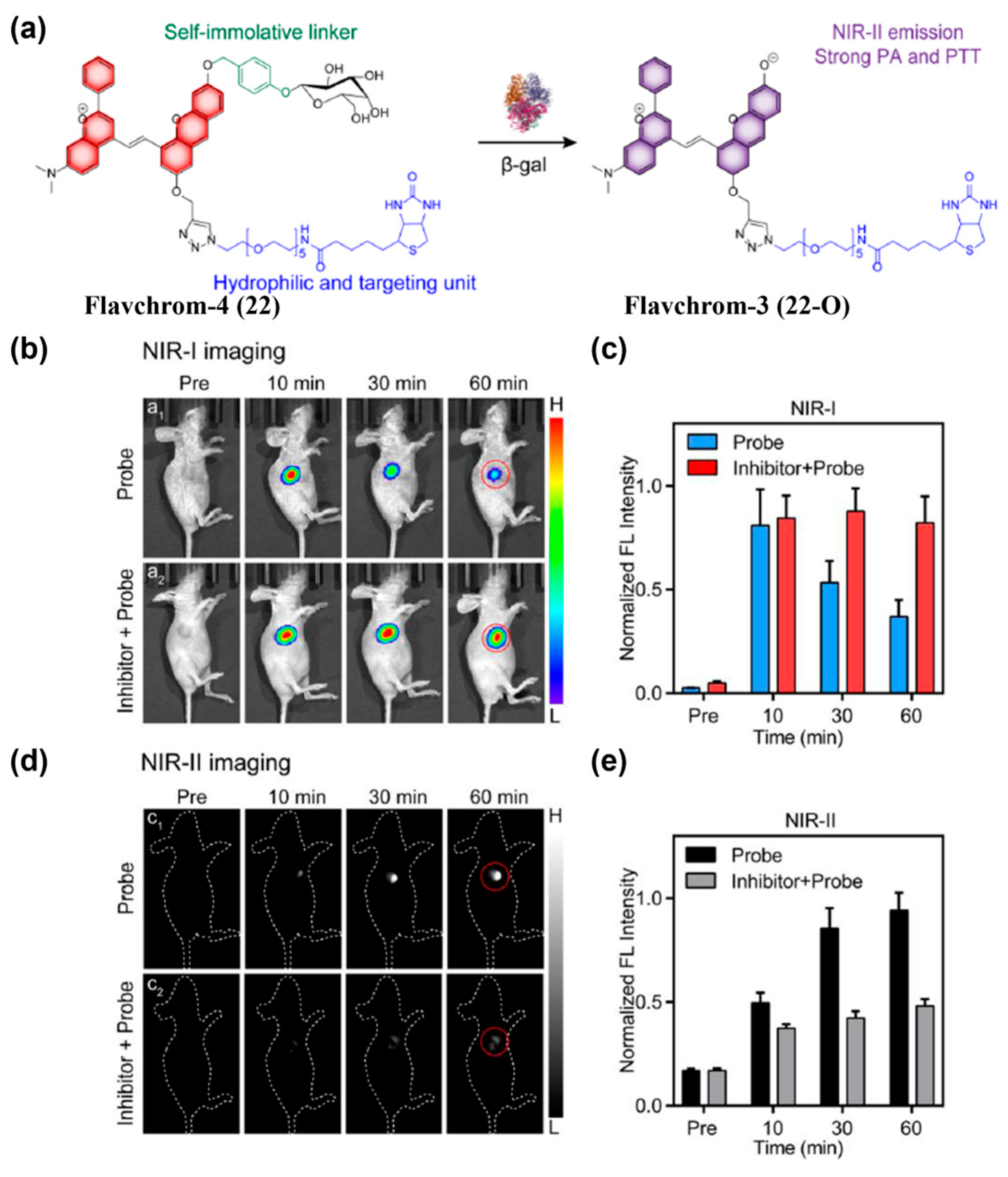
Many enzymes are upregulated in TEM, enabling enzyme-mediated activation of activatable fluorescent probes. To target the lung cancer biomarker cathepsin B (Cath B), Pu’s group constructed SWIMP (21) based on a shortwave-infrared hemicyanine-6 scaffold with three conjugated polymethine units (Figure 28a) [193]. Following intratracheal delivery of 21 into 4T1 lung metastasis murine models, in vivo fluorescence imaging in shortwave infrared window (SWIR, 900–1700 nm) was then conducted. The results showed that the SBRs at lung tumor site reached 15 (ventral) and 9.3 (dorsal) at 8 h, substantially exceeding the Rose criterion and enabling precise detection of lung metastasis (Figure 28b–d). Guo and co-workers synthesized Flavchrom-4 (22) for activatable dual-mode imaging of endogenous β-gal activity in ovarian tumors (Figure 29a) [194]. Following intratumoral administration of compound 22 in SKOV3 tumor-bearing mouse models, a rapid decrease in NIR-I emission intensity was observed at the tumor site, accompanied by a significant increase in NIR-II emission (Figure 29b–e). The NIR-II signal peaked at 60 min post-injection, exhibiting a 6.3-fold enhancement relative to the pre-injection level, thereby enabling real-time monitoring of enzymatic activity in ovarian tumors (Figure 29d,e).
PH-Activated Probes
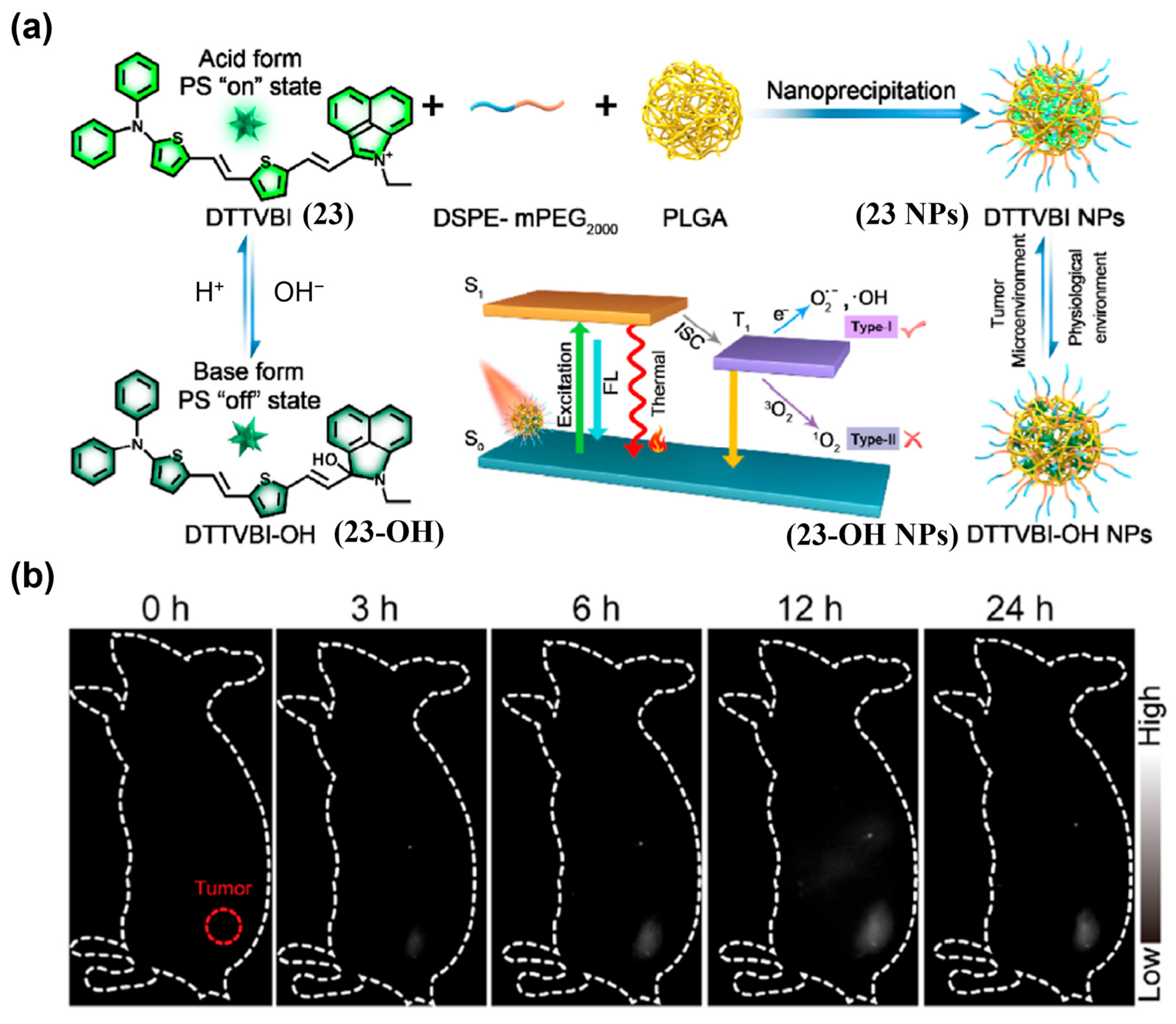
Weak acidity is an important feature of the TME. Thus, pH can be a useful target for activatable fluorescent probes. Wang et al. developed DTTVBI (23) with aggregation-induced emission (AIE) characteristics and formulated 23 NPs using DSPE-mPEG2000 and PLGA, which exhibited pH-switchable NIR-II fluorescence (Figure 30a) [195]. After i.v. injection of 23 NPs in colorectal cancer patient-derived xenograft (PDX) models, tumor NIR-II signals emerged by 3 h and peaked at 12 h, consistent with EPR-mediated accumulation (Figure 30b). Guided by the strong NIR-II fluorescence signal, synergistic type-I photodynamic therapy (PDT)/photothermal therapy (PTT) achieved substantial tumor ablation.
Dual-Activated Probes
Because many biomarkers are not tumor-exclusive, single-trigger probes can be vulnerable to nonspecific activation. Dual- or multi-targeting probes improve specificity by requiring the co-occurrence of two or more spatially co-localized stimuli to activate fluorescence [196].
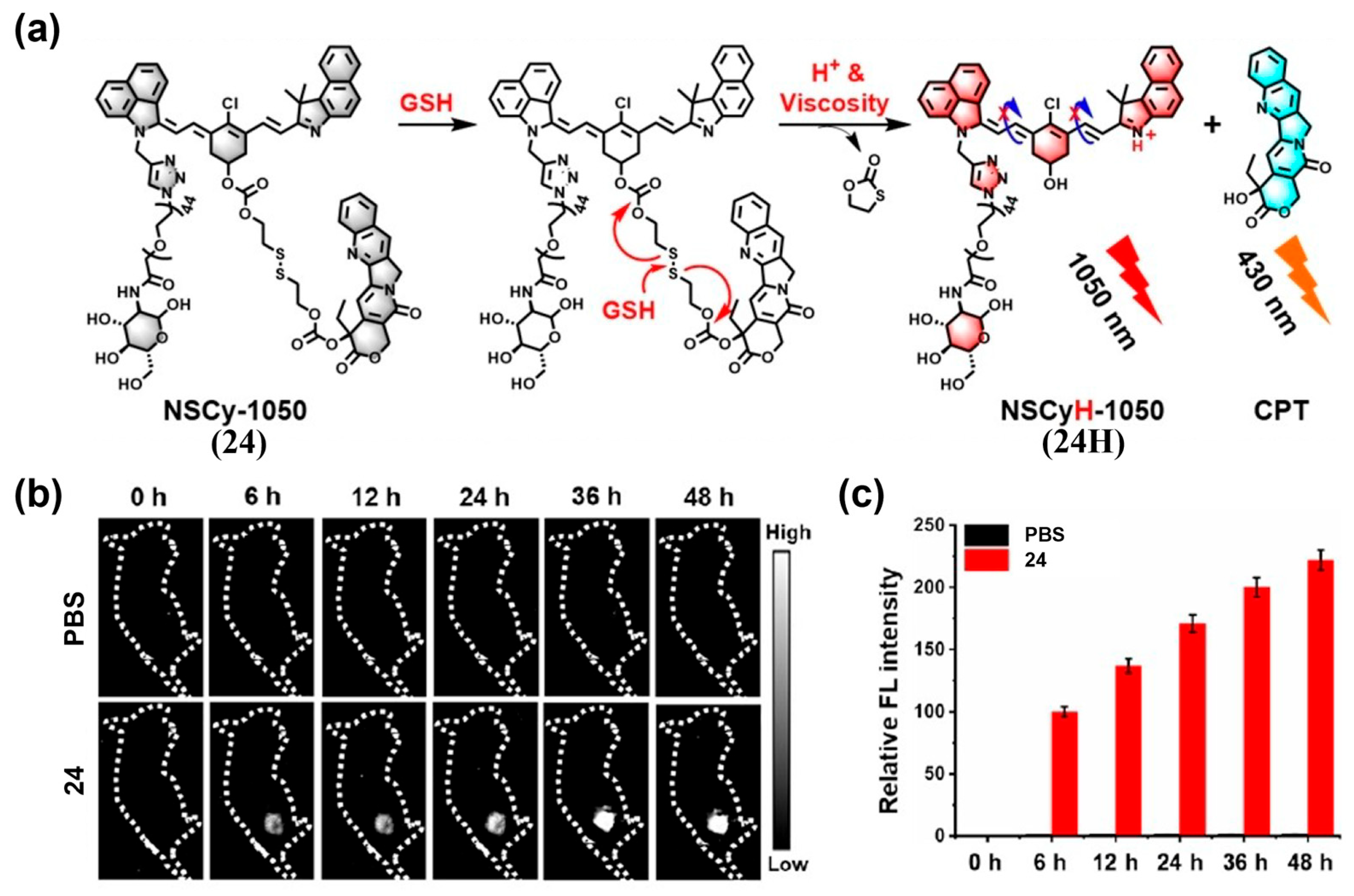
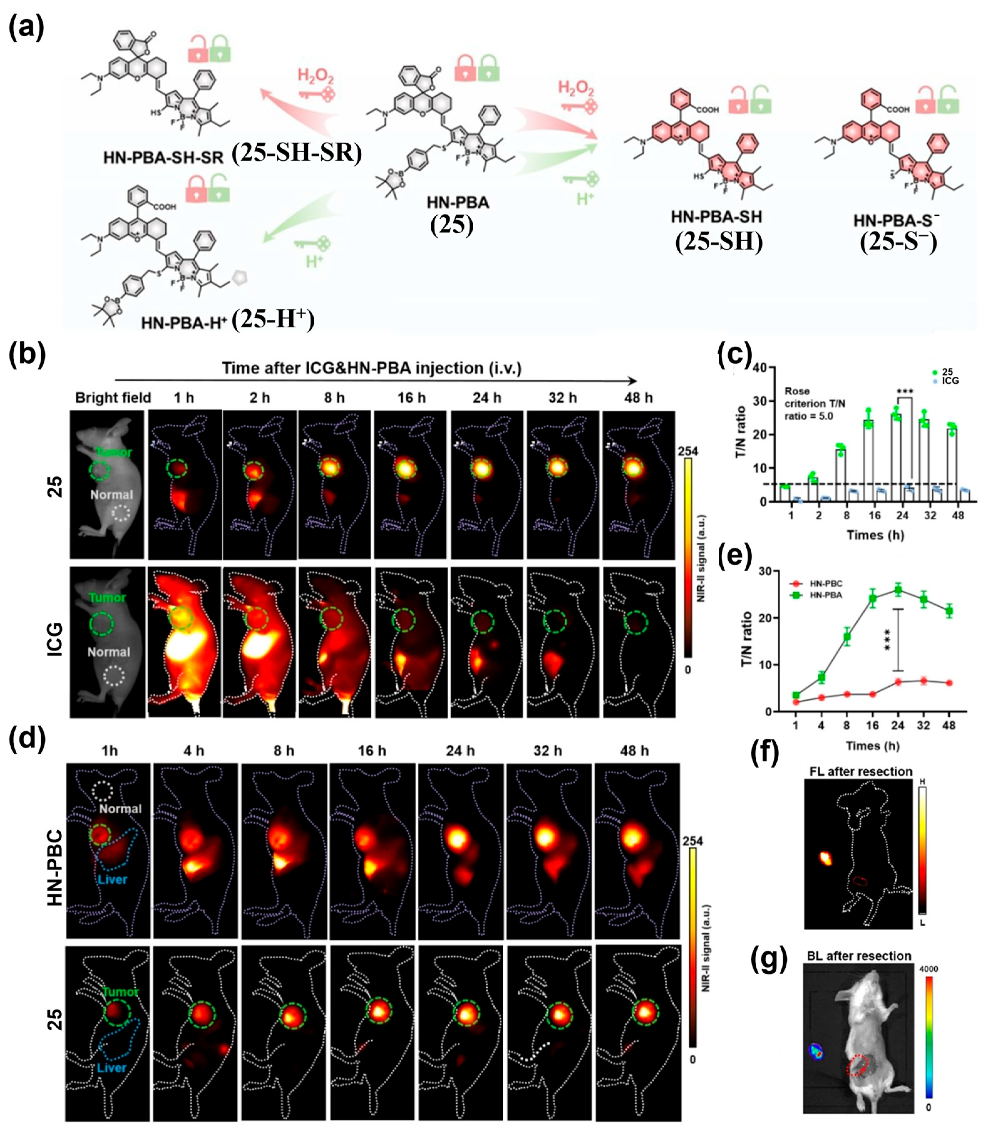
Xiong’s team developed NSCy-1050 (24), a probe that remains quenched in normal tissues but is activated by acidic TME and high intracellular viscosity—two hallmarks of many tumors—producing a strong NIR-II fluorescence signal (Figure 31a) [197]. After intravenous administration of 24 into 4T1 tumor-bearing mice, peak tumor fluorescence occurred at 48 h with a tumor-to-liver ratio (TLR) of 19.5:1, while no significant fluorescence was observed in the phosphate-buffer saline (PBS) group (Figure 31b,c). These results demonstrated the excellent NIR-II fluorescence imaging capability of 24 for tumor imaging in vivo (Figure 31b,c). Kim et al. developed HN-PBA (25), which exhibits NIR-II fluorescence activation under acidic pH and high H2O2 levels (Figure 32a) [198]. When administered intravenously to mice with subcutaneous 4T1 tumors, 25 generated a strong NIR-II fluorescence signal, with a T/N ratio 8.2-fold higher than ICG and 4.0-fold higher than the single-trigger H2O2-responsive control (HN-PBC) (Figure 32b–e). Intraoperative imaging with 25 enabled accurate identification and resection of primary tumors, achieving a T/N of 24.3 (Figure 32f,g).
Single-channel probes targeting redox species, pH, viscosity, and enzymes have demonstrated sensitive, real-time imaging in diverse tumor models. To further enhance specificity and reduce false positives, dual-/multi-stimuli designs that integrate orthogonal biochemical cues are emerging as a compelling strategy. Continued advances in probe chemistry, formulation, and pharmacokinetic tuning—alongside rigorous, quantitative imaging standards—will be critical to accelerate clinical translation.

4.2.2. Ratiometric Probes

Although activatable NIR-II single-channel probes enable selective “turn-on” of fluorescence signals, their signal intensity is susceptible to local probe concentration, instrument drift, and microenvironment fluctuations, thereby reducing detection reliability and potentially leading to false positive or false negative results [199,200]. Moreover, “always-on” and single-channel activatable NIR-II imaging typically provide qualitative readouts, limiting strict quantification. To address these limitations, ratiometric fluorescence strategies employ two emission channels to self-reference, effectively suppressing extrinsic variability and enabling accurate, quantitative detection of target analytes in complex biological environments [201].
By combining the high SNR, deep penetration, and high-fidelity capabilities of NIR-II fluorescence imaging with ratiometric reliability, various NIR-II ratiometric probes based on organic small-molecule fluorophores have been developed for sensitive in vivo imaging and accurate quantification, showing promising diagnostic potential. Depending on how the ratio is constructed, these probes can be broadly classified as dual-wavelength response probes and internally referenced probes.
Dual-Wavelength Response Probes
Dual-wavelength response probes are engineered with well-defined structural response features. Upon activation by tumor-associated biomarkers, they undergo chemical or conformational changes that cause predictable shifts in emission wavelength or intensity—such as blue- or red-shifts with increased or decreased brightness—resulting in a clear change in the fluorescence intensity ratio (FIR) between two channels. The two channels can arise from either two emission bands of a single molecule (single-fluorophore-based NIR-II ratiometric probes) or from two linked components (dual-fluorophore-based NIR-II ratiometric probes), which may be combined via chemical conjugation or co-encapsulation in a nanocarrier.
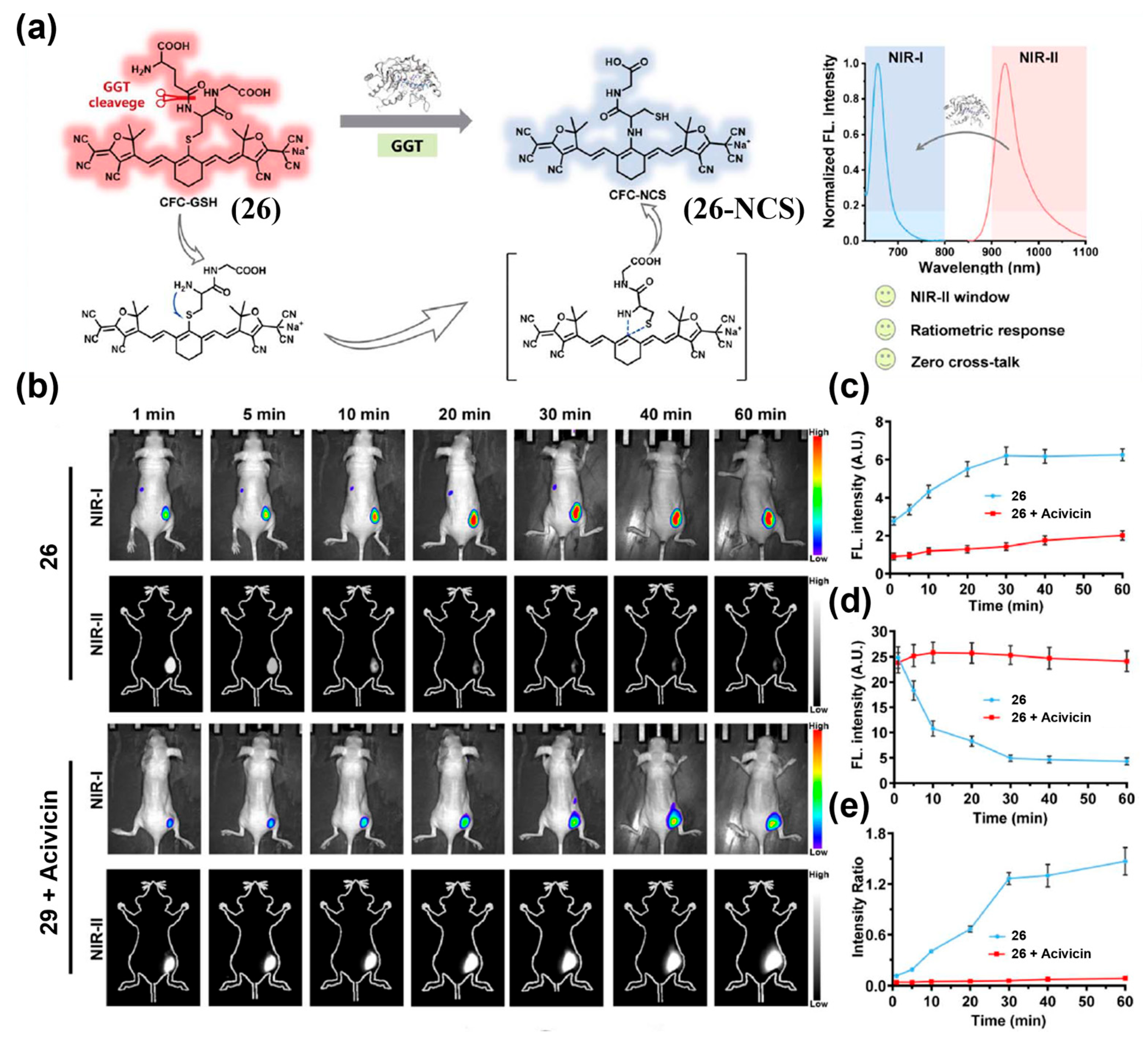
To construct a single fluorophore-based NIR-II ratiometric probe, Zeng et al. designed CFC-GSH (26) for imaging γ-Glutamyl transpeptidase (GGT) activity in tumors (Figure 33a) [202]. After intratumoral injection of 26 into a murine hepatocellular carcinoma xenograft model, a strong NIR-I fluorescence signal appeared within 1 min, peaked at 30 min, and remained stable, while the NIR-II signal decreased, causing the FIR to rise over time and plateau by about 30 min (Figure 33b–e). Pre-treatment with the GGT inhibitor acivicin markedly suppressed the ratiometric response of 26 by approximately 19.52-fold, confirming its specificity for GGT (Figure 33b–e). By self-calibration, 26 effectively mitigated interference from endogenous hepatic GGT activity, enabling accurate localization of peritoneal metastases and demonstrating potential for screening and diagnostic imaging.
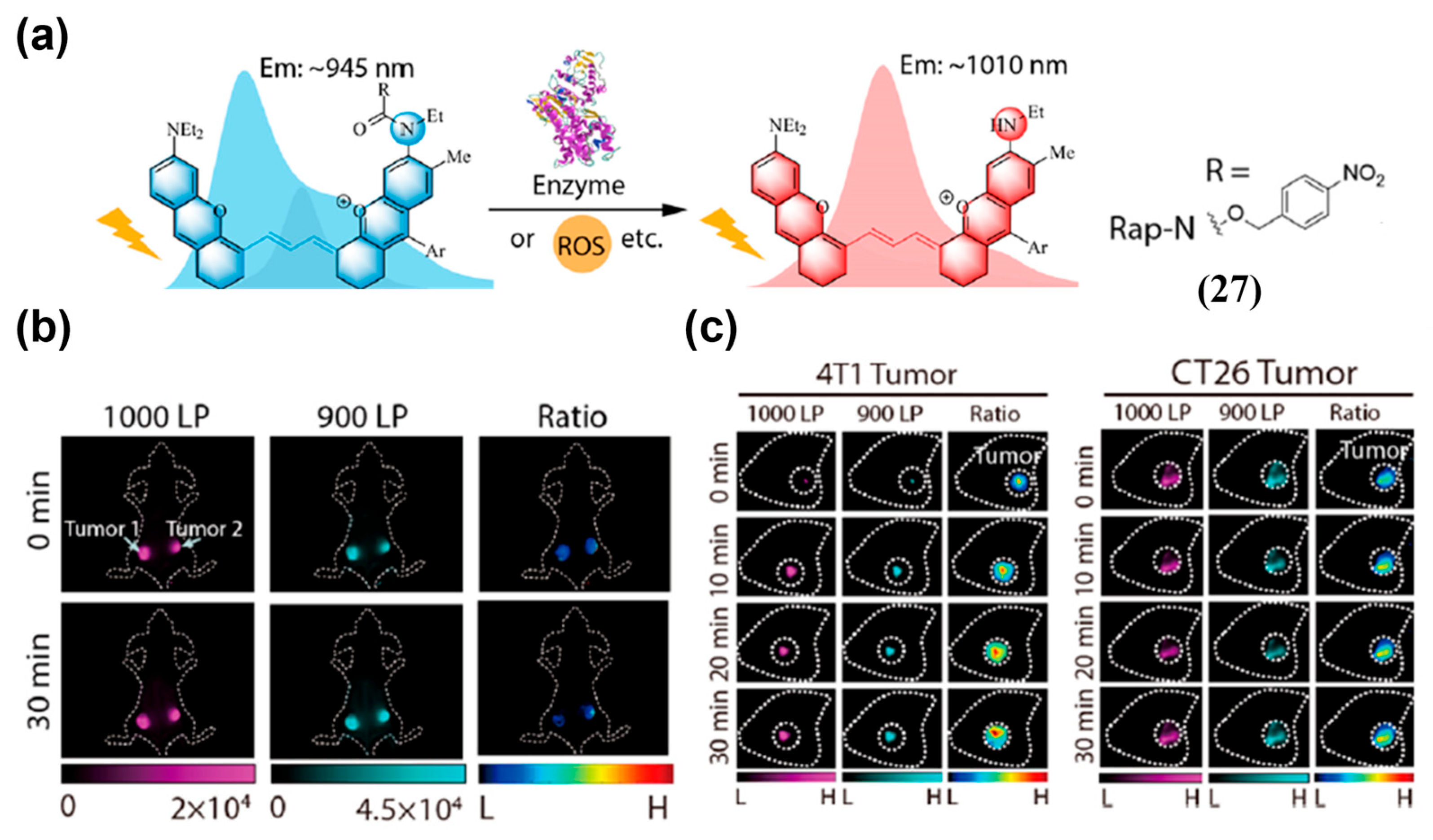
To enable NIR-II ratiometric fluorescence imaging with longer emission, Lei and colleagues synthesized Rap-N (27), based on a polymethine–thiourea scaffold for self-calibrated nitroreductase (NTR) imaging (Figure 34a) [131]. The maximal emission wavelength of 27 was 1010 nm and blue-shifted to 945 nm upon NTR-triggered acylation of its secondary amine, generating a distinct ratiometric signal (Figure 34a). In a 4T1 breast cancer model with in situ injection of 27, FIR (FL1000LP/FL900LP) of the NTR inhibitor-treated side remained stable, whereas it showed a gradual increase on the contralateral (uninhibited) side within 30 min (Figure 34b). In addition, progressive NIR-II fluorescence enhancement was observed in both 4T1 and CT-26 tumor models after in situ injection of 27, with a higher FIR in 4T1 tumors than in CT-26 tumors (~0.59 vs. ~0.53), consistent with the more severe hypoxia in 4T1 tumors (Figure 34c).
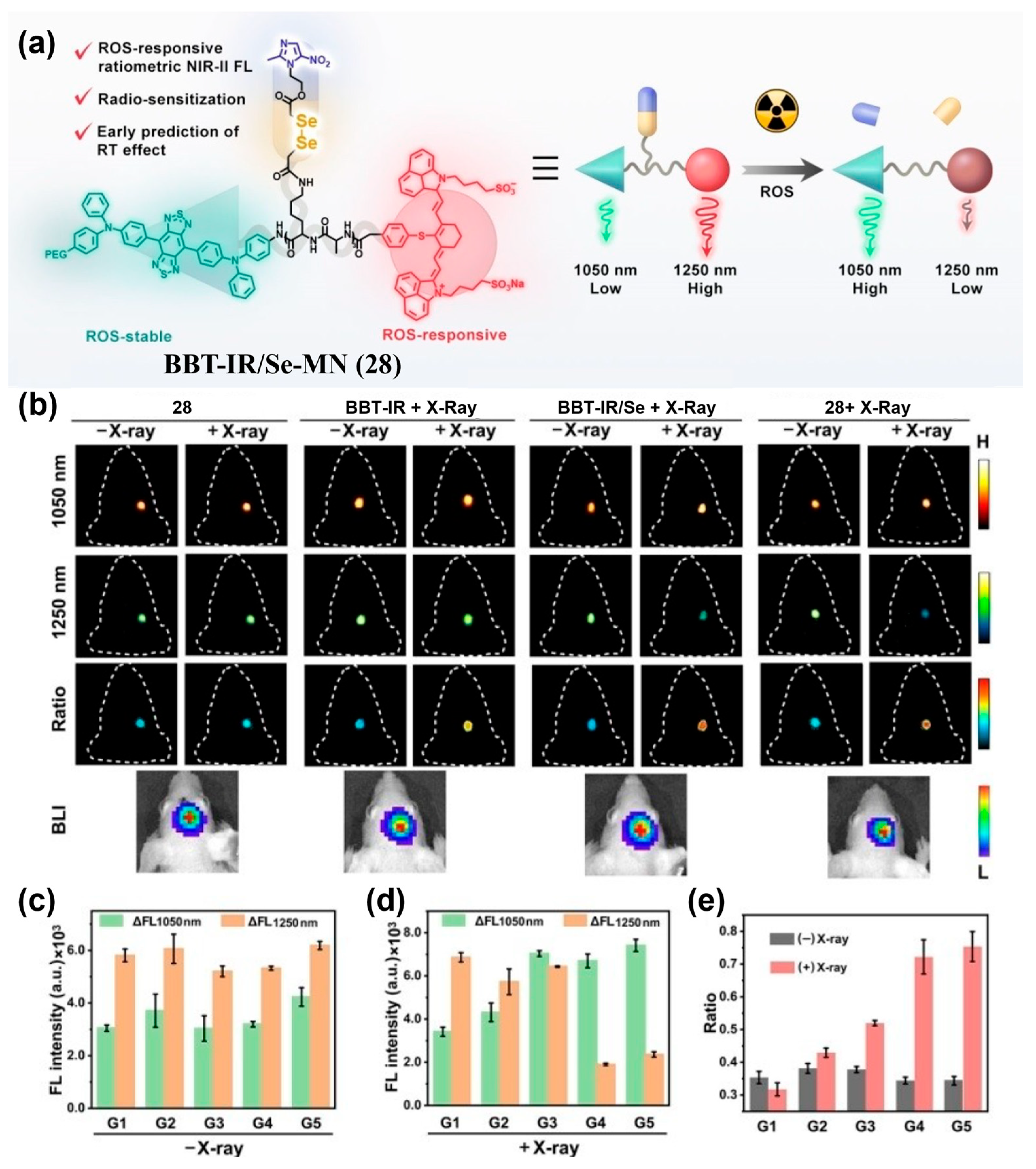
In addition to single-fluorophore-based NIR-II ratiometric probes, dual-fluorophore-based NIR-II ratiometric probes—where two channels originate from different fluorophores—are also commonly used for tumor imaging. Song et al. developed BBT-IR/Se-MN (28), covalently incorporating a ROS-insensitive D-A-D fluorophore (BBT) and a ROS-responsive cyanine dye (IR), enabling FRET from BBT to IR that quenches BBT emission at 1050 nm (Figure 35a) [203]. ROS exposure decomposed IR, disrupting FRET and restoring BBT’s NIR-II fluorescence, leading to a clear increase in the ratiometric signal (FL1050 nm/FL1250 nm). In an orthotopic U87 glioma mouse model, intravenous administration of 28 yielded immediate, high-contrast NIR-II fluorescence signals for precise tumor localization (Figure 35b). After radiotherapy, the 28-treated tumors showed significantly higher ratiometric changes compared with the controls in the groups receiving 28 + X-ray or BBT-IR/Se + X-ray (Figure 35b–e). This system enables quantitative ROS monitoring during radiotherapy, reducing tissue-specific confounders and supporting early evaluation of radiosensitivity.
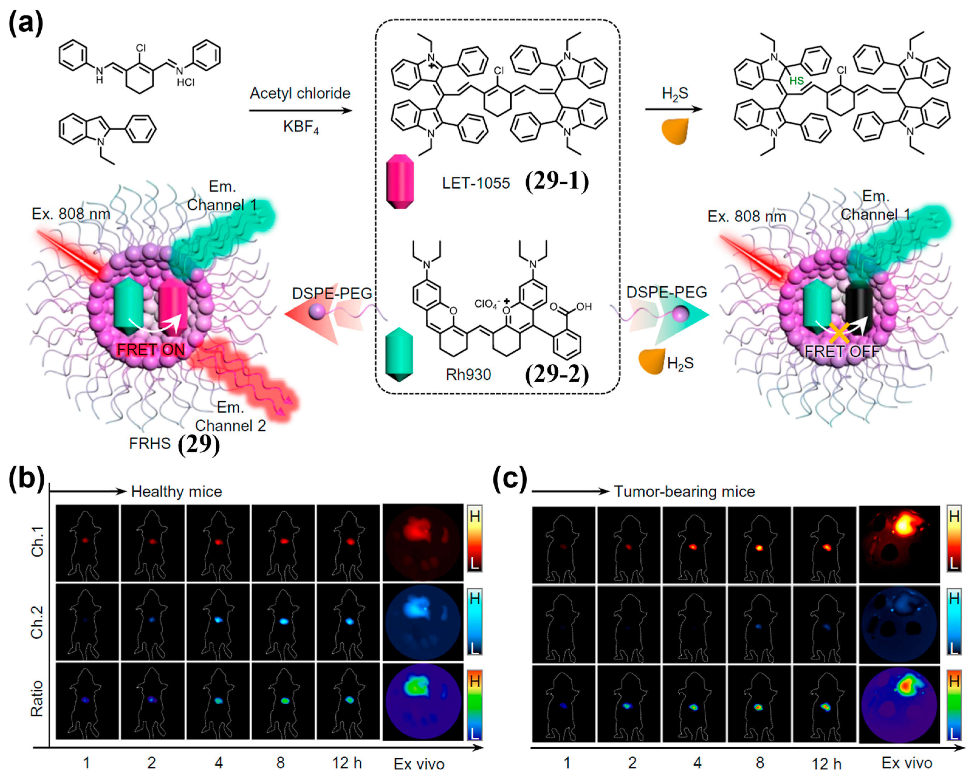
Nanoencapsulation is an effective alternative to covalent linking for incorporating two fluorophores into dual-fluorophore-based NIR-II ratiometric probes. Lin et al. developed a FRET-based H2S ratiometric probe (FRHS, 29), which was fabricated by DSPE-PEG2000-assisted encapsulation of an H2S-responsive acceptor (LET-1055, 29-1; NIR-II cyanine scaffold) and an H2S-inert donor (Rh930, 29-2; rhodamine–polymethine hybrid) (Figure 36a) [204]. Upon activation of 29 by H2S, its NIR-II fluorescence in Channel 1 (Ch.1, 900–1000 nm, attributed to 29-2) enhanced, while that in Channel 2 (Ch.2, 1000–1700 nm, attributed to 29-2) decreased (Figure 36a). After i.v. injection of 29 into orthotopic liver tumor-bearing mice, the tumor signal in Ch.1 markedly increased and that in Ch.2 slightly increased within 12 h post-injection, elevating the Ch.1/Ch.2 ratio (Figure 36c). Notably, tumor-bearing mice exhibited approximately 5.8-fold and 6.3-fold ratiometric enhancements in NIR-II fluorescence signals in the in vivo and ex vivo livers, respectively, compared to healthy mice (Figure 36b,c). These results demonstrated that 29 had the feasibility of in vivo ratiometric NIR-II fluorescence imaging of H2S.
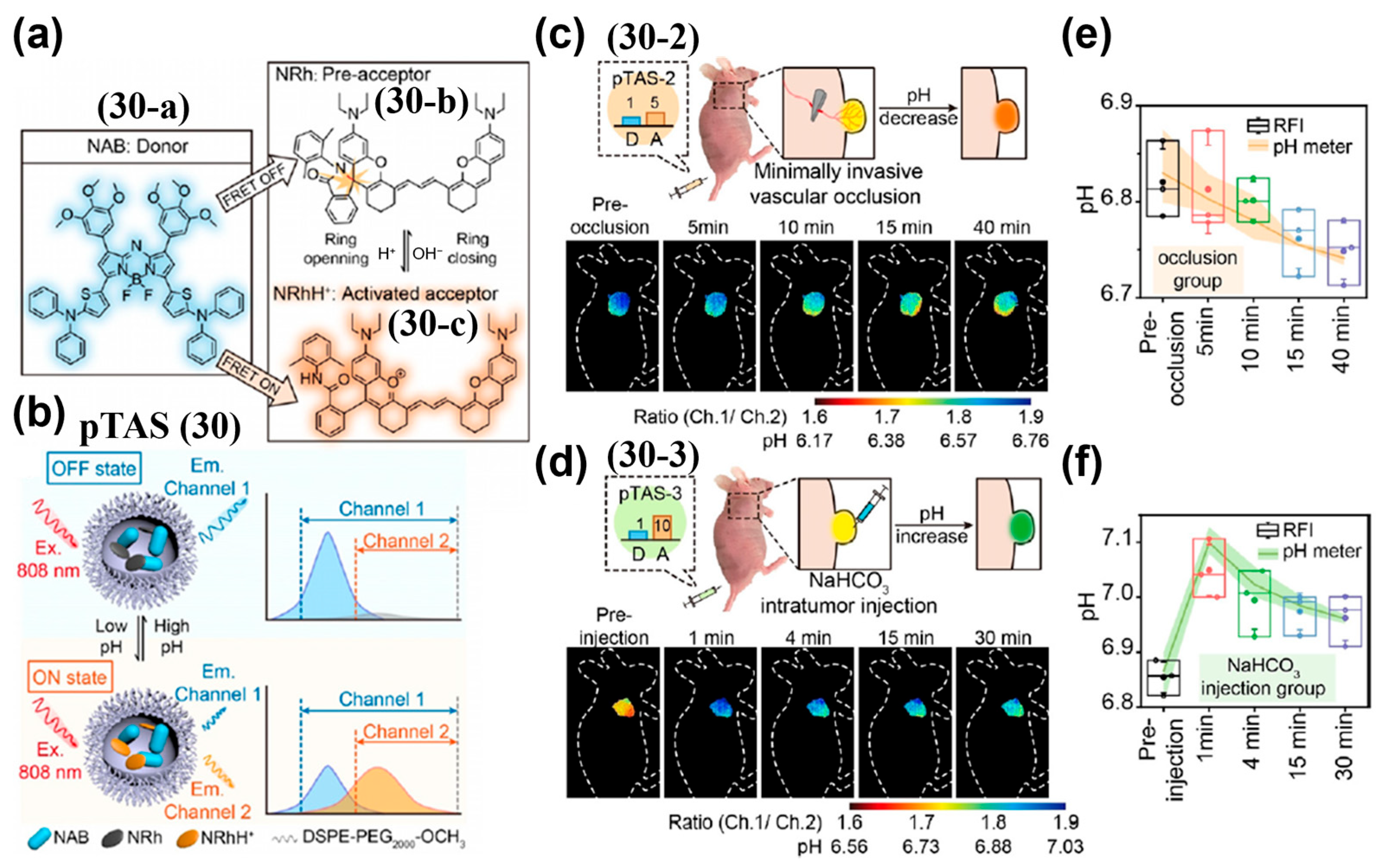
Stimuli-triggered FRET formation, like stimuli-triggered FRET disruption, is an effective approach for generating FIR and enables ratiometric fluorescence imaging. Zhang et al. developed a tunable NIR-II ratiometric pH sensor (pTAS, 30) by encapsulating the NIR-II donor aza-BODIPY (NAB, 30a) and the pH-responsive rhodamine–hydrazone polymethine pre-acceptor (NRh, 30-b) into DSPE-PEG2000-OCH3 micelles (Figure 37a,b) [205]. Under acidic conditions, 30-b was protonated to form NRhH+ (30-c), triggering FRET from 30-a to 30-c and increasing the FIR (Ch.1/Ch.2) (Figure 37a,b). Following i.v. injection into tumor-bearing mice, both pTAS-2 (30-2, 1:5 molar ration of NAB and NRh) and pTAS-3 (30-3, 1:10 molar ration of NAB and NRh) accumulated rapidly in tumors within 2 h and persisted for >24 h (Figure 37c–f). Their ratio signals stabilized at around 1.88 and 1.74, corresponding to pH 6.82 and 6.83, respectively, enabling a prolonged and stable imaging window (Figure 37e,f). These ratiometric readouts closely matched microelectrode pH measurements, with coefficients of variation below 0.31% and 0.77%, expanding the measurable range without loss of sensitivity.
Dual-wavelength response NIR-II ratiometric probes establish robust, self-calibrated readouts by translating target-triggered spectral shifts and intensity redistribution into quantitative FIR changes. Representative systems targeting key biomarkers (for example, GGT, NTR, ROS, pH, and ClO) have achieved sensitive, specific, and quantitative imaging across multiple tumor models and have facilitated therapy monitoring and intraoperative decision-making. Future priorities include scalable synthesis, pharmacokinetic optimization, and standardized ratiometric imaging workflows to accelerate clinical translation.
Internal Reference Probes
Internal-reference ratiometry uses a target-insensitive reference channel simultaneously with the target-responsive channel to enable real-time normalization. Minimizing spectral overlap between channels reduces crosstalk and improves accuracy and reproducibility of the ratiometric readout. This self-calibration strategy is conceptually straightforward and experimentally accessible, and promising for designing NIR-II ratiometric probes [201,206]. A common approach for constructing internal reference probes combines two optically complementary NIR-II emitters in a single nanoplatform, such as organic–lanthanide hybrids, organic–quantum dot composites, or all-organic assemblies—to ensure co-localization and matched pharmacokinetics in vivo.
Key design principles for robust internal-reference imaging include: (1) Spectral orthogonality—selecting two channels with well-separated excitation and emission to minimize crosstalk; (2) channel roles—combining a stable, target-insensitive reference channel under physiological conditions with a highly specific sensing channel that exhibits a strong dynamic response; (3) colocalization and pharmacokinetic (PK) matching—embedding both emitters in a single carrier to align biodistribution and clearance, enabling reliable self-calibration; (4) readout optimization—using orthogonal excitation/emission configurations or energy-transfer switches to maximize SNR and quantitative robustness. Below are representative internal-reference NIR-II ratiometric probes based on organic small-molecule fluorophores and their key performance features.
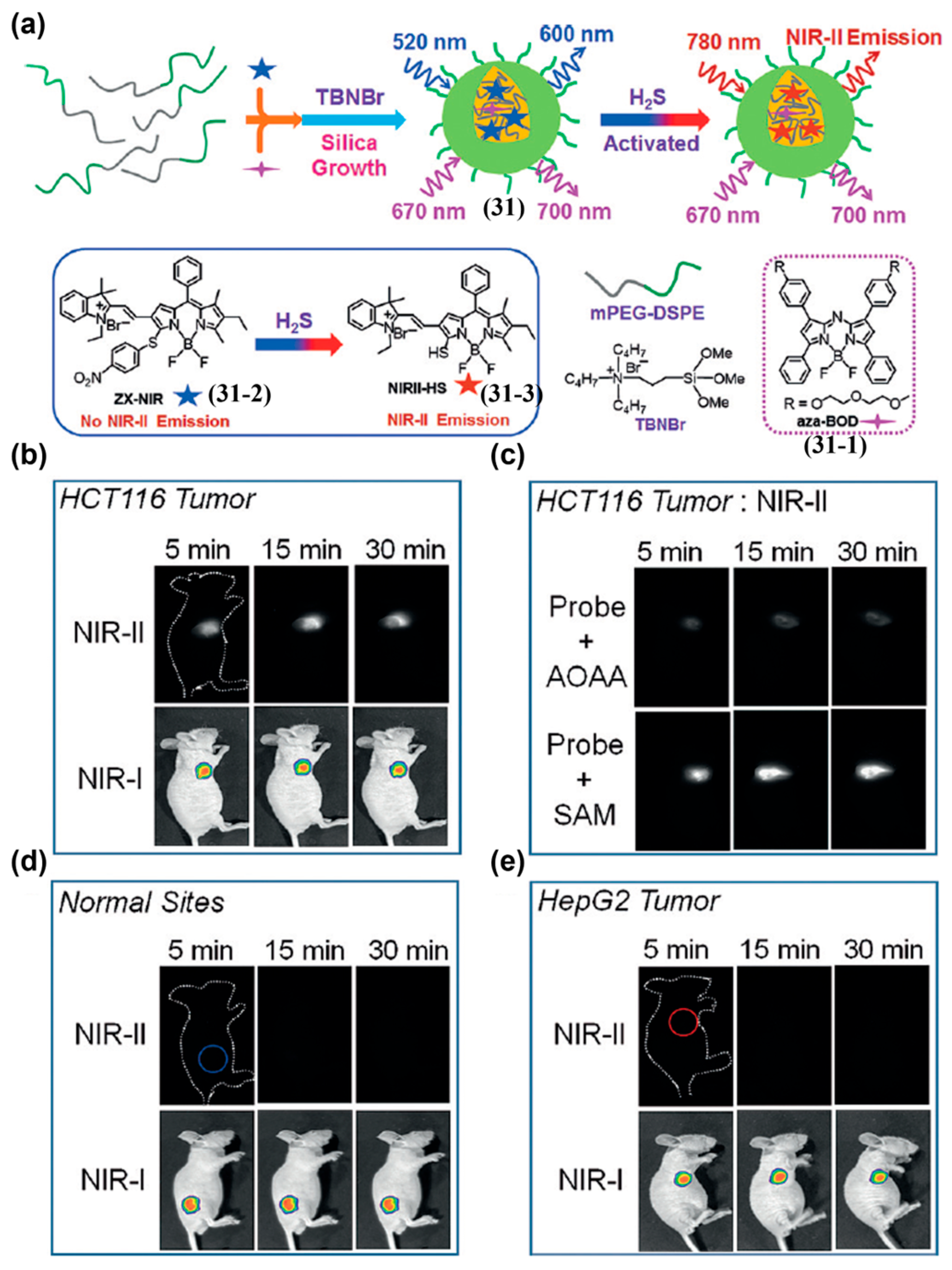
Tian and colleagues reported an H2S-activatable NIR-II ratiometric nanoprobe (31) for in vivo colorectal cancer detection (Figure 38a) [207]. This nanoprobe was constructed by encapsulating a H2S-inert aza-BODIPY dye (31-1; as the internal reference) and a H2S-responsive BODIPY dye (ZX-NIR, 31-2; for the sensing channel) within the hydrophobic interior of core–shell silica nanocomposites (Figure 38a). Upon injection, 31 produced a strong NIR-II tumor signal in HCT116 tumor-bearing mice within 1 min, stabilizing by 30 min, with a T/N ratio of approximately 5.7 (Figure 38b). The probe selectively identified H2S-rich colorectal cancer and, through dual-channel imaging, discriminated cancer types with distinct H2S levels (Figure 38b–e).
Formation of energy transfer system is a promising approach to design internal-reference NIR-II ratiometric probes. Song and co-workers designed a NO-responsive, dual-excitation NIR-II ratiometric nanoprobe (DERF-NO, 32), which was doped with BSBT as FRET acceptor and NO-responsive SIR-NO as FRET pre-donor (Figure 39a) [208]. Upon NO activation, SIR-NO transforms into SIR, triggering an intraparticle FRET system. This increases the NIR-II emission (1000–1700 nm) under 660 nm excitation (FL660Ex), while the 808 nm excited signal (FL808Ex) remains stable, enabling a reliable dual-excitation ratio (Figure 39a). After i.v. injection of 32 into 4T1 tumor-bearing mice pretreated with PBS or IFN-γ, the 660 nm–excited NIR-II signal increased continuously over 24 h in IFN-γ–treated tumor models, yielding a FL660Ex/FL808Ex ratio of 1.71 at 24 h post-injection of IFN-γ—approximately 1.39-fold higher than in PBS controls (Figure 39b). These results indicated the potential of 32 as a promising tool for visualizing and predicting macrophage polarization in tumor.
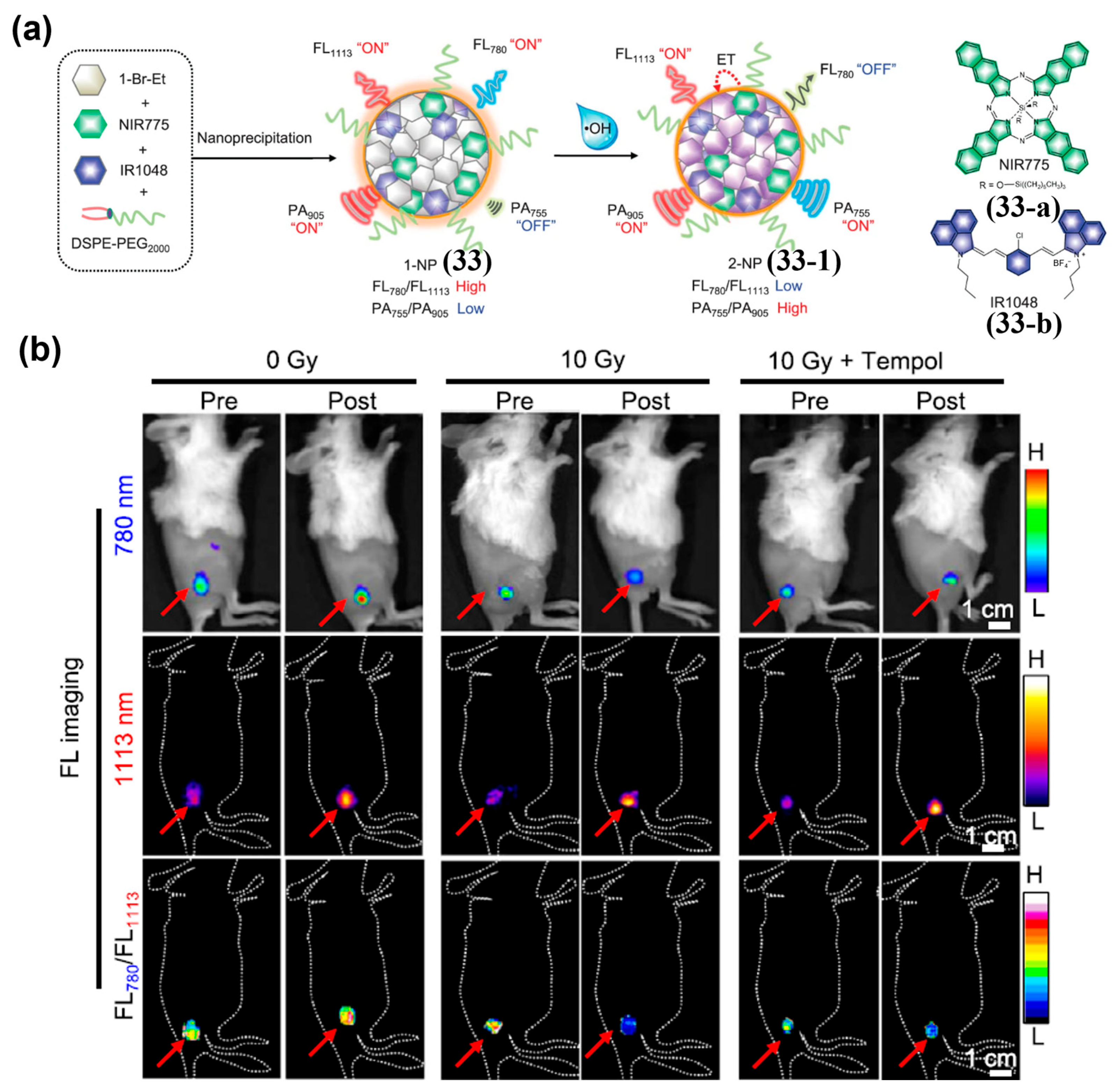
It is worth noting that in an internal-reference NIR-II ratiometric probe, the fluorophore used as the reference channel does not have to act as the donor or acceptor in the energy transfer system formed upon stimulation. Wu et al. optimized a diene optoelectronic material (1-Br-Et) and encapsulate it with a NIR-I fluorophore NIR775 and a NIR-II fluorophore IR1048 in a micellar nanoparticle to construct a hydroxyl radical (•OH )-responsive NIR ratiometric–photoacoustic (PA) probe (1-NP, 33) (Figure 40a) [209]. Oxidation of 1-Br-Et by •OH generates 2-Br-Et, which absorbs strongly at 767 nm and quenches the fluorescence of NIRI775 (FL780), while minimally affecting the NIR-II fluorescence of IR1048 (FL1113); thus, the FL780/FL1113 ratio decreases significantly (Figure 40a). After i.v. injection of 33 into 4T1 tumor-bearing mice following different radiotherapy doses, the tumor FL780 signal decreased significantly in the 10 Gy group, with a normalized ratio (ΔF780/ΔF1113 = 0.40 ± 0.07) reduced by approximately 2.7-fold, while the 0 Gy group showed minimal change (Figure 40b). These results demonstrated that 33 had the potential to predict tumor response to radiotherapy through •OH-triggered FL780/FL1113 ratio changes.
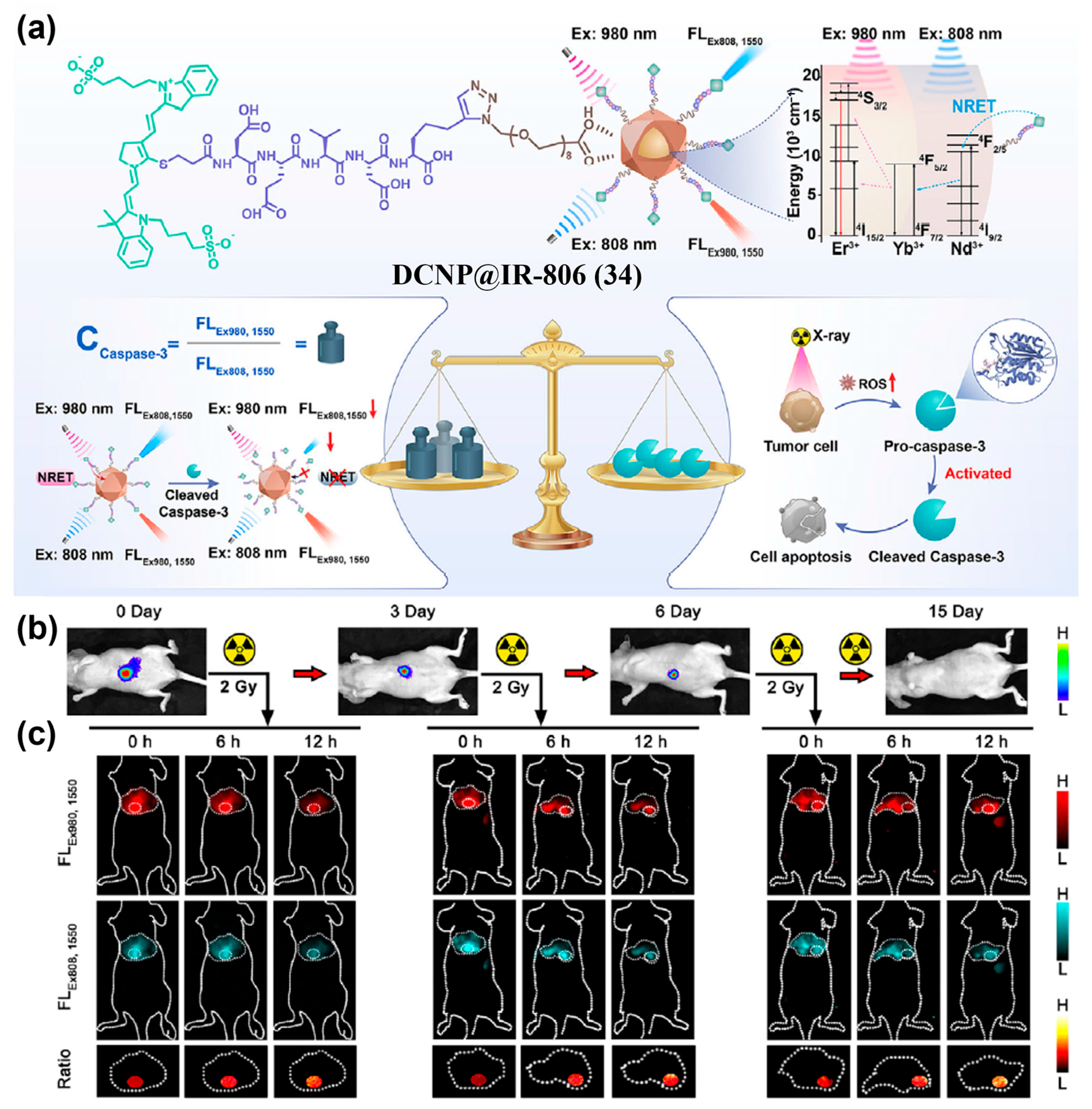
Apart from organic small-molecule fluorophores, the fluorophore used as the reference channel can also be inorganic materials. Yang et al. developed a dual-channel NIR-II ratiometric platform (DCNP@IR-806, 34) constructed by linking down-conversion nanoparticle (DCNP) and the antenna dye IR-806 via a caspase 3-responsive peptide (Figure 41a) [210]. Due to nonradiative energy transfer (NRET) from IR-806 to DCNP in 34, 1550 nm emission is detected under 808 nm excitation (FLEx808,1550), serving as the sensing channel, while 1550 nm emission under 980 nm excitation (FLEx980,1550) acts as the reference channel for self-calibration (Figure 41a). Upon cleavage of the peptide by caspase-3, the NRET system is disrupted, resulting in a decrease in the FLEx808,1550 signal, while the FLEx980,1550 signal remains stable. Therefore, the FLEx980,1550/FLEx808,1550 ratio can be used to monitor caspase-3 activity in real time. When 34 was applied to an orthotopic hepatocellular carcinoma (OHCC)-radiotherapy model, it was observed that the ratio of FLEx980,1550/FLEx808,1550 increased significantly with cumulative X-ray doses (from 64 on Day 0 to 80 on day 3 and over 100 on day 6), providing real-time feedback on treatment response and supporting timely optimization of radiotherapy regimens (Figure 41b,c). These results showed that this probe had great potential to quantitatively visualize caspase-3 activity in vivo.
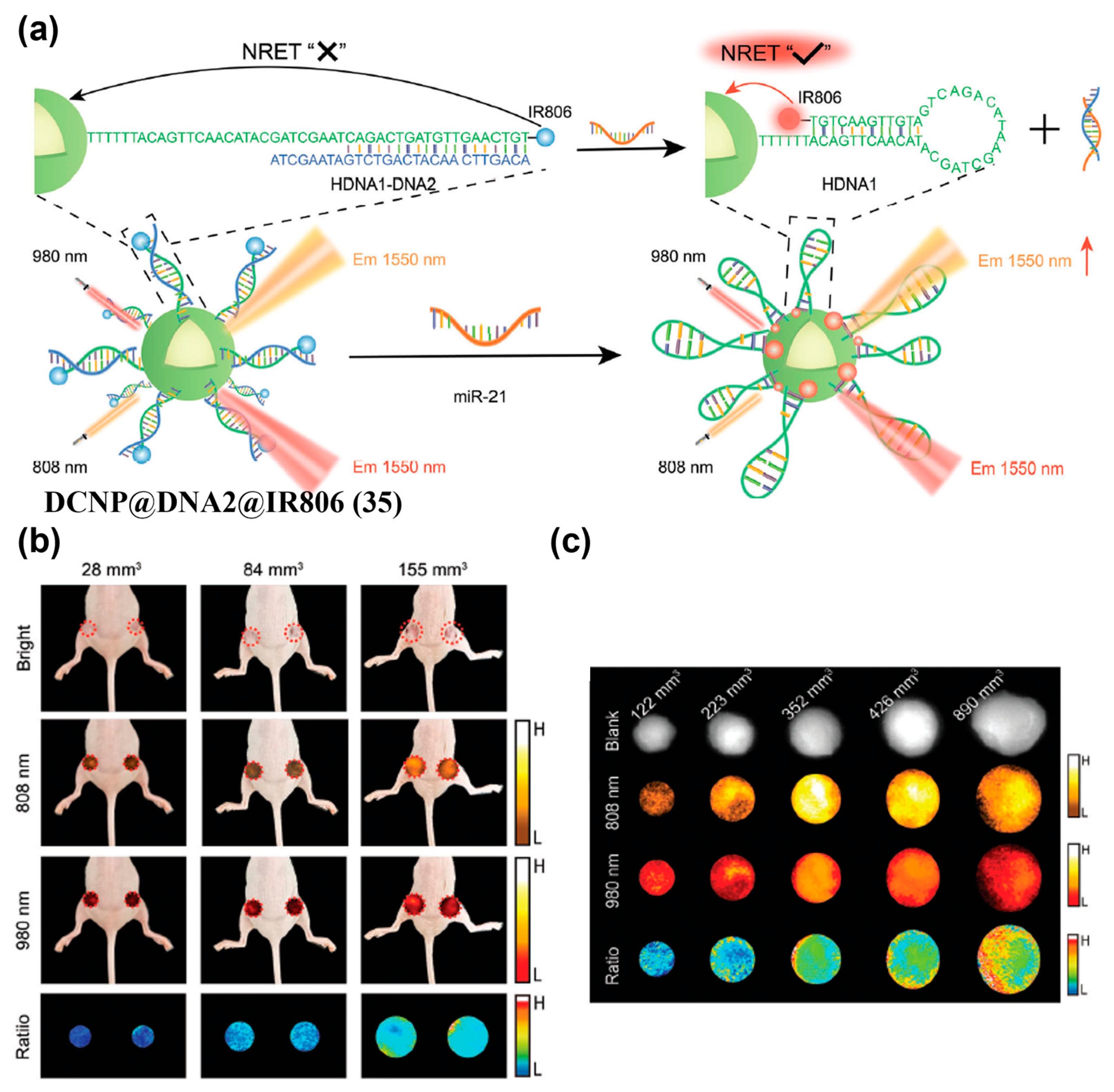
It should be noted that in a NRET effect-based internal reference probe, the NRET system may form upon response to the target stimulus. For example, Yang et al. developed a NIR-II ratiometric probe (DCNP@DNA2@IR806, 35) by covalently connecting DCNP and IR806 with miR-21-responsive DNA strand HDNA1-DNA2, enabling real-time, quantitative in vivo monitoring of miR-21 based on NRET (Figure 42a) [211]. Upon hybridization of HDNA1-DNA2 with miR-21, the proximity between IR806 and DCNP in 35 triggered a NRET effect that enhanced the 1550 nm emission of DCNP under 808 nm excitation (F1550,Ex808), while the 1550 nm emission under 980 nm excitation (F1550,Ex980) remains essentially constant (Figure 42a). At 18 h after i.v. injection of 35 into 4T1 tumor models, the ratio F1550,Ex808/F1550,Ex980 increased with tumor volume, enabling real-time tracking of tumor initiation and progression and facilitating early diagnosis (Figure 42b,c).
Internal-reference NIR-II ratiometric probes leverage a “target-sensitive plus target-inert” self-calibration framework to minimize the impact of systemic perturbations—such as probe distribution, excitation drift, and tissue optical heterogeneity—thereby enhancing the credibility of in vivo quantification. Representative systems targeting H2S, NO, •OH, miRNA, and radiotherapy-related enzyme activity have demonstrated sensitive and specific quantitative imaging across multiple tumor models, enabling therapeutic assessment and precision intervention.
Future research on internal-reference NIR-II ratiometric probes should focus on: (1) Spectral and instrument orthogonality—optimizing channel separation and device-compatible designs, such as dual-excitation or single-excitation dual-emission systems; (2) colocalization and PK matching—designing materials to ensure co-localized emitters and consistent pharmacokinetics through unified carriers and interfacial control; (3) standardized workflows—establishing consistent ratiometric imaging protocols and cross-platform calibration to enhance comparability and translational readiness.

5. Summary and Outlook

NIR-II fluorescent probes based on organic small-molecule fluorophores have emerged as a versatile platform for noninvasive, in vivo tumor imaging owing to their favorable optical properties, biocompatibility, and tunable pharmacokinetics. This review covers recent advances in molecular design and optical optimization, such as enhanced quantum yield and red-shifted emission, and their applications in D-A-D scaffolds, cyanines, BODIPY, and xanthene-based dyes. Two principal imaging strategies have matured: (1) “Always-on” probes that accumulate in tumors via the EPR effect or through active targeting (for example, antibodies, peptides); (2) “activatable” probes that respond to TME biomarkers (for example, enzymes, pH, reactive oxygen species) to achieve higher signal-to-background ratios and greater specificity. Collectively, these probes show substantial promise for delineating tumor margins, detecting micrometastases, guiding surgical resection, and enabling theranostics.
However, the reported probes all have advantages and limitations. Always-on probes, particularly those with passive targeting, are easy to prepare and convenient to apply, making them promising for clinical translation. Nevertheless, their imaging contrast is inherently limited by accumulation efficiency and is prone to high background signals. Though active-targeting probes enhance specificity and accumulation at the target site, yielding higher SBRs, their effectiveness may be affected by heterogeneous receptor expression and the possible immunogenicity of targeting ligands in the tumor microenvironment. Activatable probes represent a significant advancement in specificity, as they exhibit low background signals and generate a strong turn-on response to specific biomarkers, thereby enabling the detection of pathological activities. Nonetheless, their design is complex, and their fluorescence performance may be affected by target biomarker variability and concentration in the complex tumor microenvironment. Ratiometric probes possess a self-calibrating function that reduces interference from non-target factors and improves quantification reliability. However, this benefit requires more complex molecular or nanoscale engineering and may complicate data acquisition and interpretation. Therefore, before choosing a probe for biomedical applications, trade-offs must be considered based on the biological question, the required specificity and quantitative demands, as well as the feasibility of clinical translation.
In addition, some key challenges remain to be overcome for the clinical translation of NIR-II fluorescent probes based on organic small-molecule NIR-II fluorophores. (1) The trade-off between brightness and wavelength: Extending conjugation for longer-wavelength emission typically lowers fluorescence quantum yield, making it difficult to optimize both brightness and penetration depth simultaneously. (2) Insufficient in vivo characterization: Metabolic pathways, long-term toxicity, and immunogenicity are not fully understood, and targeted probes need improved molecular specificity. (3) TME heterogeneity: Spatial and temporal variations may lead to inconsistent responses in activatable probes, reducing reliability and generalizability.
To address the aforementioned issues, research efforts should focus on the following aspects. (1) Novel luminophores and AI-assisted design: Move beyond conventional frameworks by exploring new emissive cores and leveraging computational and AI tools to co-optimize emission wavelength and quantum yield. (2) Multimodal integration: Combine NIR-II fluorescence with PA, photothermal, ultrasound, MRI, or CT to provide complementary, multidimensional information and enhance deep-tissue quantification. (3) Logic-gated activatable probes: Implement multi-stimuli (for example, AND/OR) designs that require co-localized TME cues to enhance specificity and reduce false positives. (4) Dynamic theranostic monitoring: Develop probes to monitor treatment responses in real time, such as, apoptosis, immune infiltration, drug resistance, to support adaptive, individualized therapy. (5) Translational standardization: Establish standardized manufacturing and evaluation pipelines, conduct systematic biosafety assessments, and expand into procedural settings such as endoscopy and interventional radiology to bridge the translational gap.
In summary, NIR-II fluorescent probes based on organic small-molecule NIR-II fluorophores offer a powerful avenue for precise tumor imaging. With continued innovation in molecular engineering, intelligent imaging strategies, and standardized translational pathways, these technologies are poised to drive significant advances in early diagnosis, image-guided surgery, and personalized cancer treatment.

Author Contributions

Conceptualization, W.D.; writing—original draft preparation, M.G.; writing—review and editing, W.D., X.H. and M.G.; supervision, W.D. and X.H.; project administration, W.D. and X.H. All authors have read and agreed to the published version of the manuscript.

Funding

This work was funded by the National Natural Science Foundation of China (No. 22204021 and U24A20509), Science and Technology Innovation Joint Fund Program of Fujian Province, China (2024Y9654).

Institutional Review Board Statement

Not applicable.

Informed Consent Statement

Not applicable.

Data Availability Statement

Not applicable.

Conflicts of Interest

The authors declare no conflicts of interest.

References

  1. Ansell, S.M.; Vonderheide, R.H. Cellular Composition of the Tumor Microenvironment. Am. Soc. Clin. Oncol. Educ. Book 2013, 31, e91–e97. [Google Scholar] [CrossRef] [PubMed]
  2. Yang, F.; Yu, Y. Tumor Microenvironment--the Critical Element of Tumor Metastasis. Chin. J. Lung Cancer 2015, 18, 48–54. [Google Scholar]
  3. Wang, D.; Lu, J. Effects of Tumor Microenvironment on Tumor Angiogenesis. Tumor 2018, 38, 379–385. [Google Scholar]
  4. Satilmis, B.; Sahin, T.T.; Cicek, E.; Akbulut, S.; Yilmaz, S. Hepatocellular Carcinoma Tumor Microenvironment and Its Implications in Terms of Anti-Tumor Immunity: Future Perspectives for New Therapeutics. J. Gastrointest. Cancer 2021, 52, 1198–1205. [Google Scholar] [CrossRef]
  5. Wu, Y.; Zhang, W.; Li, J.; Zhang, Y. Optical Imaging of Tumor Microenvironment. Am. J. Nucl. Med. Molec. Imaging 2013, 3, 1–15. [Google Scholar]
  6. Yang, Y.; Jiang, Y.; Hu, X.; Shao, Z. Research Progress of Breast Tumor Microenvironment-Targeted Therapy. China Oncol. 2019, 29, 977–984. [Google Scholar]
  7. Yang, P.; Sun, Y.; Wang, Z.; Zheng, Y.; Chen, G.; Shi, Y. Research Progress on Effect of Tumor Acidic Microenvironment on Immune Cells. Chin. J. Immunol. 2021, 37, 613–617. [Google Scholar]
  8. Xiao, Y.; Yu, D. Tumor Microenvironment as A Therapeutic Target in Cancer. Pharmacol. Ther. 2021, 221, 107753. [Google Scholar] [CrossRef] [PubMed]
  9. Mieog, J.S.D.; Achterberg, F.B.; Zlitni, A.; Hutteman, M.; Burggraaf, J.; Swijnenburg, R.J.; Gioux, S.; Vahrmeijer, A.L. Fundamentals and Developments in Fluorescence-Guided Cancer Surgery. Nat. Rev. Clin. Oncol. 2022, 19, 9–22. [Google Scholar] [CrossRef]
  10. Tian, M.C.; Wu, R.L.G.; Xiang, C.H.; Niu, G.L.; Guan, W.J. Recent Advances in Fluorescent Probes for Cancer Biomarker Detection. Molecules 2024, 29, 1168. [Google Scholar] [CrossRef]
  11. Ijaz, M.; Hasan, I.; Aslam, B.; Yan, Y.Q.; Zeng, W.J.; Gu, J.S.; Jin, J.; Zhang, Y.H.; Wang, S.H.; Xing, L.; et al. Diagnostics of Brain Tumor in The Early Stage: Current Status and Future Perspectives. Biomater. Sci. 2025, 13, 2580–2605. [Google Scholar] [CrossRef]
  12. Komekbay, Z.; Shirazi, R.; Yessultanova, G.; Garifollin, A.; Tulyayeva, A.; Kereyeva, N.; Akhmetova, S.; Kaliev, A. Bibliometric Analysis of Tumor Marker Application in Gastric Cancer Diagnosis From 2019 To 2024. Front. Med. 2025, 12, 1547850. [Google Scholar] [CrossRef]
  13. Rosch, T.; Lorenz, R.; Braig, C.; Feuerbach, S.; Siewert, J.R.; Schusdziarra, V.; Classen, M. Endoscopic Ultrasound in Pancreatic Tumor-Diagnosis. Gastrointest. Endosc. 1991, 37, 347–352. [Google Scholar] [CrossRef]
  14. Raghavan, M. Conventional Modalities and Novel, Emerging Imaging Techniques for Musculoskeletal Tumors. Cancer Control 2017, 24, 161–171. [Google Scholar] [CrossRef]
  15. Savastano, L.E.; Zhou, Q.; Smith, A.; Vega, K.; Murga-Zamalloa, C.; Gordon, D.; McHugh, J.; Zhao, L.L.; Wang, M.M.; Pandey, A.; et al. Multimodal Laser-Based Angioscopy for Structural, Chemical and Biological Imaging of Atherosclerosis. Nat. Biomed. Eng. 2017, 1, 0023. [Google Scholar] [CrossRef]
  16. Ma, Y.Y.; Yan, C.X.; Guo, Z.Q.; Tan, G.; Niu, D.C.; Li, Y.S.; Zhu, W.H. Spatio-Temporally Reporting Dose-Dependent Chemotherapy via Uniting Dual-Modal MRI/NIR Imaging. Angew. Chem. Int. Ed. 2020, 59, 21143–21150. [Google Scholar] [CrossRef]
  17. Debs, P.; Ahlawat, S.; Fayad, L.M. Bone Tumors: State-of-the-Art Imaging. Skelet. Radiol. 2024, 53, 1783–1798. [Google Scholar] [CrossRef] [PubMed]
  18. Zhou, J.Y.; Lim, T.K.; Chong, V.; Huang, J. Segmentation and Visualization of Nasopharyngeal Carcinoma Using MRI. Comput. Biol. Med. 2003, 33, 407–424. [Google Scholar] [CrossRef] [PubMed]
  19. Rockall, A.G.; Reznek, R.H. Imaging of Neuroendocrine Tumours (CT/MR/US). Best Pract. Res. Clin. Endoc. Metab. 2007, 21, 43–68. [Google Scholar] [CrossRef] [PubMed]
  20. Budäus, L.; Leyh-Bannurah, S.R.; Salomon, G.; Michl, U.; Heinzer, H.; Huland, H.; Graefen, M.; Steuber, T.; Rosenbaum, C. Initial Experience of 68Ga-PSMA PET/CT Imaging in High-Risk Prostate Cancer Patients Prior to Radical Prostatectomy. Eur. Urol. 2016, 69, 393–396. [Google Scholar] [CrossRef]
  21. Cai, Q.Y.; Kim, S.H.; Choi, K.S.; Kim, S.Y.; Byun, S.J.; Kim, K.W.; Park, S.H.; Juhng, S.K.; Yoon, K.H. Colloidal Gold Nanoparticles as A Blood-Pool Contrast Agent for X-Ray Computed Tomography in Mice. Investig. Radiol. 2007, 42, 797–806. [Google Scholar] [CrossRef] [PubMed]
  22. Kim, S.H.; Lee, J.M.; Lee, M.W.; Kim, G.H.; Han, J.K.; Choi, B.I. Sonography Transmission Gel as Endorectal Contrast Agent for Tumor Visualization in Rectal Cancer. Am. J. Roentgenol. 2008, 191, 186–189. [Google Scholar] [CrossRef]
  23. Hoogstins, C.E.S.; Handgraaf, H.J.M.; Boogerd, L.S.F.; Burggraaf, J.; Vahrmeijer, A.L. Image Guided Surgery Using Near-Infrared Fluorescence: Road to Clinical Translation of Novel Probes for Real Time Tumor Visualization. In Proceedings of the Conference on Molecular-Guided Surgery—Molecules, Devices, and Applications III, San Francisco, CA, USA, 28–30 January 2017. [Google Scholar]
  24. Digital Medical Association of Chinese Medical Association; Digital Intelligent Surgery Professional Committee of Chinese Research Hospital; Liver Cancer Professional Committee of Chinese Medical Doctor Association; Clinical Precise Medicine Professional Committee; Medical Imaging and Equipment Professional Committee of China Graphics Society; Molecular Imaging Professional Committee of China Biophysical Society. Guidelines for Application of Computer-Assisted Indocyanine Green Molecular Fluorescence Imaging in Diagnosis and Surgical Navigation of Liver Tumors (2019). J. South. Med. Univ. 2019, 39, 1127–1140. [Google Scholar]
  25. Meng, X.; Pang, X.; Zhang, K.; Gong, C.; Yang, J.; Dong, H.; Zhang, X. Recent Advances in Near-Infrared-II Fluorescence Imaging for Deep-Tissue Molecular Analysis and Cancer Diagnosis. Small 2022, 18, 2202035. [Google Scholar] [CrossRef]
  26. Jo, G.; Park, Y.; Park, M.H.; Hyun, H. Rational Design of a Small Molecular Near-Infrared Fluorophore for Improved In Vivo Fluorescence Imaging. Materials 2023, 16, 7227. [Google Scholar] [CrossRef]
  27. Lim, I.; Yu Lin, E.; Garcia, J.; Jia, S.; Sommerhalter, R.E.; Ghosh, S.K.; Gladysz, J.A.; Sletten, E.M. Shortwave Infrared Fluorofluorophores for Multicolor in Vivo Imaging. Angew. Chem. Int. Ed. 2023, 62, e202215200. [Google Scholar] [CrossRef] [PubMed]
  28. Meador, W.E.; Lin, E.Y.; Lim, I.; Friedman, H.C.; Ndaleh, D.; Shaik, A.K.; Hammer, N.I.; Yang, B.; Caram, J.R.; Sletten, E.M.; et al. Silicon-Rosindolizine Fluorophores with Shortwave Infrared Absorption and Emission Profiles Enable In Vivo Fluorescence Imaging. Nat. Chem. 2024, 16, 970–978. [Google Scholar] [CrossRef]
  29. Hu, C.; Liu, H.; Zhang, Z.; Li, L.; Mao, G.-j.; Cheng, W.; Zhou, L. A Self-Calibrating Fluorescent-Photoacoustic Integrated Probe Enables Fast Visualizing Pancreatic Cancer and Imaging-Guided Tumor Surgery. Small 2025, 21, 2408527. [Google Scholar] [CrossRef] [PubMed]
  30. Lai, Y.; Dang, Y.J.; Li, F.L.; Ding, C.Y.; Yu, H.J.; Zhang, W.; Xu, Z.A. Reactive Glycolysis Metabolite-Activatable Nanotheranostics for NIR-II Fluorescence Imaging-Guided Phototherapy of Cancer. Adv. Funct. Mater. 2022, 32, 2200016. [Google Scholar] [CrossRef]
  31. Ren, H.; Zeng, X.; Zhao, X.; Hou, D.; Yao, H.; Yaseen, M.; Zhao, L.; Xu, W.; Wang, H.; Li, L. A Bioactivated In Vivo Assembly Nanotechnology Fabricated NIR Probe for Small Pancreatic Tumor Intraoperative Imaging. Nat. Commun. 2022, 13, 418. [Google Scholar] [CrossRef]
  32. Song, J.W.; Wang, H.; Meng, X.; Li, W.; Qi, J. A Hypoxia-Activated and Microenvironment-Remodeling Nanoplatform for Multifunctional Imaging and Potentiated Immunotherapy of Cancer. Nat. Commun. 2024, 15, 10395. [Google Scholar] [CrossRef]
  33. Xing, H.; Han, M.; Zhang, T.; Zeng, G.; He, J.; Xu, Z.; Kang, Y.; Xue, P. Fluorescence-Switchable Iron-Doped Nanodot Assembly as a Robust Redox Dyshomeostasis Amplifier for Noninvasive Treatment of Deep-Seated Tumors. Adv. Healthc. Mater. 2025, 14, 2500083. [Google Scholar] [CrossRef]
  34. Wan, H.; Yue, J.; Zhu, S.; Uno, T.; Zhang, X.; Yang, Q.; Yu, K.; Hong, G.; Wang, J.; Li, L.; et al. A Bright Organic NIR-II Nanofluorophore for Three-Dimensional Imaging into Biological Tissues. Nat. Commun. 2018, 9, 1171. [Google Scholar] [CrossRef] [PubMed]
  35. Zhang, X.; Li, L.; Ren, Y.; Li, M.; Tang, Y. Organic NIR-II Nanofluorophore with Ultrahigh Quantum Yield for Vessels Imaging and Fluorescence Image-Guided Surgery. Adv. Funct. Mater. 2025, 35, 2413341. [Google Scholar] [CrossRef]
  36. Kenry; Duan, Y.; Liu, B. Recent Advances of Optical Imaging in the Second Near-Infrared Window. Adv. Mater. 2018, 30, 1802394. [Google Scholar] [CrossRef]
  37. Tang, Y.; Li, Y.; Hu, X.; Zhao, H.; Ji, Y.; Chen, L.; Hu, W.; Zhang, W.; Li, X.; Lu, X.; et al. “Dual Lock-and-Key”-Controlled Nanoprobes for Ultrahigh Specific Fluorescence Imaging in the Second Near-Infrared Window. Adv. Mater. 2018, 30, 1801140. [Google Scholar] [CrossRef]
  38. Lu, L.; Li, B.; Ding, S.; Fan, Y.; Wang, S.; Sun, C.; Zhao, M.; Zhao, C.; Zhang, F. NIR-II Bioluminescence for In Vivo High Contrast Imaging and In Situ ATP-Mediated Metastases Tracing. Nat. Commun. 2020, 11, 4192. [Google Scholar] [CrossRef] [PubMed]
  39. Li, M.; Lu, Z.; Zhang, J.; Chen, L.; Tang, X.; Jiang, Q.; Hu, Q.; Li, L.; Liu, J.; Huang, W. Near-Infrared-II Fluorophore with Inverted Dependence of Fluorescence Quantum Yield on Polarity as Potent Phototheranostics for Fluorescence-Image-Guided Phototherapy of Tumors. Adv. Mater. 2023, 35, 2209647. [Google Scholar] [CrossRef]
  40. Sun, Z.; Li, T.; Wu, F.; Yao, T.; Yang, H.; Yang, X.; Yin, H.; Gao, Y.; Zhang, Y.; Li, C.; et al. Precise Synergistic Photothermal Therapy Guided by Accurate Temperature-Dependent NIR-II Fluorescence Imaging. Adv. Funct. Mater. 2024, 34, 2311622. [Google Scholar] [CrossRef]
  41. Xu, H.; Yuan, L.; Shi, Q.; Tian, Y.; Hu, F. Ultrabright NIR-II Nanoprobe for Image-Guided Accurate Resection of Tiny Metastatic Lesions. Nano Lett. 2024, 24, 1367–1375. [Google Scholar] [CrossRef]
  42. Yan, D.; Zhang, Z.; Zhang, J.; Li, X.; Wu, Q.; Gui, Y.; Zhu, J.; Kang, M.; Chen, X.; Tang, B.; et al. An All-Rounder for NIR-II Phototheranostics: Well-Tailored 1064 nm-Excitable Molecule for Photothermal Combating of Orthotopic Breast Cancer. Angew. Chem. Int. Ed. 2024, 63, e202401877. [Google Scholar] [CrossRef] [PubMed]
  43. Li, Y.; Qu, F.; Wan, F.; Zhong, C.; Rao, J.; Liu, Y.; Li, Z.; Zhu, J.; Li, Z. Aggregation Control of Anionic Pentamethine Cyanine Enabling Excitation Wavelength Selective NIR-II Fluorescence Imaging-Guided Photodynamic Therapy. Nat. Commun. 2025, 16, 762. [Google Scholar] [CrossRef]
  44. Tang, Y.; Li, Y.; He, C.; Wang, Z.; Huang, W.; Fan, Q.; Liu, B. NIR-II-Excited Off-On-Off Fluorescent Nanoprobes for Sensitive Molecular Imaging In Vivo. Nat. Commun. 2025, 16, 278. [Google Scholar] [CrossRef]
  45. Jiang, Y.; Pu, K. Dual-Targeted Molecular Fluorescence and Photoacoustic Imaging in the Second Near-Infrared Optical Window Using Organic Contrast Agents. Adv. Biosys. 2018, 2, 1700262. [Google Scholar] [CrossRef]
  46. Chen, G.; Zhang, Y.; Li, C.; Huang, D.; Wang, Q.; Wang, Q. Recent Advances in Tracking the Transplanted Stem Cells Using Near-Infrared Fluorescent Nanoprobes: Turning from the First to the Second Near-Infrared Window. Adv. Healthc. Mater. 2018, 7, 1800497. [Google Scholar] [CrossRef]
  47. Ceppi, L.; Bardhan, N.M.; Na, Y.; Siegel, A.; Rajan, N.; Fruscio, R.; Del Carmen, M.G.; Belcher, A.M.; Birrer, M.J. Real-Time Single-Walled Carbon Nanotube-Based Fluorescence Imaging Improves Survival after Debulking Surgery in an Ovarian Cancer Model. ACS Nano 2019, 13, 5356–5365. [Google Scholar] [CrossRef] [PubMed]
  48. Zebibula, A.; Alifu, N.; Xia, L.; Sun, C.; Yu, X.; Xue, D.; Liu, L.; Li, G.; Qian, J. Ultrastable and Biocompatible NIR-II Quantum Dots for Functional Bioimaging. Adv. Funct. Mater. 2018, 28, 2200386. [Google Scholar] [CrossRef]
  49. Zhang, Y.; Yang, H.; An, X.; Wang, Z.; Yang, X.; Yu, M.; Zhang, R.; Sun, Z.; Wang, Q. Controlled Synthesis of Ag2Te@Ag2S Core-Shell Quantum Dots with Enhanced and Tunable Fluorescence in the Second Near-Infrared Window. Small 2020, 16, 2001003. [Google Scholar] [CrossRef]
  50. Chen, L.; Zhao, L.; Wang, Z.; Liu, S.; Pang, D. Near-Infrared-II Quantum Dots for In Vivo Imaging and Cancer Therapy. Small 2022, 18, 2104567. [Google Scholar] [CrossRef]
  51. Zhong, Y.; Ma, Z.; Zhu, S.; Yue, J.; Zhang, M.; Antaris, A.L.; Yuan, J.; Cui, R.; Wan, H.; Zhou, Y.; et al. Boosting the Down-Shifting Luminescence of Rare-Earth Nanocrystals for Biological Imaging Beyond 1500 nm. Nat. Commun. 2017, 8, 737. [Google Scholar] [CrossRef] [PubMed]
  52. Chang, Y.; Chen, H.; Xie, X.; Wan, Y.; Li, Q.; Wu, F.; Yang, R.; Wang, W.; Kong, X. Bright Tm3+-Based Downshifting Luminescence Nanoprobe Operating Around 1800 nm for NIR-IIb and C Bioimaging. Nat. Commun. 2023, 14, 1079. [Google Scholar] [CrossRef]
  53. Tong, L.; Cao, J.; Wang, K.; Song, J.; Mu, J. Lanthanide-Doped Nanomaterials for Tumor Diagnosis and Treatment by Second Near-Infrared Fluorescence Imaging. Adv. Opt. Mater. 2024, 12, 2301767. [Google Scholar] [CrossRef]
  54. Dai, H.; Shen, Q.; Shao, J.; Wang, W.; Gao, F.; Dong, X. Small Molecular NIR-II Fluorophores for Cancer Phototheranostics. Innovation 2021, 2, 100082. [Google Scholar] [CrossRef]
  55. Liu, J.; Cheng, D.; Zhu, A.; Ding, M.; Yu, N.; Li, J. Neutrophil-Targeting Semiconducting Polymer Nanotheranostics for NIR-II Fluorescence Imaging-Guided Photothermal-NO-Immunotherapy of Orthotopic Glioblastoma. Adv. Sci. 2024, 11, 2406750. [Google Scholar] [CrossRef] [PubMed]
  56. Zhang, J.; Wang, W.; Shao, J.; Chen, J.; Dong, X. Small Molecular Cyanine Dyes for Phototheranostics. Coord. Chem. Rev. 2024, 516, 215986. [Google Scholar] [CrossRef]
  57. Mei, A.; He, X.; Lei, D.; Wang, L.; Wang, W.; Shao, J.; Shen, Q.; Jiang, F.; Dong, X. Squaraine-Based NIR Dyes for Phototheranostics. Coord. Chem. Rev. 2025, 527, 216419. [Google Scholar] [CrossRef]
  58. Yang, Q.; Hu, Z.; Zhu, S.; Ma, R.; Ma, H.; Ma, Z.; Wan, H.; Zhu, T.; Jiang, Z.; Liu, W.; et al. Donor Engineering for NIR-II Molecular Fluorophores with Enhanced Fluorescent Performance. J. Am. Chem. Soc. 2018, 140, 1715–1724. [Google Scholar] [CrossRef]
  59. Tian, R.; Ma, H.; Zhu, S.; Lau, J.; Ma, R.; Liu, Y.; Lin, L.; Chandra, S.; Wang, S.; Zhu, X.; et al. Multiplexed NIR-II Probes for Lymph Node-Invaded Cancer Detection and Imaging-Guided Surgery. Adv. Mater. 2020, 32, 1907365. [Google Scholar] [CrossRef]
  60. Mu, J.; Xiao, M.; Shi, Y.; Geng, X.; Li, H.; Yin, Y.; Chen, X. The Chemistry of Organic Contrast Agents in the NIR-II Window. Angew. Chem. Int. Ed. 2022, 61, e202114722. [Google Scholar] [CrossRef] [PubMed]
  61. Tian, R.; Feng, X.; Wei, L.; Dai, D.; Ma, Y.; Pan, H.; Ge, S.; Bai, L.; Ke, C.; Liu, Y.; et al. A Genetic Engineering Strategy for Editing Near-Infrared-II Fluorophores. Nat. Commun. 2022, 13, 2853. [Google Scholar] [CrossRef]
  62. Gao, D.; Luo, Z.; He, Y.; Yang, L.; Hu, D.; Liang, Y.; Zheng, H.; Liu, X.; Sheng, Z. Low-Dose NIR-II Preclinical Bioimaging Using Liposome-Encapsulated Cyanine Dyes. Small 2023, 19, 2206544. [Google Scholar] [CrossRef] [PubMed]
  63. Hu, Z.; Fang, C.; Li, B.; Zhang, Z.; Cao, C.; Cai, M.; Su, S.; Sun, X.; Shi, X.; Li, C.; et al. First-in-Human Liver-Tumor Surgery Guided by Multispectral Fluorescence Imaging in The Visible and Near-Infrared-I/II Windows. Nat. Biomed. Eng. 2020, 4, 259–271. [Google Scholar] [CrossRef]
  64. Li, B.; Tian, J.; Wu, C.; Li, Z.; Qiao, L.; Xie, Z.; Song, B.; Shan, Y.; Chen, S.; Tang, Y.; et al. Nitric Oxide-Activated Bioorthogonal Codelivery Nanoassembly for In Situ Synthesis of Photothermal Agent for Precise and Safe Anticancer Treatment. Adv. Mater. 2024, 36, 2405502. [Google Scholar] [CrossRef]
  65. Wang, L.; Li, N.; Wang, W.; Mei, A.; Shao, J.; Wang, W.; Dong, X. Benzobisthiadiazole-Based Small Molecular Near-Infrared-II Fluorophores: From Molecular Engineering to Nanophototheranostics. ACS Nano 2024, 18, 4683–4703. [Google Scholar] [CrossRef] [PubMed]
  66. Xu, L.; Zhang, Q.; Wang, X.; Lin, W. Biomedical Applications of NIR-II Organic Small Molecule Fluorescent Probes in Different Organs. Coord. Chem. Rev. 2024, 519, 216122. [Google Scholar] [CrossRef]
  67. Zhao, X.; Qin, Z.; Zhang, X.; Yuan, L. Research Progress in Activatable NIR-Ⅱ Small Molecule Fluorescent Probes. Chin. J. Appl. Chem. 2024, 41, 39–59. [Google Scholar]
  68. Antaris, A.L.; Chen, H.; Cheng, K.; Sun, Y.; Hong, G.; Qu, C.; Diao, S.; Deng, Z.; Hu, X.; Zhang, B.; et al. A Small-Molecule Dye for NIR-II Imaging. Nat. Mater. 2016, 15, 235–242. [Google Scholar] [CrossRef]
  69. Zeng, X.; Xiao, Y.; Lin, J.; Li, S.; Zhou, H.; Nong, J.; Xu, G.; Wang, H.; Xu, F.; Wu, J.; et al. Near-Infrared II Dye-Protein Complex for Biomedical Imaging and Imaging-Guided Photothermal Therapy. Adv. Healthc. Mater. 2018, 7, 1800589. [Google Scholar] [CrossRef]
  70. Li, B.; Lu, L.; Zhao, M.; Lei, Z.; Zhang, F. An Efficient 1064 nm NIR-II Excitation Fluorescent Molecular Dye for Deep-Tissue High-Resolution Dynamic Bioimaging. Angew. Chem. Int. Ed. 2018, 57, 7483–7487. [Google Scholar] [CrossRef]
  71. Cosco, E.D.; Caram, J.R.; Bruns, O.T.; Franke, D.; Day, R.A.; Farr, E.P.; Bawendi, M.G.; Sletten, E.M. Flavylium Polymethine Fluorophores for Near- and Shortwave Infrared Imaging. Angew. Chem. Int. Ed. 2017, 56, 13126–13129. [Google Scholar] [CrossRef] [PubMed]
  72. Bai, L.; Sun, P.; Liu, Y.; Zhang, H.; Hu, W.; Zhang, W.; Liu, Z.; Fan, Q.; Li, L.; Huang, W. Novel aza-BODIPY Based Small Molecular NIR-II Fluorophores for In Vivo Imaging. Chem. Commun. 2019, 55, 10920–10923. [Google Scholar] [CrossRef]
  73. Dou, K.; Feng, W.; Fan, C.; Cao, Y.; Xiang, Y.; Liu, Z. Flexible Designing Strategy to Construct Activatable NIR-II Fluorescent Probes with Emission Maxima beyond 1200 nm. Anal. Chem. 2021, 93, 4006–4014. [Google Scholar] [CrossRef]
  74. Lei, Z.; Sun, C.; Pei, P.; Wang, S.; Li, D.; Zhang, X.; Zhang, F. Stable, Wavelength-Tunable Fluorescent Dyes in the NIR-II Region for In Vivo High-Contrast Bioimaging and Multiplexed Biosensing. Angew. Chem. Int. Ed. 2019, 58, 8166–8171. [Google Scholar] [CrossRef] [PubMed]
  75. Liu, D.; He, Z.; Zhao, Y.; Yang, Y.; Shi, W.; Li, X.; Ma, H. Xanthene-Based NIR-II Dyes for In Vivo Dynamic Imaging of Blood Circulation. J. Am. Chem. Soc. 2021, 143, 17136–17143. [Google Scholar] [CrossRef]
  76. Qian, J.; Li, J.; Feng, Z.; Yu, X.; Wu, D.; Wu, T. Aggregation-Induced Emission Fluorophores Towards the Second Near- Infrared Optical Windows with Suppressed Imaging Background. Coord. Chem. Rev. 2022, 472, 214792. [Google Scholar]
  77. Hu, Z.; Feng, L.; Yang, P. 2, 1, 3-Benzothiadiazole Derivative Small Molecule Fluorophores for NIR-II Bioimaging. Adv. Funct. Mater. 2024, 34, 2310818. [Google Scholar] [CrossRef]
  78. Li, Y.; Zhou, H.; Bi, R.; Li, X.; Zha, M.; Yang, Y.; Ni, J.; Liew, W.H.; Olivo, M.; Yao, K.; et al. Acceptor Engineering of Small-Molecule Fluorophores for NIR-II Fluorescence and Photoacoustic Imaging. J. Mat. Chem. B 2021, 9, 9951–9960. [Google Scholar] [CrossRef]
  79. Zhou, H.; Lu, Z.; Zhang, Y.; Li, M.; Xue, D.; Zhang, D.; Liu, J.; Li, L.; Qian, J.; Huang, W. Simultaneous Enhancement of the Long-Wavelength NIR-II Brightness and Photothermal Performance of Semiconducting Polymer Nanoparticles. ACS Appl. Mater. Interfaces 2022, 14, 8705–8717. [Google Scholar] [CrossRef]
  80. Wang, Y.; Zhang, W.; Sun, P.; Cai, Y.; Xu, W.; Fan, Q.; Hu, Q.; Han, W. A Novel Multimodal NIR-II Nanoprobe for the Detection of Metastatic Lymph Nodes and Targeting Chemo-Photothermal Therapy in Oral Squamous Cell Carcinoma. Theranostics 2019, 9, 391–404. [Google Scholar] [CrossRef]
  81. Sun, P.; Chen, Y.; Sun, B.; Zhang, H.; Chen, K.; Miao, H.; Fan, Q.; Huang, W. Thienothiadiazole-Based NIR-II Dyes with D-A-D Structure for NIR-II Fluorescence Imaging Systems. ACS Appl. Bio Mater. 2021, 4, 4542–4548. [Google Scholar] [CrossRef] [PubMed]
  82. Li, Y.; Gao, J.; Wang, S.; Du, M.; Hou, X.; Tian, T.; Qiao, X.; Tian, Z.; Stang, P.; Li, S.; et al. Self-assembled NIR-II Fluorophores with Ultralong Blood Circulation for Cancer Imaging and Image-guided Surgery. J. Med. Chem. 2022, 65, 2078–2090. [Google Scholar] [CrossRef]
  83. Li, Y.; Gao, J.; Wang, S.; Li, S.; Hou, X.; Pan, Y.; Gao, J.; Qiao, X.; Tian, Z.; Chen, D.; et al. Organic NIR-II Dyes with Ultralong Circulation Persistence for Image-Guided Delivery and Therapy. J. Control. Release 2022, 342, 157–169. [Google Scholar] [CrossRef] [PubMed]
  84. Ji, A.; Lou, H.; Qu, C.; Lu, W.; Hao, Y.; Li, J.; Wu, Y.; Chang, T.; Chen, H.; Cheng, Z. Acceptor Engineering for NIR-II Dyes with High Photochemical and Biomedical Performance. Nat. Commun. 2022, 13, 3815. [Google Scholar] [CrossRef]
  85. Liu, X.; Yu, B.; Shen, Y.; Cong, H. Design of NIR-II High Performance Organic Small Molecule Fluorescent Probes and Summary of Their Biomedical Applications. Coord. Chem. Rev. 2022, 468, 214609. [Google Scholar] [CrossRef]
  86. Yi, W.; Zhou, H.; Li, A.; Yuan, Y.; Guo, Y.; Li, P.; Qi, B.; Xiao, Y.; Yu, A.; Hu, X. A NIR-II Fluorescent Probe for Articular Cartilage Degeneration Imaging and Osteoarthritis Detection. Biomater. Sci. 2019, 7, 1043–1051. [Google Scholar] [CrossRef]
  87. Fang, Y.; Shang, J.; Liu, D.; Shi, W.; Li, X.; Ma, H. Design, Synthesis, and Application of a Small Molecular NIR-II Fluorophore with Maximal Emission beyond 1200 nm. J. Am. Chem. Soc. 2020, 142, 15271–15275. [Google Scholar] [CrossRef]
  88. Guo, B.; Huang, Z.; Shi, Q.; Middha, E.; Xu, S.; Li, L.; Wu, M.; Jiang, J.; Hu, Q.; Fu, Z.; et al. Organic Small Molecule Based Photothermal Agents with Molecular Rotors for Malignant Breast Cancer Therapy. Adv. Funct. Mater. 2020, 30, 1907093. [Google Scholar] [CrossRef]
  89. Tian, R.; Ma, H.; Yang, Q.; Wan, H.; Zhu, S.; Chandra, S.; Sun, H.; Kiesewetter, D.O.; Niu, G.; Liang, Y.; et al. Rational Design of a Super-Contrast NIR-II Fluorophore Affords High-Performance NIR-II Molecular Imaging Guided Microsurgery. Chem. Sci. 2019, 10, 326–332. [Google Scholar] [CrossRef]
  90. Yang, Q.; Ma, H.; Liang, Y.; Dai, H. Rational Design of High Brightness NIR-II Organic Dyes with S-D-A-D-S Structure. Acc. Mater. Res. 2021, 2, 170–183. [Google Scholar] [CrossRef]
  91. Qu, C.; Xiao, Y.; Zhou, H.; Ding, B.; Li, A.; Lin, J.; Zeng, X.; Chen, H.; Qian, K.; Zhang, X.; et al. Quaternary Ammonium Salt Based NIR-II Probes for In Vivo Imaging. Adv. Opt. Mater. 2019, 7, 1900229. [Google Scholar] [CrossRef]
  92. Antaris, A.L.; Chen, H.; Diao, S.; Ma, Z.; Zhang, Z.; Zhu, S.; Wang, J.; Lozano, A.X.; Fan, Q.; Chew, L.; et al. A High Quantum Yield Molecule-Protein Complex Fluorophore for Near-Infrared II Imaging. Nat. Commun. 2017, 8, 15269. [Google Scholar] [CrossRef]
  93. Wu, W.; Mao, D.; Xu, S.; Kenry; Hu, F.; Li, X.; Kong, D.; Liu, B. Polymerization-Enhanced Photosensitization. Chem 2018, 4, 1937–1951. [Google Scholar] [CrossRef]
  94. Yin, S.; Song, J.; Liu, D.; Wang, K.; Qi, J. NIR-II AIEgens with Photodynamic Effect for Advanced Theranostics. Molecules 2022, 27, 6649. [Google Scholar] [CrossRef]
  95. Lei, Z.; Zhang, F. Molecular Engineering of NIR-II Fluorophores for Improved Biomedical Detection. Angew. Chem. Int. Ed. 2021, 60, 16294–16308. [Google Scholar] [CrossRef]
  96. Zhu, S.; Hu, Z.; Tian, R.; Yung, B.; Yang, Q.; Zhao, S.; Kiesewetter, D.O.; Niu, G.; Sun, H.; Antaris, A.L.; et al. Repurposing Cyanine NIR-I Dyes Accelerates Clinical Translation of Near-Infrared-II (NIR-II) Bioimaging. Adv. Mater. 2018, 30, 1802546. [Google Scholar] [CrossRef] [PubMed]
  97. Sun, C.; Li, B.; Zhao, M.; Wang, S.; Lei, Z.; Lu, L.; Zhang, H.; Feng, L.; Dou, C.; Yin, D.; et al. J-Aggregates of Cyanine Dye for NIR-II In Vivo Dynamic Vascular Imaging beyond 1500 nm. J. Am. Chem. Soc. 2019, 141, 19221–19225. [Google Scholar] [CrossRef] [PubMed]
  98. Zhao, X.; Zhang, F.; Lei, Z. The Pursuit of Polymethine Fluorophores With NIR-II Emission and High Brightness For In Vivo Applications. Chem. Sci. 2022, 13, 11280–11293. [Google Scholar] [CrossRef] [PubMed]
  99. Yang, Y.; Sun, C.; Wang, S.; Yan, K.; Zhao, M.; Wu, B.; Zhang, F. Counterion-Paired Bright Heptamethine Fluorophores with NIR-II Excitation and Emission Enable Multiplexed Biomedical Imaging. Angew. Chem. Int. Ed. 2022, 61, e202117436. [Google Scholar] [CrossRef] [PubMed]
  100. Tao, Z.; Hong, G.; Shinji, C.; Chen, C.; Diao, S.; Antaris, A.L.; Zhang, B.; Zou, Y.; Dai, H. Biological Imaging Using Nanoparticles of Small Organic Molecules with Fluorescence Emission at Wavelengths Longer than 1000 nm. Angew. Chem. Int. Ed. 2013, 52, 13002–13006. [Google Scholar] [CrossRef]
  101. Jose, D.A.; Sakla, R.; Sharma, N.; Gadiyaram, S.; Kaushik, R.; Ghosh, A. Sensing and Bioimaging of the Gaseous Signaling Molecule Hydrogen Sulfide by Near-Infrared Fluorescent Probes. ACS Sens. 2020, 5, 3365–3391. [Google Scholar] [CrossRef]
  102. Mao, Z.; Kim, J.H.; Lee, J.; Xiong, H.; Zhang, F.; Kim, J.S. Engineering of BODIPY-Based Theranostics for Cancer Therapy. Coord. Chem. Rev. 2023, 476, 214908. [Google Scholar] [CrossRef]
  103. Lee, S.; Min, S.; Kim, G.; Lee, S. Recent Advances in the Design of Organic Photothermal Agents for Cancer Treatment: A Review. Coord. Chem. Rev. 2024, 506, 215719. [Google Scholar] [CrossRef]
  104. Boens, N.; Verbelen, B.; Ortiz, M.J.; Jiao, L.J.; Dehaen, W. Synthesis of BODIPY Dyes Through Postfunctionalization of the Boron Dipyrromethene Core. Coord. Chem. Rev. 2019, 399, 213024. [Google Scholar] [CrossRef]
  105. Godard, A.; Kalot, G.; Pliquett, J.; Busser, B.; Le Guével, X.; Wegner, K.D.; Resch-Genger, U.; Rousselin, Y.; Coll, J.L.; Denat, F.; et al. Water-Soluble Aza-BODIPYs: Biocompatible Organic Dyes for High Contrast In Vivo NIR-II Imaging. Bioconjug. Chem. 2020, 31, 1088–1092. [Google Scholar] [CrossRef]
  106. Hu, X.; Fang, Z.; Zhu, C.; Yang, Y.; Yang, Z.; Huang, W. Crucial Breakthrough of BODIPY-Based NIR-II Fluorescent Emitters for Advanced Biomedical Theranostics. Adv. Funct. Mater. 2024, 34, 2401325. [Google Scholar] [CrossRef]
  107. Xu, Y.; Wang, S.; Chen, Z.; Hu, R.; Li, S.; Zhao, Y.; Liu, L.; Qu, J. Highly Stable Organic Photothermal Agent Based on Near-Infrared-II Fluorophores for Tumor Treatment. J. Nanobiotechnol. 2021, 19, 37. [Google Scholar] [CrossRef]
  108. Mao, Z.; Rha, H.; Kim, J.; You, X.; Zhang, F.; Tao, W.; Kim, J.S. THQ-Xanthene: An Emerging Strategy to Create Next-Generation NIR-I/II Fluorophores. Adv. Sci. 2023, 10, 2301177. [Google Scholar] [CrossRef] [PubMed]
  109. Liu, C.; Scott, C.N. Design Strategies to Rhodamine Analogue Fluorophores for Near-Infrared II Biological Imaging Applications. Dye. Pigment. 2021, 196, 109792. [Google Scholar] [CrossRef]
  110. Mei, Y.; Hu, Q.; Zhou, B.; Zhang, Y.; He, M.; Xu, T.; Li, F.; Kong, J. Fluorescence Quenching Based Alkaline Phosphatase Activity Detection. Talanta 2018, 176, 52–58. [Google Scholar] [CrossRef]
  111. Surender, E.M.; Bradberry, S.J.; Bright, S.A.; McCoy, C.P.; Williams, D.C.; Gunnlaugsson, T. Luminescent Lanthanide Cyclen-Based Enzymatic Assay Capable of Diagnosing the Onset of Catheter-Associated Urinary Tract Infections Both in Solution and within Polymeric Hydrogels. J. Am. Chem. Soc. 2017, 139, 381–388. [Google Scholar] [CrossRef]
  112. Xu, H.; Wang, H.; Xu, Z.; Bian, S.; Xu, Z.; Zhang, H. The Multifaceted Roles of Peptides In “Always-On” Near-Infrared Fluorescent Probes for Tumor Imaging. Bioorg. Chem. 2022, 129, 106182. [Google Scholar] [CrossRef]
  113. Wang, W.; Zhang, F.; Shi, T.; Chen, Y. Advances in Fluorescence Imaging of Malignant Tumors. J. Shanghai Jiao Tong Univ. (Med. Sci.) 2023, 43, 474–479. [Google Scholar]
  114. Pan, W.; Rafiq, M.; Haider, W.; Guo, Y.; Wang, H.; Xu, M.; Yu, B.; Cong, H.; Shen, Y. Recent Advances in NIR-II Fluorescence/Photoacoustic Dual-Modality Imaging Probes. Coord. Chem. Rev. 2024, 514, 215907. [Google Scholar] [CrossRef]
  115. Kalyane, D.; Raval, N.; Maheshwari, R.; Tambe, V.; Kalia, K.; Tekade, R.K. Employment of Enhanced Permeability and Retention Effect (EPR): Nanoparticle-Based Precision Tools for Targeting of Therapeutic and Diagnostic Agent in Cancer. Mater. Sci. Eng. C-Mater. Biol. Appl. 2019, 98, 1252–1276. [Google Scholar] [CrossRef] [PubMed]
  116. On, K.C.; Rho, J.; Yoon, H.Y.; Chang, H.; Yhee, J.Y.; Yoon, J.S.; Jeong, S.Y.; Kim, H.K.; Kim, K. Tumor-Targeting Glycol Chitosan Nanoparticles for Image-Guided Surgery of Rabbit Orthotopic VX2 Lung Cancer. Pharmaceutics 2020, 12, 621. [Google Scholar] [CrossRef]
  117. Choyke, P.L.; Kobayashi, H. Medical Uses of Fluorescence Imaging: Bringing Disease to Light. IEEE J. Sel. Top. Quantum Electron. 2012, 18, 1140–1146. [Google Scholar] [CrossRef]
  118. Zettlitz, K.A.; Tsai, W.-T.K.; Knowles, S.M.; Kobayashi, N.; Donahue, T.R.; Reiter, R.E.; Wu, A.M. Dual-Modality Immuno-PET and Near-Infrared Fluorescence Imaging of Pancreatic Cancer Using an Anti-Prostate Stem Cell Antigen Cys-Diabody. J. Nucl. Med. 2018, 59, 1398–1405. [Google Scholar] [CrossRef] [PubMed]
  119. Boogerd, L.S.F.; Boonstra, M.C.; Prevoo, H.A.J.M.; Handgraaf, H.J.M.; Kuppen, P.J.K.; van de Velde, C.J.H.; Fish, A.; Cordfunke, R.A.; Valentijn, A.R.P.M.; van Scheltinga, A.G.T.; et al. Fluorescence-Guided Tumor Detection with a Novel Anti-Epcam Targeted Antibody Fragment: Preclinical Validation. Surg. Oncol. 2019, 28, 1–8. [Google Scholar] [CrossRef]
  120. de Valk, K.S.; Deken, M.M.; Handgraaf, H.J.M.; Bhairosingh, S.S.; Bijlstra, O.D.; van Esdonk, M.J.; van Scheltinga, A.G.T.T.; Valentijn, A.R.P.M.; March, T.L.; Vuijk, J.; et al. First-in-Human Assessment of cRGD-ZW800-1, a Zwitterionic, Integrin-Targeted, Near-Infrared Fluorescent Peptide in Colon Carcinoma. Clin. Cancer Res. 2020, 26, 3990–3998. [Google Scholar] [CrossRef]
  121. Amini, A.; Safdari, Y.; Tash Shamsabadi, F. Near-Infrared Fluorescence Imaging of EGFR-Overexpressing Tumors in the Mouse Xenograft Model Using scFv-IRDye800CW and Cetuximab-IRDye800CW. Mol. Imaging 2022, 2022, 9589820. [Google Scholar] [CrossRef]
  122. Sofia Silva, A.; Silva, M.C.; Miguel, S.P.; Bonifacio, V.D.B.; Correia, I.J.; Aguiar-Ricardo, A. Nanogold Poxylation: Towards Always-On Fluorescent Lung Cancer Targeting. RSC Adv. 2016, 6, 33631–33635. [Google Scholar] [CrossRef]
  123. Xu, Y.; Wu, H.; Huang, J.; Qian, W.; Martinson, D.E.; Ji, B.; Li, Y.; Wang, Y.A.; Yang, L.; Mao, H. Probing and Enhancing Ligand-Mediated Active Targeting of Tumors Using Sub-5 nm Ultrafine Iron Oxide Nanoparticles. Theranostics 2020, 10, 2479–2494. [Google Scholar] [CrossRef]
  124. Li, D.; Pan, J.; Xu, S.; Fu, S.; Chu, C.; Liu, G. Activatable Second Near-Infrared Fluorescent Probes: A New Accurate Diagnosis Strategy for Diseases. Biosensors 2021, 11, 436. [Google Scholar] [CrossRef]
  125. Zhang, Z.; Mei, A.; Wang, W.; Xu, K.; Wang, M.; Chen, P.; Shao, J.; Dong, X. Stimuli-Responsive Organic Small-Molecule NIR-II Fluorescent Probes. Coord. Chem. Rev. 2025, 545, 217026. [Google Scholar] [CrossRef]
  126. Chen, Z.K.; Zhou, Y.J.; Li, L.; Ma, W.; Li, Y.Z.; Yang, Z. Activatable Molecular Probes with Clinical Promise for NIR-II Fluorescent Imaging. Small 2025, 21, 2411787. [Google Scholar] [CrossRef] [PubMed]
  127. Ding, F.; Feng, J.; Zhang, X.; Sun, J.; Fan, C.; Ge, Z. Responsive Optical Probes for Deep-Tissue Imaging: Photoacoustics and Second Near-Infrared Fluorescence. Adv. Drug Deliv. Rev. 2021, 173, 141–163. [Google Scholar] [CrossRef] [PubMed]
  128. Gong, L.; Shan, X.; Zhao, X.; Tang, L.; Zhang, X. Activatable NIR-II Fluorescent Probes Applied in Biomedicine: Progress and Perspectives. ChemMedChem 2021, 16, 2426–2440. [Google Scholar] [CrossRef]
  129. Wu, X.; Wang, R.; Kwon, N.; Ma, H.; Yoon, J. Activatable Fluorescent Probes for In Situ Imaging of Enzymes. Chem. Soc. Rev. 2022, 51, 450–463. [Google Scholar] [CrossRef]
  130. Zeng, Z.; Liew, S.; Wei, X.; Pu, K. Hemicyanine-Based Near-Infrared Activatable Probes for Imaging and Diagnosis of Diseases. Angew. Chem. Int. Ed. 2021, 60, 26454–26475. [Google Scholar] [CrossRef] [PubMed]
  131. Lan, Q.; Yu, P.; Yan, K.; Li, X.; Zhang, F.; Lei, Z. Polymethine Molecular Platform for Ratiometric Fluorescent Probes in the Second near-Infrared Window. J. Am. Chem. Soc. 2022, 144, 21010–21015. [Google Scholar] [CrossRef]
  132. Miao, X.; Wu, C.; Li, F.; Zhang, M. Fast and Visual Detection of Biogenic Amines and Food Freshness Based on ICT-Induced Ratiometric Fluorescent Probes. Adv. Funct. Mater. 2023, 33, 2212980. [Google Scholar] [CrossRef]
  133. Ma, Y.; Liu, L.; Ye, Z.; Xu, L.; Li, Y.; Liu, S.; Song, G.; Zhang, X. Engineering of Cyanine-Based Nanoplatform with Tunable Response Toward Reactive Species for Ratiometric NIR-II Fluorescent Imaging in Mice. Sci. Bull. 2023, 68, 2382–2390. [Google Scholar] [CrossRef]
  134. Shinde, V.R.; Revi, N.; Murugappan, S.; Singh, S.P.; Rengan, A.K. Enhanced Permeability and Retention Effect: A Key Facilitator for Solid Tumor Targeting by Nanoparticles. Photodiagn. Photodyn. Ther. 2022, 39, 102915. [Google Scholar] [CrossRef] [PubMed]
  135. Singh, R.; Norret, M.; House, M.J.; Galabura, Y.; Bradshaw, M.; Ho, D.W.; Woodward, R.C.; St Pierre, T.G.; Luzinov, I.; Smith, N.M.; et al. Dose-Dependent Therapeutic Distinction between Active and Passive Targeting Revealed Using Transferrin-Coated PGMA Nanoparticles. Small 2016, 12, 351–359. [Google Scholar] [CrossRef] [PubMed]
  136. Maruyama, K. Intracellular Targeting Delivery of Liposomal Drugs to Solid Tumors Based on EPR Effects. Adv. Drug Deliv. Rev. 2011, 63, 161–169. [Google Scholar] [CrossRef]
  137. Zhao, C.; Deng, H.; Xu, J.; Li, S.; Zhong, L.; Shao, L.; Wu, Y.; Liang, X. “Sheddable” PEG-Lipid to Balance the Contradiction of Pegylation Between Long Circulation and Poor Uptake. Nanoscale 2016, 8, 0832–10842. [Google Scholar] [CrossRef]
  138. Yao, C.; Chen, Y.; Zhao, M.; Wang, S.; Wu, B.; Yang, Y.; Yin, D.; Yu, P.; Zhang, H.; Zhang, F. A Bright, Renal-Clearable NIR-II Brush Macromolecular Probe with Long Blood Circulation Time for Kidney Disease Bioimaging. Angew. Chem. Int. Ed. 2021, 61, e202114273. [Google Scholar] [CrossRef] [PubMed]
  139. Shi, W.; Diao, S.; Liang, T.; Zhang, X.; Guo, Z.; Liu, Y.; Zhou, W.; Xie, C.; Fan, Q. A Renal-Clearable PEGylated Semiconducting Oligomer for the NIR-II Fluorescence Imaging of Tumor. ACS Appl. Bio Mater. 2022, 5, 4965–4971. [Google Scholar] [CrossRef]
  140. Ji, H.; Wang, W.; Li, X.; Han, X.; Zhang, X.; Wang, J.; Liu, C.; Huang, L.; Gao, W. Natural Small Molecules Enabled Efficient Immunotherapy through Supramolecular Self-Assembly in P53-Mutated Colorectal Cancer. ACS Appl. Mater. Interfaces 2022, 14, 2464–2477. [Google Scholar] [CrossRef]
  141. Sang, M.; Han, L.; Luo, R.; Qu, W.; Zheng, F.; Zhang, K.; Liu, F.; Xue, J.; Liu, W.; Feng, F. CD44 Targeted Redox-Triggered Self-Assembly with Magnetic Enhanced EPR Effects for Effective Amplification of Gambogic Acid to Treat Triple-Negative Breast Cancer. Biomater. Sci. 2020, 8, 212–223. [Google Scholar] [CrossRef] [PubMed]
  142. Li, T.; Li, C.; Ruan, Z.; Xu, P.; Yang, X.; Yuan, P.; Wang, Q.; Yan, L. Polypeptide-Conjugated Second Near-Infrared Organic Fluorophore for Image-Guided Photothermal Therapy. ACS Nano 2019, 13, 3691–3702. [Google Scholar] [CrossRef]
  143. Arias, J.L. Drug Targeting Strategies in Cancer Treatment: An Overview. Mini-Rev. Med. Chem. 2011, 11, 1–17. [Google Scholar] [CrossRef]
  144. Biswas, S.; Torchilin, V.P. Nanopreparations for Organelle-Specific Delivery in Cancer. Adv. Drug Deliv. Rev. 2014, 66, 26–41. [Google Scholar] [CrossRef]
  145. Zhu, L.; Kate, P.; Torchilin, V.P. Matrix Metalloprotease 2-Responsive Multifunctional Liposomal Nanocarrier for Enhanced Tumor Targeting. ACS Nano 2012, 6, 3491–3498. [Google Scholar] [CrossRef]
  146. Ran, X.; Chen, P.; Liu, Y.; Shi, L.; Chen, X.; Liu, Y.; Zhang, H.; Zhang, L.; Li, K.; Yu, X. Rational Design of Polymethine Dyes with NIR-II Emission and High Photothermal Conversion Efficiency for Multimodal-Imaging-Guided Photo-Immunotherapy. Adv. Mater. 2023, 35, 2210179. [Google Scholar] [CrossRef] [PubMed]
  147. Bhalodi, K.; Kothari, C.; Butani, S. Next-Generation Cancer Nanotherapeutics: Pluronic® F127 Based Mixed Micelles for Enhanced Drug Delivery. Naunyn-Schmiedebergs Arch. Pharmacol. 2025, 398, 3241–3270. [Google Scholar] [CrossRef]
  148. Wang, Y.; Duan, Y.; Gong, C.; Li, Y.; Xu, M.; Liu, M.; Liu, W.; Zhou, X.; Wang, L. Design of a High-Performance NIR-II Nanoprobe by Steric Regulation for In Vivo Vasculature and Tumor Imaging. Chem. Commun. 2024, 60, 8059–8062. [Google Scholar] [CrossRef]
  149. Paul, M.; Itoo, A.M.; Ghosh, B.; Biswas, S. Current Trends in the Use of Human Serum Albumin for Drug Delivery in Cancer. Expert Opin. Drug Deliv. 2022, 19, 1449–1470. [Google Scholar] [CrossRef] [PubMed]
  150. Xu, L.; Xu, X.; Xu, J.; Yu, W. Research Progress of Surface Modified Albumin Nanoparticles in Tumor Targeting Therapy. Chin. J. Clin. Pharm. Ther. 2017, 22, 948–954. [Google Scholar]
  151. Xu, J.; Du, Y.; Han, T.; Zhu, N.; Zhu, S. Protein@Cyanine-Based NIR-II Lymphography Enables the Supersensitive Visualization of Lymphedema and Tumor Lymphatic Metastasis. Adv. Healthc. Mater. 2023, 12, 2301051. [Google Scholar] [CrossRef] [PubMed]
  152. Zhang, C.; Wu, J.; Liu, W.; Zhang, W.; Lee, C.; Wang, P. New Xanthene Dyes with NIR-II Emission Beyond 1200 nm for Efficient Tumor Angiography and Photothermal Therapy. Small 2022, 18, 2202078. [Google Scholar] [CrossRef]
  153. Cao, R.; Li, R.; Shi, H.; Liu, H.; Cheng, Z. Novel HER2-Targeted Peptide for NIR-II Imaging of Tumor. Mol. Pharm. 2023, 20, 1394–1403. [Google Scholar] [CrossRef]
  154. Zhang, Y.; Wang, L.; Zhang, C.; Zhang, J.; Yuan, L.; Jin, S.; Zhou, W.; Guan, X.; Kang, P.; Zhang, C.; et al. Preclinical Assessment of Irdye800cw-Labeled Gastrin-Releasing Peptide Receptor-targeting Peptide for Near Infrared-II Imaging of Brain Malignancies. Bioeng. Transl. Med. 2023, 8, e10532. [Google Scholar] [CrossRef]
  155. Zhao, M.; Ding, J.; Mao, Q.; Zhang, Y.; Gao, Y.; Ye, S.; Qin, H.; Shi, H. A Novel αvβ3 Integrin-Targeted NIR-II Nanoprobe for Multimodal Imaging-Guided Photothermal Therapy of Tumors In Vivo. Nanoscale 2020, 12, 6953–6958. [Google Scholar] [CrossRef] [PubMed]
  156. Lyu, S.; Lu, S.; Gui, C.; Guo, C.; Han, J.; Xiao, Y.; Zhang, R.; Hong, X. A NIR-II Photoacoustic/NIR-IIa Fluorescent Probe for Targeted Imaging of Glioma under NIR-II Excitation. J. Med. Chem. 2024, 67, 1861–1871. [Google Scholar] [CrossRef]
  157. Yang, R.; Wang, P.; Lou, K.; Dang, Y.; Tian, H.; Li, Y.; Gao, Y.; Huang, W.; Zhang, Y.; Liu, X.; et al. Biodegradable Nanoprobe for NIR-II Fluorescence Image-Guided Surgery and Enhanced Breast Cancer Radiotherapy Efficacy. Adv. Sci. 2022, 9, 2104728. [Google Scholar] [CrossRef]
  158. de Jongh, S.J.; Tjalma, J.J.J.; Koller, M.; Linssen, M.D.; Vonk, J.; Dobosz, M.; Jorritsma-Smit, A.; Kleibeuker, J.H.; Hospers, G.A.P.; Havenga, K.; et al. Back-Table Fluorescence-Guided Imaging for Circumferential Resection Margin Evaluation Using Bevacizumab-800CW in Patients with Locally Advanced Rectal Cancer. J. Nucl. Med. 2020, 61, 655–661. [Google Scholar] [CrossRef]
  159. Voskuil, F.J.; de Jongh, S.J.; Hooghiemstra, W.T.R.; Linssen, M.D.; Steinkamp, P.J.; de Visscher, S.; Schepman, K.P.; Elias, S.G.; Meersma, G.J.; Jonker, P.K.C.; et al. Fluorescence-Guided Imaging for Resection Margin Evaluation in Head and Neck Cancer Patients Using Cetuximab-800CW: A Quantitative Dose-Escalation Study. Theranostics 2020, 10, 3994–4005. [Google Scholar] [CrossRef]
  160. Ogawa, M.; Kosaka, N.; Choyke, P.L.; Kobayashi, H. In Vivo Molecular Imaging of Cancer with a Quenching Near-Infrared Fluorescent Probe Using Conjugates of Monoclonal Antibodies and Indocyanine Green. Cancer Res. 2009, 69, 1268–1272. [Google Scholar] [CrossRef] [PubMed]
  161. Dréau, D.; Moore, L.J.; Das Roy, L.; Wu, S.T.; Puri, R.; Mukherjee, P. Early Detection of Mammary Tumors In Vivo Using a Highly Specific Tumor Antibody. J. Clin. Oncol. 2015, 33, 14. [Google Scholar] [CrossRef]
  162. Suo, Y.; Wu, F.; Xu, P.; Shi, H.; Wang, T.; Liu, H.; Cheng, Z. NIR-II Fluorescence Endoscopy for Targeted Imaging of Colorectal Cancer. Adv. Healthc. Mater. 2019, 8, 1900974. [Google Scholar] [CrossRef]
  163. Arsene, D.; Galais, M.P.; Bouhier-Leporrier, K.; Reimund, J.M. Recent Developments in Colorectal Cancer Treatment by Monoclonal Antibodies. Expert Opin. Biol. Ther. 2006, 6, 1175–1192. [Google Scholar] [CrossRef] [PubMed]
  164. Zhong, L.; Li, Y.; Xiong, L.; Wang, W.; Wu, M.; Yuan, T.; Yang, W.; Tian, C.; Miao, Z.; Wang, T.; et al. Small Molecules in Targeted Cancer Therapy: Advances, Challenges, and Future Perspectives. Signal Transduct. Target. Ther. 2021, 6, 201. [Google Scholar] [CrossRef]
  165. Bai, X.; Wang, C.; Bian, L.; Zhang, X. Recent Advances in the Folic Acid-Targeted Anti-Tumor Drugs. Chin. J. Med. Chem. 2021, 31, 632–649. [Google Scholar]
  166. Shi, X.; Xu, P.; Cao, C.; Cheng, Z.; Tian, J.; Hu, Z. PET/NIR-II Fluorescence Imaging and Image-Guided Surgery of Glioblastoma Using a Folate Receptor α-Targeted Dual-Modal Nanoprobe. Eur. J. Nucl. Med. Mol. Imaging 2022, 49, 325–4337. [Google Scholar] [CrossRef]
  167. Zhang, L.; Shi, X.; Li, Y.; Duan, X.; Zhang, Z.; Fu, H.; Yang, X.; Tian, J.; Hu, Z.; Cui, M. Visualizing Tumors in Real Time: A Highly Sensitive PSMA Probe for NIR-II Imaging and Intraoperative Tumor Resection. J. Med. Chem. 2021, 64, 7735–7745. [Google Scholar] [CrossRef] [PubMed]
  168. Li, M.; Wu, Y.; He, C.; Li, X.; Tao, J.; Qu, C.; Chen, W.; Cheng, Z. Mitochondria-Targeting Small-Molecule NIR-II Fluorescent Probes for Imaging and Treatment of Tumor. Adv. Therap. 2023, 6, 2300151. [Google Scholar] [CrossRef]
  169. Zhu, L.; Zhong, Y.; Wu, S.; Yan, M.; Cao, Y.; Mou, N.L.; Wang, G.X.; Sun, D.; Wu, W. Cell Membrane Camouflaged Biomimetic Nanoparticles: Focusing on Tumor Theranostics. Mater. Today Bio 2022, 14, 100228. [Google Scholar] [CrossRef]
  170. Jiang, T.; Zhan, Y.; Ding, J.; Song, Z.; Zhang, Y.; Li, J.; Su, T. Biomimetic Cell Membrane-Coated Nanoparticles for Cancer Theranostics. ChemMedChem 2024, 19, e202400410. [Google Scholar] [CrossRef] [PubMed]
  171. Peng, C.; Xu, Y.; Wu, J.; Wu, D.H.; Zhou, L.; Xia, X. TME-Related Biomimetic Strategies Against Cancer. Int. J. Nanomed. 2024, 19, 109–135. [Google Scholar] [CrossRef]
  172. Jiang, S.; Li, W.; Li, B.; Chen, S.; Lei, S.; Liu, Y.; Lin, J.; Huang, P. Albumin-Energized NIR-II Cyanine Dye for Fluorescence/Photoacoustic/Photothermal Multi-Modality Imaging-Guided Tumor Homologous Targeting Photothermal Therapy. J. Med. Chem. 2025, 68, 3324–3334. [Google Scholar] [CrossRef]
  173. Wei, Q.; Jia, H.; Zhao, H.; Tang, W.; Duan, X. A Novel Water-Soluble NIR-II Cyanine Dye for Fast Targeted Through-Skull Fluorescence Imaging of Orthotopic Glioma. Chem. Commun. 2024, 60, 14451–14454. [Google Scholar] [CrossRef]
  174. Wang, Y.; Hu, Y.; Ye, D. Activatable Multimodal Probes for In Vivo Imaging and Theranostics. Angew. Chem. Int. Ed. 2022, 61, e202209512. [Google Scholar] [CrossRef]
  175. Zhang, X.; Li, S.; Ma, H.; Wang, H.; Zhang, R.; Zhang, X. Activatable NIR-II Organic Fluorescent Probes for Bioimaging. Theranostics 2022, 12, 3345–3371. [Google Scholar] [CrossRef] [PubMed]
  176. Liu, S.; Weng, J.; Men, Y.; Yang, Y.; Ye, D. Dual-Targeted Fluorogenic Probes for Precise In Vivo Imaging: Design Principles, Activation Mechanisms, and Applications. Chin. J. Chem. 2025, 43, 2683–2701. [Google Scholar] [CrossRef]
  177. Yan, R.; Hu, Y.; Liu, F.; Wei, S.; Fang, D.; Shuhendler, A.J.; Liu, H.; Chen, H.; Ye, D. Activatable NIR Fluorescence/MRI Bimodal Probes for In Vivo Imaging by Enzyme-Mediated Fluorogenic Reaction and Self-Assembly. J. Am. Chem. Soc. 2019, 141, 10331–10341. [Google Scholar] [CrossRef]
  178. Ding, C.; Ren, T. Near Infrared Fluorescent Probes for Detecting and Imaging Active Small Molecules. Coord. Chem. Rev. 2023, 482, 215080. [Google Scholar] [CrossRef]
  179. Fernandes, R.S.; Himaja, A.; Ghosh, B.; Dey, N. Fluorescent TICT Probe for Precise Monitoring of Cellular Lipid Droplets. ACS Appl. Bio Mater. 2024, 7, 8248–8260. [Google Scholar] [CrossRef]
  180. Wang, C.; Chi, W.; Qiao, Q.; Tan, D.; Xu, Z.; Liu, X. Twisted Intramolecular Charge Transfer (TICT) and Twists Beyond TICT: From Mechanisms to Rational Designs of Bright and Sensitive Fluorophores. Chem. Soc. Rev. 2021, 50, 12656–12678. [Google Scholar] [CrossRef] [PubMed]
  181. Chi, W.; Chen, J.; Liu, W.; Wang, C.; Qi, Q.; Qiao, Q.; Tan, T.; Xiong, K.; Liu, X.; Kang, K.; et al. A General Descriptor ΔE Enables the Quantitative Development of Luminescent Materials Based on Photoinduced Electron Transfer. J. Am. Chem. Soc. 2020, 142, 6777–6785. [Google Scholar] [CrossRef] [PubMed]
  182. Niu, H.; Liu, J.; O’Connor, H.M.; Gunnlaugsson, T.; James, T.D.; Zhang, H. Photoinduced Electron Transfer (PeT) Based Fluorescent Probes for Cellular Imaging and Disease Therapy. Chem. Soc. Rev. 2023, 52, 2322–2357. [Google Scholar] [CrossRef]
  183. Xiang, X.; Dong, C.; Zhou, L.; Liu, J.; Rabinowitz, Z.M.; Zhang, Y.; Guo, H.; He, F.; Chen, X.; Wang, Y.; et al. Novel PET Imaging Probe for Quantitative Detection of Senescence In Vivo. J. Med. Chem. 2024, 67, 5924–5934. [Google Scholar] [CrossRef]
  184. Fang, B.; Shen, Y.; Peng, B.; Bai, H.; Wang, L.; Zhang, J.; Hu, W.; Fu, L.; Zhang, W.; Li, L.; et al. Small-Molecule Quenchers for Förster Resonance Energy Transfer: Structure, Mechanism, and Applications. Angew. Chem. Int. Ed. 2022, 61, e202207188. [Google Scholar] [CrossRef] [PubMed]
  185. Wu, L.; Huang, C.; Emery, B.; Sedgwick, A.C.; Bull, S.D.; He, X.; Tian, H.; Yoon, J.; Sessler, J.L.; James, T.D. Förster Resonance Energy Transfer (FRET)-Based Small-Molecule Sensors and Imaging Agents. Chem. Soc. Rev. 2020, 49, 5110–5139. [Google Scholar] [CrossRef]
  186. Yang, D.; Wu, X.; Ning, J.; Wei, B.; Miao, J.; Zhao, B.; Lin, Z. Novel Fluorescence Probe for Clo in Living Cells: Based on FRET Mechanism. Spectrochim. Acta Part A-Mol. Biomol. Spectrosc. 2024, 321, 124754. [Google Scholar] [CrossRef] [PubMed]
  187. Liu, G.; Yang, C.; Wang, X.; Chen, X.; Wang, Y.; Le, W. Oxygen Metabolism Abnormality and Alzheimer’s Disease: An Update. Redox Biol. 2023, 68, 102955. [Google Scholar] [CrossRef]
  188. Pezone, A.; Olivieri, F.; Napoli, M.V.; Procopio, A.; Avvedimento, E.V.; Gabrielli, A. Inflammation and DNA Damage: Cause, Effect or Both. Nat. Rev. Rheumatol. 2023, 19, 200–211. [Google Scholar] [CrossRef] [PubMed]
  189. Chen, D.; Zhang, T.; Jiao, A.; Mou, X.; Dong, X.; Cai, Y. Molecular Endoperoxides for Optical Imaging and Photodynamic Therapy. Coord. Chem. Rev. 2025, 523, 216258. [Google Scholar] [CrossRef]
  190. Shen, L.; Li, J.; Wen, C.; Wang, H.; Liu, N.; Su, X.; Chen, J.; Li, X. A Firm-Push-to-Open and Light-Push-to-Lock Strategy for a General Chemical Platform to Develop Activatable Dual-modality NIR-II Probes. Sci. Adv. 2024, 10, eado2037. [Google Scholar] [CrossRef]
  191. Zhang, R.; Ran, X.; Yu, K.; Zhao, Y.; Zhang, L.; Lv, X.; Zhang, H.; Yu, X.; Li, K. Rational Design of NIR-II Fluorescence/Photoacoustic Nanosensor Tailored for Mechanisms of Diabetes-Related Breast Cancer. Adv. Mater. 2025, 37, 2415891. [Google Scholar] [CrossRef]
  192. Pan, Y.; Lei, S.; Zhang, J.; Qu, J.; Huang, P.; Lin, J. Activatable NIR-II Fluorescence Probe for Highly Sensitive and Selective Visualization of Glutathione In Vivo. Anal. Chem. 2021, 93, 17103–17109. [Google Scholar] [CrossRef]
  193. Yang, H.; Li, D.; Wu, J.; Pu, K. Shortwave Infrared Hemicyanine-6 for Cancer-Activated and Shaving-Free Preclinical Imaging of Lung Metastasis. J. Am. Chem. Soc. 2025, 147, 30794–30802. [Google Scholar] [CrossRef] [PubMed]
  194. Zhang, L.; Zhang, Y.; Chi, W.; Yan, C.; Zhao, Z.; Liu, X.; Zhu, W.; Guo, Z. “Crossbreeding” Small-Molecular Weight NIR-II Flavchromenes Endows Activatable Multiplexed In Vivo Imaging. ACS Mater. Lett. 2022, 4, 1493–1502. [Google Scholar] [CrossRef]
  195. Xiao, P.; Xie, W.; Zhang, J.; Wu, Q.; Shen, Z.; Guo, C.; Wu, Y.; Wang, F.; Tang, B.; Wang, D. De Novo Design of Reversibly pH-Switchable NIR-II Aggregation-Induced Emission Luminogens for Efficient Phototheranostics of Patient-Derived Tumor Xenografts. J. Am. Chem. Soc. 2022, 145, 334–344. [Google Scholar] [CrossRef]
  196. Han, Z.; Xiong, J.; Ren, T.; Zhang, X. Recent Advances in Dual-target-activated Fluorescent Probes for Biosensing and Bioimaging. Chem.-Asian J. 2022, 17, e202200387. [Google Scholar] [CrossRef] [PubMed]
  197. Tian, Y.; Chen, Z.; Liu, S.; Wu, F.; Cao, W.; Pang, D.; Xiong, H. “Dual-Key-and-Lock” NIR-II NSCyanines Enable High-Contrast Activatable Phototheranostics in Extrahepatic Diseases. Angew. Chem. Int. Ed. 2023, 62, e202309768. [Google Scholar] [CrossRef]
  198. Dou, K.; Lu, J.; Xing, Y.; Wang, R.; Won, M.; Kim, J.; Yu, F.; Seung Kim, J. Metabolic Acidity/H2O2 Dual-Cascade-Activatable Molecular Imaging Platform Toward Metastatic Breast Tumor Malignancy. Angew. Chem. Int. Ed. 2024, 64, e202419191. [Google Scholar] [CrossRef] [PubMed]
  199. Bu, L.; Shen, B.; Cheng, Z. Fluorescent Imaging of Cancerous Tissues for Targeted Surgery. Adv. Drug Deliv. Rev. 2014, 76, 21–38. [Google Scholar] [CrossRef]
  200. Wang, K.; Liu, L.; Mao, D.; Xu, S.; Tan, C.; Cao, Q.; Mao, Z.; Liu, B. A Polarity-Sensitive Ratiometric Fluorescence Probe for Monitoring Changes in Lipid Droplets and Nucleus during Ferroptosis. Angew. Chem. Int. Ed. 2021, 60, 15095–15100. [Google Scholar] [CrossRef]
  201. Li, T.; Zhang, Y.; Wu, F.; Chen, G.; Li, C.; Wang, Q. Rational Design of NIR-II Ratiometric Fluorescence Probes for Accurate Bioimaging and Biosensing In Vivo. Small Methods 2025, 9, 2400132. [Google Scholar] [CrossRef]
  202. Chu, F.; Feng, B.; Zhou, Y.; Liu, M.; Zhang, H.; Liu, M.; Chen, Q.; Zhang, S.; Ma, Y.; Dong, J.; et al. Debut of Enzyme-Responsive Anionic Cyanine for Overlap-free NIR-II-to-I Dual-channel Tumour Imaging. Chem. Sci. 2025, 16, 4490–4500. [Google Scholar] [CrossRef] [PubMed]
  203. Ge, X.; Su, L.; Chen, Z.; Zhu, K.; Zhang, X.; Wu, Y.; Song, J. A Radio-Pharmaceutical Fluorescent Probe for Synergistic Cancer Radiotherapy and Ratiometric Imaging of Tumor Reactive Oxygen Species. Angew. Chem. Int. Ed. 2023, 62, e202305744. [Google Scholar] [CrossRef] [PubMed]
  204. Lei, S.; Jiang, K.; Zhang, C.; Sun, W.; Pan, Y.; Wang, D.; Huang, P.; Lin, J. A FRET-Based Ratiometric H2S Sensor for Sensitive Optical Molecular Imaging in Second Near-Infrared Window. Research 2023, 6, 0286. [Google Scholar] [CrossRef]
  205. Zhao, M.; Wang, J.; Lei, Z.; Lu, L.; Wang, S.; Zhang, H.; Li, B.; Zhang, F. NIR-II pH Sensor with a FRET Adjustable Transition Point for In Situ Dynamic Tumor Microenvironment Visualization. Angew. Chem. Int. Ed. 2021, 60, 5091–5095. [Google Scholar] [CrossRef] [PubMed]
  206. Huang, H.; Sun, Z.; Yang, H.; Yang, X.; Wu, F.; Sun, Y.; Li, C.; Tian, M.; Zhang, H.; Wang, Q. Precise Examination of Peripheral Vascular Disease for Diabetics with a Novel Multiplexed NIR-II Fluorescence Imaging Technology. Nano Today 2022, 43, 101378. [Google Scholar] [CrossRef]
  207. Xu, G.; Yan, Q.; Lv, X.; Zhu, Y.; Xin, K.; Shi, B.; Wang, R.; Chen, J.; Gao, W.; Shi, P.; et al. Imaging of Colorectal Cancers Using Activatable Nanoprobes with Second Near-Infrared Window Emission. Angew. Chem. Int. Ed. 2018, 57, 3626–3630. [Google Scholar] [CrossRef]
  208. Liu, Y.; Teng, L.; Zhang, B.; Zhang, X.-B.; Song, G. A Dual-excitation Ratiometric NIR-II Fluorescent Nanoplatform Enables High Contrast In Vivo Imaging. Chem. Sci. 2025, 16, 13784–13793. [Google Scholar] [CrossRef] [PubMed]
  209. Wu, L.; Ishigaki, Y.; Zeng, W.; Harimoto, T.; Yin, B.; Chen, Y.; Liao, S.; Liu, Y.; Sun, Y.; Zhang, X.; et al. Generation of Hydroxyl Radical-Activatable Ratiometric Near-Infrared Bimodal Probes for Early Monitoring of Tumor Response to Therapy. Nat. Commun. 2021, 12, 6145. [Google Scholar] [CrossRef] [PubMed]
  210. Wu, Y.; Wang, Q.; Zhu, K.; Zheng, L.; Li, Q.; Huang, W.; Du, Y.; Chen, L.; Song, J.; Yang, H. Quantitative Tracking of Apoptotic Caspase-3 In Vivo for Early Evaluation of Radiation Therapy Efficacy. Angew. Chem. Int. Ed. 2025, 64, e202502811. [Google Scholar] [CrossRef]
  211. Zheng, L.; Wu, Y.; Wang, Q.; Du, W.; Chen, L.; Song, J.; Yang, H. Quantitative Imaging of MicroRNA-21 In Vivo for Real-Time Monitoring of the Cancer Initiation and Progression. Adv. Funct. Mater. 2024, 34, 2407348. [Google Scholar] [CrossRef]
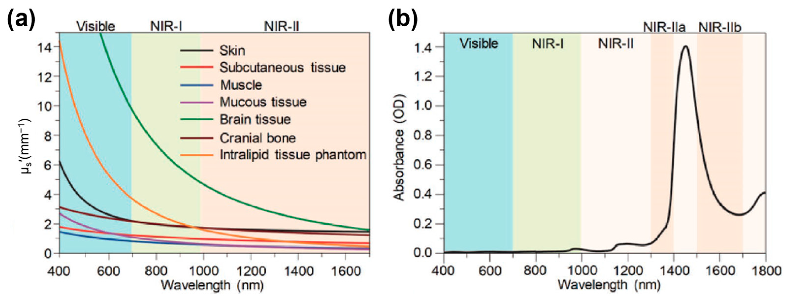
Figure 1. (a) Reduced scattering coefficients (μs′) of various tissues plotted as a function of wavelength across 400–1700 nm. (b) Visible-near-infrared (Vis–NIR) absorption spectrum of water. OD represents optical density. NIR-IIa: 1300–1400 nm; NIR-IIb: 1500–1700 nm. Reproduced from ref. [45] Copyright (2018), with permission from Wiley-VCH.
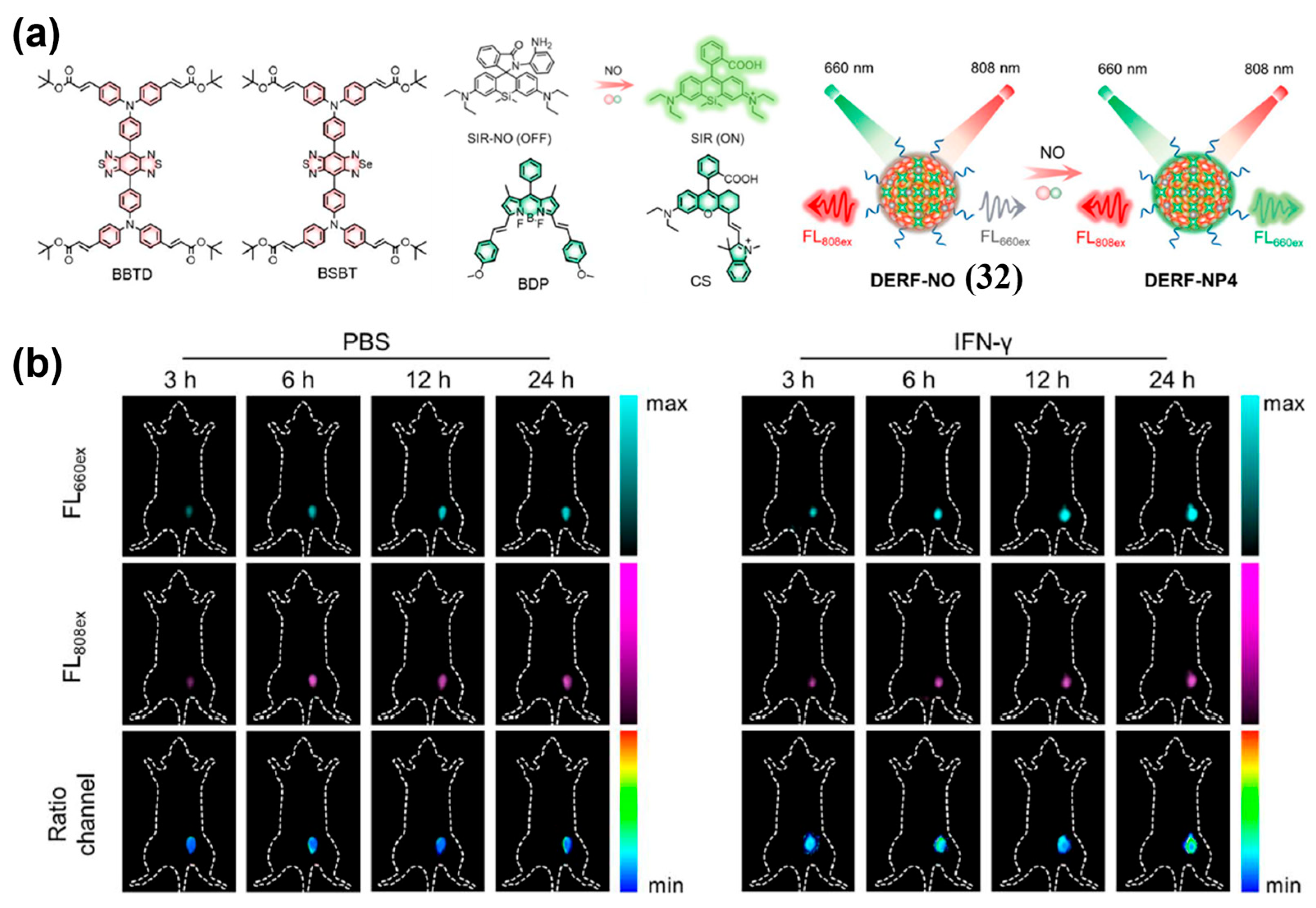
Sensors 25 07080 g001
Figure 2. D-A-D structured NIR-II organic small-molecule fluorophores.
Sensors 25 07080 g002
Figure 3. NIR-II cyanine fluorophores.
Sensors 25 07080 g003
Figure 4. NIR-II BODIPY fluorophores.
Sensors 25 07080 g004
Figure 5. NIR-II Xanthene organic fluorophores.
Sensors 25 07080 g005
Figure 6. Schematic diagram showing the two design strategies of NIR-II fluorescent probes for tumor imaging using organic small-molecule fluorophores. (a) Always-on imaging strategy, including nano-platform-based passive targeting strategy (i) and targeting group-mediated active targeting strategy (ii). (b) Activatable imaging strategy, including single-channel activatable fluorescence imaging (i) and ratiometric fluorescence imaging (ii). The wavy arrows represent the fluorescence signals at distinct wavelengths, labeled FL1 and FL2.
Sensors 25 07080 g006
Figure 7. (a) The chemical structures of FBP 912 (1) and FBPEG 912 (1a). (b,c) In vivo NIR-II fluorescence imaging of subcutaneous tumors using 1 (b) or 1a (c). (d) Ex vivo NIR-II imaging of main organs and tumors at 48 h post-injection, left: 1; right: 1a. (e,f) Normalized intensity at the tumor region (e) and tumor/liver ratio (f) at different time point after i.v. injection of 1 or 1a into mice. Reproduced from ref. [138] Copyright (2021), with permission from Wiley-VCH.
Sensors 25 07080 g007
Figure 8. (a) Chemical structure of PSO (2). (b) Schematic illustration showing the use of 2 for NIR-II fluorescence imaging of tumor. (c) Images of 2-injected tumor-bearing mice at different time points captured by the NIR-II fluorescence imaging system. The red circles in the images indicate the tumor location. Reproduced from ref. [139] Copyright (2022), with permission from American Chemical Society.
Sensors 25 07080 g008
Figure 9. (a) Schematic diagram showing the self-assembly of PF (3) into PF-NPs (3-NPs). (b) Representative in vivo NIR-II fluorescence imaging of tumor-bearing mice at different time points after i.v. injection of 3-NPs. Reproduced from ref. [142] Copyright (2019), with permission from American Chemical Society.
Sensors 25 07080 g009
Figure 10. (a) Schematic diagram showing preparation of 4-NP by encapsulating ICR-Qu (4) within DSPE-PEG2000. (b) NIR-II fluorescence imaging (using an 808 nm excitation laser) of tumor at various time points (0, 1, 6, 12, 24, and 48 h) after administration of 4-NP into 4T1 tumor-bearing BALB/c mice. (c) NIR-II fluorescence imaging-guided tumor resection (using an 808 nm excitation laser). Reproduced from ref. [146] Copyright (2023), with permission from Wiley-VCH.
Sensors 25 07080 g010
Figure 11. (a) Structural modification and assembly process of SM-16 (5) with Pluronic F127 to form SM-16 NPs (5-NPs). (b) Time-dependent NIR-II fluorescence imaging of 4T1-tumor-bearing mice i.v. injected with 5-NPs. Reproduced from ref. [148] Copyright (2024), with permission from Royal Society of Chemistry.
Sensors 25 07080 g011
Figure 12. (a) NIR-II lymphography with ICG or β-LG@IR-780 (6) probes. (b) NIR-II popliteal and sacral lymph nodes (PLN and SLN) imaging at different time points after tumor inoculation using 6. Reproduced from ref. [151] Copyright (2023), with permission from Wiley-VCH.
Sensors 25 07080 g012
Figure 13. (a) Schematic diagram of the construction of CL4/FBS (7/FBS). (b)Time-course whole-body NIR-II fluorescence images of 4T1 tumor-bearing mice and magnified images of the tumor after i.v. injection of 7/FBS or 7. (c) Quantified NIR-II fluorescence intensities of tumors in (b). Reproduced from ref. [152] Copyright (2022), with permission from Wiley-VCH.
Sensors 25 07080 g013
Figure 14. (a) Chemical structure of ICG-Herceptide probe (8). (b) NIR-II fluorescence imaging of tumor-bearing mice at 0−12 h after i.v. injection of 8 with or without the injection of trastuzumab and Herceptide. (c) NIR-II fluorescence imaging-guided surgery using 8. (d) Fluorescence intensity analysis of images in (c). *** p < 0.001. Reproduced from ref. [153] Copyright (2023), with permission from American Chemical Society.
Sensors 25 07080 g014
Figure 15. (a) Molecular structure of IRDye800-RM26 (9). (b) Ex vivo NIR-I/II imaging and cross-sectional intensity profile (blue dashed bars in images under NIR-I window, or red-dashed bars in images under NIR-II window) of glioblastoma (white dotted lines) using different dosages of 9 at NIR-I and NIR-II windows. Reproduced from ref. [154] Copyright (2023), with permission from Wiley-VCH.
Sensors 25 07080 g015
Figure 16. (a) Schematic diagram showing the self-assembly of QT-RGD (10) into nanoparticles. (b) In vivo NIR-II fluorescence images of 4T1 tumor-bearing mice at various time points (0, 1, 2, 4, 8 and 12 h) after i.v. injection of MT and 10. (c) Quantitative analysis of the fluorescence intensities of the tumor sites at different time points in (b). *** p < 0.001. Reproduced from ref. [155] Copyright (2020), with permission from Royal Society of Chemistry.
Sensors 25 07080 g016
Figure 17. (a) Chemical structure of probe IR-32p (11). (b) NIR-II and NIR-IIa fluorescence imaging of orthotopic glioma mice after i.v. injection of 11. (c) NIR-II/IIa glioma imaging (scalp removed) and ex vivo brain at 24 h p.i. (d) Ex vivo NIR-II and NIR-IIa imaging results of the main organs. Nos. 1–10: heart, liver, spleen, lung, kidney, stomach, intestine, muscle, skin, and brain, respectively. Organs were extracted at 24 h post injection. NIR-II fluorescence images were captured under 808 nm laser irradiation (300 mW cm−2, 1000 LP) at 100 ms (b,c) and 300 ms (d) exposure time. NIR-IIa images were captured under 1064 nm laser irradiation (500 mW cm−2, 1320 nm LP, 400 ms). Reproduced from ref. [156] Copyright (2024), with permission from American Chemical Society.
Sensors 25 07080 g017
Figure 18. (a) Schematic illustrating the fabrication of R&HV-Gd@ICG (12). (b) NIR-II fluorescence images of 4T1-tumor-bearing mice intravenously injected with 12, HV-Gd@ICG, or ICG, respectively. (c) TBR concentration plotted as function of time for 4T1-tumor-bearing mice (n = 4). (d) Comparison of individual 12, HV-Gd@ICG, and ICG specific tumor-targeting capabilities at maximum TBR (n = 4). * p < 0.05, ** p < 0.01. Reproduced from ref. [157] Copyright (2022), with permission from Wiley-VCH.
Sensors 25 07080 g018
Figure 19. (a) Schematic illustration showing the formation of H2a-4T@Cetuximab complex (13@cetuximab complex). (b) Representative NIR-II fluorescence images (1000 LP and 100 ms) of the HCT116 tumor model at 2, 4, 6, 8, and 20 h after tail vein injection of the 13@Cetuximab complex and 13 under an 808 nm excitation. (c) Quantification analysis of fluorescence signals of the tumor regions in (b) (n = 3). * p < 0.05, ** p < 0.01. Reproduced from ref. [69] Copyright (2018), with permission from Wiley-VCH.
Sensors 25 07080 g019
Figure 20. (a) The chemical structure of PSM-1092 (14). (b) Time-course NIR-II fluorescence images of LNCaP tumor-bearing mice after tail vein injection. (c) TNR (left Y axis, black line) and tumor uptake (right axis, blue line) versus time curve after intravenous injection of 14 in tumor-bearing mice. Reproduced from ref. [167] Copyright (2021), with permission from American Chemical Society.
Sensors 25 07080 g020
Figure 21. (a) The chemical structure of TQPTPP (15). (b) Time-course NIR-II fluorescence imaging (808 nm excitation, 1000 long-pass filter, 100 ms exposure) of tumors with tail vein injection of 15. Reproduced from ref. [168] Copyright (2023), with permission from Wiley-VCH.
Sensors 25 07080 g021
Figure 22. (a) Schematic illustration of homologous cell membrane-coated albumin-energized NIR-II Cyanine Dye LZ-1105@ Ham (16@HAm). (b) Representative NIR-II fluorescence images of A549 tumor-bearing mice at various time points after i.v. injection of 16@HSA and 16@HAm. Reproduced from ref. [172] Copyright (2025), with permission from American Chemical Society.
Sensors 25 07080 g022
Figure 23. (a) Schematic representation of the basic steps for fabrication of hT-Sulfo-1100 NPs (hT-17 NPs). (b) NIR-II fluorescence images of orthotopic glioma. The red dotted boxes mark the locations of tumors. Reproduced from ref. [173] Copyright (2024), with permission from Royal Society of Chemistry.
Sensors 25 07080 g023
Figure 24. Cartoon illustration of different mechanisms to design stimuli-activated fluorescent probes, including: (a) recovery of ICT effect, (b) restriction of TICT, (c) inhibition of PeT effect, and (d) elimination of FRET.
Sensors 25 07080 g024
Figure 25. (a) Chemical structure of H2O2-D4 (18) and the sensing mechanism towards H2O2. (b) NIR-II fluorescence images of 4T1 tumor-bearing mice before (0 h) and after intratumor treatment with 18 for 1, 3, 5, and 7 h. (c) Normalized fluorescence intensity of the tumor region in (b). Reproduced from ref. [190] Copyright (2024), with permission from AAAS.
Sensors 25 07080 g025
Figure 26. (a) Chemical structure of NO-responsive molecule DNO (19) and the response mechanism towards NO. (b) Time-course NIR-II fluorescence images of BALB/c mice with subcutaneous breast tumor (BC group) and diabetic BALB/c mice with subcutaneous breast tumor (DMBC group) after injecting DNPS under 808 nm laser irradiation. (c) Quantified data from panel (b). Reproduced from ref. [191] Copyright (2025), with permission from Wiley-VCH.
Sensors 25 07080 g026
Figure 27. (a) Illustration of a GSH-activatable NIR-II fluorescent probe LET-7 (20) for visualization of GSH. (b) Schematic diagram of probe injection in tumor-bearing mice. (c) Time-dependent NIR-II fluorescence images of 4T1 tumor-bearing mice after intratumoral (tumor side, red cycle) and subcutaneously (normal tissue side, blue cycle) injection of 20, respectively. (d) The corresponding quantitative NIR-II fluorescence intensity of tumor and normal tissues. Reproduced from ref. [192] Copyright (2021), with permission from American Chemical Society.
Sensors 25 07080 g027
Figure 28. (a) Chemical structures of SWIMP (21) and response mechanism of 21 toward Cath B for lung metastasis imaging. (b) Representative in vivo SWIR fluorescence images of living mice at different time points after intratracheal injection of 21 (left panel) with or without pretreatment by inhibitor (right panel). Excitation: 808 nm; long-pass filter: 950 nm; acquisition time: 1 s. (c) Quantification and (d) SBRs of SWIR fluorescence in the tumor region as a function of time from both the ventral (left) and dorsal (right) sides of the mice (n = 3). ** p < 0.01, **** p < 0.001. Reproduced from ref. [193] Copyright (2025), with permission from American Chemical Society.
Sensors 25 07080 g028
Figure 29. (a) Design of the β-gal-activated fluorescent probe, Flavchrom 4 (22). (b,c) Time-dependent NIR-I imaging (b) and normalized fluorescence signal values at tumor site (c) of SKOV3 tumor-bearing mouse models after intratumor injection with 22. (d,e) Time-dependent NIR-II imaging (d) and normalized fluorescence signal values at tumor site (e) of SKOV3 tumor-bearing mouse models after intratumor injection with 22. Note: red circle indicates the region of the tumor. Reproduced from ref. [194] Copyright (2022), with permission from American Chemical Society.
Sensors 25 07080 g029
Figure 30. (a) Schematic illustration of the response mechanisms of DTTVBI (23) and 23 NPs to pH. (b) NIR-II fluorescence imaging of PDX tumor-bearing mice at different time points after intravenous injection of 23 NPs. Reproduced from ref. [195] Copyright (2023), with permission from American Chemical Society.
Sensors 25 07080 g030
Figure 31. (a) The turn-on mechanism of the probe NSCy1050 (24) by H+ and high viscosity. (b) Time-dependent NIR-II fluorescence imaging of 4T1 tumor-bearing mice after i. v. injection of 200 μL PBS or 200 μL 24 (1 mM). (c) Relative fluorescence intensities on the subcutaneous 4T1 tumors in (b). Reproduced from ref. [197] Copyright (2023), with permission from Wiley-VCH.
Sensors 25 07080 g031
Figure 32. (a) Chemical structures of HN-PBA (25), HN-PBA-H+ (25-H+), HN-PBA-SH (25-SH), HN-PBA-S (25-S), and HN-PBA-SH-SR (25-SH-SR) and proposed dual-lock control and response strategy. (b) Longitudinal in vivo NIR-II fluorescence images of 4T1 tumor-bearing mice after intravenous injection (i.v.) of 25 or ICG, respectively. (c) T/N ratios of 25 and ICG over time. (d) Real-time NIR-II imaging of tumor-bearing mice after i.v. injection of 25 and the H2O2-activated control probe HN-PBC. (e) T/N ratios of 25 and HN-PBC over time. (f,g) NIR-II fluorescence (f) and bioluminescence (g) images of the tumor resected mice. *** p < 0.001. Reproduced from ref. [198] Copyright (2025), with permission from Wiley-VCH.
Sensors 25 07080 g032
Figure 33. (a) Schematic illustration of the sensing mechanism of CFC-GSH (26) for GGT. (b) NIR-I (λex = 605 nm, λem = 660 nm) and NIR-II (λex = 808 nm, 1075 nm LP) fluorescence imaging of tumor-bearing BALB/c nude mice at different time after intratumoural injection of 26 without/with pretreatment of acivicin for another 30 min. (ce) Time-dependent quantitative statistics of NIR-I (c) and NIR-II (d) fluorescence intensity, and (e) the corresponding fluorescence intensity ratio of the mice in (b). Reproduced from ref. [202] Copyright (2025), with permission from Royal Society of Chemistry.
Sensors 25 07080 g033
Figure 34. (a) Biosensing mechanism of the NIR-II ratiometric probe Rap-N (27) toward NTR. (b) Time-course NIR-II fluorescence images and corresponding ratiometric images of mice bearing 4T1 tumors, the left tumor was treated with inhibitor dicoumarol. (c) Time-course NIR-II fluorescence images of 4T1 and CT-26 tumor. Reproduced from ref. [131] Copyright (2022), with permission from Wiley-VCH.
Sensors 25 07080 g034
Figure 35. (a) Schematic illustration of ROS-responsive NIR-II ratiometric probe BBT-IR/Se-MN (28). (b) Representative NIR-II fluorescence, ratiometric fluorescence, and bioluminescence images of U87 orthotopic glioma-bearing mice under different treatments. (ce) Corresponding NIR-II fluorescence signal intensity of tumors in (b). Reproduced from ref. [203] Copyright (2023), with permission from Wiley-VCH.
Sensors 25 07080 g035
Figure 36. (a) Schematic illustration of the construction of the FRET-based ratiometric NIR-II window H2S sensor, FRHS (29). (b,c) Time-dependent NIR-II fluorescence images at Ch.1 and Ch.2, and ratiometric Ch.1/Ch.2 fluorescence images of healthy mice (b) and tumor-bearing mice (c), and corresponding ex vivo organs after i.v. injection of 29. Reproduced from ref. [204] Copyright (2023), with permission from AAAS.
Sensors 25 07080 g036
Figure 37. (a) Chemical structures of donor NAB (30-a), pre-acceptor NRh (30-b) and pH activated acceptor NRhH+ (30-c). (b) pH response illustration of pTAS (30). Channel 1–2 represent for the signal collection regions during ratiometric fluorescence imaging. (c,d) Illustrated animal models and ratiometric fluorescence images of tumors during the two pH changing processes after administration of pTAS-2 (30-2) and pTAS-3 (30-3), respectively. (e,f) Comparison of the pH changing in two physical processes by ratiometric fluorescence imaging and microelectrode pH meter. Reproduced from ref. [205] Copyright (2021), with permission from Wiley-VCH.
Sensors 25 07080 g037
Figure 38. (a) Schematic diagram showing the construction of multi-wavelength nanoprobes 31 with activatable emission in the NIR-II window. (be) Visualization and differentiation of cancers based on H2S activation of 31. (b) HCT116 tumor-bearing mice. (c) The effects of inhibitor (AOAA) and activator (SAM) in HCT116 tumor-bearing mice. (d) Normal sites. (e) HepG2 tumor-bearing mice. Reproduced from ref. [207] Copyright (2018), with permission from Wiley-VCH.
Sensors 25 07080 g038
Figure 39. (a) Chemical structures of energy acceptors (BBTD and BSBT) and donors (SIR, BDP, and CS), the chemical structure change in SIR-NO before and after reaction with NO, and schematic illustration showing the ratiometric response of DERF-NO (32) to NO with the dual-excitation ratiometric detection manner. (b) Representative fluorescence images of 4T1 tumor-bearing mice treated with PBS or IFN-γ (i.t.) and then i.v.-injected with 32. Reproduced from ref. [208] Copyright (2025), with permission from Royal Society of Chemistry.
Sensors 25 07080 g039
Figure 40. (a) Schematic illustration of preparation of 1-NP (33) via DSPE-PEG2000-assisted encapsulation of 1-BrEt, NIR775 and IR1048, and proposed conversion of 33 into 2-NP (33-1) in response to •OH, accompanying by a reduced fluorescence (FL780/FL1113) ratio but a concurrently increased photoacoustic (PA755/PA905) ratio. Right: chemical structures of NIR775 and IR1048 used for the preparation of 33. (b) Fluorescence and corresponding ratiometric fluorescence images of 4T1 tumors following indicated treatment. Red arrows and circles indicate the tumor locations. Reproduced from ref. [209] Copyright (2021), with permission from Springer Nature.
Sensors 25 07080 g040
Figure 41. (a) Schematic illustration of the ratiometric NIR-II fluorescence nanoplatform DCNP@IR 806 (34) for assessing radiotherapy effect based on accurate quantification of activated caspase-3. (b) Bioluminescence imaging of OHCC-bearing mouse at different time points after treatment. (c) NIR-II fluorescence and ratiometric imaging of OHCC-bearing mice after treatment with 34 and irradiation with X-ray doses. Reproduced from ref. [210] Copyright (2025), with permission from Wiley.
Sensors 25 07080 g041
Figure 42. (a) Schematic illustration showing the ratiometric NIR-II fluorescence detection of miR-21 using the DCNP@DNA2@IR806 (35) nanoprobe based on NRET effect. (b) NIR-II fluorescence and ratiometric NIR-II fluorescence images of 4T1 tumor-bearing mice at 18 h after i.v. injection with 35. (c) NIR-II fluorescence and ratiometric NIR-II fluorescence images for the different sized tumors (ex vivo) at 18 h post-injection with 35. Reproduced from ref. [211] Copyright (2024), with permission from Wiley.
Sensors 25 07080 g042
Table 1. The photophysical properties of representative NIR-II organic small-molecule fluorophores.
Fluorophore ClassRepresentative Exampleλabs (nm)λem (nm)SolventQY (%)Ref.
D-A-D
fluorophores
CH10557501055H2O0.03 [a][68]
H2a-4T7381024H2O0.01 [a][69]
Cyanine
fluorophores
FD108010641080H2O0.31 [a][70]
Flav 710261045H2O0.53 [a][71]
BODIPY fluorophoresNJ10609101060H2O0.50 [a][72]
WH-49601205H2O0.05 [b][73]
Xanthene fluorophoresCX-310891140CHCl30.09 [a][74]
VIX-410141210DCM0.03 [a][75]
[a] The fluorescence QY was calculated relative to IR-26 (0.05% in DCE), [b] The fluorescent QY was calculated relative to IR-1061 (1.7% in DCM).
Disclaimer/Publisher’s Note: The statements, opinions and data contained in all publications are solely those of the individual author(s) and contributor(s) and not of MDPI and/or the editor(s). MDPI and/or the editor(s) disclaim responsibility for any injury to people or property resulting from any ideas, methods, instructions or products referred to in the content.