Review

Recent Advances in Micro- and Nano-Enhanced Intravascular Biosensors for Real-Time Monitoring, Early Disease Diagnosis, and Drug Therapy Monitoring

by
Sonia Kudłacik-Kramarczyk
1,*,
Weronika Kieres
1,
Alicja Przybyłowicz
1,2,
Celina Ziejewska
2,*,
Joanna Marczyk
2 and
Marcel Krzan
1
1
Jerzy Haber Institute of Catalysis and Surface Chemistry, Polish Academy of Sciences, 8 Niezapominajek St., 30-239 Krakow, Poland
2
Faculty of Mechanical Engineering, Cracow University of Technology, 37 Jana Pawła II Av., 31-864 Krakow, Poland
*
Authors to whom correspondence should be addressed.
Sensors 2025, 25(15), 4855; https://doi.org/10.3390/s25154855
Submission received: 20 May 2025 / Revised: 30 July 2025 / Accepted: 6 August 2025 / Published: 7 August 2025
(This article belongs to the Section Biosensors)

Abstract

Intravascular biosensors have become a crucial and novel class of devices in healthcare, enabling the constant real-time monitoring of essential physiological parameters directly within the circulatory system. Recent developments in micro- and nanotechnology have relevantly improved the sensitivity, miniaturization, and biocompatibility of these devices, thereby enabling their application in precision medicine. This review summarizes the latest advances in intravascular biosensor technologies, with a special focus on glucose and oxygen level monitoring, blood pressure and heart rate assessment, and early disease diagnostics, as well as modern approaches to drug therapy monitoring and delivery systems. Key challenges such as long-term biostability, signal accuracy, and regulatory approval processes are critical considerations. Innovative strategies, including biodegradable implants, nanomaterial-functionalized surfaces, and integration with artificial intelligence, are regarded as promising avenues to overcome current limitations. This review provides a comprehensive roadmap for upcoming research and the clinical translation of advanced intravascular biosensors with a strong emphasis on their transformative impact on personalized healthcare.

Graphical Abstract

1. Introduction

A biosensor may be defined as an analytical instrument used to measure variations in biological activity—for instance, of enzymes, acids, or cells—and then transform them into quantifiable electronic signals [1]. The evolution of biosensor technologies has revolutionized healthcare in the 21st century, particularly in the field of real-time health monitoring. Nowadays, they are successfully applied in many fields, including medicine, pharmaceutics, industry, water quality management, and precision agriculture; for instance, they are used in diagnosing infections caused by bacterial and viral agents [2], detecting heavy metals [3], monitoring cholesterol levels [4], alerting personnel to water biotoxicity [5], detecting biomolecules [6], and enabling early cancer diagnosis [7]. According to the literature data, the market size of biosensors was estimated at USD 30.25 billion in 2024. Furthermore, biosensors are undoubtedly gaining increasing popularity, and this trend will continue, as indicated by the compound annual growth rate (CAGR) amounting to 8.7%, estimated from 2025 to 2034 [8]. The urgent need for reliable, real-time monitoring tools, particularly for chronic diseases, underscores the critical importance of innovations in intravascular biosensing.
Intravascular biosensors represent a groundbreaking achievement, as they bridge traditional diagnostic approaches with practical methods for the assessment of physiological parameters in patients. These devices are designed to operate within the human circulatory system, enabling unparalleled opportunities for the early detection and continuous monitoring of diseases; thus, they can significantly improve patient outcomes across various clinical settings [9,10].
The growing prevalence of chronic diseases such as diabetes, cardiovascular disorders, and respiratory conditions necessitates innovative solutions for effective patient diagnosis and treatment. Traditional methods, while reliable, often fail to deliver the rapid response needed to prevent complications or adjust therapies in real time. Intravascular biosensors, through their flexibility and high biocompatibility, offer much better integration with biological tissue [11]. This innovation is underpinned by advancements in micro- and nanotechnology, enabling the miniaturization and enhanced sensitivity of these devices [12].
Despite numerous reviews summarizing general advances in biosensor technologies, there is a notable lack of comprehensive analyses focusing specifically on intravascular biosensors and their integration with micro- and nanotechnologies. Existing reviews often address biosensors in broader contexts, without emphasizing the unique challenges and opportunities associated with implantable intravascular devices. This review aims to fill this gap by providing a critical assessment of emerging micro- and nanoscale innovations dedicated to intravascular applications, discussing not only technological advancements but also clinical translation barriers, biocompatibility concerns, and future trends such as biodegradable materials and AI-based data analysis. Our goal is to offer a structured and forward-looking perspective on how next-generation intravascular biosensors can revolutionize continuous health monitoring and personalized medicine.
The diagram below (Figure 1) presents a schematic representation of the functionality of intravascular biosensors.
This solution provides the continuous and precise monitoring of physiological parameters, which is crucial for personalized medicine.
The scope of applications for intravascular biosensors is broad and includes glucose monitoring for diabetes management, the real-time tracking of blood oxygen levels, and the assessment of cardiovascular health through continuous blood pressure and pulse measurements [13,14]. Importantly, beyond diagnostics, these biosensors play a key role in therapeutic interventions, such as drug delivery and the personalization of treatment regimens based on the patient’s dynamic physiological responses [15]. This synergy between monitoring and treatment represents a paradigm shift towards truly personalized medicine.
An interesting solution that enables the monitoring and assessment of cardiorespiratory function is a photonic smart wristband developed by Li et al. [16]. An all-polymer sensing unit forms the basis of this wearable device, which constantly measures critical health metrics, such as blood pressure, heart rate, and respiration rate. Furthermore, the conducted research showed that the biometric identification process reached a correct rate of 98.55%, confirming its applicability in personalizing healthcare services.
The table below (Table 1) provides a detailed comparison of various types of biosensors, categorized by their applications, advantages, and disadvantages, alongside relevant references for each type.
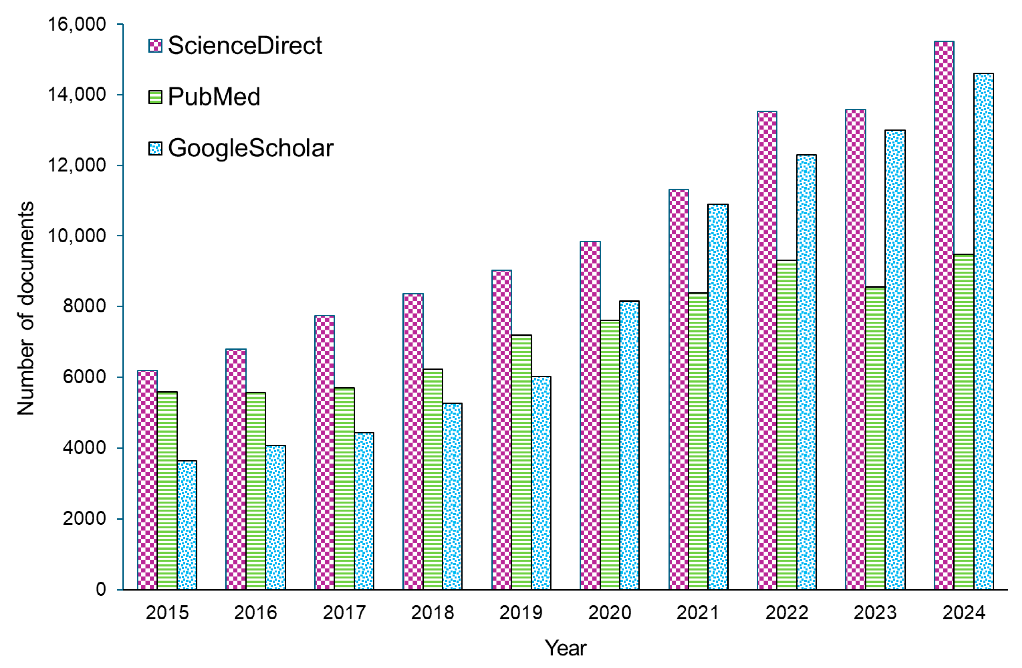
The number of published papers found in the ScienceDirect and Google Scholar databases after applying “biosensor” as a keyword has steadily increased over the last ten years, as shown in Figure 2. The graph shows different types of articles—both research and review articles, as well as other documents, including book chapters, short communications, and conference abstracts. It is worth mentioning that the number of relevant documents indexed in the database in 2025 was nearly 2.5 times higher compared to the number recorded in 2015. However, it should be noted that applying “intravascular biosensor” as a keyword yielded only 121 results in the database, dating back ten years. For instance, in 2021 and 2023, the number of documents meeting the criteria was 6 and 23, respectively.
Although researchers have presented many reviews focusing on various aspects of biosensors, there is no review relating to intravascular biosensors. Therefore, this paper provides a literature review on the current state of intravascular biosensors, with a focus on their technological foundations, clinical applications, and future potential. In more detail, following this introduction and general overview regarding biosensors (Section 1), specific areas of application, such as glucose monitoring, oxygen level assessment, and cardiovascular parameter monitoring, are described (Section 2). Progress achieved in disease diagnostics is then discussed, considering the detection of disease biomarkers and infection diagnostics (Section 3). Next, modern approaches to drug therapy monitoring and systems, as well as micro- and nanotechnology in intravascular biosensors, are presented, along with the role of emerging technologies like quantum dots and bioresorbable stents (Section 4 and Section 5). Furthermore, the possibility of applying biosensors for cancer diagnosis and treatment is discussed (Section 6). This work aims to highlight their transformative potential in modern medicine [25].

2. Monitoring of Physiological Parameters

Monitoring physiological parameters plays a crucial role in the diagnosis and treatment of various medical conditions. The analysis of biomarkers provides valuable insights into a patient’s health status, enabling timely intervention and, most importantly, personalized treatment. Advances in the field of biosensors have revolutionized healthcare through innovative solutions that combine precision and real-time monitoring. From detecting fluctuations in glucose levels in diabetic patients to assessing oxygen levels and monitoring cardiovascular parameters, biosensors address a wide range of clinical needs. These technologies not only enhance diagnostic accuracy but also improve therapeutic outcomes through integration into patient care workflows. However, their applications extend beyond acute cases, as they support long-term health monitoring and proactive disease prevention. As these systems continue to evolve, their scope of application is constantly expanding, promising a transformative impact on modern medicine. Additionally, this creates a significant research niche for scientists working on the development and optimization of these materials.

2.1. Glucose Level Monitoring

The blood glucose level is one of the most important indicators of a patient’s health status. The high prevalence of diabetes has led to extensive research into various glucose measurement methods to ensure continuous and accurate glucose monitoring. Although direct blood glucose monitoring provides precise information, many patients, particularly younger ones, are reluctant to undergo needle pricks for blood sampling due to the invasiveness of the procedure. An alternative is the use of an implanted glucose sensor. While this method is invasive, requires regular sensor replacement due to its limited lifespan, and carries the risk of microthrombus dissemination, it reduces the need for frequent and burdensome blood sampling for patients [26,27]. However, significant challenges, such as the long-term stability of sensors and calibration problems caused by the harsh environment inside the human body, must still be addressed, requiring the development of new and optimized solutions in the future [28].
Electrochemical implantable biosensors, such as subcutaneous or intravascular sensors, can provide real-time glucose level data, allowing for precise therapy adjustments [29]. David A. Gough et al. conducted a study monitoring glucose concentrations in subcutaneous tissue in diabetic patients using an implanted sensor/telemetry system. This sensor was based on membranes containing immobilized glucose oxidase and catalase, coupled with oxygen electrodes and a telemetry system integrated as an implant. Since no independent standard exists for sensor signals indicating dynamic glucose levels in tissues, a model was applied to describe the relationship between blood glucose levels and sensor signals. The tested sensor enabled long-term glucose monitoring, and the obtained results correlated with reference values, highlighting its potential to facilitate diabetes management [30]. Another study utilized an intravascular continuous glucose monitoring system in critically ill patients in intensive care units. This is particularly significant because both hyperglycemia and hypoglycemia are associated with adverse clinical outcomes in critical care patients. The study employed the GluCath System, which uses a chemical fluorescence quenching mechanism for optical blood glucose measurement via insertion into the radial artery or directly into a peripheral vein through a catheter. The GluCath System demonstrated acceptable accuracy during 48 h placement in the radial artery in post-cardiac surgery patients in intensive care units [31]. Another example of a continuous intravascular glucose monitoring system is the one developed by GlySure Ltd. This system offers continuous intravascular glucose monitoring using a diboronic acid-based receptor for precise plasma glucose measurement. Inserted via a central venous catheter, it measures glucose every 15 s. The sensor features an optical fiber within a sheath (<0.6 mm) and a chamber with glucose-detecting elements in hydrogel, protected by dialysis and microporous membranes. Advanced filtration minimizes interference from blood components, ensuring accuracy. The GlySure CIGM system has proven reliable, safe, and highly accurate in clinical settings [32]. Although progress in biosensor technology for diabetes management is encouraging, issues like precision, patient compliance, and affordability remain. Moving forward, efforts will focus on enhancing sensor precision, optimizing data analysis algorithms, and tackling challenges related to cost and accessibility [33]. While subcutaneous and intravascular sensors show promising accuracy, issues like biofouling, limited sensor lifespans, and signal drift remain unresolved, necessitating further work on antifouling coatings and sensor recalibration strategies.
A key technological parameter is the limit of detection (LOD) in intravascular blood and the linearity of the measurement. The standard physiological blood glucose concentration in a healthy person ranges from approximately 70 to 105 mg/dL [34]; therefore, sensors intended for in vivo measurement must be sensitive in the range of at least 1–10 mmol/L. Microneedle enzymatic biosensors have achieved an LOD of 1–7 µmol/L, corresponding to a concentration of 0.018–0.126 mg/dL. These sensors exhibit a linearity range of 0.05 to 9 mmol/L, which allows for the accurate mapping of glycemia changes in real time, including in situations of hypoglycemia and hyperglycemia [35].
Enzymatic and optical biosensors are most commonly used for glycemia. A more detailed discussion of biosensor technologies, including MEMS, is provided later in this section.

2.2. Oxygen Level Monitoring

Physiological processes such as circulation, respiration, digestion, and many others change the oxygen concentrations in the tissues and the blood [36]. Disturbances in the body’s homeostasis caused by reduced oxygen levels lead to respiratory and cardiovascular diseases. Hypoxia results in cellular dysfunction and organ failure, affecting critical organs such as the brain and heart. Monitoring oxygen levels in the body is a key element in patient diagnostics [37,38]. Oxygen saturation is an indicator that reflects the efficiency of oxygen transport to tissues and gas exchange [39]. The measurement is non-invasive and can be performed in both home and hospital settings. It is conducted using a pulse oximeter, which utilizes the phenomenon of spectrophotometry. The analysis is based on the absorption of red and infrared light, which penetrates tissue structures [40]. Pulse oximetry enables the diagnosis of conditions such as COVID-19 and hypoxia, which, despite low oxygen levels, may not present with symptoms like shortness of breath. Early detection helps to reduce the risk of complications [41]. The rapid and accurate monitoring of the local oxygen concentration in 3D tissue cultures allows the oxygen concentration to be controlled so that both healthy and pathological environments can be reproduced. For example, the 3D culture and oxygen monitoring system presented by Rivera et al. [36] consists of a simple design to remotely monitor oxygen concentrations during tissue culture. The researchers integrated a photonic oxygen sensor into a 3D tissue scaffold and regulated the oxygen concentration by controlling the flow of the purification gas. Although non-invasive oxygen monitoring technologies show significant promise, limitations such as calibration drift, motion artifacts, and low penetration depths in certain tissues still need to be addressed for broader clinical adoption.
Although optical photonic biosensors dominate in this case, the technologies used—as in the case of other parameters—will be discussed later in this section.

2.3. Intravascular Lactate Biosensors

Lactate is marker of hypoxia. Under non-steroidal conditions, it switches to anaerobic metabolism, which leads to the selective production of lactate by lactate dehydrogenase. Schierenbeck et al. [42] conducted a study on the implementation of intravascular microdialysis for the continuous monitoring of lactate levels in patients undergoing cardiac surgery. The continuous monitoring of blood lactate levels was performed in 80 patients undergoing cardiac surgery using microdialyzers placed in the superior vena cava. Single- or triple-lumen central catheters with microdialysis capabilities were used for this purpose. Arterial blood samples were simultaneously collected hourly for blood gas analysis, providing reference values for comparison. A total of 1601 pairs of lactate samples were collected, analyzed by microdialysis and traditional blood gas analysis. The results confirmed that central venous microdialysis is a precise and reliable method for the continuous monitoring of lactate levels in cardiac surgery patients. This technique may also be useful for the early detection of perfusion disorders in critically ill patients. Yin Ho et al. [43] developed a 5 Fr intravascular nitric oxide (NO) catheter containing a sensor for continuous lactate measurement and a port for a pressure sensor. The device exhibits antibacterial and antithrombotic properties and can be implanted intravenously. In an in vivo study of six piglets undergoing cardiac surgery with cardiopulmonary bypass (CPB), sensors were placed in the femoral vessels and in the CPB circuit. Lactate measurements obtained from the sensors in the CPB were consistent with blood gas measurements, whereas the sensors in the femoral arteries correlated well with measurements only before CPB. The pressure sensors provided accurate readings, comparable to those of FDA-approved devices. The authors recommend implanting sensors in the CPB circuit for the monitoring of lactate levels and in peripheral arteries or veins before and after CPB. This study supports the usefulness of this system for continuous metabolic monitoring during infant cardiac surgery.

2.4. Blood Pressure and Heart Rate Monitoring

Blood pressure and pulse monitoring is crucial for cardiovascular health, as it plays a vital role in diagnosing and preventing diseases that can be fatal for patients. Hypertension, often referred to as the “silent killer”, and arrhythmias remain among the leading causes of morbidity and mortality worldwide [44]. For this reason, the scientific community continues to seek solutions to enhance the fight against these conditions. Intravascular biosensors have emerged as a groundbreaking solution, offering precise and continuous monitoring capabilities that surpass those of traditional methods [45]. In recent years, intravascular blood pressure and heart rate sensors have leveraged miniaturized MEMS and piezoelectric technologies to achieve continuous real-time monitoring. For instance, sensors fabricated on a polymer substrate must be in contact with the catheter surface to achieve a better signal and greater durability. The authors of [46] describe a study in which gold patterns were fabricated on a polyimide surface, and a micrometer-sized SU-8 pressure chamber was mounted to transduce pressure. Additional gold patterns were fabricated as a resistive temperature detector, and a tensimeter was used to measure pressure. An in vivo experiment in a mouse model demonstrated that the catheter-mounted sensor could measure heart rate and carotid blood pressure in real time. A subsequent publication [47] discussed various sensors for the monitoring of cardiovascular parameters, including heart rate. These devices enable the detection of pulse waves generated by blood vessels with very high sensitivity (below 10 kPa), making them suitable for wearable applications. Triboelectric nanogenerators function as self-powered biosensors—they convert body movement into an electrical signal, enabling continuous heart rate monitoring without the need for external power. While this section outlines the clinical relevance of these parameters, the following subsection focuses in more detail on technological platforms enabling advanced hemodynamic monitoring.

2.5. Technological Platforms for Advanced Biosensing

Recent advances have led to the development of a new generation of biosensors capable of precisely monitoring physiological parameters in real time. Among the most influential technologies are MEMS, nanomaterials, quantum dots, and bioresorbable platforms, each bringing unique functionalities that enhance diagnostic accuracy and therapeutic efficacy.
Microelectromechanical systems (MEMS) represent a class of miniaturized devices that integrate mechanical and electronic components. Their unique properties, such as high sensitivity, miniaturization, and real-time signal processing, make them a versatile tool in biosensing platforms, including cardiovascular monitoring. Modern intravascular biosensors utilize advanced microelectromechanical systems (MEMS) and nanotechnology to achieve exceptional sensitivity and precision. These devices combine pressure-sensitive membranes with piezoelectric or capacitive sensing elements, which have the ability to detect subtle changes in blood vessel walls corresponding to the pulsatile blood flow [48]. Advances in wireless technology further enable real-time data transmission, facilitating seamless integration with patient monitoring systems [49].
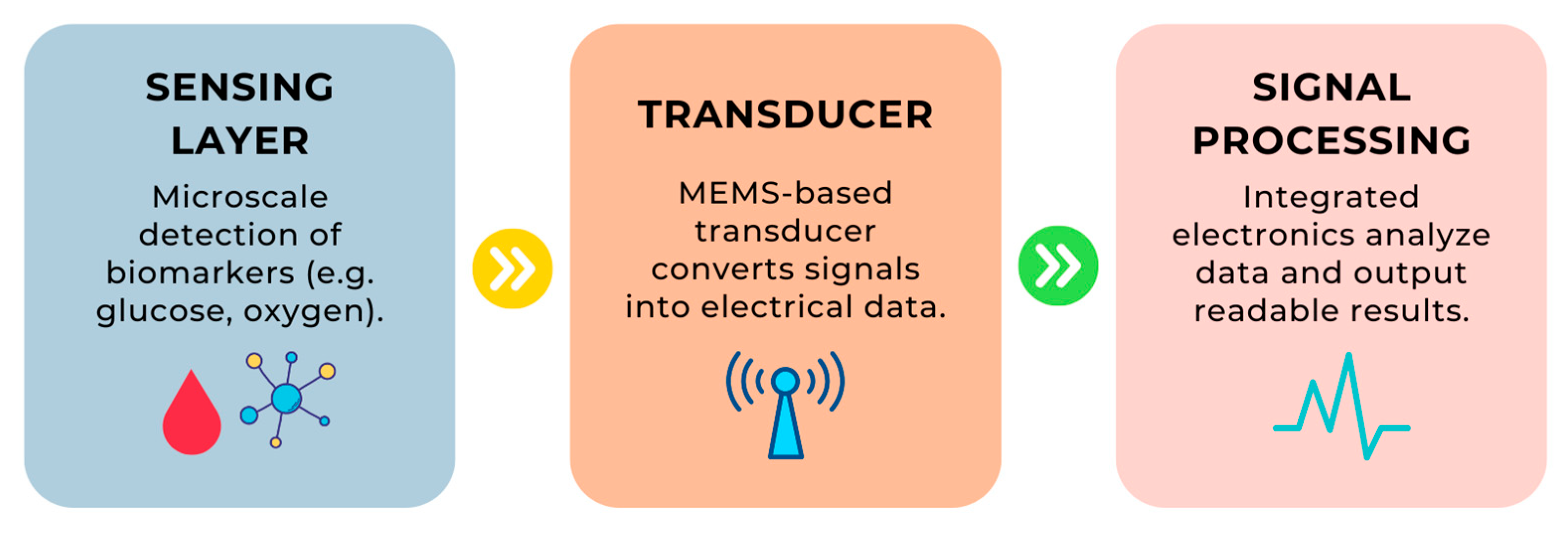
The following table (Table 2) presents a summary of various technologies used in biosensors, highlighting their applications, advantages, examples, and relevant references. It illustrates the versatility of technologies such as MEMS and nanomaterials in advancing biosensor functionalities for diverse medical and diagnostic applications. Additionally, the operation of MEMS technology in biological sensors is illustrated in Figure 3.
Biosensors integrated with bioresorbable stents can offer dual functionality: firstly, they ensure arterial patency; secondly, they enable the localized measurement of hemodynamic parameters. Such biosensors are coated with biocompatible materials, which minimize inflammatory responses, enhance patient safety, and improve device durability [58]. Such technologies highlight the shift towards multifunctional devices that align with the principles of precision medicine. Future intravascular devices will likely combine multiple functionalities, including sensing, drug delivery, and real-time AI-driven data interpretation, paving the way for fully autonomous diagnostic–therapeutic platforms.
How does it work in practice?
  • Step 1: A biomarker (e.g., a glucose molecule) interacts with the sensing layer, which recognizes its presence.
  • Step 2: Information about this interaction is converted into a signal by the MEMS transducer.
  • Step 3: The signal is processed by the signal processor, which ultimately delivers the result in an understandable format.
MEMS technology enables the miniaturization of these devices, making them suitable for high-precision medical applications, such as intravascular biosensors or medical implants. Studies have shown that the use of these biosensors enables the earlier detection of hemodynamic abnormalities, which translates into faster intervention and improved treatment outcomes [59,60].
In perioperative care, where maintaining cardiovascular stability is critical, intravascular biosensors provide real-time data, enabling the precise management of anesthesia and fluid therapy. In high-risk surgeries, such as cardiac procedures, the use of these biosensors allows for the highly accurate monitoring of hemodynamic parameters, minimizing the risk of complications [61].
Furthermore, the integration of intravascular biosensors with wearable or implantable devices supports long-term patient monitoring, which is crucial for the early detection of conditions such as heart failure or vascular stenosis. Studies indicate that the continuous monitoring of hemodynamic parameters using these biosensors enables the earlier detection of deteriorating cardiac function, facilitating the quicker implementation of appropriate therapeutic interventions [62]. Despite technological advancements, the long-term stability, miniaturization, and reliable wireless data transmission of intravascular pressure biosensors remain critical technical hurdles that need focused research efforts.
Despite their promising potential, several challenges still need to be addressed to advance diagnostics to the next level. Issues such as device miniaturization, long-term biocompatibility, and power supply limitations remain significant barriers [63]. Research into self-sustaining power mechanisms, such as harvesting energy from blood flow, represents a promising path to overcoming these obstacles [64].
Additionally, scientific reports have considered the integration of artificial intelligence and machine learning algorithms into biosensor platforms [65]. The possibility of integrating artificial intelligence algorithms with systems applied to the continuous monitoring of glucose constitutes an extremely important topic, which could change the futures of many patients with chronic diseases [66]. The analysis of huge amounts of data and identifying existing correlations and patterns afterwards can be accomplished using AI technology, whereas these tasks are usually too demanding or time-intensive for manual processing. Similarly, processing noisy and multimodal signals obtained by biosensors can be facilitated with machine learning models, which in turn improve the detection of pathological conditions [67]. Omar et al. presented an Al-based prediction model, which showed that the fusion of information coming from multifunctional sensors is possible [68]. Jin et al., in their work [69], concluded that there are obstacles that prevent biosensors from becoming popular, such as inaccuracy and consumable costs.
Moreover, the application of other biodegradable materials could offer a solution for the development of transient biosensors that degrade after fulfilling their diagnostic or therapeutic roles. The selection of appropriate materials is a key aspect. For instance, materials such as PLGA or silk fibroin are biodegradable. However, they are prone to swelling simultaneously, which hinders their application in biosensors. On the other hand, polylactic acid (PLA) or biodegradable magnesium alloys can ensure the functionality of devices within an intended lifespan, as well as being safe for the human body. This approach would eliminate the need for an additional step involving device removal [14,68,70,71].

2.6. Advantages of Intravascular Biosensors Compared to Conventional Biosensing Platforms

Intravascular biosensors, by virtue of their placement directly within blood vessels, offer several key advantages over conventional biosensing platforms, such as subcutaneous, surface-mounted, or wearable systems. Their strategic location enables the following:
  • Direct access to circulating biomarkers, allowing for more accurate and real-time measurements [32,72];
  • Faster response times, which are crucial in dynamic clinical settings such as intensive care or surgery [12];
  • Higher clinical relevance of measurements, particularly for drugs or metabolites that exhibit compartmentalization (e.g., plasma vs. interstitial fluid) [73];
  • Integration with delivery systems (e.g., infusion pumps, stents), enabling closed-loop therapies [49].
Despite these advantages, intravascular sensors face challenges related to biocompatibility, the risk of thrombosis, sensor drift, and power and communication limitations [74,75]. Table 3 below summarizes these features.
The outlined comparison sets the stage for examining specific implementations of intravascular biosensors that demonstrate their clinical relevance and technological maturity.

3. Disease Diagnostics

Disease diagnostics enables the identification and monitoring of inflammatory states and changes within the body. Access to advanced technologies enhances and expands the capabilities of diagnostic methods. Techniques such as spectroscopy, fluorescence, and DNA analysis are utilized to identify biomarkers of various conditions and differentiate between them. The fluorescent response plays a crucial role in clinical in vivo medical analysis, allowing the precise monitoring of biological processes. Two key areas of diagnostics are distinguished: the detection of biomarkers and the identification of infections caused by pathogenic agents [82,83,84].

3.1. Detection of Disease Biomarkers

Biomarkers are indicators that reflect the health status of an organism. They are used in detecting, monitoring, and evaluating responses to therapy, providing information about physiological processes occurring in the body. Biomarkers include metabolites, proteins, DNA, and RNA. They are detected in tissues and bodily fluids, primarily blood. The identification of biomarkers has been made possible through advancements in methods such as PCR, mass spectrometry, and genomic analysis, enabling rapid diagnostics in cardiovascular diseases and cancer [85,86]. Multimarker test technologies like multiplex PCR and next-generation sequencing (NGS) allow for therapy personalization by simultaneously detecting multiple biomarkers [87]. Table 4 summarizes key biomarkers and their diagnostic applications, detection methods, and associated advantages, highlighting their significance in modern healthcare. In cancer diagnostics, biomarkers are used to monitor tumor progression through next-generation sequencing, which detects genetic mutations and tumor-specific gene expression. In Alzheimer’s disease, protein analysis can enable diagnosis at the early stages of nervous system pathology. A challenge in diagnostics is that a single biomarker may be associated with multiple conditions, complicating the identification of specific diseases [88,89,90]. For example, Zhong et al. [91] have developed a fast and highly sensitive aptasensor for the quantification of biomarkers of amyloid-β (Aβ) oligomers, which are identified as reliable biomarkers for the diagnosis of Alzheimer’s disease (AD). With the advantages of low consumption, simple operation, limited time requirements, and high sensitivity, this new aptasensor driven by hyperbranched rolling circle amplification (HRCA) has great potential in the early diagnosis of AD.
The data presented in Table 4 showcase the versatility of biomarkers in diagnosing and managing various medical conditions. Glucose monitoring plays a critical role in diabetes care, leveraging electrochemical biosensors for fast and accurate detection. Troponin serves as a vital indicator for myocardial damage, offering high specificity through immunosensors. CRP facilitates the rapid detection of inflammation and infections using biochemical tests and biosensors. Additionally, genetic mutations analyzed through NGS and PCR enable early cancer diagnostics and therapy personalization. Collectively, these biomarkers underscore the transformative potential of advanced detection technologies in improving diagnostic accuracy and patient outcomes. Biosensors using surface plasmon resonance (SPR) technology are also used for CRP applications. TN detection using SPR is one of the most interesting applications in the diagnosis of acute myocardial infarction. Typically, SPR-based TN detection tools can be classified into two types: immunosensors and aptasensors. Nanostructures have an improved LOD. Furthermore, SPR-based biosensors can be fabricated using nanostructures. To date, SPR-based TN biosensors have been developed as immunosensors [101]. In modern biosensors used to measure troponin, based on field-effect transistors (FETs), a semiconductor channel material is connected between two metal electrodes, and the channel material is functionalized with receptors. The analysis time for FET-based biosensors is significantly reduced thanks to direct electrical detection without additional labeling. This simple and rapid diagnostic method is ideal for detecting a cardiac marker of myocardial infarction, where early diagnosis and rapid treatment are required. Electrochemical FET-type immunosensors based on silicon nanofibers (SiNW-FETs) can detect cTnI with an LOD of only ~5 pg/mL in buffer and 10 pg/mL in whole blood [102].

3.2. Infection Diagnostics

Human achievements in the medical field are considered to be significant. Nevertheless, currently, infectious and contagious diseases still pose a serious threat to human life. It is estimated that about 15% of deaths registered worldwide are caused by communicable diseases [103]. Some viral diseases, such as Human Papillomavirus (HPV), HIV, and SARS-CoV-2, can spread rapidly around the world, resulting in a variety of symptoms in affected individuals [104].
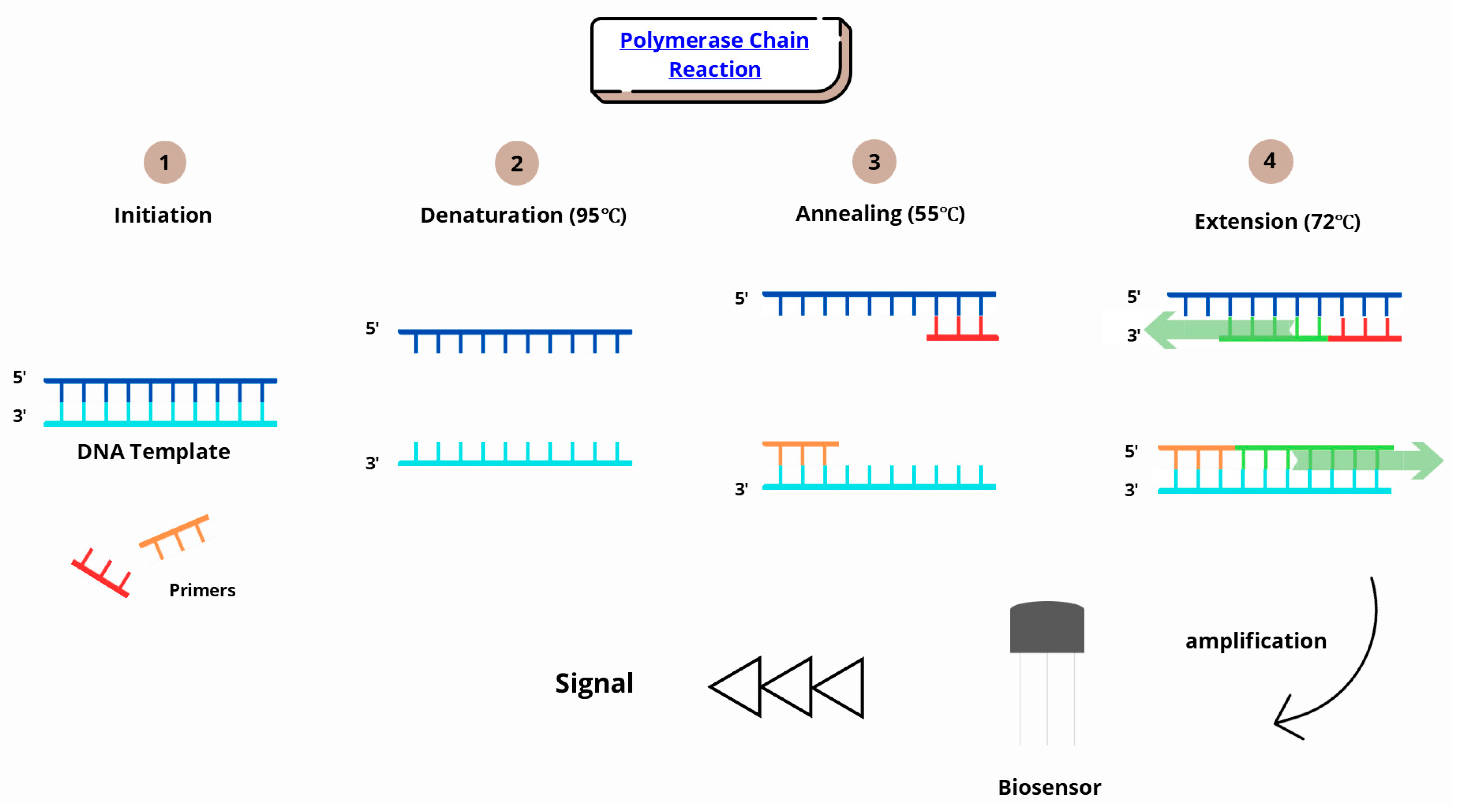
Infection diagnostics involves identifying pathogens that can lead to infections and exacerbate the occurrence of chronic diseases. Traditional diagnostic methods are time-consuming and rely on microbiological techniques, such as culturing fungi, viruses, or bacteria on selected media. Modern methods are more precise and, above all, faster. Pathogen DNA detection is enabled through rapid, highly sensitive PCR and RT-PCR tests. Multiplex PCR is also used, allowing the simultaneous detection of multiple pathogens [105,106]. Biomarkers play a crucial role in infection diagnostics, and their rapid detection is particularly important in cases of sepsis and rapidly progressing infections [107]. The PCR method involves a series of chemical reactions that lead to the copying of specific nucleic acid sequences. PCR makes it possible to identify the genetic material of a pathogen. When the sample is introduced into the biosensor and then the bioreceptor interacts with the target molecule, a transducer in the biosensor converts the changes into a signal that can be used to quantify the elements in the sample (Figure 4).
Kumar et al. [108], in their work, successfully developed and optimized an assay designed to detect anti-SARS-CoV-2 N antibodies, indicating prior SARS-CoV-2 infection. It is a visual test that uses gold nanoparticles (GNPs) functionalized with a peptide dendrimer. This idea enables direct detection with high sensitivity for breakthrough infections, not only in humans but also in animals.

4. Modern Approaches to Drug Therapy Monitoring and Systems

The world faces a significant challenge in providing healthcare and combating global threats such as Alzheimer’s disease, cancer, cardiovascular diseases, mental disorders, stroke, AIDS, and COVID-19. In addition to user-friendly diagnostics, there is an urgent need for treatment methods that are more patient-friendly. Traditional surgical treatments carry risks, require prolonged recovery periods, and incur high costs. In this context, implantable devices offer an efficient and convenient alternative [109].

4.1. Drug Therapy Monitoring

Therapeutic drug monitoring (TDM) is a clinical practice involving the measurement of pharmaceutical drug concentrations in patients’ biofluids at designated intervals to enable accurate and timely dose adjustments [110,111]. There are many drugs that have narrow and even different therapeutic windows for various cases, and one of them is methotrexate. It is used to treat patients with autoimmune diseases, such as rheumatoid arthritis (RA) [112,113].
Another instance of a drug that is characterized by a narrow therapeutic window is imatinib; this is a tyrosine kinase inhibitor that is used in the treatment of chronic myeloid leukemia. Researchers have indicated that excessive levels of imatinib increase the risk of adverse effects, whereas subtherapeutic levels may cause relapse or the development of resistance. Therefore, its clinical efficacy depends heavily on keeping the drug concentration at the proper level [114,115,116].
Biosensors represent a class of analytical tools capable of the rapid and precise determination of therapeutic drug concentrations. Significant advancements in instrumental platforms and detection methods, and the development of nanobiosensors, have greatly expanded the potential applications of these devices in drug therapy monitoring [117]. Drugs with a narrow therapeutic index often present a significant challenge because their therapeutic doses frequently overlap with toxic levels, increasing the risk of adverse effects. As a result, their administration requires meticulous attention, involving constant monitoring and the detailed analysis of their pharmacokinetic and pharmacodynamic properties [118].
Applications of biosensors in the monitoring and delivery of anticancer drugs are discussed in detail in Section 6.
In Table 5, examples of drugs administered using intravascular biosensors are summarized, along with information about the disease or condition treated, the type, and the most important features of the applied biosensor.
The table above demonstrates the versatility of intravascular biosensors in drug delivery and monitoring across various medical applications. These biosensors enable the real-time and precise control of therapeutic agents, improving treatment efficacy and minimizing adverse effects. Optical biosensors enhance immunosuppressant management in transplant medicine. These technologies demonstrate significant potential in advancing personalized medicine through precise and responsive drug delivery systems.

4.2. Drug Delivery Systems

Biosensors combined with drug delivery systems (DDS) are widely used in various diseases and health conditions, such as diabetes, Parkinson’s disease, respiratory diseases, or cardiovascular diseases, due to their ability to deliver drugs locally, the possibility for personalized therapy, fast action, and the administration of the proper dosages of drugs. This modern approach, offering combined systems, necessitates the development of a device that has a closed-loop system. For instance, in the management of diabetes, biosensors continuously monitor blood sugar levels and, on this basis, ensure the appropriate dose of insulin, thereby reducing the probability of both hyperglycemia and hypoglycemia. Similarly, one of the key challenges in Parkinson’s disease patients is to provide an appropriate supply of dopaminergic drugs, such as levodopa, to optimize symptom management [124,125,126,127].
Implantable systems that enable on-demand or self-regulated drug delivery are now feasible by utilizing internal and external stimuli for drug administration to the ocular, gastrointestinal, and subcutaneous regions [128]. One potential application of biosensors is their use in the dynamic delivery of propofol for total intravenous anesthesia. A prototype of an “intelligent” intravenous catheter equipped with a biosensor has been developed, capable of quantitatively determining the amount of propofol in the blood in real time. The proposed method and the biosensor used in the study allow for rapid and stable drug detection, and it delivers numerous valuable insights, as described in Figure 5 [81].
Bruchas et al. described wireless optofluidic probes, outlined in their article as a promising technology for precise drug delivery and the manipulation of deep brain tissue in freely moving animals. These probes combine the functions of advanced in vivo pharmacology and optogenetics into a single, soft implant. They address the limitations of traditional devices, such as metal cannulas and fiber optics, by enabling simultaneous drug and light delivery to specific areas of the brain, allowing the targeting of the same cells with both drugs and photostimulation [129].

4.3. Examples of Intravascular Sensor Implementations

The clinical relevance of intravascular biosensors is closely tied to their ability to directly access circulating biomarkers and provide real-time measurements under dynamic physiological conditions. In recent years, several systems have been developed or tested to demonstrate the viability of biosensor integration directly into the vascular system, particularly for critical care and perioperative monitoring.
One of the most prominent examples is the GluCath intravascular continuous glucose monitoring system developed by GlySure Ltd., which utilizes a fluorescence quenching-based sensing mechanism embedded in an optical fiber. The system is designed to be introduced into a central venous catheter and enables continuous glucose monitoring at the plasma level. Clinical studies have validated its performance in intensive care unit (ICU) settings, particularly in patients undergoing cardiac surgery, where strict glucose control is crucial in reducing postoperative complications [77,130]. However, the requirement for central venous catheterization limits its use to inpatient settings such as the ICU, restricting its broader applicability in outpatient or ambulatory care environments.
Beyond glycemic control, lactate monitoring via central venous microdialysis represents a critical advancement in metabolic surveillance. In a landmark study, Schierenbeck et al. implanted microdialysis catheters into the superior vena cava during cardiac surgery, enabling continuous lactate measurements. This approach provided the early detection of tissue hypoperfusion before systemic signs appeared, proving invaluable in perioperative risk management [42]. This technique, while valuable, is invasive and technically complex, which may hinder its widespread use outside of specialized surgical settings.
A further innovation involves dual-analyte electrochemical microcatheters for the simultaneous monitoring of propofol and fentanyl, as demonstrated by Moonla et al. This platform embeds two chemically distinct carbon-paste working electrodes within a narrow Teflon tube: one optimized for propofol detection in the μM range and the other for fentanyl in the nM range. Reference electrodes, antifouling coatings, and voltammetric techniques enable continuous real-time detection in artificial plasma and whole blood matrices, highlighting their feasibility for closed-loop anesthesia delivery [123]. Despite its promising sensitivity, the platform remains in the preclinical stage and faces challenges related to long-term stability, miniaturization, and clinical validation.
In addition, recent advances in transient electronics have opened the door to truly biodegradable intravascular sensors. In a foundational review, Fanelli and Ghezzi describe platforms built from biodegradable materials and designed to operate wirelessly for a limited duration before safely degrading in vivo. These systems support the transient monitoring of physiological parameters such as pressure or biochemical markers, eliminating the need for surgical retrieval and mitigating long-term implant safety concerns [131]. For example, the authors describe a fully degradable pressure sensor fabricated from magnesium and polylactic-co-glycolic acid (PLGA), capable of operating wirelessly in vivo for over two weeks before bioresorption. This system exemplifies the potential of transient electronics in vascular applications, although it remains limited by fabrication complexity and power delivery constraints.
These representative implementations clearly demonstrate the practical viability and clinical potential of intravascular biosensors. From glucose and lactate monitoring to anesthetic drug detection and the emergence of biodegradable electronic systems, recent advances underline the versatility of biosensor technologies within the vascular environment. Their ability to provide continuous, real-time, and physiologically relevant data directly from the bloodstream positions intravascular biosensors as a transformative component in the future of critical care, perioperative monitoring, and personalized medicine.

5. Micro- and Nanotechnology in Intravascular Biosensors

The application of micro- and nanotechnology enables the development of sensors with very small dimensions. An additional advantage is the possibility of directly introducing biosensors into blood vessels, which reduces the risk of complications. Nanofibers are used in electrochemical sensors to detect cardiac biomarkers, providing information about myocardial damage, while microsensors can monitor blood components [132]. Surfaces coated with nanomaterials can respond specifically to substances such as glucose or cholesterol. Carbon nanotubes and graphene, characterized by a large active surface area and excellent conductivity, are used to detect very low analyte concentrations. Micro- and nanosensors monitor critical blood parameters, such as glucose levels, oxygen concentration, and inflammatory markers. These are particularly important in diagnosing diabetes and cardiovascular diseases. Electrochemical biosensors utilize nanostructures to enhance the detection sensitivity. Upon interaction with specific biomarkers, quantum dots based on nanotechnology emit light [133,134]. Biocompatible coatings consisting of nanomaterials are hydrophobic and prevent thrombosis on sensors and protein deposition. Additionally, biosensors can deliver drugs, thus responding to changes in a patient’s health condition [135,136].

5.1. Stents and Medical Implants

Stents are a group of medical devices designed to maintain the patency of blood vessels. They are most commonly produced from polymers or metals and are used in cardiology to treat cardiovascular diseases by preventing vascular narrowing. Modern stents are additionally coated with nanoparticles to enhance their durability [137,138]. A revolutionary development is the introduction of biodegradable stents, which decompose after vessel regeneration. However, the adoption of advanced stents and the use of nanoparticles face delays in market introduction due to the lack of specific regulatory guidelines. This is a complex and layered task owing to the necessity of overcoming multiple clinical translation barriers. Existing regulations aim to ensure the safety, reliability, and effectiveness of implemented solutions. Therefore, there is an obligation to demonstrate that the applied materials do not cause toxicity, inflammatory conditions, or thrombosis during their intravascular application. Modern implants enable the monitoring of biological parameters and the early prevention of pathological developments through drug release. Furthermore, coating structures with silver or copper nanoparticles reduces the risk of infections. These coatings can also support tissue regeneration by releasing appropriate substances or inhibiting their degradation [139,140]. For example, Kim et al. [141] presented an implantable polymer-based temperature sensor for dental applications in their work. It has the advantage of being able to transmit real-time warning signals, making it possible to detect a problem before the sensor even fails. The fabricated sensor showed high linearity, repeatability, and stability under high-stress conditions caused by dynamic temperature changes. This solution ensures appropriately matched diagnostics to the detected ailment.

5.2. Nanoparticles in Imaging

Nanomaterials play a significant role in improving the sensitivity and stability of biosensors, as they provide a large active surface area for the immobilization of biomolecules. This results in the higher performance of biosensors [142]. Cui et al., in their work [143], developed a metallene structure for a gold-based electrochemiluminescence (ECL) biosensor for the detection of coronary artery calcification (CAC). Luminescent nanobubbles were synthesized from copper nanoclusters (Cu NCs). The results showed that the developed biosensor has huge potential as an auxiliary biomarker for the diagnosis of coronary artery calcification diseases. Chen et al. [144] designed and fabricated a three-dimensional gold/ferrocene/liposome nanoparticle cluster (GFLC) as a component of an electrochemical biosensor. Gold nanoparticles are the enhancement component. Liposomes are the particle recognition component, and ferrocene is the signal input component. The GFLC module has been successfully applied to the electrochemical analysis of lipopolysaccharide (LPS). This is an excellent application example that can contribute to the development of biological sensors.
Nanoparticles have found extensive application in medical imaging diagnostics. Their size, typically below 100 nm, enhances the optical and magnetic properties. Magnetic resonance imaging (MRI) utilizes superparamagnetic iron oxide nanoparticles (SPIONs) to improve the image contrast. This is particularly important in imaging soft tissues, which have homogeneous properties compared to hard tissues. Soft tissues, such as organs, muscles, and fat, contain a high and similar amount of water, resulting in minimal contrast differences during imaging. Iron nanoparticles can bind to specific cancer markers, and they are biodegradable, being converted into safe forms during metabolism. Gold nanoparticles (AuNPs) are used in highly sensitive biosensors and imaging, similarly to quantum dots. They enable the detection of specific pathogens and molecular interactions [145,146]. Sharma et al. [147], in their work, described the synthesis of zinc sulfide nanoparticles coated with benzyldihydrazone-N,N′-bis(2-hydroxy-4-diethylamino-1-formylbenzene)m(BDH-DEHB@ZnS-NP) using the co-precipitation method. At certain doses, BDH-DEHB@ZnS-NP can be used as an anticancer drug, as well as a bioimaging sensor for Hg2+ ions, without inducing cytotoxic effects.
While intravascular biosensors present numerous clinical advantages, several critical technological hurdles remain:
  • Miniaturization to avoid vascular occlusion and enable deployment via standard catheter systems. Recent reviews emphasize that implantable sensors must be dramatically miniaturized down to sub-millimeter form factors to avoid disrupting the blood flow or damaging vessel walls [148].
  • Long-term biocompatibility, requiring advanced antifouling coatings (e.g., PEG, zwitterionic hydrogels). Chronic implantation often results in biofouling and immune encapsulation, degrading sensor performance. Antifouling surfaces such as zwitterionic polymer brushes have been shown to reduce protein adsorption by over 99% and preserve sensitivity in serum for >15 days [149].
  • Reliable wireless data transmission from within deep vasculature to external receivers. Deep-tissue telemetry faces challenges related to signal attenuation and power constraints. Reviews note that implantable antennas and optical or RF-based wireless links require careful architectural design to ensure reliability [150].
  • Energy autonomy, such as harvesting energy from blood flow or inductive coupling. Techniques like inductive coupling or ultrasound-based wireless power transfer are highlighted as feasible but limited by tissue depth and alignment requirements [151].
  • Regulatory hurdles, especially for devices placed in high-risk cardiovascular sites. Implantable devices must meet stringent biocompatibility, sterilization, and safety standards, creating barriers for commercialization [80].
Recent approaches leveraging AI-assisted signal processing and biodegradable platforms show promise in extending the lifetimes and clinical reliability of intravascular devices. The comprehensive integration of sensing, data analytics, and actuation (e.g., drug delivery) will likely define the next generation of precision biosensing platforms.

6. Cancer Diagnosis and Treatment

In the current oncological sciences, it is important not only to detect cancer early but also to monitor therapy, especially for anticancer drugs acting in the therapeutic range. The real-time monitoring of in vivo drug levels enables feedback systems to regulate delivery rates, optimizing dosages to maximize efficacy and minimize toxicity. This closed-loop approach is particularly suited for prolonged intravenous treatments, such as those required in cancer therapy, although it remains underexplored in current research [125]. It has been demonstrated that combining diagnostic in vivo biosensors with drug delivery systems (DDS) for therapeutic agents allows the creation of theranostic platforms, leading to groundbreaking advances in the early detection and treatment of cancer [152]. Early detection and reliable diagnostics are key to the successful design of cancer therapies with better prognosis [153].

6.1. Biosensor Technologies and Their Diagnostic Applications

A promising diagnostic tool is represented by surface plasmon resonance-based biosensors, as they do not require the labeling or separation of cells. Cady et al. developed a lab-on-a-chip microarray biosensor using surface plasmon resonance coupled with grating and surface plasmon fluorescence to identify circulating tumor cells in a mouse model. While the focus was on blood analysis to detect circulating tumor cells, the method also allowed the analysis of many other components, such as cytokines, leukocytes, plasma antibodies, and heat shock proteins, on the same microarray [154]. This approach highlights the potential of SPR-based systems in minimally invasive cancer diagnostics.
Another interesting method was introduced by Jiang’s team, who developed an electrochemical TUNEL method using a three-dimensional biointerface for cytosensors, significantly enhancing the cell capture efficiency. By employing a quantum dot (QD)-based nanoprobe and electrochemical analysis, the sensor demonstrated high effectiveness in detecting apoptotic cells, positioning it as a promising tool for early cancer diagnostics and treatment monitoring [155].

6.2. Application of Nanotechnology in Cancer Diagnosis and Monitoring

The development of nanotechnology has led to the invention of a nanobiosensor capable of rapidly and sensitively detecting methotrexate at clinically relevant concentrations, offering potential for effective chemotherapy monitoring. Although the nanobiosensor was originally developed for drug monitoring, this approach demonstrates the sensitivity required for real-time biomarker tracking and theranostic applications in cancer care [156].
Quantum dots (QDs) are nanoscale semiconductors with tunable optical and electronic properties, making them excellent candidates for biosensing [157]. QDs conjugated with antibodies, aptamers, oligonucleotides, or peptides can target cancer markers. Their fluorescence allows QDs to serve as labels for in vitro biomarker assays and as potential in vivo imaging agents [158].
Graphene quantum dots (GQDs), a subclass of QDs, offer enhanced biocompatibility, solubility, and chemical stability. GQDs, due to their exceptional properties, are ideal for early cancer diagnosis when used with biomolecules that selectively recognize cancer biomarkers and convert them into detectable signals using optical, electrochemical, and chemiluminescent biosensors. These sensors enable the sensitive detection of key cancer biomarkers, including antigens, enzymes, hormones, and pH changes. GQD-based biosensors offer effective cancer diagnosis and enable the evaluation of anticancer therapy efficacy [159].
The application of advanced nanomotors equipped with biosensing capabilities and the ability to deliver drugs directly at the cellular level has the potential to transform chemotherapy in the near future. However, such applications remain largely unexplored and fall beyond the current diagnostic capabilities [125].

7. Conclusions

This study highlights the wide-ranging clinical applications of intravascular biosensors. Among them, advantages such as continuous glucose monitoring, oxygen saturation assessment, and cardiovascular parameter tracking can be distinguished, which significantly support the individualization of therapy in precision medicine. New developments in cancer diagnostics, such as SPR biosensors for the detection of circulating tumor cells and quantum dot cytosensors for the identification of apoptotic cells, demonstrate how intravascular biosensors are evolving to detect complex diseases. The conducted review of the world literature suggests that progress in the field of intravascular biosensors is leading to a revolution in healthcare. Technologies such as lab-on-a-chip, nanomaterial sensors (e.g., carbon and graphene quantum dots), and SPR systems are driving additional applications, particularly in oncology. Theranostic biosensors are a further development, followed by monitoring and treatment. This is possible through innovative solutions combining precision and the ability to monitor changes in real time.
Despite the immense potential of biosensor technologies, challenges remain, including long-term biocompatibility, miniaturization, and integration with existing medical systems. Key challenges remain the stability of sensor performance in vivo and protection against immunological degradation. Promising research directions include biodegradable coatings, energy-autonomous systems, and biosensors capable of wireless or integrated communication with infusion pumps. Future research should focus on overcoming these limitations, particularly in areas like autonomous powering and the integration of artificial intelligence algorithms for real-time data analysis.
In conclusion, intravascular biosensors offer remarkable opportunities for improvements in therapeutic outcomes, transforming the landscape of contemporary diagnostics and therapy through the close integration of real-time measurements and treatment. Their potential is particularly visible in personalized oncology, where technologies such as SPR-based cancer cell detectors, electrochemical cytosensors, or diagnostic platforms with GQDs can significantly improve early detection, therapy monitoring, and individualization. Their development will play a pivotal role in advancing personalized and precision medicine. Targeted multidisciplinary efforts combining materials science, bioengineering, and machine learning are urgently needed to overcome the current barriers and fully realize the potential of intravascular biosensors in clinical practice.
The examples and comparative analyses provided throughout this manuscript underscore the unique potential of intravascular biosensors to deliver high-precision, real-time data directly from within the circulatory system. This focused perspective reaffirms the relevance of micro- and nano-enhanced biosensing strategies specifically tailored to intravascular applications.

Author Contributions

Conceptualization, S.K.-K.; methodology, W.K., C.Z., J.M. and A.P.; software, W.K. and A.P.; validation, S.K.-K.; formal analysis, W.K. and A.P.; investigation, W.K., C.Z., J.M. and A.P.; resources, S.K.-K. and M.K.; data curation, W.K. and A.P.; writing—original draft preparation, W.K., A.P., S.K.-K., C.Z. and J.M.; writing—review and editing, W.K., A.P. and S.K.-K.; visualization, S.K.-K. and M.K.; supervision, S.K.-K. and M.K.; project administration, S.K.-K.; funding acquisition, S.K.-K. All authors have read and agreed to the published version of the manuscript.

Funding

This paper received funding from the National Science Centre of Poland (grant number 2024/55/D/ST8/00703).

Institutional Review Board Statement

Not applicable.

Informed Consent Statement

Not applicable.

Data Availability Statement

Data sharing is not applicable to this article.

Conflicts of Interest

The authors declare no conflicts of interest.

References

  1. Haleem, A.; Javaid, M.; Singh, R.P.; Suman, R.; Rab, S. Biosensors Applications in Medical Field: A Brief Review. Sens. Int. 2021, 2, 100100. [Google Scholar] [CrossRef]
  2. Castillo-Henríquez, L.; Brenes-Acuña, M.; Castro-Rojas, A.; Cordero-Salmerón, R.; Lopretti-Correa, M.; Vega-Baudrit, J.R. Biosensors for the Detection of Bacterial and Viral Clinical Pathogens. Sensors 2020, 20, 6926. [Google Scholar] [CrossRef]
  3. Salcedo-Arancibia, F.; Gutiérrez, M.; Chavoya, A. Design, Modeling and in Silico Simulation of Bacterial Biosensors for Detecting Heavy Metals in Irrigation Water for Precision Agriculture. Heliyon 2024, 10, e35050. [Google Scholar] [CrossRef] [PubMed]
  4. Saha, S.; Baidya, A.; Banerjee, T.; Kumar, A.; Sarkar, A.; Sar, S.; Ghosh, N. Biosensors for Cholesterol Monitoring. In Applications of Biosensors in Healthcare; Academic Press: Cambridge, MA, USA, 2025; Volume 3, pp. 283–298. [Google Scholar] [CrossRef]
  5. Su, H.; Yan, J.; Yan, X.; Zhao, Q.; Liao, C.; Li, N.; Wang, X. Highly Sensitive Standardized Toxicity Biosensors for Rapid Water Quality Warning. Bioresour. Technol. 2024, 406, 130985. [Google Scholar] [CrossRef]
  6. Thakur, A.; Kumar, A. Exploring the Potential of Ionic Liquid-Based Electrochemical Biosensors for Real-Time Biomolecule Monitoring in Pharmaceutical Applications: From Lab to Life. Results Eng. 2023, 20, 101533. [Google Scholar] [CrossRef]
  7. Wahab, M.R.A.; Palaniyandi, T.; Ravi, M.; Viswanathan, S.; Baskar, G.; Surendran, H.; Gangadharan, S.G.D.; Rajendran, B.K. Biomarkers and Biosensors for Early Cancer Diagnosis, Monitoring and Prognosis. Pathol. Res. Pract. 2023, 250, 154812. [Google Scholar] [CrossRef] [PubMed]
  8. Biosensors Market USD 69.67 Billion by 2034. Available online: https://www.novaoneadvisor.com/report/biosensors-market?utm_source= (accessed on 3 May 2025).
  9. Mayol, B.; Qubbaj, I.Z.; Nava-Granados, J.; Vasquez, K.; Keene, S.T.; Sempionatto, J.R. Aptamer and Oligonucleotide-Based Biosensors for Health Applications. Biosensors 2025, 15, 277. [Google Scholar] [CrossRef]
  10. Wang, J. Electrochemical Glucose Biosensors. Chem. Rev. 2008, 108, 814–825. [Google Scholar] [CrossRef]
  11. Yan, L.; Yang, Y.; Zhang, W.; Chen, X. Advanced Materials and Nanotechnology for Drug Delivery. Adv. Mater. 2014, 26, 5533–5540. [Google Scholar] [CrossRef]
  12. Rodbard, D. Continuous Glucose Monitoring: A Review of Successes, Challenges, and Opportunities. Diabetes Technol. Ther. 2016, 18, S2-3–S2-13. [Google Scholar] [CrossRef]
  13. Chandra, P. Personalized Biosensors for Point-of-Care Diagnostics: From Bench to Bedside Applications. Nanotheranostics 2023, 7, 210–215. [Google Scholar] [CrossRef] [PubMed]
  14. Brazaca, L.C.; Sempionatto, J.R. The Application of Biosensors in Precision Medicine. In Biosensors in Precision Medicine; Elsevier: Amsterdam, The Netherlands, 2024; pp. 133–162. [Google Scholar]
  15. Fu, J.; Gao, Q.; Li, S. Application of Intelligent Medical Sensing Technology. Biosensors 2023, 13, 812. [Google Scholar] [CrossRef]
  16. Li, W.; Long, Y.; Yan, Y.; Xiao, K.; Wang, Z.; Zheng, D.; Leal-Junior, A.; Kumar, S.; Ortega, B.; Marques, C.; et al. Wearable Photonic Smart Wristband for Cardiorespiratory Function Assessment and Biometric Identification. Opto-Electron. Adv. 2025, 8, 240254-1–240254-21. [Google Scholar] [CrossRef]
  17. Yoo, E.-H.; Lee, S.-Y. Glucose Biosensors: An Overview of Use in Clinical Practice. Sensors 2010, 10, 4558–4576. [Google Scholar] [CrossRef]
  18. Kumar, S.; Iadicicco, A.; Kim, S.; Tosi, D.; Marques, C. Introduction to the Feature Issue: Advances in Optical Biosensors for Biomedical Applications. Biomed. Opt. Express 2024, 15, 3183. [Google Scholar] [CrossRef]
  19. Youssef, K.; Ullah, A.; Rezai, P.; Hasan, A.; Amirfazli, A. Recent Advances in Biosensors for Real Time Monitoring of PH, Temperature, and Oxygen in Chronic Wounds. Mater. Today Bio 2023, 22, 100764. [Google Scholar] [CrossRef]
  20. Pereira, A.C.; Sales, M.G.F.; Rodrigues, L.R. Biosensors for Rapid Detection of Breast Cancer Biomarkers. In Advanced Biosensors for Health Care Applications; Elsevier: Amsterdam, The Netherlands, 2019; pp. 71–103. [Google Scholar] [CrossRef]
  21. Wu, K.; Saha, R.; Su, D.; Krishna, V.D.; Liu, J.; Cheeran, M.C.-J.; Wang, J.-P. Magnetic Immunoassays: A Review of Virus and Pathogen Detection Before and Amidst the Coronavirus Disease-19 (COVID-19). ACS Appl. Nano Mater. 2020, 3, 9560–9580. [Google Scholar] [CrossRef]
  22. Pohanka, M. Overview of Piezoelectric Biosensors, Immunosensors and DNA Sensors and Their Applications. Materials 2018, 11, 448. [Google Scholar] [CrossRef] [PubMed]
  23. Huang, Y.; Das, P.K.; Bhethanabotla, V.R. Surface Acoustic Waves in Biosensing Applications. Sens. Actuators Rep. 2021, 3, 100041. [Google Scholar] [CrossRef]
  24. Wang, L.; Sipe, D.M.; Xu, Y.; Lin, Q. A MEMS Thermal Biosensor for Metabolic Monitoring Applications. J. Microelectromech. Syst. 2008, 17, 318–327. [Google Scholar] [CrossRef]
  25. Boateng, E.B.; Ampofo, A.G. A Glimpse into the Future: Modelling Global Prevalence of Hypertension. BMC Public Health 2023, 23, 1906. [Google Scholar] [CrossRef]
  26. Lee, H.; Hong, Y.J.; Baik, S.; Hyeon, T.; Kim, D.H. Enzyme-Based Glucose Sensor: From Invasive to Wearable Device. Adv. Healthc. Mater. 2018, 7, 1701150. [Google Scholar] [CrossRef]
  27. Vadgama, P. Monitoring with in Vivo Electrochemical Sensors: Navigating the Complexities of Blood and Tissue Reactivity. Sensors 2020, 20, 3149. [Google Scholar] [CrossRef] [PubMed]
  28. Bobrowski, T.; Schuhmann, W. Long-Term Implantable Glucose Biosensors. Curr. Opin. Electrochem. 2018, 10, 112–119. [Google Scholar] [CrossRef]
  29. Wu, J.; Liu, H.; Chen, W.; Ma, B.; Ju, H. Device Integration of Electrochemical Biosensors. Nat. Rev. Bioeng. 2023, 1, 346–360. [Google Scholar] [CrossRef]
  30. Lucisano, J.Y.; Routh, T.L.; Lin, J.T.; Gough, D.A. Glucose Monitoring in Individuals with Diabetes Using a Long-Term Implanted Sensor/Telemetry System and Model. IEEE Trans. Biomed. Eng. 2017, 64, 1982–1993. [Google Scholar] [CrossRef]
  31. Strasma, P.J.; Finfer, S.; Flower, O.; Hipszer, B.; Kosiborod, M.; Macken, L.; Sechterberger, M.; Van Der Voort, P.H.J.; DeVries, J.H.; Joseph, J.I. Use of an Intravascular Fluorescent Continuous Glucose Sensor in ICU Patients. J. Diabetes Sci. Technol. 2015, 9, 762–770. [Google Scholar] [CrossRef] [PubMed]
  32. Crane, B.C.; Barwell, N.P.; Gopal, P.; Gopichand, M.; Higgs, T.; James, T.D.; Jones, C.M.; Mackenzie, A.; Mulavisala, K.P.; Paterson, W. The Development of a Continuous Intravascular Glucose Monitoring Sensor. J. Diabetes Sci. Technol. 2015, 9, 751–761. [Google Scholar] [CrossRef]
  33. Hemdan, M.; Ali, M.A.; Doghish, A.S.; Mageed, S.S.A.; Elazab, I.M.; Khalil, M.M.; Mabrouk, M.; Das, D.B.; Amin, A.S. Innovations in Biosensor Technologies for Healthcare Diagnostics and Therapeutic Drug Monitoring: Applications, Recent Progress, and Future Research Challenges. Sensors 2024, 24, 5143. [Google Scholar] [CrossRef]
  34. Estela, C. Blood Glucose Levels. Am. J. Grad. Math. 2011, 3, 12. [Google Scholar] [CrossRef]
  35. Wang, Y.; Wu, Y.; Lei, Y. Microneedle-Based Glucose Monitoring: A Review from Sampling Methods to Wearable Biosensors. Biomater. Sci. 2023, 11, 5727–5757. [Google Scholar] [CrossRef] [PubMed]
  36. Rivera, K.R.; Pozdin, V.A.; Young, A.T.; Erb, P.D.; Wisniewski, N.A.; Magness, S.T.; Daniele, M. Integrated Phosphorescence-Based Photonic Biosensor (IPOB) for Monitoring Oxygen Levels in 3D Cell Culture Systems. Biosens. Bioelectron. 2019, 123, 131–140. [Google Scholar] [CrossRef]
  37. Zhong, W.; Ji, Z.; Sun, C. A Review of Monitoring Methods for Cerebral Blood Oxygen Saturation. Healthcare 2021, 9, 1104. [Google Scholar] [CrossRef]
  38. Yang, M.-T. Multimodal Neurocritical Monitoring. Biomed. J. 2020, 43, 226–230. [Google Scholar] [CrossRef]
  39. Roldán, M.; Kyriacou, P.A. Near-Infrared Spectroscopy (NIRS) in Traumatic Brain Injury (TBI). Sensors 2021, 21, 1586. [Google Scholar] [CrossRef]
  40. Toffaletti, J.G.; Rackley, C.R. Monitoring Oxygen Status. In Advances in Clinical Chemistry; Elsevier: Amsterdam, The Netherlands, 2016; pp. 103–124. [Google Scholar]
  41. World Health Organization. Clinical Management of COVID-19: Interim Guidance; World Health Organization: Geneva, Switzerland, 2020. [Google Scholar]
  42. Schierenbeck, F.; Nijsten, M.W.N.; Franco-Cereceda, A.; Liska, J. Introducing Intravascular Microdialysis for Continuous Lactate Monitoring in Patients Undergoing Cardiac Surgery: A Prospective Observational Study. Crit. Care 2014, 18, R56. [Google Scholar] [CrossRef]
  43. Ho, K.K.Y.; Peng, Y.W.; Ye, M.; Tchouta, L.; Schneider, B.; Hayes, M.; Toomasian, J.; Cornell, M.; Rojas-Pena, A.; Charpie, J.; et al. Evaluation of an Anti-Thrombotic Continuous Lactate and Blood Pressure Monitoring Catheter in an In Vivo Piglet Model Undergoing Open-Heart Surgery with Cardiopulmonary Bypass. Chemosensors 2020, 8, 56. [Google Scholar] [CrossRef]
  44. Setogawa, N.; Ohbe, H.; Matsui, H.; Yasunaga, H. Amputation After Endovascular Therapy With and Without Intravascular Ultrasound Guidance: A Nationwide Propensity Score–Matched Study. Circ. Cardiovasc. Interv. 2023, 16, e012451. [Google Scholar] [CrossRef] [PubMed]
  45. Zhou, N.; Jia, P.; Liu, J.; Ren, Q.; An, G.; Liang, T.; Xiong, J. MEMS-Based Reflective Intensity-Modulated Fiber-Optic Sensor for Pressure Measurements. Sensors 2020, 20, 2233. [Google Scholar] [CrossRef]
  46. Park, J.; Seo, B.; Jeong, Y.; Park, I. A Review of Recent Advancements in Sensor-Integrated Medical Tools. Adv. Sci. 2024, 11, 2307427. [Google Scholar] [CrossRef]
  47. Tang, C.; Liu, Z.; Li, L. Mechanical Sensors for Cardiovascular Monitoring: From Battery-Powered to Self-Powered. Biosensors 2022, 12, 651. [Google Scholar] [CrossRef]
  48. Javaid, S.; Zeadally, S.; Fahim, H.; He, B. Medical Sensors and Their Integration in Wireless Body Area Networks for Pervasive Healthcare Delivery: A Review. IEEE Sens. J. 2022, 22, 3860–3877. [Google Scholar] [CrossRef]
  49. Hoare, D.; Bussooa, A.; Neale, S.; Mirzai, N.; Mercer, J. The Future of Cardiovascular Stents: Bioresorbable and Integrated Biosensor Technology. Adv. Sci. 2019, 6, 1900856. [Google Scholar] [CrossRef] [PubMed]
  50. Chircov, C.; Grumezescu, A.M. Microelectromechanical Systems (MEMS) for Biomedical Applications. Micromachines 2022, 13, 164. [Google Scholar] [CrossRef]
  51. Hou, S.; Zhang, A.; Su, M. Nanomaterials for Biosensing Applications. Nanomaterials 2016, 6, 58. [Google Scholar] [CrossRef]
  52. Geim, A.K.; Novoselov, K.S. The Rise of Graphene. Nat. Mater. 2007, 6, 183–191. [Google Scholar] [CrossRef]
  53. Huang, H.; Su, S.; Wu, N.; Wan, H.; Wan, S.; Bi, H.; Sun, L. Graphene-Based Sensors for Human Health Monitoring. Front. Chem. 2019, 7, 399. [Google Scholar] [CrossRef]
  54. Medintz, I.L.; Uyeda, H.T.; Goldman, E.R.; Mattoussi, H. Quantum Dot Bioconjugates for Imaging, Labelling and Sensing. Nat. Mater. 2005, 4, 435–446. [Google Scholar] [CrossRef]
  55. Matea, C.T.; Mocan, T.; Tabaran, F.; Pop, T.; Mosteanu, O.; Puia, C.; Iancu, C.; Mocan, L. Quantum Dots in Imaging, Drug Delivery and Sensor Applications. Int. J. Nanomed. 2017, 12, 5421–5431. [Google Scholar] [CrossRef]
  56. Napi, M.L.M.; Sultan, S.M.; Ismail, R.; How, K.W.; Ahmad, M.K. Electrochemical-Based Biosensors on Different Zinc Oxide Nanostructures: A Review. Materials 2019, 12, 2985. [Google Scholar] [CrossRef] [PubMed]
  57. Wallace, G.Q.; Lagugné-Labarthet, F. Advancements in Fractal Plasmonics: Structures, Optical Properties, and Applications. Analyst 2018, 144, 13–30. [Google Scholar] [CrossRef]
  58. Ingawale, D.S.; Iyer, D.N. Precision Health: Exploring Biosensors in Hypertension Management. In Futuristic Trends in Medical Sciences; Iterative International Publisher, Selfypage Developers Pvt Ltd.: Chikmagalur, India, 2024; Volume 3, pp. 224–236. [Google Scholar]
  59. Ojha, M.K.; Wadhwani, S.; Wadhwani, A.K.; Shukla, A. Automatic Detection of Arrhythmias from an ECG Signal Using an Auto-Encoder and SVM Classifier. Phys. Eng. Sci. Med. 2022, 45, 665–674. [Google Scholar] [CrossRef]
  60. Singh, S.; Kuschner, W.G.; Lighthall, G. Perioperative Intravascular Fluid Assessment and Monitoring: A Narrative Review of Established and Emerging Techniques. Anesthesiol. Res. Pract. 2011, 2011, 231493. [Google Scholar] [CrossRef]
  61. Dong, T.; Zhu, W.; Yang, Z.; Matos Pires, N.M.; Lin, Q.; Jing, W.; Zhao, L.; Wei, X.; Jiang, Z. Advances in Heart Failure Monitoring: Biosensors Targeting Molecular Markers in Peripheral Bio-Fluids. Biosens. Bioelectron. 2024, 255, 116090. [Google Scholar] [CrossRef] [PubMed]
  62. Crnich, C.J.; Maki, D.G. The Promise of Novel Technology for the Prevention of Intravascular Device–Related Bloodstream Infection. I. Pathogenesis and Short-Term Devices. Clin. Infect. Dis. 2002, 34, 1232–1242. [Google Scholar] [CrossRef]
  63. Shuvo, M.M.H.; Titirsha, T.; Amin, N.; Islam, S.K. Energy Harvesting in Implantable and Wearable Medical Devices for Enduring Precision Healthcare. Energies 2022, 15, 7495. [Google Scholar] [CrossRef]
  64. Jin, X.; Liu, C.; Xu, T.; Su, L.; Zhang, X. Artificial Intelligence Biosensors: Challenges and Prospects. Biosens. Bioelectron. 2020, 165, 112412. [Google Scholar] [CrossRef]
  65. Singh, R.; Bathaei, M.J.; Istif, E.; Beker, L. A Review of Bioresorbable Implantable Medical Devices: Materials, Fabrication, and Implementation. Adv. Healthc. Mater. 2020, 9, 2000790. [Google Scholar] [CrossRef]
  66. Ji, C.; Jiang, T.; Liu, L.; Zhang, J.; You, L. Continuous Glucose Monitoring Combined with Artificial Intelligence: Redefining the Pathway for Prediabetes Management. Front. Endocrinol. 2025, 16, 1571362. [Google Scholar] [CrossRef] [PubMed]
  67. Flynn, C.D.; Chang, D.; Flynn, C.D.; Chang, D. Artificial Intelligence in Point-of-Care Biosensing: Challenges and Opportunities. Diagnostics 2024, 14, 1100. [Google Scholar] [CrossRef]
  68. Omar, R.; Saliba, W.; Khatib, M.; Zheng, Y.; Pieters, C.; Oved, H.; Silberman, E.; Zohar, O.; Hu, Z.; Kloper, V.; et al. Biodegradable, Biocompatible, and Implantable Multifunctional Sensing Platform for Cardiac Monitoring. ACS Sens. 2024, 9, 126–138. [Google Scholar] [CrossRef] [PubMed]
  69. Jin, X.; Cai, A.; Xu, T.; Zhang, X. Artificial Intelligence Biosensors for Continuous Glucose Monitoring. Interdiscip. Mater. 2023, 2, 290–307. [Google Scholar] [CrossRef]
  70. Chen, X.; Manshaii, F.; Tioran, K.; Wang, S.; Zhou, Y.; Zhao, J.; Yang, M.; Yin, X.; Liu, S.; Wang, K. Wearable Biosensors for Cardiovascular Monitoring Leveraging Nanomaterials. Adv. Compos. Hybrid. Mater. 2024, 7, 97. [Google Scholar] [CrossRef]
  71. Alam, F.; Ashfaq Ahmed, M.; Jalal, A.H.; Siddiquee, I.; Adury, R.Z.; Hossain, G.M.M.; Pala, N. Recent Progress and Challenges of Implantable Biodegradable Biosensors. Micromachines 2024, 15, 475. [Google Scholar] [CrossRef]
  72. Alyami, A.M.; Kirimi, M.T.; Neale, S.L.; Mercer, J.R. Implantable Biosensors for Vascular Diseases: Directions for the Next Generation of Active Diagnostic and Therapeutic Medical Device Technologies. Biosensors 2025, 15, 147. [Google Scholar] [CrossRef]
  73. Li, J.; Centurion, F.; Chen, R.; Gu, Z. Intravascular Imaging of Atherosclerosis by Using Engineered Nanoparticles. Biosensors 2023, 13, 319. [Google Scholar] [CrossRef] [PubMed]
  74. Hosain, M.N.; Kwak, Y.S.; Lee, J.; Choi, H.; Park, J.; Kim, J. IoT-Enabled Biosensors for Real-Time Monitoring and Early Detection of Chronic Diseases. Phys. Act. Nutr. 2024, 28, 60–69. [Google Scholar] [CrossRef]
  75. Ma, C.; Matin Nazar, A.; Moradi, A.H.; Goharian, H.; Mao, G.; Yari, M.; Ji, X.; Dong, S. Advanced Triboelectric Nanogenerator Sensing Technologies for High-Efficiency Cardiovascular Monitoring. Energy Technol. 2025, 13, 2401863. [Google Scholar] [CrossRef]
  76. Manoharan Nair Sudha Kumari, S.; Thankappan Suryabai, X. Sensing the Future–Frontiers in Biosensors: Exploring Classifications, Principles, and Recent Advances. ACS Omega 2024, 9, 48918–48987. [Google Scholar] [CrossRef]
  77. Smith, J.L.; Rice, M.J. Why Have So Many Intravascular Glucose Monitoring Devices Failed? J. Diabetes Sci. Technol. 2015, 9, 782. [Google Scholar] [CrossRef]
  78. Aberer, F.; Theiler-Schwetz, V.; Ziko, H.; Hausegger, B.; Wiederstein-Grasser, I.; Hochfellner, D.A.; Eller, P.; Tomberger, G.; Ellmerer, M.; Mader, J.K.; et al. Accuracy and Stability of an Arterial Sensor for Glucose Monitoring in a Porcine Model Using Glucose Clamp Technique. Sci. Rep. 2020, 10, 6604. [Google Scholar] [CrossRef]
  79. Van Steen, S.C.J.; Rijkenberg, S.; Limpens, J.; Van Der Voort, P.H.J.; Hermanides, J.; DeVries, J.H. The Clinical Benefits and Accuracy of Continuous Glucose Monitoring Systems in Critically Ill Patients—A Systematic Scoping Review. Sensors 2017, 17, 146. [Google Scholar] [CrossRef] [PubMed]
  80. Yogev, D.; Goldberg, T.; Arami, A.; Tejman-Yarden, S.; Winkler, T.E.; Maoz, B.M. Current State of the Art and Future Directions for Implantable Sensors in Medical Technology: Clinical Needs and Engineering Challenges. APL Bioeng. 2023, 7, 031506. [Google Scholar] [CrossRef]
  81. Chaum, E.; Lindner, E. A “Smart” Biosensor-Enabled Intravascular Catheter and Platform for Dynamic Delivery of Propofol to “Close the Loop” for Total Intravenous Anesthesia. Mil. Med. 2021, 186, 370–377. [Google Scholar] [CrossRef]
  82. Moore, T.J.; Moody, A.S.; Payne, T.D.; Sarabia, G.M.; Daniel, A.R.; Sharma, B. In Vitro and In Vivo SERS Biosensing for Disease Diagnosis. Biosensors 2018, 8, 46. [Google Scholar] [CrossRef]
  83. Lv, F.; Qiu, T.; Liu, L.; Ying, J.; Wang, S. Recent Advances in Conjugated Polymer Materials for Disease Diagnosis. Small 2016, 12, 696–705. [Google Scholar] [CrossRef]
  84. Bayes-Genis, A.; Docherty, K.F.; Petrie, M.C.; Januzzi, J.L.; Mueller, C.; Anderson, L.; Bozkurt, B.; Butler, J.; Chioncel, O.; Cleland, J.G.F.; et al. Practical Algorithms for Early Diagnosis of Heart Failure and Heart Stress Using NT-ProBNP: A Clinical Consensus Statement from the Heart Failure Association of the ESC. Eur. J. Heart Fail. 2023, 25, 1891–1898. [Google Scholar] [CrossRef] [PubMed]
  85. Schmitz, J.E.; Stratton, C.W.; Persing, D.H.; Tang, Y.-W. Forty Years of Molecular Diagnostics for Infectious Diseases. J. Clin. Microbiol. 2022, 60, e02446-21. [Google Scholar] [CrossRef]
  86. Devulapally, P.R.; Bürger, J.; Mielke, T.; Konthur, Z.; Lehrach, H.; Yaspo, M.-L.; Glökler, J.; Warnatz, H.-J. Simple Paired Heavy- and Light-Chain Antibody Repertoire Sequencing Using Endoplasmic Reticulum Microsomes. Genome Med. 2018, 10, 34. [Google Scholar] [CrossRef] [PubMed]
  87. Rodríguez-Morales, A.J.; Ramírez-Vallejo, E.; Alvarado-Arnez, L.E.; Paniz-Mondolfi, A.; Zambrano, L.I.; Ko, A.I. Fatal Zika Virus Disease in Adults: A Critical Reappraisal of an under-Recognized Clinical Entity. Int. J. Infect. Dis. 2019, 83, 160–162. [Google Scholar] [CrossRef]
  88. Khan, S.; Barve, K.H.; Kumar, M.S. Recent Advancements in Pathogenesis, Diagnostics and Treatment of Alzheimer’s Disease. Curr. Neuropharmacol. 2020, 18, 1106–1125. [Google Scholar] [CrossRef] [PubMed]
  89. Twarowski, B.; Herbet, M. Inflammatory Processes in Alzheimer’s Disease—Pathomechanism, Diagnosis and Treatment: A Review. Int. J. Mol. Sci. 2023, 24, 6518. [Google Scholar] [CrossRef]
  90. Porsteinsson, A.P.; Isaacson, R.S.; Knox, S.; Sabbagh, M.N.; Rubino, I. Diagnosis of Early Alzheimer’s Disease: Clinical Practice in 2021. J. Prev. Alzheimers Dis. 2021, 8, 371–386. [Google Scholar] [CrossRef] [PubMed]
  91. Zhong, J.; Wang, Q.; Li, Q.; Cui, S.; Chen, K.; Gu, T.; Li, S.; Bai, P. Novel Hyperbranched Rolling Circle Amplification-Driven Aptasensor for Ultrasensitive and Multiplex Biomarkers Detection of Alzheimer’s Disease. Sens. Actuators B Chem. 2025, 438, 137816. [Google Scholar] [CrossRef]
  92. Wilkins, E.; Atanasov, P. Glucose Monitoring: State of the Art and Future Possibilities. Med. Eng. Phys. 1996, 18, 273–288. [Google Scholar] [CrossRef]
  93. Wang, X.-Y.; Zhang, F.; Zhang, C.; Zheng, L.-R.; Yang, J. The Biomarkers for Acute Myocardial Infarction and Heart Failure. Biomed. Res. Int. 2020, 2020, 2018035. [Google Scholar] [CrossRef]
  94. Pepys, M.B.; Hirschfield, G.M. C-Reactive Protein: A Critical Update. J. Clin. Investig. 2003, 111, 1805–1812. [Google Scholar] [CrossRef]
  95. Ng, S.B.; Turner, E.H.; Robertson, P.D.; Flygare, S.D.; Bigham, A.W.; Lee, C.; Shaffer, T.; Wong, M.; Bhattacharjee, A.; Eichler, E.E.; et al. Targeted Capture and Massively Parallel Sequencing of 12 Human Exomes. Nature 2009, 461, 272–276. [Google Scholar] [CrossRef]
  96. Mardis, E.R. The Impact of Next-Generation Sequencing Technology on Genetics. Trends Genet. 2008, 24, 133–141. [Google Scholar] [CrossRef]
  97. Wekalao, J.; Prasad Srinivasan, G.; Patel, S.K.; Ahmed Al-zahrani, F. Optimization of Graphene-Based Biosensor Design for Haemoglobin Detection Using the Gradient Boosting Algorithm for Behaviour Prediction. Measurement 2025, 239, 115452. [Google Scholar] [CrossRef]
  98. Yadav, S.; Jangra, R.; Sharma, B.R.; Sharma, M. Current Advancement in Biosensing Techniques for Determination of Alanine Aminotransferase and Aspartate Aminotransferase—A Mini Review. Process Biochem. 2022, 114, 71–76. [Google Scholar] [CrossRef]
  99. Wu, Y.; Liang, R.; Chen, W.; Wang, C.; Xing, D. The Development of Biosensors for Alkaline Phosphatase Activity Detection Based on a Phosphorylated DNA Probe. Talanta 2024, 270, 125622. [Google Scholar] [CrossRef]
  100. Wells, P.K.; Smutok, O.; Guo, Z.; Alexandrov, K.; Katz, E. Fluorometric Biosensing of α-Amylase Using an Artificial Allosteric Biosensor Immobilized on Nanostructured Interface. Talanta 2023, 255, 124215. [Google Scholar] [CrossRef]
  101. Manolov, D.E.; Röcker, C.; Hombach, V.; Nienhaus, G.U.; Torzewski, J. Plasmonic Optical Biosensors for Detecting C-Reactive Protein: A Review. Micromachines 2020, 11, 895. [Google Scholar] [CrossRef]
  102. Lee, T.; Ahn, J.H.; Choi, J.; Lee, Y.; Kim, J.M.; Park, C.; Jang, H.; Kim, T.H.; Lee, M.H. Development of the Troponin Detection System Based on the Nanostructure. Micromachines 2019, 10, 203. [Google Scholar] [CrossRef] [PubMed]
  103. Hassanzadeh, S.; Ebrahimi, F.; Saeni, A.; Kheiri, H.; Shamsara, M. CRISPR-Cas Based Biosensors as Innovative Platforms for Diagnosis of Human Papilloma Virus Infection. Microchem. J. 2025, 210, 112991. [Google Scholar] [CrossRef]
  104. Hasöksüz, M.; Kiliç, S.; Saraç, F. Coronaviruses and SARS-CoV-2. Turk. J. Med. Sci. 2020, 50, 549–556. [Google Scholar] [CrossRef]
  105. Borst, A.; Box, A.T.A.; Fluit, A.C. False-Positive Results and Contamination in Nucleic Acid Amplification Assays: Suggestions for a Prevent and Destroy Strategy. Eur. J. Clin. Microbiol. Infect. Dis. 2004, 23, 289–299. [Google Scholar] [CrossRef]
  106. Jolany Vangah, S.; Katalani, C.; Boone, H.A.; Hajizade, A.; Sijercic, A.; Ahmadian, G. CRISPR-Based Diagnosis of Infectious and Noninfectious Diseases. Biol. Proced. Online 2020, 22, 22. [Google Scholar] [CrossRef]
  107. He, R.-R.; Yue, G.-L.; Dong, M.-L.; Wang, J.-Q.; Cheng, C. Sepsis Biomarkers: Advancements and Clinical Applications—A Narrative Review. Int. J. Mol. Sci. 2024, 25, 9010. [Google Scholar] [CrossRef] [PubMed]
  108. Kumar, N.; Singh, A.; Dhaka, P.; Singh, A.; Agarwala, P.; Sharma, K.; Bhargava, A.; Bhatia, S.; Launey, T.; Kaushik, R.; et al. A Label-Free Gold Nanoparticles Functionalized Peptide Dendrimer Biosensor for Visual Detection of Breakthrough Infections in COVID-19 Vaccinated Patients. Sens. Biosensing Res. 2025, 47, 100718. [Google Scholar] [CrossRef]
  109. Li, P.; Lee, G.H.; Kim, S.Y.; Kwon, S.Y.; Kim, H.R.; Park, S. From Diagnosis to Treatment: Recent Advances in Patient-Friendly Biosensors and Implantable Devices. ACS Nano 2021, 15, 1960–2004. [Google Scholar] [CrossRef] [PubMed]
  110. Meneghello, A.; Tartaggia, S.; Alvau, M.D.; Polo, F.; Toffoli, G. Biosensing Technologies for Therapeutic Drug Monitoring. Curr. Med. Chem. 2017, 25, 4354–4377. [Google Scholar] [CrossRef]
  111. Wang, Q.; Li, S.; Chen, J.; Yang, L.; Qiu, Y.; Du, Q.; Wang, C.; Teng, M.; Wang, T.; Dong, Y. A Novel Strategy for Therapeutic Drug Monitoring: Application of Biosensors to Quantify Antimicrobials in Biological Matrices. J. Antimicrob. Chemother. 2023, 78, 2612–2629. [Google Scholar] [CrossRef]
  112. Liu, H.; Liu, Y.W.; Yang, R.Y.; Wu, M.J.; Yu, Z.W.; Han, J.W.; Zhang, C.Z.; Huang, P.F.; Liu, A.L.; Liu, M.M. Therapeutic Drug Monitoring of Methotrexate by Disposable SPCE Biosensor for Personalized Medicine. Anal. Chim. Acta 2025, 1335, 343473. [Google Scholar] [CrossRef] [PubMed]
  113. Chellal, W.; Metarfi, Y.; Ben Khadda, Z.; Hoummani, H.; Berrady, R.; Achour, S. The Interest of Therapeutic and Pharmacological Drug Monitoring of Methotrexate: A Systematic Review. Semin. Oncol. 2025, 52, 152342. [Google Scholar] [CrossRef]
  114. Eskiköy Bayraktepe, D.; Yıldız, C.; Yazan, Z. The Development of Electrochemical DNA Biosensor Based on Poly-l-Methionine and Bimetallic AuPt Nanoparticles Coating: Picomolar Detection of Imatinib and Erlotinib. Talanta 2023, 257, 124361. [Google Scholar] [CrossRef]
  115. Chen, J.; Alberi, L.; Pétermann, Y.; Buclin, T.; Guidi, M.; Carrara, S. Imatinib Detection by Memristive Biosensors for Therapeutic Drug Monitoring. Biosens. Bioelectron. X 2025, 24, 100620. [Google Scholar] [CrossRef]
  116. Qin, S.N.; Xie, H.H.; Cao, Y.J.; Wan, T.; Feng, L.; Salminen, K.; Sun, J.J. Construction of an Electrochemical Aptamer-Based Sensors for Rapid Quantification of the Anticancer Drug Imatinib in Blood to Improve Drug Bioavailability at Microdoses. Int. J. Biol. Macromol. 2024, 282, 137325. [Google Scholar] [CrossRef]
  117. McKeating, K.S.; Aubé, A.; Masson, J.F. Biosensors and Nanobiosensors for Therapeutic Drug and Response Monitoring. Analyst 2016, 141, 429–449. [Google Scholar] [CrossRef]
  118. Habet, S. Narrow Therapeutic Index Drugs: Clinical Pharmacology Perspective. J. Pharm. Pharmacol. 2021, 73, 1285–1291. [Google Scholar] [CrossRef]
  119. Scholten, K.; Meng, E. A Review of Implantable Biosensors for Closed-Loop Glucose Control and Other Drug Delivery Applications. Int. J. Pharm. 2018, 544, 319–334. [Google Scholar] [CrossRef]
  120. Psotta, C.; Cirovic, S.; Gudmundsson, P.; Falk, M.; Mandal, T.; Reichhart, T.; Leech, D.; Ludwig, R.; Kittel, R.; Schuhmann, W.; et al. Continuous Ex Vivo Glucose Sensing in Human Physiological Fluids Using an Enzymatic Sensor in a Vein Replica. Bioelectrochemistry 2023, 152, 8–10. [Google Scholar] [CrossRef]
  121. Mage, P.L.; Ferguson, B.S.; Maliniak, D.; Ploense, K.L.; Kippin, T.E.; Soh, H.T. Closed-Loop Control of Circulating Drug Levels in Live Animals. Nat. Biomed. Eng. 2017, 1, 0070. [Google Scholar] [CrossRef]
  122. Weber, S.; Tombelli, S.; Giannetti, A.; Trono, C.; O’Connell, M.; Wen, M.; Descalzo, A.B.; Bittersohl, H.; Bietenbeck, A.; Marquet, P.; et al. Immunosuppressant Quantification in Intravenous Microdialysate—Towards Novel Quasi-Continuous Therapeutic Drug Monitoring in Transplanted Patients. Clin. Chem. Lab. Med. 2021, 59, 935–945. [Google Scholar] [CrossRef]
  123. Moonla, C.; Goud, K.Y.; Teymourian, H.; Tangkuaram, T.; Ingrande, J.; Suresh, P.; Wang, J. An Integrated Microcatheter-Based Dual-Analyte Sensor System for Simultaneous, Real-Time Measurement of Propofol and Fentanyl. Talanta 2020, 218, 121205. [Google Scholar] [CrossRef] [PubMed]
  124. Ezike, T.C.; Okpala, U.S.; Onoja, U.L.; Nwike, C.P.; Ezeako, E.C.; Okpara, O.J.; Okoroafor, C.C.; Eze, S.C.; Kalu, O.L.; Odoh, E.C.; et al. Advances in Drug Delivery Systems, Challenges and Future Directions. Heliyon 2023, 9, e17488. [Google Scholar] [CrossRef] [PubMed]
  125. Cicha, I.; Priefer, R.; Severino, P.; Souto, E.B.; Jain, S. Biosensor-Integrated Drug Delivery Systems as New Materials for Biomedical Applications. Biomolecules 2022, 12, 1198. [Google Scholar] [CrossRef] [PubMed]
  126. Thankathuraipandian, S.; Greenleaf, W.; Kyani, A.; Tomlinson, T.; Balasingh, B.; Ross, E.; Pathak, Y. Development of a Remote Therapeutic Monitoring Platform: Applications for Movement Disorders. Sci. Rep. 2024, 14, 29837. [Google Scholar] [CrossRef]
  127. Ngoepe, M.; Choonara, Y.E.; Tyagi, C.; Tomar, L.K.; du Toit, L.C.; Kumar, P.; Ndesendo, V.M.K.; Pillay, V. Integration of Biosensors and Drug Delivery Technologies for Early Detection and Chronic Management of Illness. Sensors 2013, 13, 7680–7713. [Google Scholar] [CrossRef]
  128. Lee, H.J.; Choi, N.; Yoon, E.S.; Cho, I.J. MEMS Devices for Drug Delivery. Adv. Drug Deliv. Rev. 2018, 128, 132–147. [Google Scholar] [CrossRef]
  129. Jeong, J.W.; McCall, J.G.; Shin, G.; Zhang, Y.; Al-Hasani, R.; Kim, M.; Li, S.; Sim, J.Y.; Jang, K.I.; Shi, Y.; et al. Wireless Optofluidic Systems for Programmable In Vivo Pharmacology and Optogenetics. Cell 2015, 162, 662–674. [Google Scholar] [CrossRef]
  130. Diabetes Technology Society. Pre-Meeting Workshops Agenda. J. Diabetes Sci. Technol. 2012, 6, 453–461. [Google Scholar] [CrossRef]
  131. Fanelli, A.; Ghezzi, D. Transient Electronics: New Opportunities for Implantable Neurotechnology. Curr. Opin. Biotechnol. 2021, 72, 22–28. [Google Scholar] [CrossRef]
  132. Shen, L.; Wang, P.; Ke, Y. DNA Nanotechnology-Based Biosensors and Therapeutics. Adv. Healthc. Mater. 2021, 10. [Google Scholar] [CrossRef] [PubMed]
  133. Liu, S.; Jiang, Q.; Wang, Y.; Ding, B. Biomedical Applications of DNA-Based Molecular Devices. Adv. Healthc. Mater. 2019, 8, 2002205. [Google Scholar] [CrossRef]
  134. Ramesh, M.; Janani, R.; Deepa, C.; Rajeshkumar, L. Nanotechnology-Enabled Biosensors: A Review of Fundamentals, Design Principles, Materials, and Applications. Biosensors 2022, 13, 40. [Google Scholar] [CrossRef] [PubMed]
  135. Guk, K.; Han, G.; Lim, J.; Jeong, K.; Kang, T.; Lim, E.-K.; Jung, J. Evolution of Wearable Devices with Real-Time Disease Monitoring for Personalized Healthcare. Nanomaterials 2019, 9, 813. [Google Scholar] [CrossRef] [PubMed]
  136. Tovar-Lopez, F.J. Recent Progress in Micro- and Nanotechnology-Enabled Sensors for Biomedical and Environmental Challenges. Sensors 2023, 23, 5406. [Google Scholar] [CrossRef] [PubMed]
  137. Byrne, R.A.; Stone, G.W.; Ormiston, J.; Kastrati, A. Coronary Balloon Angioplasty, Stents, and Scaffolds. Lancet 2017, 390, 781–792. [Google Scholar] [CrossRef]
  138. Hauser, L.J.; Turner, J.H.; Chandra, R.K. Trends in the Use of Stents and Drug-Eluting Stents in Sinus Surgery. Otolaryngol. Clin. North. Am. 2017, 50, 565–571. [Google Scholar] [CrossRef] [PubMed]
  139. Decker, R.E.; Lamantia, Z.E.; Emrick, T.S.; Figueiredo, M.L. Sonodelivery in Skeletal Muscle: Current Approaches and Future Potential. Bioengineering 2020, 7, 107. [Google Scholar] [CrossRef]
  140. Beach, M.A.; Nayanathara, U.; Gao, Y.; Zhang, C.; Xiong, Y.; Wang, Y.; Such, G.K. Polymeric Nanoparticles for Drug Delivery. Chem. Rev. 2024, 124, 5505–5616. [Google Scholar] [CrossRef] [PubMed]
  141. Kim, J.J.; Stafford, G.R.; Beauchamp, C.; Kim, S.A. Development of a Dental Implantable Temperature Sensor for Real-Time Diagnosis of Infectious Disease. Sensors 2020, 20, 3953. [Google Scholar] [CrossRef]
  142. Ardakani, A.B.; Nayeri, M.; Nasirizadeh, N.; Ostovari, F.; Seifati, S.M. Development of an Electrochemical Biosensor Based on MoSe2 Nanoparticles and AuNPs Modified Silicon Wafer for Measuring Lung Cancer Biomarker, MiR-21. Microchem. J. 2025, 213, 113805. [Google Scholar] [CrossRef]
  143. Cui, F.; Chen, W.; Wang, P.; Fan, J.; Si, D.; Ma, Q.; Shi, J.; He, Y. Gold Metallene-Based ECL Biosensor to Detect MiRNA-126 for Coronary Artery Calcification Diagnosis. Biosens. Bioelectron. 2025, 271, 116993. [Google Scholar] [CrossRef]
  144. Chen, T.; Sheng, A.; Hu, Y.; Mao, D.; Ning, L.; Zhang, J. Modularization of Three-Dimensional Gold Nanoparticles/Ferrocene/Liposome Cluster for Electrochemical Biosensor. Biosens. Bioelectron. 2019, 124–125, 115–121. [Google Scholar] [CrossRef]
  145. Najdian, A.; Beiki, D.; Abbasi, M.; Gholamrezanezhad, A.; Ahmadzadehfar, H.; Amani, A.M.; Ardestani, M.S.; Assadi, M. Exploring Innovative Strides in Radiolabeled Nanoparticle Progress for Multimodality Cancer Imaging and Theranostic Applications. Cancer Imaging 2024, 24, 127. [Google Scholar] [CrossRef]
  146. Ou, F.-S.; Michiels, S.; Shyr, Y.; Adjei, A.A.; Oberg, A.L. Biomarker Discovery and Validation: Statistical Considerations. J. Thorac. Oncol. 2021, 16, 537–545. [Google Scholar] [CrossRef]
  147. Sharma, V.; Dutta, S.; Roy, R.K.; Manna, S.; Choudhury, S.M.; Patra, G.K. Highly Sensitive Benzildihydrazone-N,N’-Bis(2-Hydroxy-4-Diethylamino-1-Formylbenzene) Stabilized ZnS Nanoparticles as Potential Optical Chemosensors for Hg2+ Ions: Anticancer Activity and Biosensor Imaging. J. Mol. Struct. 2025, 1329, 141398. [Google Scholar] [CrossRef]
  148. He, R.; Liu, H.; Niu, Y.; Zhang, H.; Genin, G.M.; Xu, F. Flexible Miniaturized Sensor Technologies for Long-Term Physiological Monitoring. NPJ Flex. Electron. 2022, 6, 20. [Google Scholar] [CrossRef]
  149. Hu, Y.; Liang, B.; Fang, L.; Ma, G.; Yang, G.; Zhu, Q.; Chen, S.; Ye, X. Antifouling Zwitterionic Coating via Electrochemically Mediated Atom Transfer Radical Polymerization on Enzyme-Based Glucose Sensors for Long-Time Stability in 37 °C Serum. Langmuir 2016, 32, 11763–11770. [Google Scholar] [CrossRef] [PubMed]
  150. Boulogeorgos, A.A.A.; Trevlakis, S.E.; Chatzidiamantis, N.D. Optical Wireless Communications for In-Body and Transdermal Biomedical Applications. IEEE Commun. Mag. 2021, 59, 119–125. [Google Scholar] [CrossRef]
  151. Haerinia, M.; Shadid, R. Wireless Power Transfer Approaches for Medical Implants: A Review. Signals 2020, 1, 209–229. [Google Scholar] [CrossRef]
  152. Zhu, R.; Avsievich, T.; Popov, A.; Bykov, A.; Meglinski, I. In Vivo Nano-Biosensing Element of Red Blood Cell-Mediated Delivery. Biosens. Bioelectron. 2021, 175, 112845. [Google Scholar] [CrossRef]
  153. Dixit, C.K.; Kadimisetty, K.; Otieno, B.A.; Tang, C.; Malla, S.; Krause, C.E.; Rusling, J.F. Electrochemistry-Based Approaches to Low Cost, High Sensitivity, Automated, Multiplexed Protein Immunoassays for Cancer Diagnostics. Analyst 2016, 141, 536–547. [Google Scholar] [CrossRef]
  154. Mendoza, A.; Torrisi, D.M.; Sell, S.; Cady, N.C.; Lawrence, D.A. Grating Coupled SPR Microarray Analysis of Proteins and Cells in Blood from Mice with Breast Cancer. Analyst 2016, 141, 704–712. [Google Scholar] [CrossRef] [PubMed]
  155. Liu, S.; Zhang, Z.; Zhou, S.; Jiang, L.P.; Zhu, J.J. An Electrochemical-TUNEL Method for Sensitive Detection of Apoptotic Cells. Analyst 2016, 141, 567–569. [Google Scholar] [CrossRef]
  156. Yockell-Lelièvre, H.; Bukar, N.; Toulouse, J.L.; Pelletier, J.N.; Masson, J.F. Naked-Eye Nanobiosensor for Therapeutic Drug Monitoring of Methotrexate. Analyst 2016, 141, 697–703. [Google Scholar] [CrossRef]
  157. García de Arquer, F.P.; Talapin, D.V.; Klimov, V.I.; Arakawa, Y.; Bayer, M.; Sargent, E.H. Semiconductor Quantum Dots: Technological Progress and Future Challenges. Science 2021, 373, eaaz8541. [Google Scholar] [CrossRef]
  158. Wagner, M.K.; Li, F.; Li, J.; Li, X.F.; Le, X.C. Use of Quantum Dots in the Development of Assays for Cancer Biomarkers. Anal. Bioanal. Chem. 2010, 397, 3213–3224. [Google Scholar] [CrossRef] [PubMed]
  159. Iannazzo, D.; Espro, C.; Celesti, C.; Ferlazzo, A.; Neri, G. Smart Biosensors for Cancer Diagnosis Based on Graphene Quantum Dots. Cancers 2021, 13, 3194. [Google Scholar] [CrossRef] [PubMed]
Figure 1. Diagram of intravascular biosensor functionality.
Sensors 25 04855 g001
Figure 2. Number of documents collected from the ScienceDirect, PubMed, and Google Scholar databases from 2015 to 2024.
Sensors 25 04855 g002
Figure 3. A diagram illustrating micro-electromechanical systems (MEMS) technology used in biosensors, highlighting its three main operational stages.
Sensors 25 04855 g003
Figure 4. Schematic of the biosensor principle.
Sensors 25 04855 g004
Figure 5. A diagram illustrating the information obtained for the prototype of a “smart” intravenous catheter with an integrated biosensor.
Sensors 25 04855 g005
Table 1. Comparison of different types of biosensors.
Type of BiosensorApplicationsAdvantagesDisadvantagesRefs.
ElectrochemicalGlucose and blood pressure monitoringHigh sensitivity, broad applicabilitySensitivity to chemical interferences[10,17]
OpticalOxygen saturation measurement, biomarker detectionSafety, non-invasivenessLimited long-term durability[18,19]
MagneticPathogen detection, cancer biomarker, immunoassaysHigh specificity, no optical background interferenceRequires external magnet setups, limited commercial use[20,21]
Acoustic (SAW, QCM)Virus identification, small molecule and toxin sensingLabel-free, real-time, high sensitivitySensitive to environmental conditions and mechanical vibrations[22,23]
ThermalEnzyme activity, small molecule sensingSimple readout, label-freeLow sensitivity, affected by ambient temperature[24]
Table 2. Technologies used in biosensors.
TechnologyApplicationsAdvantagesExamplesRefs.
Microelectromechanical Systems (MEMS)Monitoring pressure, glucose, heart rateMiniaturization, high sensitivityReal-time monitoring in implants[50]
NanomaterialsBiocompatible coatings, biomarker detection, surface modificationReduced thrombosis, precision, biocompatibilityNanoparticles in stents and biosensors[51]
Graphene and Carbon NanotubesDetection of low analyte concentrationsHigh surface area, conductivityElectrochemical sensors in diagnostics[52,53]
Quantum DotsFluorescence, cancer diagnosticsHigh sensitivity, multifunctionalityImaging diagnostics and biomarker sensors[54,55]
Metal Oxide NanostructuresEnzyme sensors, electrochemical detectionCatalytic activity, chemical stabilityZnO nanorods, TiO2 thin films[56]
Fractal NanostructuresSurface enhancement, optical signal amplificationIncreased active surface area Fractal gold nanoarrays[57]
Table 3. Comparison of intravascular and conventional biosensors.
FeatureIntravascular BiosensorsSubcutaneous/Wearable BiosensorsRefs.
Access to biomarkersDirect and continuous access to blood plasmaIndirect via interstitial fluid; delayed correlation[76,77]
Measurement lagMinimal lag (seconds)Significant delay (minutes) due to diffusion[78,79]
Response timeRapid sampling suitable for ICU/surgery settingsSlower response not optimal for acute care[77]
Clinical relevancePlasma-level accuracy, suitable for dynamic drug/metabolite monitoringModerate/correlated to interstitial changes[79]
Biocompatibility requirementsVery high—must minimize clotting, inflammation, biofoulingModerate level for skin contact[77]
Thrombosis/infection riskElevated risk if coatings/materials are suboptimalLower risk, mainly surface exposure[80]
Integration potentialCompatible with catheters, closed-loop pumps, stent-integrated systemsPrimarily diagnostic, limited actuation capabilities[81]
Maintenance/calibrationChallenging in vivo drift, difficult recalibrationEasier; patient-controlled recalibration possible[80]
Table 4. Biomarkers and their diagnostic applications.
BiomarkerDiagnostic ApplicationsDetection MethodsAdvantagesRefs.
GlucoseDiabetes monitoringElectrochemical biosensorsFast and accurate detection[17,92]
TroponinDetection of myocardial damageImmunosensorsHigh specificity for the heart[93]
C-reactive protein (CRP)Diagnosing inflammation and infectionsBiochemical tests, biosensorsRapid inflammation detection[94]
Genetic mutations (DNA/RNA)Cancer diagnostics, genetic disordersNGS, PCR, genomic analysesTherapy personalization, early diagnostics[95,96]
Hemoglobin detectionFast and reliable blood test, tracking medical disorders, such as anemiaBiosensor grounded on metasurfacesHigh sensitivity, achieving a peak value of 267 GHzRIU−1[97]
Alanine aminotransferase (ALT), aspartate aminotransferase (AST)Diagnosis of heart failure and liver injury, as well as various tissues in the organismWorking electrode altered with nanomaterialsOpportunity to monitor, among others, liver conditions[98]
Alkaline phosphatase (ALP)Detection of diseases of bone and hepatic dysfunctionPhosphorylated DNA probeHigh sensitivity of detecting[99]
α-AmylaseDetecting acute pancreatitis and psychological stressFluorescent biosensor arraysAccurate determination of α-amylase concentrations in serum and saliva[100]
Table 5. Examples of drugs administered with intravascular biosensors.
DrugDisease/ConditionType of BiosensorDetails/OutcomesRefs.
VancomycinSevere bacterial infectionsFluorescence-based biosensorMonitors drug levels in real time, reducing risks of nephrotoxicity and ototoxicity. Allows precise dosing adjustments.[33]
InsulinDiabetes mellitusElectrochemical glucose biosensorContinuous monitoring and real-time insulin delivery to maintain glucose control.[119,120]
Chemotherapy drugs (e.g., doxorubicin)CancerElectrochemical biosensorThis feedback-loop system enables precise, patient-specific dosing of drugs within narrow therapeutic windows.[121]
Immunosuppressants (e.g., cyclosporine)Transplant medicineOptical biosensorIt combines the potential of microdialysis with an optical immunosensor in the therapeutic drug monitoring of immunosuppressants.[122]
PropofolTotal intravenous anesthesiaElectrochemical measurement using biosensor-enabled catheterThis biosensor enables the detection of the propofol present in blood, and it is characterized by the accuracy, specificity, and high stability of the emitted signal.[81]
Propofol and fentanylAnesthesiaElectrochemical sensorReal-time monitoring of the concentrations of both propofol and fentanyl simultaneously throughout surgical operations using a dual-analyte microcatheter-based system.[123]
Disclaimer/Publisher’s Note: The statements, opinions and data contained in all publications are solely those of the individual author(s) and contributor(s) and not of MDPI and/or the editor(s). MDPI and/or the editor(s) disclaim responsibility for any injury to people or property resulting from any ideas, methods, instructions or products referred to in the content.