Article

MAK-Net: A Multi-Scale Attentive Kolmogorov–Arnold Network with BiGRU for Imbalanced ECG Arrhythmia Classification

1
Guangdong Laboratory of Artificial Intelligence and Digital Economy (SZ), Shenzhen 518107, China
2
Key Laboratory of Optoelectronic Devices and Systems of Ministry of Education and Guangdong Province, College of Physics and Optoelectronic Engineering, Shenzhen University, Shenzhen 518060, China
3
Shenzhen Key Laboratory of Photonic Devices and Sensing Systems for Internet of Things, Guangdong and Hong Kong Joint Research Centre for Optical Fiber Sensors, Shenzhen University, Shenzhen 518060, China
4
Individualized Interdisciplinary Program, The Hong Kong University of Science and Technology, Hong Kong, China
5
System Hub, The Hong Kong University of Science and Technology (Guangzhou), Guangzhou 511400, China
*
Author to whom correspondence should be addressed.
Sensors 2025, 25(13), 3928; https://doi.org/10.3390/s25133928
Submission received: 18 April 2025 / Revised: 12 June 2025 / Accepted: 20 June 2025 / Published: 24 June 2025
(This article belongs to the Section Biomedical Sensors)

Abstract

Accurate classification of electrocardiogram (ECG) signals is vital for reliable arrhythmia diagnosis and informed clinical decision-making, yet real-world datasets often suffer severe class imbalance that degrades recall and F1-score. To address these limitations, we introduce MAK-Net, a hybrid deep learning framework that combines: (1) a four-branch multiscale convolutional module for comprehensive feature extraction across diverse waveform morphologies; (2) an efficient channel attention mechanism for adaptive weighting of clinically salient segments; (3) bidirectional gated recurrent units (BiGRU) to capture long-range temporal dependencies; and (4) Kolmogorov–Arnold Network (KAN) layers with learnable spline activations for enhanced nonlinear representation and interpretability. We further mitigate imbalance by synergistically applying focal loss and the Synthetic Minority Oversampling Technique (SMOTE). On the MIT-BIH arrhythmia database, MAK-Net attains state-of-the-art performance—0.9980 accuracy, 0.9888 F1-score, 0.9871 recall, 0.9905 precision, and 0.9991 specificity—demonstrating superior robustness to imbalanced classes compared with existing methods. These findings validate the efficacy of multiscale feature fusion, attention-guided learning, and KAN-based nonlinear mapping for automated, clinically reliable arrhythmia detection.

1. Introduction

Cardiovascular diseases (CVDs) represent a major threat to global public health, with rapidly rising incidence rates fueled by modern lifestyles. As the leading cause of mortality worldwide for decades, CVDs claimed 20.5 million lives in 2021—accounting for nearly one-third of all global deaths, according to the World Heart Report [1]. Cardiac arrhythmias—an important subset of cardiovascular diseases characterized by abnormal electrical activity manifesting as tachycardia, bradycardia, or irregular rhythms—pose significant diagnostic challenges due to their transient nature and morphologically diverse electrocardiographic signatures. The advent of electrocardiography (ECG) by Dutch physiologist Willem Einthoven in the early 20th century revolutionized cardiac diagnostics. Today, ECG remains the gold-standard tool for arrhythmia identification, offering temporal resolutions below 1 ms to capture fleeting electrical disturbances [2].
Traditional ECG interpretation relies heavily on expert analysis, making it susceptible to subjectivity and inefficiency, with misdiagnosis rates reaching up to 15.2% in primary care [3]. Recent advances in deep learning have significantly enhanced automated ECG analysis. Convolutional Neural Networks (CNNs) excel at morphological feature extraction [4], while recurrent architectures like LSTMs and GRUs model temporal dynamics [5,6]. Hybrid CNN-RNN models have demonstrated superior classification performance (F1-scores up to 0.923) by capturing both spatial and temporal patterns.
Prior studies have proposed various deep learning solutions: Acharya et al. developed a nine-layer CNN for arrhythmia detection [7]; Yildirim et al. added residual connections [8]; Hannun et al. introduced a deep residual network trained on a large ECG dataset [9]. Hybrid models, such as CNN-LSTM frameworks [10,11], and attention-enhanced networks [12,13], further improved performance. However, two core challenges persist: (1) performance degradation under class imbalance (F1-score drops of 12–18% for minority classes) and (2) sensitivity to noise and non-stationary waveforms, especially in ambulatory recordings [14,15,16].
Building upon the aforementioned studies, we propose a multi-scale attentive Kolmogorov–Arnold network (MAK-Net)—a novel hybrid network model that leverages multi-scale convolution and attention mechanisms to extract rich feature information from ECG signals. The architecture consists of four main modules:
  • Multi-scale CNN Module: Utilizes residual convolution with various kernel sizes (e.g., 1 × 3, 1 × 11, 1 × 21, 1 × 31) to capture both local and global morphological features.
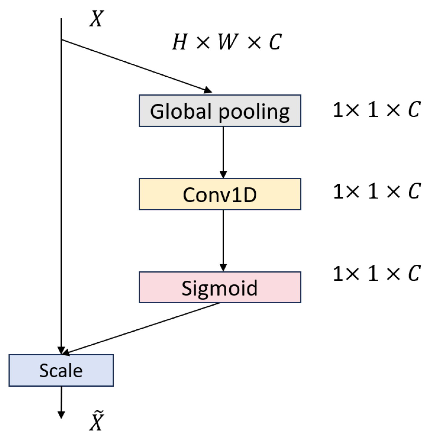
  • Attention Module: Integrates an enhanced MLPBlock with an Efficient Channel Attention (ECA) layer that uses adaptive 1D convolutions to generate channel-wise importance weights, balancing global context and local detail while adding minimal overhead.
  • BiGRU Module: Employs bidirectional Gated Recurrent Unit (GRU) layers to process ECG sequences in both forward and reverse directions, capturing long-range temporal dependencies and contextual features critical for accurate arrhythmia detection
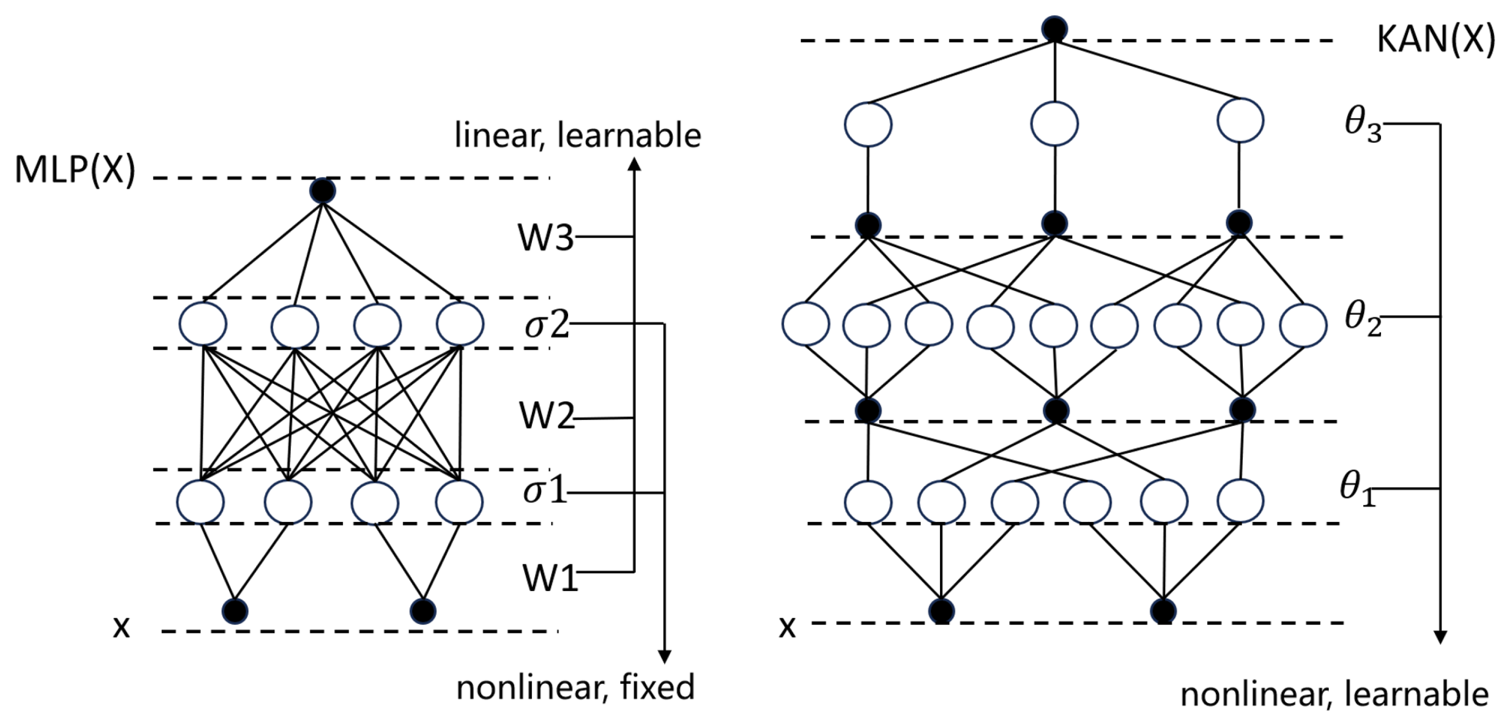
  • Kolmogorov–Arnold Network (KAN) Module: Replaces traditional MLPs to enhance nonlinear mapping.
Furthermore, the framework synergizes focal loss optimization with SMOTE-based oversampling, achieving dual mitigation of data imbalance.
The main contributions of this work are as follows:
  • Significant gains in minority-class detection: By integrating multiscale residual convolutions with dynamic attention, MAK-Net delivers state-of-the-art recall and F1-scores on imbalanced ECG datasets, overcoming the low sensitivity of existing approaches.
  • Parameter-efficient hybrid attention: Our attention module fuses the partial-convolution pruning of the 1D-conv MLPBlock with lightweight ECA channel weighting to capture both local waveform details and global context with minimal overhead, substantially boosting classification accuracy.
  • Interpretable nonlinear mapping via KAN: The KAN module replaces traditional MLP weight matrices with edge-based B-spline activation functions, yielding compact symbolic representations that enhance accuracy, interpretability, and cross-domain adaptability [17].
  • Robust class-imbalance mitigation: We jointly apply SMOTE oversampling and focal loss (γ = 2.0) to balance training distributions and emphasize hard examples, resulting in markedly improved performance on under-represented arrhythmia classes.
The structure of this paper is organized as follows: 
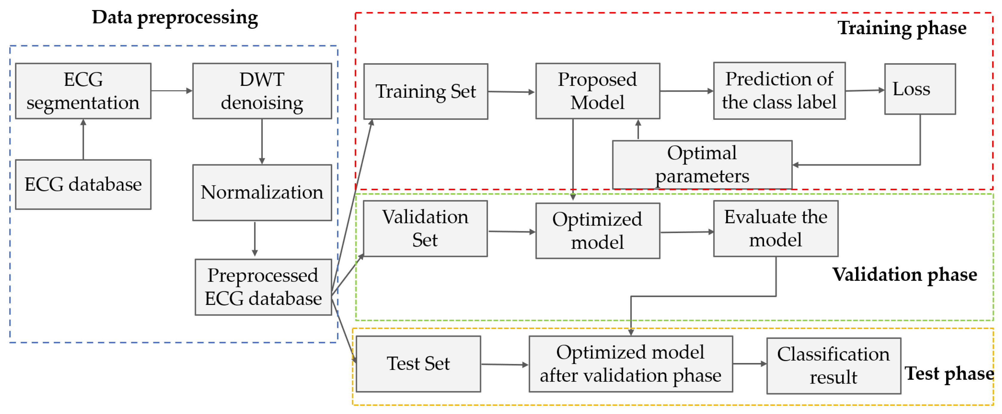
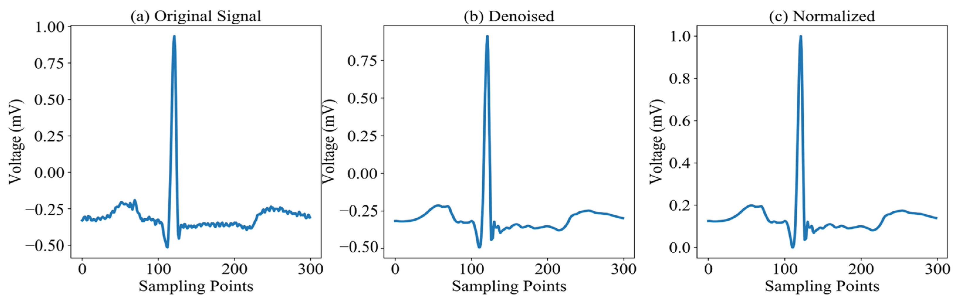
Section 2 provides a comprehensive overview of the MAK-Net architecture, details the preprocessing steps applied to the MIT-BIH dataset, and outlines the evaluation protocols employed. Section 3 presents a comparative analysis of our results against state-of-the-art methods. Finally, Section 4 discusses the clinical implications of our findings and offers concluding remarks.

2. Proposed Method

The comprehensive workflow for ECG signal classification using the MAK-Net model is depicted in Figure 1. The architecture comprises four sequential stages: (1) Data Preprocessing: Raw ECG signals undergo segmentation, denoising through wavelet decomposition and bandpass filtering, and normalization via z-score normalization. Subsequently, the dataset is partitioned into training, validation, and test sets; (2) Training Phase, Model parameters are updated by minimizing the loss function using the training set in a subject-independent (inter-patient) setting; (3) Validation Phase: Hyperparameters (e.g., learning rate, network depth) are optimized to determine the optimal configuration; and (4) Testing Phase: The final performance and generalization capability are evaluated on the test set.

2.1. ECG Preprocessing

In the preprocessing stage, raw ECG signals are first segmented by extracting 150 sampling points on either side of each R-peak to preserve complete waveform morphology. To mitigate noise sources—such as baseline wander, power-line interference, and motion artifacts from muscle activity—the segmented signals undergo denoising before any further processing, thereby improving the accuracy and robustness of downstream feature extraction and classification. In this work, we employ the Discrete Wavelet Transform (DWT) [18], which offers multiscale decomposition, precise time–frequency localization, and energy concentration properties, along with computational efficiency ideally suited for non-stationary biomedical signals [19]. By effectively suppressing noise while preserving diagnostically critical morphological features (as shown in Figure 2b), DWT enhances overall signal quality and boosts classification performance.
The final preprocessing step normalizes the denoised ECG signals to a common amplitude range, thereby accelerating model convergence and improving downstream performance. While various scaling techniques exist—such as min–max normalization and z-score standardization—this study adopts min–max normalization to rescale signal amplitudes to the [0, 1] interval. The normalized signals are shown in Figure 2c, and the transformation is defined in Equation (1):
X n e w = X X m i n X m a x X m i n
where X denotes the original signal amplitude, Xmin and Xmax are the minimum and maximum values within the segment, and Xnew is the normalized amplitude. Min–max normalization has been shown to eliminate amplitude offsets, standardize feature scales, and significantly speed up gradient-based optimization in ECG classification models [20].

2.2. MAK-Net Architecture

Figure 3 illustrates the MAK-Net architecture, which integrates four core modules in sequence: (1) a multi-scale convolutional module with residual connections and parallel kernels (1 × 3, 1 × 11, 1 × 21, 1 × 31) to capture both fine-grained P-wave details and broader RR-interval patterns [21]; (2) an attention module employing channel–spatial reweighting to amplify clinically salient regions such as ST segments and arrhythmic episodes; (3) stacked bidirectional GRU (BiGRU) layers that model long-range temporal dependencies in both forward and backward directions [17]; and (4) a Kolmogorov–Arnold Network (KAN) module that replaces conventional fully connected layers with spline-based activation functions on edges for enhanced nonlinear mapping and interpretability.
Input ECG signals first pass through a four-layer ConvBlock for initial feature extraction, then flow through the multi-scale convolutional module, attention module, BiGRU, and KAN modules in turn, whose complementary spatial, attentional, temporal, and nonlinear representations collectively drive robust arrhythmia classification.

2.2.1. Multi-Scale Convolutional Module

The multi-scale convolutional module draws directly on the InceptionTime paradigm [22], which adapts Inception’s multiplexed convolutions to time-series data by learning multiple filters of varying lengths in parallel. As shown in Figure 3, this module begins with four identical ConvBlock1 units—each comprising a 1D convolution, BatchNorm1d, ReLU activation, and MaxPool1d (kernel = 2, stride = 1, padding = 1)—to perform initial feature extraction. These feature maps then enter a multi-scale fusion block, where four parallel 1D convolutions (kernel sizes 3, 11, 21, 31) capture both fine-grained and broad morphological patterns. The resulting feature maps are combined via weighted averaging, followed by batch normalization, ReLU, dropout (p = 0.3), and max pooling to consolidate salient information. Simultaneously, a secondary ConvBlock2 processes the same inputs in parallel, enabling synergistic integration of local and multi-scale representations—an approach shown to improve ECG classification performance in multireceptive-field CNN designs [23].

2.2.2. Attention Module

As shown in Figure 3, the attention module alternates two MLPBlock modules with two ECA Layer modules in sequence. Each MLPBlock (Figure 4) employs a dual-branch design: the main branch applies successive Conv1d → BatchNorm1d → ReLU → Conv1d operations to extract local temporal features, while a parallel partial-convolution (Partial Conv) branch performs convolution on only a subset of channels, leaving the remainder unchanged. The two branch outputs are then fused via element-wise addition, leveraging parameter sharing to reduce FLOPs and preserve sequence integrity.
The ECA Layer [24] (Figure 5) implements Efficient Channel Attention by first performing channel-wise global average pooling and then using a single Conv1d (kernel size k determined adaptively) to generate channel weights without any dimensionality reduction. This lightweight recalibration mechanism highlights diagnostically salient channels—such as ST segments and arrhythmic episodes—while suppressing noise, reducing computational complexity by approximately 38% compared to standard self-attention [25].

2.2.3. BiGRU

As illustrated in Figure 6, the Bidirectional Gated Recurrent Unit (BiGRU) consists of two parallel GRU networks that process the input sequence in forward and backward directions [26]. At each time step t, the forward hidden state h t and the backward hidden state h t are computed as:
h t = G R U ( x t , h t 1 )
h t = w t h t 1 + v t h t 1 + b t
Here, x t denotes the input vector at time t, and h t is obtained by concatenating the forward and backward hidden states. Each GRU implements nonlinear gating mechanisms—parameterized by weight matrices w t , v t and bias b t —to update and reset its hidden state, thereby encoding the ECG signal into a compact representation. By integrating information from both past and future contexts, the BiGRU architecture captures long-range temporal dependencies and richer contextual cues, leading to improved model performance and classification accuracy.

2.2.4. KAN Module

The KAN network, proposed by Liu et al. [27] in 2024, is grounded in the Kolmogorov–Arnold representation theorem [28,29,30], which guarantees that any multivariate continuous function can be decomposed into a finite superposition of univariate continuous functions and addition. Departing from conventional multilayer perceptrons (MLPs) (MLPs) [31,32,33], KAN replaces fixed activation functions at neurons with learnable univariate spline functions on each edge, effectively substituting weight parameters with parametrized activation mappings. A built-in pruning strategy then extracts a compact set of symbolic functions from the trained network, enabling mechanistic interpretability and module-level analysis. Moreover, by aligning its functional basis with the periodic and trend components intrinsic to time-series data, KAN naturally embeds prior domain knowledge into its architecture, boosting performance in scientific and engineering applications.
As illustrated in Figure 7, whereas an MLP applies fixed nonlinearities after linear summation at each node, KAN nodes perform only linear aggregation, with all nonlinearity arising from the edge-based spline functions—yielding up to 15% gains in accuracy on symbolic regression benchmarks and dramatically enhanced interpretability.

3. Experimental Results

3.1. ECG Dataset

This study utilizes the MIT-BIH Arrhythmia Database [34], which comprises 48 half-hour excerpts of two-channel ambulatory ECG recordings obtained from 47 subjects studied by the BIH Arrhythmia Laboratory between 1975 and 1979. Subjects include 25 males (32–89 years) and 22 females (23–89 years). Recordings were digitized at 360 Hz with 11-bit resolution over a ±10 mV range; channel 1 is modified lead II (MLII), and channel 2 alternates among V1, V2, V4, and V5. Following Association for the Advancement of Medical Instrumentation (AAMI) recommendations and prior mapping schemes [35], the original 15 beat annotations are consolidated into 5 standard categories—normal (N), supraventricular ectopic (S), ventricular ectopic (V), fusion (F), and unknown (Q)—as detailed in Table 1.

3.2. Implementation Detail and Evaluation Metrics

We partitioned the MIT-BIH Arrhythmia Database into training (60%, 65,667 samples), validation (20%, 21,890 samples), and test (20%, 21,890 samples) sets. The training set was used to fit model parameters, the validation set to tune hyperparameters, and the test set to assess final performance. Table 2 summarizes the key hyperparameters used for training MAK-Net. We optimized model parameters using the Adam optimizer with a learning rate of 5 × 10⁻⁴ over 60 epochs and a batch size of 64. The categorical cross-entropy loss—defined in Equations (5)–(7)—served as the optimization objective.
C E ( p , y ) = log ( p ) , y = 1 log ( 1 p ) , o t h e r w i s e
Let p t be defined as:
p t = p , y = 1 1 p , o t h e r w i s e
Therefore, the cross-entropy loss can be formulated as:
C E ( p , y ) = C E ( p t ) = log ( p t )
To benchmark performance, we compared MAK-Net against six state-of-the-art methods. Classification efficacy was evaluated using five standard metrics (Equations (8)–(12))—Accuracy, Precision, Recall, F1-Score, and Specificity—to provide a comprehensive assessment of model performance.
A c c u r a c y = T P + T N T P + F P + T N + F N
P r e c i s i o n = T P T P + F P
R e c a l l = T P T P + F N
F 1 S c o r e = 2 × P r e c i s i o n × R e c a l l P r e c i s i o n + R e c a l l
S p e c i f i c i t y = T N T N + F P

3.3. Classification Performance

Under the hyperparameter configuration detailed in Table 2, MAK-Net’s classification results on the test set are summarized in Table 3, with the corresponding confusion matrix depicted in Figure 8. Table 3 reports the standard metrics—accuracy, precision, recall, F1-score, and specificity—for each arrhythmia category, while Figure 8 visualizes true positives, false positives, and false negatives to facilitate a clear understanding of per-class performance and misclassification patterns.
As Figure 8 shows, classes N, V, and Q achieve outstanding accuracies of 99.62%, 98.26%, and 99.10%, respectively, with all associated metrics (precision, recall, F1-score, specificity) exceeding 97.8%. In contrast, classes S and F register lower accuracies—92.08% and 90.20%—and correspondingly reduced F1-scores and recall values, indicating greater misclassification in these minority categories. These results underscore MAK-Net’s robust performance on predominant classes while highlighting areas for further improvement in detecting supraventricular and fusion beats.

3.4. Strategies for Mitigating Class Imbalance

The MIT-BIH Arrhythmia Database exhibits a pronounced class imbalance—normal (N), ventricular ectopic (V), and unknown (Q) beats dominate, while supraventricular ectopic (S) and fusion (F) beats are scarce. To counteract this skew, we employ two complementary strategies: (1) Focal Loss [36], which adaptively down-weights well-classified majority samples and emphasizes hard, minority-class examples; and (2) SMOTE [37], which synthetically oversamples underrepresented beats by interpolating between minority instances.

3.4.1. Focal Loss-Based Optimization for Class Imbalance

Focal Loss, first introduced by Lin et al. [36] to address extreme class imbalance in one-stage object detectors, augments the standard cross-entropy loss with a modulating factor and class-balance term to focus learning on hard-to-classify examples. Formally, for a true class probability pt, it is defined as:
F L ( p t ) = α ( 1 p t ) γ log ( p t ) = α ( 1 p t ) γ C E ( p t )
where pt is the model’s estimated probability for the ground-truth class; α ∈ [0, 1] balances the relative importance of positive versus negative samples; γ ≥ 0 is the focusing parameter that down-weights well-classified “easy” examples (pt → 1) and amplifies the loss contribution of misclassified “hard” examples (pt → 0).
By dynamically modulating the loss contribution of each example, focal loss has been demonstrated to improve model convergence and accuracy in the presence of severe class imbalance [38]. When applied to MAK-Net, it amplifies gradient signals from under-represented arrhythmia classes, thereby increasing sensitivity and reducing bias toward majority classes. Table 4 summarizes the classification performance of MAK-Net optimized with focal loss, and Figure 9 presents the corresponding confusion matrix.
As shown in Table 4, applying focal loss yields marked improvements for the underrepresented S and F classes: MAK-Net achieves accuracies of 95.71% and 92.94% for these classes—representing gains over the previous 92.08% and 90.20%, respectively—while maintaining accuracies above 98% for classes N, V, and Q. These enhancements are further corroborated by the confusion matrix in Figure 9.
To quantify the contribution of each architectural component, we perform a systematic ablation study, in which we iteratively disable or replace key modules and measure the resulting performance drop. Specifically, we (1) substitute the multiscale convolutional block with single-scale variants (kernel sizes 1 × 3, 1 × 11, 1 × 21, 1 × 31) to assess the impact of receptive-field diversity; (2) remove the spatial–channel attention module to evaluate its effect on feature reweighting; (3) omit the BiGRU layers to isolate the importance of bidirectional temporal modeling; and (4) exclude the KAN module to determine its role in nonlinear feature mapping. The observed degradation in accuracy, F1-score, and recall at each step highlights the indispensable roles of multiscale convolution, attention, temporal recurrence, and nonlinear interpretability in achieving state-of-the-art arrhythmia classification.
The ablation results in Table 5 confirm that the multiscale convolutional block consistently outperforms any single-scale variant across all metrics, echoing prior evidence that multiscale filters enhance ECG classification accuracy [39]. Sequentially disabling key modules induces further performance drops: removing the attention block yields a moderate decline, but omitting the BiGRU layers produces the most pronounced decreases in F1-score (−2.53%), precision (−2.22%), and specificity (−2.01%), underscoring the indispensable role of bidirectional temporal modeling [17]. In contrast, exclusion of the KAN module leads to the largest reductions in overall accuracy (−1.21%) and recall (−2.53%), highlighting its power in capturing nonlinear feature interactions crucial for minority-class detection.

3.4.2. SMOTE-Based Synthetic Oversampling for Class Imbalance

SMOTE (Synthetic Minority Over-sampling Technique) is a widely adopted method for addressing class imbalance by generating new, plausible samples for under-represented classes rather than simply duplicating existing ones. Originally introduced by Chawla et al. [37], SMOTE operates in feature space: for each minority-class instance, it identifies its k nearest neighbors and then synthesizes new examples by interpolating along the line segments connecting the instance to randomly chosen neighbors. This approach expands the minority-class decision region, mitigates overfitting, and improves classifier sensitivity to rare categories.
In practice, SMOTE proceeds as follows: first, for each minority sample, compute the Euclidean distances to its k nearest minority-class neighbors. Next, randomly select one neighbor and generate a synthetic point by taking a convex combination of the two feature vectors using a random weight in [0, 1]. Repeat this process until the minority class reaches the desired sample count. By filling the feature space between existing minority examples, SMOTE balances the training set and enhances the model’s ability to generalize to previously unseen minority-class patterns. To guard against overfitting from synthetic examples, SMOTE was applied only to the training folds during cross-validation, with validation and test sets left entirely unchanged to prevent any data leakage. By isolating synthetic data to the training phase, we ensure that performance metrics reflect the model’s ability to generalize to real ECG segments.
After SMOTE oversampling, each arrhythmia class is represented by approximately 54,170 samples (N: 54,166; S: 54,169; V: 54,168; F: 54,171; Q: 54,171), thereby achieving a fully balanced training set. With class frequencies equalized, we revert to the standard categorical cross-entropy loss to measure discrepancies between predicted probabilities and true labels during training. The classification performance of MAK-Net under this balanced regime is summarized in Table 6, and the corresponding confusion matrix is shown in Figure 10.
As shown in Table 6, MAK-Net trained on SMOTE-oversampled data achieves exceptional performance across all arrhythmia classes: classes N and Q both exceed 99% in accuracy and F1-score, while class V approaches 99% (98.95% accuracy, 98.98% F1-score). Notably, the previously under-represented S and F classes now reach 98.48% and 96.57% accuracy (with F1-scores of 98.81% and 97.24%, respectively), representing substantial gains over their pre-oversampling baselines.
As shown in Table 7, both focal loss and SMOTE substantially boost the accuracies of the minority classes S and F. Focal loss elevates S from 92.08% to 95.71% and F from 90.20% to 92.94%, whereas SMOTE further increases these to 98.48% and 96.57%, respectively. SMOTE also marginally enhances the majority-class accuracies—achieving 99.95% for N, 98.95% for V, and 99.62% for Q—surpassing both the baseline and focal-loss configurations. Therefore, SMOTE-based oversampling proves the most effective strategy for mitigating class imbalance and maximizing overall classification accuracy.
Table 8 compares MAK-Net’s overall metrics—accuracy, F1-score, recall, precision, and specificity—under three optimization strategies: no optimization, focal loss, and SMOTE oversampling. Both focal loss and SMOTE yield marked gains in F1-score, recall, and precision over the unoptimized baseline. SMOTE delivers the largest improvements across all metrics (accuracy: 0.9980, F1-score: 0.9888, recall: 0.9871, precision: 0.9905, specificity: 0.9991). These findings demonstrate that SMOTE-based oversampling more effectively mitigates class imbalance and maximizes overall classification performance compared to focal loss.

3.5. Performance Comprasion with Other Reported Methods

As shown in Table 9, MAK-Net outperforms all benchmark methods in accuracy (0.9980), precision (0.9905), F1-score (0.9888), and specificity (0.9991), while maintaining a competitive recall (0.9871) compared to the highest recall achieved by Ahmed et al. [40]. This balanced, state-of-the-art performance reflects several broader insights in ECG arrhythmia classification. First, hybrid architectures that combine convolutional feature extractors with recurrent or attention modules consistently surpass single-model approaches by capturing both spatial and temporal dependencies in ECG signals [41]. Second, multiscale convolutional streams integrated with lightweight attention mechanisms—also as in CNN-BiLSTM-BiGRU hybrids [42]—enhance discriminative power by jointly modeling fine-grained waveform details and global rhythm patterns. Third, embedding interpretable components, such as spline-based activation functions in KAN or hierarchical attention blocks, not only boosts accuracy but also provides mechanistic transparency, a crucial factor for clinical adoption. Together, these trends underscore the effectiveness of MAK-Net’s design in extracting rich, clinically relevant features and delivering robust arrhythmia classification.
Table 10 benchmarks MAK-Net against four canonical deep learning baselines—ResNet, RNN, LSTM, and GRU—each retrained on the MIT-BIH dataset under identical conditions. While these architectures achieve accuracies in the 0.9786–0.9824 range and F1-scores between 0.9003 and 0.9119, MAK-Net attains 0.9980 accuracy and an F1-score of 0.9888, representing gains of 1.56–1.94% in accuracy and over 7.0% in F1-score. The pronounced improvements in both precision and recall underscore MAK-Net’s balanced performance across arrhythmia types. These results indicate that, although standard CNN and RNN variants can capture general morphological or temporal patterns, they struggle with the complex, multiscale dynamics of ECG signals. In contrast, MAK-Net’s tailored integration of multiscale convolutional streams, attention reweighting, bidirectional recurrence, and nonlinear KAN modules enables richer feature extraction and more robust classification across all arrhythmia classes.

4. Discussion

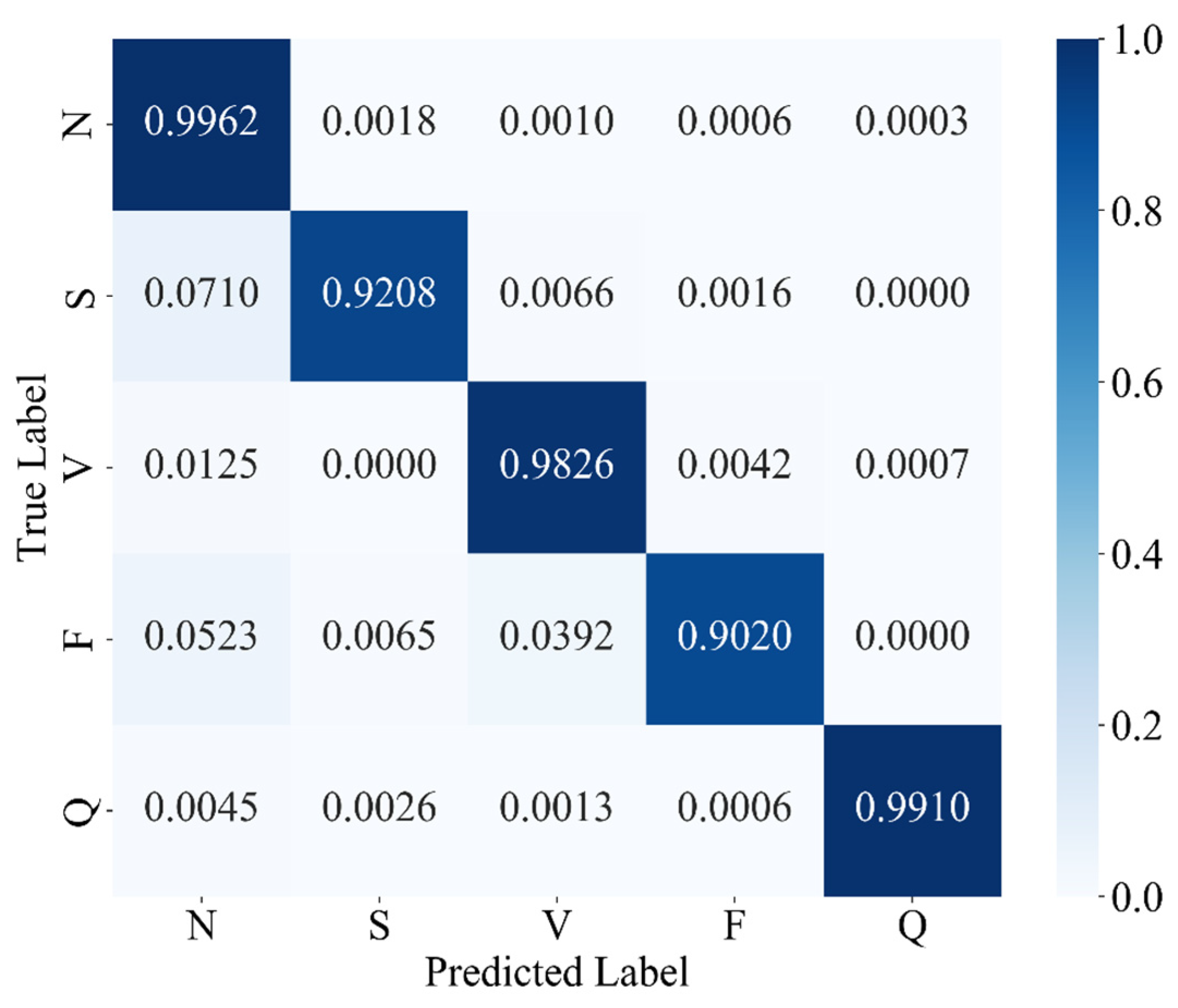
The experimental results confirm that MAK-Net delivers state-of-the-art ECG classification performance (accuracy 0.9980; F1-score 0.9888; specificity 0.9991), validating our design choices. The multiscale convolutional streams capture both fine-grained morphological features (e.g., QRS complexes) and broader rhythm patterns, while the ECA attention mechanism dynamically reweights diagnostically salient segments—analogous to a clinician’s focus on ST deviations and P-wave abnormalities [39]. The BiGRU layers encode bidirectional temporal dependencies, essential for detecting arrhythmias that evolve over extended intervals [50]. Furthermore, the KAN module’s learnable spline activations replace fixed nonlinearities, enabling post-hoc symbolic analysis of feature interactions and greatly enhancing interpretability—a critical requirement for clinical adoption [51].
Our ablation and comparative studies also highlight the pivotal role of data-level balancing. SMOTE oversampling outperforms focal-loss reweighting alone, boosting minority-class recall and F1-scores by up to 3.8% over baseline models—consistent with prior findings on class-imbalance remedies in ECG tasks [52]. This underscores that, even with advanced loss functions, equitable class representation in the training set remains indispensable for robust arrhythmia detection.
Clinically, MAK-Net’s near-perfect specificity (≥99.7%) minimizes false alarms for life-threatening conditions such as ventricular fibrillation, while its high recall (≥98.4% for rare beats) ensures reliable detection of critical but infrequent events like supraventricular ectopics. Moreover, the model’s interpretable attention maps and spline-based activations can help clinicians verify that the network’s focus aligns with established diagnostic markers, thereby fostering trust.
Nonetheless, there are still several challenges in further applying the MAK-Net in real clinical use. Generalizability to real-world ECG recordings—often corrupted by motion artifacts, electrode misplacement, and variable sampling rates—must be assessed on larger, more heterogeneous datasets [52]. The hybrid architecture’s computational complexity may also hinder deployment on wearable or edge-computing platforms, motivating future work on model compression, pruning, and quantization to retain performance while reducing inference time [53]. More specifically, we have measured MAK-Net’s computational footprint at 6.11 million parameters and an end-to-end inference time of 1.52 s for a batch of sixty-four ECG segments on a cloud server equipped with an NVIDIA RTX 3090 GPU and 14 vCPU Intel® Xeon® Platinum 8362 CPUs, corresponding to approximately 24 ms per segment. Although this complexity and latency preclude ultra-low-latency continuous monitoring, at this stage, MAK-Net is well suited for offline or semi-real-time applications requiring high diagnostic precision—such as detailed analysis of rare channelopathies (e.g., Brugada syndrome) or refined myocardial ischemia assessments. Finally, demographic biases in training data (e.g., underrepresentation of certain age or ethnic groups) necessitate rigorous fairness evaluations and bias-mitigation strategies before clinical rollout.
Looking forward, the modular nature of MAK-Net invites adaptation to other biosignal domains—such as EEG seizure detection or respiratory monitoring—where multiscale spatiotemporal modeling is equally vital. Incorporating causal inference frameworks could further refine arrhythmia precursor detection, transitioning from correlation-based classification to actionable diagnostic insights. To ensure comparability with the majority of existing studies using the MIT-BIH dataset, we adopted an intra-patient evaluation protocol, which is widely regarded as the standard approach given the dataset’s limited cohort size [54,55]. We acknowledge that inter-patient validation offers a more stringent test of generalizability, and we consider it a valuable direction for future work. In terms of the interpretability of MAK-Net, we have not included explicit visualizations or symbolic analyses of the learned spline activations; our interpretability claims are grounded in the architectural design of KANs, which employ learnable spline-based activation functions on edges, facilitating transparent and adaptable functional mappings [28,29,30]. Thus, we intend to analyze the learned spline functions to extract symbolic representations, which could offer deeper insights into the model’s decision-making process and enhance its clinical utility.
We plan to extend MAK-Net’s validation to larger, publicly available ECG repositories—such as the PTB-XL 12-lead dataset and the CPSC 2018 challenge dataset—to robustly assess cross-center generalizability. In addition, we also plan to evaluate MAK-Net’s robustness using more diverse and noise-prone datasets, such as the MIT-BIH Noise Stress Test Database, which includes recordings with baseline wander, muscle artifacts, and electrode motion artifacts, and the Brno University of Technology ECG Quality Database (BUTQDB), which comprises long-term ECG recordings collected under free-living conditions with varying signal qualities. These evaluations will help assess and enhance the model’s performance in more realistic scenarios. Additionally, in collaboration with cardiologists at the First Affiliated Hospital of Guangzhou Medical University, Guangzhou, China, we are curating a Brugada syndrome cohort, annotated for Type-1 and Type-2 ECG patterns, to evaluate MAK-Net’s sensitivity and specificity on this rare but clinically critical channelopathy. In sum, MAK-Net exemplifies how architectural innovation coupled with data-centric optimization can yield interpretable, reliable AI tools that augment, rather than replace, physician expertise in cardiovascular diagnostics.

5. Conclusions

This study introduces MAK-Net, a hybrid deep learning framework that combines multiscale convolutional streams, efficient channel attention, bidirectional GRUs, and a Kolmogorov–Arnold Network layer to deliver state-of-the-art ECG arrhythmia classification—achieving 0.9980 accuracy, a 0.9888 F1-score, 0.9871 recall, 0.9905 precision, and 0.9991 specificity on the MIT-BIH database. By integrating SMOTE-based oversampling with focal-loss optimization, MAK-Net effectively mitigates class imbalance, with SMOTE proving most impactful for minority-class detection. The model’s attention maps and spline-based activations enhance interpretability, aligning network focus with clinical markers, while its lightweight hybrid design suggests feasibility for real-time deployment. Nevertheless, further validation on heterogeneous, noisy ECG recordings and efforts in model compression and fairness assessment are essential before clinical translation. Future work will explore adapting MAK-Net’s modular architecture to other biosignals (e.g., EEG) and incorporating causal inference to advance from correlation to actionable diagnostic insights.

Author Contributions

Conceptualization, C.Z. and H.D.; methodology, B.L. and Y.X.; software, C.Z. and B.L.; validation, Y.X. and Y.W.; investigation, H.D.; resources, C.Z. and Y.W.; data curation, B.L. and Y.X.; writing—original draft preparation, C.Z. and B.L.; writing—review and editing, Y.X., Y.W. and H.D.; visualization, B.L.; supervision, C.Z. and H.D. All authors have read and agreed to the published version of the manuscript.

Funding

This research was funded by the National Natural Science Foundation of China (NSFC) (62205221 and 62405196) and Shenzhen Science and the Technology Program (Shenzhen Key Laboratory of Ultrafast Laser Micro/Nano Manufacturing ZDSYS20220606100405013).

Institutional Review Board Statement

Not applicable since the study did not involve any clinical trials.

Informed Consent Statement

Validation and testing of the developed model were conducted using a publicly accessible dataset, for which informed consent was not applicable due to the data’s public availability.

Data Availability Statement

The data may be requested by reaching out to authors through email.

Acknowledgments

The authors would like to acknowledge Qiaonan Zhong from The First Affiliated Hospital of Guangzhou Medical University, Guangzhou, China, for her professional guidance and suggestions regarding model performance evaluation and practical clinical needs in cardiology.

Conflicts of Interest

The authors declare no conflicts of interest.

Abbreviations

The following abbreviations are used in this manuscript:
AAMIAssociation for the Advancement of Medical Instrumentation
ACGANsAuxiliary Classifier Generative Adversarial Networks
BiGRUBidirectional Gated Recurrent Unit
CNNsConvolutional Neural Networks
Conv1d1-Dimensional Convolution
CVDsCardiovascular Diseases
DWTDiscrete Wavelet Transform
ECAEfficient Channel Attention
ECGElectrocardiogram
EEGElectroencephalogram
FLOPsFloating Point Operations
GRUsGated Recurrent Units
KANKolmogorov–Arnold Network
LSTMLong Short-Term Memory
MIT-BIHMassachusetts Institute of Technology-Beth Israel Hospital (Arrhythmia Database)
MLIIModified Lead II
MLPMultilayer Perceptron
ReLURectified Linear Unit
RNNsRecurrent Neural Networks
SMOTESynthetic Minority Oversampling Technique

References

  1. World Heart Report 2023. Available online: https://heartreport23.world-heart-federation.org (accessed on 15 October 2024).
  2. Bengio, Y.; Lecun, Y.; Hinton, G. Deep learning for AI. Commun. ACM 2021, 64, 58–65. [Google Scholar] [CrossRef]
  3. Chen, Y. Convolutional Neural Networks for Sentence Classification; University of Waterloo: Waterloo, ON, Canada, 2015. [Google Scholar]
  4. Elman, J.L. Finding Structure in Time. Cogn. Sci. 1990, 14, 179–211. [Google Scholar] [CrossRef]
  5. Hochreiter, S.; Schmidhuber, J. Long Short-Term Memory. Neural Comput. 1997, 9, 1735–1780. [Google Scholar] [CrossRef] [PubMed]
  6. Chung, J.; Gulcehre, C.; Cho, K.; Bengio, Y. Empirical Evaluation of Gated Recurrent Neural Networks on Sequence Modeling. arXiv 2014, arXiv:1412.3555. [Google Scholar]
  7. Acharya, U.R.; Oh, S.L.; Hagiwara, Y. A Deep Convolutional Neural Network Model to Classify Heartbeats. Comput. Biol. Med. 2017, 89, 389–396. [Google Scholar] [CrossRef] [PubMed]
  8. Yildirim, O. A novel wavelet sequence-based deep bidirectional LSTM network model for ECG signal classification. Comput. Biol. Med. 2018, 96, 189–202. [Google Scholar] [CrossRef] [PubMed]
  9. Hannun, A.Y.; Rajpurkar, P.; Haghpanahi, M. Cardiologist-level arrhythmia detection and classification in ambulatory electrocardiograms using a deep neural network. Nat. Med. 2019, 25, 65–69. [Google Scholar] [CrossRef]
  10. Ebrahimi, Z.; Loni, M.; DaneshTalab, M. A review on deep learning methods for ECG arrhythmia classification. Expert Syst. Appl. 2020, 7, 79–89. [Google Scholar] [CrossRef]
  11. Oh, S.L.; Ng, Y.K.; Tan, R.S. Automated diagnosis of arrhythmia using combination of CNN and LSTM techniques with variable-length heart beats. Comput. Biol. Med. 2018, 102, 278–287. [Google Scholar] [CrossRef]
  12. Chun, Y.C.; Yan, T.L.; Shie, J.L.; Wei, C.T. Automated ECG classification based on 1D deep learning network. Methods 2022, 202, 127–135. [Google Scholar] [CrossRef]
  13. Abdolrahman, P.; Sadasivan, P. DENS-ECG: A deep learning approach for ECG signal delineation. Expert Syst. Appl. 2021, 165, 113911. [Google Scholar]
  14. Shoughi, A.; Dowlatshahi, M.B. A practical system based on CNN-BLSTM network for accurate classification of ECG heartbeats of MIT-BIH imbalanced dataset. In Proceedings of the 2021 26th International Computer Conference, Computer Society of Iran (CSICC), Tehran, Iran, 3–4 March 2021; pp. 1–6. [Google Scholar]
  15. Samjin, C.; Hyungli, S.; Min, S.C. Performance improvement of deep learning based multi-class ECG classification model using limited medical dataset. IEEE Access 2023, 11, 53185–53194. [Google Scholar]
  16. Bechinia, H.; Benmerzoug, D.; Khlif, N. Approach Based Lightweight Custom Convolutional Neural Network and Fine-Tuned MobileNet-V2 for ECG Arrhythmia Signals Classification. IEEE Access 2024, 12, 40827–40841. [Google Scholar] [CrossRef]
  17. Bai, X.; Dong, X.; Li, Y.; Liu, R.; Zhang, H. A hybrid deep learning network for automatic diagnosis of cardiac arrhythmia based on 12-lead ECG. Sci. Rep. 2024, 14, 24441. [Google Scholar] [CrossRef]
  18. Mallat, S.G. A Theory for Multiresolution Signal Decomposition: The Wavelet Representation. IEEE Trans. Pattern Anal. Mach. Intell. 1989, 11, 674–693. [Google Scholar] [CrossRef]
  19. Zhang, D.; Wang, S.; Li, F.; Wang, J.; Sangaiah, A.K.; Sheng, V.S.; Ding, X. An ECG signal de-noising approach based on wavelet energy and sub-band smoothing filter. Appl. Sci. 2019, 9, 4968. [Google Scholar] [CrossRef]
  20. Boulif, A.; Ananou, B.; Ouladsine, M.; Delliaux, S. A literature review: Ecg-based models for arrhythmia diagnosis using artificial intelligence techniques. Bioinform. Biol. Insights 2023, 17, 11779322221149600. [Google Scholar] [CrossRef]
  21. Yang, Z.; Jin, A.; Li, Y.; Yu, X.; Xu, X.; Wang, J.; Li, Q.; Guo, X.; Liu, Y. A coordinated adaptive multiscale enhanced spatio-temporal fusion network for multi-lead electrocardiogram arrhythmia detection. Sci. Rep. 2024, 14, 20828. [Google Scholar] [CrossRef] [PubMed]
  22. Fawaz, H.I.; Lucas, B.; Forestier, G.; Pelletier, C.; Schmidt, D.F.; Weber, J.; Petitjean, F. Inceptiontime: Finding alexnet for time series classification. Data Min. Knowl. Discov. 2020, 34, 1936–1962. [Google Scholar] [CrossRef]
  23. Feyisa, D.W.; Debelee, T.G.; Ayano, Y.M.; Kebede, S.R.; Assore, T.F. Lightweight Multireceptive Field CNN for 12-Lead ECG Signal Classification. Comput. Intell. Neurosci. 2022, 2022, 8413294. [Google Scholar] [CrossRef]
  24. Wang, Q.; Wu, B.; Zhu, P.; Li, P.; Zuo, W.; Hu, Q. ECA-Net: Efficient channel attention for deep convolutional neural networks. In Proceedings of the IEEE/CVF Conference on Computer Vision and Pattern Recognition, Seattle, WA, USA, 13–19 June 2020; pp. 11534–11542. [Google Scholar]
  25. Kim, Y.; Kim, Y.K. Time-frequency multi-domain 1D convolutional neural network with channel-spatial attention for noise-robust bearing fault diagnosis. Sensors 2023, 23, 9311. [Google Scholar] [CrossRef] [PubMed]
  26. Li, C.; He, Y.; Li, X.; Jing, X. BiGRU network for human activity recognition in high resolution range profile. In Proceedings of the 2019 International Radar Conference (RADAR), Toulon, France, 23–27 September 2019; pp. 1–5. [Google Scholar]
  27. Liu, Z.; Wang, Y.; Vaidya, S.; Ruehle, F.; Halverson, J.; Soljačić, M.; Hou, T.Y.; Tegmark, M. Kan: Kolmogorov-arnold networks. arXiv 2024, arXiv:2404.19756. [Google Scholar]
  28. Kolmogorov, A.N. On the representation of continuous functions of several variables by superpositions of continuous functions of a smaller number of variables. Am. Math. Soc. 1961, 28, 55–59. [Google Scholar]
  29. Kolmogorov, A.N. On the representations of continuous functions of many variables by superposition of continuous functions of one variable and addition. Dokl. Akad. Nauk USSR 1957, 114, 953–956. [Google Scholar]
  30. Braun, J.; Griebel, M. On a constructive proof of Kolmogorov’s superposition theorem. Constr. Approx. 2009, 30, 653–675. [Google Scholar] [CrossRef]
  31. Haykin, S. Neural Networks: A Comprehensive Foundation; Prentice Hall PTR: Upper Saddle River, NJ, USA, 1994. [Google Scholar]
  32. Cybenko, G. Approximation by superpositions of a sigmoidal function. Math. Control Signals Syst. 1989, 2, 303–314. [Google Scholar] [CrossRef]
  33. Hornik, K.; Stinchcombe, M.; White, H. Multilayer feedforward networks are universal approximators. Neural Netw. 1989, 2, 359–366. [Google Scholar] [CrossRef]
  34. Moody, G.B.; Mark, R.G. The impact of the MIT-BIH arrhythmia database. IEEE Eng. Med. Biol. Mag. 2001, 20, 45–50. [Google Scholar] [CrossRef]
  35. Eleyan, A.; Alboghbaish, E. Electrocardiogram signals classification using deep-learning-based incorporated convolutional neural network and long short-term memory framework. Computers 2024, 13, 55. [Google Scholar] [CrossRef]
  36. Lin, T.Y.; Goyal, P.; Girshick, R.; He, K.; Dollár, P. Focal loss for dense object detection. In Proceedings of the IEEE International Conference on Computer Vision, Venice, Italy, 22–29 October 2017; pp. 2980–2988. [Google Scholar]
  37. Chawla, N.V.; Bowyer, K.W.; Hall, L.O.; Kegelmeyer, W.P. SMOTE: Synthetic minority over-sampling technique. J. Artif. Intell. Res. 2002, 16, 321–357. [Google Scholar] [CrossRef]
  38. Singh, J.; Beeche, C.; Shi, Z.; Beale, O.; Rosin, B.; Leader, J.; Pu, J. Batch-balanced focal loss: A hybrid solution to class imbalance in deep learning. J. Med. Imaging 2023, 10, 051809. [Google Scholar] [CrossRef] [PubMed]
  39. Guo, C.; Yin, B.; Hu, J. An electrocardiogram classification using a multiscale convolutional causal attention network. Electronics 2024, 13, 326. [Google Scholar] [CrossRef]
  40. Ahmed, A.A.; Ali, W.; Abdullah, T.A.; Malebary, S.J. Classifying cardiac arrhythmia from ECG signal using 1D CNN deep learning model. Mathematics 2023, 11, 562. [Google Scholar] [CrossRef]
  41. Sun, A.; Hong, W.; Li, J.; Mao, J. An Arrhythmia Classification Model Based on a CNN-LSTM-SE Algorithm. Sensors 2024, 24, 6306. [Google Scholar] [CrossRef]
  42. Kim, D.; Lee, K.R.; Lim, D.S.; Lee, K.H.; Lee, J.S.; Kim, D.Y.; Sohn, C.B. A novel hybrid CNN-transformer model for arrhythmia detection without R-peak identification using stockwell transform. Sci. Rep. 2025, 15, 7817. [Google Scholar] [CrossRef] [PubMed]
  43. Yamaç, M.; Duman, M.; Adalıoğlu, İ.; Kiranyaz, S.; Gabbouj, M. A personalized zero-shot ecg arrhythmia monitoring system: From sparse representation based domain adaption to energy efficient abnormal beat detection for practical ecg surveillance. arXiv 2022, arXiv:2207.07089. [Google Scholar]
  44. Pu, N.; Wu, Z.; Wang, A.; Sun, H.; Liu, Z.; Liu, H. Arrhythmia classifier based on ultra-lightweight binary neural network. In Proceedings of the 2023 15th International Conference on Electronics, Computers and Artificial Intelligence (ECAI), Bucharest, Romania, 29 June–1 July 2023; pp. 1–7. [Google Scholar]
  45. Li, J.; Pang, S.P.; Xu, F.; Ji, P.; Zhou, S.; Shu, M. Two-dimensional ECG-based cardiac arrhythmia classification using DSE-ResNet. Sci. Rep. 2022, 12, 14485. [Google Scholar] [CrossRef]
  46. Hammad, M.; Meshoul, S.; Dziwiński, P.; Pławiak, P.; Elgendy, I.A. Efficient lightweight multimodel deep fusion based on ECG for arrhythmia classification. Sensors 2022, 22, 9347. [Google Scholar] [CrossRef]
  47. He, K.; Zhang, X.; Ren, S.; Sun, J. Deep residual learning for image recognition. In Proceedings of the IEEE Conference on Computer Vision and Pattern Recognition, Las Vegas, NV, USA, 27–30 June 2016; pp. 770–778. [Google Scholar]
  48. Zaremba, W.; Sutskever, I.; Vinyals, O. Recurrent neural network regularization. arXiv 2014, arXiv:1409.2329. [Google Scholar]
  49. Graves, A.A. Long short-term memory. In Supervised Sequence Labelling with Recurrent Neural Networks; Springer: Berlin, Germany, 2012; pp. 37–45. [Google Scholar]
  50. Zhao, Y.; Ren, J.; Zhang, B.; Wu, J.; Lyu, Y. An explainable attention-based TCN heartbeats classification model for arrhythmia detection. Biomed. Signal Process. Control 2023, 80, 104337. [Google Scholar] [CrossRef]
  51. Niu, L.; Chen, C.; Liu, H.; Zhou, S.; Shu, M. A deep-learning approach to ECG classification based on adversarial domain adaptation. Healthcare 2020, 8, 437. [Google Scholar] [CrossRef] [PubMed]
  52. Bing, P.; Liu, Y.; Liu, W.; Zhou, J.; Zhu, L. Electrocardiogram classification using TSST-based spectrogram and ConViT. Front. Cardiovasc. Med. 2022, 9, 983543. [Google Scholar] [CrossRef] [PubMed]
  53. Jiang, F.; Xiao, J.; Liu, L.; Wang, C. DCETEN: A lightweight ECG automatic classification network based on Transformer model. Digit. Commun. Netw. 2024; In Press. [Google Scholar]
  54. Silva, G.; Silva, P.; Moreira, G.; Freitas, V.; Gertrudes, J.C.; Luz, E. A Systematic Review of Ecg Arrhythmia Classification: Adherence to Standards, Fair Evaluation, and Embedded Feasibility. arXiv 2025, arXiv:2503.07276. [Google Scholar]
  55. Xiao, Q.; Lee, K.; Mokhtar, S.A.; Ismail, I.; Pauzi, A.L.b.M.; Zhang, Q.; Lim, P.Y. Deep Learning-Based Ecg Arrhythmia Classification: A Systematic Review. Appl. Sci. 2023, 13, 4964. [Google Scholar] [CrossRef]
Figure 1. Overview of the MAK-Net ECG classification pipeline.
Sensors 25 03928 g001
Figure 2. ECG preprocessing pipeline and results: (a) Raw ECG segment showing baseline wander and high-frequency noise; (b) Denoised signal after discrete wavelet transform; (c) Final normalized output using min–max scaling, ready for feature extraction.
Sensors 25 03928 g002
Figure 3. MAK-Net architecture: A ConvBlock for initial feature extraction, followed by multiscale residual convolutions, an attention module, bidirectional GRUs, and a KAN module, leading to the classification output.
Sensors 25 03928 g003
Figure 4. Structure of the MLP Block.
Sensors 25 03928 g004
Figure 5. Structure of the ECA layer.
Sensors 25 03928 g005
Figure 6. Structure diagram of the bidirectional gated recurrent unit (BiGRU).
Sensors 25 03928 g006
Figure 7. Architecture of the KAN.
Sensors 25 03928 g007
Figure 8. Confusion matrix for MAK-Net with cross-entropy loss.
Sensors 25 03928 g008
Figure 9. Confusion matrix of MAK-Net optimized with focal loss.
Sensors 25 03928 g009
Figure 10. Confusion matrix of MAK-Net trained with SMOTE-oversampled data.
Sensors 25 03928 g010
Table 1. ECG class mapping using the AAMI standard.
ClassSymbolHeart BeatTraining SamplesValidation
Samples
Test
Samples
Normal beatNNormal beat54,17118,05818,116
Left bundle branch block beat
Right bundle branch block beat
Supraventricular premature beatSAtrial premature beat1807603616
Nodal (junctional) premature beat
Supraventricular premature beat
Aberrated atrial premature beat
Atrial escape beat
Nodal (junctional) escape beat
Premature ventricular contractionVPremature ventricular contraction434814491438
Ventricular escape beat
Fusion of ventricular and normal beatFFusion of ventricular and normal beat483161158
Unknown BeatQPace beat485816191562
Unclassifiable beat
Fusion of paced and normal beat
Table 2. Hyper-parameter of the model training.
Hyper-ParameterValues
Batch size64
Learning rate0.0005
Epoch60
OptimizerAdam
Loss functionCross-Entropy Loss
Table 3. MAK-Net classification performance (cross-entropy loss).
ClassAccuracyF1-ScoreRecallPrecisionSpecificity
N0.99620.99600.99620.99580.9798
S0.92080.92920.92080.93780.9982
V0.98260.98060.98260.97850.9985
F0.90200.89030.90200.87900.9991
Q0.99100.99330.99100.99550.9997
Sum0.99220.95790.95850.95730.9951
Table 4. Classification performance of MAK-Net optimized with focal loss.
ClassAccuracyF1-ScoreRecallPrecisionSpecificity
N0.99710.99720.99710.99730.9872
S0.95710.95790.95710.95880.9989
V0.98190.98090.98190.97990.9986
F0.92940.92670.92940.92400.9994
Q0.99300.99260.99300.99230.9994
Sum0.99420.97110.97170.97050.9967
Table 5. Ablation study results of MAK-Net optimized with focal loss.
ConfigurationAccuracyF1-ScoreRecallPrecisionSpecificity
Single-scale (1 × 3)0.98780.96620.96670.96580.9826
Single-scale (1 × 11)0.98670.96460.96570.96360.9836
Single-scale (1 × 21)0.98420.95830.96370.95310.9825
Single-scale (1 × 31)0.98530.95450.96450.94470.9835
Without Attention module0.98710.95660.95900.95430.9731
Without BiGRU0.98210.95290.94640.95960.9722
Without KAN0.98380.94850.94870.94830.9627
Full MAK-Net0.99420.97110.97170.97050.9967
Table 6. Classification performance of MAK-Net trained with SMOTE-oversampled data.
ClassAccuracyF1-ScoreRecallPrecisionSpecificity
N0.99950.99940.99950.99940.9970
S0.98480.98810.98470.99150.9997
V0.98950.98980.98950.99020.9993
F0.96570.97240.96580.97920.9998
Q0.99620.99420.99610.99230.9994
Sum0.99800.98880.98710.99050.9991
Table 7. Per-class accuracy of MAK-NET under different imbalance mitigation strategies.
MethodNSVFQ
No Optimization0.99620.92080.98260.90200.9910
Focal Loss 0.99710.95710.98190.92940.9930
SMOTE0.99950.98480.98950.96570.9962
Table 8. Overall performance metrics of MAK-Net under different optimization strategies.
MethodAccuracyF1-ScoreRecallPrecisionSpecificity
No Optimization0.99220.95790.95850.95730.9951
Focal Loss0.99420.97110.97170.97050.9967
SMOTE0.99800.98880.98710.99050.9991
Table 9. Comparative classification performance of MAK-Net and recent state-of-the-art methods.
AuthorsMethodAccuracyF1-ScoreRecallPrecisionSpecificity
Yamac et al. [43]1D-CNN0.98200.91900.93700.92800.9880
Pu et al. [44]1D-BNN0.97500.96400.97900.96500.9980
Eleyan et al. [35]CNN-LSTM0.97400.97300.97400.9730NA
Li et al. [45]CNN0.9810NANANA0.9870
Hammad et al. [46]Fusion CNN0.98800.98800.98800.98800.9880
Ahmed et al. [40]AAC-FFAHD0.9880NA0.9850NA0.9790
This workMAK-Net0.99800.98880.98710.99050.9991
Table 10. Performance comparison with baseline models.
AuthorMethodAccuracyF1-ScoreRecallPrecisionSpecificity
He et al. [47]ResNet0.97860.90030.90390.89710.9870
Zaremba et al. [48]RNN0.97980.90970.94000.88420.9911
Hochreiter et al. [49]LSTM0.98240.91190.93250.90680.9911
Chung et al. [6]GRU0.98060.90890.93850.88420.9916
This workMAK-Net0.99800.98880.98710.99050.9991
Disclaimer/Publisher’s Note: The statements, opinions and data contained in all publications are solely those of the individual author(s) and contributor(s) and not of MDPI and/or the editor(s). MDPI and/or the editor(s) disclaim responsibility for any injury to people or property resulting from any ideas, methods, instructions or products referred to in the content.

