

Abstract
Surface plasmon resonance (SPR) detection technology is playing an important role in various fields such as food safety and environmental monitoring due to its excellent stability and reliability. However, there is also a growing demand for higher sensitivity in SPR sensors. Therefore, this work developed an SPR sensor based on the synergistic effect of electric-field enhancement and adsorption enhancement by using amino acid-derived carbon dots (CDs). The results showed that the incorporation of amino acid CDs can generate a maximum electric-field enhancement of up to 6.44 × 10⁵ V/m in the near-field region, which is 312% of that achieved by a bare gold film. And the adsorption kinetics results indicate that the active groups on the surface of amino acid CDs exhibit a notable adsorption enhancement effect for the target molecule (NaCl), with an adsorption capacity 335% higher than that of the bare gold film. This designed SPR sensor demonstrates a detection sensitivity of 167.28 a.u./RIU for NaCl solution, representing a 247.8% improvement compared to an SPR sensor without amino acid CDs under the same conditions. This SPR sensor shows promising potential for applications in biomedical and environmental detection fields.
1. Introduction
Surface plasmon resonance (SPR) sensors, as highly advantageous analytical tools, play an indispensable role in numerous fields. In the biomedical domain, the SPR sensors can be used for disease diagnosis and drug screening [1,2]. In environmental monitoring, they enable precise identification of trace pollutants [3]. In food safety, SPR sensors facilitate the detection of harmful substances [4]. Owing to their label-free operation, real-time monitoring capability, and high sensitivity, SPR sensors have become a vital detection method in practical applications [5,6].
Nevertheless, as detection requirements toward lower concentrations and more complex systems, the sensitivity of SPR sensors faces significant challenges [7,8,9,10,11]. To meet these performance demands, researchers are actively exploring various enhancement strategies to improve SPR sensor sensitivity. Common approaches primarily include electric-field enhancement and adsorption enhancement. Electric-field enhancement typically employs nanoparticles or two-dimensional (2D) materials, such as noble metal nanoparticles and graphene. Noble metal nanoparticles leverage the coupling effect between localized surface plasmon resonance (LSPR) and the SPR, which could amplify the nearby electric-field intensity. Meanwhile, the layered stacking of 2D materials with noble metal films facilitates charge transfer between different materials, leading to localized electric-field enhancement. Adsorption enhancement, on the other hand, is achieved by increasing adsorption strength and active sites. Examples include antigen–antibody-specific binding and functionalizing the sensor surface with specific groups to adsorb more target molecules, thereby amplifying detection signals [12,13].
Carbon dots (CDs) are zero-dimensional carbon-based nanomaterials with particle sizes typically below 10 nm. The synthesis of CDs is straightforward and easy to operate. Moreover, CDs possess abundant surface functional groups, such as carboxyl and hydroxyl groups, enabling easy modification with various organic molecules or polymers. This characteristic endows them with significant potential for binding with various target analytes [14,15]. Additionally, CDs exhibit tunable optical properties, with a controllable refractive index range of 1.4–2.2 [16]. Compared to metal quantum dots, CDs demonstrate superior biocompatibility, lower cytotoxicity, and reduced environmental impact. Since their discovery in 2004, CDs have emerged as a research hotspot, showcasing tremendous application potential in materials science, biomedicine, sensing, and other fields [14,17,18,19]. Hou et al. developed an ultrasensitive optical fiber SPR sensor enhanced by an Au-NPs film modified with functionalized CDs for label-free detection of cobalt (II) ions [20]. Farokhnasab et al. found that TiO2/CDs composite materials exhibit enhanced antibacterial efficacy [21]. Alavi et al. investigated protein–DNA interactions, confirming that quantum dots can significantly amplify SPR signals [22]. Ahmad et al. demonstrated that NCQD-PVA composite films exhibit superior performance in chlorophyll detection [23].
In this work, we investigated the synergistic effect of electric-field enhancement and adsorption enhancement in SPR sensor design by leveraging the optical properties of amino acid-functionalized carbon dots (CDs), achieving a significant improvement in detection sensitivity. Experimental results demonstrate that this synergistic strategy markedly enhances the sensitivity for NaCl detection. This work provides a novel approach for optimizing SPR sensor performance, offering promising potential to advance more efficient and precise detection applications in life sciences and environmental monitoring.
2. Materials and Methods
2.1. Reagents
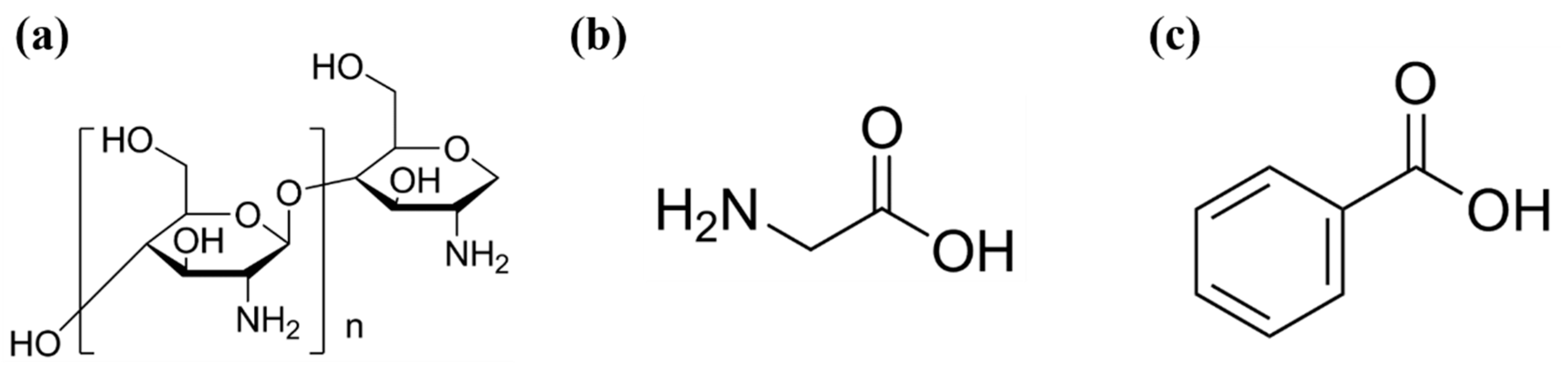
NaCl, chitosan (CS, medium molecular weight), acetic acid (assay 99.7%), glycine and benzoic acid were purchased from Sigma-Aldrich. Deionized water (18.2 MΩ·cm−1) was used throughout the experiment. The chemical structures of CS, glycine and benzoic acid are illustrated in Figure 1.
Figure 1.
The molecular structures of (a) chitosan, (b) glycine and (c) benzoic acid.
2.2. Synthesis of Carbon Dots
Typically, 1 g of carboxybenzene and 1.25 g of glycine were placed into a Teflon-lined stainless-steel autoclave followed by 25 mL of deionized water and heated at 200 °C for 48 h under static conditions. After cooling to 25 °C, the bright yellow solution was dialyzed using a 500 Da dialysis bag against deionized water to remove the impurities and obtained the CD solution [24,25].
2.3. Characterizations
The size, dimension and distribution of the synthesized CDs were characterized using a FEI/Philips Tecnai G2F20 TWIM TEM. The ultraviolet–visible (UV-Vis) spectrum of the amino acid carbon dots was characterized using a GBC UV-Vis spectrophotometer (Cintra2020, Melbourne, Australia). The Raman spectrum of the CDs was characterized using an XploRA Plus microscopic Raman spectrometer (Horiba, Ltd., Kyoto, Japan). The surface morphology of the finalized sensor chip was characterized using a Thermo Apreo 2S HiVac scanning electron microscope (Waltham, MA, USA).
2.4. Preparation of Sensing Chip
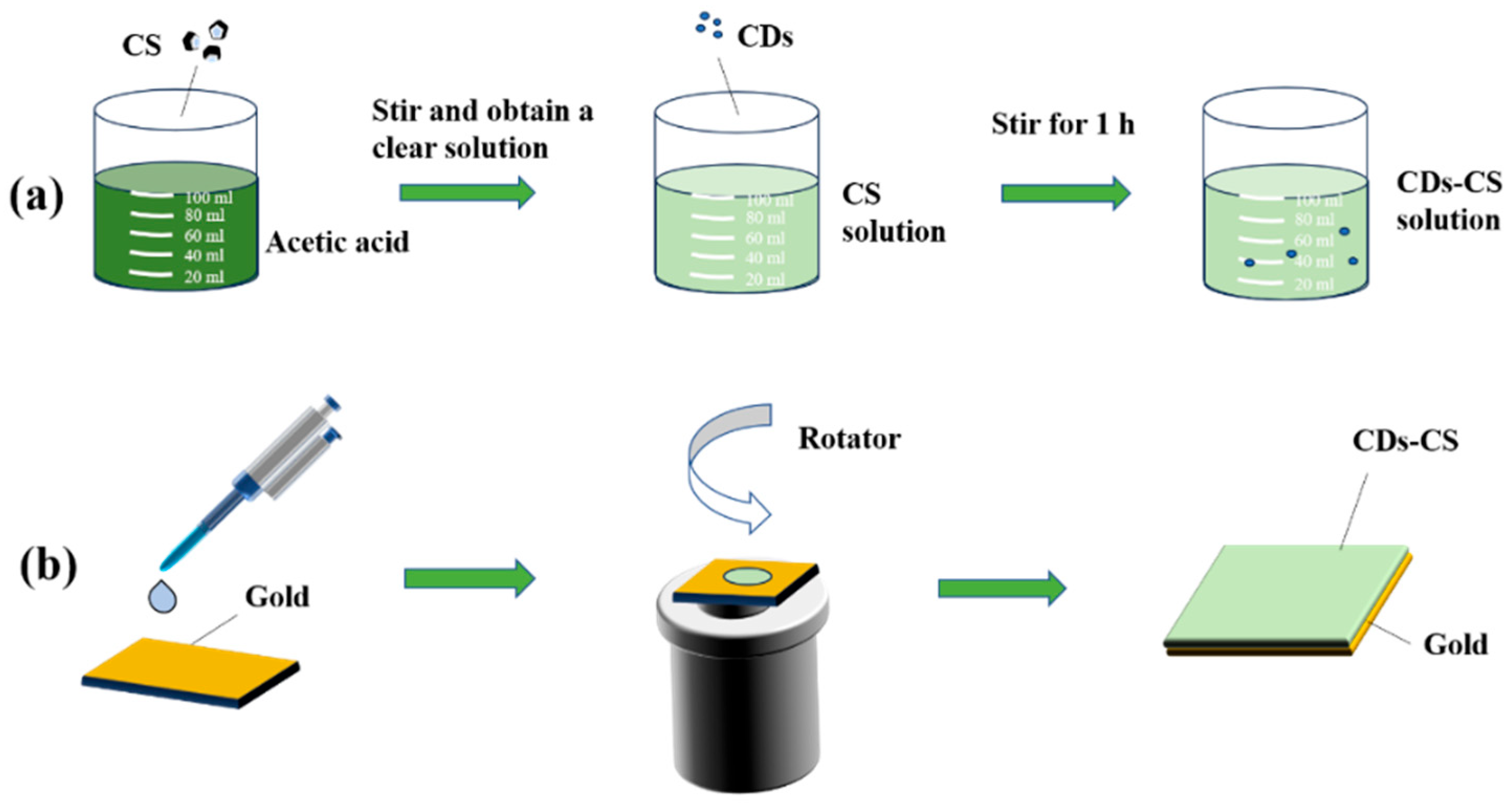
Dissolve 400 mg of CS in 50 mL of 1% acetic acid, stir it thoroughly, and then let it stand overnight at room temperature. The supernatant was taken to obtain the CS solution [26], as shown in Figure 2a. Then, add 100 μL of CDs into 5 mL of the CS solution, and stir for one hour until a uniform CDs-CS solution is formed [27]. At the same time, prepare NaCl solutions with concentrations of 0.1%, 1%, 3%, 6%, 9%, and 15%.
Figure 2.
(a) Preparation of experimental reagents; (b) fabrication of composite sensor chip.
The gold film is manufactured by Suzhou Mixin Semiconductor Co., Ltd. (Suzhou, China) according to our design by using magnetron sputtering technology. First, a 2 nm Cr layer on an SF10 glass substrate with dimensions of 1 mm × 20 mm × 20 mm, and then a 45 nm gold layer is deposited above the Cr layer. The purpose of depositing the Cr layer is to enhance the adhesion of the gold film without affecting the SPR effect. Take 0.5 mL of the above solution and use a spin coater to spin at a speed of 3000 rpm for 60 s to prepare different films on the substrate [27]. As shown in Figure 2b, a silicone microtube is used to introduce the sample solution into the flow channel through an injection pump.
2.5. Apparatus
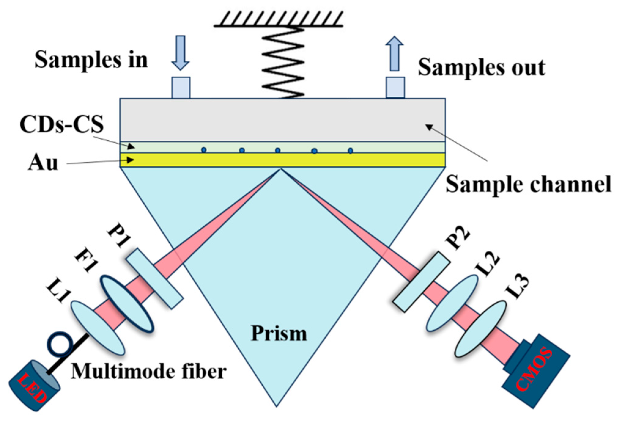
The SPR sensing experiment used a self-made intensity interrogation surface plasmon resonance imaging (SPRi) biosensing system which is based on the Kretschmann structure. The optical setup of this system is as follows: White light is generated by an LED light and introduced into the system through a multimode optical fiber. It is converted into parallel light by a collimating lens and a narrowband filter. The surface plasmon waves excited in the gold–CS–CDs interface by the parallel light are coupled with a prism. A CMOS camera receives the reflected light through an imaging lens group, thereby obtaining a sensing image. The schematic diagram is shown in Figure 3. Subsequently, with the aid of software, the average intensity value of the pixels within the region of interest is obtained to observe the dynamic changes in the intensity of the reflected light caused by the SPR effect in real time and intuitively.
Figure 3.
Schematic diagram of the sensing device: L1, L2 and L3: lenses; F1: narrowband filter; P1 and P2: polarizers.
3. Results and Discussion
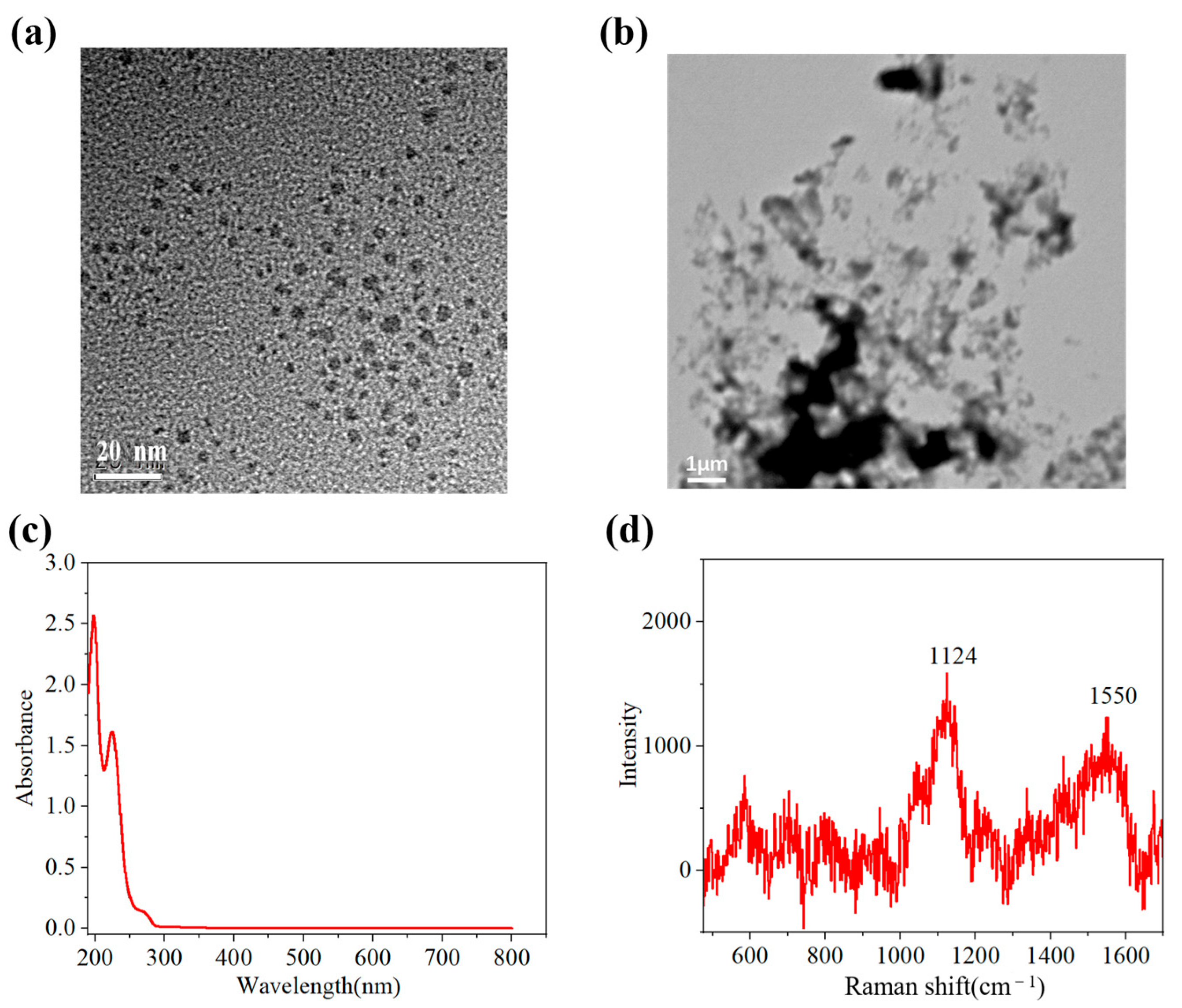
The morphology, ultraviolet–visible absorption spectrum, and Raman spectrum of the synthesized CDs are shown in Figure 4. As shown in Figure 4a, the CDs exhibited good monodispersity, and their size was approximately 5 nm. The surface morphology of the Au/CDs-CS chip is shown in Figure 4b. The spectrum of the amino acid carbon dots showed three absorption peaks at 197 nm, 224 nm, and 272 nm (Figure 4c). These CDs have a relatively low absorption intensity in the visible light region, indicating that in the SPR working area, the absorption of the CDs will not have a significant impact on the linewidth and reflectivity of the SPR spectrum, thereby ensuring good detection performance of the sensor. Since the pH was 4.14, the amino functional groups played a role in this pH environment. The peaks at the positions of 1124 cm−1 corresponded to the C-N bonds formed during the carbonization process of the amino group (-NH2) in glycine, respectively (Figure 4d). The peak at 1550 cm−1 was close to the G peak of sp2 hybridized carbon but lower than the common position of the G peak, which might imply a lower degree of order of the carbon structure or a change in the electronic structure of the carbon lattice due to nitrogen doping.
Figure 4.
(a) The TEM image of CDs. (b) The SEM image of Au/CDs-CS ship. (c) The UV–Vis absorbance spectra of CDs. (d) Raman spectra of CDs.
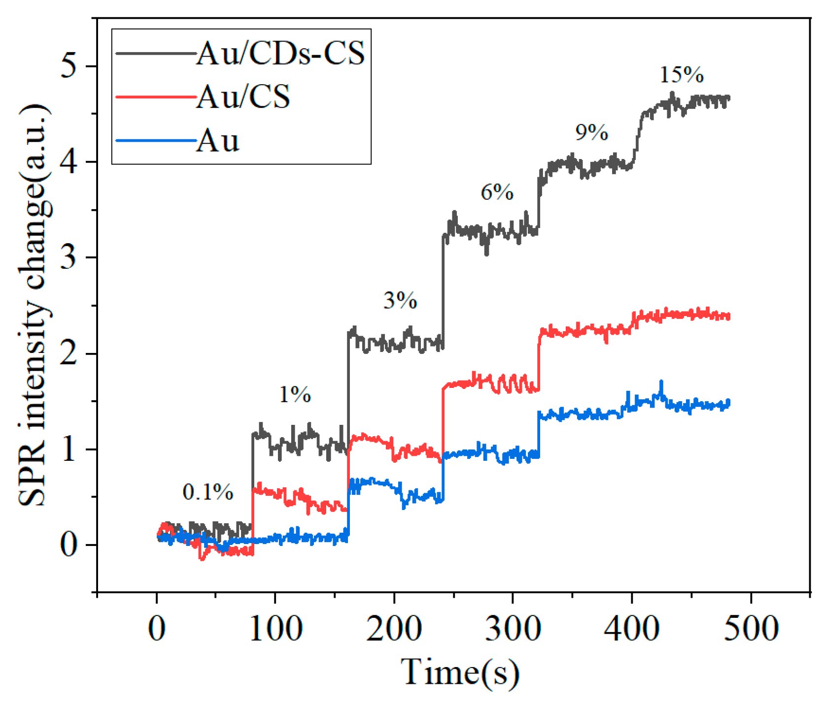
During the experiment, CDs were prepared and mixed with CS, then spin-coated on the surface of the gold film. Meanwhile, a gold film with only CS spin-coated was prepared as a control group. First, deionized water was injected into the microfluidic chamber using a syringe pump to obtain a calibration signal. Subsequently, NaCl solutions of different concentrations were injected into the microfluidic chamber in sequence, and a real-time sensing curve of intensity changing with concentration was obtained (Figure 5). To more intuitively compare the performance of different chips, the calibration signal was set to zero, and the relative intensities corresponding to different concentrations were obtained.
Figure 5.
Real-time SPR response of different chips at different NaCl concentrations.
The pKa of CS is approximately 6.5. When the pH of the sample environment is lower than 6.5, the amino groups (-NH2) on the CS molecular chain will combine with protons (H+) to form positively charged -NH3+, making the CS positively charged and capable of adsorbing Cl− [28,29,30]. As can be seen from Figure 5, under the condition of the same concentration difference, the sensing chip with CDs added has a stronger response signal, which is significantly better than the CS chip without CDs. This may be because the addition of CDs enhances the electric field at the sensing interface. At the same time, it increases the specific surface area of the chip, improves the dispersibility and stability, and is more conducive to the binding of NaCl molecules to the sites, thereby enhancing the detection signal.
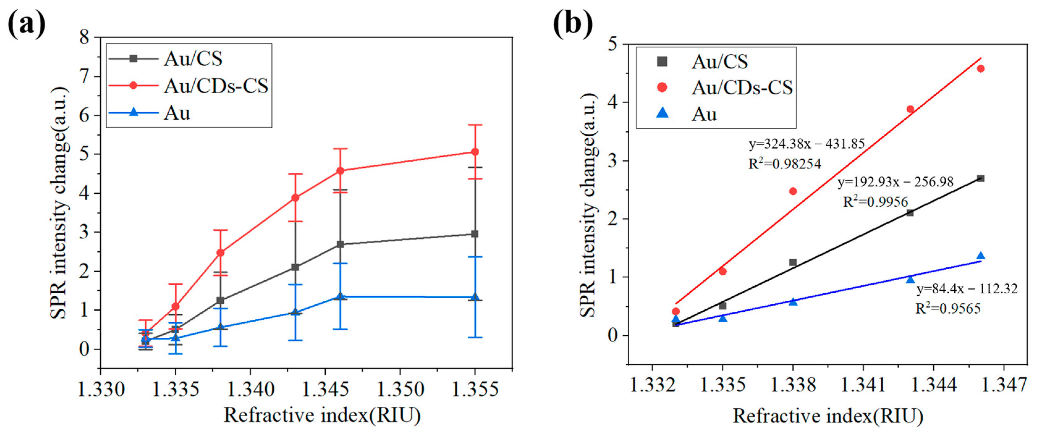
As can be seen from Figure 6, with the increase in the refractive index, the SPR intensity shows an upward trend. The growth slope of the CDs-CS curve is greater than that of the CS and bare gold film curves, indicating that under the same change in refractive index, the change in SPR intensity of the CDs-CS composite material is more significant. Moreover, at each measurement point, the SPR intensity of CDs-CS is higher than that of CS and the bare gold film. The sensitivity of the two types of chips was calculated according to Equation (1), where represents the ratio of the change in SPR intensity to the change in refractive index, represents the change in SPR intensity, and represents the change in the refractive index of the measured medium. The refractive index range of NaCl is 1.333–1.355. According to the calculation results, the sensitivity of CDs-CS is the highest, which is 167.28 a.u./RIU, that of CS is 94.96 a.u./RIU, and that of the bare gold film is 48.09 a.u./RIU. It represents a 247.8% improvement compared to an SPR sensor without amino acid CDs under the same conditions.
Figure 6.
(a) Graph of the intensity variation in different chips with the refractive index, and (b) linear response.
This article also conducts a study on the stability of the sensor chip. As can be seen from Figure 6a, compared with CS and the bare gold film, the stability of CDs-CS at high concentrations has been significantly improved. In addition, the stability of the bare gold film is better than that of CS. This may be because CS is a high-molecular polymer composed of many glucosamine units connected together. The uneven sites on the surface of the gold film lead to poor stability. After adding CDs, the dispersibility of the surface of the gold film is improved, thus enhancing the detection stability. The calibration curves were shown to verify linear response (Figure 6b). We selected a linear segment with NaCl concentrations ranging from 0.1% to 9% and removed the point with a concentration of 15%.
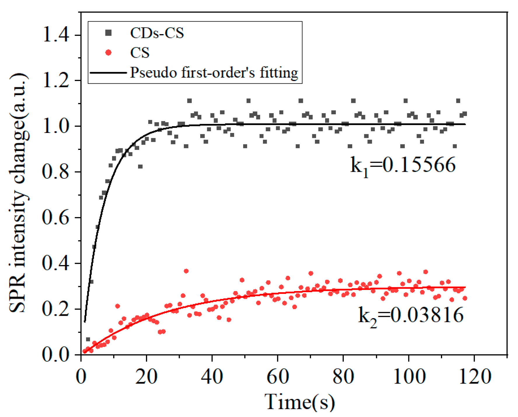
Then a pseudo-first-order kinetic adsorption analysis [31] was carried out for the NaCl concentration ranging from 9% to 15% to explore the kinetic process of the interaction between NaCl, CDs, and CS. This interaction is represented by the adsorption rate constant k, and the adsorption capacity is represented by the change in intensity, as shown in Equation (2). As can be seen from Figure 7, during the interaction process, the SPR intensity will increase as the NaCl molecules bind to the surface of the sensor and reach a stable state when the adsorption is nearly completed. After reaching equilibrium, the sensor signal remains stable, reflecting the maximum binding capacity of the sensor surface at a given concentration. A larger k value indicates a faster adsorption rate, a greater adsorption amount in the same time, and a stronger interaction. The adsorption rate constant k1 of CDs-CS is 0.15566, and the adsorption amount is 1.01051, while for CS, k2 = 0.03816, and the adsorption amount is 0.30103, indicating that the CDs-CS composite material has a stronger binding ability to NaCl. Due to the limitations of the response of the bare gold film to low concentrations of NaCl and the weak adsorption effect, it is difficult to obtain accurate data for fitting the adsorption model.
Figure 7.
Pseudo-first-order adsorption kinetic models for NaCl-CDs-CS.
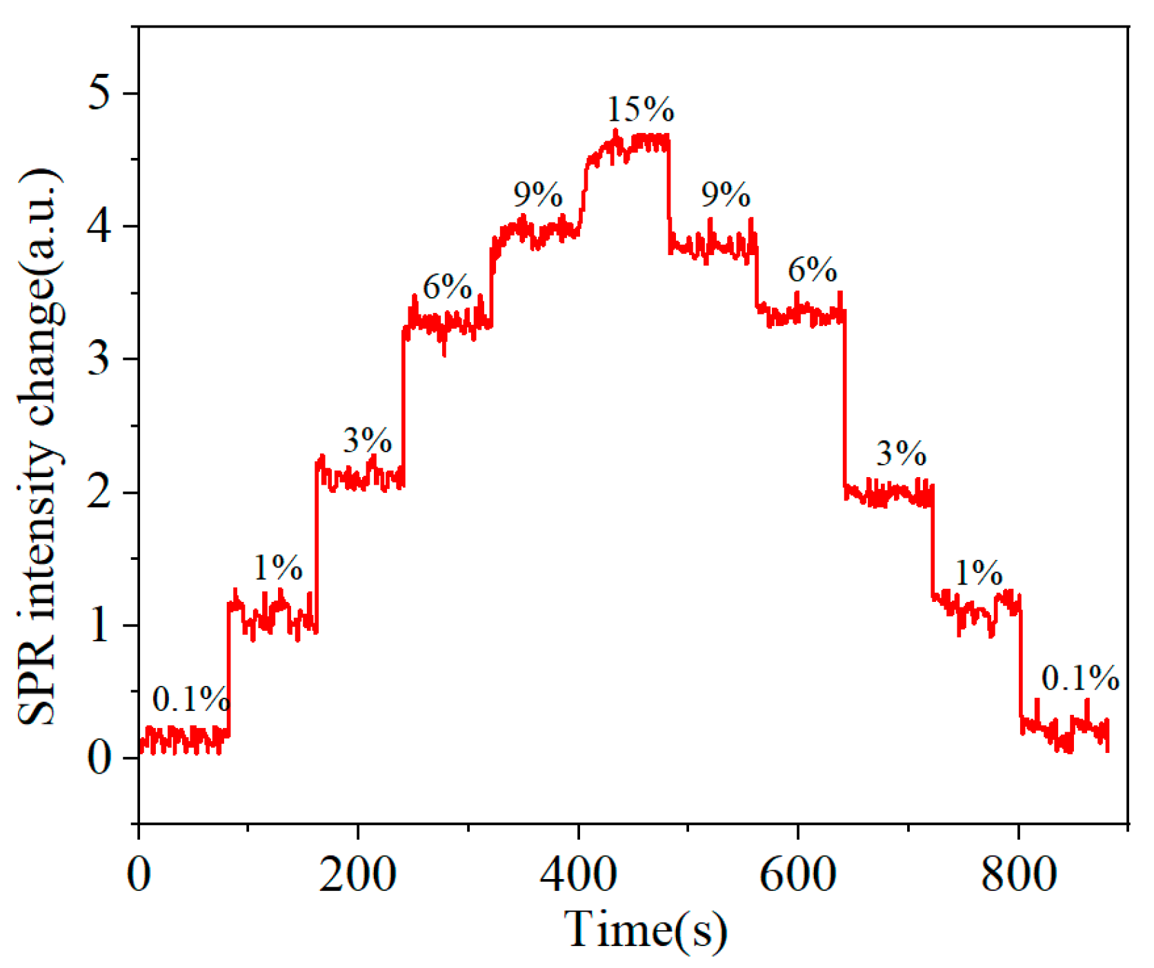
After experimentally verifying that the addition of CDs significantly enhances the sensing signal, it is necessary to study its dynamic response and reversibility. During the dynamic detection process, solutions with different refractive indices were continuously injected into the microfluidic chamber and recorded in real time. Figure 8 shows the dynamic response of the NaCl detection. The detection system exhibits good reversibility during the positive and negative cycles of the dynamic refractive index, and the average deviation of the intensity is less than 0.2 a.u., which can be regarded as the error caused by different cycle directions.
Figure 8.
Dynamic response of the CDs-CS chip detection.
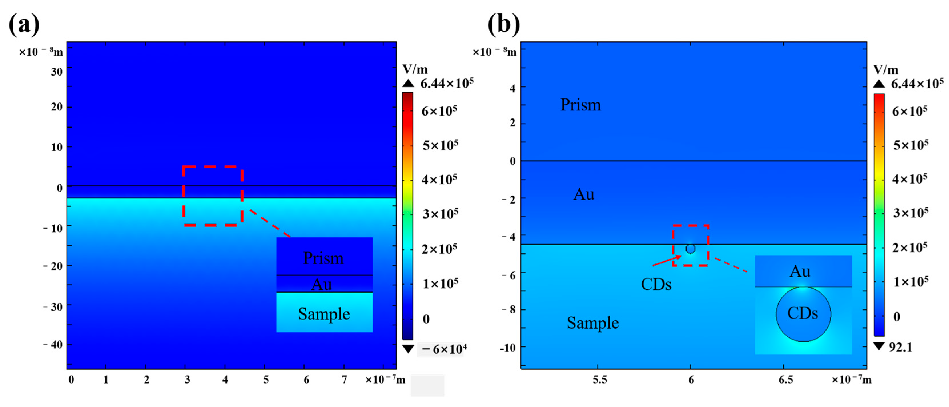
Finally, we used the finite element method to calculate the electric field distribution at the interface between CDs and the metal (Figure 9). The CDs are 5 nm away from the gold film. The refractive index of CDs was 1.592 + 0.042i, measured by ellipsometer and consistent with literature reports [16]. The electric field of the Au sensor chip is 2.06 × 10⁵ V/m, and the electric field of the Au/CDs is 6.44 × 10⁵ V/m. The simulation results show that the addition of CDs enhances the electric field of the sensor surface, increasing it by 312%, which is consistent with the experimental results.
Figure 9.
(a) The electric field distribution of the Au sensor chip. (b) The electric field distribution of the Au/CDs sensor chip.
Table 1 shows the sensitivity of the different SPR sensors using quantum dots on their own or in combination with other materials. From the table, it can be seen that the sensitivity of SPR sensors has been significantly improved after adding quantum dots.
Table 1.
Improvement in sensitivity of different SPR sensors after adding quantum dots.
4. Conclusions
In conclusion, this work has developed an SPR sensor based on an amino acid CDs-CS film, which achieved a significant improvement in detection sensitivity through synergistic effects. The results have shown that CDs enhance the adsorption capacity of the sensor surface and increase the adsorption amount. Meanwhile, the simulation results indicate that the electric field at the sensing interface is significantly enhanced, confirming the existence of the synergistic effect of CDs, which is the main reason for the improvement in sensitivity. Compared with the sensitivity of the bare gold film, the sensitivity of this sensor reaches 167.28 a.u./RIU, with an increase of 247.8%. From the perspective of practical applications, this synergistic effect of CDs provides a brand-new idea for the design of SPR sensors, which can further expand the application of SPR sensors in different fields by adjusting the preparation process of CDs and optimizing the composite method with other materials.
Author Contributions
Conceptualization, X.Y., J.Z. and M.C.; methodology, X.Y., H.L. and J.O.; software, X.Y. and Y.Y.; validation, J.O., M.W. and Z.L.; formal analysis, C.S., M.W. and L.W.; investigation, X.Y. and J.O.; data curation, X.Y. and Z.L.; writing—original draft preparation, X.Y.; writing—review and editing, X.Y. and M.C.; visualization, L.W. and J.Z.; supervision, X.Y. and M.C.; project administration, X.Y. and M.C.; funding acquisition, M.C., H.L. and Y.Y. All authors have read and agreed to the published version of the manuscript.
Funding
This work was partially supported by the National Key Research and Development Program of China “National Quality Infrastructure” (Grant No. 2021YFF0600902). Guangdong Basic and Applied Basic Research Foundation (No. 2023A1515011499 and 2023A1515140161). The Dongguan Science and Technology of Social Development Program (20231800936312).
Institutional Review Board Statement
Not applicable.
Informed Consent Statement
Not applicable.
Data Availability Statement
The data presented in this study are available on request from the corresponding author.
Acknowledgments
We thank the Priority Academic Program Development of Jiangsu Higher Education Institutions (PAPD), and the Key Laboratory of Optoelectronic Devices and Systems of Ministry of Education and Guangdong Province.
Conflicts of Interest
The authors declare that they have no known competing financial interests or personal relationships that could have appeared to influence the work reported in this paper.
References
- Omidniaee, A.; Karimi, S.; Farmani, A. Surface Plasmon Resonance-Based SiO2 Kretschmann Configuration Biosensor for the Detection of Blood Glucose. Silicon 2021, 14, 3081–3090. [Google Scholar] [CrossRef] [Scilit]
- Yanase, Y.; Sakamoto, K.; Kobayashi, K.; Hide, M. Diagnosis of immediate-type allergy using surface plasmon resonance. Opt. Mater. Express 2016, 6, 1339–1348. [Google Scholar] [CrossRef] [Scilit]
- Fauzi, N.I.M.; Fen, Y.W.; Omar, N.A.S.; Saleviter, S.; Daniyal, W.M.E.M.M.; Hashim, H.S.; Nasrullah, M. Nanostructured Chitosan/Maghemite Composites Thin Film for Potential Optical Detection of Mercury Ion by Surface Plasmon Resonance Investigation. Polymers 2020, 12, 1497. [Google Scholar] [CrossRef] [Scilit]
- Situ, C.; Mooney, M.H.; Elliott, C.T. Advances in surface plasmon resonance biosensor technology towards high-throughput, food-safety analysis. Trends Anal. Chem. 2010, 29, 1305–1315. [Google Scholar] [CrossRef] [Scilit]
- Fan, Y.; Chang, M.; Yu, X.; Zhou, J.; Shi, Y. SPR humidity dynamic monitoring method via PVA sensing membrane thickness variation and image processing techniques. Photonics Nanostruct.—Fundam. Appl. 2024, 61, 101301. [Google Scholar] [CrossRef] [Scilit]
- Yu, X.; Ouyang, J.; Li, Z.; Shi, C.; Wang, L.; Zhou, J.; Chang, M. Widening of Dynamic Detection Range in Real-Time Angular-Interrogation Surface Plasmon Resonance Biosensor Based on Anisotropic Van Der Waals Heterojunction. Biosensors 2024, 14, 601. [Google Scholar] [CrossRef] [Scilit]
- Hao, L.; Xiaolong, Q.; Wanlu, Z.; Yi, L.; Siyu, E.; Zhang, Y.N. Theoretical and experimental characterization of a salinity and temperature sensor employing optical fiber surface plasmon resonance (SPR). Instrum. Sci. Technol. 2020, 48, 601–615. [Google Scholar]
- Xiao-Li, Z.; Yunze, Y.; Shaopeng, W.; Xian-Wei, L. Surface Plasmon Resonance Microscopy: From Single-Molecule Sensing to Single-Cell Imaging. Angew. Chem. (Int. Ed. Engl.) 2020, 59, 1776–1785. [Google Scholar]
- Barnes, W.L.; Dereux, A.; Ebbesen, T.W. Surface plasmon subwavelength optics. Nature 2003, 424, 824–830. [Google Scholar] [CrossRef] [Scilit]
- Manera, M.G.; Colombelli, A.; Rella, R.; Caricato, A.; Cozzoli, P.D.; Martino, M.; Vasanelli, L. TiO2 brookite nanostructured thin layer on magneto-optical surface plasmon resonance transductor for gas sensing applications. J. Appl. Phys. 2012, 112, 053524. [Google Scholar] [CrossRef] [Scilit]
- Siew, M.G.; Susthitha, M.P.; Gurumurthy, H. ZnO for performance enhancement of surface plasmon resonance biosensor: A review. Mater. Res. Express 2020, 7, 012003. [Google Scholar]
- Fusheng, D.; Kai, Z.; Shuwen, Z.; Yufeng, Y. Sensitivity enhanced tunable plasmonic biosensor using two-dimensional twisted bilayer graphene superlattice. Nanophotonics 2023, 12, 1271–1284. [Google Scholar]
- Yangyang, Z.; Hezhen, L.; Junjie, X.; Zhongzheng, Z.; Kwangnak, K.; Hongxia, C. Controllable synthesis of multi-tip spatial gold nanostructures to facilitate SPR enhancement for exosomal PD-L1 assay. Chem. Eng. J. 2024, 481, 148137. [Google Scholar]
- Shariq, M.; Madkhli, A.Y.; Kawtherali, S.F.; Alshehri, K.; Alasmari, A.; Algarni, Z.S.; Alzahrani, H.S.; Alharbi, S.H.; Idris, M. Recent advancement in development of nitrogen-doped cqds for dye sensitized solar cell and photodetector. Rev. Surf. Interfaces 2025, 59, 105913. [Google Scholar] [CrossRef] [Scilit]
- Li, H.; Sun, C.; Zhang, M.; Yan, W.; Kang, Z. Multifunctional Thermally Activated Delayed Fluorescence Carbon Dots for Temperature-Responsive Sensor, Information Encryption, and Organelle Imaging. Adv. Funct. Mater. 2024, 34, 2405669. [Google Scholar] [CrossRef] [Scilit]
- Kumar, V.B.; Mirsky, S.K.; Shaked, N.T.; Gazit, E. High Quantum Yield Amino Acid Carbon Quantum Dots with Unparalleled Refractive Index. ACS Nano 2024, 18, 2421–2433. [Google Scholar] [CrossRef] [Scilit] [PubMed]
- Park, Y.; Yoo, J.; Lim, B.; Kwon, W.; Rhee, S.-W. Improving the functionality of carbon nanodots: Doping and surface functionalization. J. Mater. Chem. A Mater. Energy Sustain. 2016, 4, 11582–11603. [Google Scholar] [CrossRef] [Scilit]
- Qandeel, N.A.; El-Masry, A.A.; Eid, M.; Moustafa, M.A.; El-Shaheny, R. Fast one-pot microwave-assisted green synthesis of highly fluorescent plant-inspired S,N-self-doped carbon quantum dots as a sensitive probe for the antiviral drug nitazoxanide and hemoglobin. Anal. Chim. Acta 2023, 1237, 340592. [Google Scholar] [CrossRef] [Scilit]
- Adrien, R.; Clément, L.; Sandrine, I.; Emmanuelle, L.; Benoit, D.; Emmanuel, L. Surface Control of Doping in Self-Doped Nanocrystals. ACS Appl. Mater. Interfaces 2016, 8, 27122–27128. [Google Scholar]
- Hou, J.; Xia, B.; Niu, K.; Wang, J.; Li, J.; Wang, N. Ultrasensitive optical fiber SPR sensor enhanced by Au-NPs-film modified with functionalized CQDs for label-free detecting cobalt (II) ion. Anal. Chim. Acta 2024, 1320, 343030. [Google Scholar] [CrossRef] [Scilit]
- Farokhnasab, O.; Moghadam, A.; Eslamifar, Z.; Moghadam, A.H. Fabrication and characterization of chitosan-based bionanocomposite coating reinforced with TiO2 nanoparticles and carbon quantum dots for enhanced antimicrobial efficacy. Int. J. Biol. Macromol. 2025, 296, 139648. [Google Scholar] [CrossRef] [Scilit] [PubMed]
- Alavi, S.; Ghadiri, H.; Dabirmanesh, B.; Khajeh, K. The impact of quantum dot on the SPR detection improvement of molecular interactions between Rap1 interacting factor1 (Rif1) and G4. Sens. Bio-Sens. Res. 2024, 43, 100621. [Google Scholar] [CrossRef] [Scilit]
- Ahmad, N.N.A.; Hidayah, A.N.; Abu, B.M.H.; Naiim, M.N.; Sakinah, M.A.; Md, Z.A.R.; Adzir, M.M.; Gandaryus, S.A.; Kencana, W.T.D.; Yunhan, L.; et al. Polymeric carbon quantum dots as efficient chlorophyll sensor-analysis based on experimental and computational investigation. Opt. Laser Technol. 2024, 170, 110259. [Google Scholar]
- Li, H.; Ye, S.; Guo, J.; Wang, H.; Yan, W.; Song, J.; Qu, J. Biocompatible carbon dots with low-saturation-intensity and high-photobleaching-resistance for STED nanoscopy imaging of the nucleolus and tunneling nanotubes in living cells. Nano Res. 2019, 12, 3075–3084. [Google Scholar] [CrossRef] [Scilit]
- Guo, Y.; Huang, Z.; Wang, L.; Gao, X.; Chen, Y.; Lu, F.; Sun, C.; Li, H.; Li, H.; He, Y.; et al. Fluorine-Nitrogen Codoped Carbon Dots for Visualization Imaging of Nucleic Acids via Two-Photon Fluorescence Lifetime Microscopy. Anal. Chem. 2025, 97, 5744–5752. [Google Scholar] [CrossRef] [Scilit]
- Fen, Y.W.; Yunus, W.M.M.; Yusof, N.A. Surface plasmon resonance optical sensor for detection of Pb2+ based on immobilized p-tert-butylcalix[4]arene-tetrakis in chitosan thin film as an active layer. Sens. Actuators B Chem. 2012, 171–172, 287–293. [Google Scholar] [CrossRef] [Scilit]
- Bashar, K.E.F.; Wing, F.Y.; Sheh, O.N.A.; Chyi, L.J.Y.; Mohd, D.W.M.E.M. Femtomolar detection of dopamine using surface plasmon resonance sensor based on chitosan/graphene quantum dots thin film. Spectrochim. Acta Part A Mol. Biomol. Spectrosc. 2021, 263, 120202. [Google Scholar]
- Jin, Z.; Huang, G.; Song, Y.; Zheng, X.; Zhao, K. Unlocking the potential of chitosan in immunoassay sensor. Carbohydr. Polym. 2025, 350, 123024. [Google Scholar] [CrossRef] [Scilit] [PubMed]
- Li, J.; Zeng, H.; Wu, X.; Li, T.; Sun, Z.; Zhu, G.; Zhang, M.; Chen, L.; Pan, K.; Guo, X. Chitosan-based porous composites embedded with molybdenum disulfide nanosheets for removal of mercury from wastewater. Int. J. Biol. Macromol. 2024, 285, 138379. [Google Scholar] [CrossRef] [Scilit]
- Fernandes, S.C.; Vieira, I.C.; Peralta, R.A.; Neves, A. Development of a biomimetic chitosan film-coated gold electrode for determination of dopamine in the presence of ascorbic acid and uric acid. Electrochim. Acta 2010, 55, 7152–7157. [Google Scholar] [CrossRef] [Scilit]
- Gilang, G.; Silvia, C.; Ganes, S.; Aep, P.; Nugraha, N.; Joel, H.; Isa, A.; Valentino, K.Y.; Brian, Y. The revelation of glucose adsorption mechanisms on hierarchical metal-organic frameworks using a surface plasmon resonance sensor. J. Mater. Chem. B 2023, 11, 4428–4444. [Google Scholar]
- Fu, R.; Yan, X.; Hu, T.; Li, H.; Cheng, T. Carbon Dots-Based Surface Plasmon Resonance Manganese Ion Fiber Sensor for Multi-Use Scenarios. Photonic Sens. 2025, 15, 250311. [Google Scholar] [CrossRef] [Scilit]
- Rosddi, N.N.M.; Fen, Y.W.; Omar, N.A.S.; Anas, N.A.A.; Hashim, H.S.; Ramdzan, N.S.M.; Fauzi, N.I.M.; Anuar, M.F.; Daniyal, W.M.E.M.M. Glucose detection by gold modified carboxyl-functionalized graphene quantum dots-based surface plasmon resonance. Optik 2021, 239, 166779. [Google Scholar] [CrossRef] [Scilit]
Disclaimer/Publisher’s Note: The statements, opinions and data contained in all publications are solely those of the individual author(s) and contributor(s) and not of MDPI and/or the editor(s). MDPI and/or the editor(s) disclaim responsibility for any injury to people or property resulting from any ideas, methods, instructions or products referred to in the content. |
© 2025 by the authors. Licensee MDPI, Basel, Switzerland. This article is an open access article distributed under the terms and conditions of the Creative Commons Attribution (CC BY) license (https://creativecommons.org/licenses/by/4.0/).