
Validation of a Commercially Available IMU-Based System Against an Optoelectronic System for Full-Body Motor Tasks


 ,
, 
 , and
, and 
Abstract
Highlights
- The IMU-based system demonstrated high concurrent validity with a gold-standard optoelectronic motion capture system across a wide variety of full-body tasks.
- The IMU-based system achieved strong correlations (r ≥ 0.77), low RMSE values (generally < 7°), and negligible systematic biases (≤3.9°) compared to the optical reference.
- The tested IMU-based system provides clinically acceptable range-of-motion estimates and can serve as a reliable alternative to laboratory-based motion analysis tools.
- Its portability and ease of use make it particularly suited for in-clinic assessments and home-based rehabilitation programs, supporting remote patient monitoring.
Abstract
1. Introduction
2. Materials and Methods
2.1. Participants
2.2. Equipment
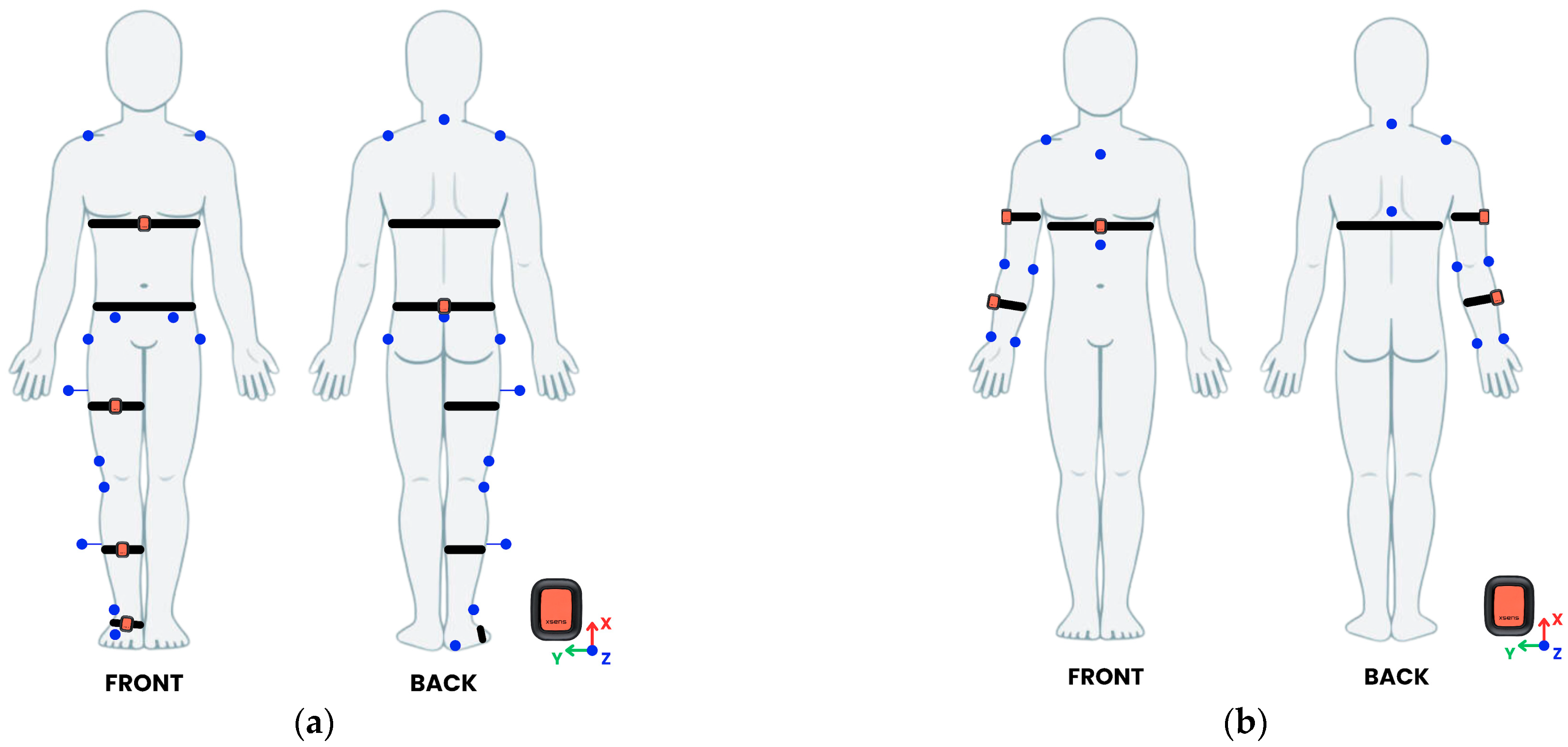
2.3. Procedure
2.4. Data Processing
2.5. Sample Size
2.6. Statistical Analysis
3. Results
3.1. Lower Body and Trunk
3.2. Upper Body
4. Discussion
5. Conclusions
Author Contributions
Funding
Institutional Review Board Statement
Informed Consent Statement
Data Availability Statement
Acknowledgments
Conflicts of Interest
Abbreviations
| IMU | Inertial Measurement Unit |
| MCap | Motion Capture |
| ROM | Range Of Motion |
| Abd | Abduction |
| Flex | Flexion |
| Ext | Extension |
| RMSE | Root Mean Square Error |
| PoS | Percentage of Similarity |
| LoAs | Limits of Agreement |
References
- Hochstenbach, J. Rehabilitation Is More than Functional Recovery. Disabil. Rehabil. 2000, 22, 201–204. [Google Scholar] [CrossRef] [PubMed]
- Andersen, L.L.; Saervoll, C.A.; Mortensen, O.S.; Poulsen, O.M.; Hannerz, H.; Zebis, M.K. Effectiveness of Small Daily Amounts of Progressive Resistance Training for Frequent Neck/Shoulder Pain: Randomised Controlled Trial. Pain 2011, 152, 440–446. [Google Scholar] [CrossRef] [PubMed]
- Latash, M.L.; Anson, J.G. Synergies in Health and Disease: Relations to Adaptive Changes in Motor Coordination. Phys. Ther. 2006, 86, 1151–1160. [Google Scholar] [CrossRef]
- Veldema, J.; Jansen, P. Resistance Training in Stroke Rehabilitation: Systematic Review and Meta-Analysis. Clin. Rehabil. 2020, 34, 1173–1197. [Google Scholar] [CrossRef]
- Nussbaumer, S.; Leunig, M.; Glatthorn, J.F.; Stauffacher, S.; Gerber, H.; Maffiuletti, N.A. Validity and Test-Retest Reliability of Manual Goniometers for Measuring Passive Hip Range of Motion in Femoroacetabular Impingement Patients. BMC Musculoskelet. Disord. 2010, 11, 194. [Google Scholar] [CrossRef] [PubMed]
- Muir, S.W.; Corea, C.L.; Beaupre, L. Evaluating Change in Clinical Status: Reliability and Measures of Agreement for the Assessment of Glenohumeral Range of Motion. N. Am. J. Sports Phys. Ther. 2010, 5, 98–110. [Google Scholar]
- Walmsley, C.P.; Williams, S.A.; Grisbrook, T.; Elliott, C.; Imms, C.; Campbell, A. Measurement of Upper Limb Range of Motion Using Wearable Sensors: A Systematic Review. Sports Med.-Open 2018, 4, 53. [Google Scholar] [CrossRef]
- Boone, D.C.; Azen, S.P.; Lin, C.M.; Spence, C.; Baron, C.; Lee, L. Reliability of Goniometric Measurements. Phys. Ther. 1978, 58, 1355–1360. [Google Scholar] [CrossRef]
- Lim, C.C.; Affandi, M.; Basah, S.N.; Din, M.Y. Evaluating Lower Limb Joint Flexion by Computerized Visual Tracking System and Compared with Electrogoniometer and Universal Goniometer. J. Telecommun. Electron. Comput. Eng. 2018, 10, 9–14. [Google Scholar]
- McGinley, J.L.; Baker, R.; Wolfe, R.; Morris, M.E. The Reliability of Three-Dimensional Kinematic Gait Measurements: A Systematic Review. Gait Posture 2009, 29, 360–369. [Google Scholar] [CrossRef]
- Colyer, S.L.; Evans, M.; Cosker, D.P.; Salo, A.I.T. A Review of the Evolution of Vision-Based Motion Analysis and the Integration of Advanced Computer Vision Methods Towards Developing a Markerless System. Sports Med.-Open 2018, 4, 24. [Google Scholar] [CrossRef] [PubMed]
- Roggio, F.; Ravalli, S.; Maugeri, G.; Bianco, A.; Palma, A.; Di Rosa, M.; Musumeci, G. Technological Advancements in the Analysis of Human Motion and Posture Management through Digital Devices. World J. Orthop. 2021, 12, 467–484. [Google Scholar] [CrossRef] [PubMed]
- Poitras, I.; Dupuis, F.; Bielmann, M.; Campeau-Lecours, A.; Mercier, C.; Bouyer, L.J.; Roy, J.-S. Validity and Reliability of Wearable Sensors for Joint Angle Estimation: A Systematic Review. Sensors 2019, 19, 1555. [Google Scholar] [CrossRef] [PubMed]
- Adesida, Y.; Papi, E.; McGregor, A.H. Exploring the Role of Wearable Technology in Sport Kinematics and Kinetics: A Systematic Review. Sensors 2019, 19, 1597. [Google Scholar] [CrossRef]
- Gu, C.; Lin, W.; He, X.; Zhang, L.; Zhang, M. IMU-Based Motion Capture System for Rehabilitation Applications: A Systematic Review. Biomim. Intell. Robot. 2023, 3, 100097. [Google Scholar] [CrossRef]
- Arlotti, J.S.; Carroll, W.O.; Afifi, Y.; Talegaonkar, P.; Albuquerque, L.; Burch, V.; Ball, J.E.; Chander, H.; Petway, A. Benefits of IMU-Based Wearables in Sports Medicine: Narrative Review. Int. J. Kinesiol. Sports 2022, 10, 36–43. [Google Scholar] [CrossRef]
- Horak, F.; King, L.; Mancini, M. Role of Body-Worn Movement Monitor Technology for Balance and Gait Rehabilitation. Phys. Ther. 2015, 95, 461–470. [Google Scholar] [CrossRef]
- Seel, T.; Raisch, J.; Schauer, T. IMU-Based Joint Angle Measurement for Gait Analysis. Sensors 2014, 14, 6891–6909. [Google Scholar] [CrossRef]
- Teufl, W.; Miezal, M.; Taetz, B.; Fröhlich, M.; Bleser, G. Validity, Test-Retest Reliability and Long-Term Stability of Magnetometer Free Inertial Sensor Based 3D Joint Kinematics. Sensors 2018, 18, 1980. [Google Scholar] [CrossRef]
- Mundt, M.; Koeppe, A.; David, S.; Witter, T.; Bamer, F.; Potthast, W.; Markert, B. Estimation of Gait Mechanics Based on Simulated and Measured IMU Data Using an Artificial Neural Network. Front. Bioeng. Biotechnol. 2020, 8, 41. [Google Scholar] [CrossRef]
- Komaris, D.-S.; Tarfali, G.; O’Flynn, B.; Tedesco, S. Unsupervised IMU-Based Evaluation of at-Home Exercise Programmes: A Feasibility Study. BMC Sports Sci. Med. Rehabil. 2022, 14, 28. [Google Scholar] [CrossRef]
- Cho, Y.-S.; Jang, S.-H.; Cho, J.-S.; Kim, M.-J.; Lee, H.D.; Lee, S.Y.; Moon, S.-B. Evaluation of Validity and Reliability of Inertial Measurement Unit-Based Gait Analysis Systems. Ann. Rehabil. Med. 2018, 42, 872–883. [Google Scholar] [CrossRef]
- De Vries, W.H.K.; Veeger, H.E.J.; Baten, C.T.M.; Van Der Helm, F.C.T. Magnetic Distortion in Motion Labs, Implications for Validating Inertial Magnetic Sensors. Gait Posture 2009, 29, 535–541. [Google Scholar] [CrossRef]
- Camomilla, V.; Bergamini, E.; Fantozzi, S.; Vannozzi, G. Trends Supporting the In-Field Use of Wearable Inertial Sensors for Sport Performance Evaluation: A Systematic Review. Sensors 2018, 18, 873. [Google Scholar] [CrossRef]
- Filippeschi, A.; Schmitz, N.; Miezal, M.; Bleser, G.; Ruffaldi, E.; Stricker, D. Survey of Motion Tracking Methods Based on Inertial Sensors: A Focus on Upper Limb Human Motion. Sensors 2017, 17, 1257. [Google Scholar] [CrossRef]
- Eichelberger, P.; Ferraro, M.; Minder, U.; Denton, T.; Blasimann, A.; Krause, F.; Baur, H. Analysis of Accuracy in Optical Motion Capture—A Protocol for Laboratory Setup Evaluation. J. Biomech. 2016, 49, 2085–2088. [Google Scholar] [CrossRef]
- Piche, E.; Guilbot, M.; Chorin, F.; Guerin, O.; Zory, R.; Gerus, P. Validity and Repeatability of a New Inertial Measurement Unit System for Gait Analysis on Kinematic Parameters: Comparison with an Optoelectronic System. Measurement 2022, 198, 111442. [Google Scholar] [CrossRef]
- Dorschky, E.; Nitschke, M.; Seifer, A.-K.; van den Bogert, A.J.; Eskofier, B.M. Estimation of Gait Kinematics and Kinetics from Inertial Sensor Data Using Optimal Control of Musculoskeletal Models. J. Biomech. 2019, 95, 109278. [Google Scholar] [CrossRef]
- Zhao, Y.; Liu, Y.; Li, J.; Wang, X.; Yang, R.; Lian, C.; Shan, P.; Wang, Y.; Zhan, Z.; Fu, C. Global Joint Information Extraction Convolution Neural Network for Parkinson’s Disease Diagnosis. Expert. Syst. Appl. 2024, 243, 122837. [Google Scholar] [CrossRef]
- Camuncoli, F.; Barni, L.; Nutarelli, S.; Rocchi, J.E.; Barcillesi, M.; Di Dio, I.; Sambruni, A.; Galli, M. Validity of the Baiobit Inertial Measurements Unit for the Assessment of Vertical Double- and Single-Leg Countermovement Jumps in Athletes. Int. J. Environ. Res. Public Health 2022, 19, 14720. [Google Scholar] [CrossRef]
- Toft Nielsen, E.; Jørgensen, P.B.; Mechlenburg, I.; Sørensen, H. Validation of an Inertial Measurement Unit to Determine Countermovement Jump Height. Asia-Pac. J. Sports Med. Arthrosc. Rehabil. Technol. 2019, 16, 8–13. [Google Scholar] [CrossRef]
- Marković, S.; Dopsaj, M.; Tomažič, S.; Kos, A.; Nedeljković, A.; Umek, A. Can IMU Provide an Accurate Vertical Jump Height Estimate? Appl. Sci. 2021, 11, 12025. [Google Scholar] [CrossRef]
- Blandeau, M.; Guichard, R.; Hubaut, R.; Leteneur, S. IMU Positioning Affects Range of Motion Measurement During Squat Motion Analysis. J. Biomech. 2023, 153, 111598. [Google Scholar] [CrossRef]
- Whelan, D.; O’Reilly, M.; Huang, B.; Giggins, O.; Kechadi, T.; Caulfield, B. Leveraging IMU Data for Accurate Exercise Performance Classification and Musculoskeletal Injury Risk Screening. In Proceedings of the 2016 38th Annual International Conference of the IEEE Engineering in Medicine and Biology Society (EMBC), Orlando, FL, USA, 16–20 August 2016; Volume 2016, p. 662. [Google Scholar]
- Kianifar, R.; Joukov, V.; Lee, A.; Raina, S.; Kulić, D. Inertial Measurement Unit-Based Pose Estimation: Analyzing and Reducing Sensitivity to Sensor Placement and Body Measures. J. Rehabil. Assist. Technol. Eng. 2019, 6, 2055668318813455. [Google Scholar] [CrossRef]
- Clemente, F.M.; Akyildiz, Z.; Pino-Ortega, J.; Rico-González, M. Validity and Reliability of the Inertial Measurement Unit for Barbell Velocity Assessments: A Systematic Review. Sensors 2021, 21, 2511. [Google Scholar] [CrossRef]
- Khuyagbaatar, B.; Tumurbaatar, M.; Tsenkherjav, K.; Purevsuren, T.; Shambaljamts, T.; Kim, K.; Danjkhuu, T.; Danaa, G.; Hyuk Kim, Y. Kinematic Comparison of Snatch and Clean Lifts in Weightlifters Using Wearable Inertial Measurement Unit Sensors. Phys. Act. Health 2024, 8, 1–9. [Google Scholar] [CrossRef]
- O’Reilly, M.A.; Whelan, D.F.; Ward, T.E.; Delahunt, E.; Caulfield, B.M. Classification of Deadlift Biomechanics with Wearable Inertial Measurement Units. J. Biomech. 2017, 58, 155–161. [Google Scholar] [CrossRef]
- Teufl, W.; Miezal, M.; Taetz, B.; Fröhlich, M.; Bleser, G. Validity of Inertial Sensor Based 3D Joint Kinematics of Static and Dynamic Sport and Physiotherapy Specific Movements. PLoS ONE 2019, 14, e0213064. [Google Scholar] [CrossRef]
- Robert-Lachaine, X.; Mecheri, H.; Larue, C.; Plamondon, A. Validation of Inertial Measurement Units with an Optoelectronic System for Whole-Body Motion Analysis. Med. Biol. Eng. Comput. 2017, 55, 609–619. [Google Scholar] [CrossRef]
- Unger, T.; De Sousa Ribeiro, R.; Mokni, M.; Weikert, T.; Pohl, J.; Schwarz, A.; Held, J.P.O.; Sauerzopf, L.; Kühnis, B.; Gavagnin, E.; et al. Upper Limb Movement Quality Measures: Comparing IMUs and Optical Motion Capture in Stroke Patients Performing a Drinking Task. Front. Digit. Health 2024, 6, 1359776. [Google Scholar] [CrossRef]
- Cutti, A.G.; Giovanardi, A.; Rocchi, L.; Davalli, A.; Sacchetti, R. Ambulatory Measurement of Shoulder and Elbow Kinematics through Inertial and Magnetic Sensors. Med. Bio Eng. Comput. 2008, 46, 169–178. [Google Scholar] [CrossRef]
- Parel, I.; Cutti, A.G.; Kraszewski, A.; Verni, G.; Hillstrom, H.; Kontaxis, A. Intra-Protocol Repeatability and Inter-Protocol Agreement for the Analysis of Scapulo-Humeral Coordination. Med. Biol. Eng. Comput. 2014, 52, 271–282. [Google Scholar] [CrossRef]
- Friesen, K.B.; Wu, L.Z.; Waslen, A.; Lang, A.E. Defining Repeatability for Scapulothoracic and Thoracohumeral Motion during the Novel Work-Related Activities and Functional Task (WRAFT) Protocol. J. Biomech. 2023, 153, 111596. [Google Scholar] [CrossRef]
- Fang, Z.; Woodford, S.; Senanayake, D.; Ackland, D. Conversion of Upper-Limb Inertial Measurement Unit Data to Joint Angles: A Systematic Review. Sensors 2023, 23, 6535. [Google Scholar] [CrossRef]
- Morrow, M.M.B.; Lowndes, B.; Fortune, E.; Kaufman, K.; Hallbeck, M. Validation of Inertial Measurement Units for Upper Body Kinematics. J. Appl. Biomech. 2017, 33, 227–232. [Google Scholar] [CrossRef]
- Li, J.; Qiu, F.; Gan, L.; Chou, L.-S. Concurrent Validity of Inertial Measurement Units in Range of Motion Measurements of Upper Extremity: A Systematic Review and Meta-Analysis. Wearable Technol. 2024, 5, e11. [Google Scholar] [CrossRef]
- Cerfoglio, S.; Capodaglio, P.; Rossi, P.; Conforti, I.; D’Angeli, V.; Milani, E.; Galli, M.; Cimolin, V. Evaluation of Upper Body and Lower Limbs Kinematics through an IMU-Based Medical System: A Comparative Study with the Optoelectronic System. Sensors 2023, 23, 6156. [Google Scholar] [CrossRef]
- Leardini, A.; Lullini, G.; Giannini, S.; Berti, L.; Ortolani, M.; Caravaggi, P. Validation of the Angular Measurements of a New Inertial-Measurement-Unit Based Rehabilitation System: Comparison with State-of-the-Art Gait Analysis. J. Neuroeng. Rehabil. 2014, 11, 136. [Google Scholar] [CrossRef]
- Felius, R.A.W.; Geerars, M.; Bruijn, S.M.; Wouda, N.C.; Van Dieën, J.H.; Punt, M. Reliability of IMU-Based Balance Assessment in Clinical Stroke Rehabilitation. Gait Posture 2022, 98, 62–68. [Google Scholar] [CrossRef]
- Pan, H.; Wang, H.; Li, D.; Zhu, K.; Gao, Y.; Yin, R.; Shull, P.B. Automated, IMU-Based Spine Angle Estimation and IMU Location Identification for Telerehabilitation. J. Neuroeng. Rehabil. 2024, 21, 96. [Google Scholar] [CrossRef]
- Lobo, P.; Morais, P.; Murray, P.; Vilaça, J.L. Trends and Innovations in Wearable Technology for Motor Rehabilitation, Prediction, and Monitoring: A Comprehensive Review. Sensors 2024, 24, 7973. [Google Scholar] [CrossRef]
- Ettefagh, A.; Roshan Fekr, A. Technological Advances in Lower-Limb Tele-Rehabilitation: A Review of Literature. J. Rehabil. Assist. Technol. Eng. 2024, 11, 20556683241259256. [Google Scholar] [CrossRef]
- Al-Amri, M.; Nicholas, K.; Button, K.; Sparkes, V.; Sheeran, L.; Davies, J.L. Inertial Measurement Units for Clinical Movement Analysis: Reliability and Concurrent Validity. Sensors 2018, 18, 719. [Google Scholar] [CrossRef]
- Niswander, W.; Wang, W.; Kontson, K. Optimization of IMU Sensor Placement for the Measurement of Lower Limb Joint Kinematics. Sensors 2020, 20, 5993. [Google Scholar] [CrossRef]
- Paulich, M.; Schepers, M.; Rudigkeit, N.; Bellusci, G. Xsens MTw Awinda: Miniature Wireless Inertial-Magnetic Motion Tracker for Highly Accurate 3D Kinematic Applications; Xsens: Enschede, The Netherlands, 2018. [Google Scholar] [CrossRef]
- Davis, R.B.; Õunpuu, S.; Tyburski, D.; Gage, J.R. A Gait Analysis Data Collection and Reduction Technique. Hum. Mov. Sci. 1991, 10, 575–587. [Google Scholar] [CrossRef]
- Goreham, J.A.; MacLean, K.F.E.; Ladouceur, M. The Validation of a Low-Cost Inertial Measurement Unit System to Quantify Simple and Complex Upper-Limb Joint Angles. J. Biomech. 2022, 134, 111000. [Google Scholar] [CrossRef]
- Mishra, P.; Pandey, C.M.; Singh, U.; Gupta, A.; Sahu, C.; Keshri, A. Descriptive Statistics and Normality Tests for Statistical Data. Ann. Card. Anaesth. 2019, 22, 67–72. [Google Scholar] [CrossRef]
- Ghasemi, A.; Zahediasl, S. Normality Tests for Statistical Analysis: A Guide for Non-Statisticians. Int. J. Endocrinol. Metab. 2012, 10, 486–489. [Google Scholar] [CrossRef]
- Cohen, J. Statistical Power Analysis for the Behavioral Sciences, 2nd ed.; L. Erlbaum Associates: Hillsdale, NJ, USA, 1988; ISBN 978-0-8058-0283-2. [Google Scholar]
- Bland, J.M.; Altman, D.G. Statistical Methods for Assessing Agreement Between Two Methods of Clinical Measurement. Lancet 1986, 1, 307–310. [Google Scholar] [CrossRef]
- Akoglu, H. User’s Guide to Correlation Coefficients. Turk. J. Emerg. Med. 2018, 18, 91–93. [Google Scholar] [CrossRef]
- Bonfiglio, A.; Petruccelli, C.; Villa, G.; Bongers, R.M.; Farella, E. Preliminary Validation of an IMU-Based Physiotherapy Assessment System for the Lower Extremities. In Pervasive Computing Technologies for Healthcare, Proceedings of the 18th EAI International Conference, PervasiveHealth 2024, Heraklion, Greece, 17–18 September 2024; Kondylakis, H., Triantafyllidis, A., Eds.; Springer Nature: Cham, Switzerland, 2025; pp. 142–158. [Google Scholar]

| Body District and Complexity | Task | Description |
|---|---|---|
| Lower body and trunk Single-joint | Hip Abd | Standing on the left leg, raise the right leg laterally away from the body at maximum ROM, keeping the pelvis stable. |
| Hip Flex | Standing on the left leg, raise the right leg forward by flexing at the hip at maximum ROM. | |
| Knee Flex | Seated on a box with legs hanging, bend and extend the right knee at maximum ROM. | |
| Ankle Flex | Seated on a box with legs hanging, perform plantar- and dorsi-flexion of the right ankle at maximum ROM. | |
| Trunk Flex | Standing upright, perform trunk flexion by hinging at the hips at approximately 45°. | |
| Lower body Multi-joint | Squat | With feet shoulder-width apart, lower the body by bending both knees and hips while keeping a straight back, then return to standing. |
| Lunge | Standing upright, step forward with the right leg and lower the body until both knees form roughly 90° angles (with the front knee aligned over the ankle), then push off to return to the starting position. | |
| Upper body Single-joint | Shoulder Flex | While standing with the arm by the side, lift the arm forward and upward at maximum ROM. |
| Shoulder Abd | While standing with the arm by the side, lift the arm laterally away from the body at maximum ROM. | |
| Elbow Flex | While standing with the arm by the side, bend the elbow to bring the forearm toward the upper arm at maximum ROM while keeping the wrist neutral. | |
| Upper body Multi-joint | Overhead Press | While standing, hold a stick with hands shoulder-width apart and push the stick upward from shoulder level until full arm extension is reached. |
| Complexity | Task | ROM (Mean ± SD) | t-Test | Accuracy | Correlation | |||
|---|---|---|---|---|---|---|---|---|
| IMU (°) | MCap (°) | p-Values | Cohen’s d | PoS (%) | RMSE (°) | r | ||
| Single-joint | Hip Abd | 27.19 ± 7.60 | 26.93 ± 8.02 | 0.795 | 0.071 | 99.0 | 3.6 | 0.89 * |
| Hip Flex | 44.51 ± 17.20 | 45.09 ± 18.64 | 0.563 | −0.172 | 98.7 | 3.1 | 0.99 * | |
| Knee Flex | 59.11 ± 15.75 | 60.33 ± 16.59 | 0.098 | −0.498 | 98.0 | 2.7 | 0.99 * | |
| Ankle Flex | 43.13 ± 17.66 | 42.56 ± 17.81 | 0.660 | 0.129 | 98.7 | 4.2 | 0.97 * | |
| Trunk Flex | 54.02 ± 11.60 | 53.04 ± 13.67 | 0.373 | 0.257 | 98.1 | 3.7 | 0.97 * | |
| Multi-joint | Squat—Hip Flex | 73.56 ± 19.40 | 73.07 ± 15.59 | 0.820 | 0.067 | 99.3 | 6.2 | 0.93 * |
| Squat—Knee Flex | 70.75 ± 20.16 | 70.58 ± 16.05 | 0.938 | 0.023 | 99.8 | 7.1 | 0.94 * | |
| Lunge—Hip Flex | 76.42 ± 14.13 | 72.53 ± 10.36 | 0.034 * | 0.661 | 94.6 | 6.9 | 0.93 * | |
| Lunge—Knee Flex | 80.51 ± 12.62 | 82.51 ± 13.72 | 0.177 | −0.398 | 97.6 | 2.2 | 0.93 * | |
| Complexity | Task | Systematic Bias (LoAs) (°) |
|---|---|---|
| Single-joint | Hip Abd | −0.3 (−7.6 to 7.0) |
| Hip Flex | 0.6 (−6.0 to 7.2) | |
| Knee Flex | 1.2 (−3.6 to 6.0) | |
| Ankle Flex | −0.6 (−9.2 to 8.1) | |
| Trunk Flex | −1.0 (−8.5 to 6.5) | |
| Multi-joint | Squat—Hip Flex | −0.5 (−15.0 to 14.0) |
| Squat—Knee Flex | −0.2 (−15.0 to 14.0) | |
| Lunge—Hip Flex | −3.9 (−15.0 to 7.6) | |
| Lunge—Knee Flex | 2.0 (−7.9 to 12.0) |
| Complexity | Task | ROM (Mean ± SD) | t-Test | Accuracy | Correlation | |||
|---|---|---|---|---|---|---|---|---|
| IMU (°) | MCap (°) | p-Values | Cohen’s d | PoS (%) | RMSE (°) | r | ||
| Single-joint | Shoulder Abd | 78.36 ± 8.83 | 79.39 ± 9.19 | 0.339 | −0.276 | 98.7 | 3.7 | 0.92 * |
| Shoulder Flex | 75.58 ± 9.87 | 74.43 ± 5.07 | 0.570 | 0.169 | 98.5 | 6.6 | 0.77 * | |
| Elbow Flex | 88.36 ± 10.84 | 86.28 ± 9.91 | 0.297 | 0.316 | 97.6 | 6.6 | 0.80 * | |
| Multi-joint | Overhead press—Shoulder Ext | 74.97 ± 17.55 | 73.87 ± 18.78 | 0.587 | 0.162 | 98.5 | 6.7 | 0.93 * |
| Overhead press—Elbow Ext | 69.86 ± 19.34 | 69.51 ± 16.81 | 0.828 | 0.062 | 99.5 | 5.5 | 0.96 * | |
| Complexity | Task | Systematic Bias (LoAs) (°) |
|---|---|---|
| Single-joint | Shoulder Abd | −1.1 (−14.0 to 12.0) |
| Shoulder Flex | 1.0 (−6.3 to 8.3) | |
| Elbow Flex | −2.1 (−15.0 to 11.0) | |
| Multi-joint | Overhead press—Shoulder Ext | −1.1 (−15.0 to 12.0) |
| Overhead press—Elbow Ext | −0.4 (−12.0 to 11.0) |
Disclaimer/Publisher’s Note: The statements, opinions and data contained in all publications are solely those of the individual author(s) and contributor(s) and not of MDPI and/or the editor(s). MDPI and/or the editor(s) disclaim responsibility for any injury to people or property resulting from any ideas, methods, instructions or products referred to in the content. |
© 2025 by the authors. Licensee MDPI, Basel, Switzerland. This article is an open access article distributed under the terms and conditions of the Creative Commons Attribution (CC BY) license (https://creativecommons.org/licenses/by/4.0/).
Share and Cite
Villa, G.; Cerfoglio, S.; Bonfiglio, A.; Capodaglio, P.; Galli, M.; Cimolin, V. Validation of a Commercially Available IMU-Based System Against an Optoelectronic System for Full-Body Motor Tasks. Sensors 2025, 25, 3736. https://doi.org/10.3390/s25123736
Villa G, Cerfoglio S, Bonfiglio A, Capodaglio P, Galli M, Cimolin V. Validation of a Commercially Available IMU-Based System Against an Optoelectronic System for Full-Body Motor Tasks. Sensors. 2025; 25(12):3736. https://doi.org/10.3390/s25123736
Chicago/Turabian StyleVilla, Giacomo, Serena Cerfoglio, Alessandro Bonfiglio, Paolo Capodaglio, Manuela Galli, and Veronica Cimolin. 2025. "Validation of a Commercially Available IMU-Based System Against an Optoelectronic System for Full-Body Motor Tasks" Sensors 25, no. 12: 3736. https://doi.org/10.3390/s25123736
APA StyleVilla, G., Cerfoglio, S., Bonfiglio, A., Capodaglio, P., Galli, M., & Cimolin, V. (2025). Validation of a Commercially Available IMU-Based System Against an Optoelectronic System for Full-Body Motor Tasks. Sensors, 25(12), 3736. https://doi.org/10.3390/s25123736