


FP-GCN: Frequency Pyramid Graph Convolutional Network for Enhancing Pathological Gait Classification
Abstract
1. Introduction
- The F-GCN significantly enhances feature extraction in gait analysis by capturing temporal dependencies within the gait cycle using frequency domain principles. This enables a more thorough analysis of walking patterns, leading to improved accuracy in pathological gait classification.
- The P-GCN refines spatial feature extraction, leveraging the richer features created by F-GCN. This multi-scale and multi-space partitioning approach further augments the accuracy of pathological gait classification.
- Multiple dataset experiments showcase the effectiveness of our methods and the superiority of our proposed approach.
2. Related Works
2.1. Non-Visual Sensor-Based Methods
2.2. Video-Based Methods
2.3. Comparative Analysis: Non-Visual and Visual Methods
3. Method
3.1. Overview
3.2. Frequency Graph Convolutional Network
3.3. Frequency Pyramid Graph Convolutional Network
4. Experiments & Results
4.1. Training and Evaluation Metric
4.2. Experiment on GIST Dataset
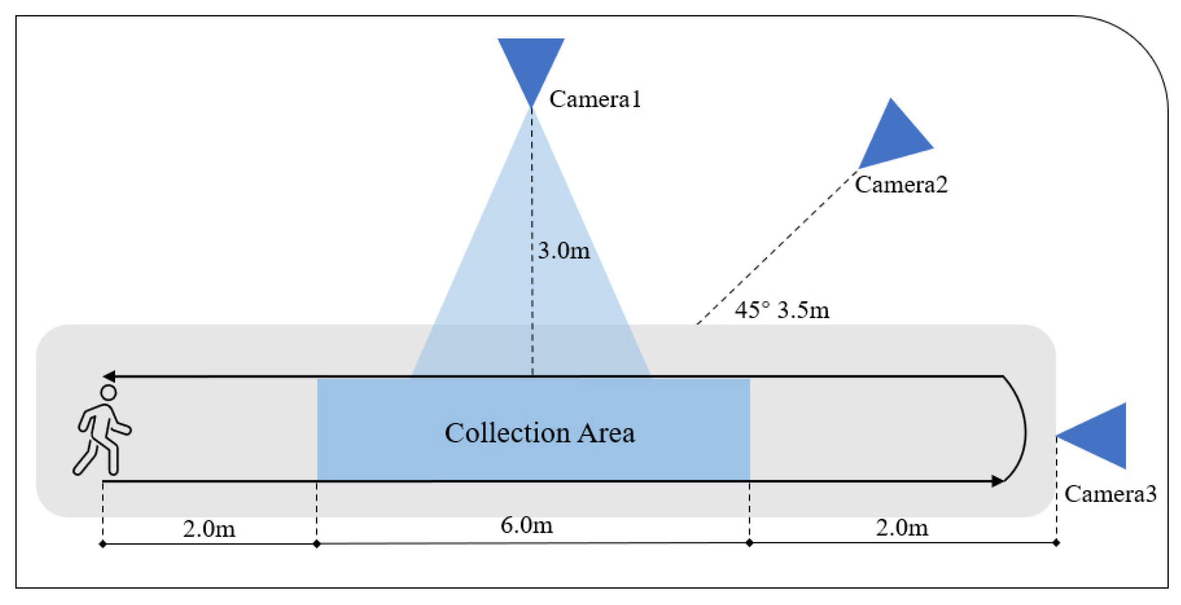
4.3. Experiment on Our Dataset
4.4. Ablation Study
5. Discussion
6. Conclusions
Author Contributions
Funding
Institutional Review Board Statement
Informed Consent Statement
Data Availability Statement
Conflicts of Interest
References
- Dingwell, J.B.; Cusumano, J.P. Nonlinear time series analysis of normal and pathological human walking. Chaos Interdiscip. J. Nonlinear Sci. 2000, 10, 848–863. [Google Scholar] [CrossRef] [PubMed]
- Zhang, Y.; Ogunbona, P.O.; Li, W.; Munro, B.; Wallace, G.G. Pathological Gait Detection of Parkinson’s Disease Using Sparse Representation. In Proceedings of the 2013 International Conference on Digital Image Computing: Techniques and Applications (DICTA), Hobart, TAS, Australia, 26–28 November 2013; pp. 1–8. [Google Scholar] [CrossRef]
- Guo, G.; Guffey, K.; Chen, W.; Pergami, P. Classification of Normal and Pathological Gait in Young Children Based on Foot Pressure Data. Neuroinformatics 2017, 15, 13–24. [Google Scholar] [CrossRef] [PubMed]
- Tao, W.; Liu, T.; Zheng, R.; Feng, H. Gait Analysis Using Wearable Sensors. Sensors 2012, 12, 2255–2283. [Google Scholar] [CrossRef] [PubMed]
- Rentz, C.; Far, M.S.; Boltes, M.; Schnitzler, A.; Amunts, K.; Dukart, J.; Minnerop, M. System Comparison for Gait and Balance Monitoring Used for the Evaluation of a Home-Based Training. Sensors 2022, 22, 4975. [Google Scholar] [CrossRef] [PubMed]
- Ivanov, K.; Mei, Z.; Lubich, L.; Guo, N.; Xile, D.; Zhao, Z.; Omisore, O.M.; Ho, D.; Wang, L. Design of a Sensor Insole for Gait Analysis. In Intelligent Robotics and Applications; Yu, H., Liu, J., Liu, L., Ju, Z., Liu, Y., Zhou, D., Eds.; Springer: Cham, Switzerland, 2019; pp. 433–444. [Google Scholar]
- Schlagenhauf, F.; Sreeram, S.; Singhose, W. Comparison of kinect and vicon motion capture of upper-body joint angle tracking. In Proceedings of the 2018 IEEE 14th International Conference on Control and Automation (ICCA), Anchorage, AK, USA, 12–15 June 2018; IEEE: Piscataway, NJ, USA, 2018; pp. 674–679. [Google Scholar]
- Albuquerque, P.; Verlekar, T.T.; Correia, P.L.; Soares, L.D. A Spatiotemporal Deep Learning Approach for Automatic Pathological Gait Classification. Sensors 2021, 21, 6202. [Google Scholar] [CrossRef]
- Ortells, J.; Herrero-Ezquerro, M.T.; Mollineda, R.A. Vision-based gait impairment analysis for aided diagnosis. Med. Biol. Eng. Comput. 2018, 56, 1553–1564. [Google Scholar] [CrossRef] [PubMed]
- Loureiro, J.; Correia, P.L. Using a Skeleton Gait Energy Image for Pathological Gait Classification. In Proceedings of the 2020 15th IEEE International Conference on Automatic Face and Gesture Recognition (FG 2020), Buenos Aires, Argentina, 16–20 November 2020; pp. 503–507. [Google Scholar] [CrossRef]
- Nghiem, A.T.; Auvinet, E.; Multon, F.; Meunier, J. Contactless abnormal gait detection. In Proceedings of the 2011 Annual International Conference of the IEEE Engineering in Medicine and Biology Society, Boston, MA, USA, 30 August–3 September 2011; pp. 5076–5079. [Google Scholar] [CrossRef]
- Bei, S.; Zhen, Z.; Xing, Z.; Taocheng, L.; Qin, L. Movement Disorder Detection via Adaptively Fused Gait Analysis Based on Kinect Sensors. IEEE Sens. J. 2018, 18, 7305–7314. [Google Scholar] [CrossRef]
- Gu, X.; Guo, Y.; Yang, G.Z.; Lo, B. Cross-Domain Self-Supervised Complete Geometric Representation Learning for Real-Scanned Point Cloud Based Pathological Gait Analysis. IEEE J. Biomed. Health Inform. 2022, 26, 1034–1044. [Google Scholar] [CrossRef] [PubMed]
- Zeng, Q.; Liu, P.; Bai, Y.; Yu, H.; Sun, X.; Han, J.; Wu, J.; Yu, N. SlowFast GCN Network for Quantification of Parkinsonian Gait Using 2D Videos. In Proceedings of the 2022 12th International Conference on CYBER Technology in Automation, Control, and Intelligent Systems (CYBER), Baishan, China, 27–31 July 2022; pp. 474–479. [Google Scholar] [CrossRef]
- Tian, H.; Ma, X.; Wu, H.; Li, Y. Skeleton-based abnormal gait recognition with spatio-temporal attention enhanced gait-structural graph convolutional networks. Neurocomputing 2022, 473, 116–126. [Google Scholar] [CrossRef]
- Jun, K.; Oh, S.; Lee, S.; Lee, D.W.; Kim, M.S. Automatic pathological gait recognition by a mobile robot using ultrawideband-based localization and a depth camera. In Proceedings of the 2022 31st IEEE International Conference on Robot and Human Interactive Communication (RO-MAN), Napoli, Italy, 29 August– 2 September 2022; pp. 652–659. [Google Scholar] [CrossRef]
- Jun, K.; Lee, Y.; Lee, S.; Lee, D.W.; Kim, M.S. Pathological Gait Classification Using Kinect v2 and Gated Recurrent Neural Networks. IEEE Access 2020, 8, 139881–139891. [Google Scholar] [CrossRef]
- Dingenen, B.; Staes, F.F.; Santermans, L.; Steurs, L.; Eerdekens, M.; Geentjens, J.; Peers, K.H.E.; Thysen, M.; Deschamps, K. Are two-dimensional measured frontal plane angles related to three-dimensional measured kinematic profiles during running? Phys. Ther. Sport 2018, 29, 84–92. [Google Scholar] [CrossRef] [PubMed]
- Stenum, J.; Rossi, C.; Roemmich, R.T. Two-dimensional video-based analysis of human gait using pose estimation. PLoS Comput. Biol. 2021, 17, e1008935. [Google Scholar] [CrossRef] [PubMed]
- Feichtenhofer, C.; Fan, H.; Malik, J.; He, K. Slowfast networks for video recognition. In Proceedings of the IEEE/CVF International Conference on Computer Vision, Seoul, Republic of Korea, 27 October–2 November 2019; pp. 6202–6211. [Google Scholar]
- Zhou, Z.; Rahman Siddiquee, M.M.; Tajbakhsh, N.; Liang, J. Unet++: A nested u-net architecture for medical image segmentation. In Proceedings of the Deep Learning in Medical Image Analysis and Multimodal Learning for Clinical Decision Support: 4th International Workshop, DLMIA 2018, and 8th International Workshop, ML-CDS 2018, Held in Conjunction with MICCAI 2018, Granada, Spain, 20 September 2018; Proceedings 4. Springer: Berlin/Heidelberg, Germany, 2018; pp. 3–11. [Google Scholar]
- Yu, F.; Wang, D.; Shelhamer, E.; Darrell, T. Deep layer aggregation. In Proceedings of the IEEE Conference on Computer Vision and Pattern Recognition, Salt Lake City, UT, USA, 18–23 June 2018; pp. 2403–2412. [Google Scholar]
- Khokhlova, M.; Migniot, C.; Morozov, A.; Sushkova, O.; Dipanda, A. Normal and pathological gait classification LSTM model. Artif. Intell. Med. 2019, 94, 54–66. [Google Scholar] [CrossRef] [PubMed]
- Yu, S.; Tan, D.; Tan, T. A framework for evaluating the effect of view angle, clothing and carrying condition on gait recognition. In Proceedings of the 18th International Conference on Pattern Recognition (ICPR’06), Hong Kong, China, 20–24 August 2006; IEEE: Piscataway, NJ, USA, 2006; Volume 4, pp. 441–444. [Google Scholar]
- Sadeghzadehyazdi, N.; Batabyal, T.; Acton, S.T. Modeling spatiotemporal patterns of gait anomaly with a CNN-LSTM deep neural network. Expert Syst. Appl. 2021, 185, 115582. [Google Scholar] [CrossRef]
- Yan, S.; Xiong, Y.; Lin, D. Spatial temporal graph convolutional networks for skeleton-based action recognition. In Proceedings of the Thirty-Second AAAI Conference on Artificial Intelligence, New Orleans, LA, USA, 2–7 February 2018. [Google Scholar]
- Kim, J.; Seo, H.; Naseem, M.T.; Lee, C.S. Pathological-Gait Recognition Using Spatiotemporal Graph Convolutional Networks and Attention Model. Sensors 2022, 22, 4863. [Google Scholar] [CrossRef] [PubMed]


| Model | Train Accuracy (%) |
|---|---|
| DNN | 85.86 |
| CNN | 86.65 |
| RNN | 86.63 |
| LSTM | 87.25 |
| CNN-LSTM [25] | 90.10 |
| GRU | 90.13 |
| ST-GCN [26] | 94.50 |
| AGS-GCN [15] | 94.56 |
| Multiple-input ST-GCN [27] | 98.34 |
| Our Model (FP-GCN) | 98.78 |
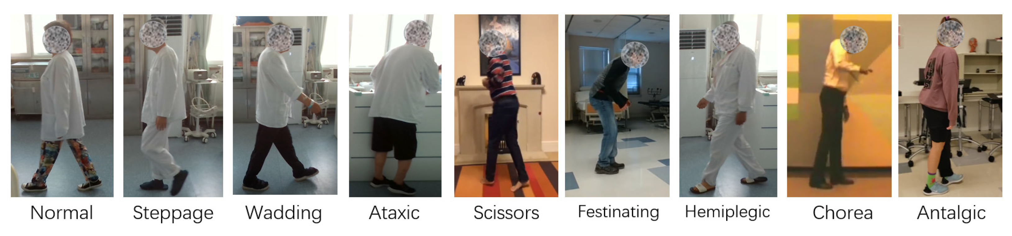
| Class | Subject | Video (Augmented) |
|---|---|---|
| Normal | 13 Web + 1 Real | 180 |
| Steppage | 8 Web + 2 Real | 200 |
| Wadding | 8 Web + 2 Real | 200 |
| Ataxic | 6 Web + 1 Real | 180 |
| Scissors | 5 Web | 180 |
| Festinating | 10 Web | 180 |
| Hemiplegic | 10 Web + 2 Real | 220 |
| Chorea | 6 Web | 144 |
| Antalgic | 8 Web | 168 |
| Total | 82 | 1644 |
| Class | ST-GCN | Our Model |
|---|---|---|
| Normal | 89.3 | 94.6 |
| Steppage | 95.6 | 96.4 |
| Wadding | 88.2 | 97.5 |
| Ataxic | 88.9 | 92.5 |
| Scissors | 82.6 | 94.5 |
| Festinating | 94.7 | 94.6 |
| Hemiplegic | 95.2 | 96.7 |
| Chorea | 94.0 | 96.5 |
| Antalgic | 94.7 | 96.2 |
| Mean Accuracy | 91.92 | 96.54 |
| Model | Accuracy (%) |
|---|---|
| F-GCN | 96.75 |
| P-GCN | 95.18 |
| FP-GCN | 98.78 |
Disclaimer/Publisher’s Note: The statements, opinions and data contained in all publications are solely those of the individual author(s) and contributor(s) and not of MDPI and/or the editor(s). MDPI and/or the editor(s) disclaim responsibility for any injury to people or property resulting from any ideas, methods, instructions or products referred to in the content. |
© 2024 by the authors. Licensee MDPI, Basel, Switzerland. This article is an open access article distributed under the terms and conditions of the Creative Commons Attribution (CC BY) license (https://creativecommons.org/licenses/by/4.0/).
Share and Cite
Zhao, X.; Li, J.; Hua, C. FP-GCN: Frequency Pyramid Graph Convolutional Network for Enhancing Pathological Gait Classification. Sensors 2024, 24, 3352. https://doi.org/10.3390/s24113352
Zhao X, Li J, Hua C. FP-GCN: Frequency Pyramid Graph Convolutional Network for Enhancing Pathological Gait Classification. Sensors. 2024; 24(11):3352. https://doi.org/10.3390/s24113352
Chicago/Turabian StyleZhao, Xiaoheng, Jia Li, and Chunsheng Hua. 2024. "FP-GCN: Frequency Pyramid Graph Convolutional Network for Enhancing Pathological Gait Classification" Sensors 24, no. 11: 3352. https://doi.org/10.3390/s24113352
APA StyleZhao, X., Li, J., & Hua, C. (2024). FP-GCN: Frequency Pyramid Graph Convolutional Network for Enhancing Pathological Gait Classification. Sensors, 24(11), 3352. https://doi.org/10.3390/s24113352