Abstract
Plant health monitoring is essential for understanding the impact of environmental stressors (biotic and abiotic) on crop production, and for tailoring plant developmental and adaptive responses accordingly. Plants are constantly exposed to different stressors like pathogens and soil pollutants (heavy metals and pesticides) which pose a serious threat to their survival and to human health. Plants have the ability to respond to environmental stressors by undergoing rapid transcriptional, translational, and metabolic reprogramming at different cellular compartments in order to balance growth and adaptive responses. However, plants’ exceptional responsiveness to environmental cues is highly complex, which is driven by diverse signaling molecules such as calcium Ca2+, reactive oxygen species (ROS), hormones, small peptides and metabolites. Additionally, other factors like pH also influence these responses. The regulation and occurrence of these plant signaling molecules are often undetectable, necessitating nondestructive, live research approaches to understand their molecular complexity and functional traits during growth and stress conditions. With the advent of sensors, in vivo and in vitro understanding of some of these processes associated with plant physiology, signaling, metabolism, and development has provided a novel platform not only for decoding the biochemical complexity of signaling pathways but also for targeted engineering to improve diverse plant traits. The application of sensors in detecting pathogens and soil pollutants like heavy metal and pesticides plays a key role in protecting plant and human health. In this review, we provide an update on sensors used in plant biology for the detection of diverse signaling molecules and their functional attributes. We also discuss different types of sensors (biosensors and nanosensors) used in agriculture for detecting pesticides, pathogens and pollutants.
1. Introduction
Modern agriculture faces several challenges like environmental stressors, climatic instability, labor shortages, and poor soil as a result of inadequate land management, which all affect agriculture productivity [1,2]. However, with the advent of sensors, many of these problems that plants face can be managed by their early detection. Modern agriculture has transformed into smart farming with the advancement in sensor-based technology [3,4]. The application of sensors (nanosensors and biosensors) that are capable of sensing changes in plant health and their morphological and physiological traits has shown to be a useful technique to boost agricultural yields [5]. Sensors used in agriculture may gather information about crops, fields, the environment, soil, and other vital characteristics, allowing farmers and other agricultural professionals to make the best decisions on how to manage their crops and fields [5,6]. In smart farming, sensor-based phenotyping has revolutionized plant research by monitoring different growth and stress traits of crops grown under different conditions [7]. Sensors play a multifaceted role to monitor different plant signaling molecules and environmental stressors in real time, which has paved the way for improving plant growth and crop yield. They also play a key role in monitoring soil variables like pH, pesticides, heavy metals, moisture, and minerals, in real time. In plant biology research, different types of sensors such as biosensors and nanosensors are used which are made up of different materials.
Plants’ sessile nature necessitates that they recognize environmental obstacles that may threaten their survival. Individual cells that detect possible hazards to neighboring cells must signal and communicate quickly in order to survive. In response to environmental stressors, plants produce diverse signaling molecules such as Ca2+, ROS, hormones, etc., which regulate diverse growth and adaptable responses [8]. For instance, Ca2+ is a well-known secondary messenger and signaling element present in both unicellular and multicellular organisms. Biotic and abiotic stresses alter cellular Ca2+ homeostasis by causing transitory fluctuations in Ca2+ concentrations in the cytosol and subcellular compartments, which are further sensed by different Ca2+ sensors regulating different growth and stress-related responses [9]. Ca2+ binding proteins such as calmodulin (CaM), CaM-like proteins (CMLs), and calcineurin B-like proteins (CBLs), detect changes in Ca2+ levels. This causes the proteins to alter shape, allowing them to interact with numerous targets and regulate downstream processes including transcription, enzymatic activity, and ion fluxes [10,11]. Plants feature particular Ca2+ active transporters like Ca2+ -ATPases and Ca2+/cation exchangers (CAX), which are present in cell membrane and intracellular organelles and contribute to the replenishment of resting [Ca2+] cyt. This cellular Ca2+ signature or cellular dynamics can be observed utilizing Ca2+ imaging methods. Earlier synthetic dyes that were Ca2+-sensitive (e.g., Ca2+ green dextran, Fura-2, Fura-2 dextran, and Indo-1) were used for the detection of cytosolic Ca2+ dynamics by fusing with Ca2+-selective chelators like EGTA and BAPTA, respectively. However, because of their limitations, the introduction of fluorescent-based genetically encoded Ca2+ indicators (GECIs) such as Aequorin, cameleon, and YC-Nano offered additional opportunities for the quantitative in vivo imaging of Ca2+ dynamics [12]. Additionally, imaging can be used to detect Ca2+ dynamics in different subcellular compartments, such as the tonoplast, the nucleus, the Golgi apparatus, mitochondria, plastids/chloroplasts, apoplastic space, and the thylakoid lumen and membrane [13,14]. Furthermore, the bioluminescence resonance energy transfer (BRET)-based GFP-aequorin reporter or sensor (i.e., G5A) solved the primary constraints of aequorin, which permitted imaging of long distance Ca2+ waves with a lower quantity of emitted light [15]. Similarly, Yellow Cameleon (YC) was developed using GECIs followed by a series of variants with higher affinity for Ca2+ as shown in Figure 1. Modern research has led to the development of highly sensitive single-fluorophore (single-FP) biosensors (GCaMP biosensors) connected with calmodulin, which have several advantages over FRET Cameleons. These include a simpler experimental design and possibly higher temporal resolution of imaging.
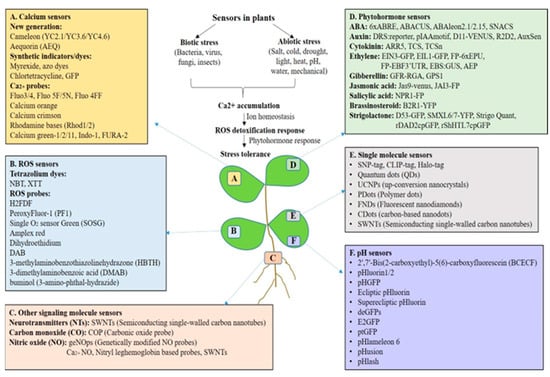
Figure 1.
Sensors used in plant biology for the detection of signaling molecules such as calcium (Ca2+), ROS, hormones, and nitric oxide (NO) which regulate diverse growth and stress responses. This Figure also shows different sensors used for monitoring pH in plants.
ROS also operate as secondary messengers in plant signal transduction altering diverse plant growth and stress-related traits. They have important roles in regulating a wide range of subcellular, cellular, and systemic signals. Additionally, ROS are vital in plant defense and acclimation responses to various biotic and abiotic environments, as well as being a critical component of several hormonal, physiological, and developmental pathways [16,17,18]. The real time monitoring of ROS in plants reveals the molecular complexity of plant signal transduction pathways related to different traits. Many sensors and reporters are used to monitor live ROS imaging in plants such as 2′,7′-dichlorofluorescin diacetate (H2DCFDA), H2DCFDA conjugated to BSA (OxyBURST), dihydroethidium (DHE) and its mitochondrion-targeted form mitoSOX, dihydro-2′,4,5,6,7,7′-hexafluorofluorescein (H2HFF), N-acetyl-3,7-dihydroxyphenoxazine (Amplex red), singlet oxygen sensor green (SOSG), and boronate-based probes such as peroxy orange-1 (PO1) [17]. These detection systems vary in detecting different ROS forms in plants.
Plant hormones play a key role in plant defense–growth tradeoffs and are key for plant survival [18]. Phytohormones are natural compounds, which control various physiological processes during stress as well as growth and development. A wide range of growth hormones [auxin (AUX), cytokinin (CK), and gibberellin (GB)] and stress hormones [abscisic acid (ABA), jasmonic acid (JA), salicylic acid (SA), ethylene (ET), brassinosteroids (BRs), strigolactones (SLs), and small peptides] have been characterized in plants [19,20]. The hormone-signaling network is very complex and interconnected. Earlier, the traditional biochemical methods such as immunohistochemistry, LC-MS or GC-MS were used to check the distribution, quantity, and identification of these hormones in plant cells and tissues [21], at high accuracy and sensitivity [22]. The advances in synthetic biology based on sensors have paved the way to broaden our knowledge of the distribution of plant hormones in different tissues, organs, and cell types. Synthetic biology offers continuous monitoring, live cell imaging, and identification of local distribution of phytohormonal concentration in plants under normal and various stress conditions. New techniques for detecting phytohormones with minimum invasion and cellular or even subcellular resolution have been made possible over the past 20 years by developments in fluorescence microscopy technology and biosensor engineering [23]. For example, to visualize AUXtransport dynamics and subcellular AUX distribution, fluorescent conjugates of AUX, such as 7-nitro-2,1,3-benzoxadiazole (NBD)-naphthalene-1-acetic acid (NAA) and NBD-indole-3-acetic acid (IAA) have been used in the model plants. Alexa Fluor 647-castasterone (AFCS) was utilized to observe BR receptor endocytosis in live Arabidopsis cells [24]. Similarly, bioactive fluorescein-labeled gibberellic acids (GA-fls) and the strigolactone (SL)-agonist probe Yoshimulactone Green (YLG) becomes activated and so fluorescent upon GA and SL application [25]. Peptide hormones were also detected using fluorescent dyes such as tetramethylrhodamine (TAMRA) to monitor their uptake and localization [26]. Another form of FRET sensor, notably ABACUS and ABAleon, a SnRK2 activity sensor (SNACS), was created and utilized to monitor ABA buildup [27]. A two-component output-sensor (TCS) was designed as a synthetic reporter to visualize the distribution of CK in Arabidopsis embryos, and its enhanced derivative, TCS-new (TCSn), was also developed. EIN3-GFP and EIL1-GFP were developed as sensors for ET. Vong et al. [28] created an enzyme-based chemical biosensor called the artificial-metalloenzyme ethylene probe (AEP) to detect ET. In addition, GPS1 which is a FRET-based GA sensor was designed to measure spatiotemporal GA distribution with high resolution [29]. Furthermore, fast changes in the quantity and distribution of plant hormones targeting specific compartments in living cells can be detected by genetically encoded biosensors (direct or indirect) [30].
The role of Ca2+ and ROS signaling responding to various external stimuli in plants is well studied but the physiological significance of pH changes is largely unknown. However, reports on pH-sensing studies based on leaf and root tissue led to the discovery of systems that sense and signal extracellular pH (pHe). In plants, pH regulates the chemistry and rheology of the cell wall to change its flexibility and govern the spatiotemporal growth of cells. pHe homeostasis is cooperatively maintained by cell wall components, enzymes that remodel the cell wall, and H+-ATPases located in the plasma membrane (PM). The pHe of plants is inherently acidic, but it fluctuates dynamically in response to environmental stimuli and physiological factors [31]. In general, use of fertilizer, the climate, and weather can vary the plant pH externally (soil), leading to a change in apoplastic pH [32], but inside, the apoplastic pH is also altered by defense and growth activities [33,34]. Recent findings demonstrate that transmembrane kinase1 (TMK1) phosphorylates and activates plasma membrane H+-ATPases, which causes apoplastic acidification, therefore modulating the AUX signal that drives cell elongation in the hypocotyl and root elongation zones [33,34]. In plants, a change in apoplast pH or extracellular alkalization is the first and most immediately observable reaction during pattern-triggered immunity (PTI), drought and salinity [35]. The secondary regulation of proton pumps or the passage of ions cause an elevation in systemic pH in plants [31]. In addition, Ca2+ transients lead to pH changes in the cytosol for distinct stimuli known as the pH–Ca2+ link. To fully comprehend the relationship between cytosolic Ca2+ transients and H+ homeostasis, various fluorescent biosensors [notably, NES-YC3.6 and pH-green fluorescent protein (GFP)] have been used to analyze pH and Ca2+ dynamics in living plant cells [36,37].
In plants, numerous GFP variations exhibit pH sensitivity due to chromophore protonation and deprotonation [38]. For example, various cytosolic and other organelle GFP-based biosensors such as pHluorin and Pt-GFP, as well as H148D pH sensors give a comprehensive set of tools for imaging fluctuations in these ions. These fluorescent probes have the limitation that they work only in cytoplasm. As a result, a genetically encoded pH indicator overcomes many of the disadvantages of traditional chemical probes. Recently, the Fluorescence Indicator reporting pH in Lysosomes (FIRE-pHLy) has been used to target lysosomal pH [39]. The use of pH-sensitive GFPs has resulted in the rather surprising finding that, rather than a tightly buffered, constant cytosolic pH, cells can display extremely dynamic pH variations in response to a wide variety of internal and external stimuli. Such pH fluctuations should have extensive impacts on cellular biochemistry, implying that pH, together with redox, might operate as a worldwide coordinator of cell activities, moving the balance of the cell between signaling/response states [38]. Biosensors are currently advancing the frontiers of our understanding of the in vivo cellular dynamics that underpin important regulatory networks at dimensions from the subcellular to the entire plant [38]. To summarize, we have shown different sensors used in plant research for detecting different signaling molecules like Ca2+, ROS, hormones, nitric oxide (NO), neurotransmitters (NTs) and pH in Figure 1.
2. Application of Nanosensors in Agriculture
Owing to their unmatched capability to sense and response to a broad spectrum of environmental stimuli and exceptional sensitivity and specificity, nanosensors have become extremely useful tools for the real-time monitoring and management of plants. Nanobiosensors are non-intrusive, sensitive devices that are developed using combined nanobiotechnological approaches to monitor a large number of environmental samples [40]. Nanobiosensors collect the information for analysis and produce signals in real-time response [41]. Different types and classes of nanomaterials including nanotubes (multi-walled and single-walled), one-dimensional nanowires, quantum dots, crystalline particles also known as nanocrystals, nanoceramics and nanocomposites and hybrid materials can be exploited to manufacture nanobiosensors. The different types of nanosensors that are used in agriculture are shown in Figure 2.
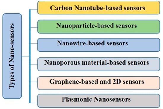
Figure 2.
Types of nanosensors made of different materials used for the detection of diverse molecules.
Nanobiosensors have an immense range of applications from household to industry which includes detection of a wide variety of fertilizers, pesticides, fungicides, pathogens, heavy metal content, and quality of soil such as pH, temperature, moisture content, soil water content and overall growth hormone level [42,43,44]. In addition, farmers utilize these portable smart sensors to monitor and manage the soil conditions locally in the agricultural industry. They record the concentrations of minerals, thus insufficiencies of particular minerals in the soil, and detection of pests, pathogens, and diseases [45]. We have shown the application of sensors in agriculture in Figure 3.
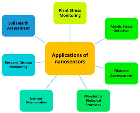
Figure 3.
The applications of nanosensors for the detection of biological processes, nutrients, and biotic and abiotic stressors, as well as soil health monitoring and disease assessment.
3. Application of Nanosensors for Pesticide Detection
Various organic and inorganic substances, including dyes, pesticides, and agricultural wastes containing hazardous materials, are introduced into soil through agricultural activities, industrial wastewater, and municipal wastewater [46]. It is essential to monitor xenobiotics in soil, particularly herbicides and soil-applied pesticides, to prevent their uptake by plants, given their detrimental effects on the environment and living organisms [47]. Due to limitations and challenges associated with conventional analytical methods for pesticides, there is a growing interest in developing new measurement techniques such as biosensors and nanobiosensors [48]. Nanosensors are small devices designed to detect specific molecules, biological components, or environmental conditions at the nanoscale [49]. They offer high specificity, portability, and superior detection capabilities compared to larger sensors [50]. Nanosensor operation typically involves three main components: sample preparation, recognition of target molecules or organisms, and signal transduction [51]. Recognition molecules such as antibodies or enzymes bind to target analytes in the sample, and signal transduction methods convert these interactions into measurable signals [45]. This allows for precise and efficient detection of a wide range of substances and environmental factors.
Significant progress has been made in the last several decades in the development of nanomaterial-based sensors for the detection of pesticide residues in soil [51]. Detecting pesticide residues is crucial for ensuring food safety, particularly in animals, as low concentration pesticides like organophosphates can accumulate [52]. Exposure to higher concentrations poses severe health risks by inhibiting enzymes such as acetylcholinesterase [53]. Thus, the development of advanced detection methods is paramount for safeguarding human health and maintaining food safety standards. Nanosensors offer innovative solutions for detecting various pesticide residues with high sensitivity and selectivity [54]. Nanoparticles serve two primary functions in these systems: signal conversion and signal enhancement. Signal conversion involves nanoparticles altering color or emitting light in response to pesticide presence, while signal enhancement utilizes nanoparticles to boost detection sensitivity through various means such as enhancing fluorescence or Raman signals [51]. Various kinds of nanosensors differing in their sensing ability to detect the herbicide, insecticide and pesticide residues within the soil samples have been introduced recently including paper-based screen-printed electrodes (SPEs) with nanostructure modifications on the transducer surface which improves both the portability and sensitivity of the electrochemical detection platform [55]. Moreover, a fluorescent nanosensor has been introduced utilizing Ytterbium Oxide nanoparticles, which have been modified using 3-aminopropyl-triethoxysilane and coated with Yb2O3, enabling the detection of imazapyr with a limit of detection of 0.2 ppm. This sensor exhibits proficient capabilities in sensing herbicides effectively [56]. Another study introduces an affinity sensor based on surface plasmon resonance (SPR) which utilizes atrazine-imprinted nanoparticles attached to a gold surface for the selective detection of atrazine, with a detection limit of 0.7134 ng/mL, while yet another SPR-based fiber-optic sensor incorporates tantalum (V) oxide (Ta2O5) nanoparticles for detecting fenitrothion with a detection limit of 38 nM [57]. Nanomaterials show considerable promise for developing non-enzymatic electrochemical sensors. These include various categories such as nanoparticles (e.g., CuO, CuO–TiO2, ZrO2, and NiO); nanocomposite (e.g., molybdenum); and peptide and carbon nanotubes, which are extensively utilized for electrochemically detecting residual pesticide particles [58]. The thorough study of residual pesticide particles using such nanomaterials is attributed to their extremely small size, large surface area, and unique electrical and chemical properties.
Recently, there has been an increasing focus on utilizing nanomaterials to improve the performance of electrode surfaces in the detection of heavy metals [59]. Electrodes modified with nanomaterials have demonstrated significant advancements in electroanalytical techniques for detecting a diverse array of heavy metals. These nanomaterials encompass metal nanoparticles, metal oxides, graphene-based materials, carbon nanotubes, and metal–organic frameworks (MOFs) [60]. Common methods for fabricating nanomaterial-modified disposable electrodes include drop casting, dip coating, spin coating, electrochemical deposition, direct growth, and screen printing [61]. One fluorescence sensor utilizes a combination of copper (II) oxide and multiwall carbon nanotubes (MWCNTs) to monitor glyphosate, achieving a limit of detection of 0.67 ppb [62]. Meanwhile, an electrochemical luminescence sensor detects glyphosate at 0.5 nM by employing composites of luminol–gold nanoparticles–L-cysteine–Cu (II) [63]. Moreover, an electrochemical sensor employing CuO-TiO2 hybrid nanocomposites detects methyl parathion at 1.21 ppb [64], while an electrochemical aptasensor detects malathion at 0.001 ng/mL utilizing CuO nanoparticle-decorated 3D graphene nanocomposites [65]. Optical nanosensors utilizing silver nanodendrites detect dimethoate at 0.002 ppm [66], and upconverting nanoparticles detect metribuzin at 6.8 × 10⁻⁸ M through ratiometric and colorimetric responses [67]. With a limit of detection of 0.01 nM in soil samples, nanosensors showcase the potential for accurate, rapid, and dependable detection of pesticide residues, thereby aiding environmental monitoring and ensuring environmental safety. Different types of nanosensors used for the detection of pesticides are shown in Table 1. Future efforts should prioritize developing simplified detection methods, including miniaturized and intelligent approaches such as colorimetry and test papers. Furthermore, there is a need to expand detection capabilities to encompass a wider range of pesticide types and enable simultaneous detection of multiple pesticides using high-throughput chip-based technologies combined with nanomaterials.

Table 1.
The applications of nanosensors for pesticide detection.
4. Application of Nanosensors for the Detection of Heavy Metals
Heavy metal pollution poses serious environmental threats, coming from industrial activities, urban runoff, and human practices. Heavy metals have the potential to bioaccumulate and biomagnify in living organisms through the food chain, posing significant health risks [70]. Studies have documented various acute and chronic toxic effects of heavy metal ions on human organs [71,72]. Exposure to heavy metal contamination can lead to oxidative stress, ecological toxicity, plant toxicity, morphological and biochemical effects, and cellular toxicity in the living organisms [66]. Moreover, increased levels of heavy metals in humans have been associated with numerous health hazards, including lower IQ in children, developmental obstacles, cancers, hypertension, weakened immune systems, cellular toxicity, oxidative damage, heart diseases, coronary artery disease, cerebrovascular disorders, and miscarriages and stillbirths, among others [72,73]. Recent incidents such as the lead tap water crisis in Flint, Michigan, highlight the critical need to be prepared for potential widespread heavy metal contaminations and the associated health risks, social consequences, and post-traumatic stress disorders [74,75].
Due to its sensitivity and convenience, electrochemical detection, especially through portable and disposable sensors, has emerged as a powerful method for monitoring heavy metals [76]. Nanomaterials, such as various oxides of metal nanoparticles, graphene-based materials, carbon nanotubes, and metal–organic frameworks (MOFs), play a crucial role in enhancing electrode surfaces for monitoring heavy metals [41]. Various nanostructure architectures have shown promising results in detecting heavy metals with high sensitivity. Metal–organic frameworks (MOFs) offer precise pore sizes and functional groups for selective sensing [77]. Chemically modified silicon membranes, electrodeposited bismuth films, and ion-imprinted polymer films have all been employed for the efficient and accurate detection of heavy metals within environmental samples [78]. These advancements underscore the potential of nanomaterial-based electrochemical sensors for effective monitoring and management of heavy metal pollution.
Significant achievements have been achieved in the advancement of analytical procedures for detecting and analyzing heavy metal ions (HMIs) in environmental samples. For heavy metal analysis, well-established methods such as graphite furnace atomic absorption spectroscopy (GF–AAS), flame atomic absorption spectroscopy (FAAS), inductively coupled plasma–mass spectroscopy (ICP–MS), atomic emission spectroscopy (AES), inductively coupled plasma–optical emission spectroscopy (ICP–OES), X-ray diffractometry, and X-ray fluorescence have been identified [79,80,81]. However, these techniques often come with drawbacks such as large sample sizes, expensive equipment, and the need for specialized training, limiting their practicality for on-site or field studies. Electrochemical and colorimetric nanosensors have emerged as promising alternatives due to their high sensitivity, specificity, affordability, mobility, and rapid detection capabilities [82]. Various electrochemical nanosensors have gained attention for their ability to identify heavy metal ions with high efficiency and specificity, making them appropriate for on-demand, in situ, and field applications [83,84]. Similarly, colorimetric nanosensors enable quick screening and visual detection for point-of-use applications. Recent reviews have highlighted the advances in nanomaterial-based optical sensors for heavy metal detection, with a focus on affordable and compact electrochemical nanosensors and smartphone-operated screen-printed electrodes (SPEs), among other technologies [85]. Developing reliable methods for heavy metal detection for environmental samples is crucial for ensuring public health safety and global homeland security.
Significant studies have been undertaken to generate sensors that are efficient in detecting heavy metal ions at trace and ultra-trace concentrations [86,87]. However, challenges such as sensitivity, selectivity, specificity, and interference persist in many available sensors [88,89]. Therefore, considerable efforts must be directed towards enhancing electrochemical and colorimetric nanosensors to achieve better efficiency, accuracy, and specificity, thus enabling reliable and expanded heavy metal ion detection capabilities [90,91]. Specifically, advancements should focus on designing and developing easily movable electrochemical sensors based on graphene, carbon nanotubes, nanostructures, carbon dots, nanomaterials, and metal–organic frameworks to facilitate accurate and specific heavy metal ion detection [92]. The advancement of portable colorimetric sensors for rapid screening and visual detection of heavy metal ions will continue to be an active area of research in the coming years [93]. Enhanced technology coupled with smartphone accessibility will open avenues for widespread adoption and enhancement of smartphone-based sensors, enabling rapid, in situ, and on-site detection of heavy metal ions [94]. We have summarized the use of various sensors for the detection of heavy metals in Table 2. Furthermore, the emergence of low-cost and disposable paper-based sensors will facilitate on-site and field detection of heavy metal ions. There will be a growing interest in microfluidic and microchip sensors to enable rapid arrays and simultaneous detection of heavy metal ions. The utilization of functionalized gold nanoparticles for fiberoptic surface plasmon resonance sensing of heavy metal ions is projected to attract great attention.
Table 2.
The roles of nanosensors in the detection of heavy metals.
5. Role of Nanosensors for the Detection of Phytopathogens and Pests
The world economy is still at risk from emerging plant diseases that continue to be a serious threat to food security and ecological stability. Plant pathogens pose a significant threat to global agriculture, with potential yield losses of up to 30% [18,102]. Fungal diseases cause enormous agricultural losses, hence effective management measures must be taken to prevent further infestation. The introduction of new technologies in disease detection and diagnosis has increased agricultural output by enabling real-time identification of plant diseases at an early stage of infection. Plant health monitoring increasingly requires early identification of plant pathogens in order to control diseases at various stages of development, reduce the risk of spreading, and prevent the entry of novel pathogens. Traditional procedures are deemed to lack the precision, accuracy, and sensitivity required to identify plant diseases. Conventional diagnostic methods use time-consuming, culture-dependent procedures that are especially difficult when it comes to dealing with biotrophic fungal pathogens. Traditional methods of plant pathogen identification mainly rely on descriptive methods that interpret visible symptoms in addition to isolation, culture, and laboratory-based methods that include physiological, biochemical, and pathogenicity tests [103]. The precision and reliability of these methods largely hinge on the expertise and proficiency of the person undertaking them. A new era in agricultural diagnostic technology was ushered in with the rapid advancement of molecular diagnostic techniques in recent decades. Plant disease identification has been made easier by the advent of novel molecular techniques which include the use of many types of the polymerase chain reaction (PCR), including nested PCR, multiplex PCR, reverse transcription (RT)-PCR, real-time PCR, and conventional PCR [104]. Critical immunological techniques include lateral flow assays (LFAs) [105], and enzyme-linked immunosorbent assays (ELISAs), plate-trapped antigen-ELISA, and double antibody sandwich-ELISA [106]. Despite these benefits, molecular detection techniques are not always able to identify pathogens at low titers in materials like seeds and insect vectors, or in the early stages of infection. Moreover, cross-contamination with PCR reagents that entirely prevent the target DNA amplification can result in false negative results, whilst cross-amplification of PCR-generated non-target DNA fragments can result in false positive results. The inability to use PCR for plant pathogen identification in the field is another restriction and thus necessitates the development of other viable detection tools [107]. In this regard, nanosensors have emerged as an important tool for the rapid detection of plant pathogens and pests in agriculture as shown in Figure 4. Different types of nanotechnology-based tools are used for plant pathogen detection which include nano-barcoding, nanobiosensors, metal nanoparticles, quantum dots, and nanodiagnostic kits. Nanomaterials such as nanoparticles, nanowires, and nanotubes have been extensively explored for the development of sensors with enhanced sensitivity and selectivity. Nanoscale sensors offer advantages such as high surface-to-volume ratios and increased signal amplification, enabling ultrasensitive detection of phytopathogens [108]. In recent times, gold nanoparticles and nanostructures have been used on a large scale for the detection of plant pathogens and disease diagnostics. For instance, Xanthomonas axonopodis pv. vesicatoria, the bacterium that causes bacterial spot disease in Solanaceae plants, has been identified using sensors that combine fluorescent silica nanoparticles with antibody molecules [109]. Similarly, phytoplasma was detected in grapes using gold nanoparticle-coated sensors [110]. In Arabidopsis thaliana, an electrochemical sensor coated with gold nanoparticles was used to detect the bacterial pathogen Pseudomonas syringae [111]. In addition to bacterial pathogens, nanosensors can also detect fungal pathogens mainly by detecting mycotoxins. For example, the biosensor 4mycosensor detects different types of fungal mycotoxins in different crops such as oat, corn, barley and wheat [112]. This nano-based diagnostic kit can assist farmers in preventing disease epidemics in the field. A previous study reported that Aspergillus spss can be accurately detected using copper oxide (CuO) nanoparticles [113]. In strawberry, carbon nanotubes were used to detect fungal pathogens such Rhizopus and Aspergillus species [114]. Another noteworthy detection method is based on surface-enhanced Raman spectroscopy (SERS), which is capable of recognizing chemical fingerprints. Using silver nanoparticles (AgNPs), this approach quickly detected Alternaria mycotoxins in pear fruit, with a limit of detection (LOD) of 1.30 μg/L [115]. For the detection of viral pathogens in plants, QD-based nanosensors have been used in different plant systems, for example, Grapevine virus A [116], Tomato ringspot virus [117], Cowpea mosaic virus [118], Cauliflower mosaic virus [119], Citrus tristeza virus [120], Bean pod mottle virus [117], and Arabis mosaic virus [117]. Sharma et al. [121] created a label-free immunosensor that detects Capsicum chlorosis virus (CaCV) in bell pepper leaves. This immunosensor was based on immobilizing viral antigens on the surfaces of gold nanoparticles (AuNPs) and multi-walled carbon nanotubes. Notably, the immunosensor had a sensitivity for CaCV detection that was 800–1000 times higher than that of DAC-ELISA. Gold nanorods (AuNRs) functionalized with antibodies specific to Cymbidium mosaic virus (CymMV) [122]. A new technique for detecting Citrus tristeza virus (CTV) using nanobiosensors was developed, which uses fluorescence emission from cadmium telluride quantum dots (CdTe-QDs) linked with CTV coat protein (CTV-CP) antibodies. Two sensitive detection approaches were discussed: Förster resonance energy transfer (FRET) biosensors and non-FRET-based biosensors for quick identification of CTV-infected plants [123].
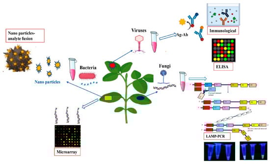
Figure 4.
Schematic illustration of different nanosensor-based pathogen detection systems in plants.
In future, different nanoscale gadgets can be customized for smart agricultural systems to detect pathogens before growers become aware of them. Nano–smart devices will therefore act as a warning system and a safety measure for disease outbreaks. Nanoscale materials are useful tools for the detection of plant disease because of the remarkable biospecificity of synthetic molecular recognition at the nanoscale, which has lately seen enormous advancements.
Pest and disease-related crop losses pose a serious threat to global food security as well as to the earnings of farmers. Globally, pests cause up to 40% of the crop yield losses each year [124]. The proper prevention of plant pests can guarantee agricultural output mainly by preventing their development and their spreading in the fields. A great way to identify, anticipate, and control plant insect pests in different crops is by remote sensing utilizing nanosensors [125,126]. With a dimension of less than 100 nm, nanosensors are potent, real-time, cost-effective, and environmentally friendly sensing devices employed to detect insects. The use of the insect’s own pheromone is paving the way for developing nanosensor-based tools for pest detection [127]. For, instance, insect sexual pheromone, methyl 2,6,10-trimethyltridecanoate, was detected using a polyaniline and multi-walled carbon nanotube (Pani/MWCNT-COOH) nanocomposite, which is a nanostructured cantilever sensor [128]. In addition, a graphene oxide and β-cyclodextrinylated cantilever nanosensor was utilized to detect the presence of Bactocera oleae in olive plants. Wehrenfennig et al. [129] used a variety of metal-oxide gas sensors including tungsten and tin oxide nanoparticles to detect the sexual pheromone of the grapevine pest (Lobesia botrana). A previous study has demonstrated the effectiveness of an oxide film nanostructure and 3-aminopropyl triethoxysilane-functionalized silicon dioxide cantilever sensor in the detection of insects’ pheromones, particularly Helicoverpa armigera (Hubner) and Scirphophaga incertulas [130]. Brezolin et al. [131] developed an extremely sensitive nanostructured cantilever sensor for Euschistus heros pheromone detection, this being a major pest of soybeans. Similarly, a variety of metal-oxide gas sensors including tungsten and tin oxide nanoparticles were employed to detect the sexual pheromone of the grapevine pest (Lobesia botrana) [129]. These studies support the notion that nanotechnology-based sensors are viable tools for the early detection and monitoring of insects in sustainable agriculture which can reduce the usage of pesticides and boost yield production.
6. Challenges and Future Perspectives of Sensor-Based Smart Farming
The current agricultural sector has several noteworthy obstacles, such as the escalating need for food, scarcities of labor, environmental stressors and climate change. A viable solution for addressing the current issues facing the agriculture sector is that of smart farming and precision agriculture [132]. Smart agricultural farming uses modern technology, such as sensors, to increase crop yield while lowering adverse environmental consequences, increasing the sustainability of farming. From the farmers’ perspective and that of the environment, smart farming has several advantages. For example, smart farming methods use sophisticated sensors to monitor biotic and abiotic stressors, soil physicochemical properties and nutrient levels. Thus a farmer is helped to apply the proper quantity of inputs, such as fertilizer, water, and pesticides at the correct time and location by utilizing data and sensors to monitor crop conditions, weather, and soil. This can lower waste and pollution while increasing crop quantity and quality. In other words, smart farming can act as an early warning system, which can help farmers to monitors and identify possible problems and provide prompt remedies in order to avoid crop yield losses [133]. Although there are many advantages to smart farming, there are also many challenges to its broad implementation. For small-scale farmers, there may be several obstacles, including the initial cost of technology, data interpretation, data privacy, and incompatibilities with current agricultural practices. In order to overcome these obstacles, education and training initiatives must be put in place to provide farmers the know-how to properly utilize these technologies [134]. On the hand, the performance of sensors is frequently impacted by environmental conditions including humidity and temperature variations. In order to provide correct results, sensors must be calibrated to offer accurate readings. Despite limitations, sensor-based smart farming has the potential to revolutionize traditional farming by making it more precise and ecofriendly.
7. Conclusions
Smart farming and precision agriculture are important to boost food security throughout the world. Over the years, agriculture has undergone a series of transformations from field to customized growth chambers which provide a precisely calibrated and controlled environment, which is essential for healthy plant growth as well as for testing and adjusting the environmental parameters [135,136] There exist numerous biophysical instruments that can automatically track, monitor, and analyze a plant’s growth and its environmental conditions [137]. With the advent of sensors, real- time monitoring of environmental stressors and plants’ responses has transformed traditional farming into more precise and smart farming. Sensors (biosensors and nanosensors) play a multifaceted role in plant research such as detection of signaling molecules and environmental stressors. Smart agriculture is rapidly transforming traditional agricultural production practices across the globe. This transformation, based on technical innovation, is critical to ensuring a viable food production. One of the primary components of smart agriculture is sensors. The use of nanosensors or biosensors in plant science provides valuable insights into the role of different signaling molecules at different cellular compartments which allows researchers to explore their in vivo distribution and transport as well as reactions to environmental variables. Sensors enable the rewiring and understanding of dynamic networks of complicated plant signal transduction and metabolism at any organizational size which play a key role in targeted metabolic engineering for crop improvement. Under field conditions, sensors are used for nutrient analysis to determine whether fortification is necessary for optimal plant development, as well as to detect infections. Sensors may give a wide range of information, allowing farmers to better care for their crops and fields. Farmers can construct a holistic image of their farms, crops, and fields by gathering a variety of data via sensors, allowing them to prepare for the future. They know which fields require special attention, which are ready for crops, which crops did well with the weather, and so on. From there, they are empowered to make plans for the future. Sensors can be used to determine soil pH, soil moisture levels, soil compaction, soil composition, weed identification, the condition of farming equipment, and even weather. Owing to their multifaceted roles, sensors can reform traditional agriculture into precise smart farming which can help to track crop performance in real time under changing environmental conditions.
Author Contributions
Conceptualization, A.T., Z.A.M. and S.A.; methodology, S A.T., Z.A.M. and S.A.; software, A.T., Z.A.M. and S.A.; validation, A.T., Z.A.M. and S.A.; formal analysis, A.T., Z.A.M. and S.A.; investigation, A.T., Z.A.M. and S.A.; resources, A.T., Z.A.M. and S.A.; data curation, A.T., Z.A.M. and S.A.; writing—original draft preparation, A.T., Z.A.M. and S.A.; writing—review and editing, A.T., Z.A.M. and S.A.; visualization, A.T., Z.A.M. and S.A.; supervision, A.T. and S.A.; project administration, A.T. and S.A.; funding acquisition, A.T. and S.A. All authors have read and agreed to the published version of the manuscript.
Funding
This research received no external funding.
Conflicts of Interest
The authors declare no conflicts of interest.
References
- Teshome, D.T.; Zharare, G.E.; Naidoo, S. The Threat of the Combined Effect of Biotic and Abiotic Stress Factors in Forestry Under a Changing Climate. Front. Plant Sci. 2020, 11, 601009. [Google Scholar] [CrossRef] [PubMed]
- Ali, S.; Tyagi, A.; Mushtaq, M.; Al-Mahmoudi, H.; Bae, H. Harnessing plant microbiome for mitigating arsenic toxicity in sustainable agriculture. Environ. Pollut. 2022, 300, 118940. [Google Scholar] [CrossRef] [PubMed]
- Saiz-Rubio, V.; Rovira-Más, F. From smart farming towards agriculture 5.0: A review on crop data management. Agronomy 2020, 10, 207. [Google Scholar] [CrossRef]
- Sabu, D.; Alagumariappan, P.; Sankaran, V.; Pittu, P.S.K.R. Design and Development of Internet of Things-Based Smart Sensors for Monitoring Agricultural Lands. Eng. Proc. 2023, 58, 13. [Google Scholar] [CrossRef]
- Ayaz, M.; Ammad-Uddin, M.; Sharif, Z.; Mansour, A.; Aggoune, E.H.M. Internet-of-Things (IoT)-based smart agriculture: Toward making the fields talk. IEEE Access 2019, 7, 129551–129583. [Google Scholar] [CrossRef]
- Dhanaraju, M.; Chenniappan, P.; Ramalingam, K.; Pazhanivelan, S.; Kaliaperumal, R. Smart farming: Internet of Things (IoT)-based sustainable agriculture. Agriculture 2022, 12, 1745. [Google Scholar] [CrossRef]
- Tanner, F.; Tonn, S.; de Wit, J.; Ackerveken, G.V.D.; Berger, B.; Plett, D. Sensor-based phenotyping of above-ground plant-pathogen interactions. Plant Methods 2022, 18, 35. [Google Scholar] [CrossRef] [PubMed]
- Ali, S.; Tyagi, A.; Bae, H. ROS interplay between plant growth and stress biology: Challenges and future perspectives. Plant Physiol. Biochem. 2023, 203, 108032. [Google Scholar] [CrossRef] [PubMed]
- Pirayesh, N.; Giridhar, M.; Khedher, A.B.; Vothknecht, U.C.; Chigri, F. Organellar calcium signaling in plants: An update. Biochim. Biophys Acta Mol. Cell Res. 2021, 1868, 118948. [Google Scholar] [CrossRef] [PubMed]
- Kudla, J.; Becker, D.; Grill, E.; Hedrich, R.; Hippler, M.; Kummer, U.; Parniske, M.; Romeis, T.; Schumacher, K. Advances and current challenges in calcium signaling. New Phytol. 2018, 218, 414–431. [Google Scholar] [CrossRef]
- Tian, W.; Wang, C.; Gao, Q.; Li, L.; Luan, S. Calcium spikes, waves and oscillations in plant development and biotic interactions. Nat. Plants 2020, 6, 750–759. [Google Scholar] [CrossRef]
- Koldenkova, V.P.; Nagai, T. Genetically encoded Ca2+ indicators: Properties and evaluation. Biochim. Biophys Acta Mol. Cell Res. 2013, 1833, 1787–1797. [Google Scholar] [CrossRef]
- Knight, H.; Trewavas, A.J.; Knight, M.R. Cold calcium signaling in Arabidopsis involves two cellular pools and a change in calcium signature after acclimation. Plant Cell 1996, 8, 489–503. [Google Scholar]
- Ordenes, V.R.; Moreno, I.; Maturana, D.; Norambuena, L.; Trewavas, A.J.; Orellana, A. In vivo analysis of the calcium signature in the plant Golgi apparatus reveals unique dynamics. Cell Calcium 2012, 52, 397–404. [Google Scholar] [CrossRef]
- Rogers, M.; Colquhoun, L.M.; Patrick, J.W.; Dani, J.A. Calcium flux through predominantly independent purinergic ATP and nicotinic acetylcholine receptors. J. Neurophysiol. 1997, 77, 1407–1417. [Google Scholar] [CrossRef]
- Ang, M.C.Y.; Saju, J.M.; Porter, T.K.; Mohaideen, S.; Sarangapani, S.; Khong, D.T.; Wang, S.; Cui, J.; Loh, S.I.; Singh, G.P.; et al. Decoding early stress signaling waves in living plants using nanosensor multiplexing. Nat. Commun. 2024, 15, 2943. [Google Scholar] [CrossRef]
- Fichman, Y.; Miller, G.; Mittler, R. Whole-plant live imaging of reactive oxygen species. Mol. Plant 2019, 12, 1203–1210. [Google Scholar] [CrossRef]
- Ali, S.; Tyagi, A.; Bae, H. Plant microbiome: An ocean of possibilities for improving disease resistance in plants. Microorganisms 2023, 11, 392. [Google Scholar] [CrossRef]
- Verma, V.; Ravindran, P.; Kumar, P.P. Plant hormone-mediated regulation of stress responses. BMC Plant Biol. 2016, 16, 86. [Google Scholar] [CrossRef]
- Stührwohldt, N.; Schaller, A. Regulation of plant peptide hormones and growth factors by post-translational modification. Plant Biol. 2019, 21, 49–63. [Google Scholar] [CrossRef]
- Nishimura, T.; Toyooka, K.; Sato, M.; Matsumoto, S.; Lucas, M.M.; Strnad, M.; Baluška, F.; Koshiba, T. Immunohistochemical observation of indole-3-acetic acid at the IAA synthetic maize coleoptile tips. Plant Signal. Behav. 2011, 6, 2013–2022. [Google Scholar] [CrossRef]
- Gemperline, E.; Keller, C.; Jayaraman, D.; Maeda, J.; Sussman, M.R.; Ané, J.-M.; Li, L. Examination of endogenous peptides in Medicago truncatula using mass spectrometry imaging. J. Proteome Res. 2016, 15, 4403–4411. [Google Scholar] [CrossRef]
- Waadt, R.; Köster, P.; Andrés, Z.; Waadt, C.; Bradamante, G.; Lampou, K.; Kudla, J.; Schumacher, K. Dual-reporting transcriptionally linked genetically encoded fluorescent indicators resolve the spatiotemporal coordination of cytosolic abscisic acid and second messenger dynamics in Arabidopsis. Plant Cell 2020, 32, 2582–2601. [Google Scholar] [CrossRef]
- Irani, N.G.; Di Rubbo, S.; Mylle, E.; Van den Begin, J.; Schneider-Pizoń, J.; Hniliková, J.; Šíša, M.; Buyst, D.; Vilarrasa-Blasi, J.; Szatmári, A.-M.; et al. Fluorescent castasterone reveals BRI1 signaling from the plasma membrane. Nat. Chem. Biol. 2012, 8, 583–589. [Google Scholar] [CrossRef]
- Shani, E.; Weinstain, R.; Zhang, Y.; Castillejo, C.; Kaiserli, E.; Chory, J.; Tsien, R.Y.; Estelle, M. Gibberellins accumulate in the elongating endodermal cells of Arabidopsis root. Proc. Natl. Acad. Sci. USA 2013, 110, 4834–4839. [Google Scholar] [CrossRef]
- Ortiz-Morea, F.A.; Savatin, D.V.; Dejonghe, W.; Kumar, R.; Luo, Y.; Adamowski, M.; Van den Begin, J.; Dressano, K.; de Oliveira, G.P.; Zhao, X.; et al. Danger-associated peptide signaling in Arabidopsis requires clathrin. Proc. Natl. Acad. Sci. USA 2016, 113, 11028–11033. [Google Scholar] [CrossRef]
- Zhang, L.; Takahashi, Y.; Hsu, P.K.; Kollist, H.; Merilo, E.; Krysan, P.J.; Schroeder, J.I. FRET Kinase Sensor Development Reveals SnRK2/OST1 Activation by ABA but Not by MeJA and High CO2 during Stomatal Closure; Bergmann, D.C., Hardtke, C.S., Leung, J., Eds.; eLife: Cambridge, UK, 2020; Volume 9, p. e56351. [Google Scholar]
- Vong, K.; Eda, S.; Kadota, Y.; Nasibullin, I.; Wakatake, T.; Yokoshima, S.; Shirasu, K.; Tanaka, K. An artificial metalloenzyme biosensor can detect ethylene gas in fruits and Arabidopsis leaves. Nat. Comm. 2019, 10, 5746. [Google Scholar] [CrossRef]
- Rizza, A.; Walia, A.; Lanquar, V.; Frommer, W.B.; Jones, A.M. In vivo gibberellin gradients visualized in rapidly elongating tissues. Nat. Plants 2017, 3, 803–813. [Google Scholar] [CrossRef]
- Walia, A.; Waadt, R.; Jones, A.M. Genetically encoded biosensors in plants: Pathways to discovery. Annu Rev. Plant Biol. 2018, 69, 497–524. [Google Scholar] [CrossRef]
- Geilfus, C.M. The pH of the apoplast: Dynamic factor with functional impact under stress. Mol. Plant 2017, 10, 1371–1386. [Google Scholar] [CrossRef]
- Tsai, H.H.; Schmidt, W. The enigma of environmental pH sensing in plants. Nat. Plants 2021, 7, 106–115. [Google Scholar] [CrossRef]
- Li, L.; Verstraeten, I.; Roosjen, M.; Takahashi, K.; Rodriguez, L.; Merrin, J.; Chen, J.; Shabala, L.; Smet, W.; Ren, H.; et al. Cell surface and intracellular auxin signalling for H+ fluxes in root growth. Nature 2021, 599, 273–277. [Google Scholar] [CrossRef]
- Lin, W.; Zhou, X.; Tang, W.; Takahashi, K.; Pan, X.; Dai, J.; Ren, H.; Zhu, X.; Pan, S.; Zheng, H.; et al. TMK-based cell-surface auxin signalling activates cell-wall acidification. Nature 2021, 599, 278–282. [Google Scholar] [CrossRef]
- Bacon, M.A.; Wilkinson, S.; Davies, W.J. pH-regulated leaf cell expansion in droughted plants is abscisic acid dependent. Plant Physiol. 1998, 118, 1507–1515. [Google Scholar] [CrossRef]
- Krebs, M.; Held, K.; Binder, A.; Hashimoto, K.; Den Herder, G.; Parniske, M.; Kudla, J.; Schumacher, K. FRET-based genetically encoded sensors allow high-resolution live cell imaging of Ca2+ dynamics. Plant J. 2012, 69, 181–192. [Google Scholar] [CrossRef]
- Fendrych, M.; Van Hautegem, T.; Van Durme, M.; Olvera-Carrillo, Y.; Huysmans, M.; Karimi, M.; Lippens, S.; Guérin, C.J.; Krebs, M.; Schumacher, K.; et al. Programmed cell death controlled by ANAC033/SOMBRERO determines root cap organ size in Arabidopsis. Curr. Biol. 2014, 24, 931–940. [Google Scholar] [CrossRef]
- Choi, W.G.; Swanson, S.J.; Gilroy, S. High-resolution imaging of Ca2+, redox status, ROS and pH using GFP biosensors. Plant J. 2012, 70, 118–128. [Google Scholar] [CrossRef]
- Chin, M.Y.; Patwardhan, A.R.; Ang, K.H.; Wang, A.L.; Alquezar, C.; Welch, M.; Nguyen, P.T.; Grabe, M.; Molofsky, A.V.; Arkin, M.R.; et al. Genetically encoded, pH-sensitive mTFP1 biosensor for probing lysosomal pH. ACS Sens. 2021, 6, 2168–2180. [Google Scholar] [CrossRef]
- Kalyani, N.; Goel, S.; Jaiswal, S. On-site sensing of pesticides using point-of-care biosensors: A review. Environ. Chem. Lett. 2021, 19, 345–354. [Google Scholar] [CrossRef]
- Thakur, A.; Kumar, A. Recent Advances on Rapid Detection and Remediation of Environmental Pollutants Utilizing Nanomaterials-Based (Bio)Sensors. Sci. Total Environ. 2022, 834, 155219. [Google Scholar] [CrossRef]
- Rai, P.; Majhi, S.M.; Yu, Y.T.; Lee, J.H. Noble metal@ metal oxide semiconductor core@ shell nano-architectures as a new platform for gas sensor applications. RSC Adv. 2015, 5, 76229–76248. [Google Scholar] [CrossRef]
- Ramnani, P.; Saucedo, N.M.; Mulchandani, A. Carbon nanomaterial-based electrochemical biosensors for label-free sensing of environmental pollutants. Chemosphere 2016, 143, 85–98. [Google Scholar] [CrossRef]
- Mondal, R.; Dam, P.; Chakraborty, J.; Paret, M.L.; Katı, A.; Altuntas, S.; Sarkar, R.; Ghorai, S.; Gangopadhyay, D.; Mandal, A.K.; et al. Potential of nanobiosensor in sustainable agriculture: The state-of-art. Heliyon 2022, 8, e12207. [Google Scholar] [CrossRef]
- Sharma, P.; Pandey, V.; Sharma, M.M.M.; Patra, A.; Singh, B.; Mehta, S.; Husen, A. A Review on Biosensors and Nanosensors Application in Agroecosystems. Nanoscale Res. Lett. 2021, 16, 1–24. [Google Scholar] [CrossRef]
- Carvalho, F.P. Pesticides, Environment, and Food Safety. Food Energy Secur. 2017, 6, 48–60. [Google Scholar] [CrossRef]
- Saad-Hussein, A.; Beshir, S.; Taha, M.M.; Shahy, E.M.; Shaheen, W.; Abdel-Shafy, E.A.; Thabet, E. Early Prediction of Liver Carcinogenicity Due to Occupational Exposure to Pesticides. Mutat. Res. Genet. Toxicol. Environ. Mutagen. 2019, 838, 46–53. [Google Scholar] [CrossRef]
- Ghormade, V.; Deshpande, M.V.; Paknikar, K.M. Perspectives for Nano-Biotechnology Enabled Protection and Nutrition of Plants. Biotechnol. Adv. 2011, 29, 792–803. [Google Scholar] [CrossRef]
- Seleiman, M.F.; Almutairi, K.F.; Alotaibi, M.; Shami, A.; Alhammad, B.A.; Battaglia, M.L. Nano-Fertilization as an Emerging Fertilization Technique: Why Can Modern Agriculture Benefit from Its Use? Plants 2020, 10, 2. [Google Scholar] [CrossRef]
- Singh, S.; Sharma, M.P.; Ahmad, A. Construction and Characterization of Protein-Based Cysteine Nanosensor for the Real Time Measurement of Cysteine Level in Living Cells. Int. J. Biol. Macromol. 2020, 143, 273–284. [Google Scholar] [CrossRef]
- Zhang, C.; Qiu, M.; Wang, J.; Liu, Y. Recent Advances in Nanoparticle-Based Optical Sensors for Detection of Pesticide Residues in Soil. Biosens 2023, 13, 415. [Google Scholar] [CrossRef]
- Qu, Y.; Min, H.; Wei, Y.; Xiao, F.; Shi, G.; Li, X.; Jin, L. Au–TiO2/Chit Modified Sensor for Electrochemical Detection of Trace Organophosphates Insecticides. Talanta 2008, 76, 758–762. [Google Scholar] [CrossRef]
- Cesarino, I.; Moraes, F.C.; Lanza, M.R.V.; MacHado, S.A.S. Electrochemical Detection of Carbamate Pesticides in Fruit and Vegetables with a Biosensor Based on Acetylcholinesterase Immobilised on a Composite of Polyaniline-Carbon Nanotubes. Food Chem. 2012, 135, 873–879. [Google Scholar] [CrossRef]
- Rhouati, A.; Majdinasab, M.; Hayat, A. A Perspective on Non-Enzymatic Electrochemical Nanosensors for Direct Detection of Pesticides. Curr. Opin. Electrochem. 2018, 11, 12–18. [Google Scholar] [CrossRef]
- Habekost, A. Rapid and Sensitive Spectroelectrochemical and Electrochemical Detection of Glyphosate and AMPA with Screen-Printed Electrodes. Talanta 2017, 162, 583–588. [Google Scholar] [CrossRef]
- Kumar, S.; Sachdeva, S.; Chaudhary, S.; Chaudhary, G.R. Assessing the Potential Application of Bio-Compatibly Tuned Nanosensor of Yb2O3 for Selective Detection of Imazapyr in Real Samples. Colloids Surfaces A Physicochem. Eng. Asp. 2020, 593, 124612. [Google Scholar] [CrossRef]
- Kant, R. Surface Plasmon Resonance Based Fiber-Optic Nanosensor for the Pesticide Fenitrothion Utilizing Ta2O5 Nanostructures Sequestered onto a Reduced Graphene Oxide Matrix. Mikrochim. Acta 2019, 187, 8. [Google Scholar] [CrossRef]
- Butmee, P.; Mala, J.; Damphathik, C.; Kunpatee, K.; Tumcharern, G.; Kerr, M.; Mehmeti, E.; Raber, G.; Kalcher, K.; Samphao, A. A Portable Selective Electrochemical Sensor Amplified with Fe3O4@Au-Cysteamine-Thymine Acetic Acid as Conductive Mediator for Determination of Mercuric Ion. Talanta 2021, 221, 121669. [Google Scholar] [CrossRef]
- Khairy, M.; Ayoub, H.A.; Banks, C.E. Non-Enzymatic Electrochemical Platform for Parathion Pesticide Sensing Based on Nanometer-Sized Nickel Oxide Modified Screen-Printed Electrodes. Food Chem. 2018, 255, 104–111. [Google Scholar] [CrossRef]
- Wen, X.; Fei, J.; Chen, X.; Yi, L.; Ge, F.; Huang, M. Electrochemical Analysis of Trifluralin Using a Nanostructuring Electrode with Multi-Walled Carbon Nanotubes. Environ. Pollut 2008, 156, 1015–1020. [Google Scholar] [CrossRef] [PubMed]
- Zubrod, J.P.; Bundschuh, M.; Arts, G.; Brühl, C.A.; Imfeld, G.; Knäbel, A.; Payraudeau, S.; Rasmussen, J.J.; Rohr, J.; Scharmüller, A.; et al. Fungicides: An Overlooked Pesticide Class? Environ. Sci. Technol. 2019, 53, 3347–3365. [Google Scholar] [CrossRef] [PubMed]
- Chang, Y.C.; Lin, Y.S.; Xiao, G.T.; Chiu, T.C.; Hu, C.C. A Highly Selective and Sensitive Nanosensor for the Detection of Glyphosate. Talanta 2016, 161, 94–98. [Google Scholar] [CrossRef] [PubMed]
- Liu, H.; Chen, P.; Liu, Z.; Liu, J.; Yi, J.; Xia, F.; Zhou, C. Electrochemical Luminescence Sensor Based on Double Suppression for Highly Sensitive Detection of Glyphosate. Sens. Actuators B Chem. 2020, 304, 121669. [Google Scholar] [CrossRef]
- Tian, X.; Liu, L.; Li, Y.; Yang, C.; Zhou, Z.; Nie, Y.; Wang, Y. Nonenzymatic Electrochemical Sensor Based on CuO-TiO2 for Sensitive and Selective Detection of Methyl Parathion Pesticide in Ground Water. Sens. Actuators B Chem. 2018, 256, 135–142. [Google Scholar] [CrossRef]
- Prabhakar, N.; Thakur, H.; Bharti, A.; Kaur, N. Chitosan-Iron Oxide Nanocomposite Based Electrochemical Aptasensor for Determination of Malathion. Anal. Chim. Acta 2016, 939, 108–116. [Google Scholar] [CrossRef] [PubMed]
- Pham, V.H.T.; Kim, J.; Chang, S.; Chung, W. Bacterial Biosorbents, an Efficient Heavy Metals Green Clean-Up Strategy: Prospects, Challenges, and Opportunities. Microorganisms 2022, 10, 610. [Google Scholar] [CrossRef] [PubMed]
- Saleh, S.M.; Alminderej, F.M.; Ali, R.; Abdallah, O.I. Optical Sensor Film for Metribuzin Pesticide Detection. Spectrochim. Acta Part A Mol. Biomol. Spectrosc. 2020, 229, 117971. [Google Scholar] [CrossRef] [PubMed]
- Yılmaz, E.; Özgür, E.; Bereli, N.; Türkmen, D.; Denizli, A. Plastic antibody based surface plasmon resonance nanosensors for selective atrazine detection. Mater. Sci. Eng. C 2017, 73, 603–610. [Google Scholar] [CrossRef] [PubMed]
- Xie, Y.; Yu, Y.; Lu, L.; Ma, X.; Gong, L.; Huang, X.; Liu, G.; Yu, Y. CuO Nanoparticles Decorated 3D Graphene Nanocomposite as Non-Enzymatic Electrochemical Sensing Platform for Malathion Detection. J. Electroanal. Chem. 2018, 812, 82–89. [Google Scholar] [CrossRef]
- Abeywickrama, C.J.; Wansapala, J. Review of organic and conventional agricultural products: Heavy metal availability, accumulation and safety. Int. J. Food Sci. Nutr. 2019, 4, 77–88. [Google Scholar]
- Samanta, S.; Kumar, V.; Nag, S.K.; Saha, K.; Sajina, A.M.; Bhowmick, S.; Paul, S.K.; Das, B.K. Assessment of Heavy Metal Contaminations in Water and Sediment of River Godavari, India. Aquat. Ecosyst. Health Manag. 2021, 24, 23–33. [Google Scholar] [CrossRef]
- Kara, H.; Demir Yetis, A.; Temel, H. Assessment of Heavy Metal Contamination in Groundwater of Diyarbakir Oil Production Area, (Turkey) Using Pollution Indices and Chemometric Analysis. Environ. Earth Sci. 2021, 80, 1–15. [Google Scholar] [CrossRef]
- Pérez-Figueroa, C.E.; Salazar-Moreno, R.; Rodríguez, E.F.; Cruz, I.L.L.; Schmidt, U.; Dannehl, D. Heavy Metals Accumulation in Lettuce and Cherry Cultivated in Cities. Pol. J. Environ. Stud. 2023, 32, 2293–2308. [Google Scholar] [CrossRef] [PubMed]
- Pieper, K.J.; Martin, R.; Tang, M.; Walters, L.; Parks, J.; Roy, S.; Devine, C.; Edwards, M.A. Evaluating Water Lead Levels during the Flint Water Crisis. Environ. Sci. Technol. 2018, 52, 8124–8132. [Google Scholar] [CrossRef] [PubMed]
- Wang, R.; Chen, X.; Li, X. Something in the Pipe: The Flint Water Crisis and Health at Birth. J. Popul. Econ. 2022, 35, 1723–1749. [Google Scholar] [CrossRef]
- Li, Z.; Xu, D.; Zhang, D.; Yamaguchi, Y. A Portable Instrument for On-Site Detection of Heavy Metal Ions in Water. Anal. Bioanal. Chem. 2021, 413, 3471–3477. [Google Scholar] [CrossRef] [PubMed]
- Mohamad Nor, N.; Ramli, N.H.; Poobalan, H.; Qi Tan, K.; Abdul Razak, K. Recent Advancement in Disposable Electrode Modified with Nanomaterials for Electrochemical Heavy Metal Sensors. Crit. Rev. Anal. Chem. 2023, 53, 253–288. [Google Scholar] [CrossRef]
- Naseri, M.; Mohammadniaei, M.; Ghosh, K.; Sarkar, S.; Sankar, R.; Mukherjee, S.; Pal, S.; Ansari Dezfouli, E.; Halder, A.; Qiao, J.; et al. A Robust Electrochemical Sensor Based on Butterfly-Shaped Silver Nanostructure for Concurrent Quantification of Heavy Metals in Water Samples. Electroanalysis 2023, 35, e202200114. [Google Scholar] [CrossRef]
- Ramdani, S.; Amar, A.; Belhsaien, K.; El Hajjaji, S.; Ghalem, S.; Zouahri, A.; Douaik, A. Assessment of Heavy Metal Pollution and Ecological Risk of Roadside Soils in Tlemcen (Algeria) Using Flame-Atomic Absorption Spectrometry. Anal. Lett. 2018, 51, 2468–2487. [Google Scholar] [CrossRef]
- Kristian, K.E.; Friedbauer, S.; Kabashi, D.; Ferencz, K.M.; Barajas, J.C.; Obrien, K. A Simplified Digestion Protocol for the Analysis of Hg in Fish by Cold Vapor Atomic Absorption Spectroscopy. J. Chem. Educ. 2015, 92, 698–702. [Google Scholar] [CrossRef]
- Yan, N.; Zhu, Z.; Jin, L.; Guo, W.; Gan, Y.; Hu, S. Quantitative Characterization of Gold Nanoparticles by Coupling Thin Layer Chromatography with Laser Ablation Inductively Coupled Plasma Mass Spectrometry. Anal. Chem. 2015, 87, 6079–6087. [Google Scholar] [CrossRef]
- Hu, T.; Lai, Q.; Fan, W.; Zhang, Y.; Liu, Z. Advances in Portable Heavy Metal Ion Sensors. Sensors 2023, 23, 4125. [Google Scholar] [CrossRef] [PubMed]
- Nayan Kumar, H.N.; Nagaraju, D.H.; Yhobu, Z.; Shivakumar, P.; Manjunatha Kumara, K.S.; Budagumpi, S.; Praveen, B.M. Recent Advances in On-Site Monitoring of Heavy Metal Ions in the Environment. Microchem. J. 2022, 182, 107894. [Google Scholar] [CrossRef]
- GadelHak, Y.; Hafez, S.H.M.; Mohamed, H.F.M.; Abdel-Hady, E.E.; Mahmoud, R. Nanomaterials-Modified Disposable Electrodes and Portable Electrochemical Systems for Heavy Metals Detection in Wastewater Streams: A Review. Microchem. J. 2023, 193, 109043. [Google Scholar] [CrossRef]
- Hajzus, J.R.; Shriver-Lake, L.C.; Dean, S.N.; Erickson, J.S.; Zabetakis, D.; Golden, J.; Pennachio, D.J.; Myers-Ward, R.L.; Trammell, S.A. Modifications of Epitaxial Graphene on SiC for the Electrochemical Detection and Identification of Heavy Metal Salts in Seawater. Sensors 2022, 22, 5367. [Google Scholar] [CrossRef]
- Bao, Q.; Li, G.; Yang, Z.; Pan, P.; Liu, J.; Li, R.; Wei, J.; Hu, W.; Cheng, W.; Lin, L. In Situ Detection of Heavy Metal Ions in Sewage with Screen-Printed Electrode-Based Portable Electrochemical Sensors. Analyst 2021, 146, 5610–5618. [Google Scholar] [CrossRef]
- Lv, H.; Zhang, G.; Yang, W.; Dai, X.; Huang, Y.; Ni, J.; Wang, Q. Portable Anti-Fouling Electrochemical Sensor for Soil Heavy Metal Ions Detection Based on the Screen-Printed Carbon Electrode Modified with Silica Isoporous Membrane. J. Electroanal. Chem. 2023, 930, 117141. [Google Scholar] [CrossRef]
- Tan, B.; Yuan, R.; Xie, X.; Qi, Y.; Qi, Z.; Wang, X. High Performance Hetero-Shelled Hollow Structure Metal-Organic Framework Hybrid Material for the Efficient Electrochemical Determination of Lead Ions. Microchem. J. 2023, 193, 109147. [Google Scholar] [CrossRef]
- Qi, Y.; Chen, X.; Liu, S.; Yang, P.; Zhang, S.; Hou, C.; Huo, D. Electrochemical Sensor for Cd2+ Detection Based on Carbon Fiber Paper Sequentially Modified With CoMOF, AuNPs, and Glutathione. J. Electrochem. Soc. 2021, 168, 067526. [Google Scholar] [CrossRef]
- Costa, M.; Di Masi, S.; Garcia-Cruz, A.; Piletsky, S.A.; Malitesta, C. Disposable Electrochemical Sensor Based on Ion Imprinted Polymeric Receptor for Cd(II) Ion Monitoring in Waters. Sens. Actuators B Chem. 2023, 383, 133559. [Google Scholar] [CrossRef]
- Bu, L.; Xie, Q.; Ming, H. Simultaneous Sensitive Analysis of Cd(II), Pb(II) and As(III) Using a Dual-Channel Anodic Stripping Voltammetry Approach. New J. Chem. 2020, 44, 5739–5745. [Google Scholar] [CrossRef]
- Rasheed, T.; Shafi, S.; Ali, J.; Sher, F.; Rizwan, K.; Khan, S. Recent Advances in Chemically and Biologically Synthesized Nanostructures for Colorimetric Detection of Heavy Metal. J. King Saud Univ.-Sci. 2022, 34, 101745. [Google Scholar] [CrossRef]
- Ghasemi, Z.; Mohammadi, A. Sensitive and Selective Colorimetric Detection of Cu (II) in Water Samples by Thiazolylazopyrimidine-Functionalized TiO2 Nanoparticles. Spectrochim. Acta. A. Mol. Biomol. Spectrosc. 2020, 239, 118554. [Google Scholar] [CrossRef] [PubMed]
- Aygun, A.; Sahin, G.; Tiri, R.N.E.; Tekeli, Y.; Sen, F. Colorimetric Sensor Based on Biogenic Nanomaterials for High Sensitive Detection of Hydrogen Peroxide and Multi-Metals. Chemosphere 2023, 339, 139702. [Google Scholar] [CrossRef] [PubMed]
- Xing, H.; Xu, J.; Zhu, X.; Duan, X.; Lu, L.; Zuo, Y.; Zhang, Y.; Wang, W. A new electrochemical sensor based on carboimidazole grafted reduced graphene oxide for simultaneous detection of Hg2+ and Pb2+. J. Electroanal. Chem. 2016, 782, 250–255. [Google Scholar] [CrossRef]
- Wang, J.; Wang, J.; Zhou, P.; Tao, H.; Wang, X.; Wu, Y. Oligonucleotide-Induced Regulation of the Oxidase-Mimicking Activity of Octahedral Mn3O4 Nanoparticles for Colorimetric Detection of Heavy Metals. Mikrochim. Acta 2020, 187, 99. [Google Scholar] [CrossRef] [PubMed]
- Qi, Y.; Zhao, J.; Weng, G.; Li, J.; Zhu, J.; Zhao, J. Modification-Free Colorimetric and Visual Detection of Hg2+ Based on the Etching from Core-Shell Structural Au-Ag Nanorods to Nanorices. Sens. Actuators B Chem. 2018, 267, 181–190. [Google Scholar] [CrossRef]
- Jimenez-Falcao, S.; Villalonga, A.; Parra-Nieto, J.; Llopis-Lorente, A.; Martinez-Ruiz, P.; Martinez-Mañez, R.; Villalonga, R. Dithioacetal-Mechanized Mesoporous Nanosensor for Hg(II) Determination. Microporous Mesoporous Mater. 2020, 297, 110054. [Google Scholar] [CrossRef]
- Satapathi, S.; Kumar, V.; Chini, M.K.; Bera, R.; Halder, K.K.; Patra, A. Highly Sensitive Detection and Removal of Mercury Ion Using a Multimodal Nanosensor. Nano-Struct. Nano-Objects 2018, 16, 120–126. [Google Scholar] [CrossRef]
- Ikram, F.; Qayoom, A.; Aslam, Z.; Shah, M.R. Epicatechin Coated Silver Nanoparticles as Highly Selective Nanosensor for the Detection of Pb 2+ in Environmental Samples. J. Mol. Liq. 2019, 277, 649–655. [Google Scholar] [CrossRef]
- Yang, C.H.; Ding, Y.L.; Qian, J. Design of Magnetic-Fluorescent Based Nanosensor for Highly Sensitive Determination and Removal of HG2+. Ceram. Int. 2018, 44, 9746–9752. [Google Scholar] [CrossRef]
- Ali, S.; Tyagi, A.; Mir, R.A.; Rather, I.A.; Anwar, Y.; Mahmoudi, H. Plant beneficial microbiome a boon for improving multiple stress tolerance in plants. Front. Plant Sci. 2023, 14, 1266182. [Google Scholar] [CrossRef] [PubMed]
- Lau, H.Y.; Wang, Y.; Wee, E.J.; Botella, J.R.; Trau, M. Field demonstration of a multiplexed point-of-care diagnostic platform for plant pathogens. Anal. Chem. 2016, 88, 8074–8081. [Google Scholar] [CrossRef] [PubMed]
- Capote, N.; Pastrana, A.M.; Aguado, A.; Sánchez-Torres, P. Molecular tools for detection of plant pathogenic fungi and fungicide resistance. Plant Pathol. 2012, 4, 151–202. [Google Scholar]
- DeBoer, S.H.; López, M.M. New grower-friendly methods for plant pathogen monitoring. Annu. Rev. Phytopathol. 2012, 8, 197–218. [Google Scholar] [CrossRef]
- Uehara-Ichiki, T.; Shiba, T.; Matsukura, K.; Ueno, T.; Hirae, M.; Sasaya, T. Detection and diagnosis of rice-infecting viruses. Front. Microbiol. 2013, 4, 289. [Google Scholar] [CrossRef] [PubMed]
- Martinelli, F.; Scalenghe, R.; Davino, S.; Panno, S.; Scuderi, G.; Ruisi, P.; Villa, P.; Stroppiana, D.; Boschetti, M.; Goulart, L.R.; et al. Advanced methods of plant disease detection: A review. Agron. Sustain. Dev. 2015, 35, 1–25. [Google Scholar] [CrossRef]
- Malik, S.; Singh, J.; Goyat, R.; Saharan, Y.; Chaudhry, V.; Umar, A.; Ibrahim, A.A.; Akbar, S.; Ameen, S.; Baskoutas, S. Nanomaterials-based biosensor and their applications: A review. Heliyon 2023, 7, e19929. [Google Scholar] [CrossRef] [PubMed]
- Yao, K.S.; Li, S.J.; Tzeng, K.C.; Cheng, T.C.; Chang, C.Y.; Chiu, C.Y.; Liao, C.Y.; Hsu, J.J.; Lin, Z.P. Fluorescence Silica Nanoprobe as a Biomarker for Rapid Detection of Plant Pathogens. Adv. Mater. Res. 2009, 79–82, 513–516. [Google Scholar] [CrossRef]
- Firrao, G.; Moretti, M.; Ruiz Rosquete, M.; Gobbi, E.; Locci, R. Nanobiotransducer for Detecting Flavescence Dorée Phytoplasma. J. Plant Pathol. 2005, 87, 101–107. [Google Scholar]
- Lau, H.Y.; Wu, H.; Wee, E.J.H.; Trau, M.; Wang, Y.; Botella, J.R. Specific and Sensitive Isothermal Electrochemical Biosensor for Plant Pathogen DNA Detection with Colloidal Gold Nanoparticles as Probes. Sci. Rep. 2017, 7, 38896. [Google Scholar] [CrossRef]
- Lattanzio, V.M.; Nivarlet, N.; Lippolis, V.; Della Gatta, S.; Huet, A.C.; Delahaut, P.; Granier, B.; Visconti, A. Multiplex Dipstick Immunoassay for Semi-Quantitative Determination of Fusarium Mycotoxins in Cereals. Anal. Chim. Acta 2012, 718, 99–108. [Google Scholar] [CrossRef]
- Etefagh, R.; Azhir, E.; Shahtahmasebi, N. Synthesis of CuO nanoparticles and fabrication of nanostructural layer biosensors for detecting Aspergillus niger fungi. Sci. Iran. 2013, 20, 1055–1058. [Google Scholar]
- Greenshields, M.W.C.C.; Cunha, B.B.; Coville, N.J.; Pimentel, I.C.; Zawadneak, M.A.C.; Dobrovolski, S.; Souza, M.T.; Hümmelgen, I.A. Fungi active microbial metabolism detection of Rhizopus sp. and Aspergillus sp. section Nigri on strawberry using a set of chemical sensors based on carbon nanostructures. Chemosensors 2016, 4, 19. [Google Scholar] [CrossRef]
- Repo, T.; Korhonen, A.; Laukkanen, M.; Lehto, T.; Silvennoinen, R. Detecting mycorrhizal colonisation in Scots pine roots using electrical impedance spectra. Biosyst. Eng. 2014, 121, 139–149. [Google Scholar] [CrossRef]
- Tereshchenko, A.; Fedorenko, V.; Smyntyna, V.; Konup, I.; Konup, A.; Eriksson, M.; Yakimova, R.; Ramanavicius, A.; Balme, S.; Bechelany, M. ZnO Films Formed by Atomic Layer Deposition as an Optical Biosensor Platform for the Detection of Grapevine Virus A-Type Proteins. Biosens. Bioelectron. 2017, 92, 763–769. [Google Scholar] [CrossRef] [PubMed]
- Zhang, M.; Chen, W.; Chen, X.; Zhang, Y.; Lin, X.; Wu, Z.; Li, M. Multiplex Immunoassays of Plant Viruses Based on Functionalized Upconversion Nanoparticles Coupled with Immunomagnetic Separation. J. Nanomater. 2013, 2013, 317437. [Google Scholar] [CrossRef]
- Medintz, I.L.; Sapsford, K.E.; Konnert, J.H.; Chatterji, A.; Lin, T.; Johnson, J.E.; Mattoussi, H. Decoration of Discretely Immobilized Cowpea Mosaic Virus with Luminescent Quantum Dots. Langmuir 2005, 21, 5501–5510. [Google Scholar] [CrossRef] [PubMed]
- Sun, W.; Zhong, J.; Qin, P.; Jiao, K. Electrochemical Biosensor for the Detection of Cauliflower Mosaic Virus 35 S Gene Sequences Using Lead Sulfide Nanoparticles as Oligonucleotide Labels. Anal. Biochem. 2008, 377, 115–119. [Google Scholar] [CrossRef] [PubMed]
- Shojaei, T.R.; Salleh, M.A.M.; Sijam, K.; Rahim, R.A.; Mohsenifar, A.; Safarnejad, R.; Tabatabaei, M. Fluorometric Immunoassay for Detecting the Plant Virus Citrus Tristeza Using Carbon Nanoparticles Acting as Quenchers and Antibodies Labeled with CdTe Quantum Dots. Microchim. Acta 2016, 183, 2277–2287. [Google Scholar] [CrossRef]
- Sharma, A.; Jindal, S.K.; Thakur, H. Phenotypic classes of leaf curl virus disease severity for nursery screening in chilli pepper. Plant Dis. Res. 2018, 33, 99–103. [Google Scholar]
- Skottrup, P.; Nicolaisen, M.; Justesen, A.F. Rapid determination of Phytophthora infestans sporangia using a surface plasmon resonance immunosensor. J. Microbiol. Methods. 2007, 68, 507–515. [Google Scholar] [CrossRef]
- Wilson, A.D. Applications of electronic-nose technologies for noninvasive early detection of plant, animal and human diseases. Chemosens 2018, 4, 45. [Google Scholar] [CrossRef]
- FAO. New Standards to Curb the Global Spread of Plant Pests and Diseases. 2021. Available online: https://www.fao.org/news/story/en/item/1187738/icode/ (accessed on 12 May 2024).
- Abd El-Ghany, N.M.; Abd El-Aziz, S.E.; Marei, S.S. A review: Application of remote sensing as a promising strategy for insect pests and diseases management. Environ. Sci. Pollut. Res. 2020, 27, 33503–33515. [Google Scholar] [CrossRef] [PubMed]
- Afsharinejad, A.; Davy, A.; Jennings, B.; Brennan, C. Performance analysis of plant monitoring nanosensor networks at THz frequencies. IEEE Internet Things J. 2015, 3, 59–69. [Google Scholar] [CrossRef]
- Martinazzo, J.; Ballen, S.C.; Steffens, J.; Steffens, C. Sensing of pheromones from Euschistus heros (F.) stink bugs by nanosensors. Sens. Actuators Rep. 2022, 4, 100071. [Google Scholar] [CrossRef]
- Brezolin, A.N.; Martinazzo, J.; Steffens, J.; Steffens, C. Nanostructured cantilever sensor using with Pani/MWCNT-COOH nanocomposites applied in the detection of pheromone. J. Mater. Sci. Mater. Electron. 2020, 31, 6008–6016. [Google Scholar] [CrossRef]
- Wehrenfennig, C.; Schott, M.; Gasch, T.; Sauerwald, T.; Düring, R.A.; Vilcinskas, A.; Kohl, C.D. Laboratory characterization of metal-oxide sensors intended for in situ analyses of pheromones—SOMMSA approach Phys. Status Solidi. 2012, 209, 935–939. [Google Scholar] [CrossRef]
- Moitra, P.; Bhagat, D.; Pratap, R.; Bhattacharya, S. A novel bio-engineering approach to generate an eminent surface-functionalized template for selective detection of female sex pheromone of Helicoverpa armigera. Sci. Rep. 2016, 6, 37355. [Google Scholar] [CrossRef] [PubMed]
- Brezolin, A.N.; Martinazzo, J.; Blassioli-Moraes, M.C.; Manzoli, A.; Steffens, J.; Steffens, C. Highly sensitive sensor for trace level detection of Euschistus heros pheromone. Ind. Biotechnol. 2019, 15, 357–364. [Google Scholar] [CrossRef]
- Javaid, M.; Haleem, A.; Singh, R.P.; Suman, R. Enhancing smart farming through the applications of Agriculture 4.0 technologies. Int. J. Intell. Netw. 2022, 3, 150–164. [Google Scholar] [CrossRef]
- Singh, G.; Sahu, R. A Bibliometric Analysis on Agriculture 4.0. NOLEGEIN-J. Oper. Res. Manag. 2019, 2, 6–13. [Google Scholar]
- Karunathilake, E.M.B.M.; Le, A.T.; Heo, S.; Chung, Y.S.; Mansoor, S. The Path to Smart Farming: Innovations and Opportunities in Precision Agriculture. Agriculture 2023, 13, 1593. [Google Scholar] [CrossRef]
- Dragavtsev, V.A. Genetic and physiological mechanisms of plant adaptation. In Adaptation in Plant Breeding; Tigerstedt, P.M.A., Ed.; Springer: Dordrecht, The Netherlands, 1997; Volume 4, pp. 59–67. [Google Scholar] [CrossRef]
- Dragavtsev, V.A. Novel Regulatory System in Plants and the Necessity of a Breeding Phytotron in the Russian Federation. Tech. Phys. 2018, 63, 1288–1292. [Google Scholar] [CrossRef]
- Dragavtsev, V.A. Epigenetics and the Engineering of Plant Varieties with Breakthrough Yield. Tech. Phys. 2022, 67, 330–339. [Google Scholar] [CrossRef]
Disclaimer/Publisher’s Note: The statements, opinions and data contained in all publications are solely those of the individual author(s) and contributor(s) and not of MDPI and/or the editor(s). MDPI and/or the editor(s) disclaim responsibility for any injury to people or property resulting from any ideas, methods, instructions or products referred to in the content. |
© 2024 by the authors. Licensee MDPI, Basel, Switzerland. This article is an open access article distributed under the terms and conditions of the Creative Commons Attribution (CC BY) license (https://creativecommons.org/licenses/by/4.0/).