Article

NiCoP/g-C3N4 Nanocomposites-Based Electrochemical Immunosensor for Sensitive Detection of Procalcitonin

1
Inner Mongolia Key Laboratory of Carbon Nanomaterials, Nano Innovation Institute (NII), College of Chemistry and Materials Science, Inner Mongolia Minzu University, Tongliao 028000, China
2
College of Bioengineering, Beijing Polytechnic, Beijing 100176, China
*
Authors to whom correspondence should be addressed.
These authors contributed equally to this work.
Sensors 2023, 23(9), 4348; https://doi.org/10.3390/s23094348
Submission received: 21 March 2023 / Revised: 22 April 2023 / Accepted: 24 April 2023 / Published: 28 April 2023
(This article belongs to the Section Nanosensors)

Abstract

:
Herein, an ultra-sensitive and facile electrochemical biosensor for procalcitonin (PCT) detection was developed based on NiCoP/g-C3N4 nanocomposites. Firstly, NiCoP/g-C3N4 nanocomposites were synthesized using hydrothermal methods and then functionalized on the electrode surface by π-π stacking. Afterward, the monoclonal antibody that can specifically capture the PCT was successfully linked onto the surface of the nanocomposites with a 1-(3-Dimethylaminopropyl)-3-ethylcarbodiimide hydrochloride (EDC) and N-Hydroxysuccinimide (NHS) condensation reaction. Finally, the modified sensor was employed for the electrochemical analysis of PCT using differential Pulse Voltammetry(DPV). Notably, the larger surface area of g-C3N4 and the higher electron transfer capacity of NiCoP/g-C3N4 endow this sensor with a wider detection range (1 ag/mL to 10 ng/mL) and an ultra-low limit of detection (0.6 ag/mL, S/N = 3). In addition, this strategy was also successfully applied to the detection of PCT in the diluted human serum sample, demonstrating that the developed immunosensors have the potential for application in clinical testing.

1. Introduction

Procalcitonin (PCT), an environmental-friendly biomarker of a variety of diseases induced by bacterial infections, has already played a significant role in clinical medicine practice [1,2,3,4,5]. Currently, various detection methods have been developed for PCT quantitative detection, such as enzyme-linked immunosorbent assays (ELISA) [6,7], protein microarrays [8,9,10], electrochemiluminescent immunosensors [11,12,13], surface plasmon resonance biosensors [14,15,16] and electrochemical biosensors [17,18,19]. Compared with other methods, the electrochemical biosensor is an effective analytical approach, and it has attracted great interest owing to its advantages of high sensitivity and selectivity, rapid response, low cost and easy miniaturization [20,21,22]. However, choosing the right electrode material remains a challenge for achieving a high-performance electrochemical sensor.
In recent years, nanocomposites based on graphene and its derivatives have drawn considerable attention in the field of sensing. Graphite-like carbon nitride (g-C3N4), a two-dimensional planar conjugation material, because of its larger surface area, unique electronic band structure, good biocompatibility, excellent electronic and physicochemical properties, has been taken as a promising alternative for SERS sensing and bioimaging, optoelectronics, direct solar water splitting and visible light photocatalytic pollutant degradation [23,24,25,26,27]. Despite these excellent properties, the application of g-C3N4 in the electrochemical sensing area is still restricted because of the large contact resistance and poor conductivity. To overcome this disadvantage, some alterations, such as doping or coupling with other nanomaterials, were adopted to improve the conductivity, making it suitable for electrochemical sensor applications.
Transition metal phosphides, such as CoP, Cu3P and Ni2P, because of their quasi-metallic properties, good conductivity, stability and high electrocatalytic activity, have been widely used in photocatalysis, supercapacitors and other research fields. However, as an electrochemical material, due to the synergy between metals, bimetallic phosphide nanomaterials showed better electrochemical properties than the corresponding monometallic [28]. Additionally, features such as high electrical conductivity, the low electronegativity of phosphorus compared to oxygen, and less negative charge of phosphorus make them particularly suitable for biosensor design. The ternary metal phosphide NiCoP has received significant attention in the field of electrocatalysis for its excellent electrical conductivity and electronic structure. In addition, factors such as a low overpotential and small charge transfer impedance make NiCoP exhibit better electrochemical performance than single [29,30,31,32]. However, to the best of our knowledge, there has been no research on using NiCoP as a sensing interface to improve sensing performance.
In this paper, a new electrochemical immunosensor based on NiCoP/g-C3N4 nanocomposite modification was for the first time constructed and used for PCT detection (Scheme 1). The formation of the NiCoP/g-C3N4 nanocomposites was examined by X-ray photoelectron spectroscopy (XPS), X-ray diffractometry (XRD), scanning electron microscope (SEM), and transmission electron microscopy (TEM), and the functionalization was confirmed using Fourier transform infrared spectroscopy (FTIR). Additionally, the electrochemical properties of this sensing platform were investigated in monitoring the PCT with different concentrations and with different backgrounds. The recovery of this sensing platform was investigated by monitoring different pulse voltammetry (DPV) of PCT in the presence of human serum blood.

2. Experiment Section

2.1. Materials

Nickel (II) Chloride Hexahydrate Puratrem (NiCl2·6H2O), Cobaltous Nitrate Hexahydrate (CoH12N2O12), Sodium hypophosphite monohydrate (NaH2PO2·H2O) and urea were purchased from Tansoole (Shanghai, China). The PCT, monoclonal antibody, Interleukin-6 (IL-6), and C-reactive protein (CRP) were purchased from Cusabio Biotech CO. (Wuhan, China). Tris-HCl buffer was purchased from Solarbio life sciences (Beijing Solarbio Science & Technology Co., Beijing, China).

2.2. Instrumentations

The structure of the nanocomposites samples was analyzed using transmission electron microscopy (TEM) (JEOL, 200 KV, MA, USA), scanning electron microscopy (SEM) (S-4800, Hitachi, Tokyo, Japan), X-ray diffraction (XRD) (Smatlab, Tokyo, Japan) and Fourier transform infrared spectroscopy (FT-IR) (Nicolet, 6700, MA, USA). The elemental compositions were examined using X-ray photoelectron spectroscopy (XPS) (ESCALAB Xi+, Thermo Fisher Scientific, MA, USA). Electrochemical measurements were performed on multi-channel electrochemical work station (VMP3, Bio-Logic Science Instruments, Seyssinet-Pariset, France) with EC-Lab software version 11.10 (bio-logic, Seyssinet-Pariset, France).

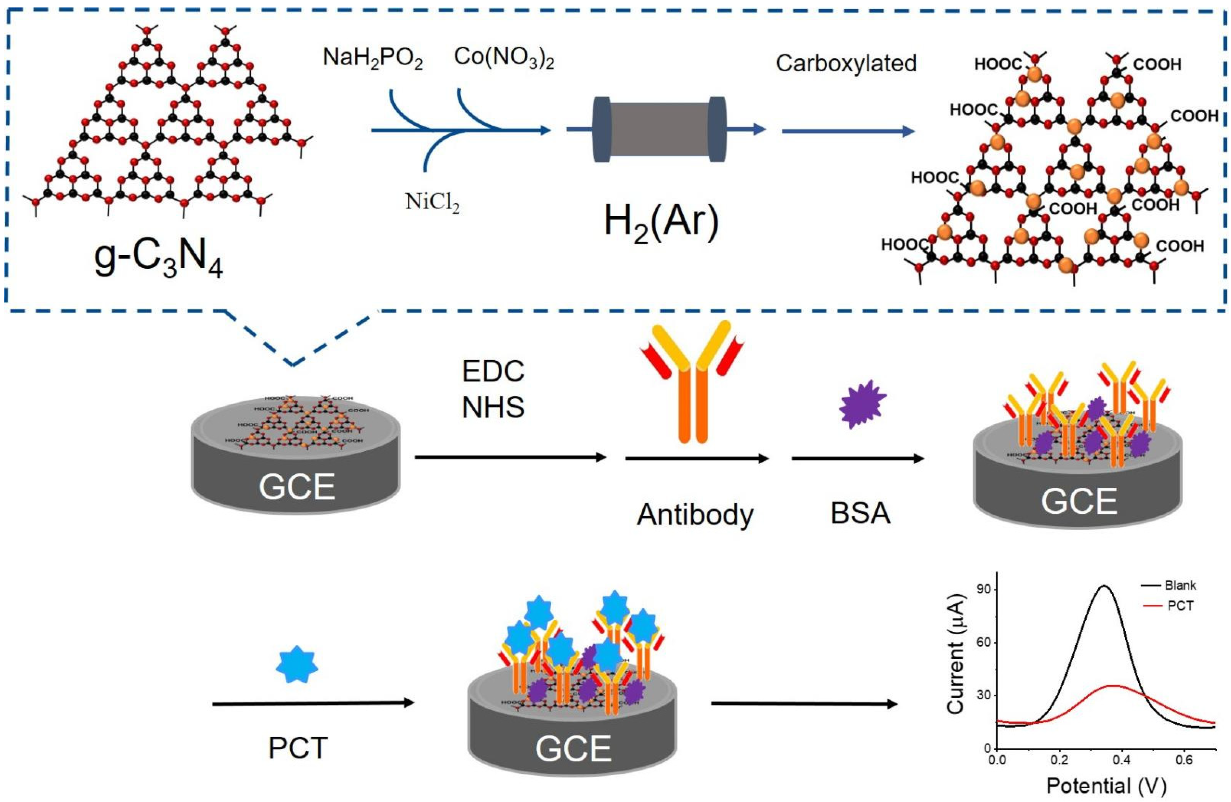
2.3. Synthesis of g-C3N4 and NiCoP/g-C3N4 Nanocomposite

The preparation method of g-C3N4 followed previously published protocols [33]. Briefly, urea was put in a crucible with a cover under ambient pressure in air. When heated to 550 °C at a ramp rate of 5 °C/min and maintained in an Argon environment for another 3 h, the yellow powder was obtained.
The synthesis of NiCoP/g-C3N4 composite also followed previously published protocols [34]. Briefly, a quantity of NiCl2·6H2O (50 mg), CoH12N2O12 (50 mg) and Sodium NaH2PO2·H2O (50 mg) was dissolved in 10 mL Milli-Q water, respectively. The amount of 300 mg g-C3N4 was added to each solution and stirred 6 h after sonication for 1 h and dried by vacuum drying. The remaining power was ground and heated at 350 °C in an Argon environment for 2 h. A white powder NiCoP/g-C3N4 nanocomposite was obtained.

2.4. Carboxylation of NiCoP/g-C3N4 Nanocomposites (COOH-NiCoP/g-C3N4)

The 1 g NiCoP/g-C3N4 nanocomposite was ground and dissolved in 100 mL of HNO3. The mixture was heated in an oil bath at 125 °C for one day. After cooling down to room temperature, the solid substance was collected and vacuumed at 35 °C overnight.

2.5. Preparation of PCT Biosensor

The bare glass carbon electrode (GCE) was polished with an alumina slurry and washed with water, then 20 μL COOH-NiCoP/g-C3N4 suspension (1 mg COOH-NiCoP/g-C3N4 dispersed into 1 mL ethanol under sonication for 30 min) was dropped on its surface and incubated for 3 h. After that, 10μL EDC and NHS mixed solution, and 10 μL and 1 μM monoclonal antibody, respectively, were dropped on the electrode surface and incubated at room temperature. Finally, BSA (2%, W/V) was used to cover the non-specified binding sites of the electrode.

2.6. General Characterization

The electrochemical measurement was performed in 5 mM K3Fe(CN)6/K4Fe(CN)6 + 1× Tris-HCl buffer. Three electrodes consisting of a NiCoP/g-C3N4 nanocomposite modified working electrode, platinum wire counter electrode and Ag/AgCl reference electrode were used. FT-IR was collected with KBr pressed as pellets on a Nicolet 6700-IR spectrophotometer in the range 400–4000 cm−1. X-ray diffraction (XRD) patterns were collected on a SmartLab instrument equipped with graphite-monochromatized Cu Ka radiation (λ = 0.1541 nm; scan speed of 6 min−1; 2θ = 10–80°) at room temperature, with XPS using Al Kα radiation as the X-ray source (1486.7 eV) with the pass energy of 30 eV.

2.7. PCT Sensing in Tris-HCl

PCT in a range from 1 ag/mL to 10 ng/mL in 1× Tris-HCl (pH 7.0) was measured by DPV in the 5 mM K3Fe(CN)6/K4Fe(CN)6. The glassy carbon electrode with NiCoP/g-C3N4 modified was acting as a working electrode, the counter electrode was platinum wire, and Ag/AgCl was the reference electrode. The mean and standard deviation were obtained from three replicated tests.

2.8. PCT Detection in Serum Sample

With the approval of the school ethics committee, the human blood samples were obtained from the Affiliated Hospital of Inner Mongolia Minzu University. Serum samples were centrifuged at 5000 rpm for 20 min and filtered with a 0.22 μm membrane prior to use. For the recovery test, the collected serum was diluted 100 times with Tris-HCl buffer, and then PCT was added to obtain the final concentration. The recoveries were determined from samples containing three concentrations of PCT in human serum.

3. Results and Discussion

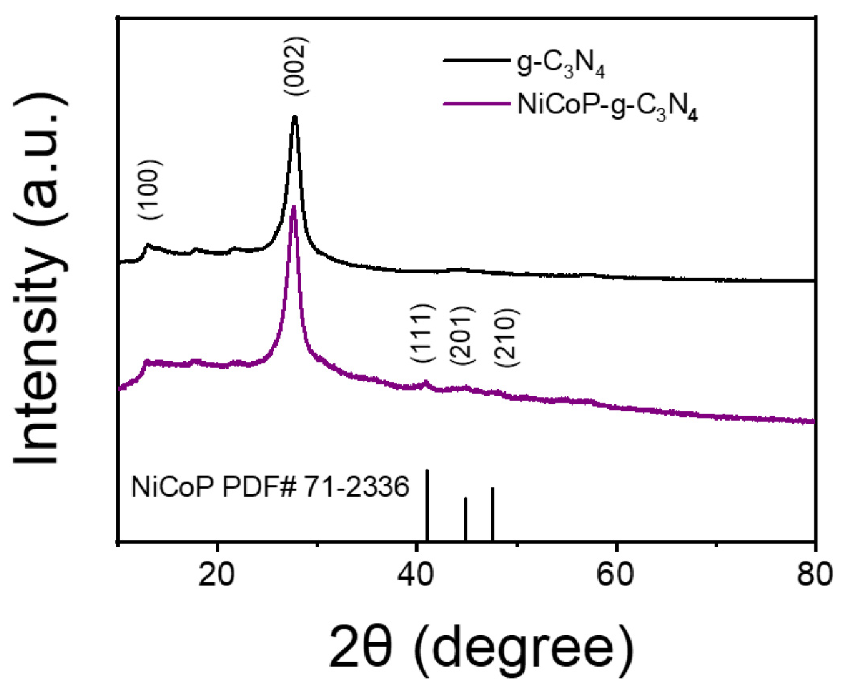
3.1. The Morphology and Structural Information

The morphologies of g-C3N4 and NiCoP/g-C3N4 nanocomposites were investigated by SEM and TEM. For the g-C3N4, they appeared layered, rippled and resembled waves of crumpled silk veil, illustrating the nanosheets structure (Figure 1A,D). Our previous research identified that this special nanosheet structure could provide more active binding sites [35]. For the NiCoP/g-C3N4 nanocomposites, there were several small nanoparticles displayed on the g-C3N4 surface, implying that NiCoP was successfully loaded onto the g-C3N4 surface (Figure 1B,E). The HRTEM image shows distinct lattice fringes with a plane spacing of 0.22 nm and 0.51nm, corresponding to the (111) and (100) planes of NiCoP (Figure 1C) [36,37,38]. In addition, the energy-dispersive X-ray spectroscopy (EDX) mapping image obtained in STEM mode demonstrated the C and N elements distributed in the substrate, and the Ni, Co and P elements throughout the structure (Figure 1F–K), further confirming the presence of NiCoP nanoparticles.
XRD was used to distinguish the phases and the structures of bare g-C3N4 and NiCoP loading. As shown in Figure 2, the lattice planes of triazine units (100) (13.1°) and the lattice planes of interlayer stacking of aromatic segments (002) (27.8°) were detected, indicating the bare g-C3N4 was successfully synthesized [35,39]. For the NiCoP/g-C3N4 samples, beside the strongest diffraction peaks of g-C3N4, several weak peaks appeared at 41°, 44.95° and 47.97°, which correspond to the (111), (201) and (210) lattice planes of NiCoP, respectively [40,41] (JCPDS No. 71-2336). These results implied that the NiCoP nanoparticles had been successfully loaded on the surface of g-C3N4 nanosheets.
Detailed information on the g-C3N4 and NiCoP/g-C3N4 nanocomposites was obtained by XPS. The survey spectra of bare g-C3N4 and NiCoP/g-C3N4 were shown in Figure 3A,D. C, N and O elements in g-C3N4 and the C, N, P, Co and Ni elements in the NiCoP/g-C3N4 sample were detected, respectively. The detected O 1s peaks in the bare g-C3N4 were mainly due to the surface-adsorbed O2 and H2O molecules. By contrast, the O 1s peak of NiCoP/g-C3N4 increased significantly, which may have been caused by the surface oxidation of NiCoP nanoclusters [36]. Four binding energy peaks were shown in the C 1s XPS spectrum of the original g-C3N4 at 284.3, 285.2, 287.5 and 288.2 eV (Figure 3B), which originated from the graphitic, C-N/C-O bindings, cyanide/cyanoquione and heptazine typed carbons, respectively. Four peaks located at 397.97 eV, 399.3 eV, 400.3 eV and 403.4 eV were well-suited for the N 1s XPS spectra of g-C3N4 (Figure 3C), which is attributed to the heptazine N, pyrrolic N, graphitic N and oxidic N, respectively [33]. By comparing the binding energy spectrum after NiCoP deposition, we found the shift in the C 1s and N 1s spectra of NiCoP/g-C3N4 (Figure 3E,F), indicating an internal force between NiCoP and g-C3N4. For the P 2p spectrum (Figure 3G), the binding energy peak was located at 127.7 eV, which was close to the P 2p3/2, indicating the presence of the P element. Moreover, the peak at 131.9 eV could be attributed to the oxidized phosphorus species in contact with air [40,42]. For Co 2p, 777.25 eV and 792.2 eV the binding peaks were attributed to Co 2p3/2 and Co 2p1/2 of metallic Co because of the formation of Co-P [43,44] (Figure 3H). Likewise, for the Ni 2p region, the strong binding energy of 852.25 eV was close to the nickel metal (852.6 eV), which implied the presence of partially charged Ni species (Figure 3I). The XPS results further confirmed that NiCoP nanoparticles had been successfully loaded onto the g-C3N4 surface.
The electrochemical properties of NiCoP/g-C3N4 nanocomposites were evaluated with electrochemical impedance spectroscopy (EIS), with [Fe(CN)6]3−/[Fe(CN)6]4− as the redox probe, and the semicircle diameter was equivalent to the electro-transfer resistance. As shown in Figure 4, compared with the bare g-C3N4, the interface electron transfer resistance (Ret) decreased significantly after the NiCoP/g-C3N4 nanocomposites were modified, indicating that the binding of NiCoP nanoparticles could change the band structure of g-C3N4 and promoted the electron transfer [45].

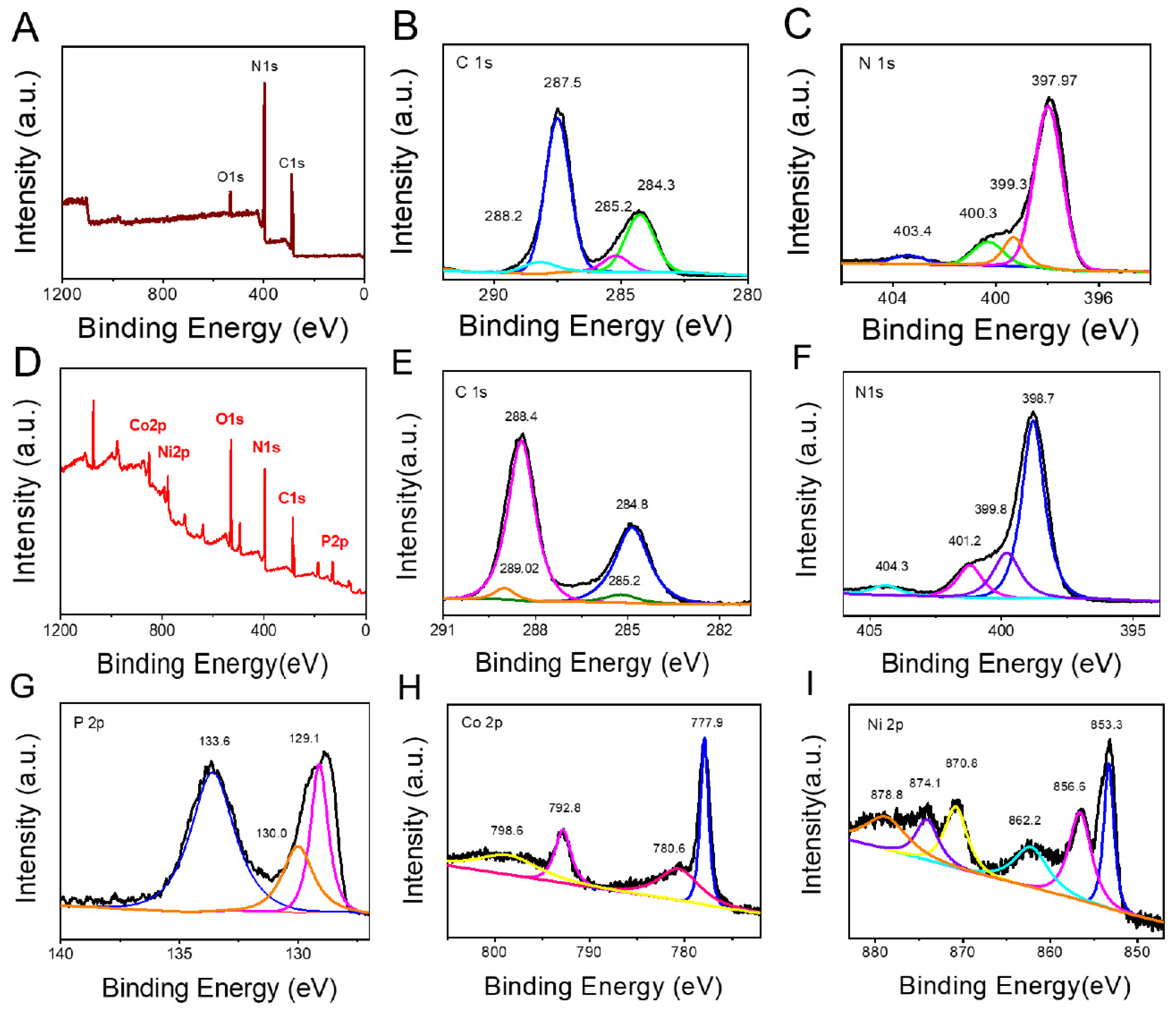
3.2. Characterization of Carboxylic NiCoP/g-C3N4 Nanocomposites

The functionalization of NiCoP/g-C3N4 nanocomposites was investigated using the FT-IR spectrum. The spectral bands at different wavelengths correspond to specific vibrations of molecular functional groups. Figure 5 shows the NiCoP/g-C3N4 before (black line) and after carboxylation (red line) in the wavelength range of 500–2500 cm−1. The band at 806 cm−1 was characteristic of tri-s-triazine, and the bands in the range of 1240–1643 cm−1 could be attributed to the C-N stretching of g-C3N4. Furthermore, the peak at 1380 cm−1 and 1577 cm−1 corresponded to the COO bending band [46]; these results indicated that the surface of NiCoP/g-C3N4 had been successfully carboxylated.

3.3. Biosensor Characterization

Cyclic voltammetry (CV) and EIS were used to verify the electrode surface modification. As shown in Figure 6 A, the bare glass carbon electrode exhibited almost a straight line, illustrating that the electro-transfer process was mainly caused by mass diffusion. The Ret increased after COOH-NiCoP/g-C3N4 nanocomposites and the antibody(Ab) were modified through π-π stacking and a zero-length amine-reactive cross-linker, EDC and NHS, respectively, which can be attributed to the COOH-NiCoP/g-C3N4 nanocomposites. The larger size of Ab blocked or hindered the diffusion of the redox probe of [Fe(CN)6]3−/4− and eventually increased the electron transfer resistance [47]. These results were consistent with those obtained from CV measurements (Figure 6B). The above results showed that the sensing interface had been successfully fabricated.

3.4. Detection Performance of the Electrochemical Immunosensor

The PCT detection performance was evaluated by exposing the sensing platform to the different concentrations of PCT under the same experimental conditions and monitored by DPV. When the PCT was captured, the electron transfer resistance on the electrode surface was enhanced, finally affecting the output of the electrochemical signal. As shown in Figure 7A, in the range of 1 ag/mL to 10 ng/mL, the electrochemical oxidation peak current dropped gradually as the PCT concentration increased. Additionally, the DPV signal and PCT concentrations showed a linear relationship; the linear equation can be described as I(μA) = 88.5–7.82 c (R2 = 0.97) (Figure 7B), and the limit of detection (LOD) was estimated to be 0.6 ag/mL (S/N = 3). The results illustrate that compared with other electrochemical methods, dynamic light scattering (DLS), SPR biosensors and enzyme-free immunosensor methods, as listed in Table 1, our sensor presents a competitive detection limit.
C-reaction protein (CRP) and Interleukin-6 (IL-6) were selected as interfering biomarkers to evaluate the selectivity of this immunosensor. CRP was used to indicate tissue damage inflammation [56], and IL-6 has been linked to various diseases such as inflammatory bowel disease, diabetes, osteoarthritis and asthma [57]. For specific detection, the concentration of PCT, CRP and IL-6 were 1, 10 and 10 pg/mL, respectively. As shown in Figure 7C, for each interfering substance, the DPV value was close to the control sample; however, after mixing with the PCT, the DPV response of the mixture was similar to PCT alone, indicating that our sensor can specifically recognize and capture the PCT. This means that the electrochemical sensor has high specificity and selectivity.

3.5. Application for the Real Sample Analysis

The practical application of NiCoP/g-C3N4 nanocomposites electrochemical PCT immunosensors was evaluated in blood PCT detection. We analyzed the PCT concentrations in human serum samples. The DPV curve in Figure 7D shows the decrease of oxidation peak current as the PCT concentrations rise from 1 pg/mL to 10 pg/mL. Through the linear relationship with the NiCoP/g-C3N4/CGE PCT sensor, the recovery of the spiked PCT (1 pg/mL, 10 pg/mL, 100 pg/mL) in 100-fold diluted human serum samples ranged from 93 to 101.2% (Table 2). These results strongly indicate that this immune sensor has great potential for analyzing PCT in real clinical samples in the future.

4. Conclusions

In this study, an electrochemical immunosensor using NiCoP/g-C3N4 composite as a sensor platform was constructed and utilized for PCT detection. This sensor showed excellent sensing performance, and the analytical performance of this sensing platform had the linearity range of (1 ag/mL to 10 ng/mL (R2 = 0.97)) and LOD of (0.6 ag/mL (S/N = 3)). At the same time, the properties of selectivity and recovery were also investigated. Those results indicated the validity of this sensing platform. In conclusion, our work presents a promising new method for PCT detection that may transform future clinical applications.

Author Contributions

Conceptualization, Y.L.; methodology, Y.L. and W.H.; validation, Y.L. and W.H.; formal analysis, F.C. and L.B.; investigation, F.C., L.B. and Y.Z.; resources, Y.L. and J.L.; data curation, Y.L.; writing—original draft preparation, Y.L.; writing—review and editing, R.W.; supervision, Y.L.; project administration, Y.L.; funding acquisition, Y.L. All authors have read and agreed to the published version of the manuscript.

Funding

This work was supported by the National Natural Science Foundation of China (22104066, 22164016 and 21901025). Innovation and entrepreneurship start-up project for students studying in Inner Mongolia (2021LXCX002). Inner Mongolia Natural Science Foundation (2020BS08007, 2020BS02007). Program for Young Talents of Science and Technology in Universities of Inner Mongolia Autonomous Region (NJYT23034) and Ph.D. Start-up Fund of Inner Mongolia Minzu University (BS515, BS453). And The APC was funded by 22104066.

Institutional Review Board Statement

Not applicable.

Informed Consent Statement

Not applicable.

Data Availability Statement

All data are included within the manuscript.

Conflicts of Interest

The authors declare no conflict of interest.

References

  1. Fang, J.; Li, J.; Feng, R.; Yang, L.; Zhao, L.; Zhang, N.; Zhao, G.; Yue, Q.; Wei, Q.; Cao, W. Dual-quenching electrochemiluminescence system based on novel acceptor CoOOH@Au NPs for early detection of procalcitonin. Sens. Actuators B Chem. 2021, 332, 129544. [Google Scholar] [CrossRef]
  2. Fang, Y.S.; Wang, H.Y.; Wang, L.S.; Wang, J.F. Electrochemical immunoassay for procalcitonin antigen detection based on signal amplification strategy of multiple nanocomposites. Biosens. Bioelectron. 2014, 51, 310–316. [Google Scholar] [CrossRef] [PubMed]
  3. BalcI, C.; Sungurtekin, H.; Gürses, E.; Sungurtekin, U.; Kaptanoğlu, B.J. Usefulness of procalcitonin for diagnosis of sepsis in the intensive care unit. Crit. Care 2002, 7, 1–6. [Google Scholar] [CrossRef] [PubMed]
  4. Gendrel, D.; Assicot, M.; Raymond, J.; Moulin, F.; Francoual, C.; Badoual, J.; Bohuon, C. Procalcitonin as a marker for the early diagnosis of neonatal infection. J. Pediatr. 1996, 128, 570–573. [Google Scholar] [CrossRef] [PubMed]
  5. Schröder, J.; Staubach, K.-H.; Zabel, P.; Stüber, F.; Kremer, B. Procalcitonin as a marker of severity in septic shock. Langenbeck’s Arch. Surg. 1999, 384, 33–38. [Google Scholar] [CrossRef]
  6. Liao, T.; Yuan, F.; Yu, H.; Li, Z. An ultrasensitive ELISA method for the detection of procalcitonin based on magnetic beads and enzyme-antibody labeled gold nanoparticles. Anal. Methods 2016, 8, 1577–1585. [Google Scholar] [CrossRef]
  7. Chiang, C.Y.; Huang, T.T.; Wang, C.H.; Huang, C.J.; Tsai, T.H.; Yu, S.N.; Chen, Y.T.; Hong, S.W.; Hsu, C.W.; Chang, T.C.; et al. Fiber optic nanogold-linked immunosorbent assay for rapid detection of procalcitonin at femtomolar concentration level. Biosens. Bioelectron. 2020, 151, 111871. [Google Scholar] [CrossRef]
  8. Buchegger, P.; Preininger, C. Four assay designs and on-chip calibration: Gadgets for a sepsis protein array. Anal. Chem. 2014, 86, 3174–3180. [Google Scholar] [CrossRef]
  9. Buchegger, P.; Sauer, U.; Toth-Székély, H.; Preininger, C. Miniaturized Protein Microarray with Internal Calibration as Point-of-Care Device for Diagnosis of Neonatal Sepsis. Sensors 2012, 12, 1494–1508. [Google Scholar] [CrossRef]
  10. Marcello, A.; Sblattero, D.; Cioarec, C.; Maiuri, P.; Melpignano, P. A deep-blue OLED-based biochip for protein microarray fluorescence detection. Biosens. Bioelectron. 2013, 46, 44–47. [Google Scholar] [CrossRef]
  11. Li, H.; Sun, Y.; Elseviers, J.; Muyldermans, S.; Liu, S.; Wan, Y. A nanobody-based electrochemiluminescent immunosensor for sensitive detection of human procalcitonin. Analyst 2014, 139, 3718–3721. [Google Scholar] [CrossRef]
  12. Song, C.; Li, X.; Hu, L.; Shi, T.; Wu, D.; Ma, H.; Zhang, Y.; Fan, D.; Wei, Q.; Ju, H. Quench-type electrochemiluminescence immunosensor based on resonance energy transfer from carbon nanotubes and Au-nanoparticles-enhanced g-C3N4 to CuO@ Polydopamine for procalcitonin detection. ACS Appl. Mater. Interfaces 2020, 12, 8006–8015. [Google Scholar] [CrossRef]
  13. Shao, X.; Song, X.; Liu, X.; Yan, L.; Liu, L.; Fan, D.; Wei, Q.; Ju, H. A dual signal-amplified electrochemiluminescence immunosensor based on core-shell CeO2-Au@Pt nanosphere for procalcitonin detection. Microchim. Acta 2021, 188, 1–9. [Google Scholar] [CrossRef]
  14. Sener, G.; Ozgur, E.; Rad, A.Y.; Uzun, L.; Say, R.; Denizli, A. Rapid real-time detection of procalcitonin using a microcontact imprinted surface plasmon resonance biosensor. Analyst 2013, 138, 6422–6428. [Google Scholar] [CrossRef]
  15. Vashist, S.K.; Schneider, E.M.; Barth, E.; Luong, J.H.T. Surface plasmon resonance-based immunoassay for procalcitonin. Anal. Chim. Acta 2016, 938, 129–136. [Google Scholar] [CrossRef]
  16. Sun, L.L.; Leo, Y.S.; Zhou, X.; Ng, W.; Wong, T.I.; Deng, J. Localized surface plasmon resonance based point-of-care system for sepsis diagnosis. Mater. Sci. Energy Technol. 2020, 3, 274–281. [Google Scholar] [CrossRef]
  17. Ge, X.Y.; Zhang, J.X.; Feng, Y.G.; Wang, A.J.; Mei, L.P.; Feng, J.J. Label-free electrochemical biosensor for determination of procalcitonin based on graphene-wrapped Co nanoparticles encapsulated in carbon nanobrushes coupled with AuPtCu nanodendrites. Microchim. Acta 2022, 189, 1–13. [Google Scholar] [CrossRef]
  18. Gupta, Y.; Pandey, C.M.; Ghrera, A.S. Development of conducting cellulose paper for electrochemical sensing of procalcitonin. Microchim. Acta 2022, 190, 32. [Google Scholar] [CrossRef]
  19. De Oliveira, P.R.; Crapnell, R.D.; Ferrari, A.G.M.; Wuamprakhon, P.; Hurst, N.J.; Dempsey-Hibbert, N.C.; Sawangphruk, M.; Janegitz, B.C.; Banks, C.E. Low-cost, facile droplet modification of screen-printed arrays for internally validated electrochemical detection of serum procalcitonin. Biosens. Bioelectron. 2023, 228, 115220. [Google Scholar] [CrossRef]
  20. Chen, Y.X.; Wu, X.; Huang, K.J. A sandwich-type electrochemical biosensing platform for microRNA-21 detection using carbon sphere-MoS2 and catalyzed hairpin assembly for signal amplification. Sens. Actuators B Chem. 2018, 270, 179–186. [Google Scholar] [CrossRef]
  21. Wang, H.; Ma, Y.; Guo, C.; Yang, Y.; Peng, Z.; Liu, Z.; Zhang, Z. Templated seed-mediated derived Au nanoarchitectures embedded with nanochitosan: Sensitive electrochemical aptasensor for vascular endothelial growth factor and living MCF-7 cell detection. Appl. Surf. Sci. 2019, 481, 505–514. [Google Scholar] [CrossRef]
  22. Li, M.; He, B. Ultrasensitive sandwich-type electrochemical biosensor based on octahedral gold nanoparticles modified poly (ethylenimine) functionalized graphitic carbon nitride nanosheets for the determination of sulfamethazine. Sens. Actuators B Chem. 2021, 329, 129158. [Google Scholar] [CrossRef]
  23. Zhang, X.; Xie, X.; Wang, H.; Zhang, J.; Pan, B.; Xie, Y. Enhanced Photoresponsive Ultrathin Graphitic-Phase C3N4 Nanosheets for Bioimaging. J. Am. Chem. Soc. 2013, 135, 18–21. [Google Scholar] [CrossRef] [PubMed]
  24. Feng, Q.M.; Shen, Y.Z.; Li, M.X.; Zhang, Z.L.; Zhao, W.; Xu, J.J.; Chen, H.Y. Dual-Wavelength Electrochemiluminescence Ratiometry Based on Resonance Energy Transfer between Au Nanoparticles Functionalized g-C3N4 Nanosheet and Ru(bpy)3(2+) for microRNA Detection. Anal. Chem. 2016, 88, 937–944. [Google Scholar] [CrossRef]
  25. Wang, Y.Z.; Hao, N.; Feng, Q.M.; Shi, H.W.; Xu, J.J.; Chen, H.Y. A ratiometric electrochemiluminescence detection for cancer cells using g-C3N4 nanosheets and Ag-PAMAM-luminol nanocomposites. Biosens. Bioelectron. 2016, 77, 76–82. [Google Scholar] [CrossRef]
  26. Medetalibeyoglu, H.; Beytur, M.; Akyıldırım, O.; Atar, N.; Yola, M.L. Validated electrochemical immunosensor for ultra-sensitive procalcitonin detection: Carbon electrode modified with gold nanoparticles functionalized sulfur doped MXene as sensor platform and carboxylated graphitic carbon nitride as signal amplification. Sens. Actuators B Chem. 2020, 319, 128195. [Google Scholar] [CrossRef]
  27. Zou, J.; Wu, S.; Liu, Y.; Sun, Y.; Cao, Y.; Hsu, J.-P.; Shen Wee, A.T.; Jiang, J. An ultra-sensitive electrochemical sensor based on 2D g-C3N4/CuO nanocomposites for dopamine detection. Carbon 2018, 130, 652–663. [Google Scholar] [CrossRef]
  28. Lin, Y.; Pan, Y.; Liu, S.; Sun, K.; Cheng, Y.; Liu, M.; Wang, Z.; Li, X.; Zhang, J. Construction of multi-dimensional core/shell Ni/NiCoP nano-heterojunction for efficient electrocatalytic water splitting. Appl. Catal. B Environ. 2019, 259, 118039. [Google Scholar] [CrossRef]
  29. Li, J.; Yan, M.; Zhou, X.; Huang, Z.Q.; Xia, Z.; Chang, C.R.; Ma, Y.; Qu, Y. Mechanistic insights on ternary Ni2−xCoxP for hydrogen evolution and their hybrids with graphene as highly efficient and robust catalysts for overall water splitting. Adv. Funct. Mater. 2016, 26, 6785–6796. [Google Scholar] [CrossRef]
  30. Li, Y.; Liu, J.; Chen, C.; Zhang, X.; Chen, J. Preparation of NiCoP hollow quasi-polyhedra and their electrocatalytic properties for hydrogen evolution in alkaline solution. ACS Appl. Mater. Interfaces 2017, 9, 5982–5991. [Google Scholar] [CrossRef]
  31. Wang, Z.; Cao, X.; Liu, D.; Hao, S.; Du, G.; Asiri, A.M.; Sun, X. Ternary NiCoP nanosheet array on a Ti mesh: A high-performance electrochemical sensor for glucose detection. Chem. Commun. 2016, 52, 14438–14441. [Google Scholar] [CrossRef]
  32. Bi, L.; Gao, X.; Zhang, L.; Wang, D.; Zou, X.; Xie, T. Enhanced Photocatalytic Hydrogen Evolution of NiCoP/g-C3N4 with Improved Separation Efficiency and Charge Transfer Efficiency. ChemSusChem 2018, 11, 276–284. [Google Scholar] [CrossRef]
  33. Liu, J.; Xie, S.; Geng, Z.; Huang, K.; Fan, L.; Zhou, W.; Qiu, L.; Gao, D.; Ji, L.; Duan, L.; et al. Carbon Nitride Supramolecular Hybrid Material Enabled High-Efficiency Photocatalytic Water Treatments. Nano Lett. 2016, 16, 6568–6575. [Google Scholar] [CrossRef]
  34. Saravanan, S.P.; Nagoor Meeran, M. Increasing the efficiency of dye-sensitized solar cells by NiCoP/g-C3N4 hybrid composite photoanodes by facile hydrothermal approach. Ionics 2021, 27, 407–416. [Google Scholar] [CrossRef]
  35. Liu, Y.; Chen, F.; Bao, L.; Hai, W. Construction of a non-enzymatic electrochemical sensor based on graphitic carbon nitride nanosheets for sensitive detection of procalcitonin. RSC Adv. 2022, 12, 22518–22525. [Google Scholar] [CrossRef]
  36. Li, C.; Wu, H.; Du, Y.; Xi, S.; Dong, H.; Wang, S.; Wang, Y. Mesoporous 3D/2D NiCoP/g-C3N4 heterostructure with dual Co–N and Ni–N bonding states for boosting photocatalytic H2 production activity and stability. ACS Sustain. Chem. Eng. 2020, 8, 12934–12943. [Google Scholar] [CrossRef]
  37. Qin, Z.; Chen, Y.; Huang, Z.; Su, J.; Guo, L. A bifunctional NiCoP-based core/shell cocatalyst to promote separate photocatalytic hydrogen and oxygen generation over graphitic carbon nitride. J. Mater. Chem. 2017, 5, 19025–19035. [Google Scholar] [CrossRef]
  38. Xing, W.; Tu, W.; Han, Z.; Hu, Y.; Meng, Q.; Chen, G. Template-induced high-crystalline g-C3N4 nanosheets for enhanced photocatalytic H2 evolution. ACS Energy Lett. 2018, 3, 514–519. [Google Scholar] [CrossRef]
  39. Ge, L. Synthesis and photocatalytic performance of novel metal-free g-C3N4 photocatalysts. Mater. Lett. 2011, 65, 2652–2654. [Google Scholar] [CrossRef]
  40. Zhu, Y.P.; Liu, Y.P.; Ren, T.Z.; Yuan, Z.Y. Self-Supported Cobalt Phosphide Mesoporous Nanorod Arrays: A Flexible and Bifunctional Electrode for Highly Active Electrocatalytic Water Reduction and Oxidation. Adv. Funct. Mater. 2015, 25, 7337–7347. [Google Scholar] [CrossRef]
  41. Wang, X.; Tong, R.; Wang, Y.; Tao, H.; Zhang, Z.; Wang, H. Surface Roughening of Nickel Cobalt Phosphide Nanowire Arrays/Ni Foam for Enhanced Hydrogen Evolution Activity. ACS Appl. Mater. Interfaces 2016, 8, 34270–34279. [Google Scholar] [CrossRef] [PubMed]
  42. Jin, C.; Xu, C.; Chang, W.; Ma, X.; Hu, X.; Liu, E.; Fan, J. Bimetallic phosphide NiCoP anchored g-C3N4 nanosheets for efficient photocatalytic H2 evolution. J. Alloys Compd. 2019, 803, 205–215. [Google Scholar] [CrossRef]
  43. Cao, S.; Chen, Y.; Hou, C.C.; Lv, X.J.; Fu, W.F. Cobalt phosphide as a highly active non-precious metal cocatalyst for photocatalytic hydrogen production under visible light irradiation. J. Mater. Chem. A 2015, 3, 6096–6101. [Google Scholar] [CrossRef]
  44. Li, Y.; Zhang, H.; Jiang, M.; Kuang, Y.; Sun, X.; Duan, X. Ternary NiCoP nanosheet arrays: An excellent bifunctional catalyst for alkaline overall water splitting. Nano Res. 2016, 9, 2251–2259. [Google Scholar] [CrossRef]
  45. Li, K.; Zhang, Y.; Lin, Y.Z.; Wang, K.; Liu, F.T. Versatile functional porous cobalt–nickel phosphide–carbon cocatalyst derived from a metal–organic framework for boosting the photocatalytic activity of graphitic carbon nitride. ACS Appl. Mater. Interfaces 2019, 11, 28918–28927. [Google Scholar] [CrossRef]
  46. Li, J.; Wang, H.; Guo, Z.; Wang, Y.; Ma, H.; Ren, X.; Du, B.; Wei, Q. A “turn-off” fluorescent biosensor for the detection of mercury (II) based on graphite carbon nitride. Talanta 2017, 162, 46–51. [Google Scholar] [CrossRef]
  47. Zhang, S.; Xia, J.; Li, X. Electrochemical Biosensor for Detection of Adenosine Based on Structure-Switching Aptamer and Amplification with Reporter Probe DNA Modified Au Nanoparticles. Anal. Chem. 2008, 80, 8382–8388. [Google Scholar] [CrossRef]
  48. Zhu, K.; Chen, J.; Hu, J.; Xiong, S.; Zeng, L.; Huang, X.; Xiong, Y. Low-sample-consumption and ultrasensitive detection of procalcitonin by boronate affinity recognition-enhanced dynamic light scattering biosensor. Biosens. Bioelectron. 2022, 200, 113914. [Google Scholar] [CrossRef]
  49. Huang, E.; Huang, D.; Wang, Y.; Cai, D.; Luo, Y.; Zhong, Z.; Liu, D. Active droplet-array microfluidics-based chemiluminescence immunoassay for point-of-care detection of procalcitonin. Biosens. Bioelectron. 2022, 195, 113684. [Google Scholar] [CrossRef]
  50. Yue, Q.; Li, X.; Xu, X.; Huang, X.; Cao, D.; Wei, Q.; Cao, W. Ratiometric electrochemical immunoassay for procalcitonin based on dual signal probes: Ag NPs and Nile blue A. Microchim. Acta 2022, 189, 126. [Google Scholar] [CrossRef]
  51. Xu, X.X.; Lei, X.L.; Ye, L.Y.; Song, S.S.; Liu, L.L.; Xu, L.G.; Xu, C.L.; Kuang, H. Gold-based paper sensor for sensitive detection of procalcitonin in clinical samples. Chin. J. Anal. Chem. 2022, 50, 100062. [Google Scholar] [CrossRef]
  52. Huang, J.; Cheng, W.; Li, Y. 3D carbonized wood-based integrated electrochemical immunosensor for ultrasensitive detection of procalcitonin antigen. Talanta 2022, 238, 122991. [Google Scholar] [CrossRef]
  53. Battaglia, F.; Baldoneschi, V.; Meucci, V.; Intorre, L.; Minunni, M.; Scarano, S. Detection of canine and equine procalcitonin for sepsis diagnosis in veterinary clinic by the development of novel MIP-based SPR biosensors. Talanta 2021, 230, 122347. [Google Scholar] [CrossRef]
  54. Li, S.; Xing, Z.; Feng, J.; Yan, L.; Wei, D.; Wang, H.; Wu, D.; Ma, H.; Fan, D.; Wei, Q. A sensitive biosensor of CdS sensitized BiVO4/GaON composite for the photoelectrochemical immunoassay of procalcitonin. Sens. Actuators B Chem. 2021, 329, 129244. [Google Scholar] [CrossRef]
  55. Jin, Y.; Wu, J.; Hu, D.; Zhang, K.; Deng, S.; Yang, L.; Hao, Y.; Wang, X.; Liu, Y.; Liu, H.; et al. Development of enzyme-free immunosensor based on nanobrush and fluorescence dye for sensitive detection of procalcitonin. Dye. Pigment. 2021, 193, 109548. [Google Scholar] [CrossRef]
  56. Tanak, A.S.; Jagannath, B.; Tamrakar, Y.; Muthukumar, S.; Prasad, S. Non-faradaic electrochemical impedimetric profiling of procalcitonin and C-reactive protein as a dual marker biosensor for early sepsis detection. Anal. Chim. Acta X 2019, 3, 100029. [Google Scholar] [CrossRef]
  57. Wu, J.; Xianyu, Y.; Wang, X.; Hu, D.; Zhao, Z.; Lu, N.; Xie, M.; Lei, H.; Chen, Y. Enzyme-Free Amplification Strategy for Biosensing Using Fe3+–Poly(glutamic acid) Coordination Chemistry. Anal. Chem. 2018, 90, 4725–4732. [Google Scholar] [CrossRef]
Scheme 1. Schematic illustration of the NiCoP/g-C3N4 nanocomposites-based electrochemical immunosensor for detecting PCT. The preparation process of the NiCoP/g-C3N4 and COOH-NiCoP/g-C3N4 was shown in dashed line box.
Sensors 23 04348 sch001
Figure 1. (A) SEM image of g-C3N4, (B) TEM image of NiCoP/g-C3N4, (C) Characteristic HRTEM image for a typical nanoparticle, (D) TEM image of g-C3N4. (E) SEM image of NiCoP/g-C3N4. (F) High-resolution STEM image, and elemental mappings of C (G), N (H), P (I), Co (J), Ni (K).
Sensors 23 04348 g001
Figure 2. XRD patterns of g-C3N4 and NiCoP/g-C3N4 nanocomposites.
Sensors 23 04348 g002
Figure 3. (A) The full XPS spectrum of g-C3N4. (B) The related high-resolution spectra of C 1s. (C) The related high-resolution spectra of N 1s. (D) The full XPS spectrum of NiCoP/g-C3N4. (E) The related high-resolution spectra of C1s. (F) The related high-resolution spectra of N 1s. (G) The related high-resolution spectra of P 2p. (H) The related high-resolution spectra of Co 2p. (I) The related high-resolution spectra of Ni 2p.
Sensors 23 04348 g003
Figure 4. EIS spectrum of g-C3N4 and NiCoP/g-C3N4 composites. The insert is the EIS spectrum of bare electrode.
Sensors 23 04348 g004
Figure 5. FT-IR spectroscopy of carboxylated NiCoP/g-C3N4.
Sensors 23 04348 g005
Figure 6. EIS spectrum (A) and CV (B) responses corresponding to electrode surface modification. Detection buffer: 1 mL 0.1M Tris-HCl containing 5 mM [Fe(CN)6]3−/4−. The insert is the EIS spectrum of bare electrode.
Sensors 23 04348 g006
Figure 7. (A) The DPV signals of the biosensor incubated with differential concentrations of PCT. Insert: linear relationship between the increased peak current and the concentration of PCT. (B) Linear plot characteristics of ΔI of sensor versus the concentration of PCT in PBS buffer. (C) Sensor selectivity: CRP (10 pg/mL), IL-6 (10 pg/mL), PCT (1 pg/mL). (D) DPV curves showing continuous monitoring for PCT concentration in the human sample using NiCoP/g-C3N4/GCE.
Sensors 23 04348 g007
Table 1. Comparison of the proposed method with previous reports on PCT detection.
MethodLinear RangeLODRefs.
Electrochemical0.0001 to 100 ng/mL0.11 pg/mL[17]
Dynamic light scattering (DLS)0.035 to 250 pg/mL0.03 pg/mL[48]
Chemiluminescence immunoassay0.044 to 100 ng/mL0.044 ng/mL[49]
Electrochemical immunoassay0.005 to 50 ng/mL1.67 pg/mL[50]
Immunochromatographic assay0.49 to 13.90 ng/mL0.1 ng/mL[51]
Electrochemical immunosensor0.05 to 90 pg/mL0.014 pg/mL[52]
SPR biosensors25 to 1000 ng/mL30 ng/mL[53]
Photoelectrochemical immunoassay0.1 pg/mL to 50 ng/mL0.03 pg/mL[54]
Enzyme-free immunosensor0.25 to 250 ng/mL0.022 ng/mL[55]
Electrochemiluminescence0.014 pg/mL to 40 ng/mL3.4 fg/mL[1]
Electrochemical immunosensor1 ag/mL to 10 ng/mL0.6 ag/mLThis work
Table 2. Recovery rate of PCT in human serum samples.
SamplesAdded (pg/mL)Found (pg/mL)Recovery (%)RSD (%) *
110.93932.36
2109.6796.71.54
3100101.2101.23.15
* RSD (%) was determined from triple parallel experiments.
Disclaimer/Publisher’s Note: The statements, opinions and data contained in all publications are solely those of the individual author(s) and contributor(s) and not of MDPI and/or the editor(s). MDPI and/or the editor(s) disclaim responsibility for any injury to people or property resulting from any ideas, methods, instructions or products referred to in the content.

