

DNA-Based Gold Nanoparticle Assemblies: From Structure Constructions to Sensing Applications
Abstract
:1. Introduction
2. DNA-Based Assembly of Gold Nanoparticles
2.1. Gold Nanoparticle Multimers
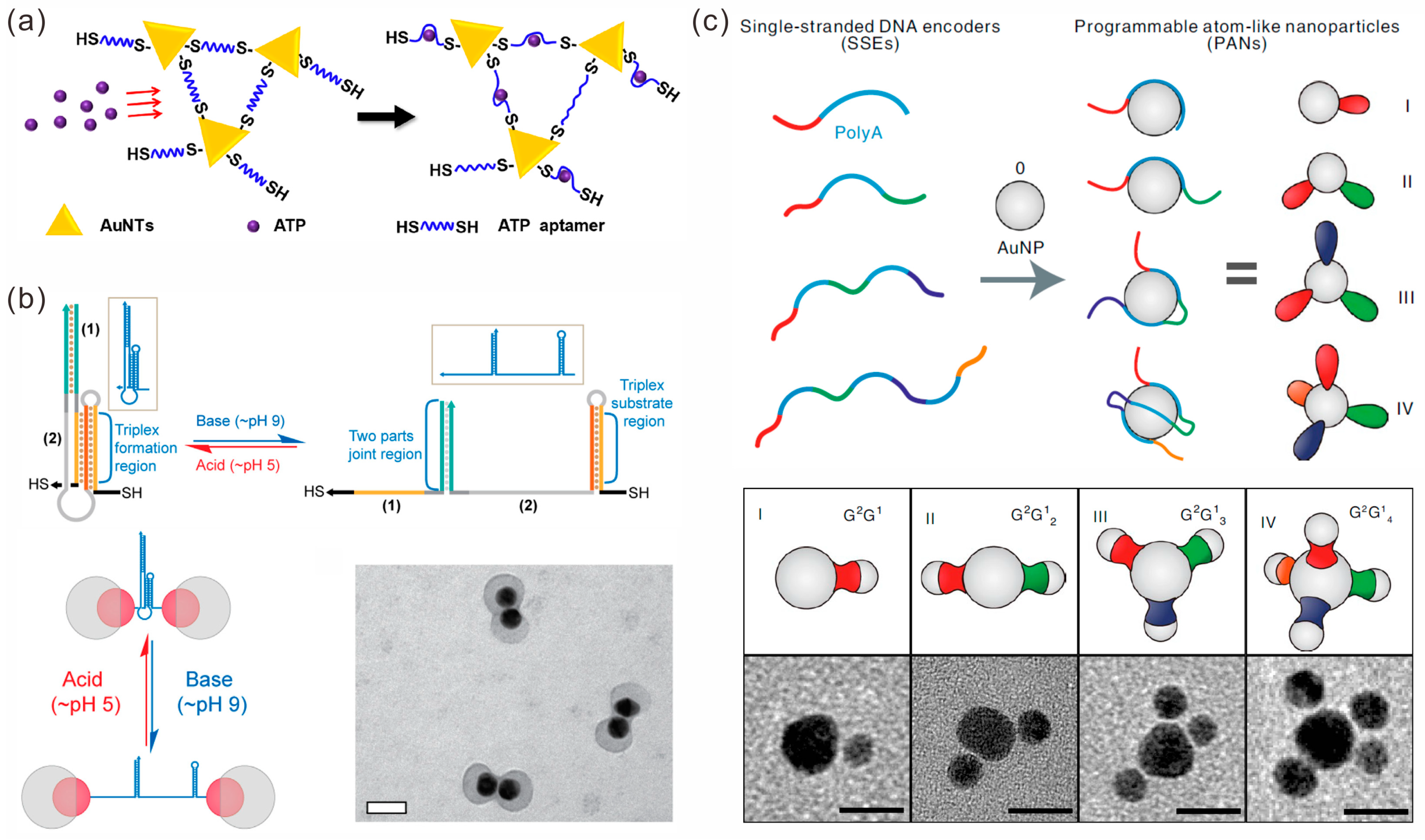
2.1.1. DNA Strand-Mediated Assembly
2.1.2. DNA Tile-Mediated Assembly
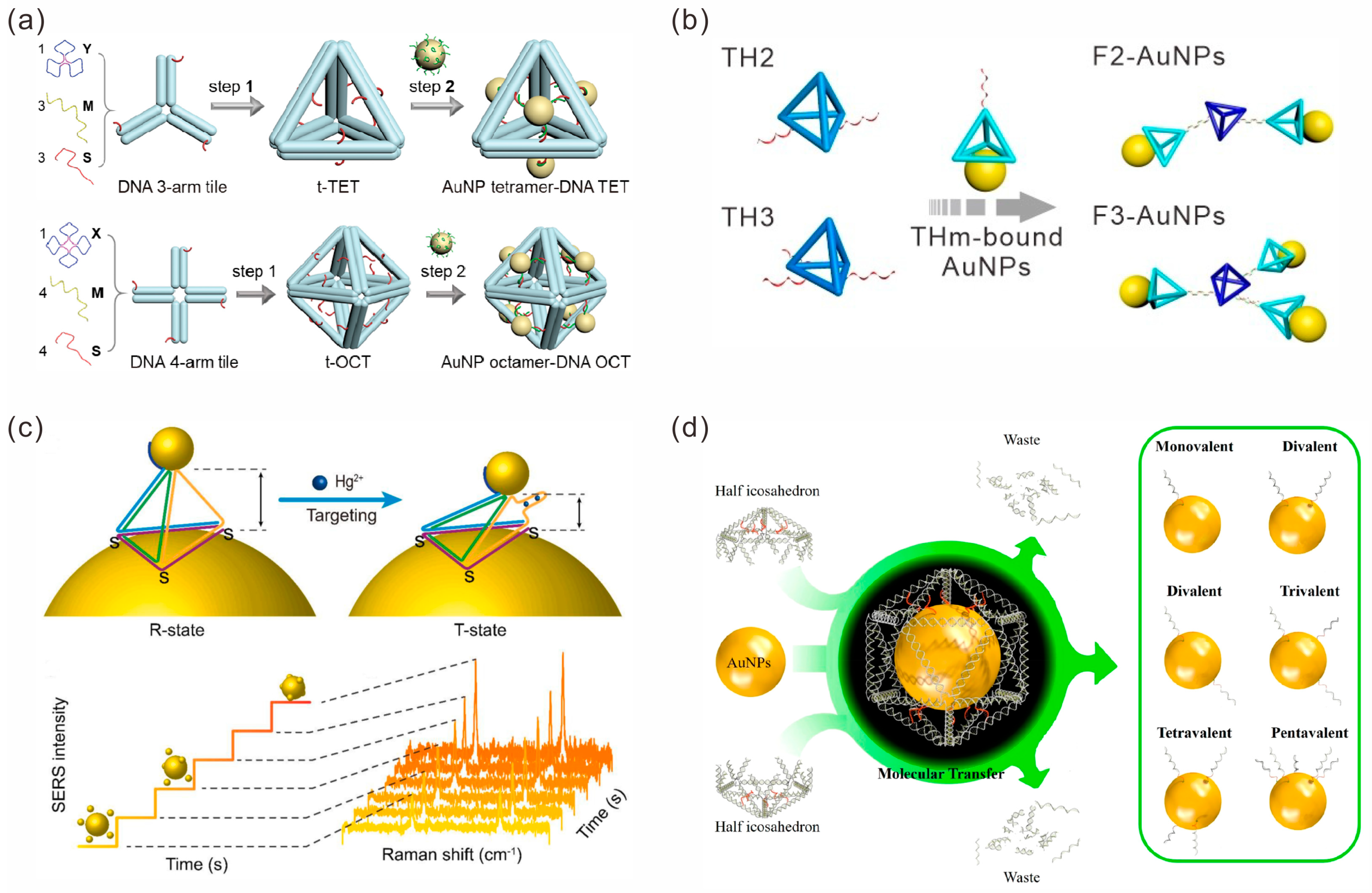
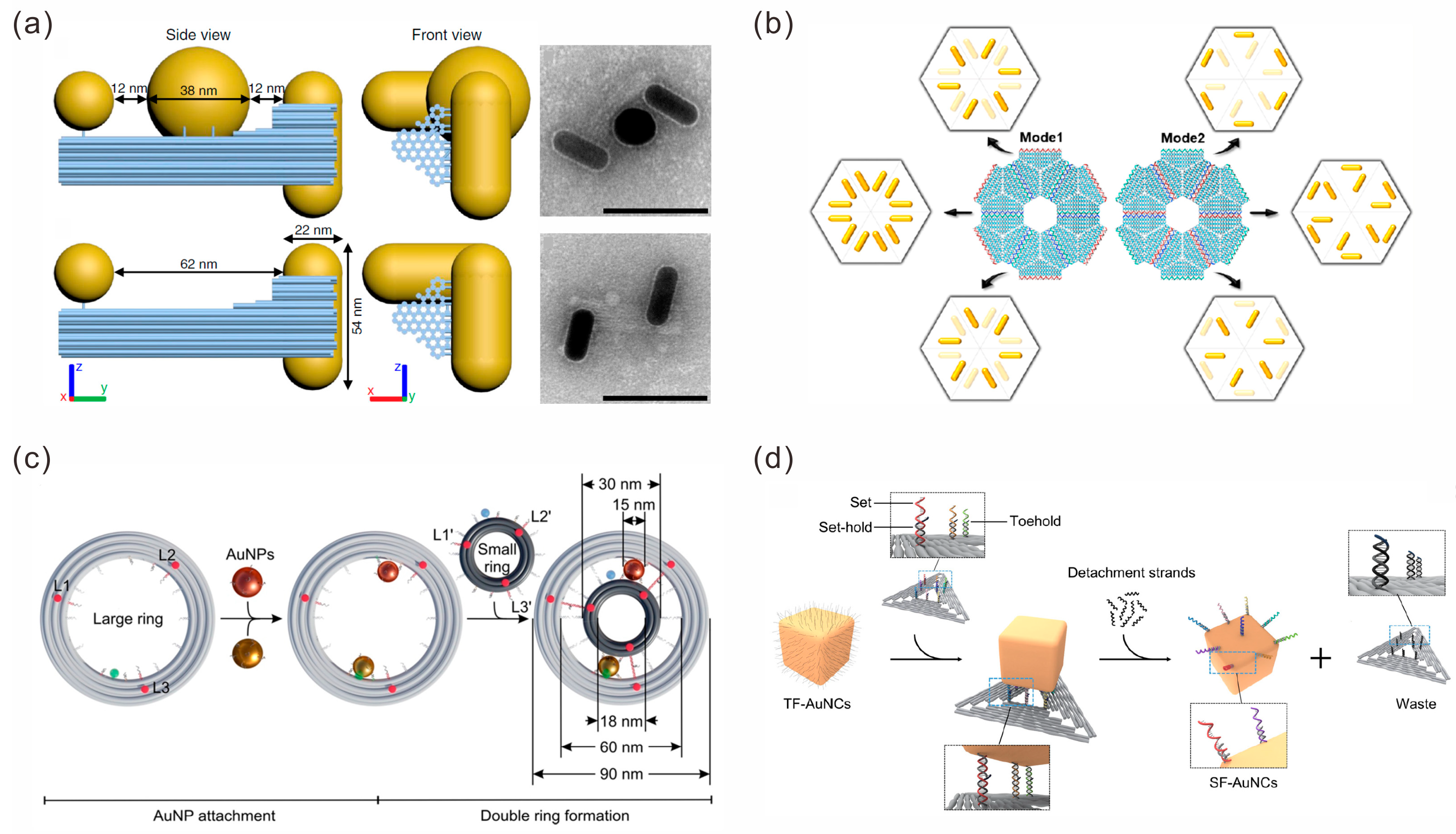
2.1.3. DNA Origami-Mediated Assembly
2.2. Gold Nanoparticle Arrays
2.2.1. 1D Arrays
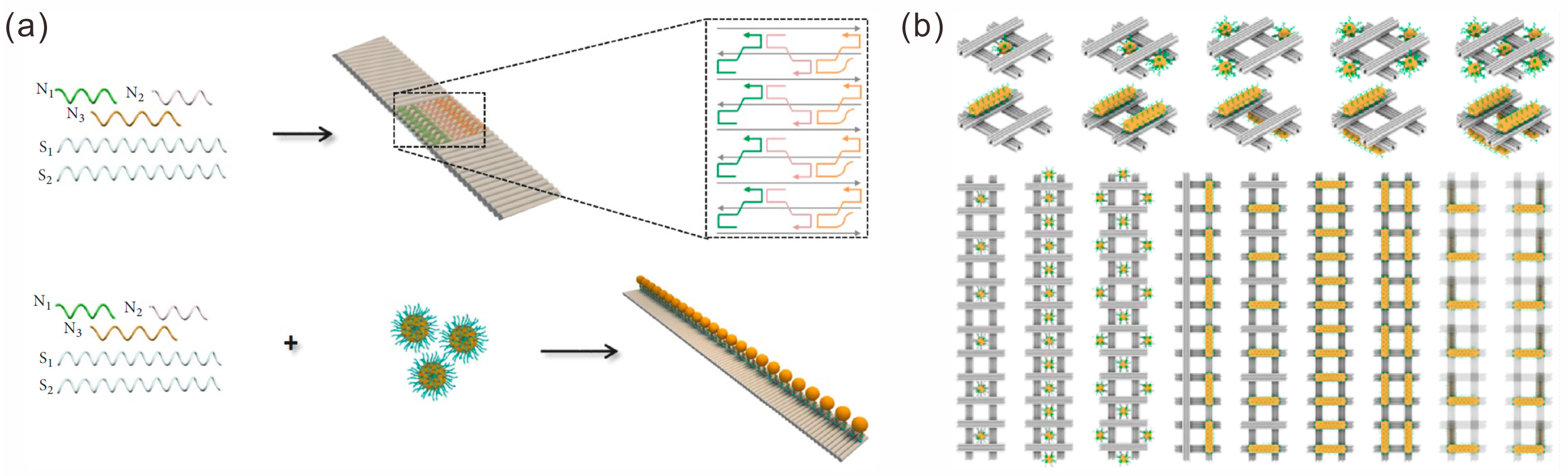
2.2.2. 2D Arrays
2.2.3. 3D Lattices
3. Sensing Applications of Gold Nanoparticle Assembly Structures
3.1. Fluorescence-Based Sensors
3.2. Chiral Sensors

3.3. SERS-Based Sensors
3.4. Other Sensors
4. Conclusions
Funding
Institutional Review Board Statement
Informed Consent Statement
Data Availability Statement
Conflicts of Interest
References
- Yaqoob, S.B.; Adnan, R.; Khan, R.M.R.; Rashid, M. Gold, Silver, and Palladium Nanoparticles: A Chemical Tool for Biomedical Applications. Front. Chem. 2020, 8, 376. [Google Scholar] [CrossRef] [PubMed]
- Jamkhande, P.G.; Ghule, N.W.; Bamer, A.H.; Kalaskar, M.G. Metal nanoparticles synthesis: An overview on methods of preparation, advantages and disadvantages, and applications. J. Drug Deliv. Sci. Tec. 2019, 53, 101174. [Google Scholar] [CrossRef]
- Sajid, M.; Plotka-Wasylka, J. Nanoparticles: Synthesis, characteristics, and applications in analytical and other sciences. Microchem. J. 2020, 154, 104623. [Google Scholar] [CrossRef]
- Sotnikov, D.V.; Berlina, A.N.; Ivanov, V.S.; Zherdev, A.V.; Dzantiev, B.B. Adsorption of proteins on gold nanoparticles: One or more layers? Colloids Surf. B 2019, 173, 557–563. [Google Scholar] [CrossRef] [PubMed]
- Chew, A.K.; Dallin, B.C.; Van Lehn, R.C. The Interplay of Ligand Properties and Core Size Dictates the Hydrophobicity of Monolayer-Protected Gold Nanoparticles. ACS Nano 2021, 15, 4534–4545. [Google Scholar] [CrossRef]
- Li, C.F.; Wang, C.L.; Ji, Z.Y.; Jiang, N.K.; Lin, W.H.; Li, D.X. Synthesis of thiol-terminated thermoresponsive polymers and their enhancement effect on optical limiting property of gold nanoparticles. Eur. Polym. J. 2019, 113, 404–410. [Google Scholar] [CrossRef]
- Fang, W.N.; Wang, J.M.; Lu, S.; Gu, Q.Y.; He, X.; Wang, F.; Wang, L.H.; Tian, Y.; Liu, H.J.; Fan, C.H. Encoding Morphogenesis of Quasi-Triangular Gold Nanoprisms with DNA. Angew. Chem. Int. Ed. 2022, 61, e202208688. [Google Scholar] [CrossRef]
- Kim, S.; Yoon, S. On the Origin of the Plasmonic Properties of Gold Nanoparticles. Bull. Korean Chem. Soc. 2021, 42, 1058–1065. [Google Scholar] [CrossRef]
- Elahi, N.; Kamali, M.; Baghersad, M.H. Recent biomedical applications of gold nanoparticles: A review. Talanta 2018, 184, 537–556. [Google Scholar] [CrossRef]
- Tanwar, S.; Kaur, V.; Kaur, G.; Sen, T. Broadband SERS Enhancement by DNA Origami Assembled Bimetallic Nanoantennas with Label-Free Single Protein Sensing. J. Phys. Chem. Lett. 2021, 12, 8141–8150. [Google Scholar] [CrossRef]
- Hao, Y.; Fang, L.; Deng, Z. Solvo-Driven Dimeric Nanoplasmon Coupling Under DNA Direction. CCS Chem. 2021, 3, 1359–1367. [Google Scholar] [CrossRef]
- Wang, P.F.; Huh, J.H.; Lee, J.; Kim, K.; Park, K.J.; Lee, S.; Ke, Y. Magnetic Plasmon Networks Programmed by Molecular Self-Assembly. Adv. Mater. 2019, 31, 1901364. [Google Scholar] [CrossRef]
- Rao, A.; Roy, S.; Pillai, P.P. Temporal Changes in Interparticle Interactions Drive the Formation of Transiently Stable Nanoparticle Precipitates. Langmuir 2021, 37, 1843–1849. [Google Scholar] [CrossRef] [PubMed]
- Amin, M.U.; Fang, J.X. Self-Assembled Gold Nano-Bipyramids for Solution-Based Surface-Enhanced Raman Spectroscopy Detection. ACS Appl. Nano Mater. 2022, 5, 10421–10430. [Google Scholar] [CrossRef]
- Jia, H.H.; Zhang, Y.F.; Zhang, C.L.; Ouyang, M.; Du, S.X. Ligand-Ligand-Interaction-Dominated Self-Assembly of Gold Nanoparticles at the Oil/Water Interface: An Atomic-Scale Simulation. J. Phys. Chem. B 2023, 127, 2258–2266. [Google Scholar] [CrossRef] [PubMed]
- Mout, R.; Moyano, D.F.; Rana, S.; Rotello, V.M. Surface functionalization of nanoparticles for nanomedicine. Chem. Soc. Rev. 2012, 41, 2539–2544. [Google Scholar] [CrossRef] [PubMed]
- Kim, M.; Lee, C.; Jeon, K.; Lee, J.Y.; Kim, Y.J.; Lee, J.G.; Kim, H.; Cho, M.; Kim, D. Harnessing a paper-folding mechanism for reconfigurable DNA origami. Nature 2023, 619, 78–86. [Google Scholar] [CrossRef]
- Pumm, A.K.; Engelen, W.; Kopperger, E.; Isensee, J.; Vogt, M.; Kozina, V.; Kube, M.; Honemann, M.N.; Bertosin, E.; Langecker, M.; et al. A DNA origami rotary ratchet motor. Nature 2022, 607, 492–498. [Google Scholar] [CrossRef]
- Ren, S.K.; Fraser, K.; Kuo, L.L.; Chauhan, N.; Adrian, A.T.; Zhang, F.M.; Linhardt, R.J.; Kwon, P.S.; Wang, X. Designer DNA nanostructures for viral inhibition. Nat. Protoc. 2022, 17, 282–326. [Google Scholar] [CrossRef]
- Mirkin, C.A.; Letsinger, R.L.; Mucic, R.C.; Storhoff, J.J. A DNA-based method for rationally assembling nanoparticles into macroscopic materials. Nature 1996, 382, 607–609. [Google Scholar] [CrossRef]
- Shen, J.L.; Jiang, X.K.; Xu, L.F.; Ge, Z.L.; Li, Q.; Song, B.; Wang, L.H.; Song, S.P. Poly-Adenine-Engineered Gold Nanogaps for SERS Nanostructures. ACS Appl. Nano Mater. 2019, 2, 3501–3509. [Google Scholar] [CrossRef]
- Seeman, N.C.; Sleiman, H.F. DNA nanotechnology. Nat. Rev. Mater. 2017, 3, 17068. [Google Scholar] [CrossRef]
- Liang, L.; Zheng, P.; Zhang, C.; Barman, I. A Programmable DNA-Silicification-Based Nanocavity for Single-Molecule Plasmonic Sensing. Adv. Mater. 2021, 33, 2005133. [Google Scholar] [CrossRef]
- Liu, H.-L.; Ahmed, S.A.; Jiang, Q.-C.; Shen, Q.; Zhan, K.; Wang, K. Gold Nanotriangle-Assembled Nanoporous Structures for Electric Field-Assisted Surface-Enhanced Raman Scattering Detection of Adenosine Triphosphate. ACS Sens. 2023, 8, 1280–1286. [Google Scholar] [CrossRef]
- Coughlin, E.E.; Hu, J.; Lee, A.; Odom, T.W. Light-Mediated Directed Placement of Different DNA Sequences on Single Gold Nanoparticles. J. Am. Chem. Soc. 2021, 143, 3671–3676. [Google Scholar] [CrossRef]
- He, M.Q.; Chen, S.; Yao, K.; Wang, K.; Yu, Y.L.; Wang, J.H. Oriented Assembly of Gold Nanoparticles with Freezing-Driven Surface DNA Manipulation and Its Application in SERS-Based MicroRNA Assay. Small Methods 2019, 3, 1900017. [Google Scholar] [CrossRef]
- Zhu, R.; Feng, H.; Li, Q.; Su, L.; Fu, Q.; Li, J.; Song, J.; Yang, H. Asymmetric Core-Shell Gold Nanoparticlesand Controllable Assemblies for SERS Ratiometric Detection of MicroRNA. Angew. Chem. Int. Ed. 2021, 60, 12560–12568. [Google Scholar] [CrossRef] [PubMed]
- Gibson, K.J.; Prominski, A.; Lee, M.S.; Cronin, T.M.; Parker, J.; Weizmann, Y. Discrete pH-Responsive Plasmonic Actuators via Site-Selective Encoding of Nanoparticles with DNA Triple Helix Motif. Cell Rep. Phys. Sci. 2020, 1, 100080. [Google Scholar] [CrossRef]
- Zhang, J.N.; Yu, J.Y.; Jin, J.; Zhou, X.; Liang, H.J.; Zhou, F.; Jiang, W. Bridge DNA guided assembly of nanoparticles to program chemical reaction networks. Nanoscale 2022, 14, 12162–12173. [Google Scholar] [CrossRef] [PubMed]
- Ye, Y.; Hou, S.; Wu, X.; Cheng, X.; He, S. Freeze-Driven Adsorption of Poly-A DNA on Gold Nanoparticles: From a Stable Biointerface to Plasmonic Dimers. Langmuir 2022, 38, 4625–4632. [Google Scholar] [CrossRef]
- Yao, G.B.; Li, J.; Li, Q.; Chen, X.L.; Liu, X.G.; Wang, F.; Qu, Z.B.; Ge, Z.L.; Narayanan, R.P.; Williams, D.; et al. Programming nanoparticle valence bonds with single-stranded DNA encoders. Nat. Mater. 2020, 19, 781–788. [Google Scholar] [CrossRef] [PubMed]
- Jiao, K.; Yan, Q.L.; Guo, L.J.; Qu, Z.B.; Cao, S.T.; Chen, X.L.; Li, Q.; Zhu, Y.; Li, J.; Wang, L.H.; et al. Compartmenting Poly-Adenine-Based Spherical Nucleic Acids for Efficient Live-Cell MicroRNA Capture. Angew. Chem. Int. Ed. 2021, 60, 14438–14445. [Google Scholar] [CrossRef] [PubMed]
- Lu, Y.X.; Tan, Y.T.; Xiao, Y.; Li, Z.X.; Sheng, E.Z.; Dai, Z.H. A silver@ gold nanoparticle tetrahedron biosensor for multiple pesticides detection based on surface-enhanced Raman scattering. Talanta 2021, 234, 122585. [Google Scholar] [CrossRef] [PubMed]
- Yu, Y.; Lei, Y.X.; Dong, Q.; Li, Y.L. Functionalization of Tile-based DNA Nanocages with Gold Nanoparticles (AuNPs) to Form AuNP Cluster-DNA Cage Hybrids. ChemNanoMat 2020, 6, 1175–1178. [Google Scholar] [CrossRef]
- He, W.; Zhao, Y.; Xing, S.; Zhang, Y.; Wang, L.; Liu, H. DNA Tetrahedron Framework Guided Conjugation and Assembly of Gold Nanoparticles. ChemPlusChem 2022, 87, e202200159. [Google Scholar] [CrossRef] [PubMed]
- Tan, Y.; Zhou, J.X.; Xing, X.T.; Wang, J.R.; Huang, J.K.; Liu, H.Y.; Chen, J.J.; Dong, M.J.; Xiang, Q.; Dong, H.F.; et al. DNA Assembly of Plasmonic Nanostructures Enables In Vivo SERS-Based MicroRNA Detection and Tumor Photoacoustic Imaging. Anal. Chem. 2023, 95, 11236–11242. [Google Scholar] [CrossRef]
- Feng, N.; Zhang, L.; Shen, J.; Hu, Y.; Wu, W.; Kouadio Fodjo, E.; Chen, S.; Huang, W.; Wang, L. SERS molecular-ruler based DNA aptamer single-molecule and its application to multi-level optical storage. Chem. Eng. J. 2022, 433, 133666. [Google Scholar] [CrossRef]
- Rizzuto, F.J.; Trinh, T.; Sleiman, H.F. Molecular Printing with DNA Nanotechnology. Chem 2020, 6, 1560–1574. [Google Scholar] [CrossRef]
- Liang, L.; Wu, L.; Zheng, P.; Ding, T.; Ray, K.; Barman, I. DNA-Patched Nanoparticles for the Self-Assembly of Colloidal Metamaterials. JACS Au 2023, 3, 1176–1184. [Google Scholar] [CrossRef]
- Edwardson, T.G.W.; Lau, K.L.; Bousmail, D.; Serpell, C.J.; Sleiman, H.F. Transfer of molecular recognition information from DNA nanostructures to gold nanoparticles. Nat. Chem. 2016, 8, 162–170. [Google Scholar] [CrossRef]
- Xie, N.L.; Liu, S.Y.; Fang, H.M.; Yang, Y.J.; Quan, K.; Li, J.; Yang, X.H.; Wang, K.M.; Huang, J. Three-Dimensional Molecular Transfer from DNA Nanocages to Inner Gold Nanoparticle Surfaces. ACS Nano 2019, 13, 4174–4182. [Google Scholar] [CrossRef] [PubMed]
- Rothemund, P.W.K. Folding DNA to create nanoscale shapes and patterns. Nature 2006, 440, 297–302. [Google Scholar] [CrossRef] [PubMed]
- Anderson, N.T.; Ren, S.K.; Chao, J.; Dinolfo, P.H.; Wang, X. Exploiting Plasmon-Mediated Energy Transfer to Enhance End-to-End Efficiency in a DNA Origami Energy Transfer Array. ACS Appl. Nano Mater. 2019, 2, 5563–5572. [Google Scholar] [CrossRef]
- Dickinson, G.D.; Mortuza, G.M.; Clay, W.; Piantanida, L.; Green, C.M.; Watson, C.; Hayden, E.J.; Andersen, T.; Kuang, W.; Graugnard, E.; et al. An alternative approach to nucleic acid memory. Nat. Commun. 2021, 12, 2371. [Google Scholar] [CrossRef]
- Peil, A.; Zhan, P.F.; Duan, X.Y.; Krahne, R.; Garoli, D.; Liz-Marzan, L.M.; Liu, N. Transformable Plasmonic Helix with Swinging Gold Nanoparticles. Angew. Chem. Int. Ed. 2022, 62, e202213992. [Google Scholar] [CrossRef] [PubMed]
- Hellmeier, J.; Platzer, R.; Mühlgrabner, V.; Schneider, M.C.; Kurz, E.; Schütz, G.J.; Huppa, J.B.; Sevcsik, E. Strategies for the Site-Specific Decoration of DNA Origami Nanostructures with Functionally Intact Proteins. ACS Nano 2021, 15, 15057–15068. [Google Scholar] [CrossRef]
- Zhan, P.F.; Urban, M.J.; Both, S.; Duan, X.Y.; Kuzyk, A.; Weiss, T.; Liu, N. DNA-assembled nanoarchitectures with multiple components in regulated and coordinated motion. Sci. Adv. 2019, 5, eaax6023. [Google Scholar] [CrossRef]
- Yeşilyurt, A.T.M.; Sanz-Paz, M.; Zhu, F.; Wu, X.; Sunil, K.S.; Acuna, G.P.; Huang, J.-S. Unidirectional Meta-Emitters Based on the Kerker Condition Assembled by DNA Origami. ACS Nano 2023, 17, 19189–19196. [Google Scholar] [CrossRef]
- Martens, K.; Binkowski, F.; Nguyen, L.; Hu, L.; Govorov, A.O.; Burger, S.; Liedl, T. Long- and short-ranged chiral interactions in DNA-assembled plasmonic chains. Nat. Commun. 2021, 12, 2025. [Google Scholar] [CrossRef]
- Liu, Y.; Ma, L.; Jiang, S.X.; Han, C.; Tang, P.; Yang, H.; Duan, X.Y.; Liu, N.; Yan, H.; Lan, X. DNA Programmable Self-Assembly of Planar, Thin-Layered Chiral Nanoparticle Superstructures with Complex Two-Dimensional Patterns. ACS Nano 2021, 15, 16664–16672. [Google Scholar] [CrossRef]
- Ma, L.; Liu, Y.; Han, C.; Movsesyan, A.; Li, P.; Li, H.; Tang, P.; Yuan, Y.; Jiang, S.; Ni, W.; et al. DNA-Assembled Chiral Satellite-Core Nanoparticle Superstructures: Two-State Chiral Interactions from Dynamic and Static Conformations. Nano Lett. 2022, 22, 4784–4791. [Google Scholar] [CrossRef] [PubMed]
- Zhou, Y.H.; Dong, J.Y.; Zhou, C.; Wang, Q.B. Finite Assembly of Three-Dimensional DNA Hierarchical Nanoarchitectures through Orthogonal and Directional Bonding. Angew. Chem. Int. Ed. 2022, 61, e202116416. [Google Scholar] [CrossRef]
- Zhan, P.F.; Both, S.; Weiss, T.; Liu, N. DNA-Assembled Multilayer Sliding Nanosystems. Nano Lett. 2019, 19, 6385–6390. [Google Scholar] [CrossRef]
- Xin, L.; Duan, X.Y.; Liu, N. Dimerization and oligomerization of DNA-assembled building blocks for controlled multi-motion in high-order architectures. Nat. Commun. 2021, 12, 3207. [Google Scholar] [CrossRef]
- Peil, A.; Xin, L.; Both, S.; Shen, L.Y.; Ke, Y.G.; Weiss, T.; Zhan, P.F.; Liu, N. DNA Assembly of Modular Components into a Rotary Nanodevice. ACS Nano 2022, 16, 5284–5291. [Google Scholar] [CrossRef]
- Niu, R.J.; Song, C.Y.; Gao, F.; Fang, W.N.; Jiang, X.Y.; Ren, S.K.; Zhu, D.; Su, S.; Chao, J.; Chen, S.F.; et al. DNA Origami-Based Nanoprinting for the Assembly of Plasmonic Nanostructures with Single-Molecule Surface-Enhanced Raman Scattering. Angew. Chem. Int. Ed. 2021, 60, 11695–11701. [Google Scholar] [CrossRef]
- Xiong, Y.; Yang, S.Z.; Tian, Y.; Michelson, A.; Xiang, S.T.; Xin, H.L.; Gang, O. Three-Dimensional Patterning of Nanoparticles by Molecular Stamping. ACS Nano 2020, 14, 6823–6833. [Google Scholar] [CrossRef]
- Zhang, T.; Hartl, C.; Frank, K.; Heuer-Jungemann, A.; Fischer, S.; Nickels, P.C.; Nickel, B.; Liedl, T. 3D DNA Origami Crystals. Adv. Mater. 2018, 30, 1800273. [Google Scholar] [CrossRef]
- Liang, H.; Jiang, L.; Li, H.; Zhang, J.; Zhuo, Y.; Yuan, R.; Yang, X. DNA-Guided One-Dimensional Plasmonic Nanostructures for the SERS Bioassay. ACS Sens. 2023, 8, 1192–1199. [Google Scholar] [CrossRef]
- Ren, S.K.; Wang, J.; Song, C.Y.; Li, Q.; Yang, Y.J.; Teng, N.; Su, S.; Zhu, D.; Huang, W.; Chao, J.; et al. Single-Step Organization of Plasmonic Gold Metamaterials with Self-Assembled DNA Nanostructures. Research 2019, 2019, 7403580. [Google Scholar] [CrossRef]
- Golla, M.; Albert, S.K.; Atchimnaidu, S.; Perumal, D.; Krishnan, N.; Varghese, R. DNA-Decorated, Helically Twisted Nanoribbons: A Scaffold for the Fabrication of One-Dimensional, Chiral, Plasmonic Nanostructures. Angew. Chem. Int. Ed. 2019, 58, 3865–3869. [Google Scholar] [CrossRef] [PubMed]
- Zhang, Y.L.; Yang, D.L.; Wang, P.F.; Ke, Y.G. Building Large DNA Bundles via Controlled Hierarchical Assembly of DNA Tubes. ACS Nano 2023, 17, 10486–10495. [Google Scholar] [CrossRef]
- Wang, P.F.; Huh, J.H.; Park, H.; Yang, D.L.; Zhang, Y.W.; Zhang, Y.L.; Lee, J.; Lee, S.; Ke, Y.G. DNA Origami Guided Self-Assembly of Plasmonic Polymers with Robust Long-Range Plasmonic Resonance. Nano Lett. 2020, 20, 8926–8932. [Google Scholar] [CrossRef]
- Johnson, J.A.; Dehankar, A.; Winter, J.O.; Castro, C.E. Reciprocal Control of Hierarchical DNA Origami-Nanoparticle Assemblies. Nano Lett. 2019, 19, 8469–8475. [Google Scholar] [CrossRef]
- Shekhirev, M.; Sutter, E.; Sutter, P. In Situ Atomic Force Microscopy of the Reconfiguration of On-Surface Self-Assembled DNA-Nanoparticle Superlattices. Adv. Funct. Mater. 2019, 29, 1806924. [Google Scholar] [CrossRef]
- Zheng, C.Y.; Palacios, E.; Zhou, W.J.; Hadibrata, W.; Sun, L.; Huang, Z.Y.; Schatz, G.C.; Aydin, K.; Mirkin, C.A. Tunable Fluorescence from Dye-Modified DNA-Assembled Plasmonic Nanocube Arrays. Adv. Mater. 2019, 31, 1904448. [Google Scholar] [CrossRef]
- Myers, B.D.; Palacios, E.; Myers, D.I.; Butun, S.; Aydin, K.; Dravid, V.P. Stimuli-Responsive DNA-Linked Nanoparticle Arrays as Programmable Surfaces. Nano Lett. 2019, 19, 4535–4542. [Google Scholar] [CrossRef] [PubMed]
- Shen, L.Y.; Pan, V.; Li, H.F.; Zhang, Y.L.; Wang, P.F.; Ke, Y.G. Programmable assembly of gold nanoparticle nanoclusters and lattices. J. Mater. Chem. B 2020, 8, 6810–6813. [Google Scholar] [CrossRef]
- Yang, S.; Liu, W.Y.; Zhang, Y.W.; Wang, R.S. Bottom-Up Fabrication of Large-Scale Gold Nanorod Arrays by Surface Diffusion-Mediated DNA Origami Assembly. ACS Appl. Mater. Interfaces 2021, 13, 50516–50523. [Google Scholar] [CrossRef] [PubMed]
- Wang, Z.H.; Liu, Z.; Dempwolf, W.; Molle, J.; Kanehira, Y.; Kogikoski, S.; Etzkorn, M.; Bald, I.; Stosch, R.; Wundrack, S. Surface-Enhanced Raman Spectroscopy on Selectively Adsorbed Plasmonic Nanostructures Using Polar Surface Arrays. ACS Appl. Nano Mater. 2023, 6, 14645–14655. [Google Scholar] [CrossRef]
- Martynenko, I.V.; Erber, E.; Ruider, V.; Dass, M.; Posnjak, G.; Yin, X.; Altpeter, P.; Liedl, T. Site-directed placement of three-dimensional DNA origami. Nat. Nanotechnol. 2023, 1–7. [Google Scholar] [CrossRef]
- Lee, S.; Zheng, C.Y.; Bujold, K.E.; Mirkin, C.A. A Cross-Linking Approach to Stabilizing Stimuli-Responsive Colloidal Crystals Engineered with DNA. J. Am. Chem. Soc. 2019, 141, 11827–11831. [Google Scholar] [CrossRef] [PubMed]
- Li, Y.; Jin, H.; Zhou, W.; Wang, Z.; Lin, Z.; Mirkin, C.A.; Espinosa, H.D. Ultrastrong colloidal crystal metamaterials engineered with DNA. Sci. Adv. 2023, 9, eadj8103. [Google Scholar] [CrossRef]
- Xiong, Q.S.; Lee, O.S.; Mirkin, C.A.; Schatz, G. Ethanol-Induced Condensation and Decondensation in DNA-Linked Nanoparticles: A Nucleosome-like Model for the Condensed State. J. Am. Chem. Soc. 2023, 145, 706–716. [Google Scholar] [CrossRef] [PubMed]
- Zhu, J.H.; Lin, H.X.; Kim, Y.; Yang, M.W.; Skakuj, K.; Du, J.S.S.; Lee, B.; Schatz, G.C.; Van Duyne, R.P.; Mirkin, C.A. Light-Responsive Colloidal Crystals Engineered with DNA. Adv. Mater. 2020, 32, 1906600. [Google Scholar] [CrossRef] [PubMed]
- Distler, M.E.; Landy, K.M.; Gibson, K.J.; Lee, B.; Weigand, S.; Mirkin, C.A. Symmetry-Breaking Dendrimer Synthons in Colloidal Crystal Engineering with DNA. J. Am. Chem. Soc. 2023, 145, 841–850. [Google Scholar] [CrossRef]
- Wang, M.Y.; Dai, L.Z.; Duan, J.L.; Ding, Z.Y.; Wang, P.; Li, Z.; Xing, H.; Tian, Y. Programmable Assembly of Nano-architectures through Designing Anisotropic DNA Origami Patches. Angew. Chem. Int. Ed. 2020, 59, 6389–6396. [Google Scholar] [CrossRef] [PubMed]
- Ma, N.N.; Dai, L.Z.; Chen, Z.; Ji, M.; Wang, Y.; Tian, Y. Environment-Resistant DNA Origami Crystals Bridged by Rigid DNA Rods with Adjustable Unit Cells. Nano Lett. 2021, 21, 3581–3587. [Google Scholar] [CrossRef]
- Wang, Y.; Yan, X.H.; Zhou, Z.Y.; Ma, N.N.; Tian, Y. pH-Induced Symmetry Conversion of DNA Origami Lattices. Angew. Chem. Int. Ed. 2022, 61, e202208290. [Google Scholar] [CrossRef]
- Yan, X.H.; Wang, Y.; Ma, N.N.; Yu, Y.F.; Dai, L.Z.; Tian, Y. Dynamically Reconfigurable DNA Origami Crystals Driven by a Designated Path Diagram. J. Am. Chem. Soc. 2023, 145, 3978–3986. [Google Scholar] [CrossRef]
- Ji, M.; Liu, J.L.; Dai, L.Z.; Wang, L.; Tian, Y. Programmable Cocrystallization of DNA Origami Shapes. J. Am. Chem. Soc. 2020, 142, 21336–21343. [Google Scholar] [CrossRef]
- Ji, M.; Zhou, Z.Y.; Cao, W.H.; Ma, N.N.; Xu, W.G.; Tian, Y. A universal way to enrich the nanoparticle lattices with polychrome DNA origami “homologs”. Sci. Adv. 2022, 8, eadc9755. [Google Scholar] [CrossRef] [PubMed]
- Wang, Y.; Dai, L.Z.; Ding, Z.Y.; Ji, M.; Liu, J.L.; Xing, H.; Liu, X.G.; Ke, Y.G.; Fan, C.H.; Wang, P.; et al. DNA origami single crystals with Wulff shapes. Nat. Commun. 2021, 12, 3011. [Google Scholar] [CrossRef] [PubMed]
- Dong, Y.X.; Liu, J.L.; Lu, X.Z.; Duan, J.L.; Zhou, L.Q.; Dai, L.Z.; Ji, M.; Ma, N.N.; Wang, Y.; Wang, P.; et al. Two-Stage Assembly of Nanoparticle Superlattices with Multiscale Organization. Nano Lett. 2022, 22, 3809–3817. [Google Scholar] [CrossRef] [PubMed]
- Tian, Y.; Lhermitte, J.R.; Bai, L.; Vo, T.; Xin, H.L.L.; Li, H.L.; Li, R.P.; Fukuto, M.; Yager, K.G.; Kahn, J.S.; et al. Ordered three-dimensional nanomaterials using DNA-prescribed and valence-controlled material voxels. Nat. Mater. 2020, 19, 789–796. [Google Scholar] [CrossRef]
- Lin, Z.W.; Emamy, H.; Minevich, B.; Xiong, Y.; Xiang, S.T.; Kumar, S.; Ke, Y.G.; Gang, O. Engineering Organization of DNA Nano-Chambers through Dimensionally Controlled and Multi-Sequence Encoded Differentiated Bonds. J. Am. Chem. Soc. 2020, 142, 17531–17542. [Google Scholar] [CrossRef]
- Julin, S.; Korpi, A.; Nonappa; Shen, B.X.; Liljestrom, V.; Ikkala, O.; Keller, A.; Linko, V.; Kostiainen, M.A. DNA origami directed 3D nanoparticle superlattice via electrostatic assembly. Nanoscale 2019, 11, 4546–4551. [Google Scholar] [CrossRef]
- Wong, A.M.; Je, K.; Zheng, C.Y.; Jibril, L.; Miao, Z.Y.; Glotzer, S.C.; Mirkin, C.A. Arrays of Colloidal Single Crystals Engineered with DNA in Lithographically Defined Microwells. Nano Lett. 2023, 23, 116–123. [Google Scholar] [CrossRef]
- Zhang, H.Y.; Lv, J.; Jia, Z.H. Efficient Fluorescence Resonance Energy Transfer between Quantum Dots and Gold Nanoparticles Based on Porous Silicon Photonic Crystal for DNA Detection. Sensors 2017, 17, 1078. [Google Scholar] [CrossRef]
- Bohlen, J.; Cuartero-Gonzalez, A.; Pibiri, E.; Ruhlandt, D.; Fernandez-Dominguez, A.I.; Tinnefeld, P.; Acuna, G.P. Plasmon-assisted Forster resonance energy transfer at the single-molecule level in the moderate quenching regime. Nanoscale 2019, 11, 7674–7681. [Google Scholar] [CrossRef]
- Dalal, S.; Sadhu, K.K. Fluorogenic response from DNA templated micrometer range self-assembled gold nanorod. J. Mater. Chem. B 2023, 11, 9019–9026. [Google Scholar] [CrossRef]
- Li, P.; Luo, C.; Chen, X.X.; Huang, C.B. An off-on fluorescence aptasensor for trace thrombin detection based on FRET between CdS QDs and AuNPs. RSC Adv. 2022, 12, 35763–35769. [Google Scholar] [CrossRef]
- Wang, C.; Nie, X.G.; Shi, Y.; Zhou, Y.; Xu, J.J.; Xia, X.H.; Chen, H.Y. Direct Plasmon-Accelerated Electrochemical Reaction on Gold Nanoparticles. ACS Nano 2017, 11, 5897–5905. [Google Scholar] [CrossRef]
- Close, C.; Trofymchuk, K.; Grabenhorst, L.; Lalkens, B.; Glembockyte, V.; Tinnefeld, P. Maximizing the Accessibility in DNA Origami Nanoantenna Plasmonic Hotspots. Adv. Mater. Interfaces 2022, 9, 2200255. [Google Scholar] [CrossRef]
- Glembockyte, V.; Grabenhorst, L.; Trofymchuk, K.; Tinnefeld, P. DNA Origami Nanoantennas for Fluorescence Enhancement. Acc. Chem. Res. 2021, 54, 3338–3348. [Google Scholar] [CrossRef]
- Hubner, K.; Pilo-Pais, M.; Selbach, F.; Liedl, T.; Tinnefeld, P.; Stefani, F.D.; Acuna, G.P. Directing Single-Molecule Emission with DNA Origami-Assembled Optical Antennas. Nano Lett. 2019, 19, 6629–6634. [Google Scholar] [CrossRef]
- Trofymchuk, K.; Kolataj, K.; Glembockyte, V.; Zhu, F.J.; Acuna, G.P.; Liedl, T.; Tinnefeld, P. Gold Nanorod DNA Origami Antennas for 3 Orders of Magnitude Fluorescence Enhancement in NIR. ACS Nano 2023, 17, 1327–1334. [Google Scholar] [CrossRef] [PubMed]
- Pfeiffer, M.; Trofymchuk, K.; Ranallo, S.; Ricci, F.; Steiner, F.; Cole, F.; Glembockyte, V.; Tinnefeld, P. Single antibody detection in a DNA origami nanoantenna. iScience 2021, 24, 103072. [Google Scholar] [CrossRef] [PubMed]
- Xin, L.; Lu, M.; Both, S.; Pfeiffer, M.; Urban, M.J.; Zhou, C.; Yan, H.; Weiss, T.; Liu, N.; Lindfors, K. Watching a Single Fluorophore Molecule Walk into a Plasmonic Hotspot. ACS Photonics 2019, 6, 985–993. [Google Scholar] [CrossRef]
- Zhou, S.; Bian, J.; Chen, P.; Xie, M.; Chao, J.; Hu, W.; Lu, Y.Q.; Zhang, W.H. Polarization-dispersive imaging spectrometer for scattering circular dichroism spectroscopy of single chiral nanostructures. Light. Sci. Appl. 2022, 11, 64. [Google Scholar] [CrossRef] [PubMed]
- Meng, D.; Ma, W.; Wu, X.L.; Xu, C.L.; Kuang, H. DNA-Driven Two-Layer Core-Satellite Gold Nanostructures for Ultrasensitive MicroRNA Detection in Living Cells. Small 2020, 16, 2000003. [Google Scholar] [CrossRef]
- Qu, A.; Sun, M.; Kim, J.-Y.; Xu, L.; Hao, C.; Ma, W.; Wu, X.; Liu, X.; Kuang, H.; Kotov, N.A.; et al. Stimulation of neural stem cell differentiation by circularly polarized light transduced by chiral nanoassemblies. Nat. Biomed. Eng. 2020, 5, 103–113. [Google Scholar] [CrossRef] [PubMed]
- Dong, J.Y.; Zhou, Y.H.; Pan, J.H.; Zhou, C.; Wang, Q.B. Assembling gold nanobipyramids into chiral plasmonic nanostructures with DNA origami. Chem. Commun. 2021, 57, 6201–6204. [Google Scholar] [CrossRef]
- Liu, F.S.; Li, N.; Shang, Y.X.; Wang, Y.M.; Liu, Q.; Ma, Z.T.; Jiang, Q.; Ding, B.Q. A DNA-Based Plasmonic Nanodevice for Cascade Signal Amplification. Angew. Chem. Int. Ed. 2022, 61, e202114706. [Google Scholar] [CrossRef]
- Avalos-Ovando, O.; Besteiro, L.V.; Movsesyan, A.; Markovich, G.; Liedl, T.; Martens, K.; Wang, Z.M.; Correa-Duarte, M.A.; Govorov, A.O. Chiral Photomelting of DNA-Nanocrystal Assemblies Utilizing Plasmonic Photoheating. Nano Lett. 2021, 21, 7298–7308. [Google Scholar] [CrossRef] [PubMed]
- Liu, T.J.; Besteiro, L.V.; Liedl, T.; Correa-Duarte, M.A.; Wang, Z.M.; Govorov, A.O. Chiral Plasmonic Nanocrystals for Generation of Hot Electrons: Toward Polarization-Sensitive Photochemistry. Nano Lett. 2019, 19, 1395–1407. [Google Scholar] [CrossRef] [PubMed]
- Ryssy, J.; Natarajan, A.K.; Wang, J.H.; Lehtonen, A.J.; Nguyen, M.K.; Klajn, R.; Kuzyk, A. Light-Responsive Dynamic DNA-Origami-Based Plasmonic Assemblies. Angew. Chem. Int. Ed. 2021, 60, 5859–5863. [Google Scholar] [CrossRef]
- Yu, Z.; Pan, L.; Ma, X.; Li, T.; Wang, F.; Yang, D.; Li, M.; Wang, P. Detection of SARS-CoV-2 RNA with a plasmonic chiral biosensor. Biosens. Bioelectron. 2023, 237, 115526. [Google Scholar] [CrossRef]
- Nguyen, M.K.; Kuzyk, A. Reconfigurable Chiral Plasmonics beyond Single Chiral Centers. ACS Nano 2019, 13, 13615–13619. [Google Scholar] [CrossRef]
- Wang, M.; Dong, J.; Zhou, C.; Xie, H.; Ni, W.; Wang, S.; Jin, H.; Wang, Q. Reconfigurable Plasmonic Diastereomers Assembled by DNA Origami. ACS Nano 2019, 13, 13702–13708. [Google Scholar] [CrossRef]
- Kaur, C.; Kaur, V.; Rai, S.; Sharma, M.; Sen, T. Selective recognition of the amyloid marker single thioflavin T using DNA origami-based gold nanobipyramid nanoantennas. Nanoscale 2023, 15, 6170–6178. [Google Scholar] [CrossRef]
- Tapio, K.; Mostafa, A.; Kanehira, Y.; Suma, A.; Dutta, A.; Bald, I. A Versatile DNA Origami-Based Plasmonic Nanoantenna for Label-Free Single-Molecule Surface-Enhanced Raman Spectroscopy. ACS Nano 2021, 15, 7065–7077. [Google Scholar] [CrossRef] [PubMed]
- Huo, B.Y.; Xia, L.; Gao, Z.X.; Li, G.K.; Hu, Y.L. ATP-Responsive Strand Displacement Coupling with DNA Origami/AuNPs Strategy for the Determination of Microcystin-LR Using Surface-Enhanced Raman Spectroscopy. Anal. Chem. 2022, 94, 11889–11897. [Google Scholar] [CrossRef] [PubMed]
- Kogikoski, S.; Tapio, K.; von Zander, R.E.; Saalfrank, P.; Bald, I. Raman Enhancement of Nanoparticle Dimers Self-Assembled Using DNA Origami Nanotriangles. Molecules 2021, 26, 1684. [Google Scholar] [CrossRef] [PubMed]
- Li, S.; Shi, B.D.; He, D.F.; Zhou, H.Y.; Gao, Z.X. DNA origami-mediated plasmonic dimer nanoantenna-based SERS biosensor for ultrasensitive determination of trace diethylstilbestrol. J. Hazard. Mater. 2023, 458, 131874. [Google Scholar] [CrossRef] [PubMed]
- Zhou, C.Y.; Yang, Y.J.; Li, H.F.; Gao, F.; Song, C.Y.; Yang, D.L.; Xu, F.; Liu, N.; Ke, Y.G.; Su, S.; et al. Programming Surface-Enhanced Raman Scattering of DNA Origami-templated Metamolecules. Nano Lett. 2020, 20, 3155–3159. [Google Scholar] [CrossRef]
- Fang, W.N.; Jia, S.S.; Chao, J.; Wang, L.Q.; Duan, X.Y.; Liu, H.J.; Li, Q.; Zuo, X.L.; Wang, L.H.; Wang, L.H.; et al. Quantizing single-molecule surface-enhanced Raman scattering with DNA origami metamolecules. Sci. Adv. 2019, 5, eaau4506. [Google Scholar] [CrossRef]
- Niu, R.; Gao, F.; Wang, D.; Zhu, D.; Su, S.; Chen, S.; YuWen, L.; Fan, C.; Wang, L.; Chao, J. Pattern Recognition Directed Assembly of Plasmonic Gap Nanostructures for Single-Molecule SERS. ACS Nano 2022, 16, 14622–14631. [Google Scholar] [CrossRef]
- Xiong, J.; Dong, C.; Zhang, J.; Fang, X.; Ni, J.; Gan, H.; Li, J.; Song, C. DNA walker-powered ratiometric SERS cytosensor of circulating tumor cells with single-cell sensitivity. Biosens. Bioelectron. 2022, 213, 114442. [Google Scholar] [CrossRef]
- Xie, Z.J.; Bao, X.Y.; Peng, C.F. Highly Sensitive and Selective Colorimetric Detection of Methylmercury Based on DNA-Functionalized Gold Nanoparticles. Sensors 2018, 18, 2679. [Google Scholar] [CrossRef]
- Kongpreecha, P.; Phanchai, W.; Puangmali, T.; Siri, S. Development of sensitive colorimetric aptasensor based on short DNA aptamer and its application to screening for paraquat residues in agricultural soil. Biotechnol. Appl. Biochem. 2022, 70, 857–869. [Google Scholar] [CrossRef]
- Le, N.H.; Ye, G.; Peng, C.; Chen, J.I.L. Metabolic mapping with plasmonic nanoparticle assemblies. Analyst 2020, 145, 2586–2594. [Google Scholar] [CrossRef] [PubMed]
- Tang, D.C.; Fan, W.J.; Xiong, M.Y.; Li, M.L.; Xiong, B.; Zhang, X.B. Topological DNA Tetrahedron Encapsulated Gold Nanoparticle Enables Precise Ligand Engineering for Targeted Cell Imaging. Anal. Chem. 2021, 93, 17036–17042. [Google Scholar] [CrossRef] [PubMed]
- Zhou, H.; Yu, Q.; Wang, H.; Zhu, W.; Liu, J.; Wang, Z. A general scattering proximity immunoassay with the formation of dimer of gold nanoparticle. Talanta 2021, 233, 122515. [Google Scholar] [CrossRef] [PubMed]
- Huang, L.; Tian, S.L.; Zhao, W.H.; Liu, K.; Ma, X.; Guo, J.H. Aptamer-based lateral flow assay on-site biosensors. Biosens. Bioelectron. 2021, 186, 113279. [Google Scholar] [CrossRef]
- Park, J.S.; Kim, S.; Han, J.; Kim, J.H.; Park, K.S. Equipment-free, salt-mediated immobilization of nucleic acids for nucleic acid lateral flow assays. Sens. Actuators B Chem. 2022, 351, 130975. [Google Scholar] [CrossRef]
- Xu, Y.; Da, J.; Lan, Q.; Luo, J.; Lu, Z.; Peng, R.; Yu, F.; Zha, Y. Engineering DNA tetrahedron as a sensing surface of lateral flow test strips and ratiometric visual detection of exosomal microRNA-150–5p. Sens. Actuators B Chem. 2023, 393, 134266. [Google Scholar] [CrossRef]
- Xu, Y.J.; Luo, J.; Lai, W.; Da, J.; Yang, B.; Luo, X.R.; Zhan, L.; Fei, Y.; Liu, L.; Zha, Y. Multiplex lateral flow test strip for detection of carbapenemase genes using barcoded tetrahedral DNA capture probe-based biosensing interface. Microchim. Acta 2023, 190, 360. [Google Scholar] [CrossRef]
- Ghotra, G.; Nguyen, B.K.; Chen, J.I.L. DNA-Functionalized Gold Nanoparticles with Toehold-Mediated Strand Displacement for Nucleic Acid Sensors. ACS Appl. Nano Mater. 2020, 3, 10123–10132. [Google Scholar] [CrossRef]
- Cheng, L.; Wang, L.; He, Z.; Sun, X.; Li, Y.; Wang, G.; Tian, Y.; Takarada, T.; Maeda, M.; Liang, X. Plasmon switching of gold nanoparticles through thermo-responsive terminal breathing of surface-grafted DNA in hydrated ionic liquids. Analyst 2021, 146, 4154–4160. [Google Scholar] [CrossRef]
- Crocker, K.; Johnson, J.; Pfeifer, W.; Castro, C.; Bundschuh, R. A quantitative model for a nanoscale switch accurately predicts thermal actuation behavior. Nanoscale 2021, 13, 13746–13757. [Google Scholar] [CrossRef] [PubMed]


| Sensor | Principle | Performance | Merits | Drawbacks |
|---|---|---|---|---|
| Fluorescence-based sensor | Interact with target analytes, resulting in changes in fluorescence signals | Limit of detection (LOD): nanomolar to picomolar | High sensitivity, high selectivity, and real-time detection | Influenced by background interference and fluorescence quenching |
| Chiral sensor | Chiral structural changes lead to chiral changes | Chiral recognition LOD: nanomolar or lower | High sensitivity, high selectivity, high specificity, and real-time detection | Vulnerable to non-specific interference |
| SERS-based sensor | AuNP structure serves as SERS substrate to enhance detection signals | LOD: femtomolar to nanomolar or molecular level detection | High sensitivity, good resolution | Susceptible to background signal interference |
| Colorimetric sensor | AuNP aggregation or dispersion cause color changes | Visually visible color changes | Simple operation, no special equipment, and low cost | Relatively low sensitivity |
| Plasmonic sensor | Different particles have different signals for dark field imaging or scattering spectra | Single particle High-contrast imaging | Microscopic-scale observation with high sensitivity | Sample bias Susceptible to background signal interference |
| Lateral flow test strips (LFTSs) | Target object interacts with the probe attached on sensor, resulting in changes in color or signal | LOD: millimolar or micromolar | Simple and rapid detection, portable and low-cost | Limited sensitivity |
| Temperature sensor | Temperature dependence of DNA strands | Sensitive to temperature changes | Reusable | Susceptible to environmental factors |
Disclaimer/Publisher’s Note: The statements, opinions and data contained in all publications are solely those of the individual author(s) and contributor(s) and not of MDPI and/or the editor(s). MDPI and/or the editor(s) disclaim responsibility for any injury to people or property resulting from any ideas, methods, instructions or products referred to in the content. |
© 2023 by the authors. Licensee MDPI, Basel, Switzerland. This article is an open access article distributed under the terms and conditions of the Creative Commons Attribution (CC BY) license (https://creativecommons.org/licenses/by/4.0/).
Share and Cite
Xie, M.; Jiang, J.; Chao, J. DNA-Based Gold Nanoparticle Assemblies: From Structure Constructions to Sensing Applications. Sensors 2023, 23, 9229. https://doi.org/10.3390/s23229229
Xie M, Jiang J, Chao J. DNA-Based Gold Nanoparticle Assemblies: From Structure Constructions to Sensing Applications. Sensors. 2023; 23(22):9229. https://doi.org/10.3390/s23229229
Chicago/Turabian StyleXie, Mo, Jinke Jiang, and Jie Chao. 2023. "DNA-Based Gold Nanoparticle Assemblies: From Structure Constructions to Sensing Applications" Sensors 23, no. 22: 9229. https://doi.org/10.3390/s23229229
APA StyleXie, M., Jiang, J., & Chao, J. (2023). DNA-Based Gold Nanoparticle Assemblies: From Structure Constructions to Sensing Applications. Sensors, 23(22), 9229. https://doi.org/10.3390/s23229229