Review

Electrochemical (Bio)Sensing Devices for Human-Microbiome-Related Biomarkers

by
Esther Sánchez-Tirado
,
Lourdes Agüí
,
Araceli González-Cortés
,
Susana Campuzano
,
Paloma Yáñez-Sedeño
* and
José Manuel Pingarrón
Department of Analytical Chemistry, Faculty of Chemistry, Universidad Complutense of Madrid, 28040 Madrid, Spain
*
Author to whom correspondence should be addressed.
Sensors 2023, 23(2), 837; https://doi.org/10.3390/s23020837
Submission received: 9 December 2022 / Revised: 2 January 2023 / Accepted: 9 January 2023 / Published: 11 January 2023
(This article belongs to the Special Issue Electrochemical Sensors for Analytical Applications)

Abstract

:
The study of the human microbiome is a multidisciplinary area ranging from the field of technology to that of personalized medicine. The possibility of using microbiota biomarkers to improve the diagnosis and monitoring of diseases (e.g., cancer), health conditions (e.g., obesity) or relevant processes (e.g., aging) has raised great expectations, also in the field of bioelectroanalytical chemistry. The well-known advantages of electrochemical biosensors—high sensitivity, fast response, and the possibility of miniaturization, together with the potential for new nanomaterials to improve their design and performance—position them as unique tools to provide a better understanding of the entities of the human microbiome and raise the prospect of huge and important developments in the coming years. This review article compiles recent applications of electrochemical (bio)sensors for monitoring microbial metabolites and disease biomarkers related to different types of human microbiome, with a special focus on the gastrointestinal microbiome. Examples of electrochemical devices applied to real samples are critically discussed, as well as challenges to be faced and where future developments are expected to go.

1. Introduction

In recent decades, research in the field of the microbiome has evolved rapidly, it currently being a topic of great interest at both the scientific and social levels [1]. The human microbiome has been defined as “an ecological community of commensal, symbiotic and pathogenic microorganisms that literally share our body space and have been all but ignored as determinants of health and disease” [2]. For instance, extensive investigations on the gut microbiome have demonstrated the association of numerous diseases and conditions (gastrointestinal or otherwise) with altered levels of intestinal microbes and related biomarkers. The results of these studies are considered an essential tool of personalized medicine, offering interesting solutions to a variety of autoimmune and metabolic illnesses and improving diagnostic and treatment strategies [3]. In a very recent review article, Aggarwal et al. [4] describe the current understanding of the relationship between the microbiome and disease as well as the therapeutic effects of microbiome modulation on the host. Microbial cells are mostly located in the gut (around 60%), with a prominent role in the microbiome gut–brain axis and the gastrointestinal tract, and to a lesser extent in the oral and nasal cavities, skin, and genital surfaces (Figure 1). Alterations in the microbiome are related to various diseases, including autoimmune and degenerative processes, as well as clinical situations and conditions. Some examples are colorectal cancer (CRC), celiac disease, cirrhosis, inflammatory bowel disease (IBD), anxiety and depression, obesity, allergies, and ageing.
A thorough understanding of the human microbiome requires identifying and monitoring of small molecules and proteins produced by microorganisms and surrounding the microbiome environment that are responsible for catalytic functions and metabolic activity. These biomolecules govern interactions with host cells and are closely related to different diseases. The microbial composition is characterized by genomic and transcriptomic sequencing, while metabolomics and other “omics” identify protein pools that can be subsequently detected and quantified using (bio)sensing tools. For example, the intestinal microbiome of insulin-resistant individuals is known to have a high capacity for the synthesis of branched-chain amino acids that are detected at high concentrations in the serum of patients [5]. Obviously, the difficulty for the diagnosis lies in obtaining the necessary “omic” information on the species to be determined, and to have both sensitive and accurate analytical methods, as well as adequate samples. With this in mind, research can be limited to simply determining the presence or the absence of specific species within or outside the microbiome locus, or to establishing the relationship of the various species to each other or to the host cells.
In the overall effort for a better understanding of the human microbiome, sensors and biosensors play an important role, since their use can help the collection of a large number and complex variety of microbiome data for the assessment of the presence and evolution of a specific disease. However, due to the wide variety of diseases associated with microbial activity and diversity, and the effect of other active biomolecules not necessarily derived from microorganisms, (bio)sensing tools with the best analytical characteristics of sensitivity, selectivity, reproducibility, accuracy and multiplexing ability are needed. Indeed, related to multiplexing, better diagnosis requires multiple detection with complex clinical samples and with the ability to seek differences in the composition of a given location in the presence or absence of the disease.
The possibility of using microbiota biomarkers to advance disease monitoring has raised great expectations, also in the field of bioelectroanalytical chemistry, which has demonstrated the ability to develop point-of-care (PoCs) detection tools for a variety of biomolecules. The inherent advantages of electrochemical transduction, such as high sensitivity, rapidity of response, and possibility of miniaturization, together with the potential of new nanomaterials to improve both the design of electrode platforms and the performance of the resulting electrochemical biosensors, make probable a huge development in the coming years, providing a better understanding of human microbiome entities.
The potential of biosensors to bring the study of the microbiome into the realms of clinical diagnostics and mass data collection was reviewed [3]. More recently, Yadav et al. [6] reviewed advances in electrochemical sensors involving nanomaterials for clinically associated human and gut metabolites, including those from the microbiome. Merkoçi’s team also discussed opportunities and challenges in developing new nanotechnology-based diagnostic devices for microbiome research [7].
However, to our knowledge, there is no review article in which the opportunities and potential for electrochemical biosensors to advance knowledge and detection of the human microbiome are critically discussed. That is why this review article aims to provide an overview of recent applications of electrochemical (bio)sensors for the monitoring of microbial metabolites and the detection of disease biomarkers related to different types of human microbiome, with a special focus on the gastrointestinal microbiome. To illustrate the critical discussion, examples of electrochemical devices applied to real samples are considered.

2. Gastrointestinal Microbiome

The study of the gastrointestinal microbiome is currently an exciting area for managing the health of the whole organism. The reason is because it modulates several physiological functions, including the immune system [8] and, through the microbiome gut–brain axis, also behaviour and mental state, causing or preventing anxiety and depression [9]. The human gut is a dynamic environment, in which microorganisms constantly interact with the host via their metabolic products. The decisive role in both processing signals from the environment and distributing them to the organism has led to the gastrointestinal microbiome being called the “fifth organ” [8]. The most recent studies on the involvement of gut microbiota in the pathogenesis of many diseases as well as the different strategies used to manipulate the gut microbiota in the prevention and treatment of disorders have been reviewed [10]. The outcomes of this complex relationship are the main pathways used to modulate the functionality of organs and systems such as the brain and the immune system, which are involved in host health and disease, and also encompass a range of activities that extend to nutrient or drug metabolism and the immune response to pathogens [11]. The gastrointestinal microbiome modulates several physiological functions such as digestion, energy metabolism, immune system development, and infection prevention [12,13,14].
Biomarkers of gastrointestinal functionality in animal nutrition and health were reviewed by Pietro et al. [15]. Metabolites produced by gut microbiota, such as trimethylamine N-oxide (TMAO), trimethylamine (TMA), short-chain fatty acids (SCFAs), and indole derivatives, contribute to various human diseases [6], such as metabolic and cardiovascular diseases [16,17], cancer and inflammation [18], depression [19], and colorectal cancer (CRC) [20].
Microbiome-derived metabolites, and in particular polyamines, are known to be involved in carcinogenesis in both animal models and humans [21]. Gut microbiota degrades nutrients rich in trimethylamine (TMA)-containing substances, such as choline, carnitine, and lecithin, to produce TMAO, which has been associated with an increased risk of developing complex illnesses such as cardiovascular diseases (CVDs), CRC [22,23], chronic kidney diseases [24], and diabetes. As a result, TMAO is considered a critical prognostic and diagnostic biomarker and its analytical monitoring is critical in health management.
As can be seen in Table S1 of the Supplementary Material, which summarizes the analytical characteristics of selected electrochemical (bio)sensors for biomarkers of the human microbiome and related biomolecules applied to clinical samples, several electrochemical (bio)sensors have been reported for the determination of TMAO. A molecularly imprinted polymer (MIP) prepared from polypyrrole (PPy) on hydrolyzed indium tin oxide (ITO)-coated glasses was reported by Lakshmi et al. [25]. The MIP was made using a chemical oxidation polymerization technique in the presence and absence of TMAO, and the detection response was provided by the peak current reduction of the target recorded by differential pulse voltammetry (DPV). The method exhibited a good sensitivity, with a LOD value of 1 μg mL−1 and a relatively narrow linear dynamic range between 1 and 15 μg mL−1, although it is claimed to be appropriate for the analysis of biological fluids. The sensor was applied to the analysis of spiked urine with recovery percentages of around 100%.
An enzyme electrochemical biosensor for TMAO was reported using an active chimeric variant of TMAO reductase in combination with formate dehydrogenase (TorA-FDH) immobilized on a glassy carbon electrode and coated with a dialysis membrane. The biosensor operated at an applied potential of −0.8 V vs. Ag/AgCl under ambient air conditions in the presence of methyl viologen as the redox mediator. A measuring range of 2–110 μM with a detection limit of 2.96 nmol L−1 TMAO was obtained, and the biosensor was applied to spiked serum samples [26]. More recently, the same group reported another enzyme biosensor for TMAO involving three enzymes: TMAO reductase (TorA), glucose oxidase (GOD) and catalase (Cat). An oxygen anti-interference membrane composed of GOD, Cat and polyvinyl alcohol (PVA) hydrogel was prepared, and the TMAO biosensor was constructed by purifying Escherichia coli TorA under anaerobic conditions and immobilizing it on the surface of a carbon electrode, which was subsequently coated with the O2 scavenging membrane. The detection potential was the same as that in the previous work, and methylviologen was also employed as the redox mediator. The sensor signal was linearly dependent on TMAO concentrations between 2 µM and 15 mM. Measurements of TMAO concentration were performed in 10% human serum, where the lowest detectable concentration was 10 µM TMAO [27]. Yi et al. [28] developed a unique TMAO detection technique based on microbial electrochemical reduction of the biomarker with Shewanella loihica PV4, which used TMAO as an electron acceptor for aerobic respiration. Direct attachment of the cells onto carbon cloth electrodes allowed the measurement of cathodic currents proportional to TMAO concentrations over a wide linear range up to 250 μM, with low LOD (5.96 μM) and high sensitivity (23.92 μA mM−1). The developed method allowed the determination to be completed in 600 s, providing an accuracy rate of 90% in serum.
Small molecules produced by microbial fermentation of carbohydrates and proteins are related to insulin resistance, obesity, and immune diseases. Among these metabolites, SCFAs and their anions and amino acid derivatives (e.g., indol) are of great interest, as they act as signaling molecules in the host–microbiota interaction [29]. For instance, butyrate and propionate anions of SCFA in feces show modification in their levels depending on the clinical state (acute or in remission phases) of patients suffering from rheumatoid arthritis (RA) [30]. Moreover, acetic, butyric, propionic, valeric, isobutyric, and isovaleric acids, and their respective anions, are the main fermentation end-products of non-digestible carbohydrates that serve as energy sources for gut epithelial cells. They modulate cytokine production and induce expansion of regulatory T cells [31] and have been associated with kidney diseases, hypertension, and inflammation [32]. Numerous efforts have been focused on providing evidence for the role of SCFAs in the relationship between the intestinal microbiome and host health [33]. Among others, the important implication of SCFAs in the development of inflammatory bowel disease (IBD) has been demonstrated. IBD is a term involving two conditions: Crohn´s disease and ulcerative colitis, both characterized by chronic inflammation of the gastrointestinal tract, whose accurate detection and diagnosis require extensive sample preparation and expensive equipment. Therefore, the development of specific (bio)sensors for SCFAs and related molecules is highly desired. However, so far, the number of methods with sufficient sensitivity and selectivity to tackle this task is small, and the few reported strategies have not been practically applied to biological samples. For example, enzymatically modified microfabricated platinum electrodes were used for the amperometric quantification of acetate and propionate, mediated by the oxidation of hydrogen peroxide [34]. As Figure 2 shows, two different enzyme systems were used: the amperometric detection of acetate was enabled by a combination of acetate kinase (AK), pyruvate kinase (PK) and pyruvate oxidase, whereas propionate CoA-transferase (PCT) and short-chain acyl-CoA oxidase (SCAOx) were used as the catalytic strategy for propionate quantification. The developed methods provided similar analytical characteristics, with linear ranges ranging up to 1.4 or 1.5 mM for acetate and propionate, respectively.
An impedimetric sensor for the real-time detection of gut-microbiota-generated SCFAs was reported by Yavarinasab et al. [35]. Figure 3 displays the electrochemical platform consisting of interdigitated gold electrodes modified with ZnO and polyvinyl alcohol (PVA). EIS measurements of the acids were performed in the liquid phase at room temperature for in vitro detection of acetic acid, propionic acid, and butyric acid, which account for more than 95% of SCFAs in the intestine at concentrations ranging from 0.5 to 10 mg mL−1. The sensor detected the level of SCFAs in bacterial isolates (L. plantarum and E. coli) and identified them with high accuracy using only 2.5 μL of sample. Other types of SCFA biosensors based on living microorganisms have also been reported, with an interesting approach of possible quantification using microbial fuel cells reported by Kaur et al. [36]. The proposed biosensor array was able to measure individual acetate, propionate, and butyrate concentrations down to 5 mg L−1 and up to 40 mg L−1. However, the detection range was rather limited for real applications in clinical samples.
Succinate is a microbiota-derived metabolite with a key role in governing intestinal homeostasis and related to a microbiome signature [37]. It is involved in several metabolic pathways, with enhanced levels derived from gut microbiome dysbiosis and increasing intestinal permeability [38]. Succinate concentration is clearly elevated in inflammatory-related health conditions, including obesity and type 2 diabetes (T2D) [39,40], and it was validated as a surrogate biomarker of poor metabolic control in patients suffering these illnesses [41]. High circulating levels of succinate in human obesity are linked to specific gut microbiota because of an abundance of succinate-producing microorganisms and scarce succinate-consuming microbes. Furthermore, alterations in circulating succinate are also related to carbohydrate metabolism and energy production [38]. The role of succinate in the regulation of intestinal inflammation has been reviewed by Connors et al. [42]. No electrochemical sensors for the determination of succinate have been found in the reviewed literature. However, the electrochemical oxidation of succinic acid in aqueous solutions using boron-doped diamond electrodes was investigated [43], although the absence of analytically useful responses precludes the preparation of a suitable sensor. Electrochemical biosensors for the detection of this species have not been developed either.
Monitoring microbiome-related biomarkers of gastrointestinal inflammation is crucial to provide relevant information on the interactions of the gastrointestinal tract with the environment and to know to what extent the functionality of the gastrointestinal barrier is maintained. However, data on the detection of gut biomarkers for these disorders are scarce. There is wide evidence that cytokines play a crucial role in the pathogenesis of IBD [44]. Relevant targets are pro-inflammatory cytokines such as IL-6, IL-12, IL-23, and IL-21, as well as anti-inflammatory cytokines, such as IL-10 and TGF-β. Pro-inflammatory interleukins and tumor growth factor (TNF-α) are known to contribute to increased intestinal permeability leading to translocation of bacteria and toxins and ultimately inflammation.
Reflecting this key role of cytokines in relation to the intestinal microbiome and derived inflammatory diseases, a large number of electrochemical (bio)sensors have been reported in recent years for the single or multiple determination of interleukins, chemokines and other cytokines in clinical samples [45,46]. However, methods devoted to interleukins directly related to the gut microbiome and IBD disorder are very scarce. The incorporation of nanomaterials in the construction of electrochemical biosensors allows the detection of ILs with high sensitivity and wide dynamic ranges in comparison with other techniques, as well as the possibility of application to body fluids others than serum, such as saliva or sweat, where these biomarkers are present in much lower concentration [47]. A general review of recent advances and possibilities with electrochemical biosensors for cytokine profiling has been published [48]. In addition, recent progress in nanomaterial-based electrochemical biosensors for the detection of interleukins has also been reviewed [49]. A recent example of electrochemical biosensors for ILs related to IBD is the impedimetric immunosensor reported by Frias et al. [50] for IL-10, an interleukin secreted in patients at the early stage of inflammation. The biosensor involves the fabrication of a microfluidic lab-on-chip device using graphene-foam flexible electrodes functionalized with pyrene carboxylic acid by π complexation, and the covalent immobilization of the anti-IL-10 antibodies. EIS measurements allowed IL-10 quantification in artificial saliva in the range from 10 to 100 fg mL−1 with a LOD value of 7.89 fg mL−1.
Along with cytokines, C-reactive protein (CRP) is also a recommended biomarker for early detection of IBD [51]. A multiplexed sensor for the continuous monitoring of IL-1β and CRP in human eccrine sweat was reported by Jagannath et al. [52]. A replaceable sweat-sensing strip functionalized for the specific targets and mounted on a wearable transducer consisting of a screen-printed two-electrode system was prepared. Impedimetric measurements were carried out upon immobilization of the respective capture antibodies using the cross-linker DTSSP (3,3′-dithiobis (sulfosuccinimidyl propionate). Dynamic ranges from 0.2 to 200 pg mL−1 IL-1β and up to 10 ng mL−1 CRP with LOD values of 0.2 pg mL−1 and 1 pg mL−1, respectively, were attained. The sensor was applied to the determination of both biomarkers in spiked sweat samples collected from healthy individuals and to continuous on-body IL-1β measurements. Table S1 summarizes the analytical characteristics claimed for other electrochemical biosensors for CRP [53,54].
Myeloperoxidase (MPO) is a specific marker of neutrophil activity [55]. The number of neutrophils in mammals has been positively correlated with tissue MPO levels, in turn related to intestinal inflammation [56] and intestinal permeability [57]. In humans, fecal MPO levels have been related to IBD disease activity [58]. A strategy for the electrochemical detection of MPO, with a microfluidic device, involved the use of streptavidin-functionalized magnetic microbeads (Strep-MBs) and biotinylated antibodies (Figure 4) [59]. Quantification of the biomarker through the measurement of its peroxidase activity allowed a LOD of 0.004 ng mL−1 MPO to be obtained. The developed method was successfully applied to human plasma from healthy individuals and patients with coronary ischemia.
More recently, a trimetallic CuPdPt nanowire network dropped onto a glassy carbon electrode was proposed as an electrochemical platform for immobilization of anti-MPO antibodies and the amperometric detection of MPO activity through the measurement of the H2O2 reduction current, which was proportional to the MPO concentration over the 100 fg mL−1 to 50 ng mL−1 range. The high sensitivity made it possible to apply the method to the analysis of human serum with good results [60]. Additionally, an electrochemical approach to measure MPO based on an immunoassay scheme involving immobilization of MPO-capture antibody on a polystyrene dipstick was implemented. After immobilization of the target, the ability of MPO to participate in enzymatic pseudohalogenation and catalase-like reactions involving, respectively, MPO/SCN/H2O2 and MPO/H2O2, was harnessed, and amperometric detection was performed by monitoring the response of H2O2 at −0.2 V vs. Ag/AgCl with a nitrogen-doped carbon-nanotube (N-CNTs) electrode. The method allowed detection of 60 μg L−1, which was suitable for the detection of MPO in human saliva [61].
Related to MPO, other intestinal enzymes such as diamine oxidase (DAO) and intestinal alkaline phosphatase (iALP) have great interest. On the one hand, mucosal damage in the small intestine and enhancement of intestinal permeability inversely correlates with DAO activity as a catalyst of diamine oxidation. This is particularly relevant in humans affected by Crohn’s disease, where mucosal DAO activity is nearly 50% lower than in healthy counterparts [62]. Moreover, this enzyme has an essential role in the degradation of exogenous histamine in the intestine, a good correlation existing between histamine intolerance and low concentration and/or activity of DAO [63]. Furthermore, iALP is involved in several physiological roles in the gastrointestinal tract, such as dephosphorylation of bacterial lipopolysaccharides (LPS) and regulation of lipid absorption, thus playing a protective role against both LPS-induced inflammation and Type 2 diabetes in humans [64]. No reports of electrochemical (bio)sensors for DAO have been found in the revised literature. However, due to its more widespread interest, there are numerous reported amperometric, impedimetric, and potentiometric biosensors applied to the determination of alkaline phosphatase in different types of samples. An overview of the application of electrochemical (and optical) biosensors for alkaline phosphatase in cell cultures has been published [65]. An illustrative example is an impedimetric immunosensor for serum alkaline phosphatase detection based on electrochemically engineered Au-nano-dendroids and graphene oxide nanocomposite [66]. It involved label-free impedance measurements on a modified SPCE with immobilized anti-ALP antibodies (Figure 5A) and provided a linear dynamic range between 100 and 1000 U L−1. The immunosensor was applied to clinical serum samples. Figure 5B shows another electrochemical biosensor that used aminoferrocene (AFC) labelled on ssDNA by conjugating it with phosphate groups as an electroactive probe for ALP activity detection. The thiolated ssDNA at 3′terminals was self-assembled on the surface of an Au electrode via S-Au bonding, and, after incubation with ALP, the removal of phosphate groups from the 5’ terminus of ssDNA was catalyzed, and the AFC probe cannot be labelled on ssDNA. This strategy provided a linear range between 20 and 100 mU mL−1 [67].
Among microbiome-produced metabolites, indole and its derivatives are relevant biomarkers for various types of inflammation, including that associated with ageing and central nervous system inflammation [68]. These biomolecules appear in the gastrointestinal microbiome as microbial metabolic products of tryptophan. Lactobacillus reuteri produces indole-3-aldehyde (I3A), whereas pathogenic E. coli strains secrete various indole derivatives, including I3A and indole-3-acetic (I3AA). In addition to these, other metabolites of indole structure associated with gut bacteria have been found to be involved in various non-infectious diseases [69]. It is important to point out that, due to the interest in the determination of these species in other fields, such as agrochemicals, the number of (bio)sensors reported in the literature for these derivatives is high. However, very few electrochemical biosensors have been applied to clinical samples. A recent example is the simple amperometric sensor for the determination of indole in plasma prepared with an SPCE modified with carbon nanotubes and chitosan (MWCNTs/CS/SPCE). This modified surface improved the electron transfer oxidation reaction of indole, providing a linear range by DPV of 5–100 μg L−1 indole and a LOD value of 0.5 μg L−1 [70]. The modified electrode was employed to determine plasma indole in healthy pregnant women and gestational diabetes mellitus (GDM) patients, with results (5.3 (4.1–7.0) μg L−1 and 7.2 (4.5–9.4) μg L−1, respectively) consistent with those obtained by a chromatographic method. The elevated indole levels in GDM patients suggested that indole might play a relevant role in diabetes mellitus. A voltametric sensor was developed by Moncer et al. [71] for the detection of 5-hydroxyindole-3-acetic acid (5-HIAA), a carcinoid cancer biomarker, in human serum, urine, and plasma, using a glassy carbon electrode modified with a molecularly imprinted polypyrrole. By recording the DPV current responses, a highly selective and sensitive method towards the target molecule was developed with a LOD value of 5 × 10⁻¹² mol L−1 and a wide linear range between 5 × 10⁻¹¹ and 5 × 10⁻⁵ mol L−1.
Among the high variety of proteins that are associated with the gastrointestinal microbiome and related diseases, those for which electrochemical (bio)sensors are available should be mentioned. For instance, intestinal fatty acids binding protein (iFABP) plays a relevant role as a biomarker of intestinal inflammation related to changes in the microbiome. Determining the levels of this protein in serum or plasma provides information about intestinal barrier dysfunction. In addition, its detection in urine or blood has been reported as a promising non-invasive method for identifying patients with acute mesenteric ischemia (AMI), provoked by an inadequate blood supply to the intestine [72]. An electrochemical biosensor for iFABP was fabricated using gold interdigitated electrodes functionalized with the specific capture antibody, sandwiching the protein with a gold-nanoparticle-modified detection antibody. A label-free impedimetric assay was implemented, providing a dynamic range encompassing the concentration of iFABP in urine below the critical concentration of 2.7 ng mL−1 and a LOD of 0.68 ng mL−1.
Other protein of interest is the one known as PAP (protein associated with pancreatitis), whose expression is proportional to the microbial response to infection and can be used as a non-invasive biomarker of the course of IBD in combination with other markers of inflammation measured in plasma such as CRP. In addition, fecal calprotectin (CALP) is a very sensitive marker for inflammation in the gastrointestinal tract, also allowing the differentiation of IBD from irritable bowel syndrome (IBS) and other diseases with different inflammatory patterns such as Crohn’s disease and ulcerative colitis [73]. A non-enzyme sandwich-like immunosensor has been reported for the determination of CALP involving immobilization of the capture antibodies on glassy carbon electrodes modified with polydopamine-decorated carbon nanotubes functionalized with gold nanoparticles (Au@MWCNTs) [74] (Figure 6). A strategy for signal amplification was employed involving the use of high-electrocatalytic PtNi nanospheres on ultrathin Cu-Fe(III) meso-tetra(4-carboxyphenyl)porphine chloride (PtNi@TCPP(Fe)) nanosheets as carrier tags for immobilization of CALP antibodies. The reduction current of H2O2 at this platform provided a calibration plot over the 200 fg mL−1 to 50 ng mL−1 antigen linear range, which is useful for the analysis of human serum.
In recent years, several studies have highlighted the role of the microbiome in the pathogenesis of autoimmune diseases [75]. Alterations in the intestinal flora and modification of the microbiome in the intestinal tract have been claimed as important factors in the pathogenesis of rheumatoid arthritis (RA) and multiple sclerosis (MS), among other diseases. There is increasing evidence that various microbial metabolites generated from carbohydrates, proteins, and bile acids profoundly regulate the immune system via host receptors and other target molecules. Importantly, microbial metabolites act bidirectionally to promote both tolerance and immunity to effectively fight infections without developing inflammatory diseases [76]. Although gastrointestinal commensal bacteria have been found to be implicated in the development of these diseases, the mechanisms underlying the relationship of human systemic autoimmunity with the microbiome have not yet been identified. Among the few biomarkers specifically derived from commensal bacteria, microbiome-associated lipopeptides are markers of neurodegeneration and related diseases such as MS [77]. For instance, a gastrointestinal- and oral-bacteria-derived lipodipeptide, Lipid 654, which functions as a Toll-like receptor 2 ligand, was found to be expressed at significantly lower levels in the serum of patients with MS than in healthy individuals [78].
The composition and status of the microbiome also have a crucial role in the initiation and progression of RA, an autoimmune disorder with increased morbidity and mortality characterized by chronic inflammation of the synovial joints leading to significant pain, swelling, and disability. Gut dysbiosis has been reported in RA and other inflammatory rheumatic diseases, including juvenile inflammatory arthritis and ankylosing spondylitis [79,80]. It has been shown that stools from patients with over-abundant Prevotella microorganisms relative to healthy controls were all seropositive for rheumatoid factor (RF) and anticitrullinated peptide autoantibodies (ACPAs) [81], both considered as specific biomarkers for this disease [82]. Examples of electrochemical biosensors for these biomarkers are given in the next section.

3. Oral Microbiome

The oral cavity has the second-largest and second-most diverse microbiota after the gut, harboring over 700 species of bacteria [83]. Therefore, the densely populated microbial communities make the oral microbiome an ideal source for the discovery of biomarkers. A proteomic analysis of saliva from healthy individuals was reported by Sivadasan et al. [84], resulting in the identification of 1256 human proteins of microbial origin. Thus, in recent years, saliva has played a central role in the diagnosis of oral and systemic diseases. There is evidence that changes in environmental conditions favor oral diseases and increase the potential for pathogenicity. Regarding this, the relationship between periodontal disease and systemic conditions including cancer, rheumatoid diseases, and diabetes mellitus has been reported in several articles [85,86,87,88].
Alteration of microenvironments in the oral cavity of normal individuals may change the microbial composition of their saliva [89], inducing pro-inflammatory responses in oral epithelial cells by activating several chemokines [90], producing short-chain organic acids and chronic inflammation caused by bacterial infection responsible for tumorigenesis [91], or resulting in the secretion of matrix metalloproteinases MMP-9 and MMP-13 (collagenase3), which contribute to oral squamous cell carcinoma (OSCC) metastasis [92] or other cancer biomarkers such as myeloid-related protein 14 (MRP14), CD59, and Mac-2-binding protein (M2BP). Several electrochemical biosensors have been reported for the detection of oral microbiome metabolites, although only a few of them have been applied to saliva. Electrochemical biosensors for the detection of matrix metalloproteinases MMPs were reviewed by Zhou [93]. An interesting example is the immunoplatform constructed for the determination of MMP-9, involving the immobilization of a capture antibody (cAb) on carboxylated magnetic microbeads (cMBs) and the implementation of a sandwich-type immunoassay using poly-HRP for signal amplification [94]. The resulting magnetically assisted immunosensor provided a linear range between 0.03 and 2 ng mL−1 and a LOD value of 13 pg mL−1MMP-9. Another MBs-based sandwich immunoassay for the amperometric determination of MMP-9 was developed where the cAb-MBs immunoconjugates were sandwiched with a biotinylated detector antibody (biotin-dAb) further labelled with a commercial streptavidin-horseradish peroxidase (Strep-HRP) polymer. The developed immunoplatform achieved a LOD value of 2.4 pg mL−1 MMP-9, and the method was applied to the determination of endogenous MMP-9 in both cancer cell lysates and serum samples of patients diagnosed with different subtypes of breast cancer [95].
Quantification of proteins derived from the oral microbiome in saliva makes it possible to detect autoimmune diseases that in some cases are in turn also related to the presence of cancer. For example, it is known that salivary levels of inflammatory cytokines involved in the immune response are significantly higher in RA and OSCC or tongue squamous cell carcinoma (TSCC) [96]. This is the case for the interleukins IL-8 or IL-1α and vascular endothelial growth factor A (VEGF-A) [97].
An overview of electrochemical sensors targeting salivary biomarkers was published by Mani et al. [98]. Several electrochemical biosensors have been developed for the detection of ILs [99] and specifically for IL-8, as a biomarker of oral cancer and other types of cancer. The normal concentration of IL-8 in human saliva is in the 200–300 pg mL−1 range, whereas patients suffering from oral cavity and oropharyngeal squamous cell carcinoma have IL-8 concentrations higher than 720 pg mL−1 [100,101]. This difference makes saliva, a harmless extraction sample, a very suitable medium for monitoring this type of disease. Figure 7 shows some representative examples of recent electrochemical biosensors for IL-8.
Bathia et al. prepared an immunosensor for salivary IL-8 involving a polyenzyme label based on biotinylated diaphorase and neutravidin. Figure 7A shows as the label was conjugated after covalent immobilization of anti-IL-8 on silane copolymer-modified ITO electrodes and implementation of a sandwich-type immunoassay with the antigen and a biotinylated detection antibody. The use of electrochemical–enzymatic redox cycling, with Os(bpy)2Cl2 as the electron mediator and NADH, provided high signal amplification as well as low nonspecific responses using chronocoulometry as the electrochemical technique, thus achieving a LOD value of 1 pg mL−1 IL-8 [101]. A label-free immunosensing approach was proposed using synthesized silver molybdate nanoparticles (β-Ag2MoO4 NPs) as the coating of ITO electrodes for covalent immobilization of anti-IL-8 (Figure 7B). The resulting immunoplatform reached a detection limit of 90 pg mL−1 and was applied to spiked saliva [102]. Verma et al. [103] developed an electrochemical immunosensor for non-invasive detection of oral cancer using ITO electrodes modified with gold nanopart-cle-reduced graphene oxide (AuNPs-rGO) as a platform for the label-free determination of IL-8 (Figure 7C). After immobilization of the specific capture anti-IL-8 antibody, the resulting immunosensor showed a linear dynamic range of 500 fg mL−1–4 ng mL−1, a LOD value of 72.73 ± 0.18 pg mL−1, and very fast detection (9 min). The immunosensor was applied to spiked human saliva. Among the various electrochemical biosensors reported by Prof. Sezgintürk’s group for the determination of IL-8 [104,105,106], Figure 7D displays a scheme of the biosensor prepared using an ITO electrode modified with 6-phosphonohexanoic acid (PHA) for the immobilization of anti-IL-8 and impedimetric detection [105]. The interest in using phosphonic acids in the preparation of electrochemical biosensors derives from their ability to spontaneously produce self-assembled monolayers (SAMs) on different electrode surfaces, including metal oxides. In addition, the SAMs of phosphonates provide a suitable matrix for immobilization of biomolecules [107]. The method developed with the IL-8-anti-IL-8-PHA-ITO immunosensor, using EIS measurements with ferro-ferricyanide as the electrochemical probe, exhibited a linear range between 0.02 pg mL−1 and 3 pg mL−1 and a low detection limit of 6 fg mL−1. The authors claimed satisfactory results in the analysis of real saliva and human serum.
Electrochemical biosensors for the determination of microbiome-related salivary cytokines other than IL-8 have also been reported. For example, Aydin and Sezgintürk proposed the use of 8-PHA-ITO electrodes for the preparation of an immunosensor for IL-1β applied to saliva and serum [108]. More recently, our group reported the application of electro-click methodology for the construction of a novel electrochemical immunosensor for IL-1β. The strategy involved the binding of ethynylated IgG to azide-MWCNT modified electrodes by an electrochemically synthesized Cu(I)-catalyzed cycloaddition reaction. Once the capture antibody was immobilized on IgG-MWCNTs, a sandwich-type immunosensor using biotinylated anti-IL-1β labelled with alkaline phosphatase-streptavidin (ALP-strept) as the detection antibody was implemented. DPV measurements with the 1-naphthylphosphate/1-naphthol system provided a LOD value of 5.2 pg mL−1, the immunosensor being applied to the determination in human saliva [109].
The ability of electrochemical biosensors to be used for multiplexed determination has been exploited for the analysis of cytokines in raw saliva. An illustrative example is the magnetically assisted bioplatform developed for the determination of IL-8 protein and its associated messenger RNA IL-8 mRNA. The strategy involved the use of carboxylated MBs, specific antibodies against IL-8, a specific hairpin DNA sequence for IL-8 mRNA, and dual screen-printed carbon electrodes (SPdCEs) [110]. Amperometric detection using the H2O2/HRP system mediated by hydroquinone (HQ) provided detection limits of 72.4 pg mL−1 IL-8 and 0.21 nmol L−1IL-8 mRNA. A dual electrochemical immunosensor was also prepared for the simultaneous determination of IL-1β and tumor necrosis factor alpha (TNF-α) in saliva and serum using SPdCEs modified with functionalized double-walled carbon nanotubes (DWCNTs) [111]. The capture antibodies were immobilized on HOOC-Phe-DWCNTs/SPdCEs by means of the polymeric coating Mix&Go™, and sandwich type immunoassays were implemented with amperometric signal amplification through the use of poly-HRP streptavidin conjugates and the H2O2/HRP/HQ enzymatic/redox system. The developed method allowed ranges of linearity extending between 0.5 and 100 pg mL−1 and from 1 to 200 pg mL−1 for IL-1β and TNF-α, respectively.
There is evidence that the oral microbiome plays a role in the etiology and progression of various autoimmune diseases, including RA and systemic lupus erythematosus (SLE) [112]. It has been demonstrated that several organisms in the oral microbiome causing periodontal infection are linked to the RA disease. The common pathogen Porphyromonas gingivalis (P. gingivalis) expresses a bacterial protein arginine deiminase that can citrullinate host peptides, these inducing the formation of anticitrullinated protein antibodies (ACPAs) [112]. This microorganism also carries heat shock proteins (HSPs) that may trigger auto-immune responses in subjects with RA. Among the different types of ACPAs that are used for the diagnosis of RA, anti-cyclic citrullinated peptide (anti-CCP) and anti-citrullinated enolase peptide (anti-CEP) can be detected in patients with both RA and periodontal disease [113].
The number of electrochemical biosensors devoted to ACPAs detection is relatively low. Furthermore, no publications on salivary applications in the context of RA have been found, although they do exist for human serum analysis, where the cut-off value for positivity is anti-CCP > 25 U mL−1. In this context, an electrochemical immunosensor was prepared using screen-printed electrodes modified with poly(aniline) (PANI) and MoS2. A citrulline-containing cyclic filaggrin peptide (21-mer) to explicitly recognize anti-CCP antibody was covalently attached to this surface, and a sandwich-type immunoassay was established with anti-CCP trapped in an interfacial polymerized PANI-AuNPs for signal amplification [114]. Using SWV, the achieved LOD value was 0.16 IU mL−1 anti-CCP.
A dual electrochemical biosensor involving carboxylated- or neutravidin-functionalized MBs and dual screen-printed carbon electrodes was developed for the simultaneous determination of anti-CCP and rheumatoid factor (RF), an autoantibody widely used as RA biomarker. Sandwich-type biosensors were constructed by involving Fc fragments of IgG Fc(IgG) and biotinylated CCP to form CCP-biotin-Neutr-MBs for the specific immobilization of RF and anti-CCP, respectively, followed by conjugation with the respective HRP-IgM and HRP-IgG. Amperometric detection using the H2O2/hydroquinone (HQ) system provided LOD values for RF and anti-CCP of 0.8 and 2.5 IU mL−1, respectively. The simultaneous determination can be completed in about two hours using a simple protocol and a sample volume (25 μL) four times smaller than that required by the ELISA method [115].

4. Nasal Microbiome

Nasal mucus and secretions constitute a first line of defense of the respiratory tract and are responsible for eliminating air pollutants and preventing microbes from entering the body [116]. At the same time, the nasal cavity is a major reservoir for pathogens that can spread from there to other sections of the respiratory tract and become involved in diseases such as asthma, allergic rhinitis, chronic rhinosinusitis (CRS) or pneumonia, among others. A healthy nasal microbiome is characterized by highly regulated microbial interactions, where a variety of immune and structural cells produce different biomarkers and reflect biological events. Some examples are monokine induced by interferon γ (MIG), IP-10, monocyte chemoattractant protein (MCP-1), eotaxin, and epidermal growth factor (EGF), in addition to several interleukins, such as IL-15, IL-8, IL-1α and IL-1β, involved in modification of proinflammatory responses [117].
An example of application of electrochemical biosensing to the analysis of nasal fluid is the method developed by Hassan-Nixon et al. [118] involving a label-free impedimetric immunosensor for the determination of the IL-8 present in the nasal epithelial lining fluid (NELF). Polyclonal anti-IL-8 antibodies were immobilized on a gold electrode modified with cysteamine and the anti-fouling zwitteronic hydrogel polycarboxybetaine methacrylate (pCBMA) prepared by photopolymerization in the presence of ethyleneglycol dimethylacrylate (EGDMA). Impedimetric responses using Fe[(CN)6]3−/4− as redox probe provided a logarithmic calibration with a linear range between 500 fg mL−1 and 50 ng mL−1 and a LOD value of 90 fg mL−1. The high sensitivity of the developed immunosensor was attributed to a superior binding affinity of the antibody due to the stabilizing effect of the ammonium and acetate ions present in the polycarboxybetaine moiety of pCBMA and to the super-hydrophilicity of the polymer, which resulted in the removal of water molecules from the hydrophobic regions of the antibody thereby increasing the protein–substrate binding affinity. The NELF samples, collected by means of a nasosorption device [119] gently introduced into the nostril lumen, were directly analyzed without the need for treatment obtaining satisfactory results.
Metabolomics has provided novel insights into the biomarkers and mechanisms of CRS [120], a chronic disease characterized by sinonasal mucosal inflammation in which commensal microbes, pathogens and their products play leading roles [121]. Two types of CRS can be distinguished: eosinophilic CRS with polyps, where interleukins IL-5 and IL-33 are mainly associated to inflammation, and non-eosinophilic CRS (without polyps), characterized by the presence of interferon gamma (IFN-γ), and the interleukins IL-17A, IL-1β and IL-8. Moreover, some of these, in particular those related to inflammation, may also be found in nasal lavages from patients of obstructive sleep apnea (OSA) [122].
A large body of epidemiologic evidence has been published linking OSA with important cardiovascular conditions, including hypertension, metabolic syndrome, coronary artery disease, arrhythmia, and heart failure [123]. Given the potentially serious consequences of untreated severe OSA, timely recognition, risk stratification, and appropriate treatment are crucial. It is well known that OSA may lead to an inflammatory response, and significant OSA is characterized by a distinct biomarker profile including significantly higher IL-6 levels after sleep in patients with moderate/severe OSA in comparison with individuals with mild or no disease [124]. Monitoring of IL-6 secretion in harvested cells and in vivo with a voltammetric immunosensor was reported [125]. A label-free configuration involving immobilization of anti-IL-6 capture antibodies on gold nanowires modified with graphene oxide (GO) and 4-aminophenyl phosphorylcholine to minimize nonspecific adsorption was implemented, and a sandwich-type assay was established by means of anti-IL-6 detection antibodies conjugated to GO and integrated with Nile blue as the redox probe. The electrochemical responses using SWV provided a linear range of 1–300 pg mL−1 with the lowest detectable concentration at 1 pg mL−1. The resulting method was applied to cell and in vivo analysis by monitoring IL-6 secretion in mouse brain.
Sinus mucosal cells also produce a large number of proteins and peptides with antimicrobial functions, including enzymes (e.g., lysozymes), defensins such as human beta-defensin-2 (hBD-2), and members of palate lung and nasal epithelium clone (PLUNC) family, whose levels are decreased in patients with nasal polyps, subsequently affecting the microbial colonization of the nose and sinuses in these individuals. Nasal epithelial cells were shown to express significantly higher levels of the pro-remodeling factors vascular endothelial growth factor (VEGF) and TGF-β cytokine compared to healthy individuals. Furthermore, local expression of the chemokines CCL-11 (eotaxin-1) and CCL-26 (eotaxin-3) is increased in asthma and allergic rhinitis. In these patients, nasal mucosa shows seasonal changes, including increased neutrophil levels expressing integrin proteins such as CD11b and glycoproteins such as CD66b and CD63.
A variety of electrochemical biosensors have been developed for the determination of VEGF. Among them, the multiplexed configuration prepared by Shen et al. [126] for the simultaneous quantification of VEGF, TGF-β and IFN-γ can be highlighted. As Figure 8 shows, a gold electrode modified with graphene oxide and streptavidin was used for the immobilization of the biotinylated aptamers respectively conjugated with anthraquinone (AQ), ferrocene (Fc), and methylene blue (MB). Binding of specific targets induced unfolding of aptamer hairpin structure, leaving the redox markers far from the electrode and reducing the electron-transfer efficiency. Thus, the redox peak currents from the electroactive labels decreased with increasing target levels in the 5–300 pg mL−1 VEGF, 5–200 pg mL−1 TGF-β, and 5–300 pg mL−1 IFN-γ ranges. This multiplexed aptasensor was applied to the analysis of sweat and serum samples. The analytical characteristics of other methods for these analytes involving electrochemical biosensors are summarized in Table S1 [127,128].

5. Concluding Remarks and Future Perspectives

In this review article, recent applications of electrochemical (bio)sensors for monitoring microbial metabolites and detecting biomarkers of diseases related to different types of human microbiome are discussed. (Bio)sensing devices have been reported for important metabolites such as trimethylamine N-oxide (TMAO), short-chain fatty acids (SFCAs), myeloperoxidase (MPO), alkaline phosphatase (ALP), metalloproteinases such as MMP-9, and cytokines such as IL-8 or VEFG (vascular endothelial growth factor), among others. The developed analytical methodologies show, in general, excellent sensitivity and rapidity of implementation, and are competitive with other commonly used methods, mainly ELISA. In addition, a remarkable advantage of electrochemical biosensors is their ability to be employed in multiplexed analyses, as has been demonstrated in cases such as the simultaneous determination of autoantibody biomarkers of rheumatoid arthritis (RF and anti-CCP) or cytokines related to inflammation (IL-1β and TNF-α). Except for a few sensors, applied to the detection of electroactive biomolecules, such as TMAO, SCFAs, indole, and 5-hydroxyindole-3-acetic acid (5-HIAA), practically all the electroanalytical methods developed are based on biosensors involving antibodies or, to a lesser extent, DNA strands or aptamers. Regarding the electrochemical techniques, the significant increase in applications of electrochemical impedance spectroscopy is noteworthy. The use of this technique makes it possible to prepare biodetection platforms with no need for enzyme labels, thus saving time and reagents. Special attention is deserved by the use of nanomaterials such as gold nanoparticles, carbon nanotubes and graphene oxide, which allow the preparation of biosensors with improved immobilization strategies and/or electron transfer rates with the electrode surface.
However, it is essential to highlight the need for future advances in the application of the developed methods to real samples. This makes it necessary to establish biosensing strategies and transduction schemes that are not only highly sensitive and selective, but also robust enough to be applied to a wide variety of samples in which biomarkers of microbial metabolism and others related can be found. It should be taken into account that such biological samples (feces, nasal fluids, tissues, mucosal and gingival fluid, among others) are much more complex and diverse than human serum or plasma, samples where electrochemical biosensors are usually validated. Therefore, adequate procedures and means to collect representative samples are also required, as well as to promote the fabrication of portable and implantable bioelectronic devices for biomarker detection, especially in the fields of oral and nasal microbiomes. Although the interest in saliva monitoring has increased enormously in recent years, relatively few studies have been focused on developing in-mouth biosensing platforms for salivary proteins related to the oral microbiome. Once the diagnostic power of saliva has been demonstrated, advances in the preparation of biocompatible materials and the use of non-fouling materials/surfaces will presumably allow rapid progress in this area of research.
The tremendous advances that electrochemical biosensors have experienced and demonstrated in recent years, as can be deduced from the examples discussed in this review article and from the recent literature, have mainly benefited the fields of oncology and immune diseases. This allows predicting that this type of biosensor will be similarly successful in contributing to advances in diseases and/or disorders related to the human microbiome. It is a matter of transferring all that has been learned and demonstrated in the field of cancer and immune diseases to the no less fascinating and complex world of the human microbiome.

Supplementary Materials

The following supporting information can be downloaded at: https://www.mdpi.com/article/10.3390/s23020837/s1, Table S1: Selected electrochemical (bio)sensors for biomarkers of some human microbiomes and related biomolecules, applied to clinical samples.

Author Contributions

Writing—review and editing, E.S.-T., L.A., A.G.-C., S.C., P.Y.-S. and J.M.P.; funding acquisition, A.G.-C., P.Y-S. and S.C. All authors have read and agreed to the published version of the manuscript.

Funding

The financial support of the PID2021-122457OB-I00 and PID2019-103899RB-I00 (Ministerio de Ciencia e Innovación) and RTI2018-096135-B-I00 (Ministerio de Ciencia, Innovación y Universidades) Research Projects and theTRANSNANOAVANSENS-CM Program from the Comunidad de Madrid (Grant S2018/NMT-4349) are gratefully acknowledged.

Institutional Review Board Statement

Not applicable.

Informed Consent Statement

Not applicable.

Data Availability Statement

Not applicable.

Conflicts of Interest

The authors declare no conflict of interest.

References

  1. Berg, G.; Rybakova, D.; Fischer, D.; Cernava, T.; Champomier Vergès, M.C.; Charles, T.; Chen, X.; Cocolin, L.; Eversole, K.; Herrero Corral, G.; et al. Microbiome definition re-visited: Old concepts and new challenges. Microbiome 2020, 8, 103. [Google Scholar] [CrossRef] [PubMed]
  2. Lederberg, J.; McCray, A. ‘Ome sweet’omics-a genealogical treasury of words. Science 2001, 15, 8. [Google Scholar]
  3. Sosnowski, K.; Akarapipad, P.; Yoon, J.-Y. The future of microbiome analysis: Biosensor methods for big data collection and clinical diagnostics. Med. Devices Sens. 2020, 3, e10085. [Google Scholar] [CrossRef]
  4. Aggarwal, N.; Shohei Kitano, V.; Ying Puah, G.R.; Kittelmann, S.; Hwang, I.Y.; Chang, M.W. Microbiome and human health: Current understanding, engineering, and enabling technologies. Chem. Rev. 2022, in press. [CrossRef] [PubMed]
  5. Yoon, M.-S. The emerging role of branched-chain amino acids in insulin resistance and metabolism. Nutrients 2016, 8, 405. [Google Scholar] [CrossRef] [Green Version]
  6. Yadav, A.K.; Verma, D.; Sajwan, R.K.; Poddar, M.; Yadav, S.K.; Verma, A.K.; Solanki, P.R. Nanomaterial-based electrochemical nanodiagnostics for human and gut metabolites diagnostics: Recent advances and challenges. Biosensors 2022, 12, 733. [Google Scholar] [CrossRef]
  7. Fuentes-Chust, C.; Parolo, C.; Rosati, G.; Rivas, L.; Perez-Toralla, K.; Simon, S.; de Lecuona, I.; Junot, C.; Trebicka, J.; Merkoçi, A. The microbiome meets nanotechnology: Opportunities and challenges in developing new diagnostic devices. Adv. Mater. 2021, 33, 2006104. [Google Scholar] [CrossRef]
  8. Dietert, R.R.; Silbergeld, E.K. Biomarkers for the 21st Century: Listening to the Microbiome. Toxicol. Sci. 2015, 144, 208. [Google Scholar] [CrossRef] [Green Version]
  9. Martin, C.R.; Osadchiy, V.; Kalani, A.; Mayer, E.A. The brain-gut-microbiome axis. Cell. Mol. Gastroenter. Hepatol. 2018, 6, 133. [Google Scholar] [CrossRef] [Green Version]
  10. Gebrayel, P.; Nicco, C.; Al Khodor, S.; Bilinski, J.; Caselli, E.; Comelli, E.M.; Egert, M.; Giaroni, C.; Karpinski, T.C.; Loniewski, I.; et al. Microbiota medicine: Towards clinical revolution. J. Transl. Med. 2022, 20, 111. [Google Scholar] [CrossRef]
  11. Naik, A.; Misra, S.K. Modern sensing approaches for predicting toxicological responses of food- and drug-based bioactives on microbiomes of gut origin. J. Agric. Food Chem. 2021, 69, 6396–6413. [Google Scholar] [CrossRef] [PubMed]
  12. Willing, B.P.; Van Kessel, A.G. Host pathways for recognition: Establishing gastrointestinal microbiota as relevant in animal health and nutrition. Livest. Sci. 2010, 133, 82–91. [Google Scholar] [CrossRef]
  13. Lee, W.-J.; Hase, K. Gut microbiota-generated metabolites in animal health and disease. Nat. Chem. Biol. 2014, 10, 416–424. [Google Scholar] [CrossRef] [PubMed]
  14. Marchesi, J.R.; Ravel, J. The vocabulary of microbiome research: A proposal. Microbiome 2015, 3, 31. [Google Scholar] [CrossRef] [Green Version]
  15. Pietro, C.; Verlhac, V.; Pérez Calvo, E.; Schmeisser, J.; Kluenter, A.-M. Biomarkers of gastrointestinal functionality in animal nutrition and health. Anim. Feed Sci. Technol. 2019, 250, 9–31. [Google Scholar]
  16. Wang, Z.; Zhao, Y. Gut microbiota derived metabolites in cardiovascular health and disease. Protein. Cell. 2018, 9, 416–431. [Google Scholar] [CrossRef] [PubMed] [Green Version]
  17. Brial, F.; Le Lay, A.; Dumas, M.E.; Gauguier, D. Implication of gut microbiota metabolites in cardiovascular and metabolic diseases. Cell. Mol. Life Sci. 2018, 75, 3977–3990. [Google Scholar] [CrossRef] [Green Version]
  18. Zeng, H.; Umar, S.; Rust, B.; Lazarova, D.; Bordonaro, M. Secondary bile acids and short chain fatty acids in the colon: A focus on colonic microbiome, cell proliferation, inflammation, and cancer. Int. J. Mol. Sci. 2019, 20, 1214. [Google Scholar] [CrossRef] [Green Version]
  19. Caspani, G.; Kennedy, S.; Foster, J.S.; Swann, J. Gut microbial metabolites in depression: Understanding the biochemical mechanisms. Microb. Cell. 2019, 6, 454. [Google Scholar] [CrossRef]
  20. Louis, P.; Hold, G.I.; Flint, H.J. The gut microbiota, bacterial metabolites and colorectal cancer. Nat. Rev. Microbiol. 2014, 12, 661. [Google Scholar] [CrossRef]
  21. Gerner, E.W.; Meyskens, F.L., Jr. Polyamines and cancer: Old molecules, new understanding. Nat. Rev. Cancer 2004, 4, 781–792. [Google Scholar] [CrossRef] [Green Version]
  22. Kalnins, G.; Kuka, J.; Grinberga, S.; Makrecka-Kuka, M.; Liepinsh, E.; Dambrova, M.; Tars, K. Structure and function of CutC Choline Lyase from human microbiota bacterium Klebsiella pneumoniae. J. Biol. Chem. 2015, 290, 21732–21740. [Google Scholar] [CrossRef] [PubMed]
  23. Thomas, A.M.; Manghi, P.; Asnicar, F.; Pasolli, E.; Armanini, F.; Zolfo, M.; Beqhini, F.; Manara, S.; Karcher, N.; Pozzi, C.; et al. Metagenomic analysis of colorectal cancer datasets identifies cross-cohort microbial diagnostic signatures and a link with choline degradation. Nat. Med. 2019, 25, 667. [Google Scholar] [CrossRef] [Green Version]
  24. Chang, Y.-C.; Chu, Y.-H.; Wang, C.-C.; Wang, C.-H.; Tain, Y.-L.; Yang, H.-W. Rapid detection of gut microbial metabolite trimethylamine N-oxide for chronic kidney disease prevention. Biosensors 2021, 11, 339. [Google Scholar] [CrossRef] [PubMed]
  25. Lakshmi, G.B.V.S.; Yadav, A.; Mehlawat, K.N.; Jalandra, R.; Solanki, P.-R.; Kumar, A. Gut microbiota derived trimethylamine N-oxide (TMAO) detection through molecularly imprinted polymer based sensor. Sci. Rep. 2021, 11, 1338. [Google Scholar] [CrossRef] [PubMed]
  26. Mitrova, B.; Waffo, A.F.T.; Kaufmann, P.; Iobbi-Nivol, C.; Leimkühler, S.; Wollenberger, U. Trimethylamine N-oxide electrochemical biosensor with a chimeric enzyme. ChemElectroChem 2019, 6, 1732–1737. [Google Scholar] [CrossRef] [Green Version]
  27. Waffo, A.F.T.; Mitrova, B.; Tiedemann, K.; Iobbi-Nivol, C.; Leimkühler, S.; Wollenberger, U. Electrochemical trimethylamine N-oxide biosensor with enzyme-based oxygen-scavenging membrane for long-term operation under ambient air. Biosensors 2021, 11, 98. [Google Scholar] [CrossRef]
  28. Yi, Y.; Liang, A.; Luo, L.; Zang, Y.; Zhao, H.; Luo, A. A novel real-time TMAO detection method based on microbial electrochemical technology. Bioelectrochem. 2022, 144, 108038. [Google Scholar] [CrossRef]
  29. Krautkramer, K.A.; Fan, J.; Bäckhed, F. Gut microbial metabolites as multi-kingdom intermediates. Nat. Rev. Microbiol. 2021, 19, 77–94. [Google Scholar] [CrossRef]
  30. Rosser, E.C.; Piper, C.J.M.; Matei, D.E.; Blair, P.A.; Rendeiro, A.F.; Orford, M.; Alber, D.G.; Krausgruber, T.; Catalan, D.; Klein, N.; et al. Microbiota-derived metabolites suppress arthritis by amplifying aryl-hidrocarbon receptor activation in regulatory B cells. Cell Metab. 2020, 31, 837–851. [Google Scholar] [CrossRef]
  31. Schirmer, M.; Smeekens, S.P.; Vlamakis, H.; Jaeger, M.; Oosting, M.; Franzosa, E.A.; Horst, R.T.; Jansen, T.; Jacobs, L.; Bonder, M.J.; et al. Linking the human gut microbiome to inflammatory cytokine production capacity. Cell 2016, 167, 1125. [Google Scholar] [CrossRef] [PubMed] [Green Version]
  32. Levy, M.; Thaiss, C.A.; Elinav, E. Metabolites: Messengers between the microbiota and the immune system. Genes Dev. 2016, 30, 1589–1597. [Google Scholar] [CrossRef] [PubMed]
  33. Den Besten, G.; van Eunen, K.; Groen, A.K.; Venema, K.; Reijmgoud, D.J.; Bakker, B.M. The role of short-chain fatty acids in the interplay between diet, gut microbiota, and host energy metabolism. J. Lipid Res. 2013, 54, 2325–2340. [Google Scholar] [CrossRef] [PubMed] [Green Version]
  34. Röhlen, D.L.; Pilas, J.; Dahmen, M.; Keusgen, M.; Selmer, T.; Schöning, M.J. Toward a hybrid biosensor system for analysis of organic and volatile fatty acids in fermentation processes. Front. Chem. 2018, 6, 284. [Google Scholar] [CrossRef] [PubMed]
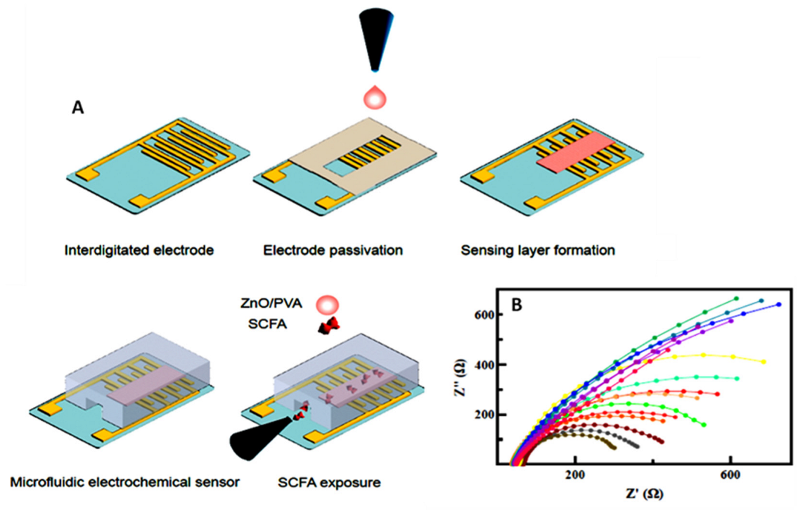
  35. Yavarinasab, A.; Filibotte, S.; Liu, S.; Tropini, C. An impedance-based chemiresistor for the real-time detection of gut-microbiota-generated short-chain fatty acids. BioRxiv 2022, 9, 507374. [Google Scholar]
  36. Kaur, A.; Kim, J.R.; Michie, I.; Dinsdale, R.M.; Guwy, A.J.; Premier, G.C. Microbial fuel cell type biosensor for specific volatile fatty acids using acclimated bacterial communities. Biosens. Bioelectron. 2013, 47, 50–55. [Google Scholar] [CrossRef]
  37. Osuna-Prieto, F.J.; Martinez-Tellez, B.; Ortiz-Alvarez, L.; Di, X.; Jurado-Fasoli, L.; Xum, H.; Ceperuelo-Mallafré, V.; Núñez-Roa, C.; Kohler, I.; Segura-Carretero, A.; et al. Elevated plasma succinate levels are linked to higher cardiovascular disease risk factors in young adults. Cardiovasc. Diabetol. 2021, 20, 151. [Google Scholar] [CrossRef]
  38. Keiran, N.; Ceperuelo-Mallafré, V.; Calvo, E.; Hernández-Álvarez, M.I.; Ejarque, M.; Núñez-Roa, C.; Horrillo, D.; Maymó-Masip, E.; Rodríguez, M.M.; Fradera, R.; et al. SUCNR1 controls an anti-inflammatory program in macrophages to regulate the metabolic response to obesity. Nat. Immunol. 2019, 20, 581–592. [Google Scholar] [CrossRef]
  39. Astiarraga, B.; Martínez, L.; Ceperuelo-Mallafré, V.; Llauradó, G.; Terrón-Puig, M.; Rodríguez, M.M.; Casajoana, A.; Pellitero, S.; Megia, A.; Vilarrasa, N.; et al. Impaired succinate response to a mixed meal in obesity and type 2 diabetes is normalized after metabolic surgery. Diabetes Care 2020, 43, 2581–2587. [Google Scholar] [CrossRef]
  40. Fernández-Veledo, S.; Vendrell, J. Gut microbiota-derived succinate: Friend or foe in human metabolic diseases? Rev. Endocr. Metab. Disord. 2019, 20, 439–447. [Google Scholar] [CrossRef] [Green Version]
  41. Serena, C.; Ceperuelo-Mallafré, V.; Keiran, N.; Queipo-Ortuño, M.I.; Bernal, R.; Gomez-Huelgas, R.; Urpi-Sarda, M.; Sabater, M.; Pérez-Brocal, V.; Andrés Lacueva, C.; et al. Elevated circulating levels of succinate in human obesity are linked to specific gut microbiota. ISME J. 2018, 12, 1642–1657. [Google Scholar] [CrossRef] [PubMed] [Green Version]
  42. Connors, J.; Dawe, N.; Van Limbergen, J. The role of succinate in the regulation of intestinal inflammation. Nutrients 2019, 11, 25. [Google Scholar] [CrossRef] [PubMed] [Green Version]
  43. Bensalah, N.; Louhichi, B.; Abdel-Wahab, A. Electrochemical oxidation of succinic acid in aqueous solutions using boron doped diamond anodes. Int. J. Environ. Sci. Technol. 2012, 9, 135–143. [Google Scholar] [CrossRef]
  44. Neurath, M.F. Cytokines in inflammatory bowel disease. Nat. Rev. Immunol. 2014, 14, 329–342. [Google Scholar] [CrossRef]
  45. Campuzano, S.; Yáñez-Sedeño, P.; Pingarrón, J.M. Revisiting electrochemical biosensing in the 21st century society for inflammatory cytokines involved in autoimmune, neurodegenerative, cardiac, viral and cancer diseases. Sensors 2021, 21, 189. [Google Scholar] [CrossRef] [PubMed]
  46. Sánchez-Tirado, E.; Agüí, L.; González-Cortés, A.; Yáñez-Sedeño, P.; Pingarrón, J.M. Biodetection techniques for quantification of chemokines. Chemosensors 2022, 10, 294. [Google Scholar] [CrossRef]
  47. Shekhar, S.; Yadav, A.K.; Khosla, A.; Solanki, P.R. Review—Interleukins profiling for biosensing applications: Possibilities and the future of disease detection. ECS Sens. Plus 2022, 1, 041601. [Google Scholar] [CrossRef]
  48. Dutta, N.; Lillehoj, P.B.; Estrela, P.; Dutta, G. Electrochemical biosensors for cytokine profiling: Recent advancements and possibilities in the near future. Biosensors 2021, 11, 94. [Google Scholar] [CrossRef]
  49. Ou, L.; Xia, N. Progress in nanomaterials-based electrochemical biosensors for the detection of interleukins. Int. J. Electrochem. Sci. 2022, 17, 220449. [Google Scholar] [CrossRef]
  50. Frias, M.; Zine, I.A.; Sigaud, N.; Lozano-Sanchez, M.; Caffio, P.; Errachid, M. Non-covalent π–π functionalized Gii-sense® graphene foam for interleukin 10 impedimetric detection. Biosens. Bioelectron. 2022, in press. [Google Scholar] [CrossRef]
  51. Barani, M.; Rahdar, A.; Sargazi, S.; Amiri, M.S.; Sharma, P.K.; Bhalla, N. Nanotechnology for inflammatory bowel disease management: Detection, imaging and treatment. Sens. Bio-Sens. Res. 2021, 32, 100417. [Google Scholar] [CrossRef]
  52. Jagannath, B.; Lin, K.-L.; Pali, M.; Sankhala, D.; Muthukumar, S.; Prasad, S. A sweat-based wearable enabling technology for real-time monitoring of IL-1β and CRP as potential markers for inflammatory bowel disease. Inflamm. Bowel Dis. 2020, 26, 1533–1542. [Google Scholar] [CrossRef] [PubMed]
  53. Thangamuthu, M.; Santschi, C.; Martin, O.J.F. Label-free electrochemical immunoassay for C-reactive protein. Biosensors 2018, 8, 34. [Google Scholar] [CrossRef]
  54. Ma, Y.; Yang, J.; Yang, T.; Deng, Y.; Gu, M.; Wang, M.; Hu, R.; Yang, Y. Electrochemical detection of C-reactive protein using functionalized iridium nanoparticles/graphene oxide as a tag. RSC Adv. 2020, 10, 9723–9729. [Google Scholar] [CrossRef] [PubMed] [Green Version]
  55. Weissmann, G.; Smolen, J.E.; Korchak, H.M. Release of inflammatory mediators from stimulated neutrophils. N. Engl. J. Med. 1980, 303, 27–34. [Google Scholar]
  56. Krawisz, J.E.; Sharon, P.; Stenson, W.F. Quantitative assay for acute intestinal inflammation based on myeloperoxidase activity: Assessment of inflammation in rat and hamster models. Gastroenterology 1984, 87, 1344–1350. [Google Scholar] [CrossRef]
  57. Kansagra, K.; Stoll, B.; Rognerud, C.; Niinikoski, H.; Ou, C.N.; Harvey, R. Total parenteral nutrition adversely affects gut barrier function in neonatal piglets. Am. J. Physiol. Gastrointest. Liver Physiol. 2003, 285, G1162. [Google Scholar] [CrossRef]
  58. Saiki, T.; Mitsuyama, K.; Toyonaka, A.; Ishida, H.; Tanikawa, K. Detection of pro- and anti-inflammatory cytokines in stools of patients with inflammatory bowel disease. Scand. J. Gastroenterol. 1998, 33, 616–622. [Google Scholar]
  59. Moral-Vico, J.; Barallat, J.; Abad, L.; Olivé-Monllau, R.; Muñoz-Pascual, F.X.; Galán Ortega, A.; del Campo, F.J.; Baldrich, E. Dual chronoamperometric detection of enzymatic biomarkers using magnetic beads and a low-cost flow cell. Biosens. Bioelectron. 2015, 69, 328–336. [Google Scholar] [CrossRef]
  60. Wen, Y.; Yuan, J.; Chen, J.; Zhao, Y.; Niu, Y.; Yu, C. Amperometric myeloperoxidase immunoassay based on the use of CuPdPt nanowire networks. Microchim. Acta 2018, 185, 55. [Google Scholar] [CrossRef]
  61. Bekhit, M.; Gorski, W. Electrochemical assays and immunoassays of the myeloperoxidase/SCN−/H2O2 system. Anal. Chem. 2019, 91, 3163–3169. [Google Scholar] [CrossRef] [PubMed]
  62. Cai, J.; Chen, H.; Weng, M.; Jiang, S.; Gao, J. Diagnostic and clinical significance of serum levels of D-lactate and diamine oxidase in patients with Crohn’s disease. Gastroenterol. Res. Pract. 2019, 2019, 8536952. [Google Scholar] [CrossRef] [PubMed]
  63. Beltrán-Ortiz, C.; Peralta, T.; Ramos, V.; Durán, M.; Behrens, C.; Maureira, D.; Guzmán, M.A.; Bastias, C.; Ferrer, P. Standardization of a colorimetric technique for determination of enzymatic activity of diamine oxidase (DAO) and its application in patients with clinical diagnosis of histamine intolerance. World Allergy Organ. J. 2020, 13, 100457. [Google Scholar] [CrossRef] [PubMed]
  64. Bilski, J.; Mazur-Bialy, A.; Wojcik, D.; Bilska, J.C.; Brzozowski, B.; Magierowski, M.; Mach, T.; Magierowska, K.; Brzozowski, T. The role of intestinal alkaline phosphatase in inflammatory disorders of gastrointestinal tract. Mediators Inflamm. 2017, 2017, 9074601. [Google Scholar] [CrossRef] [Green Version]
  65. Balbaied, T.; Moore, E. Overview of optical and electrochemical alkaline phosphatase (ALP) biosensors: Recent approaches in cells culture techniques. Biosensors 2019, 9, 102. [Google Scholar] [CrossRef]
  66. Mahato, K.; Purohit, B.; Kumar, A.; Chandra, P. Clinically comparable impedimetric immunosensor for serum alkaline phosphatase detection based on electrochemically engineered Au-nano-dendroids and graphene oxide nanocomposite. Biosens. Bioelectron. 2020, 148, 111815. [Google Scholar] [CrossRef]
  67. Wang, W.; Lu, J.; Hao, L.; Yang, H.; Song, X.; Si, F. Electrochemical detection of alkaline phosphatase activity through enzyme-catalyzed reaction using aminoferrocene as an electroactive probe. Anal. Bioanal. Chem. 2021, 413, 1827. [Google Scholar] [CrossRef] [PubMed]
  68. Wang, J.; Zhang, Z.; Childers, W.S. A biosensor for detection of indole metabolites. ACS Synth. Biol. 2021, 10, 1605. [Google Scholar] [CrossRef]
  69. Beloborodova, N.V.; Chernevskaya, E.A.; Getsina, M.L. Indolic structure metabolites as potential biomarkers of non-infectious diseases. Curr. Pharm. Des. 2021, 27, 238. [Google Scholar] [CrossRef] [PubMed]
  70. Jin, M.; Zhang, X.; Zhen, Q.; He, Y.; Chen, X.; Lyu, W.; Han, R.; Ding, M. An electrochemical sensor for indole in plasma based on MWCNTs-chitosan modified screen-printed carbon electrode. Biosens. Bioelectron. 2017, 98, 392. [Google Scholar] [CrossRef]
  71. Moncer, F.; Adhoum, N.; Catak, D.; Monser, L. Electrochemical sensor based on MIP for highly sensitive detection of 5-hydroxyindole-3-acetic acid carcinoid cancer biomarker in human biological fluids. Anal. Chim. Acta. 2021, 1181, 338925. [Google Scholar] [CrossRef] [PubMed]
  72. Abdelrasoul, G.N.; MacKay, S.; Salim, S.Y.; Ismond, K.P.; Tamura, M.; Khalifa, C.; Mannan, E.; Lin, D.; Mandal, T.; Montgomery, R.R.; et al. Non-invasive point-of-care device to diagnose acute mesenteric ischemia. ACS Sens. 2018, 3, 2296. [Google Scholar] [CrossRef] [PubMed]
  73. Pathirana, W.P.N.G.W.; Chubb, S.A.P.; Gillett, M.J.; Vasikaran, S.D. Faecal Calprotectin. Clin. Biochem. Rev. 2018, 39, 77. [Google Scholar] [PubMed]
  74. Dong, L.; Yin, L.; Tian, G.; Wang, Y.; Pei, H.; Wu, Q.; Cheng, W.; Ding, S.; Xia, Q. An enzyme-free ultrasensitive electrochemical immunosensor for calprotectin detection based on PtNi nanoparticles functionalized 2D Cumetal organic framework nanosheets. Sens. Actuators B. Chem. 2020, 308, 127687. [Google Scholar] [CrossRef]
  75. De Luca, F.; Shoenfeld, Y. The microbiome in autoimmune diseases. Clin. Exp. Immunol. 2018, 195, 74. [Google Scholar] [CrossRef] [PubMed]
  76. Kim, C.H. Immune regulation by microbiome metabolites. Immunology 2018, 154, 220. [Google Scholar] [CrossRef] [PubMed]
  77. Housley, W.J.; Pitt, D.; Hafler, D.A. Biomarkers in multiple sclerosis. Clin. Immunol. 2015, 161, 51. [Google Scholar] [CrossRef]
  78. Farrokhi, V.; Nemati, R.; Nichols, F.C.; Yao, X.; Anstadt, E.; Fujiwara, M.; Grady, J.; Wakefield, D.; Castro, W.; Donaldson, J.; et al. Bacterial lipodipeptide, Lipid 654, is a microbiome associated biomarker for multiple sclerosis. Clin. Transl. Immunol. 2013, 2, e8. [Google Scholar] [CrossRef]
  79. Bergot, A.-S.; Giri, R.; Thomas, R. The microbiome and rheumatoid arthritis. Best. Pract. Res. Clin. Rheumatol. 2019, 33, 101497. [Google Scholar] [CrossRef]
  80. Hammad, D.B.M.; Hider, S.L.; Liyanapathirana, V.C.; Tonge, D.P. Molecular characterization of circulating microbiome signatures in rheumatoid arthritis. Front. Cell. Infect. Microbiol. 2020, 9, 440. [Google Scholar] [CrossRef] [Green Version]
  81. Maeda, Y.; Kurakawa, T.; Umemoto, E.; Motooka, D.; Ito, Y.; Gotoh, K.; Hirota, K.; Matsushita, M.; Furuta, Y.; Narazaki, M.; et al. Dysbiosis contributes to arthritis development via activation of autoreactive T cells in the intestine. Arthritis Rheumathol. 2016, 68, 2646–2661. [Google Scholar] [CrossRef] [PubMed]
  82. Mobed, A.; Dolati, S.; Shakouri, S.K.; Eftekharsadat, B.; Izadseresht, B. Recent advances in biosensors for detection of osteoarthritis andrheumatoid arthritis biomarkers. Sens. Actuators A Phys. 2021, 331, 112975. [Google Scholar] [CrossRef]
  83. Deo, P.N.; Deshmukh, R. Oral microbiome: Unveiling the fundamentals. J. Oral Maxillofac. Pathol. 2019, 23, 122. [Google Scholar] [PubMed]
  84. Sivadasan, P.; Kumar Gupta, M.; Sathe, G.H.; Balakrishnan, L.; Palit, P.; Gowda, H.; Suresh, A.; Kuriakos, M.A.; Sirdeshmukh, E. Human salivary proteome—A resource of potential biomarkers for oral cancer. J. Proteom. 2015, 127, 89–95. [Google Scholar] [CrossRef]
  85. Bourgeois, D.; Inquimbert, C.; Ottolenghi, L.; Carrouel, F. Periodontal pathogens as risk factors of cardiovascular diseases, diabetes, rheumatoid arthritis, cancer, and chronic obstructive pulmonary disease—Is there cause for consideration? Microorganisms 2019, 7, 424. [Google Scholar] [CrossRef]
  86. Tar, I.; Csősz, É.; Végh, E.; Lundberg, K.; Kharlamova, N.; Soós, B.; Szekanecz, Z.; Márton, I. Salivary citrullinated proteins in rheumatoid arthritis and associated periodontal disease. Sci. Rep. 2021, 11, 13525. [Google Scholar] [CrossRef]
  87. Rabelo Buzalaf, M.A.; de Cássia Ortiz, A.; Souza Carvalho, T.; Moura Fideles, S.O.; Araújo, T.T.; Mascarenhas Moraes, S.; Rabelo Buzalaf, N.; Navas Reis, F. Saliva as a diagnostic tool for dental caries, periodontal disease and cancer: Is there a need for more biomarkers? Expert Rev. Mol. Diagn. 2020, 20, 543. [Google Scholar] [CrossRef]
  88. Nguyen, A.T.M.; Akhter, R.; Garde, S.; Scott, C.; Twigg, S.M.S.; Colagiuri, S.; Ajwani, S.; Eberhard, J. The association of periodontal disease with the complications of diabetes mellitus. A systematic review diabetes research and clinical practice. Diabetes Res. Clin. Pract. 2020, 165, 108244. [Google Scholar] [CrossRef]
  89. Chattopadhyayn, I.; Panda, M. Recent trends of saliva omics biomarkers for the diagnosis and treatment of oral cancer. J. Oral Biosci. 2019, 61, 84–94. [Google Scholar] [CrossRef]
  90. Schmidt, B.L.; Kuczynski, J.; Bhattacharya, A.; Huey, B.; Corby, P.M.; Queiroz, E.L.; Nightingale, K.; Kerr, A.R.; De Lacure, M.D.; Veeramachaneni, R.; et al. Changes in abundance of oral microbiota associated with oral cancer. PLoS One 2014, 2, e98741-53. [Google Scholar] [CrossRef]
  91. Lim, Y.; Totsika, M.; Morrison, M.; Punyadeera, C. Oral microbiome: A new biomarker reservoir for oral and oropharyngeal cancers. Theranostics 2017, 7, 4313–4321. [Google Scholar] [CrossRef] [PubMed]
  92. Gholizadeh, P.; Eslami, H.; Yousefi, M.; Asgharzadeh, M.; Kafil, S.H. Role of oral microbiome on oral cancers, a review. Biomed. Pharmacol. 2016, 84, 552–558. [Google Scholar] [CrossRef] [PubMed]
  93. Zhou, J. Electrochemical biosensors for the detection of matrix metalloproteinases. Int. J. Electrochem. Sci. 2022, 17, 221034. [Google Scholar] [CrossRef]
  94. Ruiz-Vega, G.; García-Robaina, A.; Ben Ismail, M.; Pasamar, H.; García-Berrocoso, T.; Montaner, J.; Zourob, M.; Othmane, A.; del Campo, F.J.; Baldrich, E. Detection of plasma MMP-9 within minutes. Unveiling some of the clues to develop fast and simple electrochemical magneto-immunosensors. Biosens. Bioelectron. 2018, 115, 45–52. [Google Scholar] [CrossRef] [PubMed]
  95. Arévalo, B.; ben Hassine, A.; Valverde, A.; Serafín, V.; Montero-Calle, A.; Raouafi, N.; Camps, J.; Arenas, M.; Barderas, R.; Yáñez-Sedeño, P.; et al. Electrochemical immunoplatform to assist in the diagnosis and classification of breast cancer through the determination of matrix-metalloproteinase-9. Talanta 2021, 225, 122054. [Google Scholar] [CrossRef] [PubMed]
  96. Irfan, M.; Rizantal Delgado, R.Z.; Frías-López, J. The oral microbiome and cancer. Front. Immunol. 2020, 11, 591088. [Google Scholar] [CrossRef] [PubMed]
  97. Mitsuhashi, A.; Okuma, Y. Perspective on immune oncology with liquid biopsy, peripheral blood mononuclear cells, and microbiome with non-invasive biomarkers in cancer patients. Clin. Transl. Oncol. 2018, 20, 966–974. [Google Scholar] [CrossRef]
  98. Mani, V.; Beduk, T.; Khushaim, W.; Ceylan, A.-E.; Timur, S.; Wolfbeis, O.S.; Salama, K.N. Electrochemical sensors targeting salivary biomarkers: A comprehensive review. TrAC, Trends Anal. Chem. 2021, 135, 116164. [Google Scholar] [CrossRef]
  99. McCrae, L.E.; Ting, W.-T.; Howlader, M.M.R. Advancing electrochemical biosensors for interleukin-6 detection. Biosens. Bioelectron.: X 2023, 13, 100288. [Google Scholar] [CrossRef]
  100. Yang, C.-Y.; Brooks, E.; Li, Y.; Denny, P.; Ho, C.-M.; Qi, F.W.; Shi, W.; Wolinsky, L.; Wu, B.; Wong, D.T.W.; et al. Detection of picomolar levels of interleukin-8 in human saliva by SPR. Lab Chip 2005, 5, 1017–1023. [Google Scholar] [CrossRef]
  101. Bhatia, A.; Na, H.S.; Nandhakumar, P.; Yu, B.; Jon, S.; Chung, J.; Yang, H. Electrochemical detection of interleukin-8 in human saliva using a polyenzyme label based on diaphorase and neutravidin. Sens. Actuators B Chem. 2021, 326, 128979. [Google Scholar] [CrossRef]
  102. Pachauri, N.; Lakshmi, G.B.V.S.; Sri, S.; Gupta, P.K.; Solanki, P.R. Silver molybdate nanoparticles based immunosensor for the non-invasive detection of Interleukin-8 biomarker. Mater. Sci. Eng. C 2020, 113, 110911. [Google Scholar] [CrossRef]
  103. Verma, S.; Singh, A.; Shukla, A.; Kaswan, J.; Arora, K.; Ramirez-Vick, J.; Singh, P.; Singh, S.P. Anti-IL8/AuNPs-rGO/ITO as an immunosensing platform for noninvasive electrochemical detection of oral cancer. ACS Appl. Mater. Interf. 2017, 9, 27462–27474. [Google Scholar] [CrossRef] [PubMed]
  104. Aydın, M.; Aydın, E.B.; Sezgintürk, M.K. A highly selective electrochemical immunosensor based on conductive carbon black and star PGMA polymer composite material for IL-8 biomarker detection in human serum and saliva. Biosens. Bioelectron. 2018, 117, 720. [Google Scholar] [CrossRef] [PubMed]
  105. Aydın, E.B.; Sezginturk, M.K. An impedimetric immunosensor for highly sensitive detection of IL-8 in human serum and saliva samples: A new surface modification method by 6-phosphonohexanoic acid for biosensing applications. Anal. Biochem. 2018, 554, 44. [Google Scholar] [CrossRef] [PubMed]
  106. Aydın, E.B.; Sezginturk, M.K. Fabrication of electrochemical immunosensor for detection of interleukin 8 biomarker via layer-by-layer self-assembly process on cost-effective fluorine tin oxide electrode. Electroanalysis 2021, 33, 1596. [Google Scholar] [CrossRef]
  107. Jagadeesh, R.V.; Lakshminarayanan, V. Adsorption kinetics of phosphonic acids and proteins on functionalized Indium tin oxide surfaces using electrochemical impedance spectroscopy. Electrochim. Acta 2016, 197, 1–9. [Google Scholar] [CrossRef]
  108. Aydin, E.B.; Sezgintürk, M.K. A disposable and ultrasensitive ITO based biosensor modified by 6-phosphonohexanoic acid for electrochemical sensing of IL-1β in human serum and saliva. Anal. Chim. Acta. 2018, 1039, 41–50. [Google Scholar] [CrossRef]
  109. Guerrero, S.; Agüí, L.; Yáñez-Sedeño, P.; Pingarrón, J.M. Design of electrochemical immunosensors using electro-click chemistry. Application to the detection of IL-1β cytokine in saliva. Bioelectrochemistry 2020, 133, 107484. [Google Scholar] [CrossRef]
  110. Torrente-Rodríguez, R.M.; Campuzano, S.; Ruiz-Valdepeñas Montiel, V.; Gamella, M.; Pingarrón, J.M. Electrochemical bioplatforms for the simultaneous determination of interleukin (IL)-8 mRNA and IL-8 protein oral cancer biomarkers in raw saliva. Biosens. Bioelectron. 2016, 77, 543–548. [Google Scholar] [CrossRef]
  111. Sánchez-Tirado, E.; Salvo, C.; González-Cortés, A.; Yáñez-Sedeño, P.; Langa, F.; Pingarrón, J.M. Electrochemical immunosensor for simultaneous determination of interleukin-1 beta and tumor necrosis factor alpha in serum and saliva using dual screen printed electrodes modified with functionalized double-walled carbon nanotubes. Anal. Chim. Acta 2017, 959, 66–73. [Google Scholar] [CrossRef] [PubMed]
  112. Bellando-Randone, S.; Russo, E.; Venerito, V.; Matucci-Cerinic, M.; Iannone, F.; Tangaro, S.; Amedei, A. Exploring the oral microbiome in rheumatic diseases, state of art and future prospective in personalized medicine with an AI approach. J. Pers. Med. 2021, 11, 625. [Google Scholar] [CrossRef] [PubMed]
  113. Bender, P.; Burgin, W.B.; Sculean, A.; Eick, S. Serum antibody levels against Porphyromonas gingivalis in patients with and without rheumatoid arthritis: A systematic review and meta-analysis. Clin. Oral Invest. 2017, 21, 33–42. [Google Scholar] [CrossRef]
  114. Selvam, S.P.; Chinnadayyala, S.R.; Cho, S. Electrochemical nanobiosensor for early detection of rheumatoid arthritis biomarker: Anti- cyclic citrullinated peptide antibodies based on polyaniline (PANI)/MoS2-modified screen-printed electrode with PANI-Au nanomatrix-based signal amplification. Sens. Actuators B Chem. 2021, 333, 129570. [Google Scholar] [CrossRef]
  115. Guerrero, S.; Sánchez-Tirado, E.; Martínez-García, G.; González-Cortés, A.; Yáñez-Sedeño, P.; Pingarrón, J.M. Electrochemical biosensor for the simultaneous determination of rheumatoid factor and anti-cyclic citrullinated peptide antibodies in human serum. Analyst 2020, 145, 4680. [Google Scholar] [CrossRef] [PubMed]
  116. Sung, W.-H.; Tsao, Y.-T.; Shen, C.-J.; Tsai, C.-Y.; Cheng, C.-M. Small-volume detection: Platform developments for clinically-relevant applications. J. Nanobiotechnol. 2021, 19, 114. [Google Scholar] [CrossRef]
  117. Martensson, A.; Greiff, L.; Lamei, S.S.; Lindstedt, M.; Olofsson, T.C.; Vasquez, A.; Cervin, A. Effects of a honeybee lactic acid bacterial microbiome on human nasal symptoms, commensals, and biomarkers. Int. Forum Allergy Rhinol. 2016, 6, 957–963. [Google Scholar] [CrossRef]
  118. Hassan-Nixon, H.A.G.; Singh, N.; Cass, A.E.G. A sensitive impedimetric immunosensor for the detection of Interleukin-8 in nasal epithelial lining fluid of asthma patients. Biosens. Bioelectron. X. 2022, 10, 100118. [Google Scholar]
  119. Chawes, B.L.; Edwards, M.J.; Shamji, B.; Walker, C.; Nicholson, G.C.; Tan, A.J.; Folsgaard, N.V.; Bonnelykke, K.; Bisgaard, H.; Hansel, T.T. A novel method for assessing unchallenged levels of mediators in nasal epithelial lining fluid. J. Allergy Clin. Immunol. 2010, 125, 1387–1389. [Google Scholar] [CrossRef]
  120. Li, J.-X.; Wang, Z.-Z.; Zhai, G.-T.; Chen, C.-L.; Zhu, K.-Z.; Yu, Z.; Liu, Z. Untargeted metabolomic profiling identifies disease-specific and outcome-related signatures in chronic rhinosinusitis. J. Allergy Clin. Immunol. 2022, 150, 727–735. [Google Scholar] [CrossRef]
  121. Mahdavinia, M.; Keshavarzian, A.; Tobin, M.C.; Landay, A.L.; Schleimer, R.P. A comprehensive review of the nasal microbiome in chronic rhinosinusitis (CRS). Clin. Exp. Allergy 2016, 46, 21–41. [Google Scholar] [CrossRef] [Green Version]
  122. Zhang, X.; Wang, S.; Xu, H.; Yi, H.; Guan, J.; Yin, S. Metabolomics and microbiome profiling as biomarkers in obstructive sleep apnoea: A comprehensive review. Eur. Resp. Rev. 2021, 30, 200220. [Google Scholar] [CrossRef] [PubMed]
  123. Bradley, T.D.; Floras, J.S. Obstructive sleep apnoea and its cardiovascular consequences. Lancet 2009, 373, 82–93. [Google Scholar] [CrossRef] [PubMed]
  124. Maeder, M.T.; Strobel, W.; Christ, M.; Todd, J.; Estis, J.; Wildi, K.; Thalmann, G.; Hilti, J.; Brutsche, M. Twerenbold, R.; et al. Comprehensive biomarker profiling in patients with obstructive sleep apnea. Clin. Biochem. 2015, 48, 340–346. [Google Scholar] [CrossRef] [PubMed]
  125. Qi, M.; Huang, J.; Wei, H.; Cao, C.; Feng, S.; Guo, Q.; Goldys, E.M.; Li, R.; Liu, G. Graphene oxide thin film with dual function integrated into a nanosandwich device for in vivo monitoring of interleukin-6. ACS Appl. Mater. Interfaces 2017, 9, 41659–41668. [Google Scholar] [CrossRef]
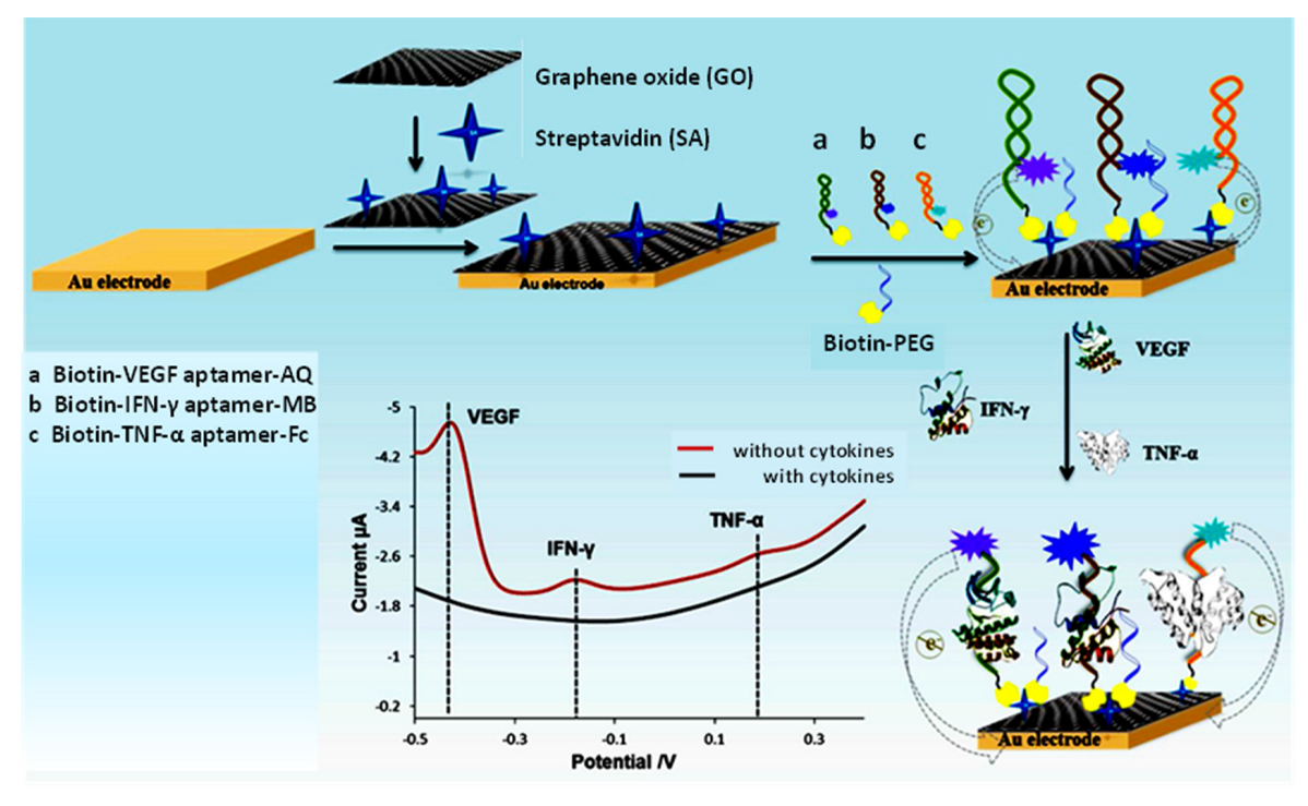
  126. Shen, Z.; Ni, S.; Yang, W.; Sun, W.; Yang, G.; Liu, G. Redox probes tagged electrochemical aptasensing device for simultaneous detection of multiple cytokines in real time. Sens. Actuators B Chem. 2021, 336, 129747. [Google Scholar] [CrossRef]
  127. Ni, S.; Qiao, L.; Shen, Z.; Gao, Y.; Liu, G. Physical absorption vs covalent binding of graphene oxide on glassy carbon electrode towards a robust aptasensor for ratiometric electrochemical detection of vascular endothelial growth factor (VEGF) in serum. Electrochim. Acta 2020, 331, 135321. [Google Scholar] [CrossRef]
  128. Johari-Ahar, M.; Karami, P.; Ghanei, M.; Afkham, A.; Bagheri, H. Development of a molecularly imprinted polymer tailored on disposable screen-printed electrodes for dual detection of EGFR and VEGF using nanoliposomal amplification strategy. Biosens. Bioelectron. 2018, 107, 26–33. [Google Scholar] [CrossRef]
Figure 1. Selected gastrointestinal, oral, and nasal microbiome biomarkers and related diseases. Figure prepared by the authors.
Sensors 23 00837 g001
Figure 2. Amperometric detection principles of (A) an acetate biosensor using acetate kinase (AK), pyruvate kinase (PK), and pyruvate oxidase (POx), and (B) a propionate biosensor using propionate CoA-transferase (PCT) and short-chain acyl-CoA oxidase (SCAOx). Adapted from [34] with permission.
Sensors 23 00837 g002
Figure 3. (A) Schematic view of the microfluidic setup and sensing layer synthesis for the determination of SCFAs and (B) Nyquist spectra of the sensing layer at various concentrations of acetic acid and propionic acid in a mixture with 0.5 mg mL−1 butyric acid. Adapted from [35] with permission.
Sensors 23 00837 g003
Figure 4. (A) Microfluidic chip and holder, and the different components of the cartridge. (B) Scheme of the magneto-immunoassay format for the determination of MPO: (1) anti-MPO-biotin; (2) biotin used as blocker; (3) anti-MPO-HRP. Adapted from [59] with permission.
Sensors 23 00837 g004
Figure 5. Schematic illustrations of: (A) the SPCE/AuNPs/Au-nano-dendroids/GO/anti-ALP probe for the determination of ALP in serum, and (B) the electrochemical assay of ALP activity based on the enzyme-catalyzed reaction. Adapted from [66,67], respectively, with permission.
Sensors 23 00837 g005
Figure 6. Illustrative schemes of: (A) the preparation procedure of PtNi@Cu-TCPP(Fe)-Ab2 bioconjugate, and (B) the construction of the sandwich immunosensor. Reprinted from [74] with permission.
Sensors 23 00837 g006
Figure 7. Schematic illustrations of some electrochemical immunosensors for the determination of IL-8: (A) a sandwich-type immunoassay using a polyenzyme label on diaphorase (DI-3) and neutravidin; (B) the synthesis of β-Ag2-MoO4 NPs and immunoelectrode fabrication; (C) fabrication of an AuNPs-rGO based immunosensor; (D) steps for preparation of an impedimetric immunosensor. Reprinted from (A) [101], (B) [102]; (C) [103] and (D) [104] with permission.
Sensors 23 00837 g007
Figure 8. Schematic of an electrochemical aptasensor for the simultaneous and real-time monitoring of VEGF, IFN-γ and TNF-α. Reproduced from [126] with permission.
Sensors 23 00837 g008
Disclaimer/Publisher’s Note: The statements, opinions and data contained in all publications are solely those of the individual author(s) and contributor(s) and not of MDPI and/or the editor(s). MDPI and/or the editor(s) disclaim responsibility for any injury to people or property resulting from any ideas, methods, instructions or products referred to in the content.

