
Paper-Based Aptasensors: Working Principles, Detection Modes, and Applications


Abstract
:1. Introduction
2. Paper-Based Devices
2.1. Paper as an Analytical Platform
- (1)
- It promotes the movement of liquids by capillary action, eliminating the need for external forces to move low volumes of samples and reagents.
- (2)
- It is highly absorbent and offers a high surface-to-volume ratio, which enables the efficient immobilization and storage of the reagents.
- (3)
- It enables the filtration of the sample.
- (4)
- It is thin, light, and conformable.
- (5)
- It is biocompatible and biodegradable.
- (6)
- It is inexpensive and easily available worldwide.
2.2. Fabrication of Paper-Based Devices
3. Aptamers and Aptasensors
3.1. Aptamers
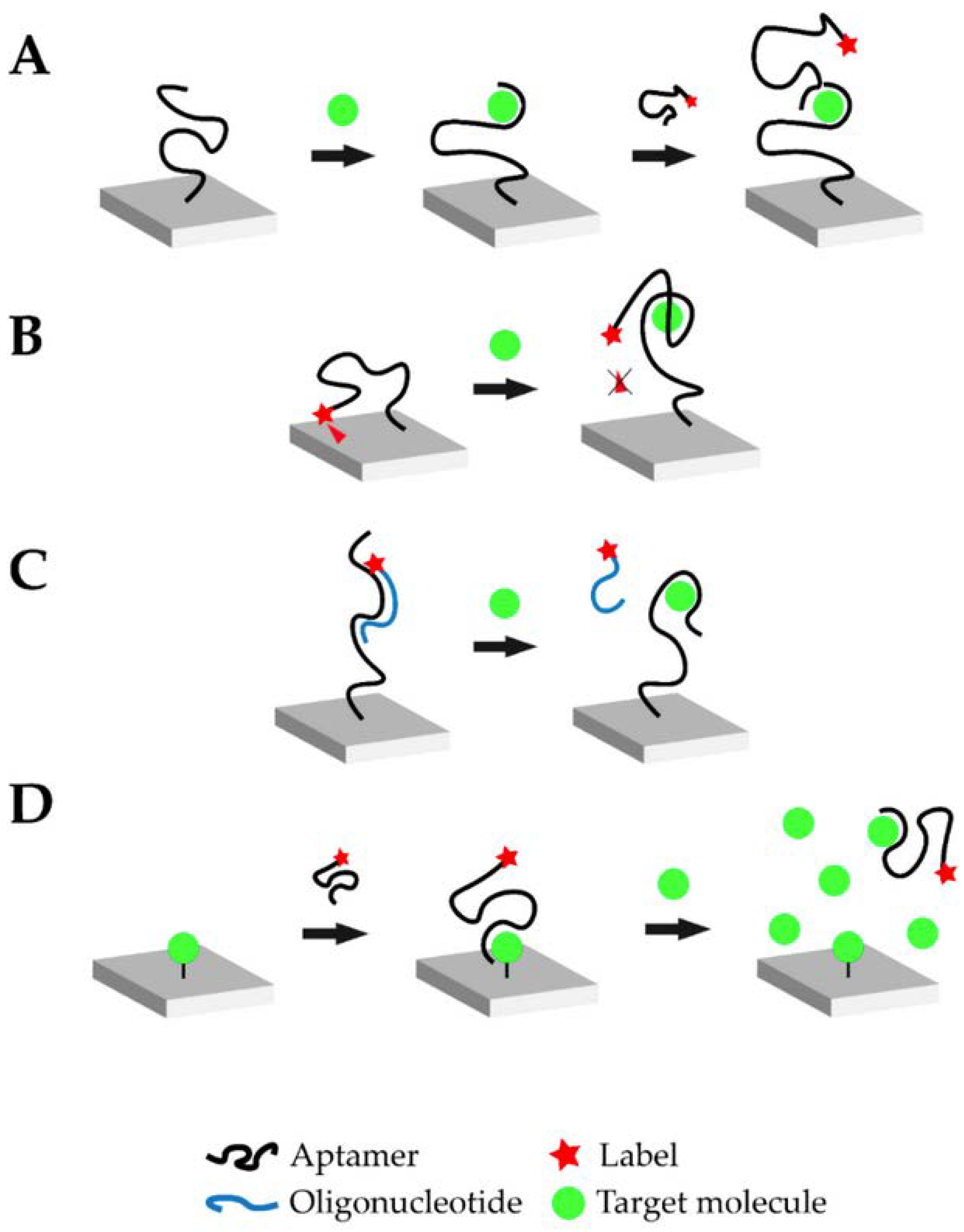
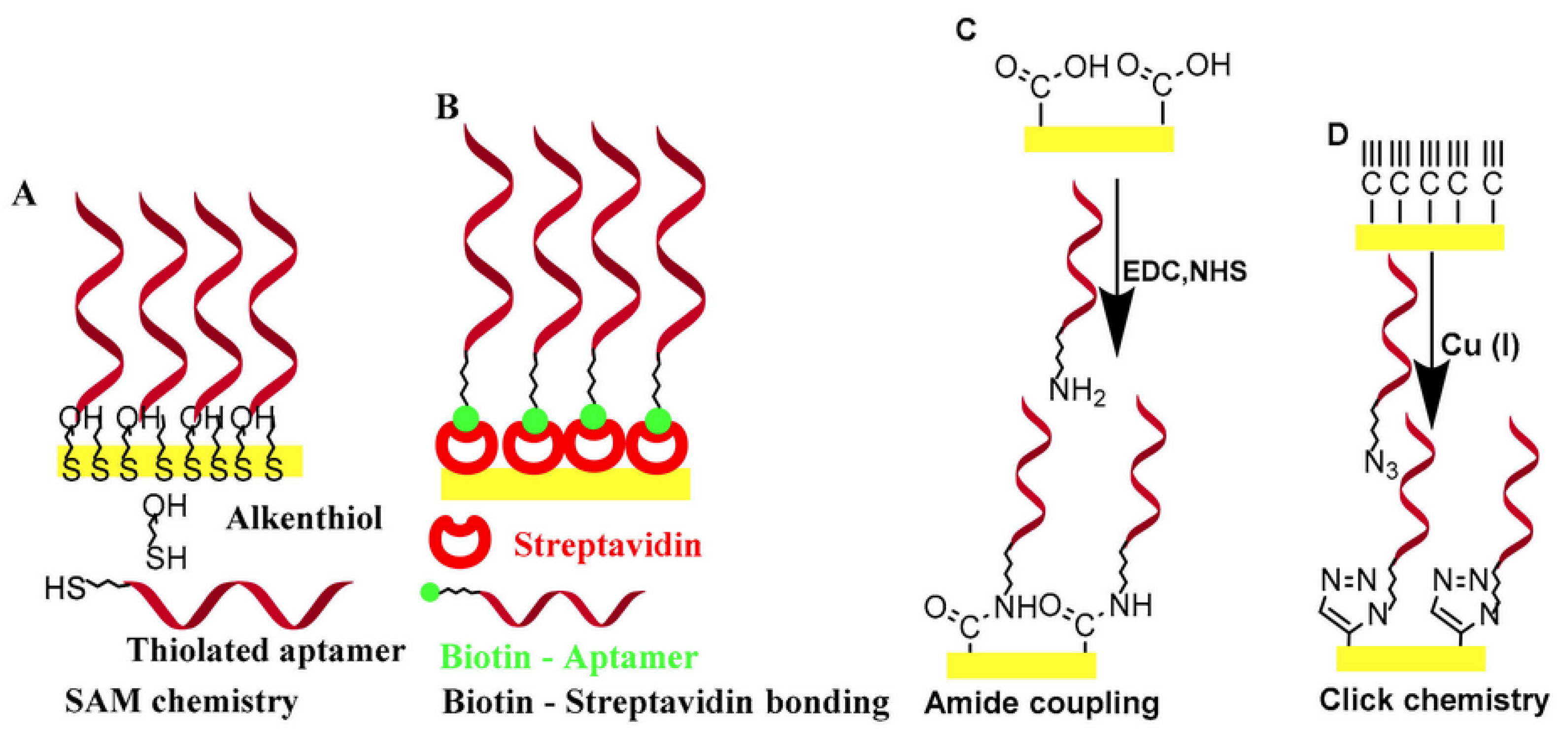
3.2. Aptasensors
4. Detection Modes in Paper-Based Aptasensors
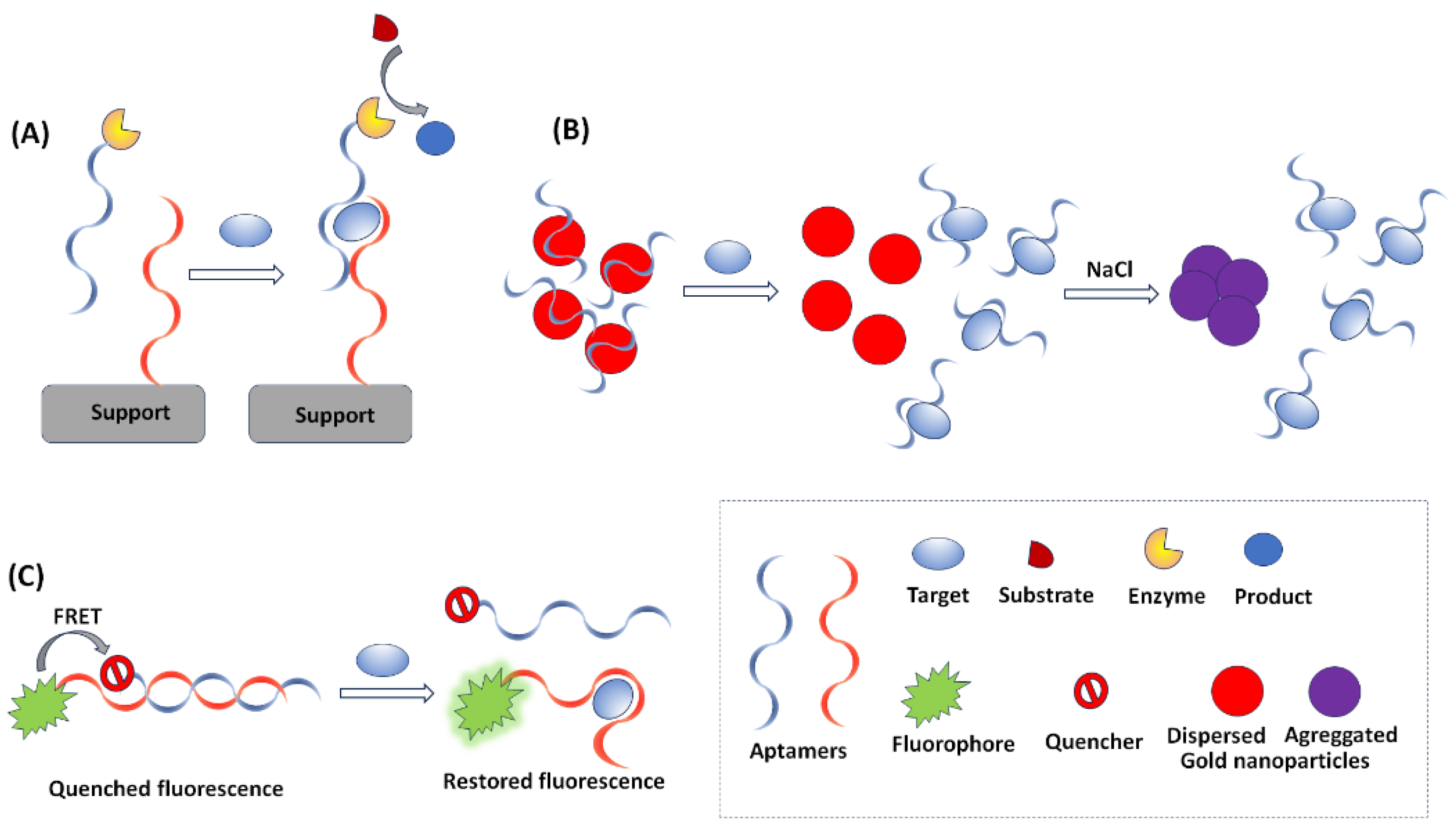
4.1. Optical Detection
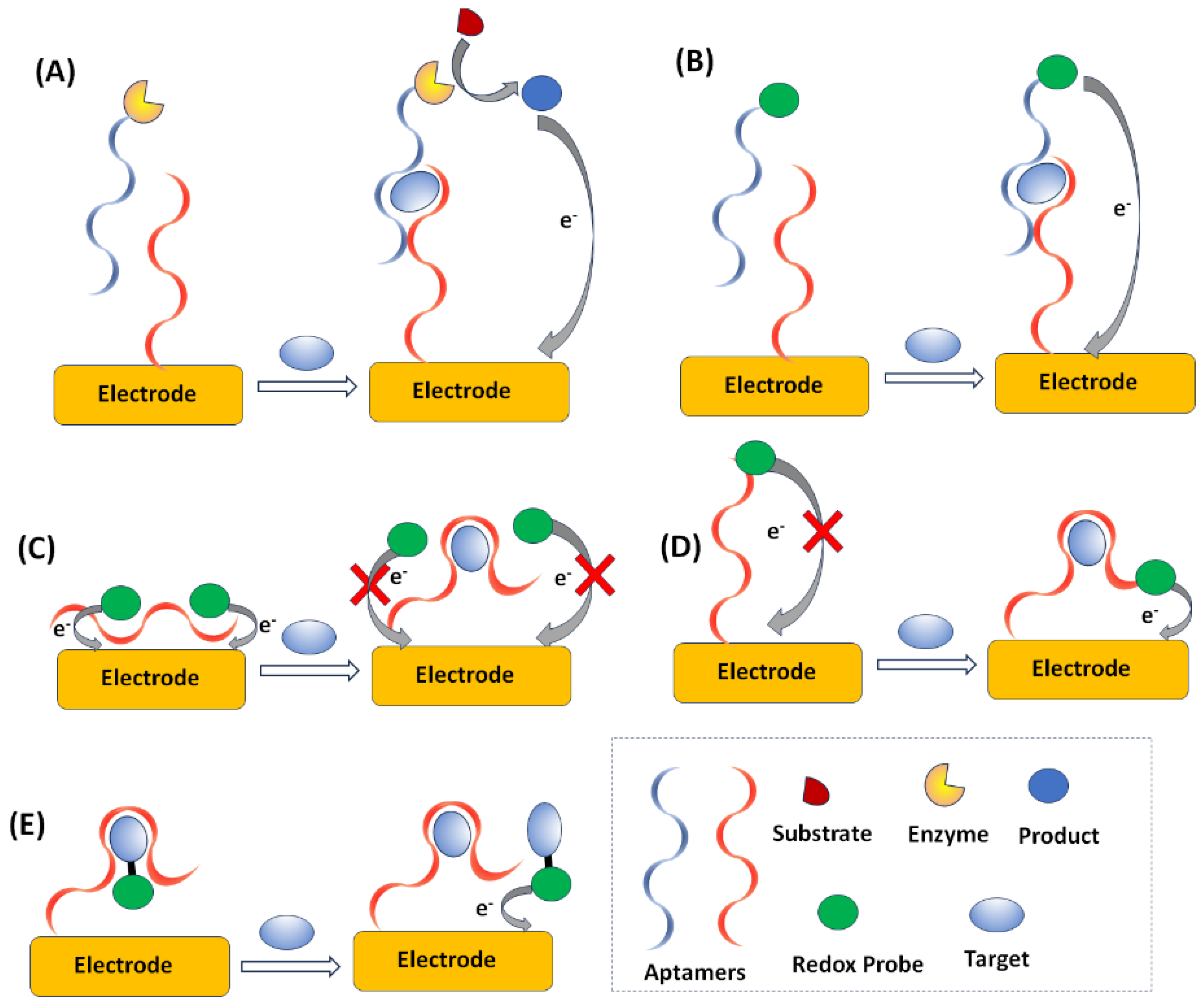
4.2. Electrochemical Detection
5. Applications of Aptasensing PADs
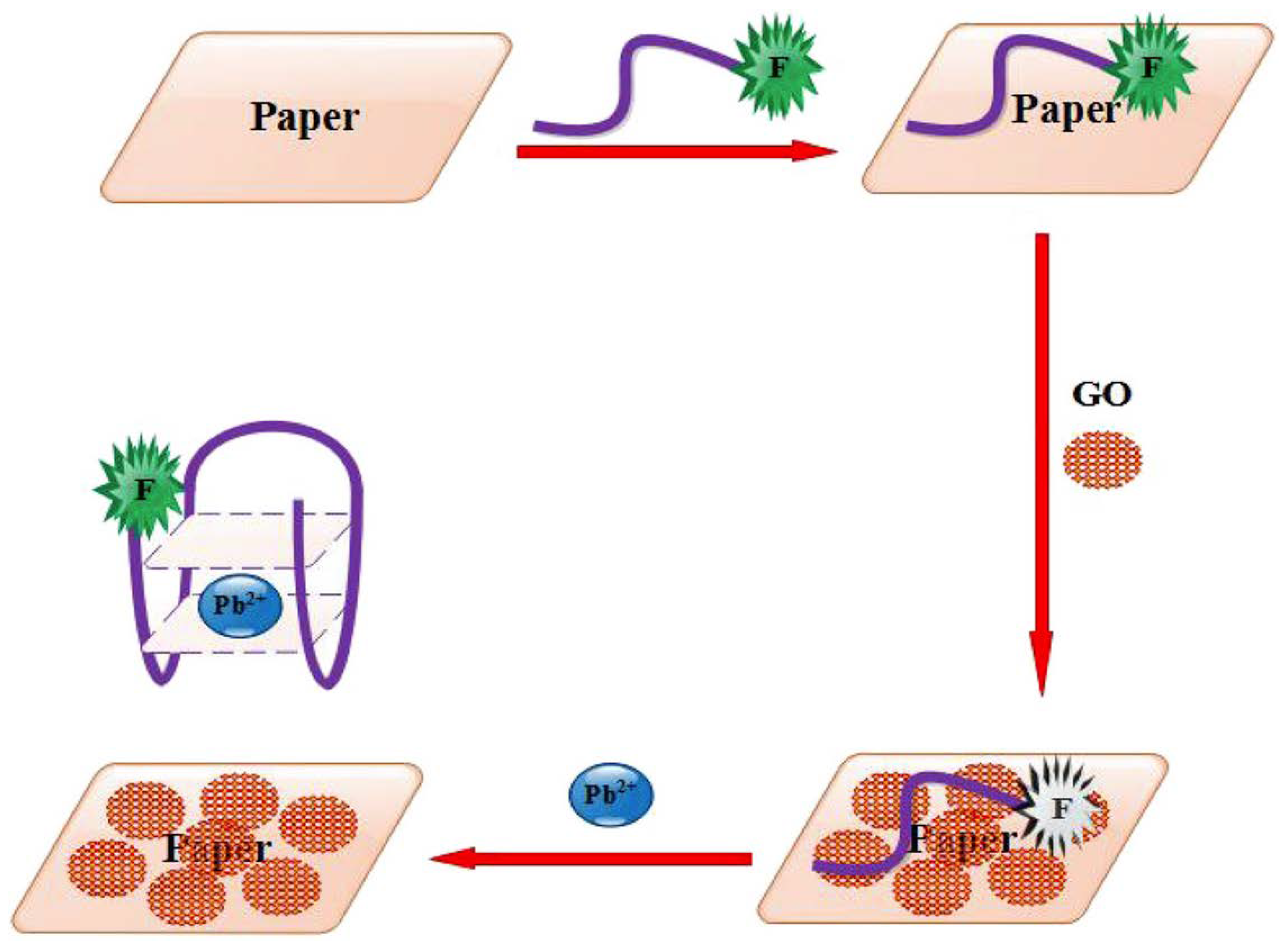
5.1. Ions
5.2. Small Molecules

| Analyte (Sample) | PAD | Type of Paper | Detection | Aptamer Sequence | Ref. |
|---|---|---|---|---|---|
| Epinephrine | NR *-wax dipping | Whatman No. 1 | Colorimetric with AuNPs | 32-mer (sequence NR *) | [69] |
| Streptomycin, tobramycin, kanamycin (milk) | Five layer-wax printing | Whatman chromatography paper No. 4, 5 | Fluorescence | Str: TAGGGAATTCGTCGACGGATGCGGGG TCTGGTGTTGTGCTTTGTTCTGTCGGG TCGTCTGCAGGTCGACGCATGCGCCG Tob: GACTAGGCACTAGTC Kana: TGGGGGTTGAGGCTAAGCCGAC 78.8 | [70] |
| Quinine, serotonin (urine, water, tomatoes, tomato juice) | Triangular-hand cutting | Glass microfiber filter paper | Paper spray-mass spectrometry | qui: 5′-GAC-AAG-GAA-AAT-CCT-TCA-ACG-AAG-TGG-GTC-3′ ser: 5′-CGA-CTG-GTA-GGC-AGA-TAG-GGG-AAG- CTG-ATT-CGA-TGC-GTG-GGT-CG-3′ | [71] |
| Tetracycline (water) Guanosine tetra- Phosphate (cell lysate) | Circular-hole punch | Whatman No. 42 | Fluorescence | tet: AUGGAAAAACAUACCAGAUUUCGAUCUGGAGAGGUGAAGAAUACGACCACCUUCCCA ppGpp: NR | [72] |
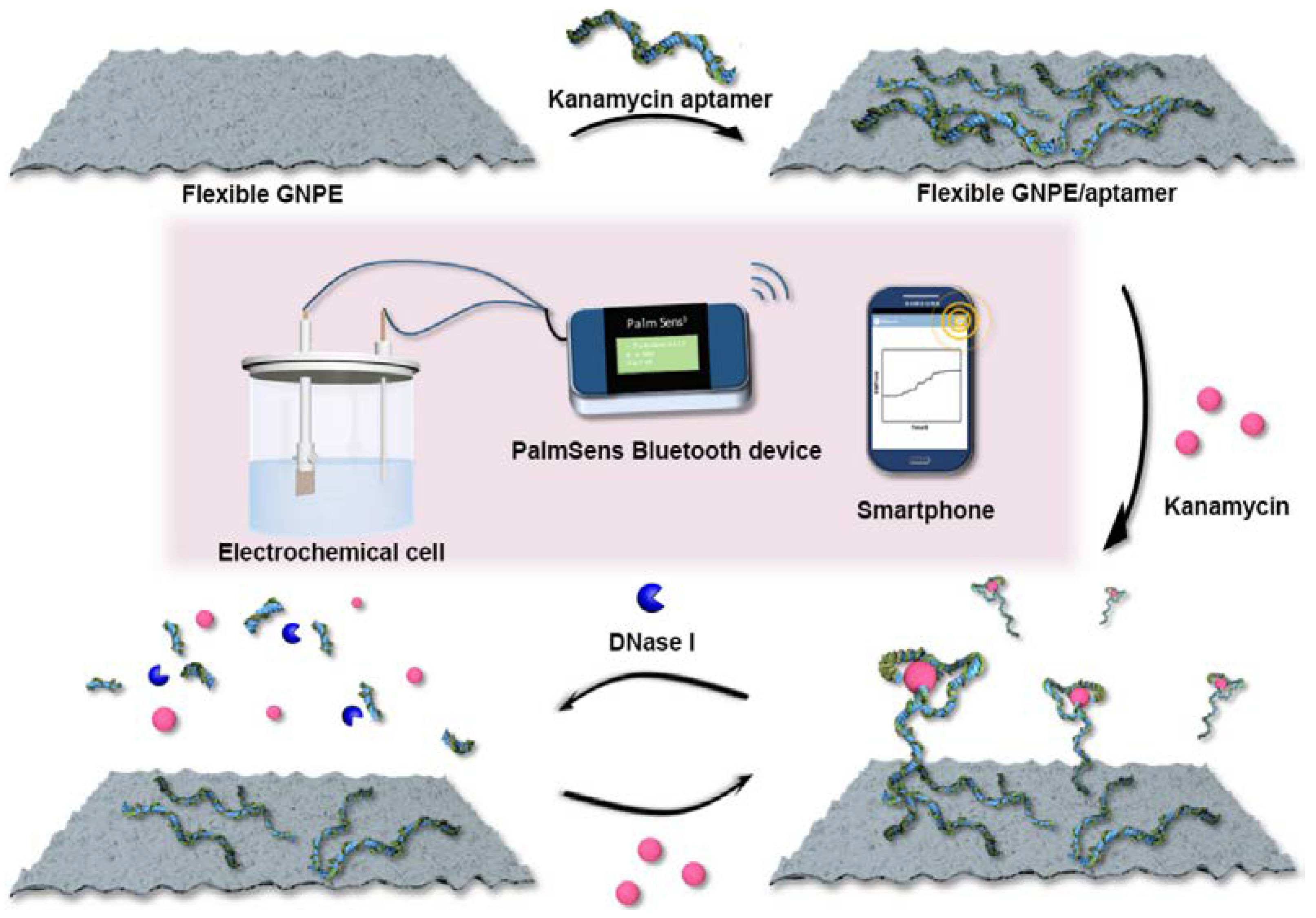
| Kanamycin (milk) | Strips-hand cutting | NR * | Potentiometry | 5′-AGATGGGGGTTGAGGCTAAGCCGA-3′ | [73] |
| Adenosine | Origami-wax printing and lamination | Whatman No. 1 | Charge | 5′-ACTCATCTGTGAAGAGAACCTGGGGGAGTATTGCGGAGGAAGGT-3′ | [74] |
| Gentamicin | Star-shaped-hand punch | Whatman Protran | Colorimetric with AuNPs | 5′-GGGACT TGGTTTAGGTAATGAGTCCC- 3′ | [75] |
| 17β-estradiol (serum) | Origami-wax printing-screen printed electrodes | Whatman No. 1 | DPV | 5′-SH-(CH2)6-GCTTCCAGCTTATTGAATTACACGCAGAGGTACGGCTCTGCGC ATTCAATTGCTGCGCGCTGAAGCGCGGAAGC-3′ | [76] |
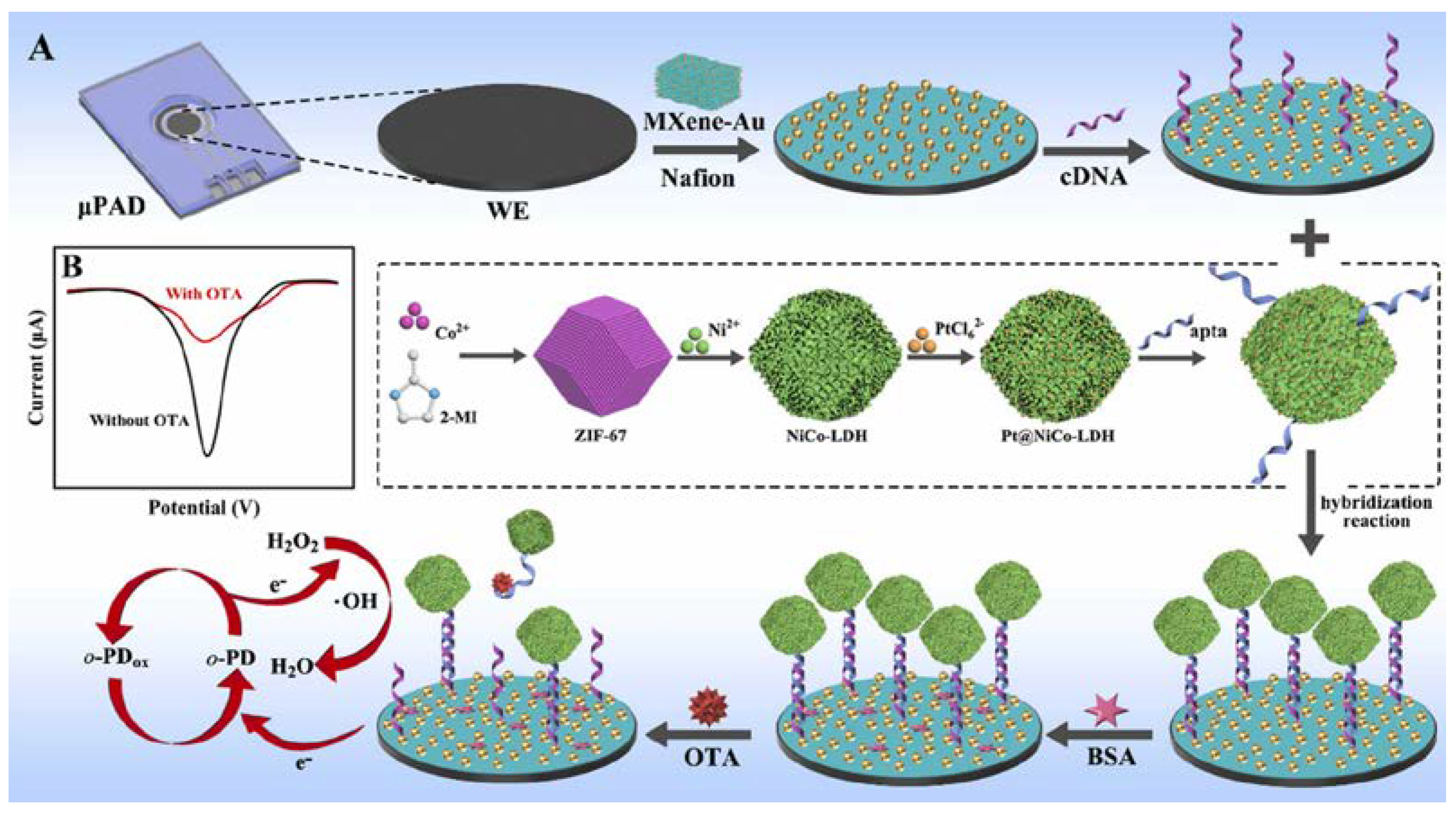
| Ochratoxin A (corn, wheat) | Circular-wax printing-screen printed electrodes | Whatman No. 1 | DPV | 5′-SH-(CH2)6-GATCGGGTGTGGGTGGCGTAAAGGGAGCATCGGACA-3′ | [77] |
| Adenosine triphosphate | Origami/wax printing/screen-printed electrodes | Whatman chromatography paper No. 1 | ECL | capture: 5′-HS-(CH2)6-ACCTGGGGGAGTAT-3′; probe: 5′-TGCGGAGGAAGGT-NH2-3′ | [78] |
| Ochratoxin A (corn, groundnut) | Microfluidic-photoresist | Whatman filter paper | Colorimetric with AuNPs | 5′-GATCGGGTGTGGGTGGCGTAAAGGGAGCATCGGACA-3′ | [79] |
5.3. Large Molecules and Proteins
| Analyte (Sample) | PAD | Type of Paper | Detection | Aptamer Sequence | Ref. |
|---|---|---|---|---|---|
| Plasmodium falciparum lactate dehydrogenase (blood) | circular-hole punch | Whatman 3MM chromatography paper | colorimetric of enzymatic activity | 5′Biotin- CTG GGC GGTAGAACCATAGTGACCCAGCCG TCTAC-3′ | [81] |
| β-bungarotoxin (venom) | circular-wax printing | Whatman filter paper No. 4 | colorimetric assay with streptavidin-HRP- TMB | 5′-CATACAAACGGAAATTCCGATTTAGTCTTTATGATCTTGATGC-3′ 5′-GGACAGAAAAAAAAAAAGACAAAGAAGAGAGAGGGAGATGGGGCTCAT-3′ | [82] |
| Osteopontin (serum) | circular-hand cutting | Fanoia S300 paper | colorimetric with Bradford reagent | 5′-Thiol-AAAAAAAAAA TGT GTG CGG CAC TCC AGT CTG TTA CGC CGC-3′ | [83] |
| platelet-derived growth factor (serum) | circular-wax printing | nitrocellulose HF 180 | colorimetric- horseradish peroxidase-mimicking DNAzyme- H2O2- hemin | 5′-ATATA GTAGA AACCA CTATC GACTC AGGCT ACGGC ACGTA GAGCA TCACC ATGAT CCTGT AGTATCAATC CTTCG CCGTC-3′ 5′-ATATA GTAGA AACCA CTATC GACTC AGGCT ACGGC ACGTA GAGCA TCACC ATGAT CCTGT AAACCCAACC CGCCC TACCC TAAA-3′ | [84] |
| arachin, β-lactoglobulin, tropomyosin (NR) | origami-wax printing | Whatman chromatography paper | colorimetric with AuNPs | arachin: TCG CAC ATT CCG CTT CTA CCG GGG GGG TCG AGC GAG TGA GCG AAT CTG TGG GTG GGC CGT AAG TCC GTG TGT GCG AA β-lactoglobulin: ATA CCA GCT TAT TCA ATT CGA CGATCG GAC CGC AGT ACC CAC CCA CCA GCC CCA ACA TCA TGC CCA TCC GTG TGT GAG ATA GTA AGT GCA ATC T Tropomyosin: TAC TAA CGG TAC AAG CTA CCA GGCCGC CAA CGT TGA CCT AGA AGC ACT GCC AGA CCC GAA CGT TGA CCT AGA AGC | [85] |
| Plasmodium lactate dehydrogenase (NR) | rectangular-hand cutting | Printer paper | FRET | 5′- GTT CGA TTG GAT TGT GCC GGA AGT GCT GGCTCG AAC—FAM—3′ | [86] |
| mucin-1 (serum) | origami-wax printing-screen-printed electrodes | Whatman chromatography paper No. 1 | ECL | 5′-GCAGTTGATCCTTTGGATACCCTGG-3′ | [87] |
| carcinoembryonic antigen (serum) | rectangular--wax printng-screen-printed electrodes | Whatman chromatography paper No. 1 | ΕCL | 5′HS-(CH2)6-ATA CCA GCT TAT TCAATT-3′ 5′HS-(CH2)6-CCC ATA GGG AAG TGG GGG A-3′ | [88] |
| immunoglobulin E (serum) | circular/marker plotting | Whatman chromatography paper No. 1 | FRET | 5′-NH2-AAAAAGGGGCACGTTTATCCGTCCCTCCTAGTGGCGTGCCCC-3′ | [89] |
| hemoglobin A1 (blood) | NR * | graphite paper | DPV with Fe(CN)63−/Fe(CN)64− probe | 5′-SH-TGGCAGGAAGACAAA- CACATCGTCGCGGCCTTAGGAGGGGCG- GACGGGGGGGGGCGTTGGTCTGTGGTGCTGT-3′ | [90] |
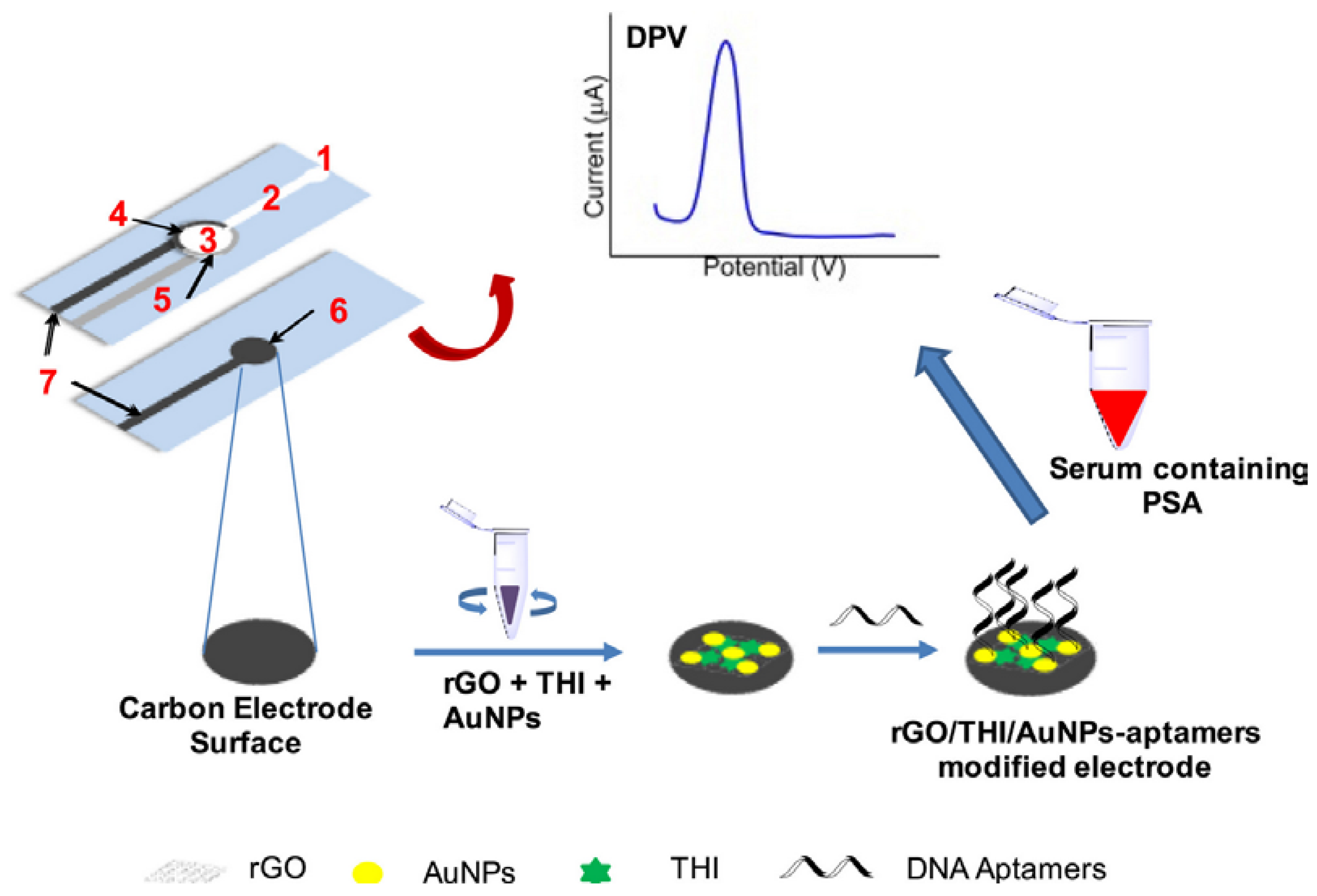
| prostate specific antigen (serum) | microfluidic/wax printing/screen-printed electrodes | Whatman chromatography paper No. 1 | DPV | 5′-ATT AAA GCT CGC CAT CAA ATA GC-3′ | [91] |
| carcinoembryonic antigen (serum) | circular/laser cutting | Whatman chromatography paper | DPV | 5′-AGATACCAGCTTATTCAATTCCGCTGCTGGTATCT-3′ | [92] |
| carcinoembryonic antigen (serum) | circular/as received | Whatman qualitative filter paper No. 3 | EIS | 5′-NH2-GAC GAT AGC GGT GAC GGC ACA GAC GTC CCG CAT CCT CCG-3′ | [93] |
| Thrombin (serum) | origami/wax printing/screen-printed electrodes | Whatman chromatography paper No. 2 | PEC | 5′-GGT TGG TGT GGT TGG AGA AGA AGG CCA ACC ACA CCA ACC GAT CC-3′ | [94] |
| prostate specific antigen (serum) | origami/wax printing/screen-printed electrodes | chromatographic paper | PEC | 5′-SH-TTAATTAAAGCTCGCCATCAAATAGC-3′ | [95] |
| vascular endothelial growth factor 165 (mesenchymal stem cells culture) | circular and microfluidic/wax printing | Whatman filter paper No. 1 | fluorescence | 5′-TGTGGGGGTGGACTGGGTGGGTACCGTCACTCGCCTCGCACCGTCC- Biotin—3′ | [96] |
| epidermal growth factor receptor (serum) | origami/wax printing/screen-printed electrodes | Whatman chromatography paper No. 1 | DPV | 5′-TAC CAG TGC GAT GCT CAG TGC CGT TTCTTC TCT TTC GCT TTT TTT GCT TTT GAG CAT GCT GAC GCA TTC GGT TGA C-3′ | [97] |
5.4. Cells and Bacteria
| Analyte (Sample) | PAD | Type of Paper | Detection | Aptamer Sequence | Ref. |
|---|---|---|---|---|---|
| human breast adenocarcinoma cells (MCF-7) (serum) | origami/wax printing/screen-printed electrodes | Whatman chromatography paper No. 114 | ECL | 5′-GCA GTT GAT CCT TTG GAT ACC CTG GTT TTT TTT TTT-HS-3′ | [98] |
| human breast adenocarcinoma cells (MCF-7) (blood) | origami/wax printing/screen-printed electrodes | Whatman chromatography paper No. 114 | ECL | 5′- GCA GTT GAT CCT TTG GAT ACC CTG GTT TTT TTT TTT -HS-3′ | [99] |
| human acute promyelocytic leukemia cells (HL 60) (NR) | origami/wax printing/screen-printed electrodes | Whatman chromatography paper No. 114 | DPV | 5′-ATCCAGAGTGACGCAGCATGCCCTAGTTACTACTACTCTTTTTAGCAAACGCCCTCGCTTTGGACACGGTGGCTTAGT-3′ | [100] |
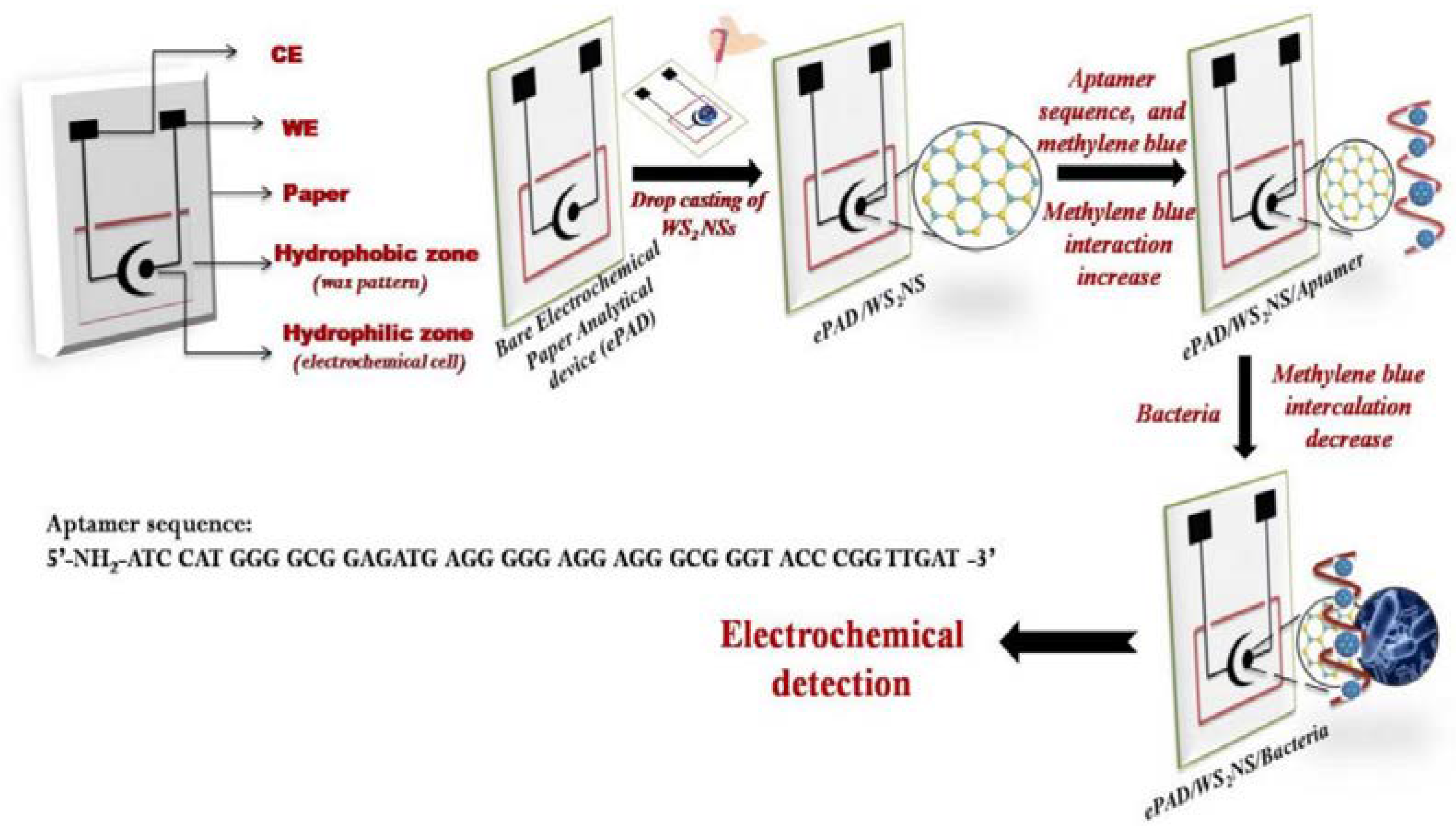
| Listeria monocytogenes (milk, cheese) | rectangular/wax printing/screen-printed electrodes | GSM 210 paper | EIS | 5′-NH2-ATC CAT GGG GCG GAGATG AGG GGG AGG AGG GCG GGT ACC CGG TTGAT-3′ | [101] |
| Zika virus (NR) | Rectangular/hand-cutting | printer paper | potentiometric | 32-mehr | [102] |
| human breast adenocarcinoma cells (MCF-7) (serum) | Origami/wax printing/screen-printed electrodes | Whatman chromatography paper No2 | DPV, colorimetric | 5′-SH-CACTACAGAGGTTGCGTCTGTCCCACGTTG TCATGGGGGGTTGGCCTG-3′ 5′-biotin-TTTTTTGCAGTTGATCCTTTGGATACCCTGGTTTGCAAAGCTTACGGCATACGT-3′ | [103] |
| MCF-7 cells, K562 cells (blood) | multi-layered/wax printing/screen-printed electrodes | NR * | DPV, colorimetric | MCF-7: 5′-NH2-C6H12-CAC TAC AGA GGT TGC GTC CCA CGT TGT CCC ACG TTG TCA TGG GGG GTT GGC CTG-3′ K562: 5′-NH2-TTT TTT TTT TAC AGC AGA TCA GTC TAT CTT CTC CTG ATG GGT TCC TAT TTA TAG GTG AAG CTGT GGC-3′ TGG CTG GGG GGC GTT | [104] |
5.5. Multiplexed Assays
| Analyte (Sample) | PAD | Type of Paper | Detection | Aptamer Sequence | Ref. |
|---|---|---|---|---|---|
| Hg2+, Ag+ (human serum, water, milk) | square/craft punch | Whatman No 1 | FRET/GO | Hg2+: 5′-FAM-TTT TTT TTT TTT-3′ Ag+: 5′-FAM-CCC CCC CCC CCC-3′ | [106] |
| lysozyme, ß-conglutin lupine, okadaic acid, brevetoxin (egg white, mussels, sausages, bread) | rectangular/hand-cutting | Whatman chromatography paper | FRET | lys: 5′-AGC AGC ACA GAG GTC AGA TG GCA GCTAAG CAG GCG GCT CAC AAA ACC ATT CGCATG CGG C CCT ATG CGT GCT ACC GTG AA-3′ ß-congl: 5′-AGC TGA CAC AGC AGG TTG GTG GGG GTGGCT TCC AGT TGG GTT GAC AAT ACG TAG GGA CAC GAA GTC CAA CCA CGA GTC GAG CAA TCT CGA AAT-3′ okadaic acid: 5′-CAG CTC AGA AGC TTG ATC CTA TTT GACCAT GTC GAG GGA GAC GCG CAG TCG CTACCA CCT GAC TCG AAG TCG TGC ATC TG-3′ brevet: 5′-ATA CCA GCT TAT TCA ATT GGC CAC CAAACC ACA CCG TCG CAA CCG CGA GAA CCG AAG TAG TGA TCA TGT CCC TGC GTG AGA TAG TAA GTG CAA TCT-3′ | [107] |
| carcinoembryonic antigen, neuron-specific enolase (serum) | multi-layer microfluidic/wax printing/screen-printed electrodes | Whatman chromatography paper No1 | DPV | CEA: 5′-ATA CCA GCT TAT TCA ATT-3′, NSE: 5′-CGG TAA TAC GGT TAT CCA CAG AAT CAG GGG-3′ | [108] |
| four glycans on K562 cell surface (cell culture) | origami/wax printing/screen-printed electrodes | Whatman chromatography paper No114 | DPV | 5′-HS-TTT TTT TTT TAC AGC AGA TCA GTC TAT CTT CTC CTG ATG GGT TCC TAT TTA TAG GTG AAG CTG T-3′ | [109] |
| E. coli O157:H7, S. Typhimurium (NR) | star-shaped/wax printing | Whatman filter paper No1 | colorimetric with AuNPs | E. coli: 5′—CCG GAC GCT TAT GCC TTG CCA TCT ACA GAG CAG GTG TGA CGG—3′ S. Typh: 5′—ACG GGC GTG GGG GCA ATG CTG CTT GTA GCC TTC CCC TGT GCG CG—3′ | [110] |
| MCF-7, HL-60, K562 cells (NR) | star-shaped/wax printing | Whatman chromatography paper No114 | FRET | MCF-7: 5′-GCA GTT GAT CCT TTG GAT ACC CTG GTT TTT TTT TTT-NH2-3′ HL-60: 5′- NH2-TTT TTT TTT ATC CAG AGT GAC GCA GCA TGC CCT AGT TAC TAC TAC TCT TTT TAG CAA AC-3′ K562 cells: 5′- NH2-TTT TTT TTT TAC AGCAGA TCA GTC TAT CTT CTC CTG ATG GGT TCC TAT TTA TAG GTG AAG CTG T-3′ | [111] |
| Acinetobacter baumannii, Escherichia coli and Staphylococcus aureus) (biological fluids) | microfluidic chip | nitrocellulose | colorimetric | A1, E27, O28 | [112] |
6. Conclusions and Future Prospects
Author Contributions
Funding
Institutional Review Board Statement
Informed Consent Statement
Data Availability Statement
Conflicts of Interest
References
- Odeh, F.; Nsairat, H.; Alshaer, W.; Ismail, M.A.; Esawi, E.; Qaqish, B.; Bawab, A.A.; Ismail, S.I. Aptamers Chemistry: Chemical Modifications and Conjugation Strategies. Molecules 2020, 25, 3. [Google Scholar] [CrossRef]
- Lim, Y.C.; Kouzani, A.Z.; Duan, W. Aptasensors: A Review. J. Biomed. Nanotechnol. 2010, 6, 93–105. [Google Scholar] [CrossRef] [PubMed]
- Yan, S.-R.; Foroughi, M.M.; Safaei, M.; Jahani, S.; Ebrahimpour, N.; Borhani, F.; Rezaei Zade Baravati, N.; Aramesh-Boroujeni, Z.; Foong, L.K. A review: Recent advances in ultrasensitive and highly specific recognition aptasensors with various detection strategies. Int. J. Biol. Macromol. 2020, 155, 184–207. [Google Scholar] [CrossRef] [PubMed]
- Akki, S.U.; Werth, C.J. Critical Review: DNA Aptasensors, Are They Ready for Monitoring Organic Pollutants in Natural and Treated Water Sources? Environ. Sci. Technol. 2018, 52, 8989–9007. [Google Scholar] [CrossRef]
- Chen, X.-F.; Zhao, X.; Yang, Z. Aptasensors for the detection of infectious pathogens: Design strategies and point-of-care testing. Microchim. Acta 2022, 189, 443. [Google Scholar] [CrossRef]
- Noviana, E.; Ozer, T.; Carrell, C.S.; Link, J.S.; McMahon, C.; Jang, I.; Henry, C.S. Microfluidic Paper-Based Analytical Devices: From Design to Applications. Chem. Rev. 2021, 121, 11835–11885. [Google Scholar] [CrossRef] [PubMed]
- Torsten, S.; Alina, E.; Thomas, S.; Johanna, W. Aptamer-based lateral flow assays. AIMS Bioeng. 2018, 5, 78–102. [Google Scholar] [CrossRef]
- Wang, T.; Chen, L.; Chikkanna, A.; Chen, S.; Brusius, I.; Sbuh, N.; Veedu, R.N. Development of nucleic acid aptamer-based lateral flow assays: A robust platform for cost-effective point-of-care diagnosis. Theranostics 2021, 11, 5174–5196. [Google Scholar] [CrossRef]
- Ming, T.; Luo, J.; Liu, J.; Sun, S.; Xing, Y.; Wang, H.; Xiao, G.; Deng, Y.; Cheng, Y.; Yang, Z.; et al. Paper-based microfluidic aptasensors. Biosens. Bioelectron. 2020, 170, 112649. [Google Scholar] [CrossRef]
- Hillscher, L.M.; Liebich, V.J.; Avrutina, O.; Biesalski, M.; Kolmar, H. Functional paper-based materials for diagnostics. ChemTexts 2021, 7, 14. [Google Scholar] [CrossRef]
- Akyazi, T.; Basabe-Desmonts, L.; Benito-Lopez, F. Review on microfluidic paper-based analytical devices towards commercialisation. Anal. Chim. Acta 2018, 1001, 1–17. [Google Scholar] [CrossRef]
- Yetisen, A.K.; Akram, M.S.; Lowe, C.R. Paper-based microfluidic point-of-care diagnostic devices. Lab Chip 2013, 13, 2210–2251. [Google Scholar] [CrossRef]
- Sinha, A.; Basu, M.; Chandna, P. Chapter Five—Paper based microfluidics: A forecast toward the most affordable and rapid point-of-care devices. In Progress in Molecular Biology and Translational Science; Pandya, A., Singh, V., Eds.; Academic Press: Cambridge, MA, USA, 2022; Volume 186, pp. 109–158. [Google Scholar]
- Nery, E.W.; Kubota, L.T. Sensing approaches on paper-based devices: A review. Anal. Bioanal. Chem. 2013, 405, 7573–7595. [Google Scholar] [CrossRef] [PubMed]
- Ozer, T.; McMahon, C.; Henry, C.S. Advances in Paper-Based Analytical Devices. Annu. Rev. Anal. Chem. 2020, 13, 85–109. [Google Scholar] [CrossRef] [PubMed]
- Lim, W.Y.; Goh, B.T.; Khor, S.M. Microfluidic paper-based analytical devices for potential use in quantitative and direct detection of disease biomarkers in clinical analysis. J. Chromatogr. B 2017, 1060, 424–442. [Google Scholar] [CrossRef]
- Busa, L.S.A.; Mohammadi, S.; Maeki, M.; Ishida, A.; Tani, H.; Tokeshi, M. Advances in Microfluidic Paper-Based Analytical Devices for Food and Water Analysis. Micromachines 2016, 7, 86. [Google Scholar] [CrossRef]
- Antonacci, A.; Scognamiglio, V.; Mazzaracchio, V.; Caratelli, V.; Fiore, L.; Moscone, D.; Arduini, F. Paper-Based Electrochemical Devices for the Pharmaceutical Field: State of the Art and Perspectives. Front. Bioeng. Biotechnol. 2020, 8. [Google Scholar] [CrossRef] [PubMed]
- Meredith, N.A.; Quinn, C.; Cate, D.M.; Reilly, T.H.; Volckens, J.; Henry, C.S. Paper-based analytical devices for environmental analysis. Analyst 2016, 141, 1874–1887. [Google Scholar] [CrossRef]
- Martinez, A.W.; Phillips, S.T.; Butte, M.J.; Whitesides, G.M. Patterned Paper as a Platform for Inexpensive, Low-Volume, Portable Bioassays. Angew. Chem. Int. Ed. 2007, 46, 1318–1320. [Google Scholar] [CrossRef]
- Modha, S.; Castro, C.; Tsutsui, H. Recent developments in flow modeling and fluid control for paper-based microfluidic biosensors. Biosens. Bioelectron. 2021, 178, 113026. [Google Scholar] [CrossRef]
- Soni, S.; Toley, B.J. Paper-based nucleic acid sample preparation for point-of-care diagnostics. Sens. Actuators B Chem. 2022, 355, 131272. [Google Scholar] [CrossRef]
- Yonet-Tanyeri, N.; Ahlmark, B.Z.; Little, S.R. Advances in Multiplexed Paper-Based Analytical Devices for Cancer Diagnosis: A Review of Technological Developments. Adv. Mater. Technol. 2021, 6, 2001138. [Google Scholar] [CrossRef]
- Li, X.; Ballerini, D.R.; Shen, W. A perspective on paper-based microfluidics: Current status and future trends. Biomicrofluidics 2012, 6, 011301. [Google Scholar] [CrossRef]
- Faheem, A.; Cinti, S. Chapter 5—Advanced techniques for manufacturing paper-based microfluidic analytical devices. In Microfluidic Biosensors; Mak, W.C., Pui Ho, A.H., Eds.; Academic Press: Cambridge, MA, USA, 2023; pp. 159–170. [Google Scholar]
- Dabbagh, S.R.; Becher, E.; Ghaderinezhad, F.; Havlucu, H.; Ozcan, O.; Ozkan, M.; Yetisen, A.K.; Tasoglu, S. Increasing the packing density of assays in paper-based microfluidic devices. Biomicrofluidics 2021, 15, 011502. [Google Scholar] [CrossRef] [PubMed]
- Lim, H.; Jafry, A.T.; Lee, J. Fabrication, Flow Control, and Applications of Microfluidic Paper-Based Analytical Devices. Molecules 2019, 24, 2869. [Google Scholar] [CrossRef]
- Anushka; Bandopadhyay, A.; Das, P.K. Paper based microfluidic devices: A review of fabrication techniques and applications. Eur. Phys. J. Spec. Top. 2023, 232, 781–815. [Google Scholar] [CrossRef]
- Tang, R.H.; Liu, L.N.; Zhang, S.F.; He, X.C.; Li, X.J.; Xu, F.; Ni, Y.H.; Li, F. A review on advances in methods for modification of paper supports for use in point-of-care testing. Microchim. Acta 2019, 186, 521. [Google Scholar] [CrossRef]
- Zhuo, Z.; Yu, Y.; Wang, M.; Li, J.; Zhang, Z.; Liu, J.; Wu, X.; Lu, A.; Zhang, G.; Zhang, B. Recent Advances in SELEX Technology and Aptamer Applications in Biomedicine. Int. J. Mol. Sci. 2017, 18, 2142. [Google Scholar] [CrossRef]
- Kohlberger, M.; Gadermaier, G. SELEX: Critical factors and optimization strategies for successful aptamer selection. Biotechnol. Appl. Biochem. 2022, 69, 1771–1792. [Google Scholar] [CrossRef] [PubMed]
- Prante, M.; Segal, E.; Scheper, T.; Bahnemann, J.; Walter, J. Aptasensors for Point-of-Care Detection of Small Molecules. Biosensors 2020, 10, 108. [Google Scholar] [CrossRef]
- Ayodele, O.O.; Adesina, A.O.; Pourianejad, S.; Averitt, J.; Ignatova, T. Recent Advances in Nanomaterial-Based Aptasensors in Medical Diagnosis and Therapy. Nanomaterials 2021, 11, 932. [Google Scholar] [CrossRef] [PubMed]
- Komarova, N.; Kuznetsov, A. Inside the Black Box: What Makes SELEX Better? Molecules 2019, 24, 3598. [Google Scholar] [CrossRef]
- Stanciu, L.A.; Wei, Q.; Barui, A.K.; Mohammad, N. Recent Advances in Aptamer-Based Biosensors for Global Health Applications. Annu. Rev. Biomed. Eng. 2021, 23, 433–459. [Google Scholar] [CrossRef] [PubMed]
- Arshavsky-Graham, S.; Heuer, C.; Jiang, X.; Segal, E. Aptasensors versus immunosensors—Which will prevail? Eng. Life Sci. 2022, 22, 319–333. [Google Scholar] [CrossRef]
- Walter, J.-G.; Stahl, F.; Scheper, T. Aptamers as affinity ligands for downstream processing. Eng. Life Sci. 2012, 12, 496–506. [Google Scholar] [CrossRef]
- Guo, W.; Zhang, C.; Ma, T.; Liu, X.; Chen, Z.; Li, S.; Deng, Y. Advances in aptamer screening and aptasensors’ detection of heavy metal ions. J. Nanobiotechnol. 2021, 19, 166. [Google Scholar] [CrossRef]
- Lou, B.; Liu, Y.; Shi, M.; Chen, J.; Li, K.; Tan, Y.; Chen, L.; Wu, Y.; Wang, T.; Liu, X.; et al. Aptamer-based biosensors for virus protein detection. TrAC Trends Anal. Chem. 2022, 157, 116738. [Google Scholar] [CrossRef]
- Cai, R.; Chen, X.; Zhang, Y.; Wang, X.; Zhou, N. Systematic bio-fabrication of aptamers and their applications in engineering biology. Syst. Microbiol. Biomanufacturing 2023, 3, 223–245. [Google Scholar] [CrossRef]
- Hua, Y.; Ma, J.; Li, D.; Wang, R. DNA-Based Biosensors for the Biochemical Analysis: A Review. Biosensors 2022, 12, 183. [Google Scholar] [CrossRef]
- Peyrin, E. Nucleic acid aptamer molecular recognition principles and application in liquid chromatography and capillary electrophoresis. J. Sep. Sci. 2009, 32, 1531–1536. [Google Scholar] [CrossRef]
- Yoo, H.; Jo, H.; Oh, S.S. Detection and beyond: Challenges and advances in aptamer-based biosensors. Mater. Adv. 2020, 1, 2663–2687. [Google Scholar] [CrossRef]
- Naresh, V.; Lee, N. A Review on Biosensors and Recent Development of Nanostructured Materials-Enabled Biosensors. Sensors 2021, 21, 1109. [Google Scholar] [CrossRef] [PubMed]
- Cho, I.-H.; Kim, D.H.; Park, S. Electrochemical biosensors: Perspective on functional nanomaterials for on-site analysis. Biomater. Res. 2020, 24, 6. [Google Scholar] [CrossRef] [PubMed]
- Azzouz, A.; Hejji, L.; Sonne, C.; Kim, K.-H.; Kumar, V. Nanomaterial-based aptasensors as an efficient substitute for cardiovascular disease diagnosis: Future of smart biosensors. Biosens. Bioelectron. 2021, 193, 113617. [Google Scholar] [CrossRef]
- Kaur, H.; Shorie, M. Nanomaterial based aptasensors for clinical and environmental diagnostic applications. Nanoscale Adv. 2019, 1, 2123–2138. [Google Scholar] [CrossRef] [PubMed]
- Kurup, C.; Tlili, C.; Zakaria, S.N.A.; Ahmed, M.U. Recent Trends in Design and Development of Nanomaterial-based Aptasensors. Biointerface Res. Appl. Chem. 2021, 11, 14057–14077. [Google Scholar] [CrossRef]
- Ghasemi, F.; Fahimi-Kashani, N.; Bigdeli, A.; Alshatteri, A.H.; Abbasi-Moayed, S.; Al-Jaf, S.H.; Merry, M.Y.; Omer, K.M.; Hormozi-Nezhad, M.R. Paper-based optical nanosensors—A review. Anal. Chim. Acta 2023, 1238, 340640. [Google Scholar] [CrossRef]
- Chiu, T.-C.; Huang, C.-C. Aptamer-Functionalized Nano-Biosensors. Sensors 2009, 9, 10356–10388. [Google Scholar] [CrossRef] [PubMed]
- Khan, N.I.; Song, E. Lab-on-a-Chip Systems for Aptamer-Based Biosensing. Micromachines 2020, 11, 220. [Google Scholar] [CrossRef]
- Kaneta, T.; Alahmad, W.; Varanusupakul, P. Microfluidic paper-based analytical devices with instrument-free detection and miniaturized portable detectors. Appl. Spectrosc. Rev. 2019, 54, 117–141. [Google Scholar] [CrossRef]
- Paschoalino, W.J.; Kogikoski, S., Jr.; Barragan, J.T.C.; Giarola, J.F.; Cantelli, L.; Rabelo, T.M.; Pessanha, T.M.; Kubota, L.T. Emerging Considerations for the Future Development of Electrochemical Paper-Based Analytical Devices. ChemElectroChem 2019, 6, 10–30. [Google Scholar] [CrossRef]
- Villalonga, A.; Mayol, B.; Villalonga, R.; Vilela, D. Electrochemical aptasensors for clinical diagnosis. A review of the last five years. Sens. Actuators B Chem. 2022, 369, 132318. [Google Scholar] [CrossRef]
- Liu, J.; Morris, M.D.; Macazo, F.C.; Schoukroun-Barnes, L.R.; White, R.J. The Current and Future Role of Aptamers in Electroanalysis. J. Electrochem. Soc. 2014, 161, H301–H313. [Google Scholar] [CrossRef]
- Abd-Ellatief, R.; Abd-Ellatief, M.R. Electrochemical Aptasensors: Current Status and Future Perspectives. Diagnostics 2021, 11, 104. [Google Scholar] [CrossRef] [PubMed]
- Xu, Y.; Cheng, G.; He, P.; Fang, Y. A Review: Electrochemical Aptasensors with Various Detection Strategies. Electroanalysis 2009, 21, 1251–1259. [Google Scholar] [CrossRef]
- Radi, A.-E. Electrochemical Aptamer-Based Biosensors: Recent Advances and Perspectives. Int. J. Electrochem. 2011, 2011, 863196. [Google Scholar] [CrossRef]
- Sassolas, A.; Blum, L.J.; Leca-Bouvier, B.D. Electrochemical Aptasensors. Electroanalysis 2009, 21, 1237–1250. [Google Scholar] [CrossRef]
- Kurup, C.P.; Mohd-Naim, N.F.; Ahmed, M.U. Recent trends in nanomaterial-based signal amplification in electrochemical aptasensors. Crit. Rev. Biotechnol. 2022, 42, 794–812. [Google Scholar] [CrossRef]
- Jin, M.; Yuan, H.; Liu, B.; Peng, J.; Xu, L.; Yang, D. Review of the distribution and detection methods of heavy metals in the environment. Anal. Methods 2020, 12, 5747–5766. [Google Scholar] [CrossRef]
- Borrill, A.J.; Reily, N.E.; Macpherson, J.V. Addressing the practicalities of anodic stripping voltammetry for heavy metal detection: A tutorial review. Analyst 2019, 144, 6834–6849. [Google Scholar] [CrossRef] [PubMed]
- Naderi, M.; Hosseini, M.; Ganjali, M.R. Naked-eye detection of potassium ions in a novel gold nanoparticle aggregation-based aptasensor. Spectrochim. Acta Part A Mol. Biomol. Spectrosc. 2018, 195, 75–83. [Google Scholar] [CrossRef]
- Fakhri, N.; Hosseini, M.; Tavakoli, O. Aptamer-based colorimetric determination of Pb2+ using a paper-based microfluidic platform. Anal. Methods 2018, 10, 4438–4444. [Google Scholar] [CrossRef]
- Khoshbin, Z.; Housaindokht, M.R.; Izadyar, M.; Verdian, A.; Bozorgmehr, M.R. A simple paper-based aptasensor for ultrasensitive detection of lead (II) ion. Anal. Chim. Acta 2019, 1071, 70–77. [Google Scholar] [CrossRef] [PubMed]
- Liu, F.; Wang, S.; Zhang, M.; Wang, Y.; Ge, S.; Yu, J.; Yan, M. Aptamer based test stripe for ultrasensitive detection of mercury(II) using a phenylene-ethynylene reagent on nanoporous silver as a chemiluminescence reagent. Microchim. Acta 2014, 181, 663–670. [Google Scholar] [CrossRef]
- Pfeiffer, F.; Mayer, G. Selection and Biosensor Application of Aptamers for Small Molecules. Front. Chem. 2016, 4, 25. [Google Scholar] [CrossRef]
- Onaş, A.M.; Dascălu, C.; Raicopol, M.D.; Pilan, L. Critical Design Factors for Electrochemical Aptasensors Based on Target-Induced Conformational Changes: The Case of Small-Molecule Targets. Biosensors 2022, 12, 816. [Google Scholar] [CrossRef] [PubMed]
- Saraf, N.; Bosak, A.; Willenberg, A.; Das, S.; Willenberg, B.J.; Seal, S. Colorimetric detection of epinephrine using an optimized paper-based aptasensor. RSC Adv. 2017, 7, 49133–49143. [Google Scholar] [CrossRef]
- Wang, L.; Zhu, F.; Zhu, Y.; Xie, S.; Chen, M.; Xiong, Y.; Liu, Q.; Yang, H.; Chen, X. Intelligent Platform for Simultaneous Detection of Multiple Aminoglycosides Based on a Ratiometric Paper-Based Device with Digital Fluorescence Detector Readout. ACS Sens. 2019, 4, 3283–3290. [Google Scholar] [CrossRef]
- Martinez Jarquin, S.; Begley, A.; Lai, Y.-H.; Bartolomeo, G.L.; Pruska, A.; Rotach, C.; Zenobi, R. Aptapaper—An Aptamer-Functionalized Glass Fiber Paper Platform for Rapid Upconcentration and Detection of Small Molecules. Anal. Chem. 2022, 94, 5657. [Google Scholar] [CrossRef]
- Shafiei, F.; McAuliffe, K.; Bagheri, Y.; Sun, Z.; Yu, Q.; Wu, R.; You, M. Paper-based fluorogenic RNA aptamer sensors for label-free detection of small molecules. Anal. Methods 2020, 12, 2674–2681. [Google Scholar] [CrossRef]
- Yao, Y.; Jiang, C.; Ping, J. Flexible freestanding graphene paper-based potentiometric enzymatic aptasensor for ultrasensitive wireless detection of kanamycin. Biosens. Bioelectron. 2019, 123, 178–184. [Google Scholar] [CrossRef]
- Liu, H.; Xiang, Y.; Lu, Y.; Crooks, R.M. Aptamer-Based Origami Paper Analytical Device for Electrochemical Detection of Adenosine. Angew. Chem. Int. Ed. 2012, 51, 6925–6928. [Google Scholar] [CrossRef] [PubMed]
- Ramalingam, S.; Collier, C.M.; Singh, A. A Paper-Based Colorimetric Aptasensor for the Detection of Gentamicin. Biosensors 2021, 11, 29. [Google Scholar] [CrossRef]
- Ming, T.; Wang, Y.; Luo, J.; Liu, J.; Sun, S.; Xing, Y.; Xiao, G.; Jin, H.; Cai, X. Folding Paper-Based Aptasensor Platform Coated with Novel Nanoassemblies for Instant and Highly Sensitive Detection of 17β-Estradiol. ACS Sens. 2019, 4, 3186–3194. [Google Scholar] [CrossRef]
- Zhang, X.; Wang, F.; Zhi, H.; Zhao, J.; Wan, P.; Feng, L. Electrochemical “signal on/off” paper-based aptasensor for ochratoxin A detection based on MXene-Au and Pt@NiCo-LDH-catalyzed signal amplification. Sens. Actuators B Chem. 2022, 368, 132161. [Google Scholar] [CrossRef]
- Yan, J.; Yan, M.; Ge, L.; Yu, J.; Ge, S.; Huang, J. A microfluidic origami electrochemiluminescence aptamer-device based on a porous Au-paper electrode and a phenyleneethynylene derivative. Chem. Commun. 2013, 49, 1383–1385. [Google Scholar] [CrossRef]
- Shahdeo, D.; Khan, A.A.; Alanazi, A.M.; Bajpai, V.K.; Shukla, S.; Gandhi, S. Molecular Diagnostic of Ochratoxin A With Specific Aptamers in Corn and Groundnut via Fabrication of a Microfluidic Device. Front. Nutr. 2022, 9, 851787. [Google Scholar] [CrossRef] [PubMed]
- Zhu, G.; Yin, X.; Jin, D.; Zhang, B.; Gu, Y.; An, Y. Paper-based immunosensors: Current trends in the types and applied detection techniques. TrAC Trends Anal. Chem. 2019, 111, 100–117. [Google Scholar] [CrossRef]
- Dirkzwager, R.M.; Liang, S.; Tanner, J.A. Development of Aptamer-Based Point-of-Care Diagnostic Devices for Malaria Using Three-Dimensional Printing Rapid Prototyping. ACS Sens. 2016, 1, 420–426. [Google Scholar] [CrossRef]
- Anand, A.; Chatterjee, B.; Dhiman, A.; Goel, R.; Khan, E.; Malhotra, A.; Santra, V.; Salvi, N.; Khadilkar, M.V.; Bhatnagar, I.; et al. Complex target SELEX-based identification of DNA aptamers against Bungarus caeruleus venom for the detection of envenomation using a paper-based device. Biosens. Bioelectron. 2021, 193, 113523. [Google Scholar] [CrossRef]
- Pereira, A.C.; Moreira, F.T.C.; Rodrigues, L.R.; Sales, M.G.F. Paper-based aptasensor for colorimetric detection of osteopontin. Anal. Chim. Acta 2022, 1198, 339557. [Google Scholar] [CrossRef]
- Li, X.; He, X.; Zhang, Q.; Chang, Y.; Liu, M. Graphene oxide-circular aptamer based colorimetric protein detection on bioactive paper. Anal. Methods 2019, 11, 4328–4333. [Google Scholar] [CrossRef]
- Tah, A.; Olmos Cordero, J.M.; Weng, X.; Neethirajan, S. Aptamer-based biosensor for food allergen determination using graphene oxide/gold nanocomposite on a paper-assisted analytical device. bioRxiv 2018, 343368. [Google Scholar] [CrossRef]
- Geldert, A.; Kenry; Lim, C.T. Paper-based MoS2 nanosheet-mediated FRET aptasensor for rapid malaria diagnosis. Sci. Rep. 2017, 7, 17510. [Google Scholar] [CrossRef] [PubMed]
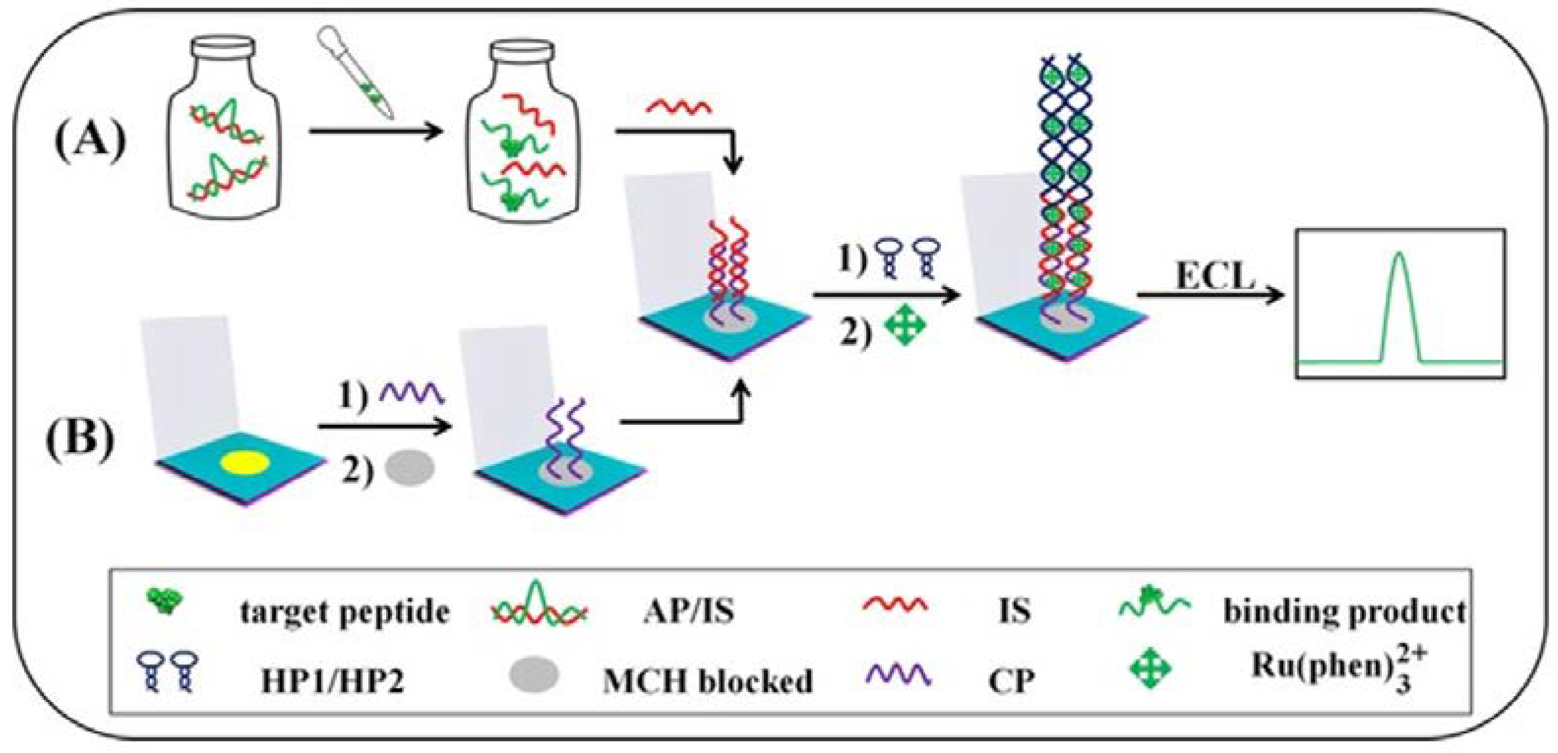
- Ma, C.; Liu, H.; Zhang, L.; Li, L.; Yan, M.; Yu, J.; Song, X. Microfluidic Paper-Based Analytical Device for Sensitive Detection of Peptides Based on Specific Recognition of Aptamer and Amplification Strategy of Hybridization Chain Reaction. ChemElectroChem 2017, 4, 1744–1749. [Google Scholar] [CrossRef]
- Zhang, X.; Bao, N.; Luo, X.; Ding, S.-N. Patchy gold coated Fe3O4 nanospheres with enhanced catalytic activity applied for paper-based bipolar electrode-electrochemiluminescence aptasensors. Biosens. Bioelectron. 2018, 114, 44–51. [Google Scholar] [CrossRef] [PubMed]
- Jiang, P.; He, M.; Shen, L.; Shi, A.; Liu, Z. A paper-supported aptasensor for total IgE based on luminescence resonance energy transfer from upconversion nanoparticles to carbon nanoparticles. Sens. Actuators B Chem. 2017, 239, 319–324. [Google Scholar] [CrossRef]
- Shajaripour Jaberi, S.Y.; Ghaffarinejad, A.; Omidinia, E. An electrochemical paper based nano-genosensor modified with reduced graphene oxide-gold nanostructure for determination of glycated hemoglobin in blood. Anal. Chim. Acta 2019, 1078, 42–52. [Google Scholar] [CrossRef]
- Wei, B.; Mao, K.; Liu, N.; Zhang, M.; Yang, Z. Graphene nanocomposites modified electrochemical aptamer sensor for rapid and highly sensitive detection of prostate specific antigen. Biosens. Bioelectron. 2018, 121, 41–46. [Google Scholar] [CrossRef]
- Liu, X.; Li, X.; Gao, X.; Ge, L.; Sun, X.; Li, F. A Universal Paper-Based Electrochemical Sensor for Zero-Background Assay of Diverse Biomarkers. ACS Appl. Mater. Interfaces 2019, 11, 15381–15388. [Google Scholar] [CrossRef]
- Yen, Y.-K.; Chao, C.-H.; Yeh, Y.-S. A Graphene-PEDOT:PSS Modified Paper-Based Aptasensor for Electrochemical Impedance Spectroscopy Detection of Tumor Marker. Sensors 2020, 20, 1372. [Google Scholar] [CrossRef] [PubMed]
- Xue, J.; Zhang, L.; Gao, C.; Zhu, P.; Yu, J. Microfluidic paper-based photoelectrochemical sensing platform with electron-transfer tunneling distance regulation strategy for thrombin detection. Biosens. Bioelectron. 2019, 133, 1–7. [Google Scholar] [CrossRef]
- Shi, H.; Ge, S.; Wang, Y.; Gao, C.; Yu, J. Wide-Spectrum-Responsive Paper-Supported Photoelectrochemical Sensing Platform Based on Black Phosphorus-Sensitized TiO2. ACS Appl. Mater. Interfaces 2019, 11, 41062–41068. [Google Scholar] [CrossRef]
- Azuaje-Hualde, E.; de Pancorbo, M.M.; Benito-Lopez, F.; Basabe-Desmonts, L. Paper based microfluidic platform for single-step detection of mesenchymal stromal cells secreted VEGF. Anal. Chim. Acta 2022, 1199, 339588. [Google Scholar] [CrossRef]
- Wang, Y.; Sun, S.; Luo, J.; Xiong, Y.; Ming, T.; Liu, J.; Ma, Y.; Yan, S.; Yang, Y.; Yang, Z.; et al. Low sample volume origami-paper-based graphene-modified aptasensors for label-free electrochemical detection of cancer biomarker-EGFR. Microsyst. Nanoeng. 2020, 6, 32. [Google Scholar] [CrossRef] [PubMed]
- Wu, L.; Ma, C.; Ge, L.; Kong, Q.; Yan, M.; Ge, S.; Yu, J. Paper-based electrochemiluminescence origami cyto-device for multiple cancer cells detection using porous AuPd alloy as catalytically promoted nanolabels. Biosens. Bioelectron. 2015, 63, 450–457. [Google Scholar] [CrossRef] [PubMed]
- Wu, L.; Zhang, Y.; Wang, Y.; Ge, S.; Liu, H.; Yan, M.; Yu, J. A paper-based electrochemiluminescence electrode as an aptamer-based cytosensor using PtNi@carbon dots as nanolabels for detection of cancer cells and for in-situ screening of anticancer drugs. Microchim. Acta 2016, 183, 1873–1880. [Google Scholar] [CrossRef]
- Su, M.; Ge, L.; Ge, S.; Li, N.; Yu, J.; Yan, M.; Huang, J. Paper-based electrochemical cyto-device for sensitive detection of cancer cells and in situ anticancer drug screening. Anal. Chim. Acta 2014, 847, 1–9. [Google Scholar] [CrossRef]
- Mishra, A.; Pilloton, R.; Jain, S.; Roy, S.; Khanuja, M.; Mathur, A.; Narang, J. Paper-Based Electrodes Conjugated with Tungsten Disulfide Nanostructure and Aptamer for Impedimetric Detection of Listeria monocytogenes. Biosensors 2022, 12, 88. [Google Scholar] [CrossRef]
- Dolai, S.; Tabib-Azar, M. Whole virus detection using aptamers and paper-based sensor potentiometry. Med. Devices Sens. 2020, 3, e10112. [Google Scholar] [CrossRef]
- Wang, H.; Zhou, C.; Sun, X.; Jian, Y.; Kong, Q.; Cui, K.; Ge, S.; Yu, J. Polyhedral-AuPd nanoparticles-based dual-mode cytosensor with turn on enable signal for highly sensitive cell evalution on lab-on-paper device. Biosens. Bioelectron. 2018, 117, 651–658. [Google Scholar] [CrossRef] [PubMed]
- Li, L.; Zhang, Y.; Ge, S.; Zhang, L.; Cui, K.; Zhao, P.; Yan, M.; Yu, J. Triggerable H2O2–Cleavable Switch of Paper-Based Biochips Endows Precision of Chemometer/Ratiometric Electrochemical Quantification of Analyte in High-Efficiency Point-of-Care Testing. Anal. Chem. 2019, 91, 10273–10281. [Google Scholar] [CrossRef]
- Grabowska, I.; Hepel, M.; Kurzątkowska-Adaszyńska, K. Advances in Design Strategies of Multiplex Electrochemical Aptasensors. Sensors 2022, 22, 161. [Google Scholar] [CrossRef]
- Khoshbin, Z.; Housaindokht, M.R.; Verdian, A. A low-cost paper-based aptasensor for simultaneous trace-level monitoring of mercury (II) and silver (I) ions. Anal. Biochem. 2020, 597, 113689. [Google Scholar] [CrossRef] [PubMed]
- Weng, X.; Neethirajan, S. Paper-based microfluidic aptasensor for food safety. J. Food Saf. 2018, 38, e12412. [Google Scholar] [CrossRef]
- Wang, Y.; Luo, J.; Liu, J.; Sun, S.; Xiong, Y.; Ma, Y.; Yan, S.; Yang, Y.; Yin, H.; Cai, X. Label-free microfluidic paper-based electrochemical aptasensor for ultrasensitive and simultaneous multiplexed detection of cancer biomarkers. Biosens. Bioelectron. 2019, 136, 84–90. [Google Scholar] [CrossRef]
- Su, M.; Ge, L.; Kong, Q.; Zheng, X.; Ge, S.; Li, N.; Yu, J.; Yan, M. Cyto-sensing in electrochemical lab-on-paper cyto-device for in-situ evaluation of multi-glycan expressions on cancer cells. Biosens. Bioelectron. 2015, 63, 232–239. [Google Scholar] [CrossRef]
- Somvanshi, S.B.; Ulloa, A.M.; Zhao, M.; Liang, Q.; Barui, A.K.; Lucas, A.; Jadhav, K.M.; Allebach, J.P.; Stanciu, L.A. Microfluidic paper-based aptasensor devices for multiplexed detection of pathogenic bacteria. Biosens. Bioelectron. 2022, 207, 114214. [Google Scholar] [CrossRef]
- Liang, L.; Su, M.; Li, L.; Lan, F.; Yang, G.; Ge, S.; Yu, J.; Song, X. Aptamer-based fluorescent and visual biosensor for multiplexed monitoring of cancer cells in microfluidic paper-based analytical devices. Sens. Actuators B Chem. 2016, 229, 347–354. [Google Scholar] [CrossRef]
- Wang, C.-H.; Wu, J.-J.; Lee, G.-B. Screening of highly-specific aptamers and their applications in paper-based microfluidic chips for rapid diagnosis of multiple bacteria. Sens. Actuators B Chem. 2019, 284, 395–402. [Google Scholar] [CrossRef]
- Naseri, M.; Ziora, Z.M.; Simon, G.P.; Batchelor, W. ASSURED-compliant point-of-care diagnostics for the detection of human viral infections. Rev. Med. Virol. 2022, 32, e2263. [Google Scholar] [CrossRef]
- Carrell, C.; Kava, A.; Nguyen, M.; Menger, R.; Munshi, Z.; Call, Z.; Nussbaum, M.; Henry, C. Beyond the lateral flow assay: A review of paper-based microfluidics. Microelectron. Eng. 2019, 206, 45–54. [Google Scholar] [CrossRef]
- Miodek, A.; Regan, E.M.; Bhalla, N.; Hopkins, N.A.E.; Goodchild, S.A.; Estrela, P. Optimisation and Characterisation of Anti-Fouling Ternary SAM Layers for Impedance-Based Aptasensors. Sensors 2015, 15, 25015–25032. [Google Scholar] [CrossRef]
- Spagnolo, S.; Davoudian, K.; De La Franier, B.; Hianik, T.; Thompson, M. Staphylococcus aureus Detection in Milk Using a Thickness Shear Mode Acoustic Aptasensor with an Antifouling Probe Linker. Biosensors 2023, 13, 614. [Google Scholar] [CrossRef] [PubMed]
- Rink, S.; Baeumner, A.J. Progression of Paper-Based Point-of-Care Testing toward Being an Indispensable Diagnostic Tool in Future Healthcare. Anal. Chem. 2023, 95, 1785–1793. [Google Scholar] [CrossRef] [PubMed]
- Cunningham, J.C.; DeGregory, P.R.; Crooks, R.M. New Functionalities for Paper-Based Sensors Lead to Simplified User Operation, Lower Limits of Detection, and New Applications. Annu. Rev. Anal. Chem. 2016, 9, 183–202. [Google Scholar] [CrossRef]


| Analyte (Sample) | PAD | Type of Paper | Detection | Aptamer Sequence | Ref. |
|---|---|---|---|---|---|
| K+ (urine) | Circular-laser printing | Glossy paper | Colorimetric with AuNPs | 5′-GGGTTAGGGTTAGGGTTAGGG-3′ | [63] |
| Pb2+ (water) | Y-shaped fluidic-laser cutting | Whatman No 1, nylon | Colorimetric with AuNPs | 5′-GGTTGGTGTGGTTGG-3′ | [64] |
| Pb2+ (tap water, lake water, milk, blood serum) | Square-craft punch | Whatman No 1 | FRET/GO | 5′-FAMGGGTGGGTGGGTGGGT-3′ | [65] |
| Hg2+ (water) | NR | Whatman No 1 | CL | (S1): 5′-NH2-(CH2)6-CAGTTTGGAC-3′ (S2): 5′-NH2-GTCCTTTCTG-3′ | [66] |
Disclaimer/Publisher’s Note: The statements, opinions and data contained in all publications are solely those of the individual author(s) and contributor(s) and not of MDPI and/or the editor(s). MDPI and/or the editor(s) disclaim responsibility for any injury to people or property resulting from any ideas, methods, instructions or products referred to in the content. |
© 2023 by the authors. Licensee MDPI, Basel, Switzerland. This article is an open access article distributed under the terms and conditions of the Creative Commons Attribution (CC BY) license (https://creativecommons.org/licenses/by/4.0/).
Share and Cite
Economou, A.; Kokkinos, C.; Bousiakou, L.; Hianik, T. Paper-Based Aptasensors: Working Principles, Detection Modes, and Applications. Sensors 2023, 23, 7786. https://doi.org/10.3390/s23187786
Economou A, Kokkinos C, Bousiakou L, Hianik T. Paper-Based Aptasensors: Working Principles, Detection Modes, and Applications. Sensors. 2023; 23(18):7786. https://doi.org/10.3390/s23187786
Chicago/Turabian StyleEconomou, Anastasios, Christos Kokkinos, Leda Bousiakou, and Tibor Hianik. 2023. "Paper-Based Aptasensors: Working Principles, Detection Modes, and Applications" Sensors 23, no. 18: 7786. https://doi.org/10.3390/s23187786
APA StyleEconomou, A., Kokkinos, C., Bousiakou, L., & Hianik, T. (2023). Paper-Based Aptasensors: Working Principles, Detection Modes, and Applications. Sensors, 23(18), 7786. https://doi.org/10.3390/s23187786