Article

Structure, Luminescence and Temperature Detection Capability of [C(NH2)3]M(HCOO)3 (M = Mg2+, Mn2+, Zn2+) Hybrid Organic–Inorganic Formate Perovskites Containing Cr3+ Ions

Włodzimierz Trzebiatowski Institute of Low Temperature and Structure Research, Polish Academy of Sciences, 50-422 Wroclaw, Poland
*
Author to whom correspondence should be addressed.
Sensors 2023, 23(14), 6259; https://doi.org/10.3390/s23146259
Submission received: 30 May 2023 / Revised: 6 July 2023 / Accepted: 7 July 2023 / Published: 9 July 2023
(This article belongs to the Special Issue Optical Thermometry: Concepts, Methods, and Applications)

Abstract

:
Metal-organic frameworks are of great interest to scientists from various fields. This group also includes organic–inorganic hybrids with a perovskite structure. Recently their structural, phonon, and luminescent properties have been paid much attention. However, a new way of characterization of these materials has become luminescence thermometry. Herein, we report the structure, luminescence, and temperature detection ability of formate organic–inorganic perovskite [C(NH2)3]M(HCOO)3 (Mg2+, Mn2+, Zn2+) doped with Cr3+ ions. Crystal field strength (Dq/B) and Racah parameters were determined based on diffuse reflectance spectra. It was shown that Cr3+ ions are positioned in the intermediate crystal field or close to it with a Dq/B range of 2.29–2.41. The co-existence of the spin-forbidden and spin-allowed transitions of Cr3+ ions enable the proposal of an approach for remote readout of the temperature. The relative sensitivity (Sr) can be easily modified by sample composition and Cr3+ ions concentration. The luminescent thermometer based on the 2E/4T2g transitions has the relative sensitivity Sr of 2.08%K−1 at 90 K for [C(NH2)3]Mg(HCOO)3: 1% Cr3+ and decrease to 1.20%K−1 at 100 K and 1.08%K−1 at 90 K for Mn2+ and Zn2+ analogs, respectively.

1. Introduction

The noticeable development of hybrid organic–inorganic perovskites (HOIPs) has been observed in recent years. The materials with the general formula ABX3, where A is an inorganic or organic cation (e.g., NH4+, (CH3)2NH+), B is a divalent metal ion (e.g., Pb2+, Zn2+), and X a monovalent anion (e.g., Cl, HCOO) have attracted increasing attention due to their extraordinary properties [1,2,3]. Hybrid materials, e.g., CH3NH3PbCl3, have been particularly implemented in state-of-the-art photovoltaic devices [4,5,6]. However, their potential usefulness is significantly greater due to their characteristics, including ferroelectricity [7,8], magnetic [9], optoelectronic [4], and luminescent properties [10,11,12,13]. The characteristics of investigated materials can be widely tuned by the replacement of A, B, and X linkers [10,14].
Among various materials, the perovskite-like metal-organic frameworks (MOFs) containing formate anions (HCOO) exhibit unique features, such as ferroelectricity, multiferroicity, and luminescence [1,10,15]. Particularly, the group of Cr-based materials shows strong luminescence and weak concentration quenching [2,10,15]. Nevertheless, temperature-dependent luminescence is one of the most outstanding phenomena. The temperature change induces the change in energy level populations, which makes formate-based compounds containing Cr3+ ions sufficient materials for non-contact temperature sensing [10].
The optical properties of the transition metals (TM), including chromium trivalent ions, can be affected by the crystal field (CF) strength [13,14,16]. The change in the CF strength leads to the change in the dominant transition type [17]. The Cr3+ ions luminescence may contain two particular emission bands: narrow spin-forbidden 2Eg4A2g (around 700 nm) and broad spin-allowed 4T2g4A2g (around 750 nm). In low temperatures, the narrow 2Eg4A2g emission is dominant. The increase in temperature induces the thermal population of the 4T2g level and, consequently, promotes the broad 4T2g4A2g emission. The narrow emission takes place in a strong CF environment. The spin-allowed emission, in turn, occurs in a weak crystal field strength. The coexistence of both types of emission indicates the intermediate CF strength. The progressive increase in temperature leads to luminescence quenching. The significant influence of temperature on spectroscopic characteristics of Cr3+-based materials has become the basis of the thermometric model development [10,13].
Luminescence temperature sensing has attracted increasing attention recently [10,18,19,20,21,22]. Non-contact thermometry has great potential for application in scientific, industrial, and biomedical areas [23,24]. Among various advantages, the high accuracy and single measurement speed are noteworthy. The possibility of the plunge measurements going beyond typical thermal imaging limitations makes this approach a promising tool for industrial process monitoring [10].
Temperature sensing is mainly based on the detectable change in the luminescent properties induced by the change in the temperature. A thermometric model can be developed by monitoring changes in lifetime, peak position, as well as the insensitivities of specific peaks [20,25]. The comparison of the intensities of two temperature-dependent emission bands allows us to determine a thermometric parameter called fluorescence intensity ratio (FIR or Δ). Such an approach is called the ratiometric method and has been the most frequently reported application recently [18]. The methods relying on FIR analysis provide high sensitivity and make it possible to implement the independent sensing ranges, which leaves room for model optimization [13,26].
The vast majority of reported thermometric compounds are based on inorganic host materials with rare-earth (RE) elements as dopants [11,25,27,28]. However, the materials containing transition metal ions exhibit promising thermometric characteristics comparable to the solutions based on RE ions [29,30]. The highly sensitive thermometric properties have been reported, inter alia, for the perovskite materials containing ethylammonium cation and Cr3+ ions [10]. The development of luminescent thermometers based solely on chromium trivalent ions is a noteworthy approach enabling to deviate from the RE-based materials. Another notable strategy for the development of the ratiometric thermometer, presented in this work, is not only considering the luminescence of Cr3+ ions but also using the luminescence of the amine group, such as guanidinium cation ([C(NH2)3]+ denoted as GA+). The multicomponent thermometric model may be a promising approach toward higher sensitivity.
Herein, we report the synthesis as well as the structural and spectroscopic properties of the first metal–organic framework luminescent thermometers based on both GA+ and Cr3+ ion luminescence. Investigated series of [GA]M1−xCrx(HCOO)3, where M = Mg2+, Mn2+, Zn2+, and x = 0, 0.01, 0.03, 0.05, have been synthesized and investigated as promising thermometric materials. The selection of three distinct cations was motivated by the fact that Mn2+ ions are the only ones that are optically active, and Zn2+ and Mg2+ ions create structures with different properties compared to transition metal ions such as Mn2+. All series exhibit outstanding temperature-dependent emission, which has become the basis of the thermometric analysis. This work is an attempt to describe the effect of the material composition on the luminescent properties with particular emphasis on luminescent thermometry. The optimization of the sensing range estimation is particularly considered.

2. Materials and Methods

The starting materials include formic acid HCOOH (POCH, ≥98%), ethanol C2H5OH (POCH, 96%), guanidine carbonate salt [GA] [C(NH2)3]2CO3, (Sigma Aldrich, 99%) (Sigma Aldrich, Saint Louis, MI, USA), zinc(II) chloride ZnCl2 (Sigma Aldrich, 99.999%) (Sigma Aldrich, Saint Louis, USA), manganese(II) perchlorate hydrate Mn(ClO4)2⋅6H2O (Sigma Aldrich, ≥99%) (Sigma Aldrich, Saint Louis, USA), magnesium(II) chloride anhydrous MgCl2 (Sigma Aldrich, 99.9%), and chromium(III) chloride CrCl3 (Sigma Aldrich, 99%). All precursors were commercially available and were used for the synthesis without any further purification. In this study, a series of [GA]M1−xCrx(HCOO)3 where M = Mn, Mg, Zn, and x = 0, 1%, 3%, 5%, were obtained by using the low-diffusion synthesis method. To grow [GA]M1−xCrx(HCOO)3 crystals, at first formic acid (8.7 mmol) and GA (4.2 mmol) was dissolved in distilled water (20 mL). This solution was added by an aqueous solution (10 mL) containing 1.0 mmol of Mn(ClO4)2⋅6H2O/ZnCl2/MgCl2 for the pure samples. The amount of Cr3+ ions was calculated based on the molarity of the M2+ ions (see Tables S1–S3). The resulting mixed solution was kept undisturbed and allowed to evaporate slowly. After two weeks, the crystals were harvested, washed with ethanol, and dried in the air. The color of the crystals was light pink for Mn or white for Mg and Zn. It also varied from green to dark green depending on the concentration of Cr3+ ions.
The powder X-ray diffraction (XRD) patterns were obtained on an X’Pert Pro X-ray diffraction system (Malvern Panalytical, Malvern, UK) equipped with a PIXcel detector (Malvern Panalytical, Malvern, UK) and using CuKα radiation (λ = 1.54056 Å). The Raman spectra were measured using a Bruker FT 110/S (Billerica, MA, USA) spectrometer operating at 1064 nm (Nd:YAG). The spectra were collected in a spectral range of 75–3200 cm−1 and with a spectral resolution of 2 cm−1. The diffuse reflectance spectra were obtained using a Varian Cary 5E UV–VIS–NIR spectrometer (Varian, Palo Alto, CA, USA). The temperature-dependent emission spectra were obtained with a Hamamatsu PMA-12 (Hamamatsu Photonics, Iwata, Japan) photonic multichannel analyzer combined with a BT-CCD sensor. As an excitation source, a 405 nm laser diode was used. The temperature was controlled by a Linkam THMS600 stage (Linkam, Tadworth, UK).

3. Results and Discussion

3.1. Structural Properties

The phase purity of all samples was confirmed by the XRD patterns with a simulation of the single-crystal structural data of [GA]Mn1−xCrx(HCOO)3 (Figure 1). The samples with Mn2+ and Zn2+ crystallized in the orthorhombic Pnna crystal structure [31], and the details of the crystal structure of analogs with Mg2+ remain unknown. In general, the formate in-connected MnO6 framework crates cavities occupied by GA+ cations (see Figure 2). The right-shifting of the diffraction lines was observed due to the partial replacement of Mn2+ (CR = 81 Å), Mg2+ (CR = 86 Å), and Zn2+ ions (CR = 88 Å) by Cr3+ ions (CR = 75.5 Å). The crystal radius (CR) was obtained from Shannon [32]. No additional phases were detected, which indicates that the Cr3+ ions were substituted by the cation M.
The Raman spectra of the [GA]M1−xCrx(HCOO)3 series, where M = Mg2+, Mn2+, Zn2+, and x = 0, 0.01, 0.03, 0.05, are marked in Figure 3a as GAMg, GAMn, and GAZn, respectively. All spectra are very similar and are consistent with the reported orthorhombic Pnna symmetry of all crystals [31,33,34,35]. However, some differences can be seen in the band shifts and the number of components, which are due to the different sizes, masses, and electronegativity of the metal cations that build the crystals. All these parameters affect the sizes of unit cells, causing Raman bands for [GA]M(HCOO)3 (M = Mg2+, Mn2+, Zn2+) to be shifted relative to each other (Figure 3b,c and Table S4). Regarding [GA]Zn(HCOO)3, the upshifts observed for [GA]Mg(HCOO)3 are most pronounced for lattice modes observed below 300 cm−1 since they are very sensitive to the long-range order in the crystal. In addition, strong shifts towards higher wavenumbers, up to 12.3 cm−1 for [GA]Mg(HCOO)3 and 10.3 cm−1 for [GA]Mg(HCOO)3, are also observed for NH stretching vibrations above 2850 cm−1 (Figure 3d), which further indicate the weakest hydrogen bonds in the [GA]Mg(HCOO)3 crystal and stronger for [GA]Zn(HCOO)3. The upshift of 7.1 cm−1 when Zn2+ ions are replaced by Mg2+ was evidenced by bands associated with vibrations of oxygen atoms directly coordinated by metal ions, i.e., ν2 + ν5 that have been assigned to symmetric C–O stretching vibrations coinciding with C–H in-plane bending modes, respectively (Figure 3b) [36]. A much weaker upshift is observed for the stretching C–N modes, reaching 3.1 cm−1 for [GA]Mg(HCOO)3 and 3.2 cm−1 for [GA]Mn(HCOO)3 related to [GA]Zn(HCOO)3 (Figure 3a). This finding indicates very similar confinement of GA+ cations and similar dynamics in the perovskite void for M = Mg2+ and Mn2+.
The introduction of Cr3+ ions into the crystal structure of [GA]M(HCOO)3 (M = Mg2+, Mn2+, Zn2+) at such low concentrations causes very subtle effects on the spectra, not exceeding 1 cm−1. This confirms that aliovalent doping up to 5 mol% does not cause significant structural changes in the orthorhombic Pnna structure.

3.2. Optical Properties and Temperature Detection

The diffuse reflectance spectra (DRS) of representative samples [GA]M1−xCrx(HCOO)3, where M = Mg2+, Mn2+, Zn2+, and x = 0.05, are shown in Figure 4. The intensity of the DRS spectrum is influenced by many factors, such as the size and position of crystallites [10]. Therefore, the DRS is used only for characterizing the localization of the energy levels of Cr3+ ions in each compound and the effect of the concentration of Cr3+ ions on the spectrum’s shape. Two primary broad bands localized around 16,828 cm−1 (594 nm) and 22,522 cm−1 (444 nm) for Zn-samples, 17,130 cm−1 (583.8 nm) and 23,162 cm−1 (431.7 nm) for Mg-samples, 17,050 cm−1 (586.5 nm) and 23,162 cm−1 (431.7 nm) for Mn-samples can be distinguished in Figure 3. These two bands are assigned to the spin-allowed transitions 4A2g4T1g and 4A2g4T2g of Cr3+ ions. In addition, a very weak and sharp peak centered at approximately 14,550 cm−1 (687.3 nm) is associated with the spin-forbidden transition from the 4A2g ground state to the 2E excited level. It was found that when the concentration of Cr3+ increases, the position of the 4A2g2E lines slightly changes (Figures S1 and S2). However, for the Zn-compounds, the 4A2g2E absorption peak is invisible (Figure S3).
Noticeably, in the spectrum of Mn-samples, very weak and sharp peaks appeared at 29,240 cm−1 (342 nm) and 24,570 cm−1 (407 nm), which are attributed to the absorption of Mn2+ ions from 6A1 ground state to 4E, 4T2, and 4A1, 4E excited levels, respectively. The intensity of these bands decreases as the content of Cr3+ increases (Figures S1–S3).
In addition, the intense band located at around 46,729 cm−1 (214 nm) can be assigned to host absorption, and it moved to 44,643 cm−1 (224 nm) for the Mn-based sample. What is more, the bad is much broader because of overlapping with the Mn-O charge transfer band (CTB) (Figure S4).
The crystal field Dq, Racah, B, and C parameters were calculated for Cr3+-doped samples (see Table 1) by using the same methodology as presented in reference [10]. Crystal field strength (CFS) Dq/B parameter is in the range of 2.29–2.39 for GAMn and 2.23–2.41 for GAMg samples. These results mean that Cr3+ ions are located in the intermediate ligand field, and energy separation between 2E and 4T2g excited levels is not significant. The Dq/B parameter is slightly higher, around 2.41–2.43 for CAZn analogs. The calculated values of Dq/B are similar to those reported recently for DMANaCr (2.29) [15]. However, for some EA and DMA analogs (EANaCr 2.18 [7], EANaAlCr 2.21 [7], DMAKCr 2.21 [37], EAKCr 2.21 [37]) reported Dq/B values are much lower than for the investigated perovskites. On the other hand, the formate perovskites with AM+ cation comprising Cr3+ ions exhibit a strong crystal field (AMNaCr 2.743 [38], AMNaAlCr 2.55 [38]).
The emission spectra of investigated hybrid organic–inorganic formates [GA]M1−xCrx(HCOO)3 (M = Mg2+, Mn2+, Zn2+, and x = 0.01, 0.03, 0.05) recorded at 80 K consists of the intense and narrow emission lines of Cr3+ ions located at 686 nm and 698 nm attributed to the spin-forbidden 2E → 4A2g transitions (Figure 5). The broad emission band, which spans from 700 nm to 1000 nm, assigned to the spin-allowed transition from the 4T2g excited level to the 4A2g ground state is also observed [11,13,16,39]. As can be seen in Figure 5b,d and Figure S5–S7, the emission intensity of GAMg and GAMn samples increased with the concentration of dopant ions, while the intensity of 1% Cr3+ and 5% Cr3+ in the GAZn analog are comparable. The samples with 3% of Cr3+ are out of the trend. However, the nature of this behavior is unspecified. The collation of the representative samples [GA]M1−xCrx(HCOO)3 (M = Mg2+, Mn2+, Zn2+, and x = 0.05) showed that the most intense luminescence exhibits a sample comprising Mg2+ ions. The emission intensity of Mn2+ and Zn2+ samples is significantly less. The substitution of different metal M2+ ions in the crystal structure of guanidine formate have an impact on the intensity relationships between spin-forbidden and spin-allowed transition of Cr3+ ions. Only for the GAMg compound 2E → 4A2g is emission more intense than spin-allowed transition; for GAMn and GAZn analogs, 4T2g4A2g transition dominates. It is worth noting that no emission of Mn2+ ions was detected, probably due to energy reabsorption by chromium ions.
The emission spectra in the function of temperature were recorded within the range of 80–300 K with 10 K steps. As can be seen in Figure 6 and Figure S8, the main component of the photoluminescence spectra belongs to the spin-allowed transitions of Cr3+ ions. Only for the GAMg sample containing 1% dopant, the 2E emission is much more intense than the band located at 795 nm. Generally, 2E → 4A2g emission quenches significantly with increasing temperature, while the 4T2g4A2g emission of Cr3+ is more stable. It is due to the thermally stimulated energy transfer from 2E to 4T2g energy level. Obtained results confirmed the occurrence of the intermediate ligand field in the nearest environment of Cr3+ ions. The mechanism of Cr3+ luminescence quenching is a well-known phenomenon in the literature and assumes crossing the 4T2g excited state parabola with the 4A2g one [11,13,16,39].
The significant dependence of photoluminescence intensity on temperature may be an interesting behavior that can be exploited for non-contact temperature readout based on luminescence. Figure 7 demonstrates a schematic representation of the approach to temperature determination. In this model, the Fluorescence Intensity Ratio (FIR) parameter can be defined as a ratio of the 2E → 4A2g (spectral range 670–710 nm marked as I1) to the 4T2g4A2g (spectral range 750–1050 nm represented as I2) transition of Cr3+ ions, respectively.
The proposed model was tested on the investigated [GA]M1−xCrx(HCOO)3 (M = Mg2+, Mn2+, Zn2+, and x = 0.01, 0.03, 0.05) hybrid organic–inorganic perovskites. It is clear that the increase in temperature causes decreasing in FIR (Figure 8), and the highest value of FIR was obtained for the GAMg: 1% Cr3+ sample. To further comparison of the observed changes in thermometric parameters and to compare their features, the absolute (Sa) and relative (Sr) sensitivities were calculated as follows:
S a = d F I R d T ,
and
S r = 1 F I R d F I R d T ,
where dFIR represents the change of fluorescence intensity ratio at temperature change ΔT. The collation of Sa and Sr changes of the investigated hybrid organic–inorganic perovskites are presented in Figure 9 and Figure S9. Generally, the Sa and Sr values are the highest at the 80–120 K range and decrease with increasing temperature. However, the sensitivity changes with sample composition and concentration of Cr3+ ions. For GAMg: Cr3+ compounds, the significant relative sensitivity exhibits sample with the lowest concentration of dopant ions and equals 2.08%K−1 at 90 K. With increasing Cr3+ ions concentration, the Sr decreased to around 1%K−1. Substitution of Mg2+ by Mn2+ caused a decrease of sensitivity to 1.20%K−1, but the optimal Cr3+ ions concentration was determined to be 3%. Similar trends are observed for GAZn: for Cr3+ analogs, however, the changes of Sr with chromium ions concentration are negligible, and the highest Sr is 1.08%K−1 at 90 K for GAZn: 1% Cr3+. Additionally, the repeatability of the thermal sensing performance of representative samples was verified by the circling heat/cool process (Figure S10). It can be seen that only a small variation from the initial value was observed, and the temperature parameter ∆ is reversed and repeated overheating/cooling cycles.
In fact, only one optical thermometer based on hybrid organic–inorganic formate perovskites [EA]2NaCr0.21Al0.79(HCOO)6 with a sensitivity Sr of 2.84%K−1 at 160 K is known [10]. Obtained values of relative sensitivities were compared with the Sr values of other inorganic and hybrid organic–inorganic luminescent thermometers (Table 2). The results show that investigated [GA]M1−xCrx(HCOO)3 (M = Mg2+, Mn2+, Zn2+, and x = 0.01, 0.03, 0.05) has the potential to be applied as a low-temperature luminescent thermometer.

4. Conclusions

Three series of samples [C(NH2)3]M(HCOO)3 (Mg2+, Mn2+, Zn2+) doped with 1%, 3%, and 5% of Cr3+ ions were synthesized using the low-diffusion synthesis method. Their structural, phonon, and luminescent properties were investigated in detail. It was shown that the incorporation of Cr3+ ions into the crystal structure of investigated hybrid organic–inorganic perovskites does not affect the phase purity of the samples. Based on diffuse reflectance spectra, crystal field strength (Dq/B) and Racah parameters were determined. It was found that Cr3+ ions are located in the intermediate crystal field or close to it with a Dq/B range of 2.29–2.41. The investigation of sample composition showed that the highest emission intensity exhibits GAMg: 5% Cr3+ sample, while the lowest one GAZn: 5% Cr3+. The presence of both the spin-forbidden and spin-allowed transitions of Cr3+ ions at a broad temperature range enables the characterization of these materials as luminescence thermometers. It turned out that the relative sensitivity of Sr depends on the sample composition and concentration of Cr3+ ions. The highest relative sensitivity Sr = 2.08%K−1 at 90 K has [GA]Mg(HCOO)3: 1% Cr3+. Replacement of Mg2+ by Mn2+ or Zn2+ reduced the sensitivity to 1.20%K−1 at 100 K and 1.08%K−1 at 90 K for [GA]Mn(HCOO)3: 3% Cr3+ and [GA]Zn(HCOO)3: 1% Cr3+, respectively.

Supplementary Materials

The following supporting information can be downloaded at: https://www.mdpi.com/article/10.3390/s23146259/s1, Figure S1. Diffuse reflectance spectra of a series of [GA]Mn1−xCrx(HCOO)3 (x = 0, 0.01, 0.03, 0.05) measured at 300 K.; Figure S2. Diffuse reflectance spectra of a series of [GA]Mg1−xCrx(HCOO)3 (x = 0, 0.01, 0.03, 0.05) measured at 300 K.; Figure S3. Diffuse reflectance spectra of a series of [GA]Zn1−xCrx(HCOO)3 (x = 0, 0.01, 0.03, 0.05) measured at 300 K.; Figure S4. Deconvolution of the absorption spectrum of [GA]Mn1−xCrx(HCOO)3 (x = 0.05) measured at 300 K.; Figure S5. Low-temperature emission spectra of [GA]Mg1−xCrx(HCOO)3 (x = 0.01, 0.03, 0.05).; Figure S6. Low-temperature emission spectra of [GA]Mn1−xCrx(HCOO)3 (x = 0.01, 0.03, 0.05).; Figure S7. Low-temperature emission spectra of [GA]Zn1−xCrx(HCOO)3 (x = 0.01, 0.03, 0.05).; Figure S8. Temperature-dependent emission spectra of [GA]Mn1−xCrx(HCOO)3 x = 0.01 (a), [GA]Mn1−xCrx(HCOO)3 x = 0.03 (b), [GA]Mg1−xCrx(HCOO)3 x = 0.01 (c), [GA]Mg1−xCrx(HCOO)3 x = 0.03 (d), [GA]Zn1−xCrx(HCOO)3 x = 0.01 (e), and [GA]Zn1−xCrx(HCOO)3 x = 0.03 (f) samples.; Figure S9. Influence of Cr3+ ions concentration on absolute sensitivity (Sa) (a–c) of [GA]M1−xCrx(HCOO)3 (M = Mg2+, Mn2+, Zn2+, and x = 0.01, 0.03, 0.05) hybrid perovskites.; Figure S10. Repeatability of ∆ temperature parameter of I1/I2 emission evaluated at 80 K and 100 K during 10 heating/cooling cycles of (a) [GA]Mg1−xCrx(HCOO)3 x = 0.01, (b) [GA]Mn1−xCrx(HCOO)3 x = 0.03, and (c) [GA]Zn1−xCrx(HCOO)3 x = 0.01.; Table S1. Quantities of precursors used for the syntheses of the series of [GA]Mn1−xCrx(HCOO)3.; Table S2. Quantities of precursors used for the syntheses of the series of [GA]Mg1−xCrx(HCOO)3.; Table S3. Quantities of precursors used for the syntheses of the series of [GA]Zn1-xCrx(HCOO)3.; Table S4. Lattice parameters and calculated factors (doct, average MII–O bond length; Voct, MIIO6 octahedral volume; σ2, bond angle variance; Δ, distortion index) [10.1107/S0021889811038970] for [GA]Mn(HCOO)3 and [GA]Zn(HCOO)3 based on the crystal data published in [10.1002/chem.200901605].; Determination of δ T . References [25,52,53] are cited in Supplementary Materials.

Author Contributions

Conceptualization, D.S.; methodology, D.S.; validation, D.S.; formal analysis, D.S., A.K., T.H.Q.V., M.A. and M.P.; investigation, D.S., A.K., T.H.Q.V., M.A. and M.P.; data curation, D.S., M.A. and M.P.; writing—original draft preparation, D.S., A.K., T.H.Q.V., M.A. and M.P.; writing—review and editing, D.S., A.K., T.H.Q.V., M.A. and M.P.; visualization, D.S., A.K., T.H.Q.V., M.A. and M.P.; supervision, D.S.; project administration, D.S.; funding acquisition, D.S. All authors have read and agreed to the published version of the manuscript.

Funding

This research was funded in whole by the National Science Centre, Poland, under project no. UMO-2020/39/D/ST5/01289. For the purpose of open access, the author has applied for a CC-BY public copyright license to any Author Accepted Manuscript (AAM) version arising from this submission.

Data Availability Statement

Experimental data: The Raman and diffuse reflectance spectra, temperature-dependent luminescence and emission maps, thermometric parameters, powder XRD data, and low-temperature emission spectra are available at 10.5281/zenodo.7970355.

Acknowledgments

The authors would also like to thank E. Bukowska for the XRD measurements and B. Macalik for the absorption measurements.

Conflicts of Interest

The authors declare no conflict of interest.

References

  1. Ptak, M.; Sieradzki, A.; Šimėnas, M.; Maczka, M. Molecular Spectroscopy of Hybrid Organic–Inorganic Perovskites and Related Compounds. Coord. Chem. Rev. 2021, 448, 214180. [Google Scholar] [CrossRef]
  2. Ptak, M.; Zarychta, B.; Stefańska, D.; Ciupa, A.; Paraguassu, W. Novel Bimetallic MOF Phosphors with an Imidazolium Cation: Structure, Phonons, High- Pressure Phase Transitions and Optical Response. Dalt. Trans. 2019, 48, 242–252. [Google Scholar] [CrossRef] [PubMed]
  3. Drozdowski, D.; Fedoruk, K.; Kabański, A.; Maczka, M.; Sieradzki, A.; Gągor, A. Broadband Yellow and White Emission from Large Octahedral Tilting in (110)-Oriented Layered Perovskites: Imidazolium-Methylhydrazinium Lead Halides. J. Mater. Chem. C 2023, 11, 4907–4915. [Google Scholar] [CrossRef]
  4. Prochowicz, D.; Franckevičius, M.; Cieślak, A.M.; Zakeeruddin, S.M.; Grätzel, M.; Lewiński, J. Mechanosynthesis of the Hybrid Perovskite CH3NH3PbI3: Characterization and the Corresponding Solar Cell Efficiency. J. Mater. Chem. A 2015, 3, 20772–20777. [Google Scholar] [CrossRef]
  5. Marimuthu, T.; Yuvakkumar, R.; Kumar, P.S.; Vo, D.V.N.; Xu, X.; Xu, G. Two-Dimensional Hybrid Perovskite Solar Cells: A Review. Environ. Chem. Lett. 2022, 20, 189–210. [Google Scholar] [CrossRef]
  6. Kim, J.Y.; Lee, J.W.; Jung, H.S.; Shin, H.; Park, N.G. High-Efficiency Perovskite Solar Cells. Chem. Rev. 2020, 120, 7867–7918. [Google Scholar] [CrossRef] [PubMed]
  7. Ptak, M.; Maczka, M.; Gagor, A.; Sieradzki, A.; Bondzior, B.; Dereń, P.; Pawlus, S. Phase Transitions and Chromium(III) Luminescence in Perovskite-Type [C2H5NH3][Na0.5CrxAl0.5- x(HCOO)3] (x = 0, 0.025, 0.5), Correlated with Structural, Dielectric and Phonon Properties. Phys. Chem. Chem. Phys. 2016, 18, 29629–29640. [Google Scholar] [CrossRef]
  8. Huang, C.R.; Luo, X.; Chen, X.G.; Song, X.J.; Zhang, Z.X.; Xiong, R.G. A Multiaxial Lead-Free Two-Dimensional Organic-Inorganic Perovskite Ferroelectric. Natl. Sci. Rev. 2021, 8, nwaa232. [Google Scholar] [CrossRef]
  9. Wang, Z.C.; Rogers, J.D.; Yao, X.; Nichols, R.; Atay, K.; Xu, B.; Franklin, J.; Sochnikov, I.; Ryan, P.J.; Haskel, D.; et al. Colossal Magnetoresistance without Mixed Valence in a Layered Phosphide Crystal. Adv. Mater. 2021, 33, 2005755. [Google Scholar] [CrossRef]
  10. Kabański, A.; Ptak, M.; Stefańska, D. Metal-Organic Framework Optical Thermometer Based on Cr3+ Ion Luminescence. ACS Appl. Mater. Interfaces 2023, 15, 7074–7082. [Google Scholar] [CrossRef]
  11. Stefańska, D.; Bondzior, B.; Vu, T.H.Q.; Grodzicki, M.; Dereń, P.J. Temperature Sensitivity Modulation through Changing the Vanadium Concentration in a La2MgTiO6:V5+,Cr3+ double Perovskite Optical Thermometer. Dalt. Trans. 2021, 50, 9851–9857. [Google Scholar] [CrossRef]
  12. Wu, Y.; Fan, W.; Gao, Z.; Tang, Z.; Lei, L.; Sun, X.; Li, Y.; Cai, H.L.; Wu, X. New Photoluminescence Hybrid Perovskites with Ultrahigh Photoluminescence Quantum Yield and Ultrahigh Thermostability Temperature up to 600 K. Nano Energy 2020, 77, 105170. [Google Scholar] [CrossRef]
  13. Stefańska, D.; Vu, T.H.Q.; Dereń, P.J. Multiple Ways for Temperature Detection Based on La2MgTiO6 Double Perovskite Co-Doped with Mn4+ and Cr3+ Ions. J. Alloys Compd. 2023, 938, 22–26. [Google Scholar] [CrossRef]
  14. Ptak, M.; Dziuk, B.; Stefańska, D.; Hermanowicz, K. The Structural, Phonon and Optical Properties of [CH3NH3]M0.5Cr:XAl0.5-x(HCOO)3 (M = Na, K.; X = 0, 0.025, 0.5) Metal-Organic Framework Perovskites for Luminescence Thermometry. Phys. Chem. Chem. Phys. 2019, 21, 7965–7972. [Google Scholar] [CrossRef]
  15. Mączka, M.; Bondzior, B.; Dereń, P.; Sieradzki, A.; Trzmiel, J.; Pietraszko, A.; Hanuza, J. Synthesis and Characterization of [(CH3)2NH2][Na0.5Cr0.5(HCOO)3]: A Rare Example of Luminescent Metal-Organic Frameworks Based on Cr(III) Ions. Dalt. Trans. 2015, 44, 6871–6879. [Google Scholar] [CrossRef] [PubMed]
  16. Dereń, P.J.; Malinowski, M.; Strȩk, W. Site Selection Spectroscopy of Cr3+ in MgAl2O4 Green Spinel. J. Lumin. 1996, 68, 91–103. [Google Scholar] [CrossRef]
  17. Lin, H.; Bai, G.; Yu, T.; Tsang, M.K.; Zhang, Q.; Hao, J. Site Occupancy and Near-Infrared Luminescence in Ca3Ga2Ge3O12: Cr3+ Persistent Phosphor. Adv. Opt. Mater. 2017, 5, 1700227. [Google Scholar] [CrossRef]
  18. Bolek, P.; Zeler, J.; Carlos, L.D.; Zych, E. Mixing Phosphors to Improve the Temperature Measuring Quality. Opt. Mater. 2021, 122, 111719. [Google Scholar] [CrossRef]
  19. Yin, H.Q.; Yin, X.B. Metal-Organic Frameworks with Multiple Luminescence Emissions: Designs and Applications. Acc. Chem. Res. 2020, 53, 485–495. [Google Scholar] [CrossRef]
  20. Maturi, F.E.; Brites, C.D.S.; Ximendes, E.C.; Mills, C.; Olsen, B.; Jaque, D.; Ribeiro, S.J.L.; Carlos, L.D. Going Above and Beyond: A Tenfold Gain in the Performance of Luminescence Thermometers Joining Multiparametric Sensing and Multiple Regression. Laser Photonics Rev. 2021, 15, 2100301. [Google Scholar] [CrossRef]
  21. Sójka, M.; Brites, C.D.S.; Carlos, L.D.; Zych, E. Exploiting Bandgap Engineering to Finely Control Dual-Mode Lu2(Ge,Si)O5:Pr3+ luminescence Thermometers. J. Mater. Chem. C 2020, 8, 10086–10097. [Google Scholar] [CrossRef]
  22. Sójka, M.; Runowski, M.; Zheng, T.; Shyichuk, A.; Kulesza, D.; Zych, E.; Lis, S. Eu2+ emission from Thermally Coupled Levels—New Frontiers for Ultrasensitive Luminescence Thermometry. J. Mater. Chem. C 2022, 10, 1220–1227. [Google Scholar] [CrossRef]
  23. del Rosal, B.; Ximendes, E.; Rocha, U.; Jaque, D. In Vivo Luminescence Nanothermometry: From Materials to Applications. Adv. Opt. Mater. 2017, 5, 1600508. [Google Scholar] [CrossRef]
  24. Marciniak, L.; Bednarkiewicz, A. Nanocrystalline NIR-to-NIR Luminescent Thermometer Based on Cr3+,Yb3+ Emission. Sens. Actuators B Chem. 2017, 243, 388–393. [Google Scholar] [CrossRef]
  25. Brites, C.D.S.; Millán, A.; Carlos, L.D. Lanthanides in Luminescent Thermometry. Handb. Phys. Chem. Rare Earths 2016, 49, 339–427. [Google Scholar] [CrossRef]
  26. Łukaszewicz, M.; Tomala, R.; Lisiecki, R. From Upconversion to Thermal Radiation: Spectroscopic Properties of a Submicron Y2O3:Er3+,Yb3+ Ceramic under IR Excitation in an Extremely Broad Temperature Range. J. Mater. Chem. C 2020, 8, 1072–1082. [Google Scholar] [CrossRef]
  27. Gavrilović, T.V.; Jovanović, D.J.; Lojpur, V.; Dramićanin, M.D. Multifunctional Eu3+- and Er3+/Yb3+-Doped GdVO4 Nanoparticles Synthesized by Reverse Micelle Method. Sci. Rep. 2014, 4, 4209. [Google Scholar] [CrossRef] [Green Version]
  28. Łukaszewicz, M.; Klimesz, B.; Szmalenberg, A.; Ptak, M.; Lisiecki, R. Neodymium-Doped Germanotellurite Glasses for Laser Materials and Temperature Sensing. J. Alloys Compd. 2021, 860, 157923. [Google Scholar] [CrossRef]
  29. Piotrowski, W.; Kniec, K.; Marciniak, L. Enhancement of the Ln3+ Ratiometric Nanothermometers by Sensitization with Transition Metal Ions. J. Alloys Compd. 2021, 870, 159386. [Google Scholar] [CrossRef]
  30. Marciniak, L.; Kniec, K.; Elżbieciak-Piecka, K.; Trejgis, K.; Stefanska, J.; Dramićanin, M. Luminescence Thermometry with Transition Metal Ions. A Review. Coord. Chem. Rev. 2022, 469, 214671. [Google Scholar] [CrossRef]
  31. Hu, K.L.; Kurmoo, M.; Wang, Z.; Gao, S. Metal-Organic Perovskites: Synthesis, Structures, and Magnetic Properties of [C(NH2)3][MII(HCOO)3] (M = Mn, Fe, Co, Ni, Cu, and Zn; C(NH2)3=guanidinium). Chem.—A Eur. J. 2009, 15, 12050–12064. [Google Scholar] [CrossRef] [PubMed]
  32. Shannon, R.D. Revised Effective Ionic Radii and Systematic Studies of Interatomic Distances in Halides and Chalcogenides. Acta Crystallogr. Sect. A 1976, 32, 751–767. [Google Scholar] [CrossRef]
  33. Gui, D.; Ji, L.; Muhammad, A.; Li, W.; Cai, W.; Li, Y.; Li, X.; Wu, X.; Lu, P. Jahn-Teller Effect on Framework Flexibility of Hybrid Organic-Inorganic Perovskites. J. Phys. Chem. Lett. 2018, 9, 751–755. [Google Scholar] [CrossRef]
  34. Collings, I.E.; Hill, J.A.; Cairns, A.B.; Cooper, R.I.; Thompson, A.L.; Parker, J.E.; Tang, C.C.; Goodwin, A.L. Compositional Dependence of Anomalous Thermal Expansion in Perovskite-like ABX3 Formates. Dalt. Trans. 2016, 45, 4169–4178. [Google Scholar] [CrossRef] [Green Version]
  35. Rossin, A.; Chierotti, M.R.; Giambastiani, G.; Gobetto, R.; Peruzzini, M. Amine-Templated Polymeric Mg Formates: Crystalline Scaffolds Exhibiting Extensive Hydrogen Bonding. CrystEngComm 2012, 14, 4454–4460. [Google Scholar] [CrossRef]
  36. Mączka, M.; Ptak, M.; Macalik, L. Infrared and Raman Studies of Phase Transitions in Metal-Organic Frameworks of [(CH3)2NH2][M(HCOO)3] with M=Zn, Fe. Vib. Spectrosc. 2014, 71, 98–104. [Google Scholar] [CrossRef]
  37. Ptak, M.; Gągor, A.; Sieradzki, A.; Bondzior, B.; Dereń, P.; Ciupa, A.; Trzebiatowska, M.; Mączka, M. The Effect of K+ Cations on the Phase Transitions, and Structural, Dielectric and Luminescence Properties of [Cat][K0.5Cr0.5(HCOO)3], Where Cat Is Protonated Dimethylamine or Ethylamine. Phys. Chem. Chem. Phys. 2017, 19, 12156–12166. [Google Scholar] [CrossRef] [Green Version]
  38. Ptak, M.; Stefańska, D.; Gagor, A.; Svane, K.L.; Walsh, A.; Paraguassu, W. Heterometallic Perovskite-Type Metal-Organic Framework with an Ammonium Cation: Structure, Phonons, and Optical Response of [NH4]Na0.5Cr:XAl0.5−x(HCOO)3 (x = 0, 0.025 and 0.5). Phys. Chem. Chem. Phys. 2018, 20, 22284–22295. [Google Scholar] [CrossRef] [Green Version]
  39. Mikenda, W.; Preisinger, A. N-Lines in the Luminescence Spectra of Cr3+ -Doped Spinels (II) Origins of N-Lines. J. Lumin. 1981, 26, 67–83. [Google Scholar] [CrossRef]
  40. Li, L.; Zhu, Y.; Zhou, X.; Brites, C.D.S.; Ananias, D.; Lin, Z.; Paz, F.A.A.; Rocha, J.; Huang, W.; Carlos, L.D. Visible-Light Excited Luminescent Thermometer Based on Single Lanthanide Organic Frameworks. Adv. Funct. Mater. 2016, 26, 8677–8684. [Google Scholar] [CrossRef]
  41. Liu, W.; Liu, L.; Wang, Y.; Chen, L.; McLeod, J.A.; Yang, L.; Zhao, J.; Liu, Z.; Diwu, J.; Chai, Z.; et al. Tuning Mixed-Valent Eu2+/Eu3+in Strontium Formate Frameworks for Multichannel Photoluminescence. Chemistry 2016, 22, 11170–11175. [Google Scholar] [CrossRef] [PubMed]
  42. Paquin, F.; Rivnay, J.; Salleo, A.; Stingelin, N.; Silva, C. Multi-Phase Semicrystalline Microstructures Drive Exciton Dissociation in Neat Plastic Semiconductors. J. Mater. Chem. C 2015, 3, 10715–10722. [Google Scholar] [CrossRef] [Green Version]
  43. Kaczmarek, A.M.; Liu, Y.Y.; Wang, C.; Laforce, B.; Vincze, L.; Van Der Voort, P.; Van Deun, R. Grafting of a Eu3+-Tfac Complex on to a Tb3+-Metal Organic Framework for Use as a Ratiometric Thermometer. Dalt. Trans. 2017, 46, 12717–12723. [Google Scholar] [CrossRef] [PubMed]
  44. Cui, Y.; Song, R.; Yu, J.; Liu, M.; Wang, Z.; Wu, C.; Yang, Y.; Wang, Z.; Chen, B.; Qian, G. Dual-Emitting MOF⊃dye Composite for Ratiometric Temperature Sensing. Adv. Mater. 2015, 27, 1420–1425. [Google Scholar] [CrossRef] [PubMed]
  45. Back, M.; Trave, E.; Ueda, J.; Tanabe, S. Ratiometric Optical Thermometer Based on Dual Near-Infrared Emission in Cr3+-Doped Bismuth-Based Gallate Host. Chem. Mater. 2016, 28, 8347–8356. [Google Scholar] [CrossRef]
  46. Back, M.; Ueda, J.; Xu, J.; Asami, K.; Brik, M.G.; Tanabe, S. Effective Ratiometric Luminescent Thermal Sensor by Cr3+-Doped Mullite Bi2Al4O9 with Robust and Reliable Performances. Adv. Opt. Mater. 2020, 8, 2000124. [Google Scholar] [CrossRef]
  47. Wang, Q.; Liang, Z.; Luo, J.; Yang, Y.; Mu, Z.; Zhang, X.; Dong, H.; Wu, F. Ratiometric Optical Thermometer with High Sensitivity Based on Dual Far-Red Emission of Cr3+ in Sr2MgAl22O36. Ceram. Int. 2020, 46, 5008–5014. [Google Scholar] [CrossRef]
  48. Ueda, J.; Back, M.; Brik, M.G.; Zhuang, Y.; Grinberg, M.; Tanabe, S. Ratiometric Optical Thermometry Using Deep Red Luminescence from 4T2 and 2E States of Cr3+ in ZnGa2O4 Host. Opt. Mater. 2018, 85, 510–516. [Google Scholar] [CrossRef]
  49. Yang, S.H.; Lee, Y.C.; Hung, Y.C. Thermometry of Red Nanoflaked SrAl12O19:Mn4+ Synthesized with Boric Acid Flux. Ceram. Int. 2018, 44, 11665–11673. [Google Scholar] [CrossRef]
  50. Trejgis, K.; Maciejewska, K.; Bednarkiewicz, A.; Marciniak, L. Near-Infrared-to-Near-Infrared Excited-State Absorption in LaPO4:Nd3+Nanoparticles for Luminescent Nanothermometry. ACS Appl. Nano Mater. 2020, 3, 4818–4825. [Google Scholar] [CrossRef]
  51. Glais, E.; Dordević, V.; Papan, J.; Viana, B.; Dramićanin, M.D. MgTiO3:Mn4+ a Multi-Reading Temperature Nanoprobe. RSC Adv. 2018, 8, 18341–18346. [Google Scholar] [CrossRef] [PubMed]
  52. Reshchikov, M.A. Mechanisms of Thermal Quenching of Defect-Related Luminescence in Semiconductors. Phys. Status Solidi A 2020, 218, 2000101. [Google Scholar] [CrossRef]
  53. Shionoya, S. Photoluminescence. In Luminescence of Solids; Springer: Boston, MA, USA, 2021. [Google Scholar] [CrossRef]
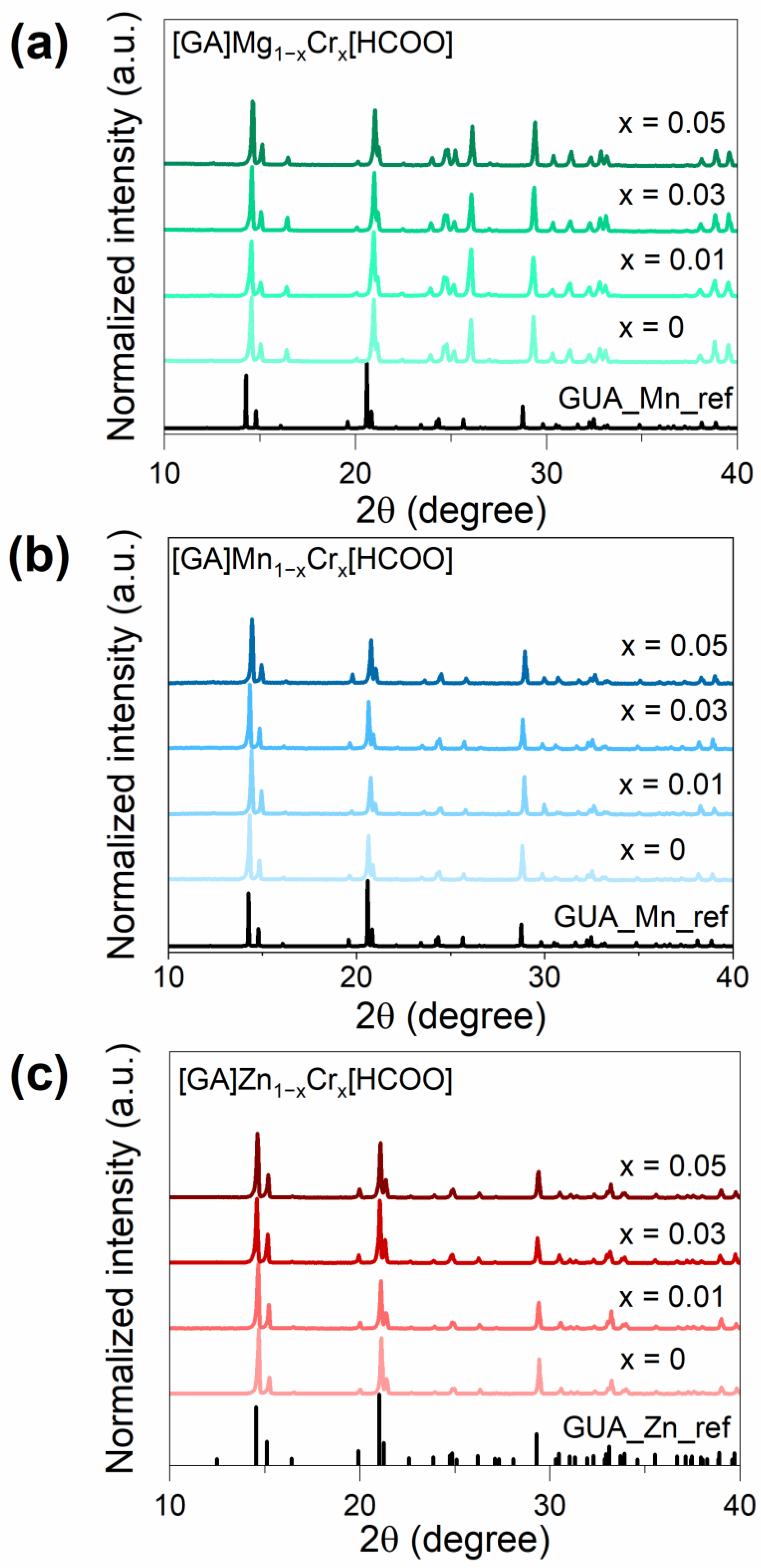
Figure 1. XRD patterns for a series of [GA]Mg1−xCrx(HCOO)3 (x = 0, 0.01, 0.03, 0.05) (a), [GA]Mn1−xCrx(HCOO)3 (x = 0, 0.01, 0.03, 0.05 (b), and [GA]Zn1−xCrx(HCOO)3 (x = 0, 0.01, 0.03, 0.05 (c).
Sensors 23 06259 g001
Figure 2. The crystal structure of [GA]Mn(HCOO)3 based on data presented in [31]. The dashed lines present HBs between GA+ cations and the manganese-formate framework.
Sensors 23 06259 g002
Figure 3. The Raman spectra of the [GA]M1−xCrx(HCOO)3 series, where M = Mg2+, Mn2+, Zn2+, and x = 0, 0.01, 0.03, and 0.05 (a) and the enlarged areas with bands corresponding to stretching C–N (b), symmetric C–O stretching and C–H in-plane bending (ν2 + ν5) (c), and stretching N–H modes (d).
Sensors 23 06259 g003
Figure 4. Diffuse reflectance spectra of representative samples [GA]M1−xCrx(HCOO)3 (M = Mg2+, Zn2+, Mn2+ and x = 0.05) measured at 300 K.
Sensors 23 06259 g004
Figure 5. Emission spectra of [GA]M1−xCrx(HCOO)3 (M = Mg2+, Mn2+, Zn2+, and x = 0.05) at 80 K (a) and influence of Cr3+ ions concentration of emission intensity (bd) of the investigated samples.
Sensors 23 06259 g005
Figure 6. Temperature-dependent emission spectra and thermal evolution of emission intensity of [GA]Mg(HCOO)3: 5% Cr3+ (a), [GA]Mn(HCOO)3: 5% Cr3+ (b), and [GA]Zn(HCOO)3: 5% Cr3+ (c) representative samples, respectively.
Sensors 23 06259 g006
Figure 7. Graphical representation of way for the temperature detection in hybrid organic–inorganic formate perovskites [GA]M1−xCrx(HCOO)3 (M = Mg2+, Mn2+, Zn2+, and x = 0.01, 0.03, 0.05).
Sensors 23 06259 g007
Figure 8. Influence of Cr3+ ions concentration on Fluorescence Intensity Ratio (FIR) (ac) of [GA]M1−xCrx(HCOO)3 (M = Mg2+, Mn2+, Zn2+, and x = 0.01, 0.03, 0.05) hybrid perovskites.
Sensors 23 06259 g008
Figure 9. Influence of Cr3+ ions concentration on relative sensitivity Sr (ac) of [GA]M1−xCrx(HCOO)3 (M = Mg2+, Mn2+, Zn2+, and x = 0.01, 0.03, 0.05) hybrid perovskites.
Sensors 23 06259 g009
Table 1. The collation of crystal field parameters and energies of electron transitions of the investigated series of [GA]M1−xCrx(HCOO)3 (M = Mg2+, Mn2+, Zn2+, and x = 0.01, 0.03, 0.05).
ParametersGAMn:GAMg:GAZn:
1%Cr3+3%Cr3+5%Cr3+1%Cr3+3%Cr3+5%Cr3+1%Cr3+3%Cr3+5%Cr3+
4A2g2E (cm−1)14,53514,53614,53714,55214,55214,54714,54014,53914,540
4A2g4T2g (cm−1)15,54515,95915,73515,82815,91716,25915,64015,54415,500
4A2g4T1g (cm−1)21,97222,15622,43922,70322,68222,95222,06221,90121,869
Dq (cm−1)155515551574158315921626156415541550
B (cm−1)650675686709692676648641643
Dq/B2.392.302.292.232.302.412.412.432.41
C (cm−1)324231903166312231573184324732643259
C/B4.134.254.624.404.574.715.015.095.07
Table 2. Collation of exemplary luminescent thermometers with their highest relative sensitivity (Sr) at working temperature (T) 1.
CompoundSr (%K−1)T (K)Reference
[GA]Mg(HCOO)3: 1% Cr3+2.0890This work
[GA]Zn(HCOO)3: 1% Cr3+1.0890This work
[GA]Mn(HCOO)3: 3% Cr3+1.20100This work
[EA]2NaCr0.21Al0.79(HCOO)62.84160[10]
(Me2NH2)3[Eu3(FDC)4(NO3)4]·4H2O2.7170[40]
Sr(HCOO)2:Eu2+/Eu3+3.8293[41]
Ln-cpda (Ln = Eu, Tb)16300[42]
TbMOF@3%Eu-tfac2.59225[43]
[Eu2(qptca)(NO3)2(DMF)4](CH3CH2OH)3perylene1.28293[44]
Bi2Ga4O9:Cr3+0.7290[45]
Bi2Al4O9:Cr3+1.24290[46]
Sr2MgAl22O36:Cr3+1.7310[47]
ZnGa2O4:Cr3+2.8310[48]
SrAl12O19:Mn4+0.27393[49]
LaPO4:Nd3+7.19303[50]
MgTiO3:Mn4+1.293[51]
La2MgTiO6: Cr3+, V4+1.96165[11]
1 GA—guanidine, EA—ethylammonium, H2FDC—9-fluorenone-2,7-dicarboxylic acid, H3cpda—5-(4-carboxyphenyl)-2,6-pyridinedicarboxylic acid, TbMOF—[Tb2(bpydc)3(H2O)3nDMF, H2bpydc—2,2-bipyridine-5,5′-dicarboxylic acid, tfac—trifluoroacetylacetonate, H4qptca—1,1′:4′,1′′:4′′,1′′′-quaterphenyl-3,3′′′,5,5′′′-tetracarboxylic acid, DMF—dimethylformamide.
Disclaimer/Publisher’s Note: The statements, opinions and data contained in all publications are solely those of the individual author(s) and contributor(s) and not of MDPI and/or the editor(s). MDPI and/or the editor(s) disclaim responsibility for any injury to people or property resulting from any ideas, methods, instructions or products referred to in the content.