
Two-Dimensional Wavenumber Analysis Implemented in Ultrasonic Vector Doppler Method with Focused Transmit Beams


Abstract
1. Introduction
2. Materials and Methods
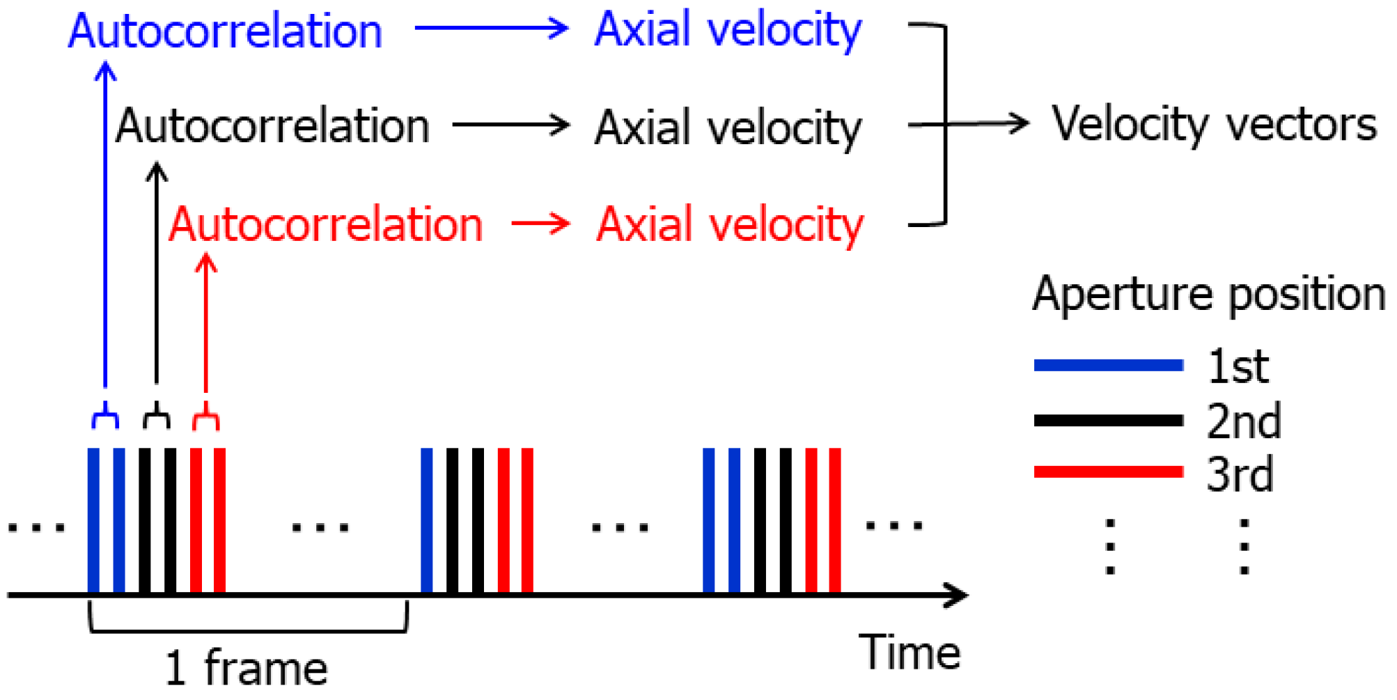
2.1. Tx-Rx Sequence and Beamforming
2.2. Principles for Velocity Estimation
2.3. Consideration on Beam Steering Angle in Estimation of Axial Velocity
2.4. Clutter Filtering
2.5. Numerical Simulations
2.6. Experimental Setup
2.7. Metrics for Evaluation of Accuracy in Velocity Estimation
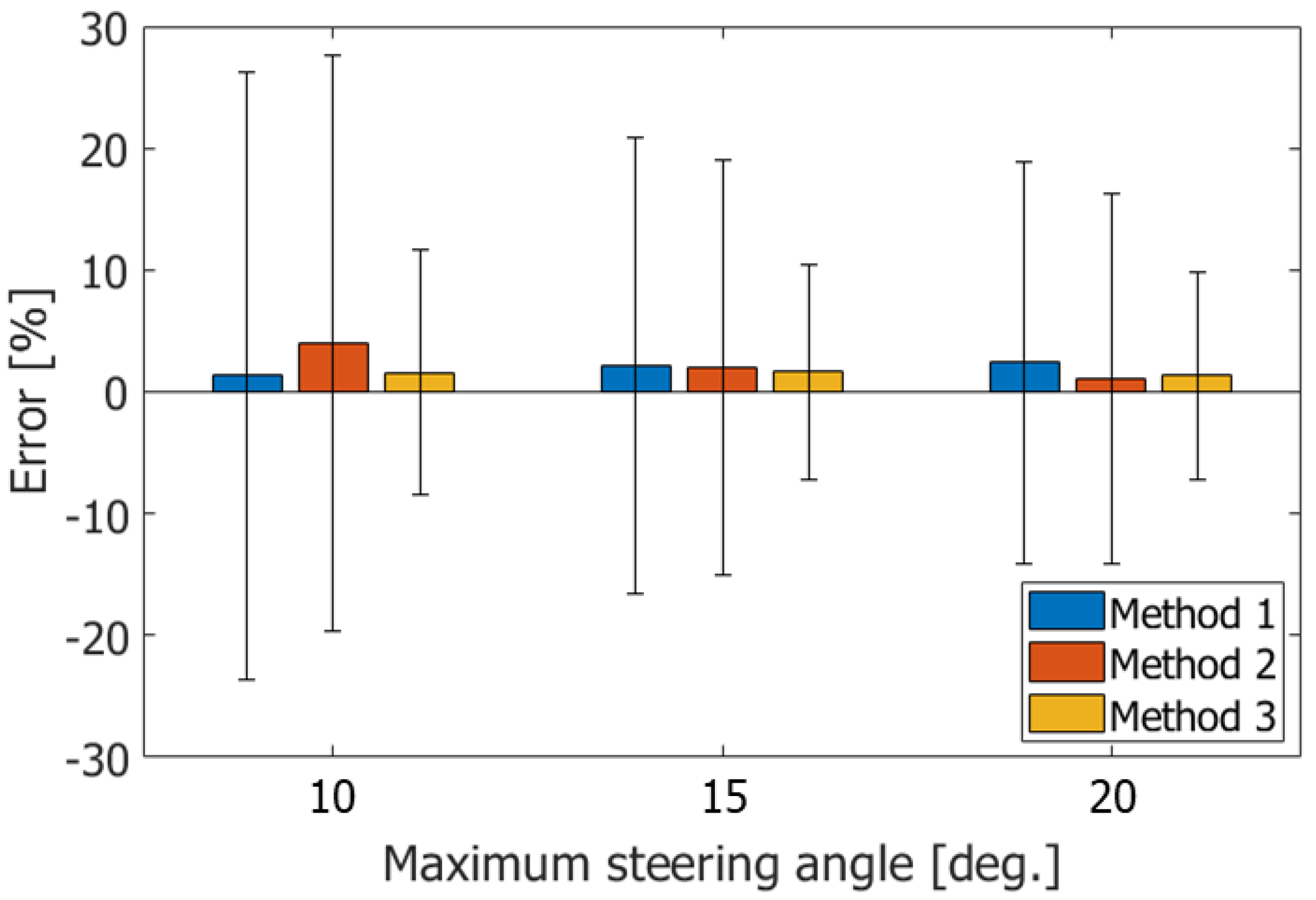
- Method 1: w/o estimation, ,
- Method 2: w/ estimation by Equation (3),
- Method 3: w/ estimation by Equation (3), from Equation (4)
3. Results
3.1. Simulations
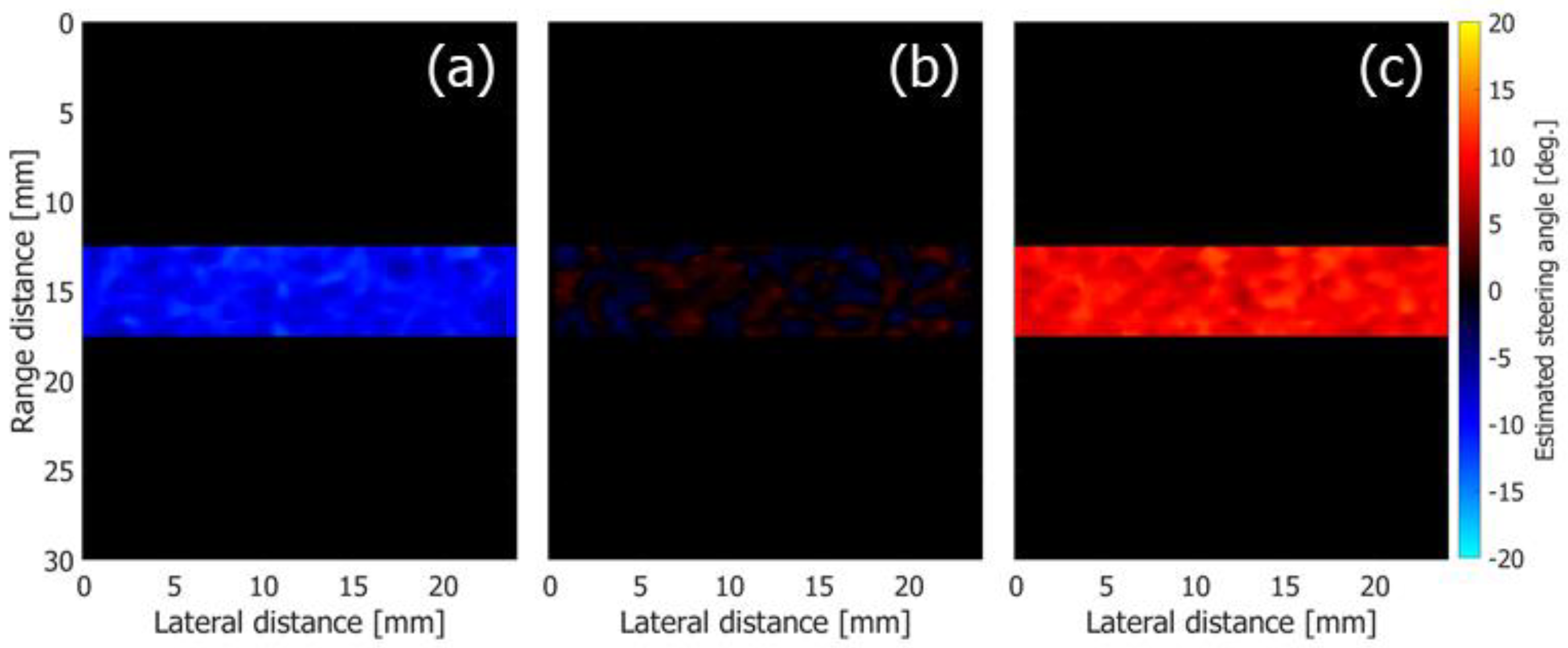
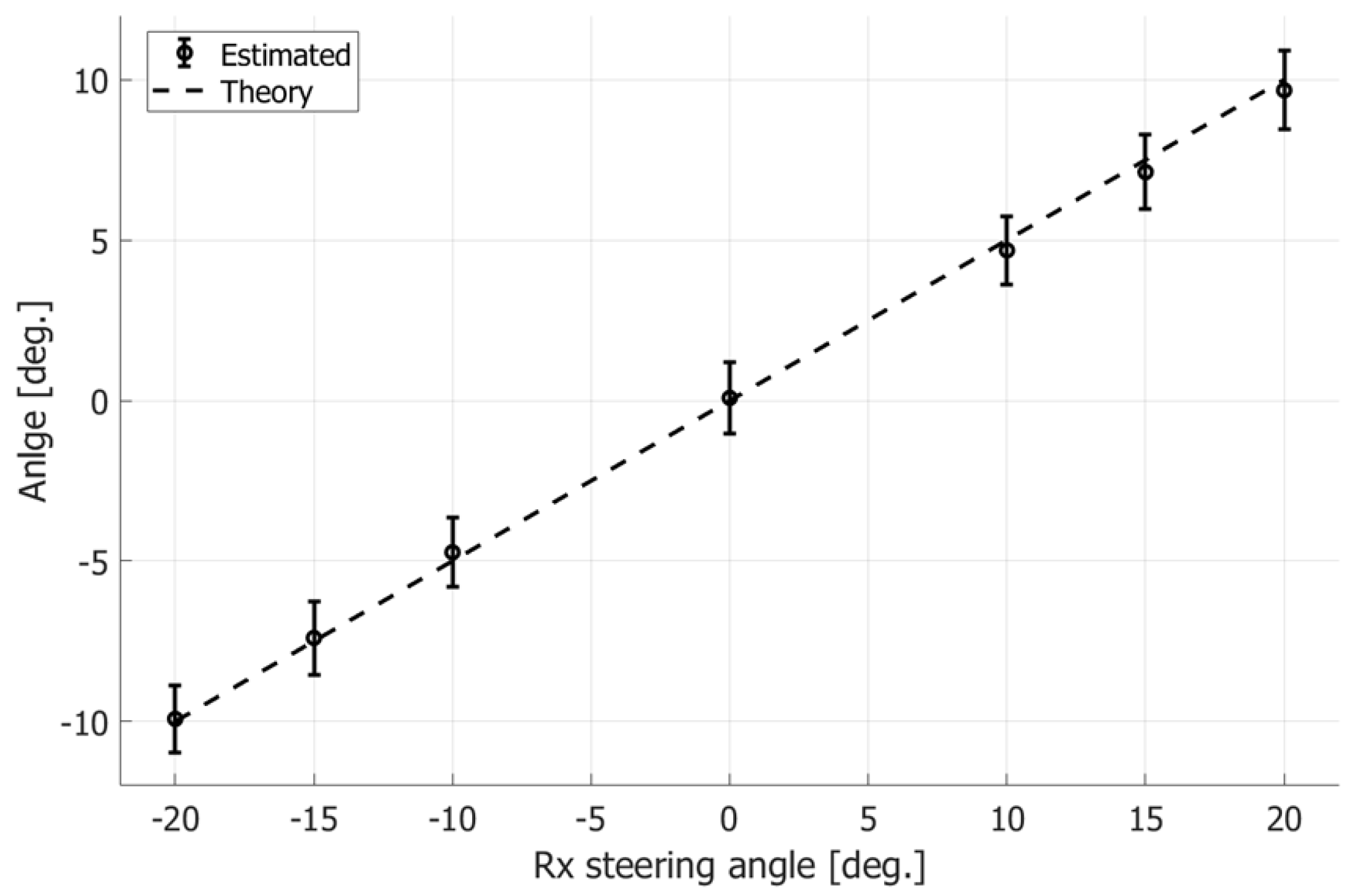
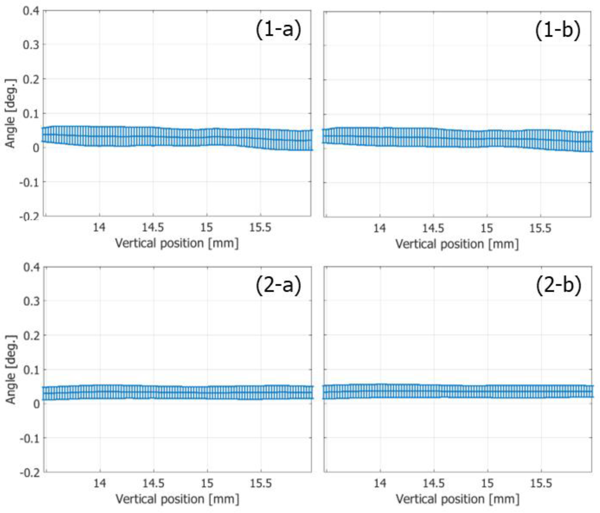
3.1.1. Estimation of Tilt Angle of Wavefront
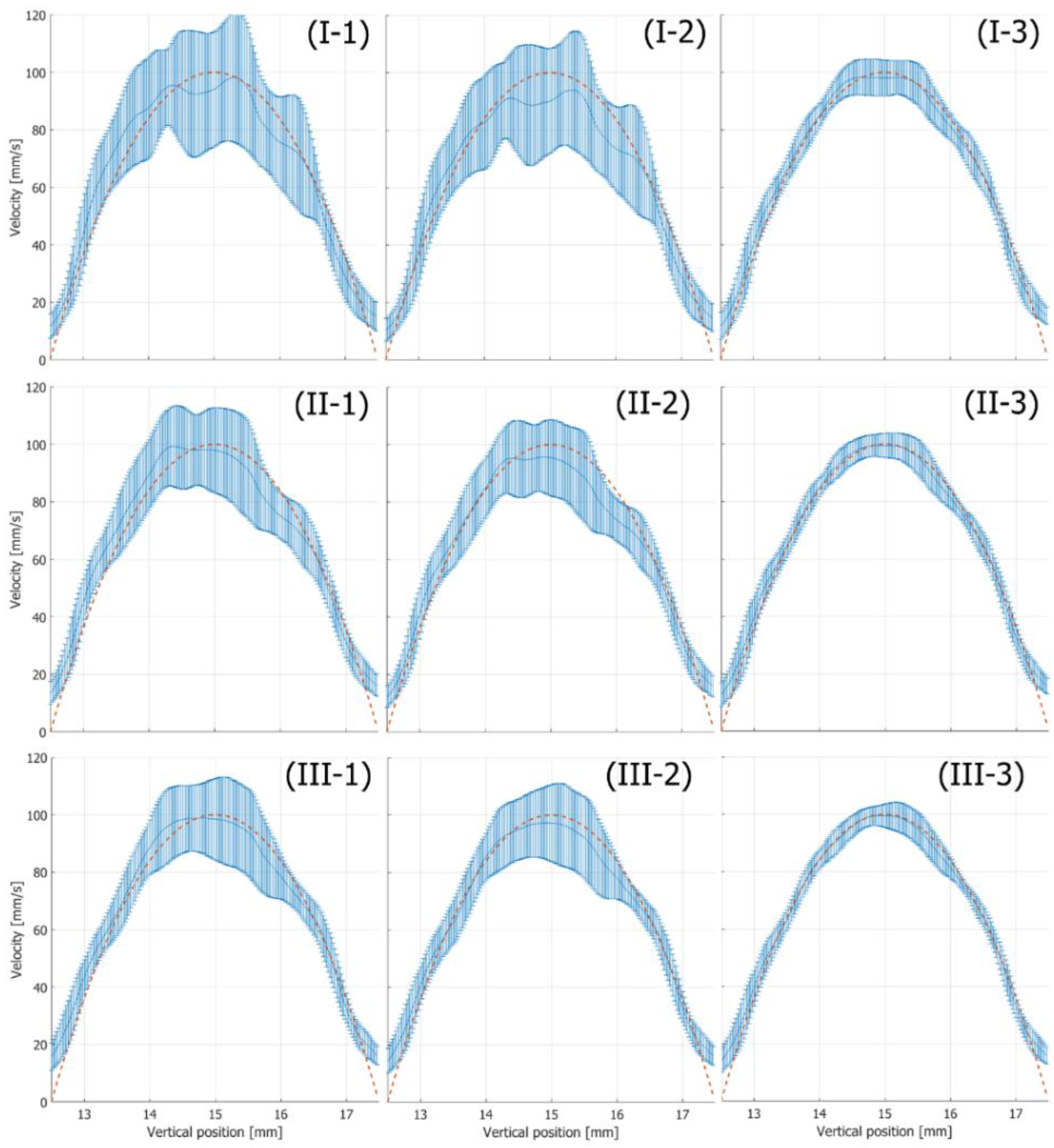
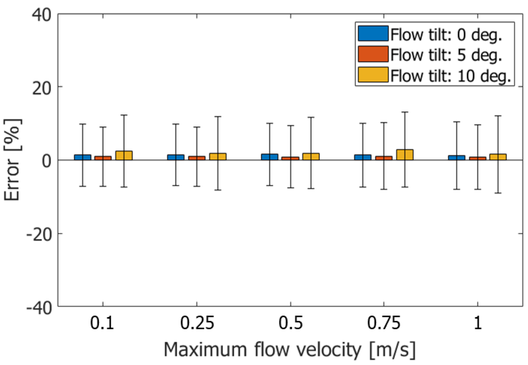
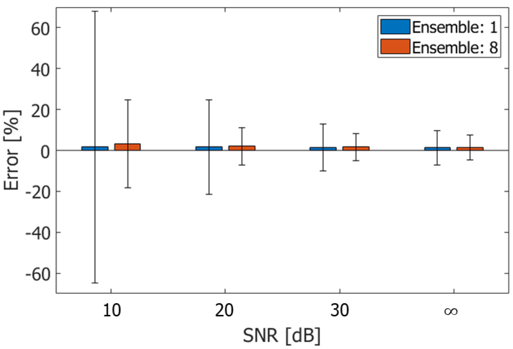
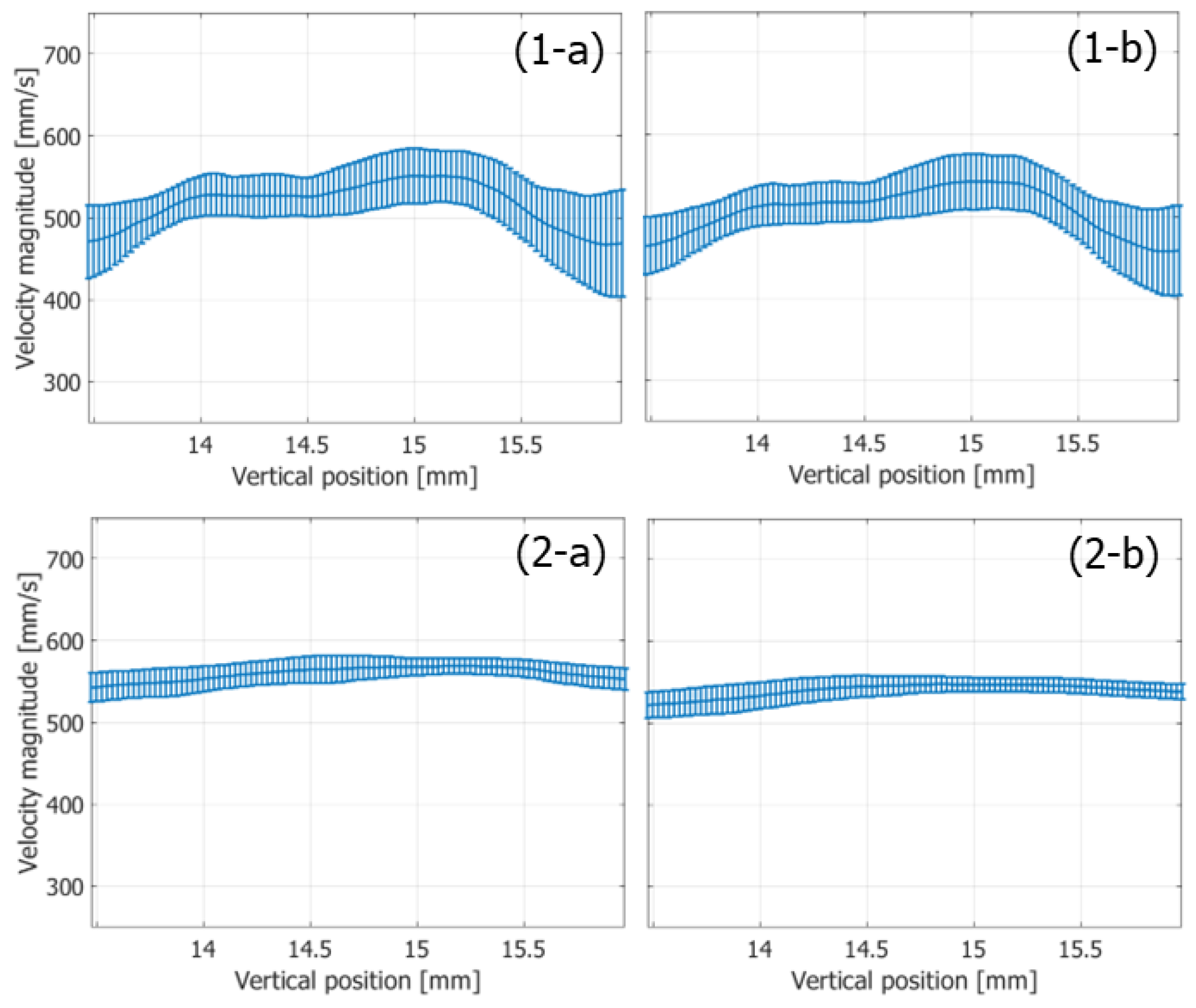
3.1.2. Accuracy in Velocity Estimation
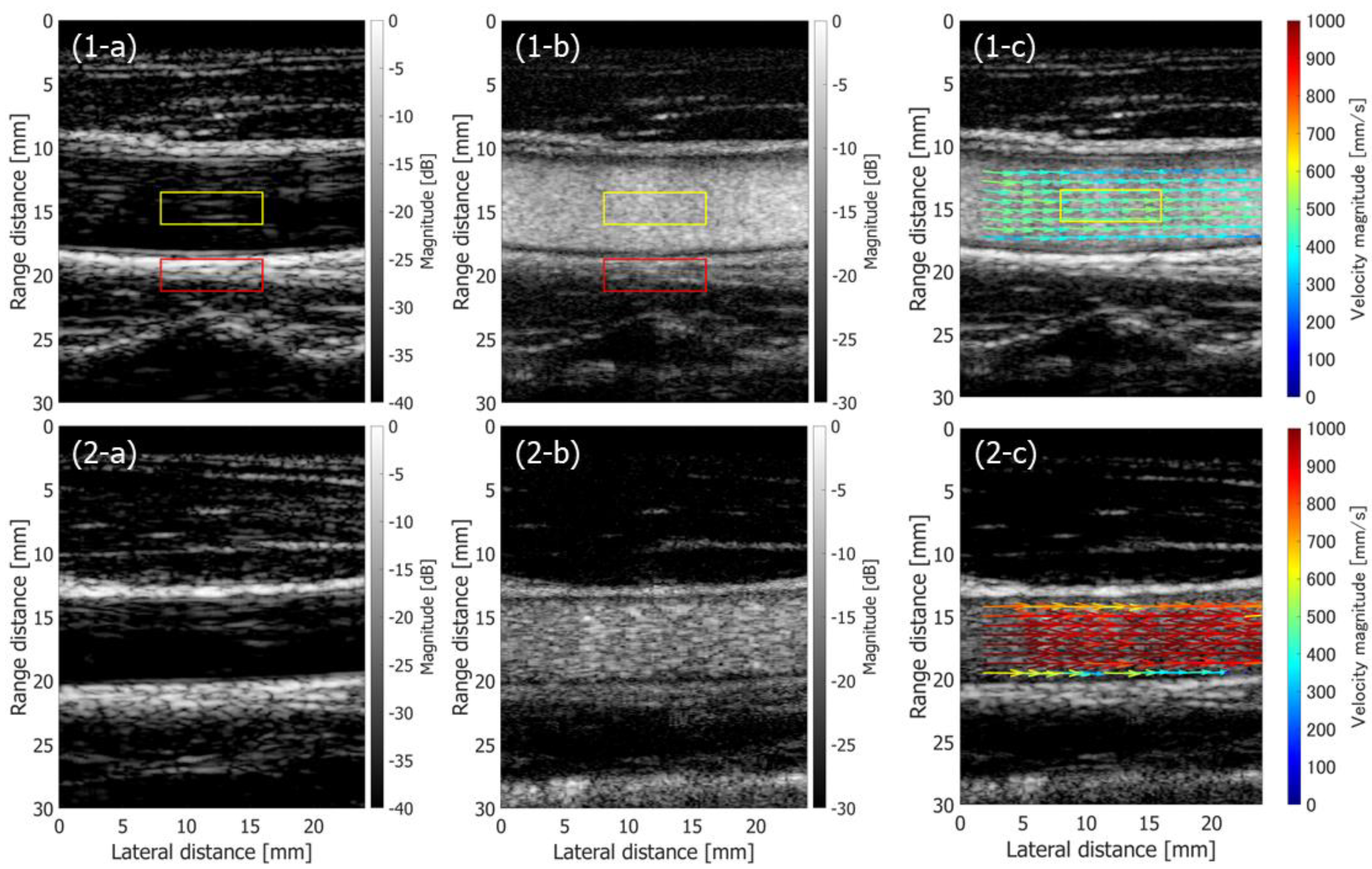
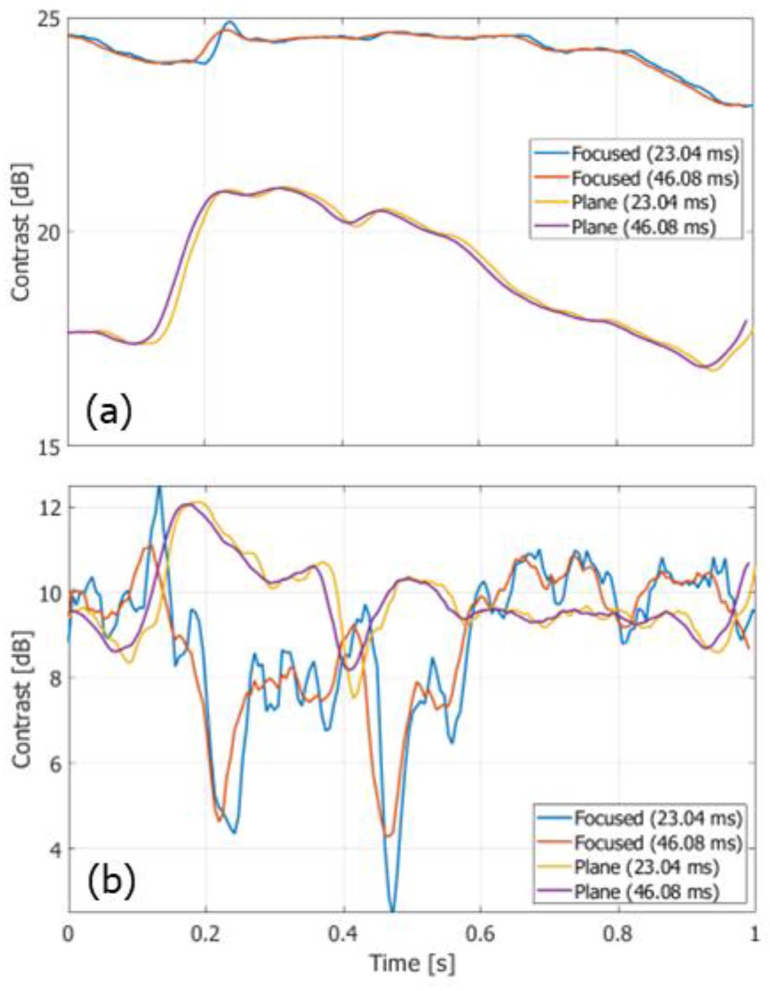
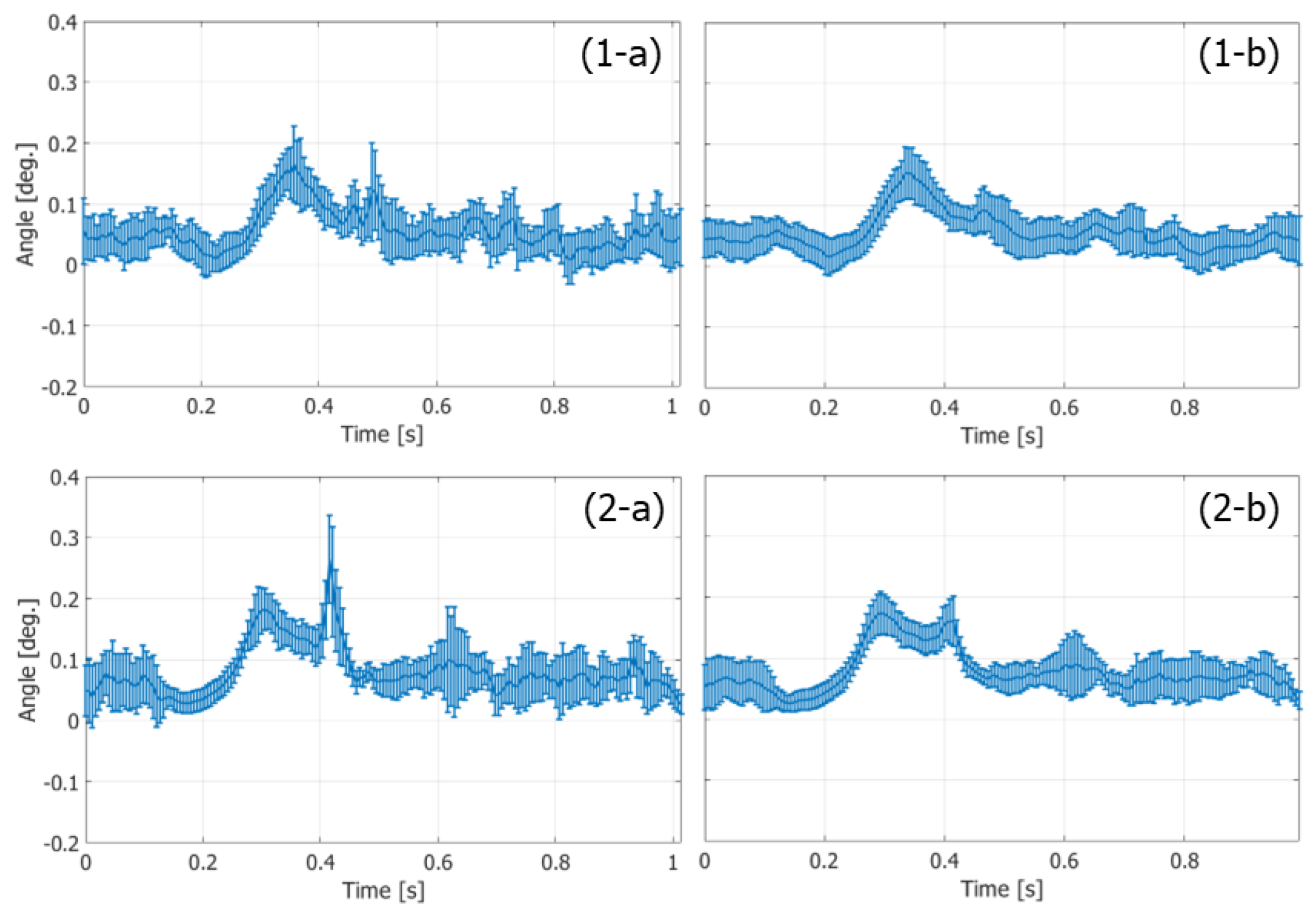
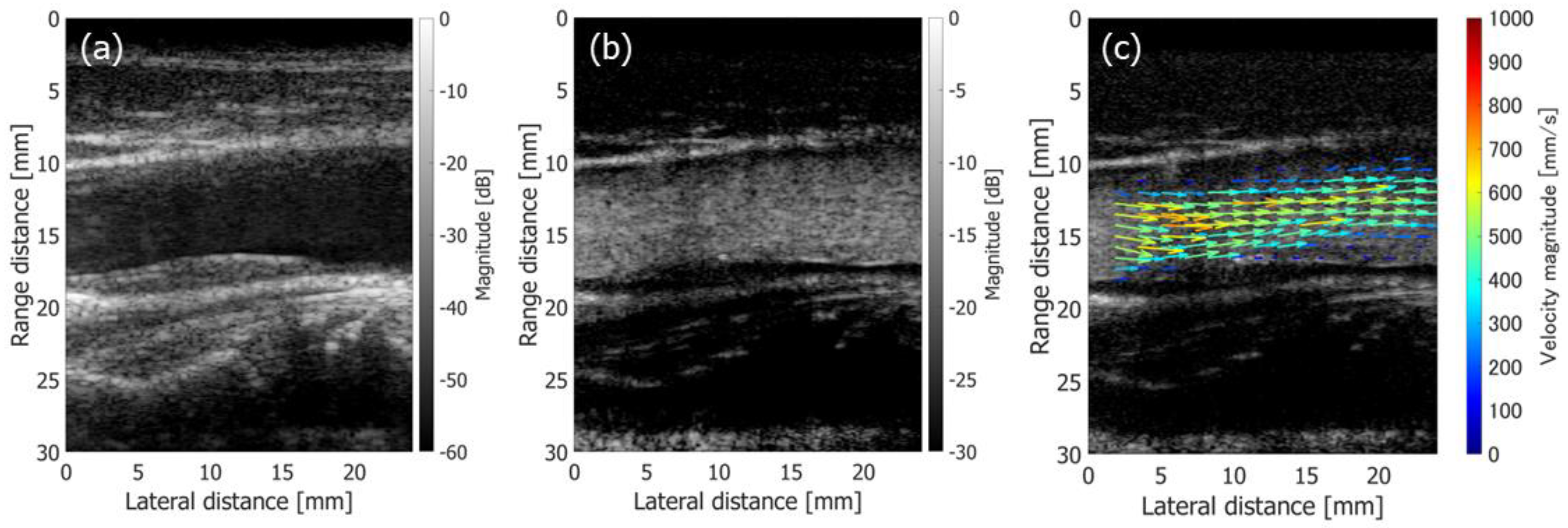
3.2. In Vivo Measurements
4. Discussion
5. Conclusions
Author Contributions
Funding
Institutional Review Board Statement
Informed Consent Statement
Data Availability Statement
Acknowledgments
Conflicts of Interest
References
- Kasai, C.; Namekawa, K.; Koyano, A.; Omoto, R. Real-time two-dimensional blood flow imaging using an autocorrelation technique. IEEE Trans. Sonics Ultrason. 1985, 32, 458–464. [Google Scholar] [CrossRef]
- Asami, R.; Tanaka, T.; Shimizu, M.; Seki, Y.; Nishiyama, T.; Sakashita, H.; Okada, T. Ultrasonic vascular vector flow mapping for 2-D flow estimation. Ultrasound Med. Biol. 2019, 45, 1663–1674. [Google Scholar] [CrossRef] [PubMed]
- Trahey, G.E.; Allison, J.W.; von Ramm, O.T. Angle independent ultrasonic detection of blood flow. IEEE Trans. Biomed. Eng. 1987, 34, 965–967. [Google Scholar] [CrossRef] [PubMed]
- Bohs, L.N.; Friemel, B.H.; Trahey, G.E. Experimental velocity profiles and volumetric flow via two-dimensional speckle tracking. Ultrasound Med. Biol. 1995, 21, 885–898. [Google Scholar] [CrossRef]
- Takahashi, H.; Hasegawa, H.; Kanai, H. Echo speckle imaging of blood particles with high-frame-rate echocardiography. Jpn. J. Appl. Phys. 2014, 53, 07KF08. [Google Scholar] [CrossRef]
- Fadnes, S.; Nyrnes, S.A.; Torp, H.; Lovstakken, L. Shunt flow evaluation in congenital heart disease based on two-dimensional speckle tracking. Ultrasound Med. Biol. 2014, 40, 2379–2391. [Google Scholar] [CrossRef]
- Takahashi, H.; Hasegawa, H.; Kanai, H. Echo motion imaging with adaptive clutter filter for assessment of cardiac blood flow. Jpn. J. Appl. Phys. 2015, 54, 07HF09. [Google Scholar] [CrossRef]
- Nyrnes, S.A.; Fadnes, S.; Wigen, M.S.; Mertens, L.; Lovstakken, L. Blood speckle-tracking based on high-frame rate ultrasound imaging in pediatric cardiology. J. Am. Soc. Echocardiogr. 2020, 33, 493–503. [Google Scholar] [CrossRef]
- Dunmire, B.; Beach, K.W.; Labs, K.H.; Plett, M.; Strandness, D.E., Jr. Cross-beam vector Doppler ultrasound for angle-independent velocity measurements. Ultrasound Med. Biol. 2000, 26, 1213–1235. [Google Scholar] [CrossRef]
- Fahrbach, K.K. Apparatus for Measuring the Speed of Flowing Media. U.S. Patent 3,766,517, 16 October 1973. [Google Scholar]
- Peronneau, P.; Bournat, J.P.; Bugnon, A.; Barbet, A.; Xhaard, M. Theoretical and practical aspects of pulsed Doppler flowmetry real-time application to the measure of instantaneous velocity profiles in vitro and in vivo. In Cardiovascular Applications of Ultra-Sound; Reneman, R.S., Ed.; North Holland Publishing: Amsterdam, The Netherlands, 1974; pp. 66–84. [Google Scholar]
- Peronneau, P.; Sandman, W.; Xhaard, M. Blood flow patterns in large arteries. In Ultrasound in Medicine; White, D.N., Brown, R.E., Eds.; Plenum Press: New York, NY, USA, 1977; pp. 1193–1208. [Google Scholar]
- Fox, M.D. Multiple crossed-beam ultrasound Doppler velocimetry. IEEE Trans. Son. Ultrason. 1978, 25, 281–286. [Google Scholar] [CrossRef]
- Steel, R.; Ramnarine, K.V.; Davidson, F.; Fish, P.J.; Hoskins, P.R. Angle-independent estimation of maximum velocity through stenoses using vector Doppler ultrasound. Ultrasound Med. Biol. 2003, 29, 575–584. [Google Scholar] [CrossRef]
- Tortoli, P.; Dallai, A.; Boni, E.; Francalanci, L.; Ricci, S. An automatic angle tracking procedure for feasible vector Doppler blood velocity measurements. Ultrasound Med. Biol. 2010, 36, 488–496. [Google Scholar] [CrossRef]
- Jensen, J.A.; Munk, P. A new method for estimation of velocity vectors. IEEE Trans. Ultrason. Ferroelectr. Freq. Control 1998, 45, 837–851. [Google Scholar] [CrossRef]
- Tanter, M.; Bercoff, J.; Sandrin, L.; Fink, M. Ultrafast compound imaging for 2-D motion vector estimation: Application to transient elastography. IEEE Trans. Ultrason. Ferroelectr. Freq. Control 2002, 49, 1363–1374. [Google Scholar] [CrossRef] [PubMed]
- Udesen, J.; Gran, F.; Hansen, K.; Jensen, J.A.; Thomsen, C.; Nielsen, M.B. High frame-rate blood vector velocity imaging using plane waves: Simulations and preliminary experiments. IEEE Trans. Ultrason. Ferroelectr. Freq. Control 2008, 55, 1729–1743. [Google Scholar] [CrossRef] [PubMed]
- Hasegawa, H.; Kanai, H. Simultaneous imaging of artery-wall strain and blood flow by high frame rate acquisition of RF signals. IEEE Trans. Ultrason. Ferroelectr. Freq. Control 2008, 55, 2626–2639. [Google Scholar] [CrossRef] [PubMed]
- Montaldo, G.; Tanter, M.; Bercoff, J.; Benech, N.; Fink, M. Coherent plane-wave compounding for very high frame rate ultrasonography and transient elastography. IEEE Trans. Ultrason. Ferroelectr. Freq. Control 2009, 56, 489–506. [Google Scholar] [CrossRef] [PubMed]
- Bercoff, J.; Montaldo, G.; Loupas, T.; Savery, D.; Mézière, F.; Fink, M.; Tanter, M. Ultrafast compound Doppler imaging: Providing full blood flow characterization. IEEE Trans. Ultrason. Ferroelectr. Freq. Control 2011, 58, 134–147. [Google Scholar] [CrossRef] [PubMed]
- Ricci, S.; Bassi, L.; Tortoli, P. Real-time vector velocity assessment through multigate Doppler and plane waves. IEEE Trans. Ultrason. Ferroelectr. Freq. Control 2014, 61, 314–324. [Google Scholar] [CrossRef]
- Ricci, S.; Ramalli, A.; Bassi, L.; Boni, E.; Tortoli, P. Real-time blood velocity vector measurement over a 2-D region. IEEE Trans. Ultrason. Ferroelectr. Freq. Control 2018, 65, 201–209. [Google Scholar] [CrossRef]
- Rossi, S.; Ramalli, A.; Tortoli, P. On the depth-dependent accuracy of plane-wave-based vector velocity measurements with linear arrays. IEEE Trans. Ultrason. Ferroelectr. Freq. Control 2021, 68, 2707–2715. [Google Scholar] [CrossRef] [PubMed]
- Ekroll, I.K.; Swillens, A.; Segers, P.; Dahl, T.; Torp, H.; Lovstakken, L. Simultaneous quantification of flow and tissue velocities based on multi-angle plane wave imaging. IEEE Trans. Ultrason. Ferroelectr. Freq. Control 2013, 60, 727–738. [Google Scholar] [CrossRef] [PubMed]
- Yiu, B.Y.S.; Lai, S.S.M.; Yu, A.C.H. Vector projectile imaging: Time-resolved dynamic visualization of complex flow patterns. Ultrasound Med. Biol. 2014, 40, 2295–2309. [Google Scholar] [CrossRef] [PubMed]
- Yiu, B.Y.S.; Yu, A.C.H. Least-squares multi-angle Doppler estimators for plane wave vector flow imaging. IEEE Trans. Ultrason. Ferroelectr. Freq. Control 2016, 63, 1733–1744. [Google Scholar] [CrossRef] [PubMed]
- Fadnes, S.; Ekroll, I.K.; Nyrnes, S.A.; Torp, H.; Lovstakken, L. Robust angle-independent blood velocity estimation based on dual-angle plane wave imaging. IEEE Trans. Ultrason. Ferroelectr. Freq. Control 2015, 62, 1757–1767. [Google Scholar] [CrossRef]
- Saris, A.E.C.M.; Hansen, H.H.G.; Fekkes, S.; Nillesen, M.M.; Rutten, M.C.M.; de Korte, C.L. A comparison between compounding techniques using large beam-steered plane wave imaging for blood vector velocity imaging in a carotid artery model. IEEE Trans. Ultrason. Ferroelectr. Freq. Control 2016, 63, 1758–1771. [Google Scholar] [CrossRef]
- Karageorgos, G.M.; Apostolakis, I.Z.; Nauleau, P.; Gatti, V.; Weber, R.; Kemper, P.; Konofagou, E.E. Pulse wave imaging coupled with vector flow mapping: A phantom, simulation, and in vivo Study. IEEE Trans. Ultrason. Ferroelectr. Freq. Control 2021, 68, 2516–2531. [Google Scholar] [CrossRef]
- Lenge, M.; Ramalli, A.; Tortoli, P.; Cachard, C.; Liebgott, H. Plane wave transverse oscillation for high-frame-rate 2-D vector flow imaging. IEEE Trans. Ultrason. Ferroelectr. Freq. Control 2015, 62, 2126–2137. [Google Scholar] [CrossRef]
- Perrot, V.; Ekroll, I.K.; Avdal, J.; Saxhaug, L.M.; Dalen, H.; Vray, D.; Løvstakken, L.; Liebgott, H. Translation of simultaneous vessel wall motion and vectorial blood flow imaging in healthy and diseased carotids to the clinic: A pilot study. IEEE Trans. Ultrason. Ferroelectr. Freq. Control 2021, 68, 558–569. [Google Scholar] [CrossRef]
- Jensen, J.; Hoyos, C.A.V.; Stuart, M.B.; Ewertsen, C.; Nielsen, M.B.; Jensen, J.A. Fast plane wave 2-D vector flow imaging using transverse oscillation and directional beamforming. IEEE Trans. Ultrason. Ferroelectr. Freq. Control 2017, 64, 1050–1062. [Google Scholar] [CrossRef]
- Lenge, M.; Ramalli, A.; Boni, E.; Liebgott, H.; Cachard, C.; Tortoli, P. High-frame-rate 2-D vector blood flow imaging in the frequency domain. IEEE Trans. Ultrason. Ferroelectr. Freq. Control 2014, 61, 1504–1514. [Google Scholar] [CrossRef] [PubMed]
- Kaburaki, K.; Mozumi, M.; Hasegawa, H. Estimation of two-dimensional motion velocity using ultrasonic signals beamformed in Cartesian coordinate for measurement of cardiac dynamics. Jpn. J. Appl. Phys. 2018, 57, 07LF03. [Google Scholar] [CrossRef]
- Tong, L.; Gao, H.; Choi, H.F.; D’hooge, J. Comparison of conventional parallel beamforming with plane wave and diverging wave imaging for cardiac applications: A simulation study. IEEE Trans. Ultrason. Ferroelectr. Freq. Control 2012, 59, 1654–1663. [Google Scholar] [CrossRef] [PubMed]
- Tong, L.; Gao, H.; D’hooge, J. Multi-transmit beam forming for fast cardiac imaging-a simulation study. IEEE Trans. Ultrason. Ferroelectr. Freq. Control 2013, 60, 1719–1731. [Google Scholar] [CrossRef] [PubMed]
- Tong, L.; Ramalli, A.; Jasaityte, R.; Tortoli, P.; D’hooge, J. Multi-transmit beam forming for fast cardiac imaging—Experimental validation and in vivo application. IEEE Trans. Med. Imaging 2014, 33, 1205–1219. [Google Scholar] [CrossRef]
- Tong, L.; Ramalli, A.; Tortoli, P.; Fradella, G.; Caciolli, S.; Luo, J.; D’hooge, J. Wide-angle tissue Doppler imaging at high frame rate using multi-line transmit beamforming: An experimental validation in vivo. IEEE Trans. Med. Imaging 2015, 35, 521–528. [Google Scholar] [CrossRef]
- Ramalli, A.; Dallai, A.; Guidi, F.; Bassi, L.; Boni, E.; Tong, L.; Fradella, G.; D’hooge, J.; Tortoli, P. Real-time high-frame-rate cardiac B-mode and tissue Doppler imaging based on multiline transmission and multiline acquisition. IEEE Trans. Ultrason. Ferroelectr. Freq. Control 2018, 65, 2030–2041. [Google Scholar] [CrossRef]
- Hasegawa, H.; Mozumi, M.; Omura, M.; Nagaoka, R.; Saito, K. Preliminary study on estimation of flow velocity vectors using focused transmit beams. Jpn. J. Appl. Phys. 2022, 61, SG1026. [Google Scholar] [CrossRef]
- Loupas, T.; Powers, J.T.; Gill, R.W. An axial velocity estimator for ultrasound blood flow imaging, based on a full evaluation of the Doppler equation by means of a two-dimensional autocorrelation approach. IEEE Trans. Ultrason. Ferroelectr. Freq. Control 1995, 42, 672–688. [Google Scholar] [CrossRef]
- Rabben, S.I.; Baerum, S.; Sorhus, V.; Torp, H. Ultrasound-based vessel wall tracking: An auto-correlation technique with RF center frequency estimation. Ultrasound Med. Biol. 2002, 28, 507–517. [Google Scholar] [CrossRef]
- Hasegawa, H.; Kanai, H. Modification of the phased-tracking method for reduction of artifacts in estimated artery wall deformation. IEEE Trans. Ultrason. Ferroelectr. Freq. Control 2006, 53, 2050–2064. [Google Scholar] [CrossRef] [PubMed]
- Hasegawa, H.; Kanai, H.; Koiwa, Y. Modified phased tracking method for measurement of change in thickness of arterial wall. Jpn. J. Appl. Phys. 2002, 41, 3563–3571. [Google Scholar] [CrossRef][Green Version]
- Hasegawa, H.; Kanai, H. Reduction of influence of variation in center frequencies of RF echoes on estimation of artery-wall strain. IEEE Trans. Ultrason. Ferroelectr. Freq. Control 2008, 55, 1921–1934. [Google Scholar] [CrossRef] [PubMed]
- Hasegawa, H.; Nagaoka, R.; Omura, M.; Mozumi, M.; Saito, K. Measurement of flow velocity vectors in carotid artery by plane wave imaging with repeated transmit sequence. J. Med. Ultrason. 2021, 48, 417–427. [Google Scholar] [CrossRef] [PubMed]
- Hasegawa, H.; Omura, M.; Nagaoka, R. On the investigation of autocorrelation-based vector Doppler method with plane wave imaging. IEEE Trans. Ultrason. Ferroelectr. Freq. Control 2022, 69, 1301–1311. [Google Scholar] [CrossRef]
- Jensen, J.A. A model for the propagation and scattering of ultrasound in tissue. J. Acoust. Soc. Am. 1991, 89, 182–190. [Google Scholar] [CrossRef]
- Jensen, J.A.; Svendsen, N.B. Calculation of pressure fields from arbitrarily shaped, apodized, and excited ultrasound transducers. IEEE Trans. Ultrason. Ferroelectr. Freq. Control 1992, 39, 262–267. [Google Scholar] [CrossRef]
- Podkowa, A.S.; Oelze, M.L.; Ketterling, J.A. High-frame-rate Doppler ultrasound using a repeated transmit sequence. Appl. Sci. 2018, 8, 227. [Google Scholar] [CrossRef]
- Mahafza, B.R. Introduction to Radar Analysis; CRC Press: Boca Raton, FL, USA, 1998. [Google Scholar]
- Demené, C.; Deffieux, T.; Pernot, M.; Osmanski, B.F.; Biran, V.; Gennisson, J.L.; Sieu, L.A.; Bergel, A.; Franqui, S.; Correas, J.M.; et al. Spatiotemporal clutter filtering of ultrafast ultrasound data highly increases Doppler and fultrasound sensitivity. IEEE Trans. Med. Imaging 2015, 34, 2271–2285. [Google Scholar] [CrossRef]
- Zócalo, Y.; Bia, D. Sex- and age-related physiological profiles for brachial, vertebral, carotid, and femoral arteries blood flow velocity parameters during growth and aging (4–76 years): Comparison with clinical cut-off levels. Front. Physiol. 2021, 12, 729309. [Google Scholar] [CrossRef]
- Park, M.Y.; Jung, S.E.; Byun, J.Y.; Kim, J.H.; Joo, G.E. Effect of beam-flow angle on velocity measurements in modern Doppler ultrasound systems. Am. J. Roentgenol. 2012, 198, 1139–1143. [Google Scholar] [CrossRef] [PubMed]
- Mozumi, M.; Nagaoka, R.; Hasegawa, H. Utilization of singular value decomposition in high-frame-rate cardiac blood flow imaging. Jpn. J. Appl. Phys. 2019, 58, SGGE02. [Google Scholar] [CrossRef]


| Parameters | Value |
|---|---|
| Tx center Frequency | 4.8 MHz |
| Element pitch | 0.2 mm |
| PRF | 10 kHz (simulation) 10.417 kHz (in vivo) |
| Sampling frequency | 31.25 MHz |
| Rx F-number | 2.34 |
| Correlation kernel size | x: 1.0 mm, z: 0.825 mm |
| Ensemble size | 1–8 (simulation), 4, 8 (in vivo) |
Publisher’s Note: MDPI stays neutral with regard to jurisdictional claims in published maps and institutional affiliations. |
© 2022 by the authors. Licensee MDPI, Basel, Switzerland. This article is an open access article distributed under the terms and conditions of the Creative Commons Attribution (CC BY) license (https://creativecommons.org/licenses/by/4.0/).
Share and Cite
Hasegawa, H.; Omura, M.; Nagaoka, R.; Saito, K. Two-Dimensional Wavenumber Analysis Implemented in Ultrasonic Vector Doppler Method with Focused Transmit Beams. Sensors 2022, 22, 9787. https://doi.org/10.3390/s22249787
Hasegawa H, Omura M, Nagaoka R, Saito K. Two-Dimensional Wavenumber Analysis Implemented in Ultrasonic Vector Doppler Method with Focused Transmit Beams. Sensors. 2022; 22(24):9787. https://doi.org/10.3390/s22249787
Chicago/Turabian StyleHasegawa, Hideyuki, Masaaki Omura, Ryo Nagaoka, and Kozue Saito. 2022. "Two-Dimensional Wavenumber Analysis Implemented in Ultrasonic Vector Doppler Method with Focused Transmit Beams" Sensors 22, no. 24: 9787. https://doi.org/10.3390/s22249787
APA StyleHasegawa, H., Omura, M., Nagaoka, R., & Saito, K. (2022). Two-Dimensional Wavenumber Analysis Implemented in Ultrasonic Vector Doppler Method with Focused Transmit Beams. Sensors, 22(24), 9787. https://doi.org/10.3390/s22249787