
Progress in Biosensors for the Point-of-Care Diagnosis of COVID-19


Abstract
:1. Introduction
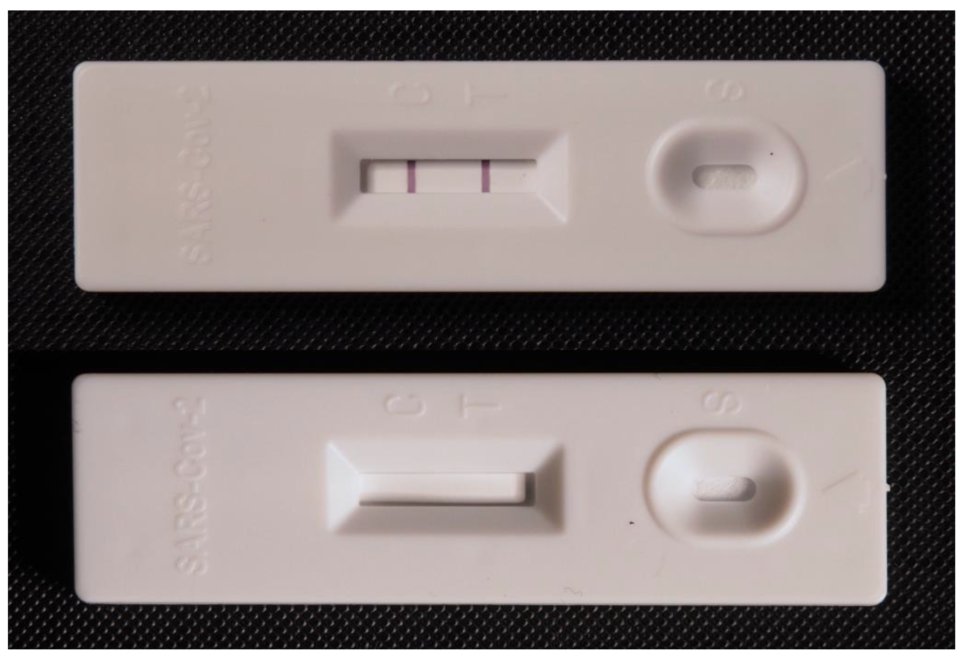
2. Current Point-of-Care Tests for COVID-19
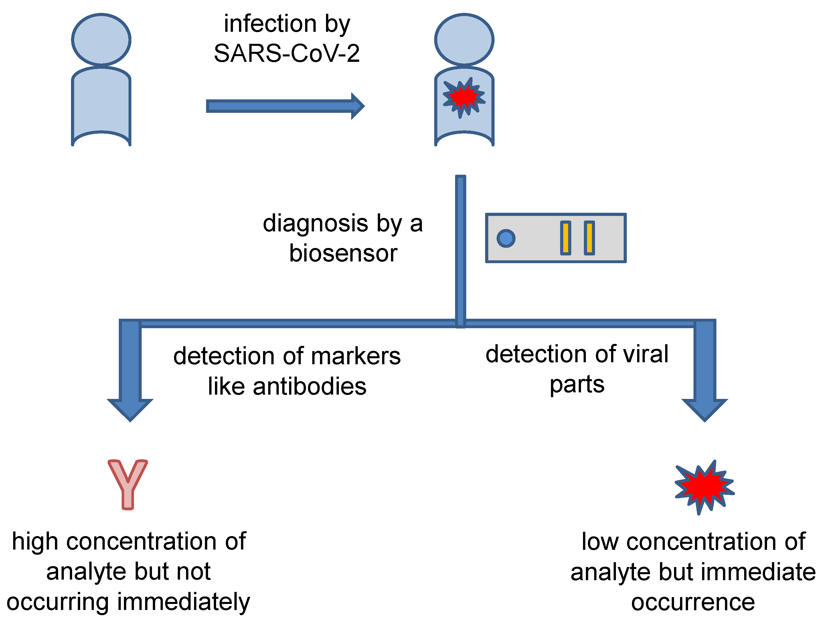
3. Concept of Point-of-Care Biosensors for COVID-19 Diagnosis
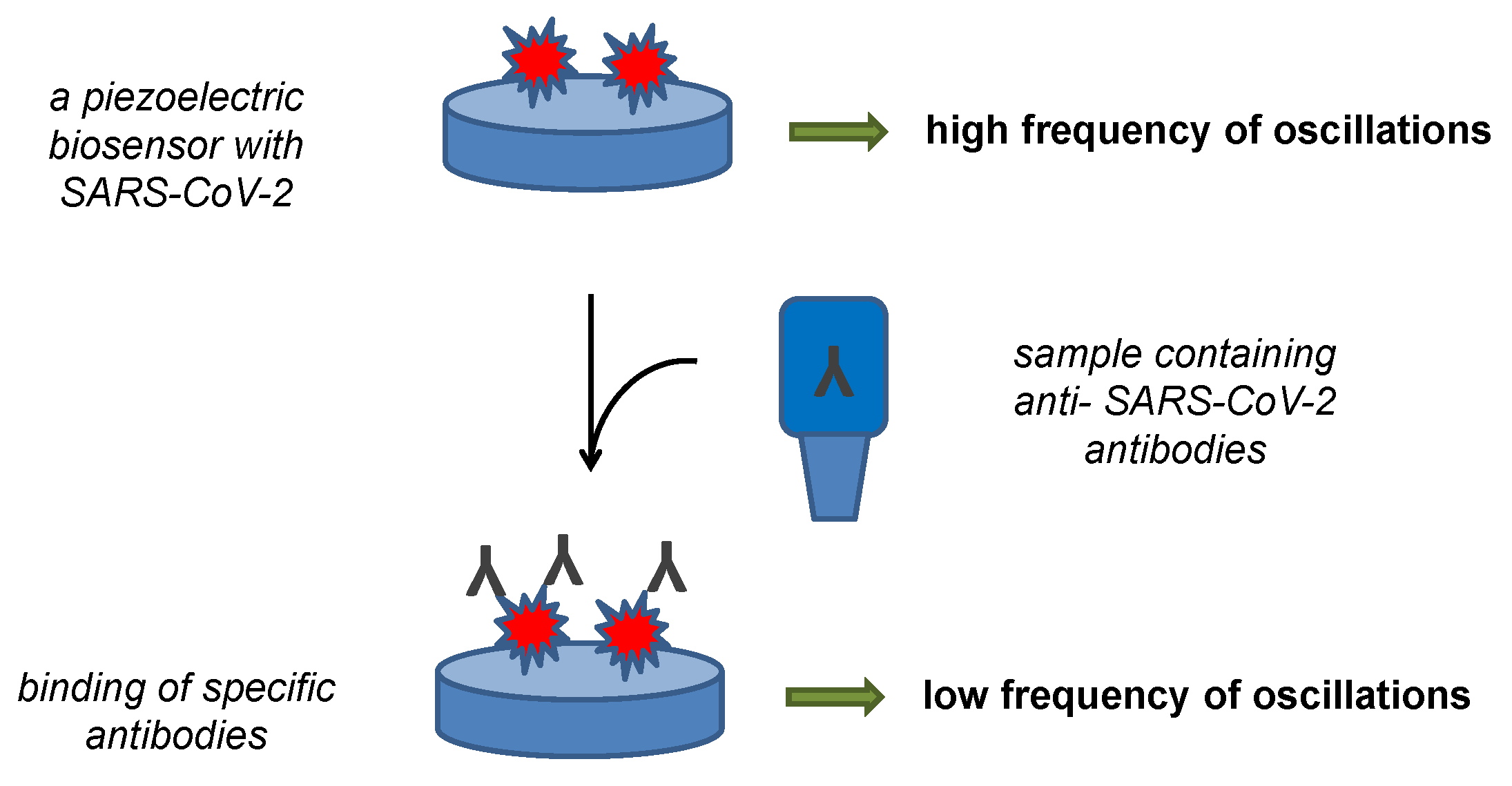
4. Biosensors for Measuring Anti-COVID-19 Antibodies
5. Biosensors for Measuring SARS-CoV-2 and Its Determining Parts
6. Conclusions
Funding
Institutional Review Board Statement
Informed Consent Statement
Data Availability Statement
Conflicts of Interest
References
- Eibensteiner, F.; Ritschl, V.; Stamm, T.; Cetin, A.; Schmitt, C.P.; Ariceta, G.; Bakkaloglu, S.; Jankauskiene, A.; Klaus, G.; Paglialonga, F.; et al. Countermeasures against COVID-19: How to navigate medical practice through a nascent, evolving evidence base—A European multicentre mixed methods study. BMJ Open 2021, 11, 11. [Google Scholar] [CrossRef] [PubMed]
- Kong, L.C.; Hu, Y.; Wang, Q.; Chen, X.D.; Yao, T.; Wang, Y.; Jin, H.; Fan, L.J.; Du, W. Could COVID-19 pandemic be stopped with joint efforts of travel restrictions and public health countermeasures? A modelling study. BMJ Open 2021, 11, 8. [Google Scholar] [CrossRef] [PubMed]
- Vichiensan, V.; Hayashi, Y.; Kamnerdsap, S. COVID-19 Countermeasures and Passengers’ Confidence of Urban Rail Travel in Bangkok. Sustainability 2021, 13, 9377. [Google Scholar] [CrossRef]
- Si, R.S.; Yao, Y.M.; Zhang, X.Q.; Lu, Q.; Aziz, N. Investigating the Links Between Vaccination Against COVID-19 and Public Attitudes Toward Protective Countermeasures: Implications for Public Health. Front. Public Health 2021, 9, 11. [Google Scholar] [CrossRef] [PubMed]
- Zhu, H.; Lu, H.Z. The development of a quarantine strategy is an important path to a normalized response to COVID-19. BioSci. Trends 2020, 14, 396–398. [Google Scholar] [CrossRef]
- Traylor, A.M.; Tannenbaum, S.I.; Thomas, E.J.; Salas, E. Helping Healthcare Teams Save Lives During COVID-19: Insights and Countermeasures From Team Science. Am. Psychol. 2021, 76, 1–13. [Google Scholar] [CrossRef]
- Mohsin, A.K.M.; Lei, H.Z.; Hossain, S.F.A. Impact of COVID-19 Pandemic on Consumer Economy: Countermeasures Analysis. Sage Open 2021, 11, 10. [Google Scholar] [CrossRef]
- Wang, L.J.; Li, C.D.; Chen, X.H.; Zhu, L.L. Causal Relationship Between the Spread of the COVID-19 and Geopolitical Risks in Emerging Economies. Front. Public Health 2020, 8, 626055. [Google Scholar] [CrossRef]
- Butt, A.S. Supply chains and COVID-19: Impacts, countermeasures and post-COVID-19 era. Int. J. Logist. Manag. 2022. [Google Scholar] [CrossRef]
- Liu, Y.W.; Cui, Q.; Liu, Y.; Zhang, J.Z.; Zhou, M.F.; Ali, T.; Yang, L.Y.; Feng, K.S.; Hubacek, K.; Li, X.B. Countermeasures against economic crisis from COVID-19 pandemic in China: An analysis of effectiveness and trade-offs. Struct. Chang. Econ. Dyn. 2021, 59, 482–495. [Google Scholar] [CrossRef]
- Li, Y.; Wang, X.J.; Wang, W. The Impact of COVID-19 on Cancer. Infect. Drug Resist. 2021, 14, 3809–3816. [Google Scholar] [CrossRef] [PubMed]
- Zhao, Z.; Huang, C.F.; Huang, Z.Y.; Lin, F.J.; He, Q.L.; Tao, D.; Jaffrezic-Renault, N.; Guo, Z.Z. Advancements in electrochemical biosensing for respiratory virus detection: A review. Trac-Trends Anal. Chem. 2021, 139, 16. [Google Scholar] [CrossRef] [PubMed]
- Tran, V.V.; Tran, N.H.T.; Hwang, H.S.; Chang, M. Development strategies of conducting polymer-based electrochemical biosensors for virus biomarkers: Potential for rapid COVID-19 detection. Biosens. Bioelectron. 2021, 182, 21. [Google Scholar] [CrossRef] [PubMed]
- Choi, J.R. Development of Point-of-Care Biosensors for COVID-19. Front. Chem. 2020, 8, 517. [Google Scholar] [CrossRef]
- Yue, J.; Xu, H.; Zhou, Y.; Liu, W.; Han, X.F.; Mao, Q.; Li, S.X.; Tam, L.S.; Ma, J.; Liu, W. Dyslipidemia Is Related to Mortality in Critical Patients With Coronavirus Disease 2019: A Retrospective Study. Front. Endocrinol. 2021, 12, 611526. [Google Scholar] [CrossRef] [PubMed]
- Gonzalez-Garcia, N.; Miranda-Lora, A.L.; Garduno-Espinosa, J.; Granados-Riveron, J.T.; Mendez-Galvan, J.F.; Nieto-Zermeno, J.; Castilla-Peon, M.F. International heterogeneity in coronavirus disease 2019 pediatric mortality rates. Bol. Med. Hosp. Infant. Mex. 2021, 78, 24–28. [Google Scholar] [CrossRef] [PubMed]
- Hernandez-Romieu, A.C.; Adelman, M.W.; Hockstein, M.A.; Robichaux, C.J.; Edwards, J.A.; Fazio, J.C.; Blum, J.M.; Jabaley, C.S.; Caridi-Scheible, M.; Martin, G.S.; et al. Timing of Intubation and Mortality Among Critically Ill Coronavirus Disease 2019 Patients: A Single-Center Cohort Study. Crit. Care Med. 2020, 48, E1045–E1053. [Google Scholar] [CrossRef] [PubMed]
- Barioni, E.M.S.; Nascimento, C.; Amaral, T.L.M.; Ramalho Neto, J.M.; Prado, P.R.D. Clinical indicators, nursing diagnoses, and mortality risk in critically ill patients with COVID-19: A retrospective cohort. Rev. Esc. Enferm. USP 2022, 56, e20210568. [Google Scholar] [CrossRef] [PubMed]
- Cui, D.; Wang, Y.M.; Huang, L.X.; Gu, X.Y.; Huang, Z.S.; Mu, S.R.; Wang, C.; Cao, B. Rheumatic Symptoms Following Coronavirus Disease 2019 (COVID-19): A Chronic Post-COVID-19 Condition. Open Forum Infect. Dis. 2022, 9, ofac170. [Google Scholar] [CrossRef] [PubMed]
- Kadirvelu, B.; Burcea, G.; Quint, J.K.; Costelloe, C.E.; Faisal, A.A. Variation in global COVID-19 symptoms by geography and by chronic disease: A global survey using the COVID-19 Symptom Mapper. EClinicalMedicine 2022, 45, 15. [Google Scholar] [CrossRef]
- Galal, I.; Hussein, A.; Amin, M.T.; Saad, M.M.; Zayan, H.E.E.; Abdelsayed, M.Z.; Moustafa, M.M.; Ezzat, A.R.; Helmy, R.E.D.; Abd Elaal, H.K.; et al. Determinants of persistent post-COVID-19 symptoms: Value of a novel COVID-19 symptom score. Egypt. J. Bronchol. 2021, 15, 10. [Google Scholar] [CrossRef]
- Luo, Y.M.; Wu, J.; Lu, J.Y.; Xu, X.; Long, W.; Yan, G.J.; Tang, M.Y.; Zou, L.; Xu, D.Z.; Zhuo, P.; et al. Investigation of COVID-19-related symptoms based on factor analysis. Ann. Pallliat. Med. 2020, 9, 1851–1858. [Google Scholar] [CrossRef] [PubMed]
- Fernandez-de-las-Penas, C.; Martin-Guerrero, J.D.; Pellicer-Valero, O.J.; Navarro-Pardo, E.; Gomez-Mayordomo, V.; Cuadrado, M.L.; Arias-Navalon, J.A.; Cigaran-Mendez, M.; Hernandez-Barrera, V.; Arendt-Nielsen, L. Female Sex Is a Risk Factor Associated with Long-Term Post-COVID Related-Symptoms but Not with COVID-19 Symptoms: The LONG-COVID-EXP-CM Multicenter Study. J. Clin. Med. 2022, 11, 413. [Google Scholar] [CrossRef] [PubMed]
- Soma, P.; Bester, J. Pathophysiological Changes in Erythrocytes Contributing to Complications of Inflammation and Coagulation in COVID-19. Front. Physiol. 2022, 13, 899629. [Google Scholar] [CrossRef]
- Lamb, L.E.; Dhar, N.; Timar, R.; Wills, M.; Dhar, S.; Chancellor, M.B. COVID-19 inflammation results in urine cytokine elevation and causes COVID-19 associated cystitis (CAC). Med. Hypotheses 2020, 145, 110375. [Google Scholar] [CrossRef]
- Wong, R.S.Y. Inflammation in COVID-19: From pathogenesis to treatment. Int. J. Clin. Exp. Pathol. 2021, 14, 831–844. [Google Scholar]
- Tan, Q.Q.; He, L.J.; Meng, X.J.; Wang, W.; Pan, H.D.; Yin, W.G.; Zhu, T.C.A.; Huang, X.; Shan, H. Macrophage biomimetic nanocarriers for anti-inflammation and targeted antiviral treatment in COVID-19. J. Nanobiotechnol. 2021, 19, 173. [Google Scholar] [CrossRef]
- Mester, A.; Benedek, I.; Rat, N.; Tolescu, C.; Polexa, S.A.; Benedek, T. Imaging Cardiovascular Inflammation in the COVID-19 Era. Diagnostics 2021, 11, 1114. [Google Scholar] [CrossRef]
- Johnson, J.N.; Loriaux, D.B.; Jenista, E.; Kim, H.W.; Baritussio, A.; De Garate Iparraguirre, E.; Bucciarelli-Ducci, C.; Denny, V.; O’Connor, B.; Siddiqui, S.; et al. Society for Cardiovascular Magnetic Resonance 2021 cases of SCMR and COVID-19 case collection series. J. Cardiovasc. Magn. Reson. 2022, 24, 42. [Google Scholar] [CrossRef]
- Shim, S.R.; Kim, S.J.; Hong, M.; Lee, J.; Kang, M.G.; Han, H.W. Diagnostic Performance of Antigen Rapid Diagnostic Tests, Chest Computed Tomography, and Lung Point-of-Care-Ultrasonography for SARS-CoV-2 Compared with RT-PCR Testing: A Systematic Review and Network Meta-Analysis. Diagnostics 2022, 12, 1302. [Google Scholar] [CrossRef]
- Barbas, C.S.V. Thoracic Computed Tomography to Assess ARDS and COVID-19 Lungs. Front. Physiol. 2022, 13, 829534. [Google Scholar] [CrossRef] [PubMed]
- Jenkins, H.H.; Lopez, A.A.T.; Tarantini, F.S.; Tomlin, H.; Scales, D.; Lee, I.N.; Wu, S.; Hyde, R.; Lis-Slimak, K.; Byaruhanga, T.; et al. Performance evaluation of a non-invasive one-step multiplex RT-qPCR assay for detection of SARS-CoV-2 direct from saliva. Sci. Rep. 2022, 12, 11553. [Google Scholar] [CrossRef] [PubMed]
- Mannan, N.; Raihan, R.; Parvin, U.S.; Fazle Akbar, S.M.; Reza, M.S.; Islam, S.; Kundu, J.; Noman, A.A.; Fakhruddin, M.; Billaha, M.; et al. Detection of SARS-CoV-2 RNA by Reverse Transcription-Polymerase Chain Reaction (RT-PCR) on Self-Collected Nasal Swab Compared With Professionally Collected Nasopharyngeal Swab. Cureus 2022, 14, e25618. [Google Scholar] [CrossRef] [PubMed]
- Ali, H.; Alkhaursi, K.; Holton, T. Development of a colorimetric RT-LAMP assay for the detection of SARS-COV-2 isolated from Oman. J. Infect. Dev. Ctries. 2022, 16, 952–958. [Google Scholar] [CrossRef] [PubMed]
- Khan, S.H.; Zaidi, S.K.; Gilani, M. PCR to CRISPR: Role of Nucleic Acid Tests (NAT) in detection of COVID-19. J. Pak. Med. Assoc. 2022, 72, 1166–1174. [Google Scholar] [CrossRef]
- Yu, Z.; Xu, L.; Lyu, W.; Shen, F. Parallel multistep digital analysis SlipChip demonstrated with the quantification of nucleic acid by digital LAMP-CRISPR. Lab Chip 2022, 22, 2954–2961. [Google Scholar] [CrossRef]
- Kashir, J.; Yaqinuddin, A. Loop mediated isothermal amplification (LAMP) assays as a rapid diagnostic for COVID-19. Med. Hypotheses 2020, 141, 109786. [Google Scholar] [CrossRef]
- Augustine, R.; Hasan, A.; Das, S.; Ahmed, R.; Mori, Y.; Notomi, T.; Kevadiya, B.D.; Thakor, A.S. Loop-Mediated Isothermal Amplification (LAMP): A Rapid, Sensitive, Specific, and Cost-Effective Point-of-Care Test for Coronaviruses in the Context of COVID-19 Pandemic. Biology 2020, 9, 182. [Google Scholar] [CrossRef]
- Balck, A.; Föh, B.; Borsche, M.; Rahmöller, J.; Vollstedt, E.J.; Waldeck, F.; Käding, N.; Twesten, C.; Mischnik, A.; Gillessen-Kaesbach, G.; et al. Protocol of the Luebeck longitudinal investigation of SARS-CoV-2 infection (ELISA) study—A prospective population-based cohort study. BMC Public Health 2022, 22, 1305. [Google Scholar] [CrossRef]
- Ramos, A.; Araújo, B.; Lacerda, L.; Flora, A.L.; Ribeiro, L.; Patrício, E.; Cardoso, M.J.; Guimarães, J.T. SARS-CoV-2 seroprevalence in healthcare workers: The experience of a Portuguese COVID-19 front-line hospital during the 1st pandemic wave. Porto. Biomed. J. 2022, 7, e166. [Google Scholar] [CrossRef]
- Donoso Mantke, O.; Corman, V.M.; Taddei, F.; McCulloch, E.; Niemeyer, D.; Grumiro, L.; Dirani, G.; Wallace, P.S.; Drosten, C.; Sambri, V.; et al. Importance of external quality assessment for SARS-CoV-2 antigen detection during the COVID-19 pandemic. J. Clin. Virol. 2022, 154, 105222. [Google Scholar] [CrossRef] [PubMed]
- Schwarze, M.; Krizsan, A.; Brakel, A.; Pohl, F.; Volke, D.; Hoffmann, R. Cross-Reactivity of IgG Antibodies and Virus Neutralization in mRNA-Vaccinated People Against Wild-Type SARS-CoV-2 and the Five Most Common SARS-CoV-2 Variants of Concern. Front. Immunol. 2022, 13, 915034. [Google Scholar] [CrossRef] [PubMed]
- Sakyi, A.; Laing, E.; Ephraim, R.; Asibey, O.; Sadique, O. Evaluation of analytical errors in a clinical chemistry laboratory: A 3 year experience. Ann. Med. Health Sci. Res. 2015, 5, 8–12. [Google Scholar] [CrossRef] [PubMed]
- Al-Ghaithi, H.; Pathare, A.; Al-Mamari, S.; Villacrucis, R.; Fawaz, N.; Alkindi, S. Impact of Educational Activities in Reducing Pre-Analytical Laboratory Errors: A quality initiative. Sultan Qaboos Univ. Med. J. 2017, 17, e309–e313. [Google Scholar] [CrossRef]
- Sturgeon, C.M. External quality assessment of hormone determinations. Best Pract. Res. Clin. Endocrinol. Metab. 2013, 27, 803–822. [Google Scholar] [CrossRef]
- Lee, G.R.; Fitzgibbon, M.C.; O’Shea, P. Laboratory services: Regaining and maintaining control. Int. J. Health Care Qual. Assur. 2016, 29, 507–522. [Google Scholar] [CrossRef]
- Wallace, P.S.; MacKay, W.G. Quality in the molecular microbiology laboratory. Methods Mol. Biol. 2013, 943, 49–79. [Google Scholar] [CrossRef]
- Somborac Bačura, A.; Dorotić, M.; Grošić, L.; Džimbeg, M.; Dodig, S. Current status of the lateral flow immunoassay for the detection of SARS-CoV-2 in nasopharyngeal swabs. Biochem. Med. 2021, 31, 020601. [Google Scholar] [CrossRef]
- Zhou, Y.; Wu, Y.; Ding, L.; Huang, X.; Xiong, Y. Point-of-care COVID-19 diagnostics powered by lateral flow assay. Trends Analyt. Chem. 2021, 145, 116452. [Google Scholar] [CrossRef]
- Wang, J.J.; Zhang, N.; Richardson, S.A.; Wu, J.V. Rapid lateral flow tests for the detection of SARS-CoV-2 neutralizing antibodies. Expert. Rev. Mol. Diagn. 2021, 21, 363–370. [Google Scholar] [CrossRef]
- Pohanka, M. Point-of-Care Diagnoses and Assays Based on Lateral Flow Test. Int. J. Anal. Chem. 2021, 2021, 6685619. [Google Scholar] [CrossRef]
- Choi, S.; Choi, E.Y.; Kim, D.J.; Kim, J.H.; Kim, T.S.; Oh, S.W. A rapid, simple measurement of human albumin in whole blood using a fluorescence immunoassay (I). Clin. Chim. Acta 2004, 339, 147–156. [Google Scholar] [CrossRef] [PubMed]
- Wu, Y.H.; Zhou, Y.F.; Leng, Y.K.; Lai, W.H.; Huang, X.L.; Xiong, Y.H. Emerging design strategies for constructing multiplex lateral flow test strip sensors. Biosens. Bioelectron. 2020, 157, 13. [Google Scholar] [CrossRef] [PubMed]
- Ngom, B.; Guo, Y.C.; Wang, X.L.; Bi, D.R. Development and application of lateral flow test strip technology for detection of infectious agents and chemical contaminants: A review. Anal. Bioanal. Chem. 2010, 397, 1113–1135. [Google Scholar] [CrossRef] [PubMed]
- Machiesky, L.; Cote, O.; Kirkegaard, L.H.; Mefferd, S.C.; Larkin, C. A rapid lateral flow immunoassay for identity testing of biotherapeutics. J. Immunol. Methods 2019, 474, 112666. [Google Scholar] [CrossRef]
- Hendrickson, O.D.; Byzova, N.A.; Zvereva, E.A.; Zherdev, A.V.; Dzantiev, B.B. Sensitive lateral flow immunoassay of an antibiotic neomycin in foodstuffs. J. Food Sci. Technol. 2021, 58, 292–301. [Google Scholar] [CrossRef]
- Beloglazova, N.V.; Shmelin, P.S.; Eremin, S.A. Sensitive immunochemical approaches for quantitative (FPIA) and qualitative (lateral flow tests) determination of gentamicin in milk. Talanta 2016, 149, 217–224. [Google Scholar] [CrossRef]
- Dou, L.N.; Zhao, B.X.; Bu, T.; Zhang, W.T.; Huang, Q.; Yan, L.Z.; Huang, L.J.; Wang, Y.R.; Wang, J.L.; Zhang, D.H. Highly sensitive detection of a small molecule by a paired labels recognition system based lateral flow assay. Anal. Bioanal. Chem. 2018, 410, 3161–3170. [Google Scholar] [CrossRef]
- Alnajrani, M.N.; Alsager, O.A. Lateral flow aptasensor for progesterone: Competitive target recognition and displacement of short complementary sequences. Anal. Biochem. 2019, 587, 113461. [Google Scholar] [CrossRef]
- Yang, H.L.; Wang, Y.R.; Liu, S.Y.; Ouyang, H.; Lu, S.G.; Li, H.T.; Fu, Z.F. Lateral flow assay of methicillin-resistant Staphylococcus aureus using bacteriophage cellular wall-binding domain as recognition agent. Biosens. Bioelectron. 2021, 182, 113189. [Google Scholar] [CrossRef]
- Lee, K.W.; Yu, Y.C.; Chun, H.J.; Jang, Y.H.; Han, Y.D.; Yoon, H.C. Instrumentation-Free Semiquantitative Immunoanalysis Using a Specially Patterned Lateral Flow Assay Device. Biosensors 2020, 10, 87. [Google Scholar] [CrossRef] [PubMed]
- Soleimani, R.; Deckers, C.; Huang, T.D.; Bogaerts, P.; Evrard, S.; Wallemme, I.; Habib, B.; Rouze, P.; Denis, O. Rapid COVID-19 antigenic tests: Usefulness of a modified method for diagnosis. J. Med. Virol. 2021, 93, 5655–5659. [Google Scholar] [CrossRef] [PubMed]
- Frnda, J.; Durica, M. On Pilot Massive COVID-19 Testing by Antigen Tests in Europe. Case Study: Slovakia. Infect. Dis. Rep. 2021, 13, 45–57. [Google Scholar] [CrossRef] [PubMed]
- Candel, F.J.; Barreiro, P.; San Roman, J.; Abanades, J.C.; Barba, R.; Barberan, J.; Bibiano, C.; Canora, J.; Canton, R.; Calvo, C.; et al. Recommendations for use of antigenic tests in the diagnosis of acute SARS-CoV-2 infection in the second pandemic wave: Attitude in different clinical settings. Rev. Esp. Quim. 2020, 33, 466–484. [Google Scholar] [CrossRef] [PubMed]
- Scohy, A.; Anantharajah, A.; Bodeus, M.; Kabamba-Mukadi, B.; Verroken, A.; Rodriguez-Villalobos, H. Low performance of rapid antigen detection test as frontline testing for COVID-19 diagnosis. J. Clin. Virol. 2020, 129, 104455. [Google Scholar] [CrossRef] [PubMed]
- Yamayoshi, S.; Sakai-Tagawa, Y.; Koga, M.; Akasaka, O.; Nakachi, I.; Koh, H.; Maeda, K.; Adachi, E.; Saito, M.; Nagai, H.; et al. Comparison of Rapid Antigen Tests for COVID-19. Viruses 2020, 12, 1420. [Google Scholar] [CrossRef]
- Kyosei, Y.; Yamura, S.; Namba, M.; Yoshimura, T.; Watabe, S.; Ito, E. Antigen tests for COVID-19. Biophys. Physicobiol. 2021, 18, 28–39. [Google Scholar] [CrossRef]
- Siddiqui, Z.K.; Chaudhary, M.; Robinson, M.L.; McCall, A.B.; Peralta, R.; Esteve, R.; Callahan, C.W.; Manabe, Y.C.; Campbell, J.D.; Johnson, J.K.; et al. Implementation and Accuracy of BinaxNOW Rapid Antigen COVID-19 Test in Asymptomatic and Symptomatic Populations in a High-Volume Self-Referred Testing Site. Microbiol. Spectr. 2021, 9, e0100821. [Google Scholar] [CrossRef]
- Fearon, E.; Buchan, I.E.; Das, R.; Davis, E.L.; Fyles, M.; Hall, I.; Hollingsworth, T.D.; House, T.; Jay, C.; Medley, G.F.; et al. SARS-CoV-2 antigen testing: Weighing the false positives against the costs of failing to control transmission. Lancet Respir. Med. 2021, 9, 685–687. [Google Scholar] [CrossRef]
- Shey, M.S.; Schmidt, B.M.; Wiysonge, C.S. Antibody tests for diagnosing COVID-19: How relevant are they? Pan Afr. Med. J. 2020, 37, 4. [Google Scholar] [CrossRef]
- Zhao, J.; Yuan, Q.; Wang, H.; Liu, W.; Liao, X.; Su, Y.; Wang, X.; Yuan, J.; Li, T.; Li, J.; et al. Antibody Responses to SARS-CoV-2 in Patients With Novel Coronavirus Disease 2019. Clin. Infect. Dis. 2020, 71, 2027–2034. [Google Scholar] [CrossRef] [PubMed]
- Ong, D.S.Y.; Fragkou, P.C.; Schweitzer, V.A.; Chemaly, R.F.; Moschopoulos, C.D.; Skevaki, C. How to interpret and use COVID-19 serology and immunology tests. Clin. Microbiol. Infect. 2021, 27, 981–986. [Google Scholar] [CrossRef] [PubMed]
- Zollner, A.; Watschinger, C.; Rössler, A.; Farcet, M.R.; Penner, A.; Böhm, V.; Kiechl, S.J.; Stampfel, G.; Hintenberger, R.; Tilg, H.; et al. B and T cell response to SARS-CoV-2 vaccination in health care professionals with and without previous COVID-19. EBioMedicine 2021, 70, 103539. [Google Scholar] [CrossRef] [PubMed]
- Zurac, S.; Nichita, L.; Mateescu, B.; Mogodici, C.; Bastian, A.; Popp, C.; Cioplea, M.; Socoliu, C.; Constantin, C.; Neagu, M. COVID-19 vaccination and IgG and IgA antibody dynamics in healthcare workers. Mol. Med. Rep. 2021, 24, 578. [Google Scholar] [CrossRef] [PubMed]
- Schrezenmeier, E.; Bergfeld, L.; Hillus, D.; Lippert, J.D.; Weber, U.; Tober-Lau, P.; Landgraf, I.; Schwarz, T.; Kappert, K.; Stefanski, A.L.; et al. Immunogenicity of COVID-19 Tozinameran Vaccination in Patients on Chronic Dialysis. Front. Immunol. 2021, 12, 690698. [Google Scholar] [CrossRef] [PubMed]
- Wang, J.; Hou, Z.; Liu, J.; Gu, Y.; Wu, Y.; Chen, Z.; Ji, J.; Diao, S.; Qiu, Y.; Zou, S.; et al. Safety and immunogenicity of COVID-19 vaccination in patients with non-alcoholic fatty liver disease (CHESS2101): A multicenter study. J. Hepatol. 2021, 75, 439–441. [Google Scholar] [CrossRef]
- Tretyn, A.; Szczepanek, J.; Skorupa, M.; Jarkiewicz-Tretyn, J.; Sandomierz, D.; Dejewska, J.; Ciechanowska, K.; Jarkiewicz-Tretyn, A.; Koper, W.; Pałgan, K. Differences in the Concentration of Anti-SARS-CoV-2 IgG Antibodies Post-COVID-19 Recovery or Post-Vaccination. Cells 2021, 10, 1952. [Google Scholar] [CrossRef]
- Kocagoz, T.; Can, O.; Yurttutan Uyar, N.; Aksoy, E.; Polat, T.; Cankaya, D.; Karakus, B.; Mozioglu, E.; Kocagoz, S. Simple concentration method enables the use of gargle and mouthwash instead of nasopharyngeal swab sampling for the diagnosis of COVID-19 by PCR. Eur. J. Clin. Microbiol. Infect. Dis. 2021, 40, 2617–2622. [Google Scholar] [CrossRef]
- Pohanka, M. Point-of-care diagnosis of COVID-19 disease based on antigen tests. Bratisl. Med. J. 2021, 122, 763–770. [Google Scholar] [CrossRef]
- Pohanka, M. COVID-19 molecular level laboratory diagnoses. Bratisl. Med. J. 2021, 122, 11–17. [Google Scholar] [CrossRef]
- Pandolfi, L.; Fossali, T.; Frangipane, V.; Bozzini, S.; Morosini, M.; D’Amato, M.; Lettieri, S.; Urtis, M.; Di Toro, A.; Saracino, L.; et al. Broncho-alveolar inflammation in COVID-19 patients: A correlation with clinical outcome. BMC Pulm. Med. 2020, 20, 301. [Google Scholar] [CrossRef] [PubMed]
- Zhang, Y.; Ong, C.M.; Yun, C.; Mo, W.; Whitman, J.D.; Lynch, K.L.; Wu, A.H.B. Diagnostic Value of Nucleocapsid Protein in Blood for SARS-CoV-2 Infection. Clin. Chem. 2021, 68, 240–248. [Google Scholar] [CrossRef] [PubMed]
- Thudium, R.F.; Stoico, M.P.; Høgdall, E.; Høgh, J.; Krarup, H.B.; Larsen, M.A.H.; Madsen, P.H.; Nielsen, S.D.; Ostrowski, S.R.; Palombini, A.; et al. Early Laboratory Diagnosis of COVID-19 by Antigen Detection in Blood Samples of the SARS-CoV-2 Nucleocapsid Protein. J. Clin. Microbiol. 2021, 59, e0100121. [Google Scholar] [CrossRef] [PubMed]
- Cady, N.C.; Tokranova, N.; Minor, A.; Nikvand, N.; Strle, K.; Lee, W.T.; Page, W.; Guignon, E.; Pilar, A.; Gibson, G.N. Multiplexed detection and quantification of human antibody response to COVID-19 infection using a plasmon enhanced biosensor platform. Biosens. Bioelectron. 2021, 171, 112679. [Google Scholar] [CrossRef] [PubMed]
- Schasfoort, R.B.M.; van Weperen, J.; van Amsterdam, M.; Parisot, J.; Hendriks, J.; Koerselman, M.; Karperien, M.; Mentink, A.; Bennink, M.; Krabbe, H.; et al. High throughput surface plasmon resonance imaging method for clinical detection of presence and strength of binding of IgM, IgG and IgA antibodies against SARS-CoV-2 during CoViD-19 infection. MethodsX 2021, 8, 17. [Google Scholar] [CrossRef] [PubMed]
- Masterson, A.N.; Sardar, R. Selective Detection and Ultrasensitive Quantification of SARS-CoV-2 IgG Antibodies in Clinical Plasma Samples Using Epitope-Modified Nanoplasmonic Biosensing Platforms. ACS Appl. Mater. Interfaces 2022, 14, 26517–26527. [Google Scholar] [CrossRef]
- Bao, L.L.; Park, J.; Shim, S.; Yoneda, M.; Kai, C.; Kim, B.; Ieee. A rapid COVID-19 diagnostic device integrating porous microneedles and the paper-based immunoassay biosensor. In Proceedings of the 10th IEEE CPMT Symposium Japan (ICSJ), Kyoto, Japan, 10–12 November 2021; pp. 164–167. [Google Scholar] [CrossRef]
- Mattioli, I.A.; Castro, K.R.; Macedo, L.J.A.; Sedenho, G.C.; Oliveira, M.N.; Todeschini, I.; Vitale, P.M.; Ferreira, S.C.; Manuli, E.R.; Pereira, G.M.; et al. Graphene-based hybrid electrical-electrochemical point-of-care device for serologic COVID-19 diagnosis. Biosens. Bioelectron. 2022, 199, 113866. [Google Scholar] [CrossRef]
- Song, D.; Liu, J.Y.; Xu, W.J.; Han, X.Z.; Wang, H.L.; Cheng, Y.; Zhuo, Y.X.; Long, F. Rapid and quantitative detection of SARS-CoV-2 IgG antibody in serum using optofluidic point-of-care testing fluorescence biosensor. Talanta 2021, 235, 122800. [Google Scholar] [CrossRef]
- Xu, W.J.; Liu, J.Y.; Song, D.; Li, C.S.; Zhu, A.N.; Long, F. Rapid, label-free, and sensitive point-of-care testing of anti-SARS-CoV-2 IgM/IgG using all-fiber Fresnel reflection microfluidic biosensor. Microchim. Acta 2021, 188, 261. [Google Scholar] [CrossRef]
- Chen, M.R.; Cui, D.Z.; Zhao, Z.Y.; Kang, D.; Li, Z.; Albawardi, S.; Alsageer, S.; Alamri, F.; Alhazmi, A.; Amer, M.R.; et al. Highly sensitive, scalable, and rapid SARS-CoV-2 biosensor based on In2O3 nanoribbon transistors and phosphatase. Nano Res. 2022, 15, 5510–5516. [Google Scholar] [CrossRef]
- Yang, H.S.; Racine-Brzostek, S.E.; Karbaschi, M.; Yee, J.; Dillard, A.; Steel, P.A.D.; Lee, W.T.; McDonough, K.A.; Qiu, Y.; Ketas, T.J.; et al. Testing-on-a-probe biosensors reveal association of early SARS-CoV-2 total antibodies and surrogate neutralizing antibodies with mortality in COVID-19 patients. Biosens. Bioelectron 2021, 178, 113008. [Google Scholar] [CrossRef] [PubMed]
- Racine-Brzostek, S.E.; Karbaschi, M.; Gaebler, C.; Klasse, P.J.; Yee, J.; Caskey, M.; Yang, H.S.; Hao, Y.; Sukhu, A.; Rand, S.; et al. TOP-Plus Is a Versatile Biosensor Platform for Monitoring SARS-CoV-2 Antibody Durability. Clin. Chem. 2021, 67, 1249–1258. [Google Scholar] [CrossRef] [PubMed]
- Pohanka, M. Piezoelectric Immunosensor for the Determination of Immunoglobulin G. Int. J. Electrochem. Sc. 2018, 13, 8784–8791. [Google Scholar] [CrossRef]
- Li, H.; Long, M.; Su, H.Y.; Tan, L.; Shi, X.W.; Du, Y.M.; Luo, Y.; Deng, H.B. Carboxymethyl chitosan assembled piezoelectric biosensor for rapid and label-free quantification of immunoglobulin Y. Carbohydr. Polym. 2022, 290, 119482. [Google Scholar] [CrossRef]
- Zhou, L.J.; Kato, F.; Ogi, H. Sensitive label-free immunoglobulin G detection using a MEMS quartz crystal microbalance biosensor with a 125 MHz wireless quartz resonator. Jpn. J. Appl. Phys. 2021, 60, 4. [Google Scholar] [CrossRef]
- Liu, Y.; Yu, X.; Zhao, R.; Shangguan, D.H.; Bo, Z.Y.; Liu, G.Q. Real time kinetic analysis of the interaction between immunoglobulin G and histidine using quartz crystal microbalance biosensor in solution. Biosens. Bioelectron. 2003, 18, 1419–1427. [Google Scholar] [CrossRef]
- Mandal, D.; Indaleeb, M.M.; Younan, A.; Banerjee, S. Piezoelectric point-of-care biosensor for the detection of SARS-COV-2 (COVID-19) antibodies. Sens. Bio-Sens. Res. 2022, 37, 100510. [Google Scholar] [CrossRef]
- Kim, W.; Kim, S.; Han, J.; Kim, T.G.; Bang, A.; Choi, H.W.; Min, G.E.; Shin, J.H.; Moon, S.W.; Choi, S. An excitation wavelength-optimized, stable SERS biosensing nanoplatform for analyzing adenoviral and AstraZeneca COVID-19 vaccination efficacy status using tear samples of vaccinated individuals. Biosens. Bioelectron. 2022, 204, 114079. [Google Scholar] [CrossRef]
- Wu, Q.; Wu, W.; Chen, F.F.; Ren, P. Highly sensitive and selective surface plasmon resonance biosensor for the detection of SARS-CoV-2 spike S1 protein. Analyst 2022, 147, 2809–2818. [Google Scholar] [CrossRef]
- Kumar, A.; Kumar, A.; Srivastava, S.K. Silicon Nitride-BP-Based Surface Plasmon Resonance Highly Sensitive Biosensor for Virus SARS-CoV-2 Detection. Plasmonics 2022, 17, 1065–1077. [Google Scholar] [CrossRef]
- Saad, Y.; Gazzah, M.H.; Mougin, K.; Selmi, M.; Belmabrouk, H. Sensitive Detection of SARS-CoV-2 Using a Novel Plasmonic Fiber Optic Biosensor Design. Plasmonics 2022, 17, 1489–1500. [Google Scholar] [CrossRef] [PubMed]
- Cennamo, N.; Pasquardini, L.; Arcadio, F.; Lunelli, L.; Vanzetti, L.; Carafa, V.; Altucci, L.; Zeni, L.G. SARS-CoV-2 spike protein detection through a plasmonic D-shaped plastic optical fiber aptasensor. Talanta 2021, 233, 122532. [Google Scholar] [CrossRef] [PubMed]
- Zheng, Y.Q.; Bian, S.M.; Sun, J.C.; Wen, L.Y.; Rong, G.G.; Sawan, M. Label-Free LSPR-Vertical Microcavity Biosensor for On-Site SARS-CoV-2 Detection. Biosensors 2022, 12, 151. [Google Scholar] [CrossRef] [PubMed]
- Peng, Y.; Pan, Y.H.; Sun, Z.W.; Li, J.L.; Yi, Y.X.; Yang, J.; Li, G.X. An electrochemical biosensor for sensitive analysis of the SARS-CoV-2 RNA. Biosens. Bioelectron. 2021, 186, 113309. [Google Scholar] [CrossRef]
- Gutierrez-Galvez, L.; del Cano, R.; Menendez-Luque, I.; Garcia-Nieto, D.; Rodriguez-Pena, M.; Luna, M.; Pineda, T.; Pariente, F.; Garcia-Mendiola, T.; Lorenzo, E. Electrochemiluminescent nanostructured DNA biosensor for SARS-CoV-2 detection. Talanta 2022, 240, 123203. [Google Scholar] [CrossRef]
- Forinova, M.; Pilipenco, A.; Visova, I.; Kuncak, J.; Lynn, N.S.; Yudin, P.; Dostalek, J.; Honig, V.; Palus, M.; Maskova, H.; et al. Biosensor for rapid detection of SARS-CoV-2 in real-world samples. In Proceedings of the 20th IEEE Sensors Conference, Sydney, Australia, 31 October–3 November 2021. [Google Scholar] [CrossRef]
- Park, S.; Kim, H.; Woo, K.; Kim, J.M.; Jo, H.J.; Jeong, Y.; Lee, K.H. SARS-CoV-2 Variant Screening Using a Virus-Receptor-Based Electrical Biosensor. Nano Lett. 2022, 22, 50–57. [Google Scholar] [CrossRef]
- Lee, J.H.; Jung, Y.; Lee, S.K.; Kim, J.; Lee, C.S.; Kim, S.; Lee, J.S.; Kim, N.H.; Kim, H.G. Rapid Biosensor of SARS-CoV-2 Using Specific Monoclonal Antibodies Recognizing Conserved Nucleocapsid Protein Epitopes. Viruses 2022, 14, 255. [Google Scholar] [CrossRef]
- Kim, H.Y.; Lee, J.H.; Kim, M.J.; Park, S.C.; Choi, M.; Lee, W.; Ku, K.B.; Kim, B.T.; Park, E.C.; Kim, H.G.; et al. Development of a SARS-CoV-2-specific biosensor for antigen detection using scFv-Fc fusion proteins. Biosens. Bioelectron. 2021, 175, 112868. [Google Scholar] [CrossRef]
- Kim, S.; Ryu, H.; Tai, S.; Pedowitz, M.; Rzasa, J.R.; Pennachio, D.J.; Hajzus, J.R.; Milton, D.K.; Myers-Ward, R.; Daniels, K.M. Real-time ultra-sensitive detection of SARS-CoV-2 by quasi-freestanding epitaxial graphene-based biosensor. Biosens. Bioelectron. 2022, 197, 113803. [Google Scholar] [CrossRef]
- Yakoh, A.; Pimpitak, U.; Rengpipat, S.; Hirankarn, N.; Chailapakul, O.; Chaiyo, S. Paper-based electrochemical biosensor for diagnosing COVID-19: Detection of SARS-CoV-2 antibodies and antigen. Biosens. Bioelectron. 2021, 176, 112912. [Google Scholar] [CrossRef]


| Device | Type of Technique | Type of Detected Antibodies | Specifications | References |
|---|---|---|---|---|
| multiplexed grating-coupled fluorescent plasmonics biosensor with immobilized S and N proteins | spectroscopic technique | anti S and N proteins Ig | selectivity 100% and sensitivity 87% | [84] |
| surface plasmon resonance biosensor containing S protein | spectroscopic technique | IgG, IgM, and IgA specific to receptor binding domain | sensitivity was equal to 93% and specificity to 100% | [85] |
| nanoplasmonic biosensing platform with immobilized S protein on gold triangular nanoprisms | spectroscopic technique | IgG specific to S protein | limit of detection equal 30 amol/L, specificity 90%, sensitivity 100% | [86] |
| colorimetric vertical-flow immunoassay biosensor containing gold nanoparticles labeled antibodies | spectroscopic technique | IgG and IgM specific to S protein | assay time 2 min, applicable for interstitial fluid as a sample | [87] |
| voltammetric biosensor with receptor binding domain bioconjugate with SARS-CoV-2 antigen | electrochemical technique | IgG against receptor binding domain | limit of detection 1.0 pg/mL, sample size 40 µL, assay time 15 min | [88] |
| optofluidic fluorescence biosensor containing S protein | spectroscopic technique | IgG specific against S protein | limit of detection 12.5 ng/mL, sample sized 100 µL, assay lasting 25 min | [89] |
| Fresnel reflection microfluidic biosensor | spectroscopic technique | IgG and IgM specific to S protein | limit of detection 0.82 ng/mL for IgM and 0.45 ng/mL for IgG | [90] |
| field effect transistor voltammetric recording formation of an immunocomplex | electrochemical technique | IgG specific to S protein | limit of detection 1 pg/mL | [91] |
| avidity testing-on-a-probe biosensor | spectroscopic technique | total antibodies specific to S protein | approximate limit of detection 2.5 µg/mL of IgG or IgM | [92] |
| piezoelectric biosensor | piezoelectrical technique | total antibodies specific to S protein | N/A | [98] |
| surface-enhanced Raman scattering biosensing platform | spectroscopic technique | IgG created after the AZD1222 vaccine application | limit of detection 10−14 mol/L, the relative standard deviation was under 3% | [99] |
| Device | Type of Technique | Detected Part of SARS-CoV-2 | Specifications | References |
|---|---|---|---|---|
| electrochemical biosensor with oligonucleotide immobilized on gold nanostructures | electrochemical technique | specific oligonucleotide | limit of detection 514 amol/L | [106] |
| quartz crystal microbalance biosensor with bound antibody | piezoelectrical technique | N protein | limit of detection 6.7 × 10³ PFU/mL | [107] |
| potentiometric biosensor with dual gate field-effect transistor, immobilized ACE2, and an antibody | electrochemical technique | S protein via interaction with ACE2 or an antibody | limit of detection 165 viral particles/mL, assay time 20 min | [108] |
| lateral flow test with antibodies labeled by colored cellulose nanobeads and line analyzer | spectroscopic technique | N protein | limit of detection 100 pg of N protein respectively 1400 TCID50, assay time 15 min | [109] |
| lateral flow test with fragmented antibodies labeled by colored cellulose nanobeads and line analyzer | spectroscopic technique | N protein | limit of detection 2 ng for N protein or 2.5 × 10⁴ PFU | [110] |
| electrochemical biosensor with graphene on silicon carbide and immobilized anti S1 protein antibody | electrochemical technique | S1 protein | limit of detection 60 virus copies/mL or 1 ag/mL for pure S1 protein | [111] |
| paper electrochemical biosensor containing either anti S protein antibody (or S protein) | electrochemical technique | S protein and possible assay of anti S protein antibodies | limit of detection 0.96 ng/mL for IgG, 0.14 for IgM, 0.11 ng/mL for S protein | [112] |
Publisher’s Note: MDPI stays neutral with regard to jurisdictional claims in published maps and institutional affiliations. |
© 2022 by the author. Licensee MDPI, Basel, Switzerland. This article is an open access article distributed under the terms and conditions of the Creative Commons Attribution (CC BY) license (https://creativecommons.org/licenses/by/4.0/).
Share and Cite
Pohanka, M. Progress in Biosensors for the Point-of-Care Diagnosis of COVID-19. Sensors 2022, 22, 7423. https://doi.org/10.3390/s22197423
Pohanka M. Progress in Biosensors for the Point-of-Care Diagnosis of COVID-19. Sensors. 2022; 22(19):7423. https://doi.org/10.3390/s22197423
Chicago/Turabian StylePohanka, Miroslav. 2022. "Progress in Biosensors for the Point-of-Care Diagnosis of COVID-19" Sensors 22, no. 19: 7423. https://doi.org/10.3390/s22197423
APA StylePohanka, M. (2022). Progress in Biosensors for the Point-of-Care Diagnosis of COVID-19. Sensors, 22(19), 7423. https://doi.org/10.3390/s22197423