Review

Robotic Biofeedback for Post-Stroke Gait Rehabilitation: A Scoping Review

by
Cristiana Pinheiro
1,2,
Joana Figueiredo
1,2,
João Cerqueira
3,4 and
Cristina P. Santos
1,2,4,*
1
Center for MicroElectroMechanical Systems (CMEMS), University of Minho, 4800-058 Guimarães, Portugal
2
LABBELS-Associate Laboratory, University of Minho, 4800-058 Guimarães, Portugal
3
Life and Health Sciences Research Institute (ICVS), University of Minho, 4710-057 Braga, Portugal
4
Clinical Academic Center (2CA-Braga), Hospital of Braga, 4710-243 Braga, Portugal
*
Author to whom correspondence should be addressed.
Sensors 2022, 22(19), 7197; https://doi.org/10.3390/s22197197
Submission received: 16 August 2022 / Revised: 14 September 2022 / Accepted: 20 September 2022 / Published: 22 September 2022
(This article belongs to the Special Issue Sensorimotor and Cognitive Wearable Augmentation Devices)

Abstract

:
This review aims to recommend directions for future research on robotic biofeedback towards prompt post-stroke gait rehabilitation by investigating the technical and clinical specifications of biofeedback systems (BSs), including the complementary use with assistive devices and/or physiotherapist-oriented cues. A literature search was conducted from January 2019 to September 2022 on Cochrane, Embase, PubMed, PEDro, Scopus, and Web of Science databases. Data regarding technical (sensors, biofeedback parameters, actuators, control strategies, assistive devices, physiotherapist-oriented cues) and clinical (participants’ characteristics, protocols, outcome measures, BSs’ effects) specifications of BSs were extracted from the relevant studies. A total of 31 studies were reviewed, which included 660 stroke survivors. Most studies reported visual biofeedback driven according to the comparison between real-time kinetic or spatiotemporal data from wearable sensors and a threshold. Most studies achieved statistically significant improvements on sensor-based and clinical outcomes between at least two evaluation time points. Future research should study the effectiveness of using multiple wearable sensors and actuators to provide personalized biofeedback to users with multiple sensorimotor deficits. There is space to explore BSs complementing different assistive devices and physiotherapist-oriented cues according to their needs. There is a lack of randomized-controlled studies to explore post-stroke stage, mental and sensory effects of BSs.

1. Introduction

Gait disabilities, mainly caused by strokes, compromise daily independence, quality of life, professional and social inclusion, and increase the risk of falling in adults [1,2]. Stroke survivors may regain their quality of life through neuroplasticity phenom, elicited by biofeedback systems (BSs) [3,4,5].
In the context of this review, a BS is a robotic device that measures gait-related unconscious parameters through sensors and feedback in real-time this information to users through visual, auditory, or haptic cues, using appropriate actuators [6]. Therefore, patients are aware of their abnormal behaviour, and they are intensively and repetitively encouraged to self-control it (fostering neuroplasticity) towards recovery.
A review on BSs designed for post-stroke gait rehabilitation is essential to find evidence on the BSs’ rehabilitation effects and to identify the challenges and the limitations to be tackled in future research. The most recent reviews on this topic [7,8,9,10] revealed the promising impact of biofeedback on motor recovery. Linda van Gelder et al. [7] suggested future directions on biofeedback research to improve gait function, although they had considered a large variety of participant groups (healthy, runners, stroke/hemiplegia, Parkinson’s, incomplete spinal cord injuries, cerebral palsy, multiple sclerosis, amputees, diabetics, and knee injuries). Similarly, Thomas Bowman et al. [9] analyzed the BSs’ effects in heterogeneous participants, including patients with Parkinson’s disease, stroke, and mild cognitive impairment. It is important to note that the unique post-stroke sensorimotor deficits suggest that a review on the clinical effects of BSs’ merits should be pathology-specific rather than crossing different pathologies. However, the reviews [7,9] do not focus on post-stroke participants, who are the target users for several BSs developed for gait training [7,8,10,11]. On the other hand, Rosalyn Stanton et al. [8] investigated the efficacy of biofeedback to improve performance in lower limb activities (namely, sitting, sit to stand, standing, and walking) after stroke compared to conventional therapy. This review was not specific for gait rehabilitation. Since gait recovery, due to its complexity, requires balance and coordinated activation of muscles, a review focusing on gait rehabilitation is valuable [12]. Jacob Spencer et al. [10] reviewed current evidence and future research directions related to post-stroke gait biofeedback, but they excluded studies involving biofeedback in adjunction with robotic assistive gait training (also not found in [8,9]). However, biofeedback in adjunction with assistive gait training can encourage patients’ active participation, preventing motor dependence on assistive devices [13].
Therefore, there is a need to conduct a scoping review and appoint future research directions on the design of BSs exclusively related to post-stroke gait rehabilitation, including the adjunctive use with assistive devices such as exoskeletons. Moreover, physiotherapist-oriented sensory cues provided according to biofeedback parameters should be innovatively studied once the physiotherapist’s involvement can assure effective use of the robotic devices and foster the patient’s motivation [14].
This study reviewed the technical and clinical specifications of BSs developed for post-stroke gait rehabilitation, including the complementary use with assistive devices and/or physiotherapist-oriented cues, advancing the current literature [7,8,9,10]. This work aims to review the sensors, actuators, and control strategies used to drive the sensory cues according to the sensor’s output (technical specifications); and clinical protocols and evidence concerning the system’s efficacy on post-stroke recovery (clinical specifications). Further, it recommends directions for future research regarding the design of both BSs and clinical studies towards effective and prompt post-stroke gait rehabilitation. The following question was investigated: Which are the technical and clinical specifications of BSs that should be followed in future research to achieve efficacy in post-stroke rehabilitation?

2. Materials and Methods

2.1. Data Sources and Search Strategy

The studies presented in this review were searched in the following six databases: Cochrane (source: “all except PubMed and Embase”), Embase, PubMed, PEDro, Scopus, and Web of Science, using search field “Title Abstract Keyword” from January 2019 to September 2022. For the search strategy, we used the following keywords: “(biofeedback OR neurofeedback OR (EMG AND feedback)) AND (stroke OR post-stroke) AND (gait OR walk*) AND (rehabilitation OR recovery)”. Studies published in the English language were filtered to those between 2015 and 2021. The reference lists of the relevant studies and reviews were checked.

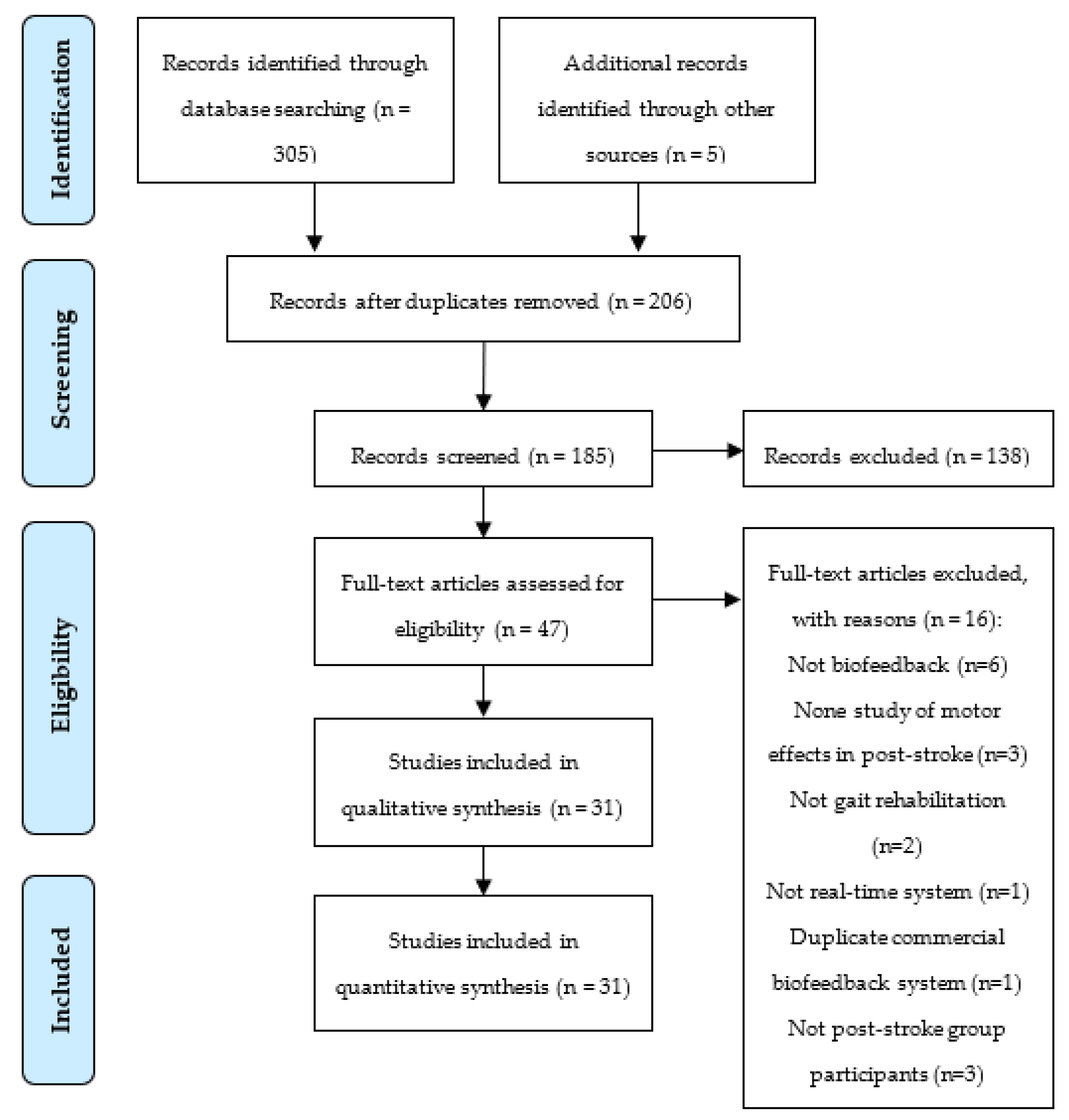
2.2. Study Selection

The studies were checked and selected by the primary researcher (C.P.) according to the following inclusion criteria: original studies; identification of the used technology in the BS; clinical intervention with post-stroke patients; BS designed for gait rehabilitation; clinical assessment of motor effects. The following exclusion criteria were applied: not biofeedback; no study of motor effects in post-stroke; not gait rehabilitation; feedback was not in real-time; duplicate commercial BS; not post-stroke participants.

2.3. Data Extraction and Synthesis

The primary researcher (C.P) extracted the relevant specifications of BSs from all eligible studies. A specification was considered relevant when mentioned in more than one study. A second researcher (J.F.) checked the reliability of data extraction. A third researcher (C.S.) allowed a consensus between researchers. Data regarding technical and clinical specifications were extracted from the relevant studies. Technical specifications are the following: sensors used to provide biofeedback and biofeedback parameters; actuators to provide sensory cues; control strategies to manage sensory cues according to sensor’s measures; assistive devices used in adjunction with BSs; physiotherapist-oriented sensory cues controlled according to biofeedback parameters. Clinical specifications address the following: post-stroke participants’ characteristics; clinical protocols employed for BSs’ validation and clinical intervention; sensor-based and clinical outcome measures used to assess BSs’ effects on post-stroke recovery; BSs’ effects on post-stroke recovery. Data extracted were summarized by counting the number of studies in each category for each specification. For example, regarding the specification of sensors used in the BSs, the number of studies using each sensor was counted, grouping the studies using the same sensor.

2.4. Methodological Quality Assessment

The quality assessment of the reviewed studies was performed through the Effective Public Health Practice Project (EPHPP) tool for quantitative studies [15]. This tool allows the quality assessment of mixed-methods studies, evaluating the following components as “strong”, “moderate” or “weak”: “selection bias”, “study design”, “cofounders”, “blinding”, “data collection methods”, and “withdrawals and drop-outs”. The global rating is considered “strong” if no component is “weak”, “moderate” if only one component is “weak”, or “weak” if two or more components are “weak”.

3. Results

The search strategy (Figure 1) yielded 305 studies, resulting in 206 studies after duplicates’ removal. Then, 185 studies’ titles and abstracts were screened for relevance, and 47 studies were full-text assessed for eligibility, excluding 138 and 16 studies, respectively. A total of 31 studies were included in this review.

3.1. Technical Specifications

Table 1 details the technical specifications (sensors and related biofeedback parameters, actuators and related sensory cues, adjunctive assistive devices, physiotherapist-oriented sensory cues) of the reviewed studies.

3.1.1. Sensors and Biofeedback Parameters

Sensors were used in BSs to measure in real-time biofeedback parameters. Biofeedback parameters addressed neuro-biomechanical data (measured by the sensors) related to motor function in the field of gait rehabilitation. The sensors’ devices, related biofeedback parameters, and the locations where the sensors were placed on the human body (in wearable devices) were reviewed.
Four studies used an electromyography (EMG) system [21,22,25,39] or inertial measurement units (IMUs) [31,40,43,46]. EMG-based systems measured electromyographic signals on tibialis anterior (four studies), quadriceps femoris (two studies), gastrocnemius lateralis, soleus, vastus lateralis, rectus femoris, and biceps femoris muscles. IMUs were usually placed on feet [31,40,46]. Three studies referred force sensors [16,18,19], load sensors [22,26,41], pressure sensors [31,32,35], a camera [27,29,34], an optical motion capture system [23,30,45], or an electroencephalography (EEG) system [20,37,42]. Force sensors were allocated on first (two studies) and fifth metatarsal heads (two studies), toe and heel of feet. Besides the feet (two studies), load and pressure sensors were alternatively placed on an exoskeleton’s knee and hip joints [22] or on a cane [32], respectively. Regarding the EEG, two studies discriminated the following target locations: Oz, Cz, ipsilesional hand motor cortex area, and Broadmann area 10. Two studies mentioned force platforms integrated into treadmills [17,38].
The sensors can be conjugated in the same BS. Only two studies included more than one sensor in the same BS, conjugating pressure sensors with IMUs [31] and magnetic resonance imaging (MRI) with EEG system [37]. Non-wearable sensors were employed in 13 studies, namely force platforms, optical motion capture systems, cameras, strain gauges, balance board, infrared sensors, plantar pressure measurement mat, and MRI system (Table 1).
Different sensors were used to measure the same biofeedback parameter. Kinetic parameters were evaluated in 12 studies by pressure, force, or load sensors, force platforms, pneumatic insoles, plantar pressure measurement mats, or balance boards [16,17,22,26,28,29,31,35,36,38,41] Spatiotemporal parameters were monitored in 12 studies by force sensor, load sensors, strain gauges, IMUs, cameras, optical motion capture systems, or infrared sensors [18,19,24,26,27,29,30,31,33,34,40,46]. Physiological parameters were measured in eight studies by EMG, EEG, fNIRS, or MRI systems [20,21,22,25,37,39,42,44]. Kinematic parameters were assessed in five studies by IMUs or optical motion capture systems [23,31,40,43,45].

3.1.2. Actuators and Biofeedback Mode

According to the sensor’s data, actuators were used in BSs to provide sensory cues (i.e., biofeedback mode) to the users. The actuators’ devices, biofeedback modes, and location where the actuators were placed on the human body (in wearable devices) were reviewed.
The following three biofeedback modes were reported: visual (25 studies), auditory (13 studies), and haptic (4 studies). A total of 11 studies combined different biofeedback modes, conjugating visual with auditory cues (9 studies), auditory with haptic, and visual with haptic.
Visual cues were provided by non-wearable devices as screens in 24 studies, using computers, monitors, televisions, tablets, or projectors; or as lights. Auditory cues were supplied by speakers in 11 studies, headphones [24], earphones [40], or an indicator placed on the waist [32]. Speakers were usually integrated into non-wearable devices such as computers, televisions, mobile phones, or tablets, but they were also placed on the human body, namely the waist and foot. Haptic cues were delivered by vibrators (two studies) placed on the paretic wrist or leg, programmable plates integrated into a robotic assistive device, or an electrotactile system using electrodes placed on the user’s unaffected thigh. The electrotactile system provided haptic cues without stimulating any unintended muscular activity that can alter the patient’s gait [19].

3.1.3. Biofeedback Control Strategies

Biofeedback control strategies are described below, explaining how and when the sensory cues (visual, auditory, and/or haptic cues) were provided to the users according to the real-time data from the sensors.
Visual cues were provided as follows. J Jung et al. [36] exhibited a color map of the feet, setting the color from light blue to red and yellow as the pressure load increased. Byl et al. [31] flashed foot pressing indicators and the values of step length, stride width, and toe-off angle, adding statements on the screen according to them (e.g., “you are doing perfectly”, “twist your feet inwards”, “step further on the left”). Bae et al. [27] showed a motion observation screen of simultaneously a reference model and the real-time video of the participant’s ankle from the side.
Tamburella et al. [22] displayed blue, red, and white colored stripes portioned into stages within the gait cycle representing over-activation, under-activation, and optimal muscle contraction, respectively, according to a targeted reference muscle activation profile [22]. Moreover, weighted averages of the joint human–robot interaction (HRI) torques were exhibited in an array of line graphs (1 line/gait phase) [22]. Day et al. [23], Nagano et al. [30], and Park et al. [45] overlaid a trace/round points representing a one-dimensional summary of performance, MFC, and position of ankle markers during the swing phase, respectively, on a goal zone (horizontal lines in [23,30] and vertical bars in [45]). Ochi et al. [26] and Kim et al. [35] activated lights and modulated a graphical depiction, respectively, according to the real-time load of the user’s weight on each limb during the stance phase.
Relatively to auditory cues, K-S Jung et al. [32] produced a sound whenever the peak vertical force on a cane was higher than a threshold. Choi et al. [41] generated a beep sound when a pre-defined load was detected in the stance phase. Hankinson et al. [43] produced a target beat per minute that was silenced (and then a metronome plays instead) when the participant did not perform a target angle range of movement.
Concerning haptic cues, Ma et al. [16] activated a vibrator at 220 Hz and 1 G when the medial plantar force was less than a threshold (50% lateral plantar force). Afzal et al. [18] enabled a vibrotactile cue at 100% intensity (1.5 G) when the paretic leg’s swing phase started and ended after 200–500 ms (time increased or decreased with the error between desired and actual time symmetry ratio) or the end of the swing phase, whichever came first [18]. On the other hand, the vibration was enabled at 25–100% intensity (intensity increased or decreased with the error) during the paretic leg’s swing phase [18].
Visual and auditory cues were conjugated as follows. Genthe et al. [17] displayed horizontal a green and a black line with a cursor (X) indicating the target and actual AGRF, respectively. Guzik et al. [24] presented feet characters according to the measured step length, a rectangle character indicating the area where the feet should be placed, and the values of recent and target step lengths. Hsu et al. [28], Mihara et al. [44], and Surucu et al. [25] presented vertical bars that change their height to reflect the magnitude of the participant’s weight-bearing on the paretic leg, SMA activation status, and muscle activity, respectively. Arpa et al. [21] and Tsaih et al. [39] demonstrated muscle contraction amplitude over time through line graphs. These studies enabled auditory cues (audible tone [17] and beep sound [28,44]) when the target was achieved.
Givon et al. [29] presented the virtual-reality games from Microsoft Xbox Kinect (as in Song et al. [34]), Nintendo Wii Fit, and SeeMe VR system. Skvortsov et al. [46] slowed down the movement around the virtual environment when an out-of-range stance time is detected. Moreover, a vertical bar (with marked target range) that changes its height to reflect the stance time was presented.
Shin et al. [33] displayed green, blue, or red colors when the deviation of the actual and step length from the target is within 10 cm, ranged from 10 to 30 cm, is more than 30 cm, respectively. Additionally, auditory cues were provided corresponding to the target step cycle initiation [33]. Schlieβman et al. [40] referred to a colored graph comprising consecutively green, yellow, and red ranges selected to match the comparison between calculated parameters and reference values. Furthermore, three auditory instructions were provided using the Android-OS default Google text-to-speech engine: “stance duration correct”, “stance duration shorter”, and “stance duration much shorter” for green, yellow, and red ranges, respectively [40].
Relatively to auditory and haptic cues conjugation, Khoo et al. [19] gave an audio tone at 200 Hz and electrotactile stimulation at 80–250 µs pulse width, 250 Hz, and up to 115 mA (using 500 Ω test load) when the swing time of the affected limb exceeded a threshold [19]. Moreover, the audio tone was activated when the affected leg established a heel-strike and stopped at a pre-defined time [19].
Concerning visual and haptic cues conjugation, Boehm et al. [38] displayed the vertical force distribution on each foot and induced a vibratory cue from the force plates when the users supported most of their weight with the stance foot. From the reviewed studies, we verified that the sensory cues were usually provided to the users following principles of a Finite State Machine (FSM) approach, which compared the user’s current state (based on real-time data from sensors) with a threshold or reference. Details about the controllers (linear, nonlinear, intelligent, adaptive) were not explicitly mentioned. Study [22] did not use and seven studies did not mention a threshold or reference in their biofeedback control strategies. The remaining studies defined a threshold or reference according to the non-paretic limb (six studies), a baseline trial performed prior training without sensory cues (nine studies), data from healthy subjects (six studies), maximum voluntary contraction (two studies) in the case of EMG-based biofeedback, body weight (two studies), and the paretic limb. Regarding the baseline trial approach, the user tried some pre-defined thresholds consecutively, and the difficulty to achieve them was evaluated [17], or the threshold was directly calculated from baseline data. Six studies have updated the threshold during training agreeing to the user’s performance [20,32,37,40,43,46]. User’s performance was usually defined according to the frequency of achieving the threshold or reference in all studies. The time when the sensory cues are enabled depends on the control’s periodicity and the type of reinforcement intended (positive and negative reinforcement to enable the sensory cue at a condition that must be learned or avoided, respectively). The periodicity of the biofeedback control strategies was not mentioned in 14 studies. The remaining 16 studies controlled sensory cues at each gait cycle (14 studies), a fixed time in the case of EEG-based biofeedback (2 studies), or at each third stride. Three studies reported the update of the periodicity during training, usually according to the user’s performance [17,39,40]. Positive and negative reinforcement was provided in 24 and 13 studies, respectively.

3.1.4. Assistive Devices Used in Adjunction with BSs

Assistive devices were used in adjunction with BSs to accelerate gait rehabilitation once they provided user-oriented intensive and repetitive training. A total of 13 studies reported the complementary use of assistive devices with BSs, namely, treadmills (11 studies), a cane, hip and knee exoskeleton, ankle-foot orthosis, gait-assistance robot with four robotic arms to control both thighs and legs, EMG-Functional Electrical Stimulation (FES) system targeting tibialis anterior muscle, kinetic immersive interface for neuromuscular coordination enhancement (KIINCE), and a cable-driven robotic system to correct force on the pelvis in the lateral direction towards the paretic side. Four studies assisted with the treadmill and another assistive device in adjunction with BSs, that was, the hip and knee exoskeleton [22], the ankle-foot orthosis [45], the gait-assistance robot [26], and the cable-driven robotic system [28]. In total, 14 studies included non-wearable assistive devices, and the remaining study did not mention wearability.

3.1.5. Physiotherapist-Oriented Sensory Cues

Only four studies stated physiotherapist-oriented sensory cues, being all visual ones. Physiotherapist-oriented sensory cues were provided along with patient-oriented cues, allowing physiotherapists to follow the biofeedback training. Consequently, they supplied adequate instructions to the patients combining their expert visual inspection with objective data from sensors, aiming to accelerate the recovery. A total of 26 studies did not refer to the assessment of the sensory cues by physiotherapists (NM in Table 1) but implemented patient-oriented cues that physiotherapists can simultaneously assess due to the nature of the actuators’ device, namely, screen, light, speaker, indicator, or programmable plate. Five studies did not allow physiotherapists to assess patient-oriented cues (NA in Table 1) because of the nature of the actuators’ device, specifically, headphone, earphone, vibrator, and/or electrotactile system.

3.2. Clinical Specifications

Table 2 summarizes the clinical specifications (sample size, study design, training dosing, and evaluation time points) found in the reviewed studies.

3.2.1. Post-Stroke Participants

The review englobes a sample size of 660 post-stroke participants, with 17 post-stroke participants being the median sample size of the reviewed studies. The maximum and minimum sample sizes were found in Guzik et al. and Nan et al. [20,24] with 50 and 2 post-stroke subjects, respectively.
The post-stroke participants are mostly characterized by age (30 studies), gender (30 studies) [36,37,38,39,40,41,42,44,45,46], time post stroke (27 studies) [16,17,18,21,22,23,24,25,26,27,28,29,30,31,32,33,34,36,37,38,39,40,41,42,44,45,46], hemiplegic side (26 studies) [16,17,18,21,22,23,24,25,26,27,30,31,32,33,34,35,36,37,38,39,40,41,42,44,45,46], stroke aetiology (18 studies) [16,18,20,22,25,26,27,29,30,32,33,35,36,37,38,40,41,42], and body mass (15 studies) [16,18,20,22,27,28,30,32,33,34,36,38,39,41,46].

3.2.2. Study Design

The review includes 15 uncontrolled, 11 randomized controlled, 4 non-randomized controlled, and 1 randomized cross-over study (Table 2). A total of 10 controlled studies were balanced on the number of participants between experimental and control groups (Table 2).
The clinical protocols of uncontrolled studies were based on training and control or baseline procedures. The training procedure allowed the users to follow sensory cues provided by the BS towards recovery. The control procedure was similar to the training one, except for the use of biofeedback. Therefore, the control procedure is used to control the biofeedback effects within the participant. Additionally, the control procedure was used to specify training thresholds.
Five uncontrolled studies only performed training and control procedures, randomly assigning the order between participants to limit carry-over, practice, and order effect [16,18,33,37,38]. Moreover, two studies implemented a wash-out period (i.e., a period without biofeedback; 1 h [33] and 1 month [37] considering a training dosing of 5 min and twice a week over 2 months for 50 min, respectively) between control and training procedures to exclude the carry-over, practice, and order effect.
Three uncontrolled studies complemented biofeedback training with conventional physiotherapy exercises [24,37] or robotic assistive devices [28,38]. Hsu et al. performed training with biofeedback only and training by combining pelvic corrective force and biofeedback, randomly assigning the order between participants [28]. The control procedure did not include any type of biofeedback and robotic assistive device [28]. In opposition to Hsu et al., Boehm et al. implemented a control procedure without the biofeedback but with the robotic assistive device [38].
The cross-over study from Tamburella et al. randomly created two groups of participants to perform cross-over training with two BSs complemented with robotic assistive devices and conventional rehabilitation [22]. The control procedure was executed with the robotic assistive device [22].
Controlled studies included two groups of participants who executed specific interventions: experimental and control groups. Experimental groups trained with the biofeedback system. Additionally, experimental groups accompanied biofeedback training with conventional rehabilitation exercises (12 studies) [19,21,25,26,27,31,32,35,39,41,43,44] and robotic assistive devices (2 studies) [26,27]. Control groups usually did not perform biofeedback training, only addressing conventional rehabilitation. However, the control groups from Song et al. followed distinct biofeedback from the experimental group [34], and Lee et al. and Mihara et al. implemented sham neurofeedback [42,44]. Bae et al. and Ochi et al. included and did not include, respectively, the robotic assistive devices during control intervention [26,27].

3.2.3. Clinical Protocols

This section reviews the existence and duration of the familiarization prior to the training, the motor task executed during familiarization and biofeedback training, the assistance provided by the assistive devices (in case of their use), and the training dosing.
Three studies mentioned familiarization of the BS prior to training (10 min in [16], 2 min in [23], the value was not mentioned in [18]) while study [28] performed familiarization during training. Regarding the motor task, 20 studies addressed walking (10 studies walking on treadmill and the remaining walking on level-ground) while the remaining tackled sitting (8 studies) [20,21,25,27,37,39,42,44] or standing (2 studies) [29,34]. Training during sitting was performed using EMG-feedback, neurofeedback (motor imagery), and biofeedback complemented with EMG-FES. Virtual-reality games included standing training. For gait training, only two studies did not implement a self-selected walking speed [22,26].
Some studies evaluated the effects of biofeedback training during motor assistance driven by assistive devices. The exoskeleton from Tamburella et al. applied body-weight support equal to 50% of the participant’s body weight, 100% guidance assistance, and 1.3 km/h speed [22]. The gait-assistance robot from Ochi et al. was predominantly used in active-assistive mode reaching a 0.75 km/h speed, 10–30° hip ROM, and 5–10° ankle ROM [26]. The EMG-FES system from Bae et al. stimulated 1–50 mA by 0.1 s rise, 5 s on, and 2 s decay according to the subject’s ankle joint [27]. The cable-driven robotic system from Hsu et al. employed a pelvic corrective force equal to 9% of participants’ body weight at the timing of heel strike of the paretic leg and lasted for 400 ms [28]. The KIINCE from Boehm et al. moved its plates forward and afterward out of phase in a simplified walking motion [38]. The treadmill speed from Guzik et al. increased by 5 to 10% during consecutive training sessions [24].
The training dosing usually implied multiple sessions with a fixed frequency per week. The maximum, minimum, and median training dosing were twice a week for 12 weeks at 60 min/session in Givon et al., two 15 min sessions in Day et al., and three times a week for 4 weeks at 30 min/session in Kim et al., respectively. Only five studies, all uncontrolled ones, completed the training procedure on a single day.

3.2.4. Sensor-Based and Clinical Outcomes

The evaluation time points were mostly pre- and post-training (31 studies) to assess sensor-based (29 studies) and clinical (22 studies) outcomes (Table 3). Retention evaluation time points were executed immediately after training (immediate post-training) and follow-up in 28 and 9 studies, respectively. The later, earlier, and usual follow-up evaluations were performed 3 months [21,29], 2 min [17], and 1 month [21,30,37,40] post-training (Table 2), respectively.
The evaluated sensor-based outcomes were categorized on spatiotemporal (18 studies), kinetic (13 studies), kinematic (10 studies), and physiological (8 studies) outcomes.S patiotemporal outcomes mostly included step length (8 studies) and walking speed (9 studies). Mostly addressed kinetic outcomes were plantar pressure (2 studies), peak vertical force (2 studies), peak joint torque (2 studies), and grip strength (2 studies). Joint ROM was the most evaluated kinematic outcome (5 studies) [21,23,25,31]. Physiological ones usually included EMG- (5 studies) and EEG-based (3 studies) outcomes. EMG-based outcome was muscle activation. EEG-based outcomes comprised relative individual alpha-band (IAB) amplitude, sensorimotor rhythm (SMR) waves amplitude, and alpha-band weighted node degree of functional connectivity.
Clinical outcomes addressed post-stroke stage (7 studies), motor (19 studies), mental (8 studies), and sensory (2 studies) effects on post-stroke participants. Post-stroke stage was mostly evaluated by Fugl–Meyer (FM) (6 studies). Motor effects were mostly assessed by 10 m walk test (12 studies) and Timed Up and Go (TUG) (13 studies). Mental effects were determined by Quebec User Evaluation of Satisfaction with Assistive Technology 2.0 (QUEST 2.0) (2 studies), Visual Analogue Scale (VAS) (2 studies) of motivation, mood, satisfaction, and pain, and Likert-type satisfaction scale (2 studies). Sensory effects were revealed by proprioception test and Pin Prick (PP) test for haptic effects and by Start Cancellation Test for visual effects.

3.2.5. BS Effects on Post-Stroke Recovery

Table 3 shows the BS effects on post-stroke recovery based on the parametric and non-parametric statistical tests performed by the reviewed studies to conclude the existence of statistically significant differences (alpha set to 0.05) on the measured outcomes between the evaluation time points on both experimental and control groups.
Regarding sensor-based outcomes, 15, 12, 8, and 5 studies reached statistically significant improvements on spatiotemporal, kinetic, kinematic, and physiological outcomes, respectively, between at least two evaluation time points. Studies from Givon et al., Nagano et al., Hsu et al., Skvortzov et al., Park et al., and Mottaz et al. [28,29,30,37,45,46] did not find statistically significant improvements between all evaluation time points. Nan et al. [20] did not evaluate physiological outcomes using statistical tests. Moreover, 8, 6, 6, and 2 controlled studies exhibited higher statistically significant improvements on the experimental group than the control group concerning spatiotemporal, kinetic, kinematic, and physiological outcomes, respectively.
For the clinical outcomes, four, five, and four studies achieved statistically significant improvements on post-stroke stage, motor, and mental function, respectively, between at least two evaluation time points. Two, two, four, and two studies did not evaluate post-stroke stage, motor, mental, and sensory effects, respectively, using statistical tests. Givon et al., Hankinson et al., and Schlieβmann et al. [29,40,43] did not find statistically significant improvements on clinical outcomes between all evaluation time points. Additionally, three, five, and two controlled studies demonstrated higher significant improvements on the experimental group than the control group concerning post-stroke stage, motor, and mental effects, respectively.

3.3. Methodological Quality Assessment

Table 4 shows the results of the EPHPP tool. The “selection bias” is “weak” (15 studies) or “moderate” (13 studies) in most studies. The “study design” is “strong” or “moderate” in 16 and 15 studies, respectively. The “confounders”, “blinding”, and “withdrawals and drop-outs” components are “strong” in most studies (29, 24, and 21 studies, respectively), being “weak” in 2, 7, and 10 studies, respectively. The “data collection methods” are “strong” in all studies. The global rating is “strong”, “moderate”, or “weak” in 5, 12, and 14 studies, respectively.

4. Discussion

This review aims to investigate the technical and clinical specifications of BSs for gait rehabilitation that should be followed in future research to achieve efficacy for post-stroke recovery. Despite the current promising research, more scientific evidence is needed to rate the efficacy of biofeedback accurately [8]. The research question is answered in the following sections.

4.1. Technical Specifications

There was no sensor technology found that was common to most studies, as appointed in [9]. In of spite that, sensors that measure kinetic and spatiotemporal parameters were usually employed in the reviewed studies. Pressure sensors and force platforms evaluated kinetic biofeedback parameters, and force and load sensors additionally measured spatiotemporal biofeedback parameters. IMUs and motion capture systems determined kinematic and spatiotemporal biofeedback parameters. Cameras assessed the body’s movement. EMG and EEG systems measured muscular activation from the tibialis anterior and quadriceps femoris muscles and alpha-band EEG signals, respectively.
Even though most of the studies implied only one sensor on the BS, Byl et al. and Mottaz et al. [31,37] combined pressure sensors and MRI with IMUs and EEG, respectively. As reviewed in Byl et al. and Mottaz et al. [31,37], future research should study the effectiveness of combining multiple biomechanical and physiological sensors to personalize biofeedback for post-stroke users given their variable motor deficits. Further, the use of multimodal sensors may enable a more holistic BS-based gait training and assessment, attending to intra- and inter-subject motor variability [7,10].
Regarding sensors’ wearability, wearable sensors were used in most studies. Wearability allows unique assessments of body motion during ambulatory training in a non-fixed facility [10,47]. Thus, allowing the users to practice in multiple spaces, encouraging training dosing to increase in everyday scenarios and, consequently, accelerating recovery [7,48]. However, EMG and EEG sensors required a time-consuming preparation in opposition to IMUs, pressure, force, and load sensors that were fast positioned on the feet. This finding guides future research to select wearable sensors with fast positioning.
Concerning biofeedback mode, most studies reported visual biofeedback mode, as concluded in [9], using screens from monitors, televisions, tablets, or projectors. Auditory and haptic biofeedback modes were usually applied using speakers integrated into computers, televisions, or tablets, and vibrators, respectively. Even most studies have implied one mode, visual and auditory cues were combined once multimodal biofeedback can reduce the user’s cognitive load compared to a single-mode [7]. Therefore, there is space to explore auditory and haptic modes, as retained in [7,10], so that post-stroke users with multiple sensorial deficits can take advantage of multimodal biofeedback rehabilitation. In this manner, the physiotherapist has the necessary resources to personalize the training according to the patient’s imminent sensorial deficits.
Regarding actuators wearability, only haptic biofeedback was provided using wearable actuators in most studies, allowing ambulatory practice on daily-like scenarios as overground walking. Thus, future research should study the impact of using wearable actuators as earphones and augmented reality glasses for auditory and visual modes, respectively, benchmarking the results with non-wearable solutions. Haptic actuators were placed on both upper or lower limbs, fostering the conclusion that haptic feedback on the body, either at or away from the desired body segment to be changed, can improve motor performance [48].
Most biofeedback control strategies compared sensor data with a threshold or reference obtained from the user’s limbs, a baseline trial, maximum voluntary contraction, body weight, or data from healthy subjects, having a need to benchmark these methods. Regarding periodicity, most BSs for gait training have controlled the sensory cues at each gait cycle. Studies exploring EMG- and EEG-based biofeedback, performed during sitting, did not mention the control’s periodicity or attended a fixed-time control, respectively. Future studies should clearly state the control’s details (type, quality, and periodicity), as concluded in [7], fostering the research’s reproducibility. Less than half of the studies updated the threshold or reference and the control’s periodicity during training agreeing with the time for achieving the threshold or reference, intended as the user’s imminent disability level. In this sense, the rehabilitation was personalized according to the user’s imminent disability level, avoiding frustration and dependence on the sensory cues, respectively [37,49]. However, there is space to continue the investigation of biofeedback control strategies’ personalization according to the user’s imminent disability level, reporting significant evidence of this adaptation.
Positive reinforcement was employed in most biofeedback control strategies, as referred in the systematic review of Bowman et al. [9], drawing the user’s attention to a condition that should be learned and repeated. Even most studies with multiple biofeedback modes applied positive reinforcement on both. In this manner, there is space to explore if combining both positive and negaztive reinforcements, taking into account the cognitive effort, accelerates the relearning process by aware the patients of motor conditions that must be repeated and avoided, respectively.
Visual cues were usually active during training, which can lead to visual reliance and high cognitive effort [50,51]. They provided detailed biofeedback through graphs and scenarios modulated on shape, color, and size according to the control strategy. Auditory and haptic cues were usually inactive, being enabled according to the control strategy at a fixed intensity once intensity adaptation may not be perceived by post-stroke users due to their sensory deficits [52].
Non-wearable assistive devices, mostly treadmills, were used in adjunction with BSs. Treadmills encourage an intensive practice of walking at a controlled and stable gait speed [11]. Robotic assistive devices such as exoskeletons, robotic arms, FES systems, robotic platforms, or cable-driven robotic systems were also applied. They were usually controlled to provide 100% guidance assistance. These robotic systems intensively and repetitively assisted patients on motor tasks while BSs fostered active participation during training [22]. The future directions should continue exploring the contribution of BSs, as an alternative and complementary medicine approach, behind different closed-loop controlled wearable assistive devices.
Physiotherapists guide and instruct the patients according to their specialized knowledge towards recovery [53]. Additionally, physiotherapists’ intervention during training allows the safe and effective use of BSs and robotic assistive devices [6]. Although it was not stated in most studies, BSs could provide objective real-time information about the patients’ motor behaviour to the physiotherapists during the training, complementing visual inspection of the patients, as in [22,25,31,42]. Moreover, BSs could fulfil the lack of physical contact between the physiotherapists and patients during training with robotic assistive devices [54]. Future research should explore the design of physiotherapist-oriented sensory cues according to the physiotherapist’s needs to enrich their contribution to rehabilitation.

4.2. Clinical Specifications

Clinical studies related to the effects of BSs on post-stroke recovery had been carried out with a median sample size of 17 post-stroke participants, serving as a reference for future research. Post-stroke participants were selected and characterized mostly by age, gender, time post-stroke, hemiplegic side, stroke aetiology, and body mass, avoiding influencing the research evidence. Future clinical studies may include quantitative characterization of participants’ disability level before the intervention, increasing the reliability of their conclusions.
Literature includes randomized balanced controlled and uncontrolled studies as high-quality and proof of concept research designs, respectively, addressing a median training dose of three times a week for 4 weeks at 30 min/session. Familiarization was typically not stated. However, future research should appoint the existence and duration of this procedure once it can influence the research reproducibility. Moreover, further randomized controlled studies need to be conducted to find clinical evidence regarding the efficacy of biofeedback on post-stroke motor recovery; and potentiate benchmarking of biofeedback technologies and the standardization of their technical and clinical specifications [7,9,10].
Biofeedback training usually involved walking at a self-selected speed, but neurofeedback and biofeedback complemented with EMG-FES were trained during sitting and virtual reality games during standing. Evaluation time points occurred at pre-training and post-training in most studies, evaluating the effects of BSs on post-stroke recovery considering sensor-based and clinical outcomes. Retention procedures were also performed to evaluate motor learning immediately (in most studies) and follow-up post-training. Follow-up evaluations usually occurred 1 month post-training to assess the long-term benefits of the intervention.
In comparison with kinematic and physiological outcomes, spatiotemporal and kinetic ones were the most evaluated sensor-based outcomes. It was expected that most studies would apply kinetic or spatiotemporal biofeedback parameters. Step length and walking speed were highlighted between spatiotemporal outcomes. Clinical outcomes assessed motor effects in most of the studies using the 10 m walk test and TUG. Less than half of the studies also measured the post-stroke stage, mental and sensory effects through clinical outcomes. BSs exert direct action on cognitive and sensory functions once the patients are encouraged to self-control their motor behavior according to the coding scheme of the sensory inputs [13]. Therefore, future studies should fulfil this gap by conducting a user-specific holistic assessment, also including kinematic and physiological sensor-based outcomes, post-stroke stage, mental and sensory clinical effects.
Most studies achieved statistically significant improvements on at least one spatiotemporal, kinetic, kinematic, and physiological sensor-based outcomes, post-stroke stage, motor and mental clinical effects between at least two evaluation time points. Although most studies implied kinetic or spatiotemporal biofeedback parameters, they obtained promising results concerning kinematic and physiological effects and clinical-based outcomes. Moreover, most controlled studies exhibited higher statistically significant improvements on the experimental group than the control group concerning spatiotemporal, kinetic, and physiological sensor-based outcomes, motor and mental clinical effects. In this manner, these positive trends indicate the promise of the efficacy of biofeedback on rehabilitation, as concluded in [7,8,9,10]. Future research should evaluate clinical sensory effects using statistical tests to power the research conclusions.

4.3. Future Directions and Challenges to Overcome

There is space to explore: (i) the effectiveness of combining multimodal wearable sensors to cope with variable sensorimotor deficits of stroke survivors; (ii) auditory and haptic biofeedback modes so that post-stroke patients who exhibit multiple sensorial deficits can take advantage of biofeedback rehabilitation; (iii) the impact of using wearable actuators such as earphones and augmented reality glasses, benchmarking the results with the standard non-wearable solutions; (iv) the contribution of BSs behind different robotic assistive devices; (v) the design of physiotherapist-oriented sensory cues according to the physiotherapist’s needs to enhance the physiotherapist–patient interaction; and, (vi) the effects of biofeedback, using statistical tests to power the research conclusions, on kinematic and physiological sensor-based outcomes, post-stroke stage, mental and sensory clinical outcomes. Moreover, future studies on robotic biofeedback for post-stroke gait rehabilitation should: (i) clearly state the biofeedback control’s details (type, quality, and periodicity) and appoint the existence and duration of the familiarization procedure to foster the research’s reproducibility and benchmarking; (ii) include quantitative characterization of participants’ disability level before the intervention to increase the reliability of the conclusions; (iii) biofeedback control strategies’ personalized according to the user’s imminent disability level and the combination of positive and negative reinforcement to accelerate the relearning process; and, (iv) conduct randomized controlled studies to find high-quality clinical evidence regarding the efficacy of biofeedback on post-stroke motor recovery and potentiate benchmarking of biofeedback technologies.

4.4. Study Limitations

The main limitation of this review is the moderate or weak quality in most of the reviewed studies mainly due to the rate of the components “selection bias” and “withdrawals and dropouts” (Table 4). Future research should focus on planning studies with strong quality to power their results and conclusions, as concluded in [7,9].

5. Conclusions

Most BSs measured kinetic and spatiotemporal parameters using wearable sensors and compared these real-time data with a reference or threshold to control non-wearable actuators capable of providing visual biofeedback following a positive reinforcement. Clinical protocols were executed with a median of 15 post-stroke participants performing walking training three times a week for 4 weeks at 30 min/session. Evaluation of spatiotemporal and kinetic sensor-based outcomes and clinical motor effects were usually performed at pre-training, post-training, and 1-month follow-up. Most controlled studies exhibited higher statistically significant improvements on the experimental group than the control group.
Future studies should report the control’s periodicity and familiarization procedures and explore kinematic and physiological sensor-based outcomes, post-stroke stage, mental and sensory clinical effects. There is space to explore the effectiveness of using multiple wearable sensors and actuators to provide biofeedback; the effects of combining both positive and negative reinforcement on biofeedback control strategies; the contribution of BSs behind different robotic assistive devices; physiotherapist-oriented sensory cues according to the physiotherapist’s needs; and the personalization of biofeedback training according to the user’s imminent disability level.

Author Contributions

The idea for the article: C.P., J.F., C.P.S. and J.C.; literature search and data analysis: C.P. and J.F.; draft of the work: C.P.; revision of the work: all authors (C.P., J.F., C.P.S. and J.C.). All authors have read and agreed to the published version of the manuscript.

Funding

This work has been supported in part by the FEDER Funds through the COMPETE 2020—Programa Operacional Competitividade e Internacionalização (POCI) and P2020 with the Reference Project SmartOs Grant POCI-01-0247-FEDER-039868, and by FCT national funds, under the national support to R&D units grant, through the reference project UIDB/04436/2020 and UIDP/04436/2020, under scholarship reference 2020.05709.BD, and under Stimulus of Scientific Employment with the grant 2020.03393.CEECIND.

Institutional Review Board Statement

Not applicable.

Informed Consent Statement

Not applicable.

Conflicts of Interest

The authors declare no conflict of interest.

Abbreviations

This appendix comprises a glossary of the terms used in this paper, as follows:
5XSSTFive Times Sit-to-Stand Test
9HPTNine-Hole Peg Test
ARATAction Research Arm Test
BBSBerg Balance Scale
BBTBox and Block Test
BDIBeck Depression Inventory
BIBarthel Index
BS biofeedback systems
CESDCenter for Epidemiologic Studies Depression Scale
DGIDynamic Gait Index
EPHPP Effective Public Health Practice Project
EEG electroencephalography
EMG electromyographic
ESSEuropean Stroke Scale
FACFunctional Ambulation Category
FES Functional Electrical Stimulation
FGAFunctional Gait Assessment
FIMFunctional Independence Measure
FM Fugl–Meyer
fNIRSfunctional near-infrared spectroscopy
FRTFunctional Reach Test
GDSGeriatric Depression Scale
HADSHospital Anxiety and Depression Scales
HAIHauser Ambulance Index
IAB Individual Alpha-Band
IADLInstrumental Activities of Daily Living
IMU Inertial Measurement Unit
KIINCE Kinetic Immersive Interface for Neuromuscular Coordination Enhancement
LOSLimit of Stability
MALMotor Activity Log
MASModified Ashworth Scale
MMTManual Muscle Test
MoCAMontreal Cognitive Assessment
MRCMedical Research Council
MRI Magnetic Resonance Imaging
NASA-TLXNational Aeronautics and Space Administration Task Load Index
PP Pin Prick
QCMQuestionnaire for Current Motivation
QUEST Quebec User Evaluation of Satisfaction with Assistive Technology
RCSRelationship Change Scale
SBTStanding Balance Test
SMR sensorimotor rhythm
SMAsupplementary motor area
TGATinetti Gait Assessment
TISTrunk Impairment Scale
TUG Timed Up and Go
VAS Visual Analogue Scale
WBLTWeight-Bearing Lunge Test
WISCIWalking Index for Spinal Cord Injury

References

  1. Luengo-Fernandez, R.; Leal, J.; Candio, P.; Violato, M.; Stroke Alliance for Europe. Economic Impact of Stroke. 2019. Available online: https://www.safestroke.eu/economic-impact-of-stroke/ (accessed on 24 April 2020).
  2. Johnson, C.O.; Nguyen, M.; Roth, G.A.; Nichols, E.; Alam, T.; Abate, D.; Abd-Allah, F.; Abdelalim, A.; Abraha, H.N.; Abu-Rmeileh, N.M.; et al. Global, regional, and national burden of stroke, 1990–2016: A systematic analysis for the Global Burden of Disease Study 2016. Lancet Neurol. 2019, 18, 439–458. [Google Scholar] [CrossRef]
  3. Wolf, S.L. Electromyographic Biofeedback Applications to Stroke Patients. Phys. Ther. 1983, 63, 1448–1459. [Google Scholar] [CrossRef] [PubMed]
  4. Huang, H.; Wolf, S.L.; He, J. Recent developments in biofeedback for neuromotor rehabilitation. J. Neuroeng. Rehabil. 2006, 3, 11. [Google Scholar] [CrossRef] [PubMed]
  5. FlintRehab. Neuroplasticity After Stroke: The Single Most Important Key to Recovery. 2018. Available online: https://www.flintrehab.com/2018/neuroplasticity-after-stroke/ (accessed on 27 May 2019).
  6. Association for Applied Psychophysiology and Biofeedback. Standards for Performing Biofeedback. 2013. Available online: https://www.aapb.org/i4a/pages/index.cfm?pageid=3678#I (accessed on 20 March 2020).
  7. van Gelder, L.M.A.; Barnes, A.; Wheat, J.S.; Heller, B.W. The use of biofeedback for gait retraining: A mapping review. Clin. Biomech. 2018, 59, 159–166. [Google Scholar] [CrossRef]
  8. Stanton, R.; Ada, L.; Dean, C.M.; Preston, E. Biofeedback improves performance in lower limb activities more than usual therapy in people following stroke: A systematic review. J. Physiother. 2017, 63, 11–16. [Google Scholar] [CrossRef] [PubMed]
  9. Bowman, T.; Gervasoni, E.; Arienti, C.; Lazzarini, S.G.; Negrini, S.; Crea, S.; Cattaneo, D.; Carrozza, M.C. Wearable Devices for Biofeedback Rehabilitation: A Systematic Review and Meta-Analysis to Design Application Rules and Estimate the Effectiveness on Balance and Gait Outcomes in Neurological Diseases. Sensors 2021, 21, 3444. [Google Scholar] [CrossRef]
  10. Spencer, J.; Wolf, S.L.; Kesar, T.M. Biofeedback for Post-stroke Gait Retraining: A Review of Current Evidence and Future Research Directions in the Context of Emerging Technologies. Front. Neurol. 2021, 12, 637199. [Google Scholar] [CrossRef]
  11. Mikolajczyk, T.; Ciobanu, I.; Badea, D.I.; Iliescu, A.; Pizzamiglio, S.; Schauer, T.; Seel, T.; Seiciu, P.L.; Turner, D.L.; Berteanu, M. Advanced technology for gait rehabilitation: An overview. Adv. Mech. Eng. 2018, 10, 1–19. [Google Scholar] [CrossRef]
  12. Perry, J. Gait Analysis: Normal and Pathological Function; SLACK Incorporated: West Deptford, NJ, USA, 1992. [Google Scholar]
  13. Morone, G.; Spitoni, G.F.; de Bartolo, D.; Ghooshchy, S.G.; di Iulio, F.; Paolucci, S.; Zoccolotti, P.; Iosa, M. Rehabilitative devices for a top-down approach. Expert Rev. Med. Devices 2019, 16, 187–195. [Google Scholar] [CrossRef] [PubMed]
  14. Ganesh, G.; Takagi, A.; Osu, R.; Yoshioka, T.; Kawato, M.; Burdet, E. Two is better than one: Physical interactions improve motor performance in humans. Sci. Rep. 2015, 4, 3824. [Google Scholar] [CrossRef] [Green Version]
  15. Effective Public Healthcare Panacea Project. Quality Assessment Tool for Quantitative Studies. Available online: https://www.ephpp.ca/quality-assessment-tool-for-quantitative-studies/ (accessed on 25 January 2020).
  16. Ma, C.Z.-H.; Zheng, Y.-P.; Lee, W.C.-C. Changes in gait and plantar foot loading upon using vibrotactile wearable biofeedback system in patients with stroke. Top. Stroke Rehabil. 2018, 25, 20–27. [Google Scholar] [CrossRef] [PubMed]
  17. Genthe, K.; Schenck, C.; Eicholtz, S.; Zajac-Cox, L.; Wolf, S.; Kesar, T.M. Effects of real-time gait biofeedback on paretic propulsion and gait biomechanics in individuals post-stroke. Top. Stroke Rehabil. 2018, 25, 186–193. [Google Scholar] [CrossRef]
  18. Afzal, M.R.; Lee, H.; Eizad, A.; Lee, C.H.; Oh, M.-K.; Yoon, J. Effects of Vibrotactile Biofeedback Coding Schemes on Gait Symmetry Training of Individuals With Stroke. IEEE Trans. Neural Syst. Rehabil. Eng. 2019, 27, 1617–1625. [Google Scholar] [CrossRef]
  19. Khoo, I.-H.; Marayong, P.; Krishnan, V.; Balagtas, M.; Rojas, O.; Leyba, K. Real-time biofeedback device for gait rehabilitation of post-stroke patients. Biomed. Eng. Lett. 2017, 7, 287–298. [Google Scholar] [CrossRef]
  20. Nan, W.; Dias, A.P.B.; Rosa, A.C. Neurofeedback Training for Cognitive and Motor Function Rehabilitation in Chronic Stroke: Two Case Reports. Front. Neurol. 2019, 10, 800. [Google Scholar] [CrossRef]
  21. Arpa, S.; Ozcakir, S. Does electromyographic biofeedback improve exercise effects in hemiplegic patients? A pilot randomized controlled trial. J. Rehabil. Med. 2019, 51, 109–112. [Google Scholar] [CrossRef]
  22. Tamburella, F.; Moreno, J.C.; Valenzuela, D.S.H.; Pisotta, I.; Iosa, M.; Cincotti, F.; Mattia, D.; Pons, J.L.; Molinari, M. Influences of the biofeedback content on robotic post-stroke gait rehabilitation: Electromyographic vs. joint torque biofeedback. J. Neuroeng. Rehabil. 2019, 16, 95. [Google Scholar] [CrossRef]
  23. Day, K.A.; Cherry-Allen, K.M.; Bastian, A.J. Individualized feedback to change multiple gait deficits in chronic stroke. J. Neuroeng. Rehabil. 2019, 16, 158. [Google Scholar] [CrossRef]
  24. Guzik, A.; Drużbicki, M.; Kwolek, A.; Przysada, G.; Brzozowska-Magoń, A.; Wolan-Nieroda, A.; Ćwirlej-Sozańska, A.; Wiśniowska-Szurlej, A.; Wyszyńska, J. Analysis of the association between selected factors and outcomes of treadmill gait training with biofeedback in patients with chronic stroke. J. Back Musculoskelet. Rehabil. 2020, 33, 159–168. [Google Scholar] [CrossRef]
  25. Dost Sürücü, G.; Tezen, Ö. The effect of EMG biofeedback on lower extremity functions in hemiplegic patients. Acta Neurol. Belg. 2021, 121, 113–118. [Google Scholar] [CrossRef]
  26. Ochi, M.; Wada, F.; Saeki, S.; Hachisuka, K. Gait training in subacute non-ambulatory stroke patients using a full weight-bearing gait-assistance robot: A prospective, randomized, open, blinded-endpoint trial. J. Neurol. Sci. 2015, 353, 130–136. [Google Scholar] [CrossRef] [PubMed]
  27. Bae, S.; Lee, J.; Lee, B.-H. Effect of an EMG–FES Interface on Ankle Joint Training Combined with Real-Time Feedback on Balance and Gait in Patients with Stroke Hemiparesis. Healthcare 2020, 8, 292. [Google Scholar] [CrossRef] [PubMed]
  28. Hsu, C.-J.; Kim, J.; Roth, E.J.; Rymer, W.Z.; Wu, M. Use of Pelvic Corrective Force With Visual Feedback Improves Paretic Leg Muscle Activities and Gait Performance After Stroke. IEEE Trans. Neural Syst. Rehabil. Eng. 2019, 27, 2353–2360. [Google Scholar] [CrossRef] [PubMed]
  29. Givon, N.; Zeilig, G.; Weingarden, H.; Rand, D. Video-games used in a group setting is feasible and effective to improve indicators of physical activity in individuals with chronic stroke: A randomized controlled trial. Clin. Rehabil. 2016, 30, 383–392. [Google Scholar] [CrossRef]
  30. Nagano, H.; Said, C.M.; James, L.; Begg, R.K. Feasibility of Using Foot–Ground Clearance Biofeedback Training in Treadmill Walking for Post-Stroke Gait Rehabilitation. Brain Sci. 2020, 10, 978. [Google Scholar] [CrossRef]
  31. Byl, N.; Zhang, W.; Coo, S.; Tomizuka, M. Clinical impact of gait training enhanced with visual kinematic biofeedback: Patients with Parkinson’s disease and patients stable post stroke. Neuropsychologia 2015, 79, 332–343. [Google Scholar] [CrossRef]
  32. Jung, K.-S.; Bang, H.; In, T.-S.; Cho, H.-Y. Gait training with auditory feedback improves trunk control, muscle activation and dynamic balance in patients with hemiparetic stroke: A randomized controlled pilot study. J. Back Musculoskelet. Rehabil. 2020, 33, 1–6. [Google Scholar] [CrossRef]
  33. Shin, J.; Chung, Y. Influence of visual feedback and rhythmic auditory cue on walking of chronic stroke patient induced by treadmill walking in real-time basis. NeuroRehabilitation 2017, 41, 445–452. [Google Scholar] [CrossRef]
  34. Song, G.B.; Park, E.C. Effect of virtual reality games on stroke patients’ balance, gait, depression, and interpersonal relationships. J. Phys. Ther. Sci. 2015, 27, 2057–2060. [Google Scholar] [CrossRef]
  35. Kim, J.-S.; Oh, D.-W. Use of real-time visual feedback during overground walking training on gait symmetry and velocity in patients with post-stroke hemiparesis: Randomized controlled, single-blind study. Int. J. Rehabil. Res. 2020, 43, 247–254. [Google Scholar] [CrossRef]
  36. Jung, J.; Choi, W.; Lee, S. Immediate augmented real-time forefoot weight bearing using visual feedback improves gait symmetry in chronic stroke. Technol. Health Care 2020, 28, 733–741. [Google Scholar] [CrossRef] [PubMed]
  37. Mottaz, A.; Corbet, T.; Doganci, N.; Magnin, C.; Nicolo, P.; Schnider, A.; Guggisberg, A.G. Modulating functional connectivity after stroke with neurofeedback: Effect on motor deficits in a controlled cross-over study. NeuroImage Clin. 2018, 20, 336–346. [Google Scholar] [CrossRef] [PubMed]
  38. Boehm, W.L.; Gruben, K.G. Development of KIINCE: A kinetic feedback-based robotic environment for study of neuromuscular coordination and rehabilitation of human standing and walking. J. Rehabil. Assist. Technol. Eng. 2018, 5, 205566831879358. [Google Scholar] [CrossRef] [PubMed]
  39. Tsaih, P.-L.; Chiu, M.-J.; Luh, J.-J.; Yang, Y.-R.; Lin, J.-J.; Hu, M.-H. Practice Variability Combined with Task-Oriented Electromyographic Biofeedback Enhances Strength and Balance in People with Chronic Stroke. Behav. Neurol. 2018, 2018, 7080218. [Google Scholar] [CrossRef] [PubMed]
  40. Schließmann, D.; Nisser, M.; Schuld, C.; Gladow, T.; Derlien, S.; Heutehaus, L.; Weidner, N.; Smolenski, U.; Rupp, R. Trainer in a pocket-proof-of-concept of mobile, real-time, foot kinematics feedback for gait pattern normalization in individuals after stroke, incomplete spinal cord injury and elderly patients. J. Neuroeng. Rehabil. 2018, 15, 44. [Google Scholar] [CrossRef]
  41. Choi, Y.-H.; Kim, J.-D.; Lee, J.-H.; Cha, Y.-J. Walking and balance ability gain from two types of gait intervention in adult patients with chronic hemiplegic stroke: A pilot study. Assist. Technol. 2019, 31, 112–115. [Google Scholar] [CrossRef]
  42. Lee, Y.-S.; Bae, S.-H.; Lee, S.-H.; Kim, K.-Y. Neurofeedback Training Improves the Dual-Task Performance Ability in Stroke Patients. Tohoku J. Exp. Med. 2015, 236, 81–88. [Google Scholar] [CrossRef]
  43. Hankinson, K.; Shaykevich, A.; Vallence, A.-M.; Rodger, J.; Rosenberg, M.; Etherton-Beer, C. A Tailored Music-Motor Therapy and Real-Time Biofeedback Mobile Phone App (‘GotRhythm’) to Promote Rehabilitation Following Stroke: A Pilot Study. Neurosci. Insights 2022, 17, 26331055221100587. [Google Scholar] [CrossRef]
  44. Mihara, M.; Fujimoto, H.; Hattori, N.; Otomune, H.; Kajiyama, Y.; Konaka, K.; Watanabe, Y.; Hiramatsu, Y.; Sunada, Y.; Miyai, I.; et al. Effect of Neurofeedback Facilitation on Poststroke Gait and Balance Recovery. Neurology 2021, 96, e2587–e2598. [Google Scholar] [CrossRef]
  45. Park, S.; Liu, C.; Sánchez, N.; Tilson, J.K.; Mulroy, S.J.; Finley, J.M. Using Biofeedback to Reduce Step Length Asymmetry Impairs Dynamic Balance in People Poststroke. Neurorehabil. Neural Repair 2021, 35, 738–749. [Google Scholar] [CrossRef]
  46. Skvortsov, D.V.; Kaurkin, S.N.; Ivanova, G.E. A Study of Biofeedback Gait Training in Cerebral Stroke Patients in the Early Recovery Phase with Stance Phase as Target Parameter. Sensors 2021, 21, 7217. [Google Scholar] [CrossRef]
  47. Lünenburger, L.; Colombo, G.; Riener, R. Biofeedback for robotic gait rehabilitation. J. Neuroeng. Rehabil. 2007, 4, 1. [Google Scholar] [CrossRef] [PubMed]
  48. Saichi, K.; Yasuda, K.; Kitaji, Y.; Kaibuki, N.; Iwata, H. Development and pilot clinical evaluation of a haptic-based perception-empathy biofeedback device for gait rehabilitation. In Proceedings of the 2016 38th Annual International Conference of the IEEE Engineering in Medicine and Biology Society (EMBC), Orlando, FL, USA, 16–20 August 2016; Volume 2016, pp. 6158–6161. [Google Scholar] [CrossRef]
  49. Giggins, O.M.; Persson, U.; Caulfield, B. Biofeedback in rehabilitation. J. Neuroeng. Rehabil. 2013, 10, 60. [Google Scholar] [CrossRef] [PubMed]
  50. Yasuda, K.; Saichi, K.; Kaibuki, N.; Harashima, H.; Iwata, H. Haptic-based perception-empathy biofeedback system for balance rehabilitation in patients with chronic stroke: Concepts and initial feasibility study. Gait Posture 2018, 62, 484–489. [Google Scholar] [CrossRef]
  51. Chan, A.H.S.; Ng, A.W.Y. Finger response times to visual, auditory and tactile modality stimuli. Lect. Notes Eng. Comput. Sci. 2012, 2196, 1449–1454. [Google Scholar]
  52. Afzal, M.R.; Oh, M.-K.; Lee, C.-H.; Park, Y.S.; Yoon, J. A Portable Gait Asymmetry Rehabilitation System for Individuals with Stroke Using a Vibrotactile Feedback. Biomed. Res. Int. 2015, 2015, 375638. [Google Scholar] [CrossRef]
  53. Klamroth-Marganska, V. Stroke Rehabilitation: Therapy Robots and Assistive Devices. Adv. Exp. Med. Biol. 2018, 1065, 579–587. [Google Scholar]
  54. Stoller, O.; Waser, M.; Stammler, L.; Schuster, C. Evaluation of robot-assisted gait training using integrated biofeedback in neurologic disorders. Gait Posture 2012, 35, 595–600. [Google Scholar] [CrossRef]
Figure 1. Flow diagram of search strategy based on PRISMA.
Sensors 22 07197 g001
Table 1. Technical specifications (NA—Not Applied, NM—Not Mentioned, PR—Positive Reinforcement, NR—Negative Reinforcement).
Studies (Year)SensorActuatorAssistive DeviceTherapist-Oriented CueControl (Threshold Source, Periodicity, Reinforcement)
DeviceBiofeedback ParameterLocationDeviceModeLocation
Ma et al. (2018) [16]2 thin-film force sensitive resistors (FSRs) (A301, Tekscan Co., Ltd., Wood Dale IL, USA)medial and lateral plantar forcesParetic first and fifth metatarsal heads1 vibrator (XY-B1027-DX, Xiongying electronics Co., Ltd., China)hapticparetic wristNANAparetic limb, gait cycle, PR
Genthe et al. (2018) [17]force platforms (Bertec Corporation, Columbus, OH, USA)anterior–posterior ground reaction forcedual-belt treadmillscreen and speaker (MotionMonitor, Illinois, USA)visual, auditorynon-wearabletreadmillNMbaseline, gait cycle, PR
Afzal et al. (2019) [18]4 FSRs (Tekscan, A401)time symmetry ratiotoe, metatarsal 1, metatarsal 5, and heel of feetvibrotactor array (6 units, Precision Microdrives 310-101)hapticparetic legNANAhealthy, gait cycle, PR and NR
Khoo et al. (2017) [19]6 FSRs (TekScan)heel-strike and toe-off events, stance and swing times3 at the front towards the toe and 3 at the back towards the heel of feetpiezo speaker (NM), electrotactile systemauditory, hapticwaist, electrode placed on the user’s thigh on the unaffected sideNANM, NAnon-paretic limb, gait cycle, NR
Nan et al. (2019) [20]EEG system (Compact 823, Meditron, Electromedicina Ltd.a, São Paulo, Brazil)relative individual alpha band amplitude in the target location (Cz, Oz)headcomputer screen (NM)visualnon-wearableNANMbaseline, 2 s, PR
Arpa et al. (2019) [21]EMG system (Neurotrac ETS Simplex 2005)EMG signaltibialis anterior and quadriceps femoris musclescomputer monitor, speaker (NM)visual, auditorynon-wearableNANMmaximum voluntary contraction, NM, PR
Tamburella et al. (2019) [22]BS1: EMG system (g.tec, Austria); BS2: load cells (Lokomat)BS1: EMG signal; BS2: weighted averages of the joint human–robot interaction torqueBS1: tibialis anterior, gastrocnemius lateralis, soleus, vastus lateralis, rectus femoris, biceps femoris of the affected leg; BS2: hip and knee joints of exoskeletonBS1: computer screen (NM); BS2: screen (NM)visualnon-wearabletreadmill, hip and knee exoskeleton (Lokomat)BS1: NM; BS2: visualBS1: healthy, gait cycle, PR and NR; BS2: NA, gait cycle
Day et al. (2019) [23]Optotrak Certus optical motion capture system (Northern Digital, Waterloo, ON, Canada)information from sagittal plane hip and knee angles was condensed to a one-dimensional summary of performance markers attached on bilateral lower limbstv screen (NM)visualnon-wearabletreadmill (Woodway, WI)NMhealthy, gait cycle, PR and NR
Guzik et al. (2020) [24]strain gauge (NM)step lengthtreadmillscreen, headphones (NM)visual, auditorynon-wearableGait trainer 2 treadmill (Biodex)NM, NANM, gait cycle, PR
Surucu et al. (2021) [25]EMG system (Electronica Pagani Italy Modular Biofeedback)EMG signaltibialis anterior musclecomputer screen, speaker (NM)visual, auditorynon-wearableNAvisualNM, PR
Ochi et al. (2015) [26]load sensors (NM)stance phase and load amountinserted between the sole of the foot and the foot bed of the shoe (feet)lights (NM)visualnon-wearablegait-assistance robot (4 robotic arms control both thighs and legs), treadmillNMbody weight, gait cycle, NM
Bae et al. (2020) [27]web cam (NM)participants’ ankle from the sidenon-wearabletv screen (NM)visualnon-wearableEMG-FES (tibialis anterior muscle)NMhealthy, NM, PR and NR
Hsu et al. (2019) [28]pneumatic insole (NM)vertical forceunder feettv screen (NM)visualnon-wearabletreadmill (Woodway, WI), cable-driven robotic system (corrective force on pelvis)NMbody weight, gait cycle, PR
Givon et al. (2016) [29]BS1 and BS3: camera (Microsoft Xbox Kinect, SeeMe VR system); BS2: balance board (Nintendo Wii Fit)BS1 and BS3: body movement; BS2: centre of pressurenon-wearabletv screen, speakers (NM)visual, auditorynon-wearableNANMNM
Nagano et al. (2020) [30]Optotrak Certus optical motion capture system (Northern Digital, ON)minimum foot clearanceone marker on the toe of the affected limbmonitor (NM)visualnon-wearabletreadmillNMbaseline, gait cycle, PR and NR
Byl et al. (2015) [31]barometric pressure sensors (NM); IMUs (accelerometer, magnetometer, gyroscope; NM)ground reaction forces (foot pressing indicators); step lengths, stride widths, and toe-out anglestoe, the first and second metatarsophalangeal joint, the fourth and fifth metatarsophalangeal joint, and the heel of feet; feetiPad screen (Apple, USA)visualnon-wearabletreadmillvisualNM
K-S Jung et al. (2020) [32]pressure sensor (NM)peak vertical forcecaneindicator (NM)auditorywaistcaneNMbaseline, NM, PR
Shin et al. (2017) [33]infrared sensors (NM)step length, step cycle initiationon the rail and both sides of treadmilltv screen, speakers (NM)visual, auditorynon-wearabletreadmill (Motorika Reoambulator)NMnon-paretic limb, gait cycle, PR and NR
Song et al. (2015) [34]camera (Microsoft Xbox Kinect)body movementnon-wearabletv screen, speakers (NM)visual, auditorynon-wearableNANMNM
Kim et al. (2020) [35]pressure sensors (F-scan system, Teckscan Inc., USA)pressure loadfeetcomputer monitor (NM)visualnon-wearableNANMnon-paretic limb, NM, PR and NR
J Jung et al. (2020) [36]plantar pressure measurement mat with smart socks made of conductive material (GAITRite, CIR System Inc., USA)pressure loadfeetbeam projector and screen (NM)visualnon-wearableNANMnon-paretic limb, NM, PR and NR
Mottaz et al. (2018) [37]MRI system (3T Siemens Trio TIM scanner, Siemens Medical Solutions, Germany), EEG system (BioSemi ActiveTwo, BioSemi B. V., Amsterdam, The Netherlands)realistic head geometry, alpha-band weighted node degree of functional connectivity using absolute imagery part of coherency in the target area (ipsilesional hand motor cortex area, Brodmann area 10)non-wearable, headcomputer screen (NM)visualnon-wearableNANMNM, 0.3 s, PR
Boehm et al. (2018) [38]force plates (NM)vertical force distribution on each footKIINCEscreen, programable plates (NM)visual, hapticnon-wearablekinetic immersive interface for neuromuscular coordination enhancement (KIINCE)NMnon-paretic limb, gait cycle, PR
Tsaih et al. (2018) [39]EMG system (NM)EMG signaltibialis anterior musclemonitor, speaker (NM)visual, auditorynon-wearableNANMmaximum voluntary contraction, NM, PR
Schlieβmann et al. (2018) [40]IMUs (accelerometer, gyroscope, magnetometer; NM)foot-to-ground angle at heel-strike, stride length, stance duration, swing durationfeettablet, speaker or earphones (NM)visual, auditorynon-wearable, earsNANM, NAhealthy, third stride, PR and NR
Choi et al. (2019) [41]loading sensor (NM)user’s weightcenter of the metatarsal heads under the paralyzed footSpeaker (NM)auditoryfootNANMnon-paretic limb, gait cycle, PR
Lee et al. (2015) [42]EEG system (QEEG-8 LXE3208, LAXHA Inc., Daejeon, Korea)sensorimotor rhythm waves amplitudeheadcomputer screen (NM)visualnon-wearableNAvisualhealthy, NM, PR
Hankinson et al. (2022) [43]IMUs (Mbientlab Inc., San Francisco, CA, USA)yaw, pitch, roll anglesupper or lower limbsmobile phone speakerauditorynon-wearableNANMbaseline, NM, PR and NR
Mihara et al. (2021) [44]dunctional near-infrared spectroscopy (fNIRS) (OMM-3000, Shimadzu Corp., Tokyo, Japan)supplementary motor area (SMA) activationheadscreenvisualnon-wearableNANMNM
Park et al. (2021) [45]Qualisys Oqus motion capture systemposition of reflective markers during swing phaseanklescreenvisualnon-wearabletreadmill, ankle-foot orthosisNMbaseline, gait cycle, PR and NR
Skvortsov et al. (2021) [46]Stadis system (Neurosoft, Ivanovo, Russia)stance timeanklescreenvisualnon-wearabletreadmillNMbaseline, NM, NR
Table 2. Clinical specifications (RCT: Randomized Controlled Trial, EG: Experimental Group, CG: Control Group).
StudiesStudy DesignSample Size (EG1, EG2/CG)Lower Limb ActivityTraining DosingEvaluation Time Points
Ma et al. (2018) [16]uncontrolled design8 (8/0)walking7 m-long walkway five times during procedures
Genthe et al. (2018) [17]uncontrolled design9 (9/0)walking6 min three times pre-, post-control and training procedures, 2, 15, 30 min follow-up
Afzal et al. (2019) [18]uncontrolled design8 (8/0)walkingnot mentionedduring procedures
Khoo et al. (2017) [19]non-randomized controlled design6 (2,2/2)walking20 min twice a week for 8 weeks pre-, mid-, and post-training
Nan et al. (2019) [20]uncontrolled design2 (2/0)sitting60 min twice a week until 15 sessionspre-, during, and post-training
Arpa et al. (2019) [21]RCT34 (17/17)sitting15 min five times a week for 2 weeks pre-training, immediate post-training, 1-, 3-month follow-up
Tamburella et al. (2019) [22]randomized cross-over design10 (5,5/0)walking40 min three times a week until 12 sessions pre- and post-training
Day et al. (2019) [23]uncontrolled design10 (10/0)walking15 min two sessions at least 3 days apart during procedures, pre-, and post-training
Guzik et al. (2020) [24]uncontrolled design50 (50/0)walking20 min five times a week for 2 weeks pre- and post-training
Surucu et al. (2021) [25]non-randomized controlled design40 (20/20)sitting20 min five times a week for 3 weeks pre- and post-training
Ochi et al. (2015) [26]RCT26 (13/13)walking20 min five times a week for 4 weeks pre- and post-training
Bae et al. (2020) [27]RCT26 (13/13)sitting40 min five times a week for 4 weeks pre- and post-training
Hsu et al. (2019) [28]uncontrolled design15 (15/0)walking5-minpre-, early-, late-, and post-training
Givon et al. (2016) [29]RCT47 (23/24)standing1 h twice a week for 3 months pre-training, immediate post-training, and 3-month follow-up
Nagano et al. (2020) [30]uncontrolled design6 (6/0)walking10 min eight sessions over 4 weeks pre-training, immediate post-training, and 1-month follow-up
Byl et al. (2015) [31]non-randomized controlled design12 (7/4)walking30 min 12 sessions over 6–8 weeks pre- and post-training
K-S Jung et al. (2020) [32]RCT20 (10/10)walking30 min five times a week for 4 weeks pre- and post-training
Shin et al. (2017) [33]uncontrolled design17 (17/0)walking5 minduring procedures, post-training
Song et al. (2015) [34]non-randomized controlled design40 (20/20)standing30 min five times a week for 8 weeks pre- and post-training
Kim et al. (2020) [35]RCT24 (12/12)walking30 min three times a week for 4 weeks pre-training, immediate post-training, and three times per week for 2 weeks follow-up
J Jung et al. (2020) [36]uncontrolled design10 (10/0)walkingnot mentionedduring procedures
Mottaz et al. (2018) [37]uncontrolled design10 (10/0)sitting50 min twice a week over 2 months pre-training, immediate post-training, and 1-month follow-up
Boehm et al. (2018) [38]uncontrolled design10 (10/0)walking30-sduring procedures
Tsaih et al. (2018) [39]RCT33 (13,11/9)sitting40 min 18 sessions over 6 weeks pre-training, 1-day, 2-week, and 6-week follow-up
Schlieβmann et al. (2018) [40]uncontrolled design11 (11/0)walking15 min three consecutive sessions pre-training, immediate post-training, and 4-week follow-up
Choi et al. (2019) [41]RCT24 (12/12)walking20 min three times a week for 6 weeks pre- and post-training
Lee et al. (2015) [42]RCT20 (20/0)sitting30 min three times a week for 8 weeks pre- and post-training
Hankinson et al. (2022) [43]RCT22 (10/12)upper or lower limb tasks20 min three times a week for 6 weekspre-, mid-, and post-training
Mihara et al. (2021) [44]RCT54 (28/26)sitting6 min three times a week for 2 weekspre-training, immediate post-training, and 2-week follow-up
Park et al. (2021) [45]uncontrolled design36walking3 min three timesduring procedures
Skvortsov et al. (2021) [46]uncontrolled design20walking18 min eight to eleven timespre- and post-training
Table 3. Outcomes and BS effects on post-stroke recovery: grey indicates non-measured outcomes, blue shows the non-statistically evaluated outcomes, green addresses the condition when at least one measured outcome statistically significantly improved between at least two evaluation time points, dark green corresponds to the condition when at least one measured outcome statistically significantly improved between at least two evaluation time points and the improvement was higher on the experimental group than the control group, and red refers to the condition when none of the measured outcomes demonstrated statistically significant improvements between all evaluation time points.
StudiesSensor-Based OutcomesClinical Outcomes
SpatiotemporalKineticKinematicPhysiologicalPost-Stroke StageMotorMentalSensory
Ma et al. [16]Walking speed
Stance time
Stride time
Plantar pressure
Foot-floor contact area
Peak joint angle
Genthe et al. [17]Step length
Spatial asymmetry
Peak joint moment
Peak AGRF
AGRF deficit
Peak joint angle
Peak joint angle deficit
Afzal et al. [18]Walking speed
Time asymmetry
Khoo et al. [19]Time asymmetry
Nan et al. [20] IAB amplitude BBS
10-m walk test
TUG
HADS
MMSE
Brief Aphasia Evaluation
Arpa et al. [21] Peak joint momentJoint ROMMuscle activation 10 m walk test
MAS
BI
Tamburella et al. [22] Human–robot interaction torque BBS
MAS
BI
MMT
FAC
Trunk Control Test
QUEST 2.0
VAS of motivation, mood, satisfaction
NASA-TLX
QCM
CESD
Day et al. [23]Walking speed
Foot clearance
Joint ROM
One-dimensional summary of performance from joint angles
Performance deficit
FM
ESS
MoCAProprioception test
Start Cancellation Test
Guzik et al. [24] 10 m walk test
TUG
FIM
BI
2 min walk test
Surucu et al. [25] Joint ROMMuscle activationBrunnstrom neurophysiological assessmentMAS
Modified Motor Assessment Scale
Ochi et al. [26] Muscle torque FM10 m walk test
FIM
FAC
Bae et al. [27]Step length
Walking speed
Stride length
Stance time
Swing time
Cadence
TUG
Tardieu scale
WBLT
Hsu et al. [28]Step length
Walking speed
Spatial asymmetry
Peak vertical force Muscle activation
Givon et al. [29]Number of steps walked per dayGrip strength 10 m walk test
FIM
ARAT
FRT
IADL questionnaire
Likert-type satisfaction scale
Session attendance
GDS
Nagano et al. [30]Step length
Foot clearance
Step time
Step width
Peak joint angle at MFC
Byl et al. [31]Step length Joint ROM FM10 m walk test
TUG
BBS
MMT
6 min walk test
TGA
5XSST
DGI
Number of falls experienced before admitted and during training
VAS of pain
K-S Jung et al. [32] Peak vertical force Muscle activation TUG
TIS
Shin et al. [33]Step length
Spatial asymmetry
Step length deficit
Likert-type satisfaction scale
Song et al. [34] 10 m walk test
TUG
LOS test
BDI
RCS
Kim et al. [35]Step length
Walking speed
Stride length
Spatial asymmetry
Single support time
Double support time
TUG
J Jung et al. [36]Walking speed
Stride length
Stance time
Swing time
Time asymmetry
Cadence
Single support time
Double support time
Toe-only time
Heel-only time
Mottaz et al. [37] Grip strength Alpha-band weighted node degree of functional connectivityFM10 m walk test
TUG
MAS
MAL
MRC muscle scale
9HPT
BBT
Boehm et al. [38] Force asymmetry
Tsaih et al. [39] Muscle strength 10 m walk test
TUG
LOS test
6 min walk test
Schlieβmann et al. [40]Stride length
Swing time
Peak joint angle at heel-strike 10 m walk test
TUG
MMT
WISCI II
QUEST 2.0PP test
Choi et al. [41] Centre of pressure 10 m walk test
TUG
FGA
Lee et al. [42]Stance time
Cadence
Plantar pressure SMR waves amplitude 10 m walk test
Dual task error
Hankinson et al. (2022) [43] FM
Mihara et al. (2021) [44]Walking speed FMTUG
FIM
BBS
Park et al. (2021) [45]Step length asymmetry Whole-body angular momentum
Skvortsov et al. (2021) [46]Walking speed
Foot clearance
Stance time
Single support time
Double support time
Joint anglesMuscle activation TUG
BBS
Hauser Ambulance Index (HAI)
Standing Balance Test (SBT)
Table 4. Quality assessment results through EPHPP tool for each study.
StudiesComponent RatingsGlobal Rating
Selection BiasStudy DesignConfoundersBlindingData Collection MethodsWithdrawals and Drop-outs
Ma et al. [16]moderatemoderatestrongstrongstrongweakmoderate
Genthe et al. [17]weakmoderatestrongstrongstrongweakweak
Afzal et al. [18]weakmoderatestrongstrongstrongweakweak
Khoo et al. [19]moderatestrongweakweakstrongweakweak
Nan et al. [20]weakmoderatestrongstrongstrongweakweak
Arpa et al. [21]weakstrongstrongstrongstrongstrongmoderate
Tamburella et al. [22]moderatestrongstrongstrongstrongstrongstrong
Day et al. [23]weakmoderatestrongstrongstrongweakweak
Guzik et al. [24]moderatemoderatestrongstrongstrongweakmoderate
Surucu et al. [25]moderatestrongstrongweakstrongstrongmoderate
Ochi et al. [26]strongstrongstrongstrongstrongstrongstrong
Bae et al. [27]moderatestrongstrongweakstrongstrongmoderate
Hsu et al. [28]weakmoderatestrongstrongstrongweakweak
Givon et al. [29]weakstrongstrongstrongstrongstrongmoderate
Nagano et al. [30]weakmoderatestrongstrongstrongweakweak
Byl et al. [31]moderatestrongstrongweakstrongweakweak
K-S Jung et al. [32]weakstrongstrongstrongstrongweakweak
Shin et al. [33]moderatemoderatestrongstrongstrongweakmoderate
Song et al. [34]moderatestrongstrongweakstrongweakweak
Kim et al. [35]weakstrongstrongstrongstrongstrongmoderate
J Jung et al. [36]moderatemoderatestrongstrongstrongweakmoderate
Mottaz et al. [37]weakmoderatestrongstrongstrongweakweak
Boehm et al. [38]weakmoderatestrongstrongstrongweakweak
Tsaih et al. [39]moderatestrongstrongstrongstrongstrongstrong
Schlieβmann et al. [40]weakmoderatestrongstrongstrongstrongmoderate
Choi et al. [41]moderatestrongstrongstrongstrongweakmoderate
Lee et al. [42]moderatestrongstrongstrongstrongstrongstrong
Hankinson et al. (2022) [43]strongstrongweakstrongstrongstrongmoderate
Mihara et al. (2021) [44]strongstrongstrongstrongstrongstrongstrong
Park et al. (2021) [45]weakmoderatestrongweakstrongstrongweak
Skvortsov et al. (2021) [46]weakmoderatestrongweakstrongstrongweak
Publisher’s Note: MDPI stays neutral with regard to jurisdictional claims in published maps and institutional affiliations.

