Metabolic Syndrome—An Emerging Constellation of Risk Factors: Electrochemical Detection Strategies

Abstract
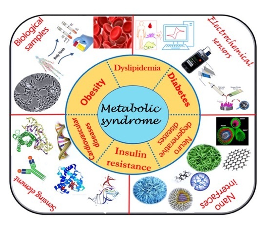
1. Introduction
2. Diagnosis of Metabolic Syndrome (MetS)
3. Sensors for Metabolic Syndrome
4. Evolution of Electrochemical Biosensors
5. Electrochemical Detection Strategies for Multi-Analyte Detection
6. Conclusions and Future Directions
Author Contributions
Funding
Acknowledgments
Conflicts of Interest
References
- Robbins, G.R.; Wen, H.; Ting, J.P.Y. Inflammasomes and metabolic disorders: Old genes in modern diseases. Mol. Cell 2014, 54, 297–308. [Google Scholar] [CrossRef] [PubMed]
- What Is Metabolic Syndrome? National Heart, Lung, and Blood Institute: Bethesda, MD, USA, 2016.
- Oladejo, A.O. Overview of the metabolic syndrome; an emerging pandemic of public health significance. Ann. Ib. Postgrad. Med. 2011, 9, 78–82. [Google Scholar] [PubMed]
- Alberti, K.G.M.M.; Zimmet, P.; Shaw, J. The metabolic syndrome—A new worldwide definition. Lancet 2005, 366, 1059–1062. [Google Scholar] [CrossRef]
- Sarafidis, P.A.; Nilsson, P.M. The metabolic syndrome: A glance at its history. J. Hypertens. 2006, 24, 621–626. [Google Scholar] [CrossRef] [PubMed]
- Oda, E. Metabolic syndrome: Its history, mechanisms, and limitations. Acta Diabetol. 2012, 49, 89–95. [Google Scholar] [CrossRef]
- Eckel, R.H.; Alberti, K.G.M.M.; Grundy, S.M.; Zimmet, P.Z. The metabolic syndrome. Lancet 2010, 365, 1–72. [Google Scholar] [CrossRef]
- Bonomini, F.; Rodella, L.F.; Rezzani, R. Metabolic Syndrome, Aging and Involvement of Oxidative Stress. Aging Dis. 2015, 6, 109. [Google Scholar] [CrossRef]
- Park, Y.; Zhu, S.; Palaniappan, L.; Heshka, S. The Metabolic Syndrome. Arch. Intern. Med. 2003, 163, 427–436. [Google Scholar] [CrossRef]
- Koroshi, A. Microalbuminuria, is it so important? Hippokratia 2007, 11, 105–107. [Google Scholar]
- Patel, J.L.; Suthar, A.M.; Dalsaniya, V.B.; Parikh, A.P.; Suthar, N.N.; Patel, K.L. A study of metabolic syndrome and its components in Type 2 Diabetes Mellitus subjects and their asymptomatic First degree relatives. Indian J. Clin. Pract. 2013, 23, 520–533. [Google Scholar]
- Verma, S.; Hussain, M.E. Obesity and diabetes: An update. Diabetes Metab. Syndr. Clin. Res. Rev. 2017, 11, 73–79. [Google Scholar] [CrossRef] [PubMed]
- Grundy, S.M. Metabolic syndrome: A multiplex cardiovascular risk factor. J. Clin. Endocrinol. Metab. 2007, 92, 399–404. [Google Scholar] [CrossRef] [PubMed]
- Yazdanpanah, S.; Rabiee, M.; Tahriri, M.; Abdolrahim, M.; Tayebi, L. Glycated hemoglobin-detection methods based on electrochemical biosensors. TrAC Trends Anal. Chem. 2015, 72, 53–67. [Google Scholar] [CrossRef]
- Heart online. Common Investigations In Cardiovascular Disease. Blood tests Full Blood Count (FBC). 2014, pp. 1–3. Available online: https://www.heartonline.org.au/media/DRL/Investigations_in_cardiovascular_disease.pdf (accessed on 6 December 2019).
- Leaf, D.A. Hypertriglyceridemia: A Guide to Assessment and Treatment. Hosp. Physician 2008, 32, 17–24. [Google Scholar]
- Laterza, O.F.; Price, C.P.; Scott, M.G. Cystatin C: An improved estimator of glomerular filtration rate? Clin. Chem. 2002, 48, 699–707. [Google Scholar]
- Herget-Rosenthal, S. Imaging Techniques in the Management of Chronic Kidney Disease: Current Developments and Future Perspectives. Semin. Nephrol. 2011, 31, 283–290. [Google Scholar] [CrossRef]
- Sangachin, M.G.; Cavuoto, L.A.; Wang, Y. Use of various obesity measurement and classification methods in occupational safety and health research: a systematic review of the literature. BMC Obes. 2018, 5, 28. [Google Scholar] [CrossRef]
- Douyon, L.; Schteingart, D.E. Effect of obesity and starvation on thyroid hormone, growth hormone, and cortisol secretion. Endocrinol. Metab. Clin. N. Am. 2002, 31, 173–189. [Google Scholar] [CrossRef]
- Yoo, E.-H.; Lee, S.-Y. Glucose Biosensors: An Overview of Use in Clinical Practice. Sensors 2010, 10, 4558–4576. [Google Scholar] [CrossRef]
- Monošík, R.; Streďanský, M.; Šturdík, E. Biosensors—Classification, characterization and new trends. Acta Chim. Slovaca 2012, 5, 109–120. [Google Scholar] [CrossRef]
- Justino, C.I.L.; Duarte, A.C.; Rocha-Santos, T.A.P. Critical overview on the application of sensors and biosensors for clinical analysis. TrAC Trends Anal. Chem. 2016, 85, 36–60. [Google Scholar] [CrossRef]
- Palchetti, I.; Mascini, M.; Chimica, D.; Lastruccia, V. Biosensor Technology: A Brief History. In Sensors and Microsystems; Springer: Dordrecht, The Netherlands, 2010; Volume 54, pp. 15–23. [Google Scholar]
- Qureshi, A.; Gurbuz, Y.; Niazi, J.H. Biosensors for cardiac biomarkers detection: A review. Sens. Actuators B Chem. 2012, 171–172, 62–76. [Google Scholar] [CrossRef]
- Park, S.; Boo, H.; Chung, T.D. Electrochemical non-enzymatic glucose sensors. Anal. Chim. Acta 2006, 556, 46–57. [Google Scholar] [CrossRef] [PubMed]
- Murugaiyan, S.B.; Ramasamy, R.; Gopal, N.; Kuzhandaivelu, V. Biosensors in clinical chemistry: An overview. Adv. Biomed. Res. 2014, 3, 67. [Google Scholar]
- Holade, Y.; Tingry, S.; Servat, K.; Napporn, T.; Cornu, D.; Kokoh, K. Nanostructured Inorganic Materials at Work in Electrochemical Sensing and Biofuel Cells. Catalysts 2017, 7, 31. [Google Scholar] [CrossRef]
- Milano, F.; Giotta, L.; Chirizzi, D.; Papazoglou, S.; Kryou, C.; De Bartolomeo, A.; De Leo, V.; Guascito, M.R.; Zergioti, I. Phosphate modified screen printed electrodes by lift treatment for glucose detection. Biosensors 2018, 8, 91. [Google Scholar] [CrossRef] [PubMed]
- Niu, X.; Chen, C.; Zhao, H.; Chai, Y.; Lan, M. Novel snowflake-like Pt-Pd bimetallic clusters on screen-printed gold nanofilm electrode for H2O2 and glucose sensing. Biosens. Bioelectron. 2012, 36, 262–266. [Google Scholar] [CrossRef]
- Kimmel, D.W.; LeBlanc, G.; Meschievitz, M.E.; Cliffel, D.E. Chemical Sensors and Biosensors. Anal. Chem. 2007, 84, 685–707. [Google Scholar] [CrossRef]
- Asefa, T.; Duncan, C.T.; Sharma, K.K. Recent advances in nanostructured chemosensors and biosensors. Analyst 2009, 134, 1980–1990. [Google Scholar] [CrossRef]
- Chen, S.; Yuan, R.; Chai, Y.; Hu, F. Electrochemical sensing of hydrogen peroxide using metal nanoparticles: A review. Microchim. Acta 2012, 180, 15–32. [Google Scholar] [CrossRef]
- Chauhan, N.; Chawla, S.; Pundir, C.S.; Jain, U. An electrochemical sensor for detection of neurotransmitter-acetylcholine using metal nanoparticles, 2D material and conducting polymer modified electrode. Biosens. Bioelectron. 2017, 89, 377–383. [Google Scholar] [CrossRef]
- Zeng, Q.; Cheng, J.S.; Liu, X.F.; Bai, H.T.; Jiang, J.H. Palladium nanoparticle/chitosan-grafted graphene nanocomposites for construction of a glucose biosensor. Biosens. Bioelectron. 2011, 26, 3456–3463. [Google Scholar] [CrossRef] [PubMed]
- Lin, C.-Y.; Chang, C.-T. Iron oxide nanorods array in electrochemical detection of H2O2. Sens. Actuators B Chem. 2015, 220, 695–704. [Google Scholar] [CrossRef]
- Ibupoto, Z.; Khun, K.; Beni, V.; Willander, M. Non-Enzymatic Glucose Sensor Based on the Novel Flower Like Morphology of Nickel Oxide. Soft Nanosci. Lett. 2013, 3, 46–50. [Google Scholar] [CrossRef]
- Guascito, M.R.; Chirizzi, D.; Malitesta, C.; Siciliano, T.; Tepore, A. Te oxide nanowires as advanced materials for amperometric nonenzymatic hydrogen peroxide sensing. Talanta 2013, 115, 863–869. [Google Scholar] [CrossRef] [PubMed]
- Marie, M.; Mandal, S.; Manasreh, O. An Electrochemical Glucose Sensor Based on Zinc Oxide Nanorods. Sensors 2015, 15, 18714–18723. [Google Scholar] [CrossRef] [PubMed]
- Ma, Y.; Di, J.; Yan, X.; Zhao, M.; Lu, Z.; Tu, Y. Direct electrodeposition of gold nanoparticles on indium tin oxide surface and its application. Biosens. Bioelectron. 2009, 24, 1480–1483. [Google Scholar] [CrossRef] [PubMed]
- Wang, J.X.; Sun, X.W.; Wei, A.; Lei, Y.; Cai, X.P.; Li, C.M.; Dong, Z.L. Zinc oxide nanocomb biosensor for glucose detection. Appl. Phys. Lett. 2006, 88. [Google Scholar] [CrossRef]
- Wu, W.; He, Q.; Jiang, C. Magnetic iron oxide nanoparticles: Synthesis and surface functionalization strategies. Nanoscale Res. Lett. 2008, 3, 397–415. [Google Scholar] [CrossRef]
- Kaushik, A.; Solanki, P.R.; Ansari, A.A.; Sumana, G.; Ahmad, S.; Malhotra, B.D. Iron oxide-chitosan nanobiocomposite for urea sensor. Sens. Actuators B Chem. 2009, 138, 572–580. [Google Scholar] [CrossRef]
- Batchelor-McAuley, C.; Wildgoose, G.G.; Compton, R.G.; Shao, L.; Green, M.L.H. Copper oxide nanoparticle impurities are responsible for the electroanalytical detection of glucose seen using multiwalled carbon nanotubes. Sens. Actuators B Chem. 2008, 132, 356–360. [Google Scholar] [CrossRef]
- Vabbina, P.K.; Kaushik, A.; Pokhrel, N.; Bhansali, S.; Pala, N. Electrochemical cortisol immunosensors based on sonochemically synthesized zinc oxide 1D nanorods and 2D nanoflakes. Biosens. Bioelectron. 2015, 63, 124–130. [Google Scholar] [CrossRef] [PubMed]
- Hahn, Y.-B.; Ahmad, R.; Tripathy, N. Chemical and biological sensors based on metal oxide nanostructures. Chem. Commun. 2012, 48, 10369. [Google Scholar] [CrossRef] [PubMed]
- Ates, M. A review study of (bio)sensor systems based on conducting polymers. Mater. Sci. Eng. C 2013, 33, 1853–1859. [Google Scholar] [CrossRef] [PubMed]
- Dhand, C.; Solanki, P.R.; Datta, M.; Malhotra, B.D. Polyaniline/Single-Walled Carbon Nanotubes Composite Based Triglyceride Biosensor. Electroanalysis 2010, 22, 2683–2693. [Google Scholar] [CrossRef]
- Jeykumari, D.R.S.; Narayanan, S.S. Functionalized carbon nanotube-bienzyme biocomposite for amperometric sensing. Carbon N.Y. 2009, 47, 957–966. [Google Scholar] [CrossRef]
- Sotiropoulou, S.; Chaniotakis, N.A. Carbon nanotube array-based biosensor. Anal. Bioanal. Chem. 2003, 375, 103–105. [Google Scholar] [CrossRef]
- Rivas, G.A.; Rubianes, M.D.; Rodríguez, M.C.; Ferreyra, N.F.; Luque, G.L.; Pedano, M.L.; Miscoria, S.A.; Parrado, C. Carbon nanotubes for electrochemical biosensing. Talanta 2007, 74, 291–307. [Google Scholar] [CrossRef]
- Electrochemical, O.N. Graphene and Other Nanomaterial-Based Electrochemical Aptasensors. Biosensors 2012, 2, 1–14. [Google Scholar]
- Kumar, V.; Shorie, M.; Ganguli, A.K.; Sabherwal, P. Graphene-CNT nanohybrid aptasensor for label free detection of cardiac biomarker myoglobin. Biosens. Bioelectron. 2015, 72, 56–60. [Google Scholar] [CrossRef]
- Artiles, M.S.; Rout, C.S.; Fisher, T.S. Graphene-based hybrid materials and devices for biosensing. Adv. Drug Deliv. Rev. 2011, 63, 1352–1360. [Google Scholar] [CrossRef]
- Zhang, X.; Liao, Q.; Chu, M.; Liu, S.; Zhang, Y. Structure effect on graphene-modified enzyme electrode glucose sensors. Biosens. Bioelectron. 2014, 52, 281–287. [Google Scholar] [CrossRef] [PubMed]
- Liu, Y.; Dong, X.; Chen, P. Biological and chemical sensors based on graphene materials. Chem. Soc. Rev. 2012, 41, 2283. [Google Scholar] [CrossRef] [PubMed]
- Zhao, J.; Chen, G.; Zhu, L.; Li, G. Graphene quantum dots-based platform for the fabrication of electrochemical biosensors. Electrochem. Commun. 2011, 13, 31–33. [Google Scholar] [CrossRef]
- Razmi, H.; Mohammad-Rezaei, R. Graphene quantum dots as a new substrate for immobilization and direct electrochemistry of glucose oxidase: Application to sensitive glucose determination. Biosens. Bioelectron. 2013, 41, 498–504. [Google Scholar] [CrossRef]
- Han, E.; Yang, Y.; He, Z.; Cai, J.; Zhang, X.; Dong, X. Development of tyrosinase biosensor based on quantum dots/chitosan nanocomposite for detection of phenolic compounds. Anal. Biochem. 2015, 486, 102–106. [Google Scholar] [CrossRef]
- Chen, Z.; Ren, X.; Meng, X.; Chen, D.; Yan, C.; Ren, J.; Yuan, Y.; Tang, F. Optical detection of choline and acetylcholine based on H2O2-sensitive quantum dots. Biosens. Bioelectron. 2011, 28, 50–55. [Google Scholar] [CrossRef]
- Khan, S.A.; Smith, G.T.; Seo, F.; Ellerbee, A.K. Label-free and non-contact optical biosensing of glucose with quantum dots. Biosens. Bioelectron. 2015, 64, 30–35. [Google Scholar] [CrossRef]
- Li, L.; Wang, Y.; Pan, L.; Shi, Y.; Cheng, W.; Shi, Y.; Yu, G. A Nanostructured Conductive Hydrogels-Based Biosensor Platform for Human Metabolite Detection. Nano Lett. 2015, 15. [Google Scholar] [CrossRef]
- Kestwal, R.M.; Bagal-Kestwal, D.; Chiang, B.-H. Fenugreek hydrogel-agarose composite entrapped gold nanoparticles for acetylcholinesterase based biosensor for carbamates detection. Anal. Chim. Acta 2015, 886, 143–150. [Google Scholar] [CrossRef]
- Liu, A.; Chen, X.; Wang, K.; Wei, N.; Sun, Z.; Lin, X.; Chen, Y.; Du, M. Electrochemical DNA biosensor based on aldehyde-agarose hydrogel modified glassy carbon electrode for detection of PML/RARa fusion gene. Sens. Actuators B Chem. 2011, 160, 1458–1463. [Google Scholar] [CrossRef]
- Buenger, D.; Topuz, F.; Groll, J. Hydrogels in sensing applications. Prog. Polym. Sci. 2012, 37, 1678–1719. [Google Scholar] [CrossRef]
- Song, H.S.; Kwon, O.S.; Kim, J.H.; Conde, J.; Artzi, N. 3D hydrogel scaffold doped with 2D graphene materials for biosensors and bioelectronics. Biosens. Bioelectron. 2017, 89, 187–200. [Google Scholar] [CrossRef] [PubMed]
- Razmi, H.; Habibi, E. Nanomolar detection of hydrogen peroxide at a new polynuclear cluster of tin pentacyanonitrosylferrate nanoparticle-modified carbon ceramic electrode. Anal. Biochem. 2009, 392, 126–132. [Google Scholar] [CrossRef] [PubMed]
- Warsinke, A. Point-of-care testing of proteins. Anal. Bioanal. Chem. 2009, 393, 1393–1405. [Google Scholar] [CrossRef] [PubMed]
- Luo, X.; Morrin, A.; Killard, A.J.; Smyth, M.R. Application of nanoparticles in electrochemical sensors and biosensors. Electroanalysis 2006, 18, 319–326. [Google Scholar] [CrossRef]
- Hayat, A.; Catanante, G.; Marty, J.L. Current trends in nanomaterial-based amperometric biosensors. Sensors 2014, 14, 23439–23461. [Google Scholar] [CrossRef]
- Madhurantakam, S.; Jayanth Babu, K.; Balaguru Rayappan, J.B.; Krishnan, U.M. Fabrication of mediator-free hybrid nano-interfaced electrochemical biosensor for monitoring cancer cell proliferation. Biosens. Bioelectron. 2017, 87, 832–841. [Google Scholar] [CrossRef]
- Larin, A.; Womble, P.C.; Dobrokhotov, V. Hybrid SnO2/TiO2 nanocomposites for selective detection of ultra-low hydrogen sulfide concentrations in complex backgrounds. Sensors 2016, 16, 1373. [Google Scholar] [CrossRef]
- Zhu, C.; Yang, G.; Li, H.; Du, D.; Lin, Y. Electrochemical Sensors and Biosensors Based on Nanomaterials and Nanostructures. Anal. Chem. 2015, 87, 230–249. [Google Scholar] [CrossRef]
- Pijl, H.; Romijn, J.A. Obesity, dopamine and the metabolic syndrome: Potential of dopaminergic agents in the control of metabolism. Curr. Opin. Endocrinol. Diabetes 2006, 13, 179–184. [Google Scholar] [CrossRef]
- Wang, G.J.; Volkow, N.D.; Logan, J.; Pappas, N.R.; Wong, C.T.; Zhu, W.; Netusil, N.; Fowler, J.S. Brain dopamine and obesity. Lancet 2001, 357, 354–357. [Google Scholar] [CrossRef]
- Limberg, J.K.; Kellawan, J.M.; Harrell, J.W.; Johansson, R.E.; Eldridge, M.W.; Proctor, L.T.; Sebranek, J.J.; Schrage, W.G. Exercise-mediated vasodilation in human obesity and metabolic syndrome: Effect of acute ascorbic acid infusion. AJP Heart Circ. Physiol. 2014, 307, H840–H847. [Google Scholar] [CrossRef] [PubMed]
- Ellulu, M.S.; Rahmat, A.; Patimah, I.; Khaza’Ai, H.; Abed, Y. Effect of vitamin C on inflammation and metabolic markers in hypertensive and/or diabetic obese adults: A randomized controlled trial. Drug Des. Devel. Ther. 2015, 9, 3405–3412. [Google Scholar] [CrossRef] [PubMed]
- Johnson, R.J.; Nakagawa, T.; Jalal, D.; Sánchez-Lozada, L.G.; Kang, D.H.; Ritz, E. Uric acid and chronic kidney disease: Which is chasing which? Nephrol. Dial. Transplant. 2013, 28, 2221–2228. [Google Scholar] [CrossRef] [PubMed]
- Mohandas, R.; Johnson, R.J. Uric Acid Levels Increase Risk for New-Onset Kidney Disease. J. Am. Soc. Nephrol. 2008, 19, 2251–2253. [Google Scholar] [CrossRef] [PubMed]
- Tang, C.F.; Kumar, S.A.; Chen, S.M. Zinc oxide/redox mediator composite films-based sensor for electrochemical detection of important biomolecules. Anal. Biochem. 2008, 380, 174–183. [Google Scholar] [CrossRef]
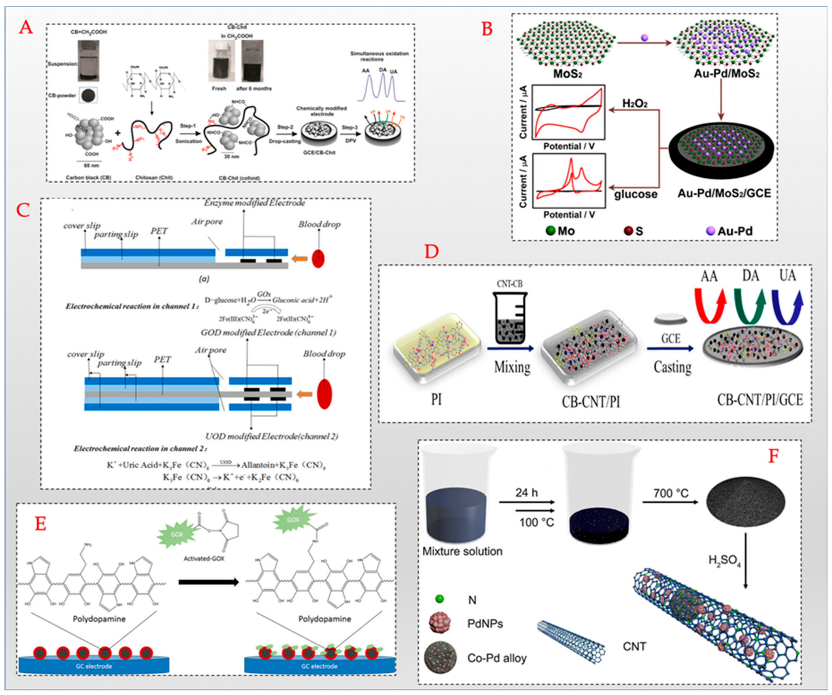
- Wang, Y.; Yang, T.; Hasebe, Y.; Zhang, Z.; Tao, D. Carbon black-carbon nanotube co-doped polyimide sensors for simultaneous determination of ascorbic acid, uric acid, and dopamine. Materials 2018, 11, 1691. [Google Scholar] [CrossRef]
- Dinesh, B.; Saraswathi, R.; Senthil Kumar, A. Water based homogenous carbon ink modified electrode as an efficient sensor system for simultaneous detection of ascorbic acid, dopamine and uric acid. Electrochim. Acta 2017, 233, 92–104. [Google Scholar] [CrossRef]
- Moser, I.; Jobst, G. Pre-calibrated biosensors for single-use applications. Chemie-Ingenieur-Technik 2013, 85, 172–178. [Google Scholar] [CrossRef]
- Canevari, T.C.; Raymundo-Pereira, P.A.; Landers, R.; Benvenutti, E.V.; Machado, S.A.S. Sol-gel thin-film based mesoporous silica and carbon nanotubes for the determination of dopamine, uric acid and paracetamol in urine. Talanta 2013, 116, 726–735. [Google Scholar] [CrossRef]
- Huang, Q.; An, Y.; Tang, L.; Jiang, X.; Chen, H.; Bi, W.; Wang, Z.; Zhang, W. A dual enzymatic-biosensor for simultaneous determination of glucose and cholesterol in serum and peritoneal macrophages of diabetic mice: Evaluation of the diabetes-accelerated atherosclerosis risk. Anal. Chim. Acta 2011, 707, 135–141. [Google Scholar] [CrossRef] [PubMed]
- Kouassi, G.K.; Irudayaraj, J.; McCarty, G. Activity of glucose oxidase functionalized onto magnetic nanoparticles. Biomagn. Res. Technol. 2005, 3, 1. [Google Scholar] [CrossRef] [PubMed]
- Gao, J.; Huang, W.; Chen, Z.; Yi, C.; Jiang, L. Simultaneous detection of glucose, uric acid and cholesterol using flexible microneedle electrode array-based biosensor and multi-channel portable electrochemical analyzer. Sens. Actuators B Chem. 2019, 287, 102–110. [Google Scholar] [CrossRef]
- Teymourian, H.; Salimi, A.; Khezrian, S. Fe3O4 magnetic nanoparticles/reduced graphene oxide nanosheets as a novel electrochemical and bioeletrochemical sensing platform. Biosens. Bioelectron. 2013, 49, 1–8. [Google Scholar] [CrossRef] [PubMed]
- Jaime, J.; Rangel, G.; Muñoz-Bonilla, A.; Mayoral, A.; Herrasti, P. Magnetite as a platform material in the detection of glucose, ethanol and cholesterol. Sens. Actuators B Chem. 2017, 238, 693–701. [Google Scholar] [CrossRef]
- Mahjoub, S.; Masrour-Roudsari, J. Role of oxidative stress in pathogenesis of metabolic syndrome. Caspian J. Intern. Med. 2012, 3, 386–396. [Google Scholar]
- Li, X.; Du, X. Molybdenum disulfide nanosheets supported Au-Pd bimetallic nanoparticles for non-enzymatic electrochemical sensing of hydrogen peroxide and glucose. Sens. Actuators B Chem. 2017, 239, 536–543. [Google Scholar] [CrossRef]
- Guo, J.; Ma, X. Simultaneous Monitoring of Glucose and Uric Acid On a Single Test Strip with Dual Channels. Biosens. Bioelectron. 2017, 94, 415–419. [Google Scholar] [CrossRef]
- Huang, B.; Wang, Y.; Lu, Z.; Du, H.; Ye, J. One pot synthesis of palladium-cobalt nanoparticles over carbon nanotubes as a sensitive non-enzymatic sensor for glucose and hydrogen peroxide detection. Sens. Actuators B Chem. 2017, 252, 1016–1025. [Google Scholar] [CrossRef]
- Yang, H.; Wang, Z.; Li, C.; Xu, C. Nanoporous PdCu alloy as an excellent electrochemical sensor for H2O2 and glucose detection. J. Colloid Interface Sci. 2017, 491, 321–328. [Google Scholar] [CrossRef]
- George, G.; Anandhan, S. Tuning characteristics of Co3O4 nanofiber mats developed for electrochemical sensing of glucose and H2O2. Thin Solid Films 2016, 610, 48–57. [Google Scholar] [CrossRef]
- Kucherenko, D.Y.; Kucherenko, I.S.; Soldatkin, O.O.; Topolnikova, Y.V.; Dzyadevych, S.V.; Soldatkin, A.P. A highly selective amperometric biosensor array for the simultaneous determination of glutamate, glucose, choline, acetylcholine, lactate and pyruvate. Bioelectrochemistry 2019, 128, 100–108. [Google Scholar] [CrossRef] [PubMed]
- Zhang, L.; Li, H.; Ni, Y.; Li, J.; Liao, K.; Zhao, G. Porous cuprous oxide microcubes for non-enzymatic amperometric hydrogen peroxide and glucose sensing. Electrochem. Commun. 2009, 11, 812–815. [Google Scholar] [CrossRef]
- Wu, S.; Zhao, H.; Ju, H.; Shi, C.; Zhao, J. Electrodeposition of silver-DNA hybrid nanoparticles for electrochemical sensing of hydrogen peroxide and glucose. Electrochem. Commun. 2006, 8, 1197–1203. [Google Scholar] [CrossRef]
- Zhao, C.; Wu, X.; Li, P.; Zhao, C.; Qian, X. Hydrothermal deposition of CuO/rGO/Cu2O nanocomposite on copper foil for sensitive nonenzymatic voltammetric determination of glucose and hydrogen peroxide. Microchim. Acta 2017, 184, 2341–2348. [Google Scholar] [CrossRef]
- Liu, A.; Lang, Q.; Liang, B.; Shi, J. Sensitive detection of maltose and glucose based on dual enzyme-displayed bacteria electrochemical biosensor. Biosens. Bioelectron. 2017, 87, 25–30. [Google Scholar] [CrossRef]
- Gao, Z.F.; Chen, D.M.; Lei, J.L.; Luo, H.Q.; Li, N.B. A regenerated electrochemical biosensor for label-free detection of glucose and urea based on conformational switch of i-motif oligonucleotide probe. Anal. Chim. Acta 2015, 897, 10–16. [Google Scholar] [CrossRef]
- He, J.; Sunarso, J.; Zhu, Y.; Zhong, Y.; Miao, J.; Zhou, W.; Shao, Z. High-performance non-enzymatic perovskite sensor for hydrogen peroxide and glucose electrochemical detection. Sens. Actuators B Chem. 2017, 244, 482–491. [Google Scholar] [CrossRef]
- Wu, W.; Yu, B.; Wu, H.; Wang, S.; Xia, Q.; Ding, Y. Synthesis of tremella-like CoS and its application in sensing of hydrogen peroxide and glucose. Mater. Sci. Eng. C 2017, 70, 430–437. [Google Scholar] [CrossRef]
- Liu, M.; Liu, R.; Chen, W. Graphene wrapped Cu2O nanocubes: Non-enzymatic electrochemical sensors for the detection of glucose and hydrogen peroxide with enhanced stability. Biosens. Bioelectron. 2013, 45, 206–212. [Google Scholar] [CrossRef]
- Kumar-Krishnan, S.; de Fuentes, O.A.; Prokhorov, E.; Luna-Barcenas, J.G. Electrochemical detection of hydrogen peroxide and glucose based on chitosan stabilized silver nanowire modified electrodes. In Proceedings of the 2014 IEEE 9th IberoAmerican Congress on Sensors, Bogota, Colombia, 15–18 October 2014; pp. 5–8. [Google Scholar]
- Zhang, Y.; Zhou, Q.; Zhao, W.; Chu, W.; Zheng, J. Array of recessed gold nanoelectrodes formed with polymethylmethacrylate for individual detection of ascorbic acid, dopamine and uric acid. Electrochim. Acta 2016, 212, 25–31. [Google Scholar] [CrossRef]
- Jin, J.; Liu, W.; Deng, S.; Han, Y.; Cai, Y.; Hui, G. A non-enzyme electrochemical qualitative and quantitative analyzing method for glucose, D-fructose, and sucrose utilizing Cu foam material. Electrochim. Acta 2015, 153, 594–601. [Google Scholar] [CrossRef]


| Year | Nomenclature | Risk Factors Included | Proposed By |
|---|---|---|---|
| 1923 | Hypertoni–Hyperglycemi–Hyperurikemi syndrome | Hypertension, hyperglycemia, hyperurecemia | Kylin |
| 1966 | Trisyndrome metabolique | Gout, diabetes, hyperlipidemia | Camus |
| 1967 | Plurimetabolic syndrome | Hyperlipidemia, obesity, diabetes, hypertension, coronary heart disease | Avogaro and Crepaldi |
| 1968 | Wohlstands-syndrom (Syndrome of affluence) | Hyperlipidemia, obesity, diabetes, hypertension, coronary heart disease | Mehnert and Kuhlmann |
| 1981 | Metabolische-syndrom (Metabolic syndrome) | Hyperlipidemia, hyperinsulinemia, obesity, diabetes, hypertension, gout, thrombophilia | Hanefeld and Leonhardt |
| 1988 | Syndrome X | Impaired glucose tolerance, hyperinsulinemia, very low-density lipoprotein (VLDL), triglycerides, cholesterol, hypertension, low high-density lipoprotein (HDL) | G.M. Reaven |
| 1989 | Deadly quartet | Central adiposity, impaired glucose tolerance, hypertriglyceridemia, hypertension | Kaplan |
| 1991–1992 | Insulin resistance syndrome | Insulin resistance, diabetes, hypertriglyceridemia | DeFronzo and Ferranini, Haffner |
| 1994 | Visceral fat syndrome | Visceral fat, diabetes, dyslipidemia | Nakamura and Matsuzawa |
| Agency | Risk Factor | |||||
|---|---|---|---|---|---|---|
| Body Weight | Insulin Resistance | Lipids | Blood Pressure | Glucose | Others | |
| World Health Organization (WHO), 1998 | Waist/hip >0.9 (men) >0.85 (women) or body mass index (BMI) >30 kg/m2 | IGT/IFG/type 2 diabetes or lower insulin sensitivity + any 2 of the other factors | TG ≥150 mg/dL and/or HDL <35 mg/dL (men) <39 (women) | ≥140/90 mm Hg | IGT/IFG/type 2 diabetes | Micro-albuminuria Urinary excretion rate >20 mg/min or albumin/creatinine >30 mg/g |
| European Group for the study of Insulin Resistance (EGIR), 1999 | Waist circumference ≥94 cm (men) ≥80 cm (men) | Plasma insulin >75th percentile | TG ≥ 150 mg/dL and/or HDL <39 mg/dL | ≥140/90 mm Hg | IGT/fasting plasma glucose >110 mg/dL | None |
| National Cholesterol Education Programme/Adult Treatment Panel III (NCEP/ATP III), 2001 | Waist circumference ≥102 cm (men) ≥8 cm (men) | Any three of the five factors listed | TG ≥150 mg/dL and/or HDL <40 mg/dL (men) <50 (women) | ≥130/85 mm Hg | >110 mg/dL | None |
| American Association of Clinical Endocrinologists (AACE), 2003 | BMI ≥25 kg/m2 | IGT/IFG + any of the other factors | TG ≥ 150 mg/dL and/or HDL <35 mg/dL (men) <39 (women) | ≥130/85 mm Hg | Fasting plasma glucose 110–126 mg/dL; post-prandial 140–200 mg/dL | None |
| International Diabetes Federation (IDF), 2005 | Ethnicity based values for waist circumference >94 cm (Euro men) >80 cm (Euro women) >90 cm (Asian men) >80 cm (Asian women) | Not listed | TG ≥ 150 mg/dL and/or HDL <40 mg/dL (men) <50 (women) | ≥130/85 mm Hg | >100 mg/dL | None |
| Analyte | Nano-Interface | Enzymes Used | Technique | Ref. | |
|---|---|---|---|---|---|
| Dual Analytes | Glucose and H2O2 | Pt–Pd bimetallic clusters | Yes | CV, Amp | [30] |
| Glucose and Cholesterol | Poly-thionine film | No | CV, Amp | [85] | |
| Glucose and H2O2 | Au–Pd bimetallic nanoparticles | No | CV, Amp | [91] | |
| Glucose and Uric acid | Carbon ink | Yes | CA | [92] | |
| Glucose and H2O2 | Pd-CoCNTs | No | CV, Amp, EIS | [93] | |
| Glucose and H2O2 | PdCu alloy | No | CV, Amp | [94] | |
| Glucose and H2O2 | Co3O4 | No | CV, Amp | [95] | |
| Glucose and H2O2 | Cu2O | No | CV, Amp, EIS | [97] | |
| Glucose and H2O2 | Silver–DNA hybrid nanoparticles | Yes | CV, Amp | [98] | |
| Glucose and H2O2 | CuO/rGO/Cu2O | No | CV, Amp | [99] | |
| Glucose and Maltose | MWCNTs | No | CV, Amp | [100] | |
| Glucose and Urea | E-DNA | No | CV, Amp, EIS | [101] | |
| Glucose and H2O2 | Perovskite | No | Amp | [102] | |
| Glucose and H2O2 | CoS | No | CV, Amp, EIS | [103] | |
| Glucose and H2O2 | Graphene wrapped CuO nanocubes | No | CV, Amp | [104] | |
| Glucose and H2O2 | Ag nanowires-CS | Yes | CV, Amp | [105] | |
| Triple Analytes | Uric Acid, Dopamine, Ascorbic Acid | Carbon black–carbon nanotube/polyimide composite | No | CV, DPV, Amp | [81] |
| Ascorbic Acid, Dopamine and Uric Acid | Water-soluble homogenous carbon black–chitosan ink | No | CV, DPV, Amp | [82] | |
| Glucose, Uric Acid, Cholesterol | Gold/titanium electrodeposited with polyaniline on platinum nanoparticles | Yes | Amp | [87] | |
| Ascorbic acid, Dopamine and Uric acid | Gold electrode patterned on polymethylmethacrylate | No | CV, DPV | [106] | |
| Glucose, Ethanol and Cholesterol | Polydopamine-coated magnetic nanoparticles | Yes | CV, Amp | [89] | |
| Glucose, D-Fructose, Sucrose | 3-D Cu foam | No | CV, A | [107] |
© 2019 by the authors. Licensee MDPI, Basel, Switzerland. This article is an open access article distributed under the terms and conditions of the Creative Commons Attribution (CC BY) license (http://creativecommons.org/licenses/by/4.0/).
Share and Cite
Sasya, M.; Devi, K.S.S.; Babu, J.K.; Balaguru Rayappan, J.B.; Krishnan, U.M. Metabolic Syndrome—An Emerging Constellation of Risk Factors: Electrochemical Detection Strategies. Sensors 2020, 20, 103. https://doi.org/10.3390/s20010103
Sasya M, Devi KSS, Babu JK, Balaguru Rayappan JB, Krishnan UM. Metabolic Syndrome—An Emerging Constellation of Risk Factors: Electrochemical Detection Strategies. Sensors. 2020; 20(1):103. https://doi.org/10.3390/s20010103
Chicago/Turabian StyleSasya, Madhurantakam, K. S. Shalini Devi, Jayanth K. Babu, John Bosco Balaguru Rayappan, and Uma Maheswari Krishnan. 2020. "Metabolic Syndrome—An Emerging Constellation of Risk Factors: Electrochemical Detection Strategies" Sensors 20, no. 1: 103. https://doi.org/10.3390/s20010103
APA StyleSasya, M., Devi, K. S. S., Babu, J. K., Balaguru Rayappan, J. B., & Krishnan, U. M. (2020). Metabolic Syndrome—An Emerging Constellation of Risk Factors: Electrochemical Detection Strategies. Sensors, 20(1), 103. https://doi.org/10.3390/s20010103