Enhancement of the Humidity Sensing Performance in Mg-Doped Hexagonal ZnO Microspheres at Room Temperature


Abstract
:1. Introduction
2. Materials and Methods
2.1. Materials
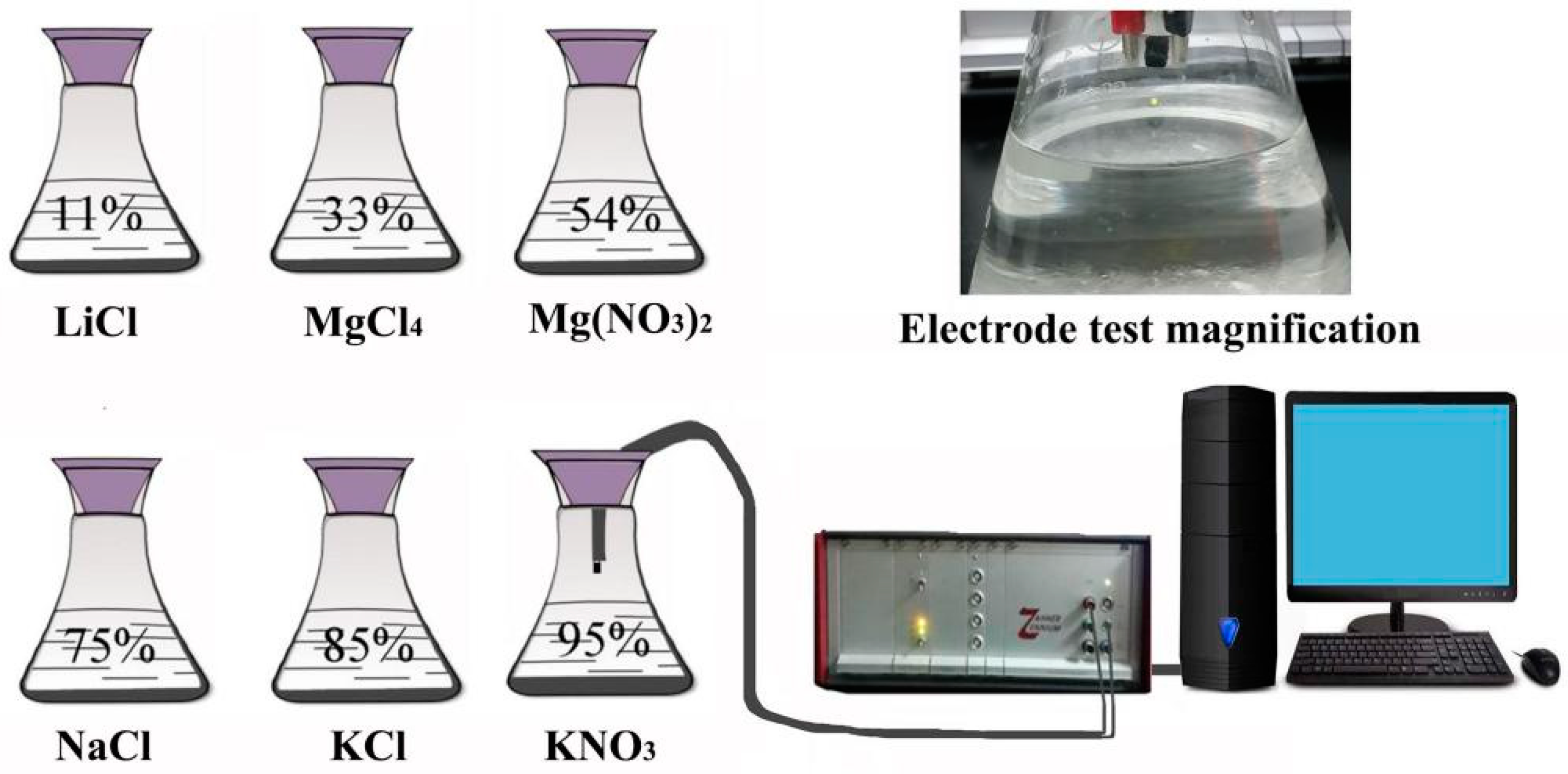
2.2. Apparatus
2.3. Synthesis of Mg-Doped ZnO Microspheres
2.4. Preparation of Humidity Sensors
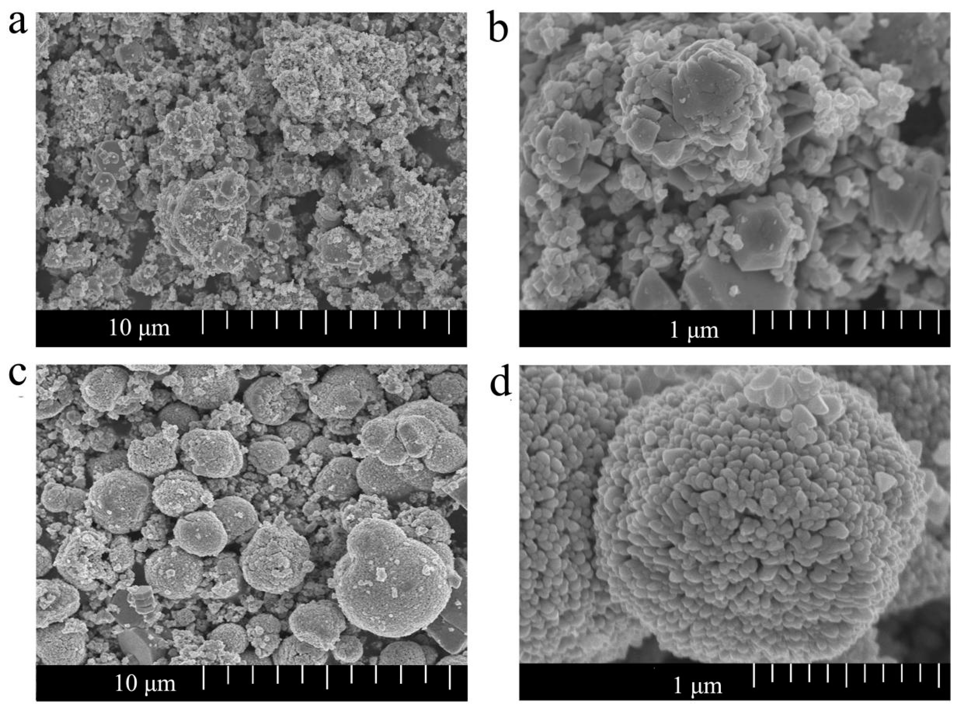
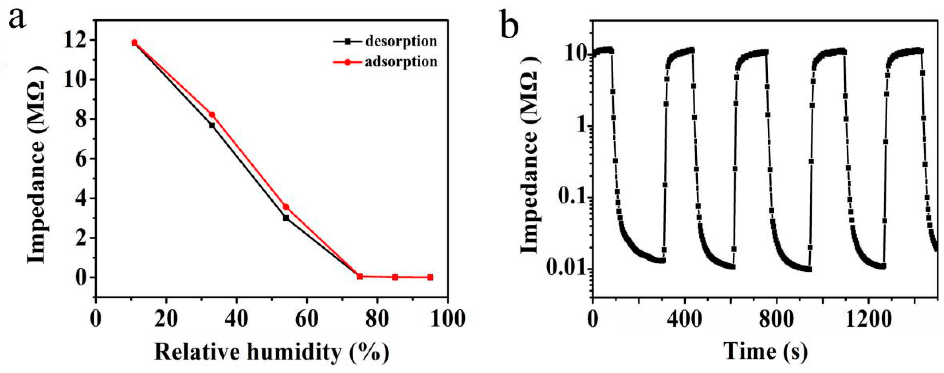
3. Results and Discussion
4. Conclusions
Author Contributions
Funding
Acknowledgments
Conflicts of Interest
References
- Wang, W.; Li, Z.; Liu, L.; Zhang, H.; Zheng, W.; Wang, Y.; Huang, H.; Wang, Z.; Wang, C. Humidity sensor based on LiCl-doped ZnO electropunk nanofibers. Sens. Actuators B Chem. 2009, 141, 404–409. [Google Scholar] [CrossRef]
- Qi, Q.; Zhang, T.; Yu, Q.; Wang, R.; Zeng, Y.; Liu, L.; Yang, H. Properties of humidity sensing ZnO nanorods-base sensor fabricated by screen-printing. Sens. Actuators B Chem. 2008, 133, 638–643. [Google Scholar] [CrossRef]
- Fei, T.; Jiang, K.; Liu, S.; Zhang, T. Humidity sensors based on Li-loaded nanoporous polymers. Sens. Actuators B Chem. 2014, 190, 523–528. [Google Scholar] [CrossRef]
- Tsai, F.S.; Wang, S.J. Enhanced sensing performance of relative humidity sensors using laterally grown ZnO nanosheets. Sens. Actuators B 2014, 193, 280–287. [Google Scholar] [CrossRef]
- Toloman, D.; Popa, A.; Stan, M.; Socaci, C.; Biris, A.R.; Katona, G.; Tudorache, F.; Petrila, I.; Iacomi, F. Reduced graphene oxide decorated with Fe doped SnO2 nanoparticles for humidity sensor. Appl. Surf. Sci. 2017, 402, 410–417. [Google Scholar] [CrossRef]
- Zhu, Y.; Li, Q.; Wang, P.; Zang, W.; Xing, L.; Xue, X. Enhanced piezo-humidity sensing of Sb-doped ZnO nanowire arrays as self-powered/active humidity sensor. Mater. Lett. 2015, 154, 77–80. [Google Scholar] [CrossRef]
- Li, Y.; Hong, L.; Yang, M. Crosslinked and quaternized poly (4-vinylpyridine)/polypyrrole composite as a potential candidate for the detection of low humidity. Talanta 2008, 75, 412–417. [Google Scholar] [CrossRef]
- Yuan, Q.; Li, N.; Tu, J.; Li, X.; Wang, R.; Zhang, T.; Shao, C. Preparation and humidity sensitive property of mesoporous ZnO–SiO2 composite. Sens. Actuators B Chem. 2010, 149, 413–419. [Google Scholar] [CrossRef]
- Zhang, T.; Wang, R.; Geng, W.; Li, X.; Qi, Q.; He, Y.; Wang, S. Study on humidity sensing properties based on composite, materials of Li-doped mesoporous silica A-SBA-15. Sens. Actuators B Chem. 2008, 128, 482–487. [Google Scholar] [CrossRef]
- Qi, Q.; Zhang, T.; Wang, S.; Zheng, X. Humidity sensing properties of KCl-doped ZnO nanofibers with super-rapid response and recovery. Sens. Actuators B Chem. 2009, 137, 649–655. [Google Scholar] [CrossRef]
- Zhao, J.; Liu, Y.; Li, X.; Lu, G.; You, L.; Liang, X.; Liu, F.; Zhang, T.; Du, Y. Highly sensitive humidity sensor based on high surface area mesoporous LaFeO3 prepared by a nanocasting routen. Sens. Actuators B Chem. 2013, 181, 802–809. [Google Scholar] [CrossRef]
- Turgut, G.; Duman, S.; Sonmez, E.; Ozcelik, F.S. A study of Eu incorporated ZnO thin films: An application of Al/ZnO:Eu/p-Si heterojunction diode. Mater. Sci. Eng. B 2016, 206, 9–16. [Google Scholar] [CrossRef]
- Zhang, Y.; Liu, Y.; Wu, L.; Li, H.; Han, L.; Wang, B.; Xie, E. Effect of annealing atmosphere on the photoluminescence of ZnO nanospheres. Appl. Surf. Sci. 2009, 255, 4801–4805. [Google Scholar] [CrossRef]
- Khataee, A.R.; Karimi, A.; Soltani, R.D.C.; Safarpour, M.; Hanifehpour, Y.; Joo, S.W. Europium-doped ZnO as a visible light responsive nanocatalyst: Sonochemical synthesis, characterization and response surface modeling of photocatalytic process. Appl. Catal. A Gen. 2014, 488, 160–170. [Google Scholar] [CrossRef]
- Zhang, H.; Zhang, M.; Lin, C.; Zhang, J. AuNPs Hybrid Black ZnO Nanorods Made by a Sol-Gel Method for Highly Sensitive Humidity Sensing. Sensors 2018, 18, 218. [Google Scholar] [CrossRef]
- Tomer, V.K.; Duhan, S.; Sharma, A.K.; Malik, R.; Nehra, S.P. One pot synthesis of mesoporous ZnO-SiO2 nanocomposites as high performance humidity sensor. Colloids Surf. A Physicochem. Eng. Asp. 2015, 483, 121–128. [Google Scholar] [CrossRef]
- Zhang, M.; Zhang, H.; Li, L.; Tuokedaerhan, K.; Jia, Z. Er-enhanced humidity sensing performance in black ZnO-based sensor. J. Alloys Compd. 2018, 744, 364–369. [Google Scholar] [CrossRef]
- Chang, S.-P.; Chang, S.-J.; Lu, C.-Y.; Li, M.-J.; Hsu, C.-L.; Chiou, Y.-Z.; Hsueh, T.-J.; Chen, I.C. A ZnO nanowire-based humidity sensor. Superlattices Microstruct. 2010, 47, 772–778. [Google Scholar] [CrossRef]
- Chen, X.; Liu, L.; Peter, Y.Y.; Mao, S.S. Increasing solar absorption for photocatalysis with black hydrogenated titanium dioxide nanocrystals. Science 2011, 331, 746–750. [Google Scholar] [CrossRef]
- Zhu, D.; Hu, T.; Zhao, Y.; Zang, W.; Xing, L.; Xue, X. High-performance self-powered/active humidity sensing of Fe-doped ZnO nanoarray nanogenerator. Sens. Actuators B Chem. 2015, 213, 382–389. [Google Scholar] [CrossRef]
- Yu, S.; Zhang, H.; Lin, C.; Bian, M. The enhancement of humidity sensing performance based on Eu-doped ZnO. Curr. Appl. Phys. 2019, 19, 82–88. [Google Scholar] [CrossRef]
- Ohtomo, A.; Kawasaki, M.; Koida, T.; Masubuchi, K.; Koinuma, H.; Sakurai, Y.; Yoshida, Y.; Yasuda, T.; Segawa, Y. MgxZn1−xO as II–VI widegap semiconductor alloy. Curr. Appl. Phys. 1998, 72, 2466–2468. [Google Scholar] [CrossRef]
- Chen, C.; Yu, W.; Liu, T.; Cao, S.; Tsang, Y. Graphene oxide/WS2/Mg-doped ZnO nanocomposites for solar-light catalytic and antibacterial applications. Sol. Energy Mater. Sol. Cells 2017, 160, 43–53. [Google Scholar] [CrossRef]
- Jia, Y.; Sun, H.; Liang, H.; Ji, H.; Song, L.; Gao, C.; Xu, H. A highly efficient metal-free oxygen reduction electrocatalyst assembled from carbon nanotubes and graphene. Adv. Mater. 2016, 28, 4606–4613. [Google Scholar]
- Giri, P.; Chakrabarti, P. Effect of Mg doping in ZnO buffer layer on ZnO thin film devices for electronic applications. Superlattices Microstruct. 2016, 93, 248–260. [Google Scholar] [CrossRef]
- Ivetić, T.B.; Dimitrievska, M.R.; Finčurb, N.L.; Đačanin, L.R.; Gúth, I.O.; Abramović, B.F.; Lukić-Petrović, S.R. Effect of annealing temperature on structural and optical properties of Mg-doped ZnO nanoparticles and their photocatalytic efficiency in alprazolam degradation. Ceram. Int. 2014, 40, 1545–1552. [Google Scholar] [CrossRef]
- Iqbal, J.; Jan, T.; Ismail, M.; Ahmad, N.; Arif, A.; Khan, M.; Adil, M.; Arsha, A. Influence of Mg doping level on morphology, optical, electrical properties and antibacterial activity of ZnO nanostructures. Ceram. Int. 2014, 40, 7487–7493. [Google Scholar] [CrossRef]
- Zeng, W.; Yang, X.; Shang, M.; Xu, X.; Yang, W.; Hou, H. Fabrication of Mg-doped ZnO nanofibers with high purities and tailored band gaps. Ceram. Int. 2016, 42, 10021–10029. [Google Scholar] [CrossRef]
- Nishant, K.; Anchal, S. Green photoluminescence and photoconductivity from screen-printed Mg doped Zn O films. J. Alloys Compd. 2018, 735, 312–318. [Google Scholar]
- Samanta, A.; Goswami, M.N. Optical properties and enhanced photocatalytic activity of Mg-doped ZnO nanoparticles. Phys. E Low-Dimens. Syst. Nanostruct. 2018, 104, 254–260. [Google Scholar] [CrossRef]
- Arshad, M.; Ansari, M.M.; Ahmed, A.S.; Tripathi, P.; Ashraf, S.S.Z.; Naqvi, A.H.; Azam, A. Band gap engineering and enhanced photoluminescence of Mg doped ZnO nanoparticles synthesized by wet chemical route. J. Lumin. 2015, 161, 275–280. [Google Scholar] [CrossRef]
- Yang, J.; Wang, Y.; Kong, J.; Yu, M.; Jin, H. Synthesis of Mg-doped hierarchical ZnO nanostructures via hydrothermal method and their optical properties. J. Alloys Compd. 2016, 657, 261–267. [Google Scholar] [CrossRef]
- Wu, S.; Chen, Z.; Wang, T.; Ji, X. A facile approach for the fabrication of Au/ZnO-hollow-sphere-monolayer thin films and their photocatalytic properties. Appl. Surf. Sci. 2017, 412, 67–69. [Google Scholar] [CrossRef]
- Aksoy, S.; Caglar, Y.; Ilican, S. Sol–gel derived Li–Mg co-doped Zn O films: Preparation and characterization via XRD, XPS, FESEM. J. Alloys Compd. 2012, 512, 171–178. [Google Scholar] [CrossRef]
- Chen, M.; Wang, X.; Yu, Y.H.; Pei, Z.L.; Bai, X.D.; Sun, C.; Huang, R.F.; Wen, L.S. X-ray photoelectron spectroscopy and auger electron spectroscopy studies of A1- doped ZnO films. Appl. Surf. Sci. 2000, 158, 134–140. [Google Scholar] [CrossRef]
- Manzhia, P.; Kumarib, R.; Alamb, M.B. Mg-doped ZnO nanostructures for efficient Organic Light Emitting Diode. Vacuum 2018. [Google Scholar] [CrossRef]
- Gong, M.; Li, Y.; Guo, Y.; Lv, X.; Dou, X. 2D TiO2 nanosheets for ultrasensitive humidity sensing application Benefited by abundant surface oxygen vacancy defects. Sens. Actuators B Chem. 2018, 262, 350–358. [Google Scholar] [CrossRef]
- Yu, X.X.; Wu, Y.; Dong, B.; Dong, Z.F.; Yang, X. Enhanced solar light photocatalytic properties of ZnO nanocrystals by Mg-doping via polyacrylamide polymer method. J. Photochem. Photobiol. A Chem. 2015, 124, 275–286. [Google Scholar] [CrossRef]
- Lu, Z.; Gong, Y.; Li, X.; Zhang, Y. MoS2-modified ZnO quantum dots nanocomposite: Synthesis and ultrafast humidity response. Appl. Surf. Sci. 2017, 399, 330–336. [Google Scholar]
- Hsu, N.F.; Chang, M.; Hsu, K.T. Rapid synthesis of ZnO dandelion-like nanostructures and their applications in humidity sensing and photocatalysis. Mater. Sci. Semicond. Process. 2014, 21, 200–205. [Google Scholar] [CrossRef]
- Brouri, T.; Lescop, B.; Elies, P.; Rioual, S. Interplay effects of humidity and UV light sensitivities of Zn0.9Mg0.1O nanogranular thin film. Appl. Surf. Sci. 2015, 353, 933–938. [Google Scholar] [CrossRef]
- Kannan, P.K.; Saraswathi, R.; Rayappan, J.B.B. A highly sensitive humidity sensor based on DC reactive magnetron sputtered zinc oxide thin film. Sens. Actuators A 2010, 164, 8–14. [Google Scholar] [CrossRef]


© 2019 by the authors. Licensee MDPI, Basel, Switzerland. This article is an open access article distributed under the terms and conditions of the Creative Commons Attribution (CC BY) license (http://creativecommons.org/licenses/by/4.0/).
Share and Cite
Lin, C.; Zhang, H.; Zhang, J.; Chen, C. Enhancement of the Humidity Sensing Performance in Mg-Doped Hexagonal ZnO Microspheres at Room Temperature. Sensors 2019, 19, 519. https://doi.org/10.3390/s19030519
Lin C, Zhang H, Zhang J, Chen C. Enhancement of the Humidity Sensing Performance in Mg-Doped Hexagonal ZnO Microspheres at Room Temperature. Sensors. 2019; 19(3):519. https://doi.org/10.3390/s19030519
Chicago/Turabian StyleLin, Cunchong, Hongyan Zhang, Jun Zhang, and Chu Chen. 2019. "Enhancement of the Humidity Sensing Performance in Mg-Doped Hexagonal ZnO Microspheres at Room Temperature" Sensors 19, no. 3: 519. https://doi.org/10.3390/s19030519
APA StyleLin, C., Zhang, H., Zhang, J., & Chen, C. (2019). Enhancement of the Humidity Sensing Performance in Mg-Doped Hexagonal ZnO Microspheres at Room Temperature. Sensors, 19(3), 519. https://doi.org/10.3390/s19030519