Review

Point-of-Care Strategies for Detection of Waterborne Pathogens

1
Department of Bio and Nano Technology, Guru Jambheshwar University of Science and Technology, Hisar-Haryana 125001, India
2
Department of Chemistry “Ugo Schiff”, University of Florence, Via della Lastruccia 3, 50019 Sesto Fiorentino, Florence, Italy
3
Department of Natural Sciences, Florida Polytechnic University, Lakeland, FL 33805-8531, USA
*
Authors to whom correspondence should be addressed.
Sensors 2019, 19(20), 4476; https://doi.org/10.3390/s19204476
Submission received: 20 September 2019 / Revised: 11 October 2019 / Accepted: 13 October 2019 / Published: 16 October 2019
(This article belongs to the Special Issue Sensors for Water Monitoring)

Abstract

:
Waterborne diseases that originated due to pathogen microorganisms are emerging as a serious global health concern. Therefore, rapid, accurate, and specific detection of these microorganisms (i.e., bacteria, viruses, protozoa, and parasitic pathogens) in water resources has become a requirement of water quality assessment. Significant research has been conducted to develop rapid, efficient, scalable, and affordable sensing techniques to detect biological contaminants. State-of-the-art technology-assisted smart sensors have improved features (high sensitivity and very low detection limit) and can perform in a real-time manner. However, there is still a need to promote this area of research, keeping global aspects and demand in mind. Keeping this view, this article was designed carefully and critically to explore sensing technologies developed for the detection of biological contaminants. Advancements using paper-based assays, microfluidic platforms, and lateral flow devices are discussed in this report. The emerging recent trends, mainly point-of-care (POC) technologies, of water safety analysis are also discussed here, along with challenges and future prospective applications of these smart sensing technologies for water health diagnostics.

1. Introduction

Water is the most essential component for subsistence of life on the planet. The water resources worldwide are drowned due to a variety of pollutants, either biological or nonbiological. As per the World Health Organization (WHO) reports 2019 [1], more than one billion people are unable to access clean water sources. The water contaminants, i.e., physical, chemical, and biological substances, beyond certain levels may be harmful [2,3,4]. Globally, approximately 1.6 million people pass away yearly from waterborne diseases caused by biological contaminants. Such contaminated water is continuously affecting younger children (under the age of five years) severely. Most biological contaminants are organisms including bacteria, viruses, protozoans, and helminths. These microorganisms can enter human body through direct consumption of contaminated water and, as a result, cause adverse health risks.
Various countries have their legally enforceable standards for public water systems that are intended to limit contaminant levels in drinking water to ensure public health protection. As per WHO regulations regarding bacterial contaminants in drinking water, the total number of general bacteria and coliforms should not be detectable in any 100 mL volume of water. It has been observed through various reports that the risk of waterborne infection increases with the level of biological contamination. The major biological water contaminants are pathogens e.g., Campylobacter, Clostridium, Salmonella, Staphylococcous, colon bacillus, enteroviruses, human caliciviruses, Entamoeba histolytica, Giardia lamblia, microsporidia spp., Anabaena, Microcystis, Schistosoma mansoni, and Taenia saginata. These bacteria infect water rapidly. There are various pathways of pathogen entry inside the human body, either directly through drinking water or indirectly through food [5,6]. Further, these can be released into environment with excreta, knowingly or unknowingly, thereby contaminating lakes, rivers, and reservoirs.
Many incidents caused by water-borne diseases, such as cholera and dysentery, claimed several lives from the 19th to the 20th century [7]. In 1991, a major cholera outbreak accounted for large number of deaths in America and Africa. The reason behind this outbreak was poor wastewater treatment systems that led to Vibrio cholera contamination. In recent years, Escherichia coli (E. coli) O157 and other coliform bacteria have been found to be the major causes of water-borne infections [8]. E. coli O157 produces verotoxin, which may cause hemorrhagic colitis, thrombocytic thromhemolytic uraemia, and hemolytic uraemia syndrome [9,10]. Therefore, monitoring of biological contaminants is of utmost importance to clean the drinking water and eliminate the life-threatening diseases associated with polluted water. Conventional detection methods for biological contaminants include microbiological [11], nucleic acid amplification and hybridization [12], protein signature analysis [13], and immunological assays [14]. Some commercial devices, such as MicroChemLab, fatty acid methyl esters (FAME) analyzer, etc., are also available based on conventional detection methods. Although these conventional methods are accurate and highly sensitive, the instrumentation is bulky and the process requires trained personnel and sample pretreatment. Moreover, these devices cannot be applied for on-site detection of contaminants. Therefore, miniaturized, easy to operate, and affordable point-of-care (POC) devices exhibiting high specificity and sensitivity facilitating on-site detection are attracting research orientation.
The present, careful and critical, review recapitulates reported water contaminant detection platforms for water quality assessment. Conventional methods are summarized with scientific discussion associated with their limitations. Further, the advanced POC technologies for rapid and effective monitoring of biological contaminants are explored in terms of their different formats. Additionally, the challenges and future prospects of POCs for biological contaminants are also presented in this report to serve as a guideline to plan future research to develop next-generation sensors for water health assessment.

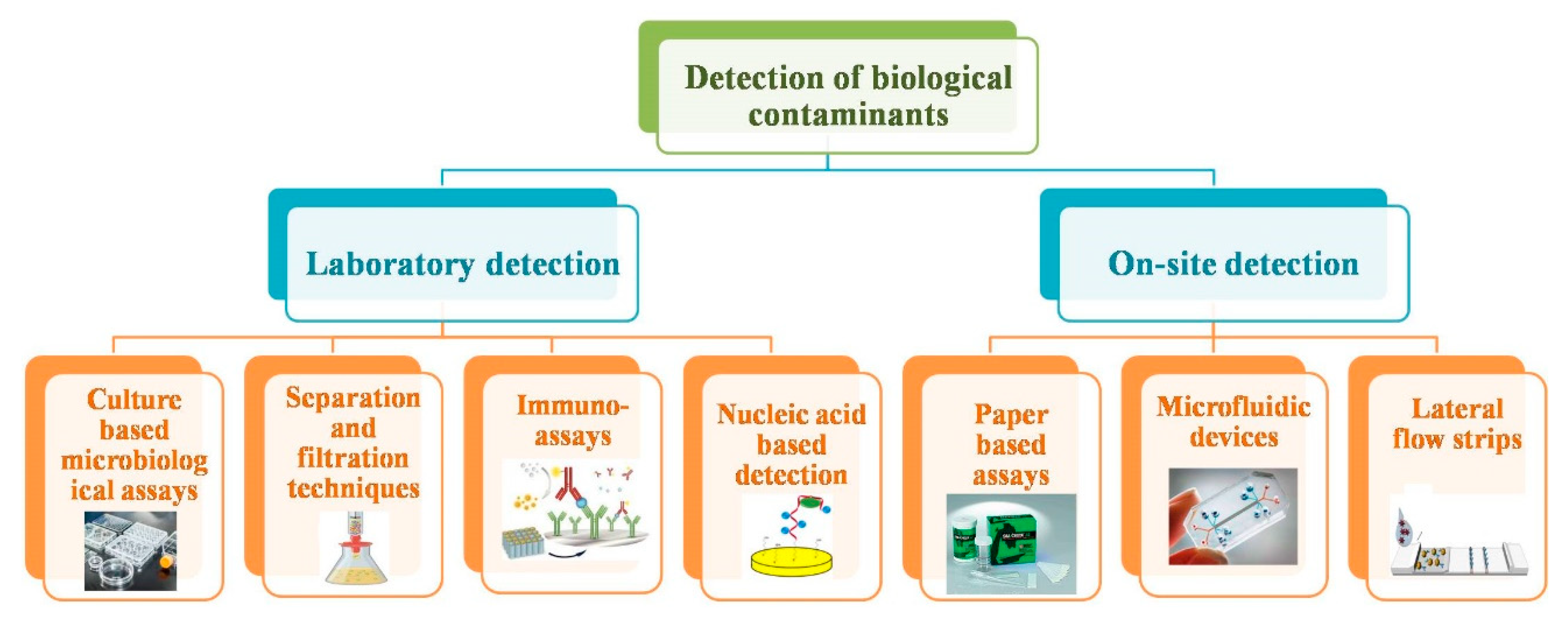
2. Conventional Methods to Detect Biological Contaminants

Water quality monitoring is the primary factor towards prevention of illness and infections caused by microorganisms. In the 20th century, due consideration has been given to water quality monitoring for pathogen detection. The various detection methods for biological contaminants are discussed here in detail (Figure 1). The conventional approaches are majorly focused on culture-based testing, separation and filtration of microbes, immunological methods, and nucleic acid-based detection. The continuous improvement in detection approaches can be observed, keeping in view the nature and origin of microorganisms to be detected.

2.1. Microbiological Assays Technique

The conventional microbiological methods rely on the growth and isolation of pathogens on the specific media. Some common microbiological diagnostics are based on microbiological cultures using medium (either selective or differential), Gram-staining, microscopy of specimens, and biochemical tests [15]. Culture-based microbiological assays, generally known as the golden standard, were the first developed methods for the qualitative and quantitative detection of biological contaminants i.e., pathogens [16]. The basic steps involved in the detection of pathogens via culturing are: enrichment, plating, incubation, colony counting, biochemical screening, and serological confirmation [17]. Culture-based methods are sensitive, inexpensive, and provide accurate information on the nature and number of microorganisms present in the sample [18]. However, these methods require several days to weeks to produce results, depending on the ability of the pathogen to multiply into visible colonies. Further, the steps of culture medium preparation, inoculation, and colony counting make the process labor intensive. Moreover, these methods have reached their limit in growing specific microorganisms in artificial media [19]. Despite these limitations, these conventional culture-based methods are still in clinical practice because they can provide diagnosis of acute infections in early stages in comparison to other technological advances.

2.2. Separation and Filtration Techniques

In order to minimize the time period required for culture-based plating assays, research efforts have been directed towards the development of alternatives for specific detection of pathogens and also to avoid chances of the false-negative results. In this regard, different separation and filtration techniques (i.e., chemical, physical, and antibody-based techniques) have been explored to recover and concentrate the microbes from contaminated water [20,21,22]. In order to spot the very low concentration of pathogens in water, an initial step of concentration or enrichment is required for their detection. After concentrating the target organism from a portion of a large quantity of material, a shortened and efficient detection of pathogenic microbes can be achieved. Further, membrane-based direct epifluorescent filtration technique (DEFT) is another approach for enumeration of microbes, depending upon the binding properties of the fluorescent dye. DEFT is a direct method to concentrate the target pathogens from a large sample volume [23]. These conventional approaches suffer with the use of harsh chemical reagents that can cause cell injury.
Another technique to shorten the isolation stage is immunomagnetic separation (IMS), in which the selective enrichment stage can be replaced with nongrowth-related procedures [24]. In this approach, superparamagnetic particles (such as iron oxide-coated polystyrene beads) coated with target organism-specific antibodies are used to isolate the pathogens from the aqueous solution containing a mixed population of microorganisms [25]. Over the past few years, the IMS technique has been reported in combination with polymerase chain reaction (PCR) assays to improve their sensitivity for detection of microorganisms [26,27]. This combination offered a rapid (<7 h) and simultaneous detection of different microorganisms (i.e., Listeria monocytogenes [L. monocytogenes] and Listeria ivanovii) without any enrichment process from the sample [28]. The major limitation with these chemical and physical methods is the interference with surface properties of microorganisms during their recovery and concentration from aqueous solutions.

2.3. Immunoassays Approach

Immunoassays are based on the specific interaction between an antigen (i.e., biological contaminant) and an antibody to measure the concentration of microbes in a solution. Different formats and variations of immunological assays have been reported, which may involve the multiple steps of addition and separation of reagents [12,29,30]. In conventional homogeneous immunoassays formats (such as agglutination, immunodiffusion, and turbidemetry), the bound–unbound antibodies are not separated and the formed antibody–antigen complex is easily visible in short incubation times [31]. However, this approach does not facilitate appropriate quantification and detection limits. The heterogeneous immunoassay, in the form of highly specific and sensitive enzyme-linked immunosorbent assay (ELISA), and its varied modifications are now the most prevalent methods on commercial levels for detection of biological contaminants [32]. In this method, unbound antibodies are separated from the bound ones, which helps in improving the detection limit i.e., ranges from 103–105 cfu/mL for whole cells and few ng/mL for proteins/toxins [33]. Although the method is accurate, the direct detection of microbes is not possible; enrichment is required for achieving the required quantification of analytes. The specificity of immunoassays motivates the researchers to explore further modifications in order to achieve higher sensitivity, portability, ease of use, and reusability.

2.4. Nucleic Acid-Based Detection

The advancements in biotechnology have resulted in the development of a variety of nucleic acid-based rapid detection platforms for biological contaminants. These methods exhibit higher sensitivity and specificity than culture-based assays, along with drastic reduction in the detection time [34]. The major strategies involved in nucleic acid-based detection of pathogens are hybridization and amplification. Nucleic acid hybridization involves identification of biological contaminants through complementary binding of single-stranded nucleic acid probes (labeled with radioactive or fluorescent compounds) with target organism genes [35,36]. In the presence of complementary target genes in the sample to be tested, the probe forms hydrogen bonds, facilitating its detection through autoradiography or fluorescent signals from the probe. This approach has a limitation in terms of the requirement of a high concentration of target nucleic acid to achieve detectable signals. This limitation can be overcome by nucleic acid amplification-based assays (like PCR), which increase the number of specific target nucleic acid in a sample [37]. PCR can generate multiple copies of a target DNA sequence via different processing steps such as (a) repeated cycles of denaturation of target DNA, (b) annealing of synthetic oligonucleotide primers to denatured strands, and (c) polymerization of target DNA as a template using enzymes. The repeated cycles of PCR result in million-fold amplification of a single copy of target DNA in less than 2 h, thereby eliminating the need for culture enrichment [38]. Cell concentration up to 101–106 cfu/mL has been detected with PCR methods in combination with nucleic acid extraction approaches [39]. Besides the revolutionary nature of PCR in terms high specificity and selectivity, there are still limitations in terms of nonreliable detection and quantification of infectious or viable organisms. Furthermore, its routine use is also not feasible due to requirement of costly reagents, trained personnel, and a very clean working environment.

3. Point-Of-Care (POC) Devices for Biological Contaminants

The laboratory-based technologies (such as PCR and ELISA) are not accessible or affordable in resource-limited areas lacking basic infrastructure and/or trained operators. POC devices have emerged to facilitate the on-site detection of different analytes (Table 1) [40,41]. The POC devices are replacing culture-based and other cumbersome commercial methods, which is attributable to widened accessibility to detection, rapid analysis time, and cost effectiveness [42]. Various POCs are majorly developed on microfluidic [43,44,45] or lateral flow systems [46,47,48] using glass; paper; silk fibroin; flexible graphite; or polymer substrates, such as polydimethylsiloxane (PDMS), poly(3,4-ethylene dioxythiophene):poly(styrene sulfonate) (PEDOT:PSS)), poly(methyl methacrylate) (PMMA), and polyethylene terephthalate (PET), etc. Further, the detection approaches are dependent upon fluorescence [49], colorimetry [50], and electrochemistry [51,52,53] to achieve rapid and easy to interpret signals for water safety monitoring.

3.1. Paper-Based Assay Methodology

Paper-based devices are attractive candidates for quantitative POC detection of biological contaminants in water as they offer low cost development, simple operation, portability, and robust instrumentation [68]. The main advantages of paper-based sensing devices include (i) high surface area to volume ratio; (ii) efficient adsorption; (iii) biocompatibility; (iv) ease of functionalization for stable immobilization of bioprobes; (v) straightforward sterilization; and (vi) easy disposal via incineration [69,70]. Also, the paper materials are lightweight and easily accessible worldwide with minimal chemical handling required due to convenient storage and transport of reagents within the matrix [71]. A paper-based handheld culture device has been reported for E. coli detection in water [54]. The strategy exploited recombinant reported lacZ T4 (carrying β-galactosidase gene) bacteriophage along with bioluminescent (6-O-β-galactopyranosyl-luciferin, Beta-Glo(®)) β-galactosidase indicator substrate to monitor β-galactosidase release post phage-mediated cell lysis, as shown in Figure 2. For detection of contaminants, the filtration of water samples was carried out using membrane filters (having 0.45 µm pore size) to concentrate bacteria followed by incubation of filter papers on nutrient medium-coated paper at 37 °C for 4 h. Next, the bacteriophage and respective indicator substrates were incorporated into the device resulting in cell lysis. The lysed cells released β-galactosidase enzyme, which catalyzed the conversion of chromogenic products into fluorescent product. The fluorescence was detected using a portable luminescence imaging device attached to culture-based paper. The sensor demonstrated a visual detection limit of < 10 CFU/mL within a time period of 5.5 h with high specificity in the presence of other nonspecific bacteria, such as Enterobacter cloacae, Aeromonas hydrophila, and Salmonella Typhimurium.
The wide characteristics of nanomaterials have been studied for water and wastewater treatment such as catalysis, high reactivity, and adsorption [72,73,74]. Incorporation of advanced nanomaterials with paper-based assays have significantly improved the sensitivity of sensors [75]. A group of researchers has explored graphene oxide (GO), a two-dimensional nanomaterial acting as an excellent quencher to fluorescent quantum dots (QDs) for highly sensitive detection of Salmonella spp [55]. The turn ON/OFF testing strips were designed by decorating the test line with (i) CdSe@ZnS QDs as capture probes and (ii) GO as an agent for target revealing. The quenching of fluorescence of the test line occurs in the absence of the target pathogen due to energy transfer between donor Ab-QDs and acceptor GO, resulting in the turn OFF state of the sensor. However, the fluorescence of the test line is restored in the presence of target Salmonella spp. due to selective binding of bacteria to Ab-QDs on the test line, which increased the distance (> 20 nm) between donor and acceptor to interrupt the resonance energy transfer. The turn ON state of the test line fluorescence indicated the presence of Salmonella with 100 CFU/mL detection limit in milk and water samples. Apart from fluorescence, high surface area and excellent electrical conductivity of graphene have also been investigated for imparting improved sensitivity of paper-based sensors for biological contaminants [76]. The sensing platform employing hierarchical structure of graphene-wrapped copper oxide-cysteine has achieved a detection limit of 3.8 CFU/mL for E. coli O157:H7 in fruit juice, milk, and water samples [77].
Ma and coworkers patterned the screen-printed polydimethylsiloxane (PDMS) paper chip-based visible immunoassay for bacterial detection (i.e., E. coli) using antibody-immobilized detection zones, where detection antibody-functionalized gold nanoparticles (AuNPs) act as a signal reporter [56]. Following the specific immunological reactions, a silver enhancer solution was added onto the detection zone. AuNPs concentrated on the detection zone catalyzed reduction of silver ions to make the visualized black spots. The silver-stained chips were then scanned using a commercial digital camera and/or scanner to obtain the quantitative results. The calibration of gray value of silver-stained spots with bacterial concentration provided the information on density of E. coli in water samples. The sensitivity of the developed strips was found to be comparable to conventional ELISA, but with reduced cost due to lesser consumption of time and reagents, pointing towards its potential for in situ monitoring of water quality.
Another litmus paper-based DipTest has been developed for E. coli detection in aqueous solutions, in which enzymatic reactions were performed directly on porous cellulose blotting paper [57]. The long strips of paper were coated with a hydrophobic barrier at the top edge, chemoattractant at the bottom edge, and customized chemical reagents immediately below the top edge, forming the reaction zone. During the detection process, the paper strip was dipped in water samples (contaminated with E. coli), where the bacterial cells were chemotactically attracted towards the paper strip, followed by their ascent towards the reaction zone through capillary wicking. Thereafter, the concentrated E. coli at the paper strip (particularly at the reaction zone) reacts with chemical reagents to produce pink to red color, indicating the presence of contamination in the tested samples. The fabricated paper strips have been tested for sensitivity and specificity, on the basis of which their potential for efficient on-site screening of water samples for biological contamination has been established.
Further, efforts have been focused on achieving improvements in sensitivity of the paper-based sensor for pathogen detection via different techniques like electrochemical assay [78,79]. Rengaraj et al. [58] have explored a novel paper-based electrochemical sensor for impedimetric sensing of bacterial contamination (refer to Figure 3). The electrochemical device is composed of screen-printed carbon electrodes fabricated onto hydrophobic paper with incorporated functionality. Concanavalin A has been employed as a recognition element instead of costly antibody probes to reduce the cost of the sensor significantly. Concanavalin exhibits high specificity for mono or oligo-saccharides present on the cell envelope of bacteria. The selection of Concanavalin A as the recognition agent was carried out due to its high selectivity towards oligosaccharides or monosaccharides on bacteria. The electrochemical sensor detected bacteria down to a concentration of 2 × 10³ CFU/mL, which is sufficiently low to advocate applicability in real samples. Besides these advancements, there are still several challenges associated with commercial application of paper-based assays such as accuracy, sensitivity, and multiple analytes detection at same time.
Despite the promising properties and applications of paper-based sensors, there are some associated limitations in terms of accuracy, resolution, sensitivity, and multiplexed detection. However, the efforts can be diverted towards patterned fabrication and employment of other advanced detection approaches (for e.g., chemiluminescence and electrochemiluminescence) [68,80].

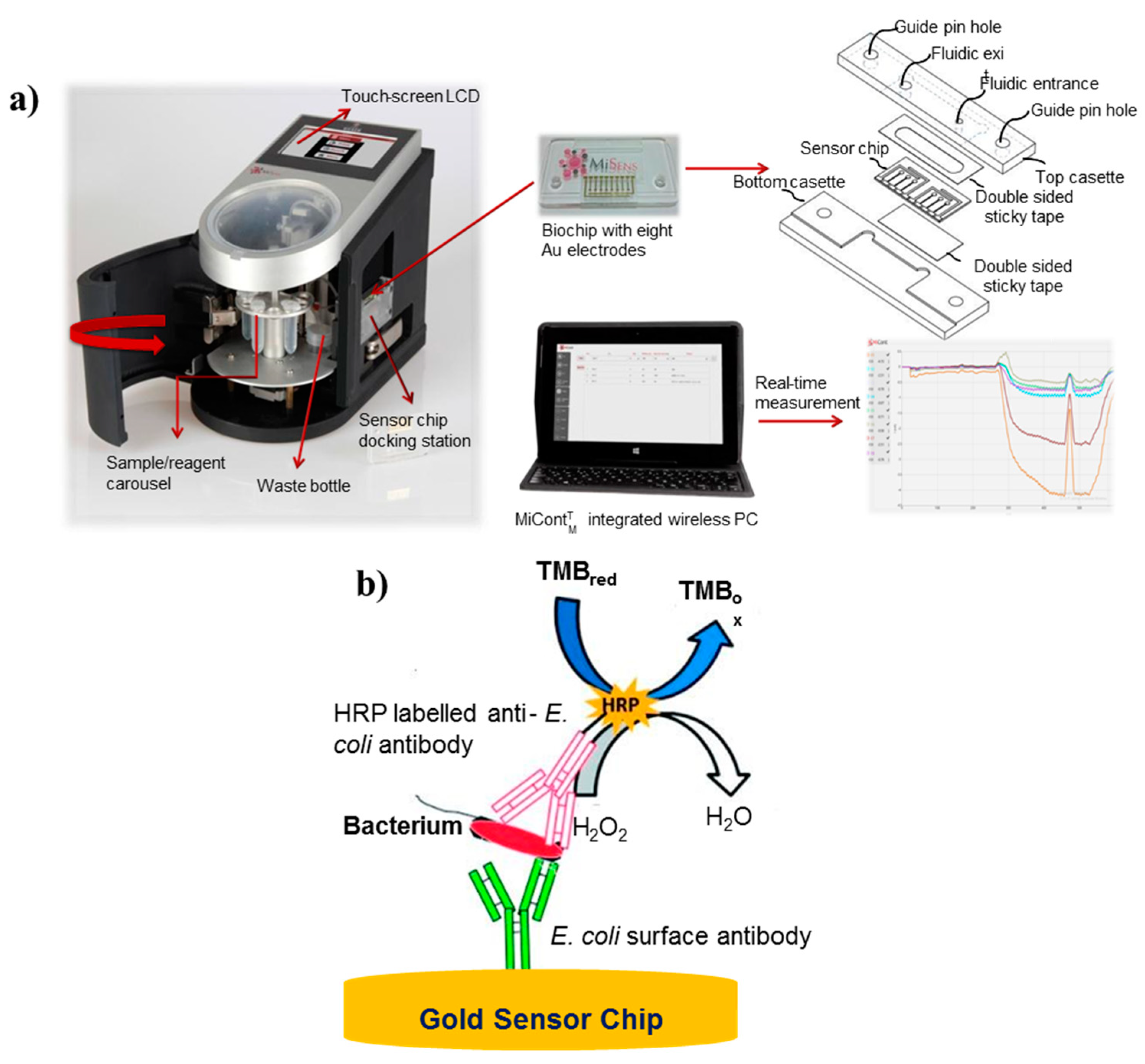
3.2. Microfluidic Detection Platforms for Water Quality Assessment

Microfluidic-based analytical devices are receiving research and commercial orientation for sensing of biological contaminants in environmental samples (i.e., water and food) due to low cost, high throughput, simplicity, minimal sample requirement, rapidity, and on-site applicability [81]. Different works incorporating microfluidic platforms with different analytical approaches have also surfaced for detection of biological contaminants such as PCR [82,83,84], loop-mediated isothermal amplification (LAMP) [85,86], mass spectrometry [87,88], and electrochemistry [89,90]—in particular, a disposable PCR microfluidic device composed of an array of closed reactors preloaded with different pairs of primers for real-time monitoring of multiple water-borne pathogens (coliform bacteria) [91]. PCR detects the genes specific to the organisms through amplification. Capillary flow scheme has been used as single step for PCR mixture loading and isolation of the reactors incorporated to minimize the evaporative losses, reducing the complexity and cost of the device, with a detection limit of 51 CFU/mL. Using the similar microfluidic quantitative PCR (MFQPCR) system, the human viral and enteric bacterial pathogens have been detected down to concentration of 2 copies/μl of cDNA/DNA [92] and 100 cells/liter [93], respectively, in wastewater treatment plants. However, further research efforts are needed to resolve the major limitation of microfluidic PCR (such as bubble generation and reagent evaporation) through appropriate modeling of microfluidic platforms for controlled fluid transfer [94].
Microfluidic paper-based analytical devices (µPADs) have been investigated for the detection of different pathogens in environmental samples, such as L. monocytogenes [95,96], E. coli [97], Salmonella [98], etc. Jokerst and coworkers reported a series of colorimetric spot tests using bioactive paper for sensing of L. monocytogenes, E. coli, and S. enteric in water samples from agricultural lands [59]. The pathogen detection is based on the catalytic conversion of dried compounds predeposited on the µPAD with the enzyme produced by the target bacteria grown in the culture media. For E. coli determination, the reaction between chlorophenol red β-galactopyranoside and β-galactosidase results in color transition from yellow to red. Determination of L. monocytogenes was facilitated by the production of a blue color on reaction of 5-bromo-4-chloro-3-indolyl-myo-inositol phosphate with phosphatidylinositol-specific phospholipase C. Lastly, the catalytic action of esterase on 5-bromo-6-chloro-3-indolyl caprylate resulted in the formation of a purple colored product, leading to detection of S. enteric. The detection limit of 10 CFU/cm2 was achieved after 4 to 12 h of enrichment process, which is comparatively faster than culture-based methods. Further, µPAD based on similar colorimetric detection of pathogen Crronobacter spp. has been reported [99]. A micro-spot μPAD was fabricated using a polyvinyl chloride (PVC) pad with filter paper. In the presence of Cronobacter spp., the reaction of microbe-specific enzymes with chromogenic substrates can be observed in terms of color change, i.e., from colorless to indigo. This color change was measured and co-related to quantify the present analyte with an achievable detection limit of 10 CFU/cm2 in 10 h at low-cost.
Park et al. have also demonstrated on-field sensing of E. coli in aqueous samples using paper microfluidics and smartphones [60]. A three-channel device was fabricated with different types of anti-E. coli-conjugated beads (for both low and high concentrations) and also bovine serum albumin (BSA)-conjugated beads preloaded in two detection and negative control channels, respectively. In the presence of E. coli antigens, immune agglutination occurred with specific antibody-conjugated beads preloaded on the paper fibers, while BSA-conjugated beads remained unreacted. The quantification of the extent of immune agglutination was achieved by evaluation of digital images for Mie scatter intensity at an optimized distance and angle using gyroscope installed in smartphones. The assay results were found to be in validation with conventional culture-based methods. The combination of microfluidics and smartphones enabled the on-site, easy, and highly specific detection of E. coli with sufficiently high sensitivity with much lesser analysis time of 90 s. Microfluidic chip-based electrochemical sensors have also been intensively explored for biological contaminant detection in food and water samples with high sensitivity and rapid response [100,101]. The progress in the area of fabrication and microelectronics has facilitated the incorporation of electrochemical assay into microfluidic platforms with minimal sample, time, and cost requirements [102]. In microfluidic chip-based electrochemical sensors, the probe composed of either bare conductive substrate or biorecognition molecules immobilized the substrate on the electrode surface, as well as the analyte to be detected; the analyte brings the measurable changes in electrochemical properties of the electrode [103]. Altintas et al. [61] fabricated a fully automated and real-time electrochemical detection platform for pathogens, refer to Figure 4. The custom-designed biosensing platform offered sensitive quantification of E. coli with a detection limit of 50 CFU/mL. The regeneration ability of sensor surface and high specificity towards E. coli detection in the presence of other pathogens (i.e., Shigella, S. spp., S. aureus, and S. typhimurium) make it a promising tool for pathogen monitoring in water samples.
Kim and coworkers have developed a label-free impedance-based positive dielectrophoretic microfluidic detection platform for assessment of E. coli contamination in drinking water [62]. The chip is composed of a cell-focusing electrode for sorting of bacterial cells from water and a sensing electrode for subsequent capture and sensing of bacterial analyte. The binding of bacterial cells resulted in the change in resistance to charge transfer at the electrode surface and these impedance changes were then used to enumerate E. coli bacterial cells down to the detection limit of 300 CFU/mL in less than 1 min. The major limitation with label-free detection platforms is lack of specificity, which can generate false positive results on the attachment of nonspecific microorganisms to the electrode surface. Therefore, a group of researchers have used specifically tagged target biological contaminants prior to impedance-based electrochemical detection [63]. The microfluidic chip-based immunosensor is composed of anti-E. coli O157:H7 antibodies immobilized on to nanoporous alumina membrane. The explored strategy resulted in improved sensitivity with a relatively lower detection limit of 100 CFU/mL and negligible interference from nontarget bacteria. The integration of microfluidic chips with different analytical techniques is contributing towards the progress of microfluidics for pathogenic microbe detection in human body fluids, food, water, and air.

3.3. Lateral Flow Devices for Contaminant Detection

Lateral flow devices are one of the most commercialized POC testing methods, attributable to low-cost, easy interpretation, high specificity, and portability for detection and quantification of different analytes in complex mixtures. In the modern era, lateral flow assay-based rapid tests have been developed and widely accepted by regulatory authorities for screening of diseases [104], chemicals [105], pathogens [106], water pollutants [107], and toxins [108]. Lateral flow assays offer one-step detection, without the involvement of washing steps, in a simple manner, with relatively short development time, low-cost production, and low sample volume requirements. A novel lateral flow device for ultrasensitive and highly specific multiplexed sensing of pathogenic as well as nonpathogenic E. coli based on intracellular enzymatic activity of β-glucuronidase (GUS) or β-galactosidase (β-GAL) has been reported on bioactive strips [64]. The test strips were printed with sol-gel-derived silica ink using ink-jet printing technology, entrapping chlorophenol red β-galactopyranoside (CPRG), 5-bromo-4-chloro-3-indolyl-β-d-glucuronide sodium salt (XG), or both and FeCl3 at different zones. For detection, the sample was lysed and transported through the metal salt zone to the different substrate zones via lateral flow. On reaching the substrate, enzymatic hydrolysis of the substrate is initiated by the intracellular enzymes, resulting in production of blue colored and yellow to red-magenta product for XG hydrolysis by GUS, indicative of nonpathogenic E. coli, and CPRG hydrolysis by β-GAL, indicative of total coliform bacteria, respectively. The immunomagnetic nanoparticles were also employed for further selective preconcentration, which helped in achieving a very low detection limit of 5 CFU/mL and 20 CFU/mL for E. coli O157:H7 and E. coli BL21, respectively within 30 min with high specificity. In this work, the inclusion of the bacterial culturing step allowed detection of 8 CFU/ 100 mL E. coli cells in 8 h.
The major challenge of POC devices in food and water safety applications is lack of multiplexed detection, with reduced risk of cross-reactivity among various target analytes. Chen et al. [65] reported the fabrication of lateral flow test strips containing multiple sensing zones for detection of Pseudomonas aeruginosa (P. aeruginosa) toxin genes (i.e., ecfX, ExoU, and ExoS) in drinking water samples. The different capturing molecules were embedded into multiple detection zones to detect anti-hex (homeobox protein), anti-FITC (fluorescein isothiocyanate), and anti-digoxin antibody-tagged LAMP amplicons. Another study on the multiplexed detection of different pathogens using a 10-channel lateral flow device has been reported through upconverting phosphor technology-based fluorescence [109]. For this, 10 individual paper strips were each immobilized with antibodies specific to different pathogens, including S. typhimurium, E. coli O157:H7, Vibrio cholera O1, V. cholera O139, V. parahaemolyticus, S. paratyphi A, S. paratyphi B, S. paratyphi C, S. choleraesuis, and S. enteritidis. Further, the integration of individual strips was carried out by a single lateral flow device. The multiplexed sensor achieved detection limits in the range of 104–105 CFU/mL for different pathogens without the need of sample enrichment. The proposed device holds greater promise for rapid safety assessment of food and water resources.
The integration of lateral flow assay with smartphones is paving the way for simultaneous detection of multiple analytes, depending upon the different analytes (such as ions, bacteria, and small molecules) and recognition element reactions in the same sample, refer to Figure 5 [66]. The detection probe consisting of nanoparticles with different emission bands can resolve the issue of crossover reactions from multiple analytes. However, some fundamental features of lateral flow assays must be improved in terms of reproducibility and accuracy comparable to laboratory-based detection systems.
Researchers are exploring various ways to integrate different analytical techniques into one system to develop paper-based sample-to-answer devices for qualitative and quantitative determination of biological contaminants. For instance, LAMP, nucleic acid testing (NAT), and colorimetric detection methods have been orchestrated into a robust, simple, four-layered lateral flow device [67]. Flinders Technology Associates (FTA) cards and glass fibers have been used as extraction and amplification substrates for nucleic acid, respectively. The separation of paper layers by polyvinyl chloride (PVC) acted as valves for controlling the sample transportation from extraction to amplification and further to lateral flow zones. The fully integrated device was able to detect E. coli down to concentration as low as 10−100 CFU/mL, supporting its efficient use in safety monitoring of food and water samples. Overall, lateral flow assays provide cheap, rapid, and easy point-of-care testing of different analytes, but, require improvements in a variety of areas such as signal amplification, precise sample volume accuracy, limit of sensitivity, operation without good antibody preparation, dependence of analysis time on nature of sample, pore obstruction due to matrix obstruction, and simple procedure for sample pretreatment [110].

4. Conclusion and Future Prospects

In summary, this review presented an overview of recent advancements in the area of developing devices (from conventional approaches to POC) to detect biological contaminants such as pathogens, their components, and toxic secretions, largely in water samples. The presence of these contaminants is responsible for a variety of acute and chronic health hazards, along with major economic losses. Therefore, the qualitative and quantitative estimation of biological contamination is of esteemed importance. Conventionally, culture-based microbiological assays, molecular detection platforms, ELISA, separation, and filtration techniques are used on commercial levels. These methods are specific and accurate, but suffer from limitations of long analysis time, high cost, requirement of heavy instrumentation and trained personnel, and nonapplicability for on-site detection. Therefore, portable POC devices are being explored to enable highly sensitive, specific, and personalized detection of environmental contaminants. The majority of POC devices employ either antibody–antigen interaction-based immunoassays, or nucleic acid-based hybridization and amplification assays. The most common transduction techniques incorporated in these devices are visible colorimetry, fluorescence, or electrochemical detection. Further, it has been elucidated that the POC devices are fabricated on paper, microfluidic chip, or lateral flow platforms.
While POC devices are being extensively studied, several formidable challenges require addressing for their translation into commercial field applications. Efforts are required to achieve enhanced sensitivity, multiplexing, portability, and ease of quantification in POC devices for water and food safety assessment. For improved sensitivity, signal amplification using advanced nanostructures exhibiting intrinsic extraordinary optoelectronic and catalytic properties can be integrated into chip- and paper-based devices. Incorporation of smartphones and other similar readers can facilitate portability, easy understanding of results, and wide real field applicability. Also, the exploration of methods of fabricating POC devices with multiplexed detection requires considerable scientific attention in order to reduce cost and assay time, and increase assay productivity. The integration of multiple key detection methods into a single system is an emerging platform for simultaneous detection of multiple analytes. However, the area is still at its infancy and requires research orientation. The advent of such improvement in POC devices will aid in the achievement of water and food safety analysis and quality control in an easy, rapid, inexpensive, and effective manner.

Funding

This research was funded by HSCST, Govt. of Haryana, India (research grant vide letter No. HSCST/R&D/2018/2103 dated 01-08-2018) and DST-PURSE sanctioned to GJUS&T, Hisar under PURSE program No. SR/PURSE Phase 2/40(G). JM is grateful to CSIR for SRA fellowship (No. B-12587 Dates 01-08-2019).

Conflicts of Interest

Authors declare no conflict of interest.

References

  1. WHO Report. 2019. Available online: https://www.who.int/news-room/fact-sheets/detail/drinking-water (accessed on 17 August 2019).
  2. Kumar, S.; Bhanjana, G.; Dilbaghi, N.; Kumar, R.; Umar, A. Fabrication and characterization of highly sensitive and selective arsenic sensor based on ultra-thin graphene oxide nanosheets. Sens. Actuators B Chem. 2016, 227, 29–34. [Google Scholar] [CrossRef]
  3. Halden, R.U. Epistemology of contaminants of emerging concern and literature meta-analysis. J. Hazard. Mater. 2015, 282, 2–9. [Google Scholar] [CrossRef] [PubMed]
  4. Kumar, S.; Bhanjana, G.; Dilbaghi, N.; Umar, A. Zinc oxide nanocones as potential scaffold for the fabrication of ultra-high sensitive hydrazine chemical sensor. Ceram. Int. 2015, 41, 3101–3108. [Google Scholar] [CrossRef]
  5. Gruenberg, J.; van der Goot, F.G. Mechanisms of pathogen entry through the endosomal compartments. Nat. Rev. Mol. Cell Biol. 2006, 7, 495–504. [Google Scholar] [CrossRef]
  6. Cabral, J.P. Water microbiology. Bacterial pathogens and water. Int. J. Environ. Res. Public Health 2010, 7, 3657–3703. [Google Scholar] [CrossRef]
  7. Ali, M.; Nelson, A.R.; Lopez, A.L.; Sack, D.A. Updated global burden of cholera in endemic countries. PLoS Negl. Trop. Dis. 2015, 9, e0003832. [Google Scholar] [CrossRef]
  8. Park, J.; Kim, J.S.; Kim, S.; Shin, E.; Oh, K.H.; Kim, Y.; Kim, C.H.; Hwang, M.A.; Jin, C.M.; Na, K.; et al. A waterborne outbreak of multiple diarrhoeagenic Escherichia coli infections associated with drinking water at a school camp. Int. J. Infect. Dis. 2018, 66, 45–50. [Google Scholar] [CrossRef]
  9. Valilis, E.; Ramsey, A.; Sidiq, S.; DuPont, H.L. Non-O157 shiga toxin-producing Escherichia coli-a poorly appreciated enteric pathogen: Systematic review. Int. J. Infect. Dis. 2018, 76, 82–87. [Google Scholar] [CrossRef]
  10. Karmali, M.A. Factors in the emergence of serious human infections associated with highly pathogenic strains of shiga toxin-producing Escherichia coli. Int. J. Med. Microbiol. 2018, 308, 1067–1072. [Google Scholar] [CrossRef]
  11. Van Nevel, S.; Koetzsch, S.; Proctor, C.R.; Besmer, M.D.; Prest, E.I.; Vrouwenvelder, J.S.; Knezev, A.; Boon, N.; Hammes, F. Flow cytometric bacterial cell counts challenge conventional heterotrophic plate counts for routine microbiological drinking water monitoring. Water Res. 2017, 113, 191–206. [Google Scholar] [CrossRef] [Green Version]
  12. Rajapaksha, P.; Elbourne, A.; Gangadoo, S.; Brown, R.; Cozzolino, D.; Chapman, J. A review of methods for the detection of pathogenic microorganisms. Analyst 2019, 144, 396–411. [Google Scholar] [CrossRef] [PubMed]
  13. Nesatyy, V.J.; Suter, M.J.F. Proteomics for the analysis of environmental stress responses in organisms. Environ. Sci. Technol. 2007, 41, 6891–6900. [Google Scholar] [CrossRef] [PubMed]
  14. Párraga-Niño, N.; Quero, S.; Ventós-Alfonso, A.; Uria, N.; Castillo-Fernandez, O.; Ezenarro, J.J.; Muñoz, F.X.; Garcia-Nuñez, M.; Sabrià, M. New system for the detection of Legionella pneumophila in water samples. Talanta 2018, 189, 324–331. [Google Scholar] [CrossRef] [PubMed]
  15. Ferrer, O.; Casas, S.; Galvañ, C.; Lucena, F.; Bosch, A.; Galofré, B.; Mesa, J.; Jofre, J.; Bernat, X. Direct ultrafiltration performance and membrane integrity monitoring by microbiological analysis. Water Res. 2015, 83, 121–131. [Google Scholar] [CrossRef]
  16. Kim, U.; Ghanbari, S.; Ravikumar, A.; Seubert, J.; Figueira, S. Rapid, affordable, and point-of-care water monitoring via a microfluidic DNA sensor and a mobile interface for global health. IEEE J. Transl. Eng. Health Med. 2013, 1, 3700207. [Google Scholar]
  17. Clark, K.D.; Purslow, J.A.; Pierson, S.A.; Nacham, O.; Anderson, J.L. Rapid preconcentration of viable bacteria using magnetic ionic liquids for PCR amplification and culture-based diagnostics. Anal. Bioanal. Chem. 2017, 409, 4983–4991. [Google Scholar] [CrossRef]
  18. Aw, T.G.; Rose, J.B. Detection of pathogens in water: From phylochips to qPCR to pyrosequencing. Curr. Opin. Biotechnol. 2012, 23, 422–430. [Google Scholar] [CrossRef]
  19. Zulkifli, S.N.; Rahim, H.A.; Lau, W.J. Detection of contaminants in water supply: A review on state-of-the-art monitoring technologies and their applications. Sens. Actuators B Chem. 2018, 255, 2657–2689. [Google Scholar] [CrossRef]
  20. Jimenez, M.; Miller, B.; Bridle, H.L. Efficient separation of small microparticles at high flowrates using spiral channels: Application to waterborne pathogens. Chem. Eng. Sci. 2017, 157, 247–254. [Google Scholar] [CrossRef] [Green Version]
  21. Hassan, A.H.A.; Bergua, J.F.; Morales-Narváez, E.; Mekoçi, A. Validity of a single antibody-based lateral flow immunoassay depending on graphene oxide for highly sensitive determination of E. coli O157: H7 in minced beef and river water. Food Chem. 2019, 297, 124965. [Google Scholar] [CrossRef]
  22. Wang, Y.; Salazar, J.K. Culture-independent rapid detection methods for bacterial pathogens and toxins in food matrices. Compr. Rev. Food Sci. Food Saf. 2016, 5, 183–205. [Google Scholar] [CrossRef]
  23. Mandal, P.K.; Biswas, A.K.; Choi, K.; Pal, U.K. Methods for rapid detection of foodborne pathogens: An overview. Am. J. Food Technol. 2011, 6, 87–102. [Google Scholar] [CrossRef]
  24. Xu, M.; Wang, R.; Li, Y. Rapid detection of Escherichia coli O157: H7 and Salmonella Typhimurium in foods using an electrochemical immunosensor based on screen-printed interdigitated microelectrode and immunomagnetic separation. Talanta 2016, 148, 200–208. [Google Scholar] [CrossRef] [PubMed]
  25. Skjerve, E.; Rørvik, L.M.; Olsvik, O. Detection of Listeria monocytogenes in foods by immunomagnetic separation. Appl. Environ. Microbiol. 1990, 56, 3478–3481. [Google Scholar] [PubMed]
  26. Hyeon, J.Y.; Deng, X. Rapid detection of Salmonella in raw chicken breast using real-time PCR combined with immunomagnetic separation and whole genome amplification. Food Microbiol. 2017, 63, 111–116. [Google Scholar] [CrossRef] [PubMed]
  27. Liu, P.; Kim, M.; Schlesinger, D.; Kranz, C.; Ha, S.; Ha, J.; Slauch, J.; Baek, S.; Moe, C. Immunomagnetic separation combined with RT-qPCR for determining the efficacy of disinfectants against human noroviruses. J. Infect. Public Health 2015, 8, 145–154. [Google Scholar] [CrossRef] [Green Version]
  28. Mao, Y.; Huang, X.; Xiong, S.; Xu, H.; Aguilar, Z.P.; Xiong, Y. Large-volume immunomagnetic separation combined with multiplex PCR assay for simultaneous detection of Listeria monocytogenes and Listeria ivanovii in lettuce. Food Control 2016, 59, 601–608. [Google Scholar] [CrossRef]
  29. Dwivedi, H.P.; Jaykus, L.A. Detection of pathogens in foods: The current state-of-the-art and future directions. Crit. Rev. Microbiol. 2011, 37, 40–63. [Google Scholar] [CrossRef]
  30. Musso, D.; La Scola, B. Laboratory diagnosis of leptospirosis: A challenge. J. Microbiol. Immunol. Infect. 2013, 46, 245–252. [Google Scholar] [CrossRef] [Green Version]
  31. Connelly, J.T.; Baeumner, A.J. Biosensors for the detection of waterborne pathogens. Anal. Bioanal. Chem. 2012, 402, 117–127. [Google Scholar] [CrossRef]
  32. Jain, S.; Chattopadhyay, S.; Jackeray, R.; Abid, C.Z.; Kohli, G.S.; Singh, H. Highly sensitive detection of Salmonella typhi using surface aminated polycarbonate membrane enhanced-ELISA. Biosens. Bioelectron. 2012, 31, 37–43. [Google Scholar] [CrossRef] [PubMed]
  33. Hsu, H.L.; Chuang, C.C.; Liang, C.C.; Chiao, D.J.; Wu, H.L.; Wu, Y.P.; Lin, F.P.; Shyu, R.H. Rapid and sensitive detection of Yersinia pestis by lateral-flow assay in simulated clinical samples. BMC Infect. Dis. 2018, 18, 402. [Google Scholar] [CrossRef] [PubMed]
  34. Jofre, J.; Blanch, A.R. Feasibility of methods based on nucleic acid amplification techniques to fulfil the requirements for microbiological analysis of water quality. J. Appl. Microbiol. 2010, 109, 1853–1867. [Google Scholar] [CrossRef] [PubMed]
  35. Saccà, M.L.; Ferrero, V.E.V.; Loos, R.; Di Lenola, M.; Tavazzi, S.; Grenni, P.; Ademollo, N.; Patrolecco, L.; Huggett, J.; Caracciolo, A.B.; et al. Chemical mixtures and fluorescence in situ hybridization analysis of natural microbial community in the Tiber river. Sci. Total Environ. 2019, 673, 7–19. [Google Scholar] [CrossRef] [PubMed]
  36. Wang, C.; Yang, H.; Wu, S.; Liu, Y.; Wei, W.; Zhang, Y.; Wei, M.; Liu, S. Manifold methods for telomerase activity detection based on various unique probes. TrAC Trends Anal. Chem. 2018, 105, 404–412. [Google Scholar] [CrossRef]
  37. Silva, D.M.; Domingues, L. On the track for an efficient detection of Escherichia coli in water: A review on PCR-based methods. Ecotoxicol. Environ. Saf. 2015, 113, 400–411. [Google Scholar] [CrossRef] [PubMed] [Green Version]
  38. Saiki, R.K.; Gelfand, D.H.; Stoffel, S.; Scharf, S.J.; Higuchi, R.; Horn, G.T.; Mullis, K.B.; Erlich, H.A. Primer-directed enzymatic amplification of DNA with a thermostable DNA polymerase. Science 1988, 239, 487–491. [Google Scholar] [CrossRef]
  39. Brandt, J.; Albertsen, M. Investigation of detection limits and the influence of DNA extraction and primer choice on the observed microbial communities in drinking water samples using 16S rRNA gene amplicon sequencing. Front. Microbiol. 2018, 9, 2140. [Google Scholar] [CrossRef]
  40. Meredith, N.A.; Quinn, C.; Cate, D.M.; Reilly, T.H.; Volckens, J.; Henry, C.S. Paper-based analytical devices for environmental analysis. Analyst 2016, 141, 1874–1887. [Google Scholar] [CrossRef]
  41. Carrell, C.; Kava, A.; Nguyen, M.; Menger, R.; Munshi, Z.; Call, Z.; Nussbaum, M.; Henry, C. Beyond the lateral flow assay: A review of paper-based microfluidics. Microelectron. Eng. 2019, 206, 45–54. [Google Scholar] [CrossRef]
  42. Ding, X.; Mauk, M.G.; Yin, K.; Kadimisetty, K.; Liu, C. Interfacing pathogen detection with smartphones for point-of-care applications. Anal. Chem. 2018, 91, 655–672. [Google Scholar] [CrossRef] [PubMed]
  43. Almeida, M.I.G.; Jayawardane, B.M.; Kolev, S.D.; McKelvie, I.D. Developments of microfluidic paper-based analytical devices (μPADs) for water analysis: A review. Talanta 2018, 177, 176–190. [Google Scholar] [CrossRef] [PubMed]
  44. Kaneta, T.; Alahmad, W.; Varanusupakul, P. Microfluidic paper-based analytical devices with instrument-free detection and miniaturized portable detectors. Appl. Spectrosc. Rev. 2019, 54, 117–141. [Google Scholar] [CrossRef]
  45. Nasseri, B.; Soleimani, N.; Rabiee, N.; Kalbasi, A.; Karimi, M.; Hamblin, M.R. Point-of-care microfluidic devices for pathogen detection. Biosens. Bioelectron. 2018, 117, 112–128. [Google Scholar] [CrossRef]
  46. Zhao, F.; Ge, L.; Zhao, X.; Zhang, H.; Huang, X.; Zeng, J.; Lv, J. Establishment of a highly sensitive lateral-flow test method for the determination of Vibrio parahemolyticus. J. Food Saf. Qual. 2018, 9, 910–913. [Google Scholar]
  47. Melnik, S.; Neumann, A.C.; Karongo, R.; Dirndorfer, S.; Stübler, M.; Ibl, V.; Niessner, R.; Knopp, D.; Stoger, E. Cloning and plant-based production of antibody MC 10E7 for a lateral flow immunoassay to detect [4-arginine] microcystin in freshwater. Plant Biotechnol. J. 2018, 16, 27–38. [Google Scholar] [CrossRef]
  48. Akter, S.; Kustila, T.; Leivo, J.; Muralitharan, G.; Vehniäinen, M.; Lamminmäki, U. Noncompetitive chromogenic lateral-flow immunoassay for simultaneous detection of microcystins and nodularin. Biosensors 2019, 9, 79. [Google Scholar] [CrossRef]
  49. Taitt, C.R.; Anderson, G.P.; Ligler, F.S. Evanescent wave fluorescence biosensors: Advances of the last decade. Biosens. Bioelectron. 2016, 76, 103–112. [Google Scholar] [CrossRef]
  50. Kudo, H.; Yamada, K.; Watanabe, D.; Suzuki, K.; Citterio, D. Paper-based analytical device for zinc ion quantification in water samples with power-free analyte concentration. Micromachines 2017, 8, 127. [Google Scholar] [CrossRef]
  51. Mettakoonpitak, J.; Boehle, K.; Nantaphol, S.; Teengam, P.; Adkins, J.A.; Srisa-Art, M.; Henry, C.S. Electrochemistry on paper-based analytical devices: A review. Electroanalysis 2016, 28, 1420–1436. [Google Scholar] [CrossRef]
  52. Bhanjana, G.; Dilbaghi, N.; Kumar, R.; Kumar, S. Zinc oxide quantum dots as efficient electron mediator for ultrasensitive and selective electrochemical sensing of mercury. Electrochim. Acta 2015, 178, 361–367. [Google Scholar] [CrossRef]
  53. Bhanjana, G.; Rana, P.; Chaudhary, G.R.; Dilbaghi, N.; Kim, K.H.; Kumar, S. Manganese oxide nanochips as a novel electrocatalyst for direct redox sensing of hexavalent chromium. Sci. Rep. 2019, 9, 8050. [Google Scholar] [CrossRef] [PubMed]
  54. Burnham, S.; Hu, J.; Anany, H.; Brovko, L.; Deiss, F.; Derda, R.; Griffiths, M.W. Towards rapid on-site phage-mediated detection of generic Escherichia coli in water using luminescent and visual readout. Anal. Bioanal. Chem. 2014, 406, 5685–5693. [Google Scholar] [CrossRef] [PubMed]
  55. Morales-Narváez, E.; Naghdi, T.; Zor, E.; Merkoçi, A. Photoluminescent lateral-flow immunoassay revealed by graphene oxide: Highly sensitive paper-based pathogen detection. Anal. Chem. 2015, 87, 8573–8577. [Google Scholar] [CrossRef]
  56. Ma, S.; Tang, Y.; Liu, J.; Wu, J. Visible paper chip immunoassay for rapid determination of bacteria in water distribution system. Talanta 2014, 120, 135–140. [Google Scholar] [CrossRef]
  57. Gunda, N.S.K.; Dasgupta, S.; Mitra, S.K. DipTest: A litmus test for E. coli detection in water. PLoS ONE 2017, 12, e0183234. [Google Scholar] [CrossRef]
  58. Rengaraj, S.; Cruz-Izquierdo, Á.; Scott, J.L.; Di Lorenzo, M. Impedimetric paper-based biosensor for the detection of bacterial contamination in water. Sens. Actuators B Chem. 2018, 265, 50–58. [Google Scholar] [CrossRef]
  59. Jokerst, J.C.; Adkins, J.A.; Bisha, B.; Mentele, M.M.; Goodridge, L.D.; Henry, C.S. Development of a paper-based analytical device for colorimetric detection of select foodborne pathogens. Anal. Chem. 2012, 84, 2900–2907. [Google Scholar] [CrossRef]
  60. San Park, T.; Yoon, J.Y. Smartphone detection of Escherichia coli from field water samples on paper microfluidics. IEEE Sens. J. 2014, 15, 1902–1907. [Google Scholar] [CrossRef]
  61. Altintas, Z.; Akgun, M.; Kokturk, G.; Uludag, Y. A fully automated microfluidic-based electrochemical sensor for real-time bacteria detection. Biosens. Bioelectron. 2018, 100, 541–548. [Google Scholar] [CrossRef]
  62. Kim, M.; Jung, T.; Kim, Y.; Lee, C.; Woo, K.; Seol, J.H.; Yang, S. A microfluidic device for label-free detection of Escherichia coli in drinking water using positive dielectrophoretic focusing, capturing, and impedance measurement. Biosens. Bioelectron. 2015, 74, 1011–1015. [Google Scholar] [CrossRef] [PubMed]
  63. Tian, F.; Lyu, J.; Shi, J.; Tan, F.; Yang, M. A polymeric microfluidic device integrated with nanoporous alumina membranes for simultaneous detection of multiple foodborne pathogens. Sens. Actuators B Chem. 2016, 225, 312–318. [Google Scholar] [CrossRef]
  64. Hossain, S.Z.; Ozimok, C.; Sicard, C.; Aguirre, S.D.; Ali, M.M.; Li, Y.; Brennan, J.D. Multiplexed paper test strip for quantitative bacterial detection. Anal. Bioanal. Chem. 2012, 403, 1567–1576. [Google Scholar]
  65. Chen, Y.; Cheng, N.; Xu, Y.; Huang, K.; Luo, Y.; Xu, W. Point-of-care and visual detection of P. aeruginosa and its toxin genes by multiple LAMP and lateral flow nucleic acid biosensor. Biosens. Bioelectron. 2016, 81, 317–323. [Google Scholar] [CrossRef] [PubMed]
  66. Jin, B.; Yang, Y.; He, R.; Park, Y.I.; Lee, A.; Bai, D.; Li, F.; Lu, T.J.; Xu, F.; Lin, M. Lateral flow aptamer assay integrated smartphone-based portable device for simultaneous detection of multiple targets using upconversion nanoparticles. Sens. Actuators B Chem. 2018, 276, 48–56. [Google Scholar] [CrossRef]
  67. Choi, J.R.; Yong, K.W.; Tang, R.; Gong, Y.; Wen, T.; Li, F.; Pingguan-Murphy, B.; Bai, D.; Xu, F. Advances and challenges of fully integrated paper-based point-of-care nucleic acid testing. TrAC Trends Anal. Chem. 2017, 93, 37–50. [Google Scholar] [CrossRef]
  68. Singh, A.; Lantigua, D.; Meka, A.; Taing, S.; Pandher, M.; Camci-Unal, G. Paper-based sensors: Emerging themes and applications. Sensors 2018, 18, 2838. [Google Scholar] [CrossRef]
  69. Cate, D.M.; Adkins, J.A.; Mettakoonpitak, J.; Henry, C.S. Recent developments in paper-based microfluidic devices. Anal. Chem. 2014, 87, 19–41. [Google Scholar] [CrossRef]
  70. Pelton, R. Bioactive paper provides a low-cost platform for diagnostics. TrAC Trends Anal. Chem. 2009, 28, 925–942. [Google Scholar] [CrossRef]
  71. Nery, E.W.; Kubota, L.T. Sensing approaches on paper-based devices: A review. Anal. Bioanal. Chem. 2013, 405, 7573–7595. [Google Scholar] [CrossRef]
  72. Alhan, S.; Nehra, M.; Dilbaghi, N.; Singhal, N.K.; Kim, K.H.; Kumar, S. Potential use of ZnO@ activated carbon nanocomposites for the adsorptive removal of Cd2+ ions in aqueous solutions. Environ. Res. 2019, 173, 411–418. [Google Scholar] [CrossRef]
  73. Madhura, L.; Singh, S.; Kanchi, S.; Sabela, M.; Bisetty, K. Nanotechnology-based water quality management for wastewater treatment. Environ. Chem. Lett. 2019, 17, 65–121. [Google Scholar] [CrossRef]
  74. Kumar, S.; Nehra, M.; Kedia, D.; Dilbaghi, N.; Tankeshwar, K.; Kim, K.H. Nanodiamonds: Emerging face of future nanotechnology. Carbon 2019, 143, 678–699. [Google Scholar] [CrossRef]
  75. Vikesland, P.J. Nanosensors for water quality monitoring. Nat. Nanotechnol. 2018, 13, 651–660. [Google Scholar] [CrossRef] [PubMed]
  76. Artiles, M.S.; Rout, C.S.; Fisher, T.S. Graphene-based hybrid materials and devices for biosensing. Adv. Drug Deliv. Rev. 2011, 63, 1352–1360. [Google Scholar] [CrossRef]
  77. Pandey, C.M.; Tiwari, I.; Singh, V.N.; Sood, K.N.; Sumana, G.; Malhotra, B.D. Highly sensitive electrochemical immunosensor based on graphene-wrapped copper oxide-cysteine hierarchical structure for detection of pathogenic bacteria. Sens. Actuators B Chem. 2017, 238, 1060–1069. [Google Scholar] [CrossRef]
  78. Lu, J.; Ge, S.; Ge, L.; Yan, M.; Yu, J. Electrochemical DNA sensor based on three-dimensional folding paper device for specific and sensitive point-of-care testing. Electrochim. Acta 2012, 80, 334–341. [Google Scholar] [CrossRef]
  79. Ali, M.M.; Brown, C.L.; Jahanshahi-Anbuhi, S.; Kannan, B.; Li, Y.; Filipe, C.D.; Brennan, J.D. A printed multicomponent paper sensor for bacterial detection. Sci. Rep. 2017, 7, 12335. [Google Scholar] [CrossRef]
  80. Gosselin, D.; Belgacem, M.N.; Joyard-Pitiot, B.; Baumlin, J.M.; Navarro, F.; Chaussy, D.; Berthier, J. Low-cost embossed-paper micro-channels for spontaneous capillary flow. Sens. Actuators B 2017, 248, 395–401. [Google Scholar] [CrossRef]
  81. Busa, L.; Mohammadi, S.; Maeki, M.; Ishida, A.; Tani, H.; Tokeshi, M. Advances in microfluidic paper-based analytical devices for food and water analysis. Micromachines 2016, 7, 86. [Google Scholar] [CrossRef]
  82. Zhang, H.; Huang, F.; Cai, G.; Li, Y.; Lin, J. Rapid and sensitive detection of Escherichia coli O157: H7 using coaxial channel-based DNA extraction and microfluidic PCR. J. Dairy Sci. 2018, 101, 9736–9746. [Google Scholar] [CrossRef]
  83. Salman, A.; Carney, H.; Bateson, S.; Ali, Z. Shunting microfluidic PCR device for rapid bacterial detection. Talanta 2020, 207, 120303. [Google Scholar] [CrossRef] [PubMed]
  84. Li, Z.; Ju, R.; Sekine, S.; Zhang, D.; Zhuang, S.; Yamaguchi, Y. All-in-one microfluidic device for on-site diagnosis of pathogens based on integrated continuous flow PCR and electrophoresis biochip. Lab Chip 2019, 19, 2663–2668. [Google Scholar] [CrossRef] [PubMed]
  85. Azizi, M.; Zaferani, M.; Cheong, S.H.; Abbaspourrad, A. Pathogenic bacteria detection using RNA-based loop-mediated isothermal-amplification-assisted nucleic acid amplification via droplet microfluidics. ACS Sens. 2019, 4, 841–848. [Google Scholar] [CrossRef] [PubMed]
  86. Wang, S.; Zheng, L.; Cai, G.; Liu, N.; Liao, M.; Li, Y.; Zhang, X.; Lin, J. A microfluidic biosensor for online and sensitive detection of Salmonella typhimurium using fluorescence labeling and smartphone video processing. Biosens. Bioelectron. 2019, 140, 111333. [Google Scholar] [CrossRef] [PubMed]
  87. Zhou, W.; Le, J.; Chen, Y.; Cai, Y.; Hong, Z.; Chai, Y. Recent advances in microfluidic devices for bacteria and fungus research. TrAC Trends Anal. Chem. 2019, 112, 175–195. [Google Scholar] [CrossRef]
  88. Yew, M.; Ren, Y.; Koh, K.S.; Sun, C.; Snape, C. A review of state-of-the-art microfluidic technologies for environmental applications: Detection and remediation. Glob. Chall. 2019, 3, 1800060. [Google Scholar] [CrossRef]
  89. Fernández-la-Villa, A.; Pozo-Ayuso, D.F.; Castaño-Álvarez, M. Microfluidics and electrochemistry: An emerging tandem for next-generation analytical microsystems. Curr. Opin. Electrochem. 2019, 15, 175–185. [Google Scholar] [CrossRef]
  90. Loo, J.F.; Ho, A.H.; Turner, A.P.; Mak, W.C. Integrated printed microfluidic biosensors. Trends Biotechnol. 2019, 37, 1104–1120. [Google Scholar] [CrossRef]
  91. Ramalingam, N.; Rui, Z.; Liu, H.B.; Dai, C.C.; Kaushik, R.; Ratnaharika, B.; Gong, H.Q. Real-time PCR-based microfluidic array chip for simultaneous detection of multiple waterborne pathogens. Sens. Actuators B Chem. 2010, 145, 543–552. [Google Scholar] [CrossRef]
  92. Ishii, S.; Kitamura, G.; Segawa, T.; Kobayashi, A.; Miura, T.; Sano, D.; Okabe, S. Microfluidic quantitative PCR for simultaneous quantification of multiple viruses in environmental water samples. Appl. Environ. Microbiol. 2014, 80, 7505–7511. [Google Scholar] [CrossRef]
  93. Ishii, S.; Segawa, T.; Okabe, S. Simultaneous quantification of multiple food-and waterborne pathogens by use of microfluidic quantitative PCR. Appl. Environ. Microbiol. 2013, 79, 2891–2898. [Google Scholar] [CrossRef] [PubMed]
  94. Lee, S.H.; Song, J.; Cho, B.; Hong, S.; Hoxha, O.; Kang, T.; Kim, D.; Lee, L.P. Bubble-free rapid microfluidic PCR. Biosens. Bioelectron. 2019, 126, 725–733. [Google Scholar] [CrossRef] [PubMed]
  95. Liu, F.; Zhang, C. A novel paper-based microfluidic enhanced chemiluminescence biosensor for facile, reliable and highly-sensitive gene detection of Listeria monocytogenes. Sens. Actuators B Chem. 2015, 209, 399–406. [Google Scholar] [CrossRef]
  96. Zhang, D.; Bi, H.; Liu, B.; Qiao, L. Detection of pathogenic microorganisms by microfluidics based analytical methods. Anal. Chem. 2018, 90, 5512–5520. [Google Scholar] [CrossRef]
  97. Safavieh, M.; Ahmed, M.U.; Tolba, M.; Zourob, M. Microfluidic electrochemical assay for rapid detection and quantification of Escherichia coli. Biosens. Bioelectron. 2012, 31, 523–528. [Google Scholar] [CrossRef]
  98. San Park, T.; Li, W.; McCracken, K.E.; Yoon, J.Y. Smartphone quantifies Salmonella from paper microfluidics. Lab Chip 2013, 13, 4832–4840. [Google Scholar] [CrossRef]
  99. Sun, L.; Jiang, Y.; Pan, R.; Li, M.; Wang, R.; Chen, S.; Fu, S.; Man, C. A novel, simple and low-cost paper-based analytical device for colorimetric detection of Cronobacter spp. Anal. Chim. Acta 2018, 1036, 80–88. [Google Scholar] [CrossRef]
  100. Luka, G.; Ahmadi, A.; Najjaran, H.; Alocilja, E.; DeRosa, M.; Wolthers, K.; Malki, A.; Aziz, H.; Althani, A.; Hoorfar, M. Microfluidics integrated biosensors: A leading technology towards lab-on-a-chip and sensing applications. Sensors 2015, 15, 30011–30031. [Google Scholar] [CrossRef]
  101. Mirasoli, M.; Guardigli, M.; Michelini, E.; Roda, A. Recent advancements in chemical luminescence-based lab-on-chip and microfluidic platforms for bioanalysis. J. Pharm. Biomed. Anal. 2014, 87, 36–52. [Google Scholar] [CrossRef]
  102. Xu, M.; Wang, R.; Li, Y. Electrochemical biosensors for rapid detection of Escherichia coli O157: H7. Talanta 2017, 162, 511–522. [Google Scholar] [CrossRef]
  103. Yao, L.; Wang, L.; Huang, F.; Cai, G.; Xi, X.; Lin, J. A microfluidic impedance biosensor based on immunomagnetic separation and urease catalysis for continuous-flow detection of E. coli O157: H7. Sens. Actuators B Chem. 2018, 259, 1013–1021. [Google Scholar] [CrossRef]
  104. Banerjee, R.; Jaiswal, A. Recent advances in nanoparticle-based lateral flow immunoassay as a point-of-care diagnostic tool for infectious agents and diseases. Analyst 2018, 143, 1970–1996. [Google Scholar] [CrossRef] [PubMed]
  105. Dalirirad, S.; Steckl, A.J. Aptamer-based lateral flow assay for point of care cortisol detection in sweat. Sens. Actuators B Chem. 2019, 283, 79–86. [Google Scholar] [CrossRef]
  106. Ma, Q.; Yao, J.; Yuan, S.; Liu, H.; Wei, N.; Zhang, J.; Shan, W. Development of a lateral flow recombinase polymerase amplification assay for rapid and visual detection of Cryptococcus neoformans/C. gattii in cerebral spinal fluid. BMC Infect. Dis. 2019, 19, 108. [Google Scholar] [CrossRef] [PubMed]
  107. Pöhlmann, C.; Dieser, I.; Sprinzl, M. A lateral flow assay for identification of Escherichia coli by ribosomal RNA hybridisation. Analyst 2014, 139, 1063–1071. [Google Scholar] [CrossRef] [PubMed]
  108. Raeisossadati, M.J.; Danesh, N.M.; Borna, F.; Gholamzad, M.; Ramezani, M.; Abnous, K.; Taghdisi, S.M. Lateral flow based immunobiosensors for detection of food contaminants. Biosens. Bioelectron. 2016, 86, 235–246. [Google Scholar] [CrossRef]
  109. Zhao, Y.; Wang, H.; Zhang, P.; Sun, C.; Wang, X.; Wang, X.; Yang, R.; Wang, C.; Zhou, L. Rapid multiplex detection of 10 foodborne pathogens with an up-converting phosphor technology-based 10−channel lateral flow assay. Sci. Rep. 2016, 6, 21342. [Google Scholar] [CrossRef]
  110. Koczula, K.M.; Gallotta, A. Lateral flow assays. Essays Biochem. 2016, 60, 111–120. [Google Scholar] [CrossRef]
Figure 1. Detection methods for biological contaminants.
Sensors 19 04476 g001
Figure 2. Principles for the detection of E. coli bacterial cells using a paper-based handheld culture device.
Sensors 19 04476 g002
Figure 3. Schematic representation of paper-based biosensors for bacterial contaminant sensing from water. (a) Multiple steps of modification of the electrode surface, followed by a working principle of bacteria sensing; (b) layout of final paper-based screen-printed carbon electrode (reprinted with permission from [58]).
Sensors 19 04476 g003
Figure 4. Schematic illustration of microfluidic-based electrochemical biosensor. (a) Fully automated detection system and (b) assay for pathogen detection (reprinted with permission from [61]).
Sensors 19 04476 g004
Figure 5. Schematic representation of lateral flow aptamer assay for simultaneous sensing of multiple analytes. (A) Structure of lateral flow aptamer assay, (B) hybridization of upconversion nanoparticles probe with complementary DNA in the absence of target analytes, (C) decrease in fluorescence of test zones due to aptamer bonding with target analytes, (D) display of detection results using smartphone-based portable device, and (E) schematic view of smartphone-based portable device (reprinted with permission from [66]).
Sensors 19 04476 g005
Table 1. List of various platforms available for on-site detection of biological contaminants.
S. No.TypeAnalyteSubstrateTransduction PlatformDetection Limit (CFU/mL)Analysis TimeCostLifetimeRef.
1Paper-basedE. coliFilter paperFluorescence105.5 hModerateSingle use[54]
2Salmonella spp.CdSe@ZnS QDs decorated paper stripsFluorescence3.8-ModerateSingle use[55]
3E. coliAuNP decorated PDMS paper chipsOptical immunoassay57-HighReusable[56]
4E. coliLitmus paperColorimetry DipTest2 × 105 to 4 × 10⁴-Very lowSingle use[57]
5Bacterial ContaminantScreen printed carbon electrodeElectrochemical impedance2 × 103 ModerateReusable[58]
6MicrofluidicL. monocytogenes, E. coli, S. entericColor-producing compounds deposited on µPADColorimetry104−12 hModerateReusable[59]
7E. coliPaper fibersGyroscope installed in smartphone1090 sModerateReusable[60]
8E. coliAuNP-coated biochipsCyclic voltammetry and amperometry508 minHighReusable[61]
9E. coliDieletrophoretic microfluidic chipElectrochemical impedance300< 1 minHighReusable[62]
10E. coliNanoporous alumina membraneElectrochemical impedance100-ModerateReusable[63]
11Lateral flow E. coliSol-gel-derived silica ink-coated test stripsColorimetry530 minModerateReusable[64]
12Psuedomonas aeruginosaAuNP-conjugated nitrocellulose membraneVisual detection2050 minLowSingle use[65]
13SalmonellaUpconverting nanoparticles-coated paper stripsColorimetry8530 minHighSingle use[66]
14E. coliFlinders Technology Associates (FTA) cards and glass fibersColorimetry10−100-HighReusable[67]

