Passive Gamma-Ray and Neutron Imaging Systems for National Security and Nuclear Non-Proliferation in Controlled and Uncontrolled Detection Areas: Review of Past and Current Status


Abstract
1. Introduction
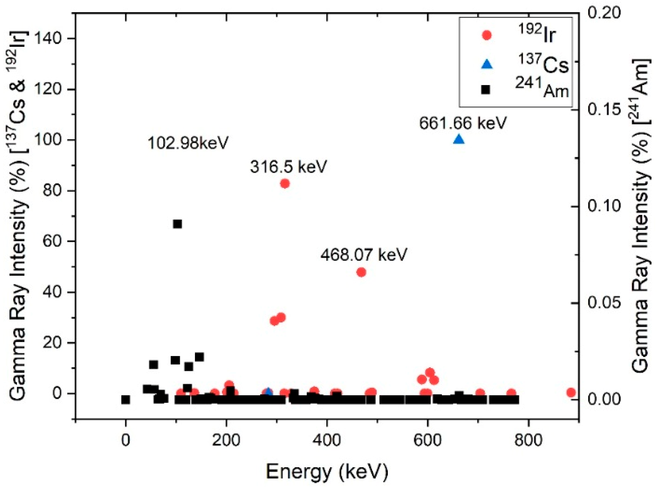
2. Radioactive Materials, Nuclear Materials and Radiation Sources:
- Any plutonium isotope concentration except that with 80% or more of 238Pu,
- Uranium enriched in the isotopes 233U or 235U,
- Uranium containing the mixture of isotopes as occurring in nature other than in the form of ore or ore-residue,
- Any material containing one or more of the above [4].
3. Problem Definition and Authorities’ Requirements
- At a mean dose rate of 0.2 μSv/h, the alarm of the system should be activated when the dose rate increases in a period of 1 s by 0.1 μSv/h for a pocket size instrument, by 0.05 μSv/h for a handheld instrument and 0.1 μSv/h for a fixed-installation instrument, for a duration of one second with 99% detection accuracy.
- False alarm rate should be minimal, with background measures of 0.2 μSv/h, with a false alarm rate of less than one every 12 h for pocket size instruments, less than six per hour for handheld instruments and less than one per day for fixed-installation instruments.
- The alarm of the system should be activated above a threshold of 20,000 n/s with a source to detector distance of 0.25 m for handheld instruments and 20,000 n/s in 5 s with source to detector distance of 2.0 m for fixed-installation instruments, using a system with 99% detection accuracy.
- False alarm rate should be minimal with less than six per hour for handheld instruments and one per day for fixed-installation instruments.
- The alarm of the system should be activated when the count increases above the background level by 0.5 μSv/h in 2 s for Radionuclide Identification Devices (RIDs) in the pocket and handheld size categories.
- The alarm of the system should be activated with 232Th, 137Cs, and 133Ba, 60Co and 57Co sources moving past the system at a speed of 2.22 m/s and distance of closest approach of 3 m for RIDs in the fixed installation size category.
- False alarm rate should be minimal with less than one every 10 h for pocket size and handheld instruments and less than one every two hours for fixed-installation size instruments.
- The alarm of the system should be activated when the exposure is above the threshold of 20,000 n/s in 2 s with 252Cf sources with a source to detector distance of 0.25 m for RIDs in the pocket size and handheld size categories.
- For a moving 252Cf source with activity of 20,000 n/s and moving past the system at a speed of 2.22 m/s at a distance of closest approach of 3 m, the system has to be able to detect the source with up to 1 cm steel or 0.5 cm of lead of shielding for RIDs in the fixed installation size category.
- False alarm rate should be minimal with less than one every 10 h for pocket size and handheld instruments and less than one every two hours for fixed-installation size instruments.
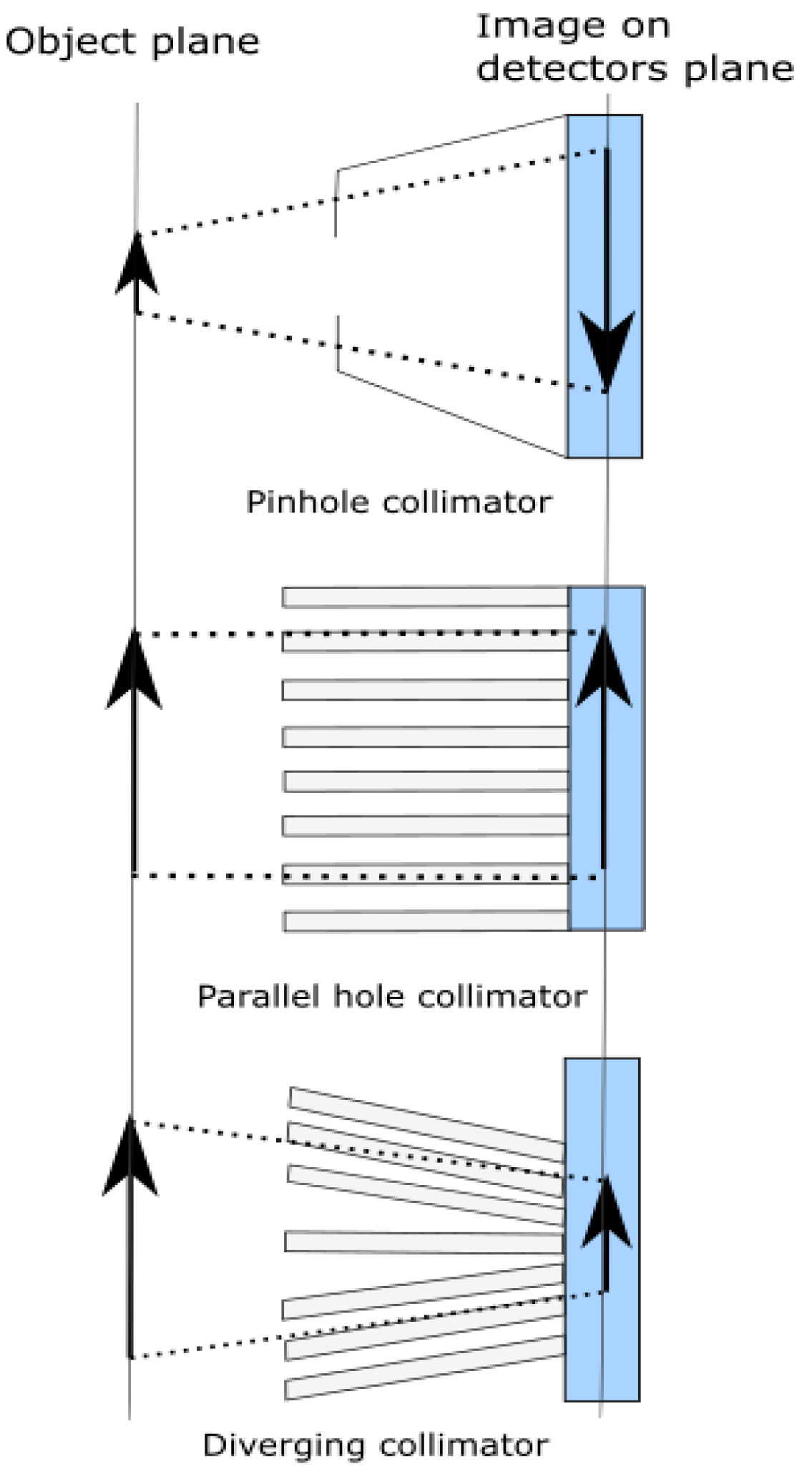
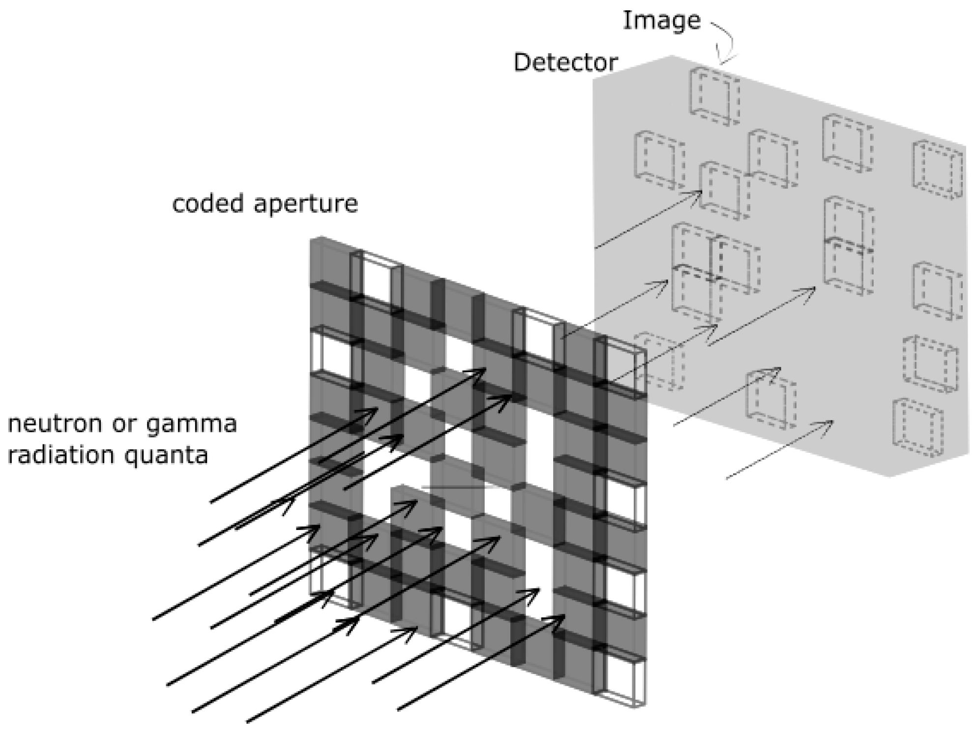
4. Physical and Electronic Collimations
4.1. Physical Collimation
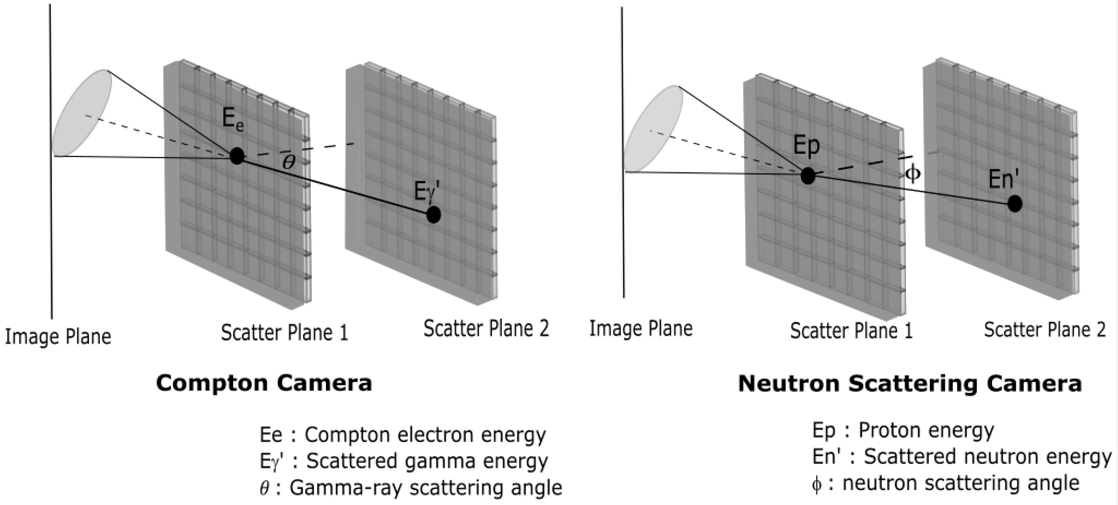
4.2. Electronic Collimation: Compton Camera and Neutron Scattering Camera
5. Passive Detection Systems of Illicit Radioactive Materials
5.1. Gamma-Ray Detection Systems
5.2. Neutron Detection Systems
5.3. Dual Gamma-Ray and Neutron Detection Systems
6. Conclusions
- Security agencies and legislation bodies requirements,
- Areas under surveillance and place of implementation,
- Image quality requirement,
- Timing and speed requirements.
Funding
Conflicts of Interest
References
- Zaitseva, L.; Hand, K. Nuclear smuggling chains. Suppliers, intermediaries, and end-users. Am. Behav. Sci. 2003, 46, 822–844. [Google Scholar] [CrossRef]
- IAEA. Inicdents and Trafficking Database (ITDB) Incidents of Nuclear and Other Radioactive Material Out of Regulatory Control; International Atomic Energy Agency: Vienna, Austria, 2018. [Google Scholar]
- Byrd, R.C.; Moss, J.M.; Priedhorsky, W.C.; Pura, C.A.; Richter, G.W.; Saeger, K.J.; Scarlett, W.R.; Scott, S.C.; Wagner, R.L. Nuclear detection to prevent or defeat clandestine nuclear attack. IEEE Sens. J. 2005, 5, 593–609. [Google Scholar] [CrossRef]
- IAEA. Safety Glossary Terminology Used in Nuclear, Radiation, Radioactive Waste and Transport Safety Version 2.0; Department of Nuclear Safety and Security; International Atomic Energy Agency: Vienna, Austria, 2006. [Google Scholar]
- Glossary of Terms for Nuclear, Biological, and Chemical Agents and Defense Equipment; U.S. Army Medical Department and U.S. Army Medical Department, Army Public Health Center: Washington, DC, USA, 2001.
- Gozani, T. Active Nondestructive Assay of Nuclear Materials, Principles and Applications; US Nuclear Regulatory Commission: Washington, DC, USA, 1981.
- Reilly, D.; Ensslin, N.; Smith, H.; Kreiner, S. Passive Nondestructive Assay of Nuclear Materials; National Technical Information Service, U.S. Department of Commerce: Washington, DC, USA, 1991.
- Milbrath, B.D.; Peurrung, A.J.; Bliss, M.; Weber, W.J. Radiation detector materials: An overview. J. Mater. Res. 2008, 23, 2561–2581. [Google Scholar] [CrossRef]
- Gamma-Ray Spectrometry Catalog; The Idaho National Laboratory: Idaho Falls, ID, USA, 1999.
- Gozani, T. Fission signatures for nuclear material detection. IEEE Trans. Nucl. Sci. 2009, 56, 736–741. [Google Scholar] [CrossRef]
- Stewart, L. Neutron Spectrum and Absolute Yield of a Plutonium-Beryllium Source. Phys. Rev. 1955, 98, 740–743. [Google Scholar] [CrossRef]
- Krane, K.S. Introductry Nuclear Physics; John Wiley and Sons: New York, NY, USA, 1988. [Google Scholar]
- Knoll, G.F. Radiation Detection and Measurment, 4th ed.; John Wiley and Sons: New York, NY, USA, 2010. [Google Scholar]
- Yapıcı, H.; Şahin, N.; Bayrak, M. Investigation of neutronic potential of a moderated (D–T) fusion driven hybrid reactor fueled with thorium to breed fissile fuel for LWRs. Energy Convers. Manag. 2000, 41, 435–447. [Google Scholar] [CrossRef]
- Knaster, J.; Arbeiter, F.; Cara, P.; Chel, S.; Facco, A.; Heidinger, R.; Ibarra, A.; Kasugai, A.; Kondo, H.; Micciche, G.; et al. IFMIF, the European–Japanese efforts under the Broader Approach agreement towards a Li(d,xn) neutron source: Current status and future options. Nucl. Mater. Energy 2016, 9, 46–54. [Google Scholar] [CrossRef]
- Mansur, L.K.; Rowcliffe, A.F.; Nanstad, R.K.; Zinkle, S.J.; Corwin, W.R.; Stoller, R.E. Materials needs for fusion, Generation IV fission reactors and spallation neutron sources–similarities and differences. J. Nucl. Mater. 2004, 329–333, 166–172. [Google Scholar] [CrossRef]
- Nea, O. Java-Based Nuclear Data Information System. Available online: http://www.oecd-nea.org/janis/ (accessed on 10 February 2016).
- Terrell, J. Distributions of Fission Neutron Numbers. Phys. Rev. 1957, 108, 783. [Google Scholar] [CrossRef]
- Herzo, D.; Koga, R.; Millard, W.A.; Moon, S.; Ryan, J.; Wilson, R.; Zych, A.D.; White, R.S. A Large Double Scatter Telescope for Gamma Rays and Neutrons. Nucl. Instrum. Methods 1975, 123, 583–597. [Google Scholar] [CrossRef]
- Wunderer, C.B.; Holslin, D.; Macri, J.R.; McConnell, M.; Ryan, J.M. SONTRAC-a low background, large area solar neutron spectrometer. In Proceedings of the Conference on the High Energy Radiation Background in Space, Snowmass, CO, USA, 22–23 July 1997; pp. 73–76. [Google Scholar]
- Ryan, J.M.; Desorgher, L.; Flückiger, E.O.; Macri, J.R.; McConnell, M.L.; Miller, R.S. SONTRAC: An imaging spectrometer for solar neutrons. In Proceedings of the Proceedings of SPIE—The International Society for Optical Engineering, Waikoloa, HI, USA, 11 February 2003; pp. 399–410. [Google Scholar]
- Marleau, P.; Brennan, J.; Krenz, K.; Mascarenhas, N.; Mrowka, S. Advances in imaging fission neutrons with a neutron scatter camera. In Proceedings of the IEEE Nuclear Science Symposium Conference Record, Honolulu, HI, USA, 26 October–3 November 2007; pp. 170–172. [Google Scholar]
- Mascarenhas, N.; Brennan, J.; Krenz, K.; Lund, J.; Marleau, P.; Rasmussen, J.; Ryan, J.; Macri, J. Development of a neutron scatter camera for fission neutrons. In Proceedings of the IEEE Nuclear Science Symposium Conference Record, San Diego, CA, USA, 29 October–1 November 2006; pp. 185–188. [Google Scholar]
- IAEA. Combating Ilicit Trafficking in Nuclear and Other Radioactive Materials; International Atomic Energy Agency, IAEA: Vienna, Austria, 2007. [Google Scholar]
- JRC: 20 Years Combating Illicit Trafficking of Nuclear Materials. Available online: https://ec.europa.eu/jrc/en/news/jrc-20-years-combating-illicit-trafficking-nuclear-materials-7023 (accessed on 7 March 2019).
- Radiological and Nuclear Prevention. Available online: https://www.interpol.int/en/Crimes/Terrorism/Radiological-and-Nuclear-terrorism/Radiological-and-nuclear-prevention (accessed on 7 March 2019).
- Treaty on the Non-Proliferation of Nuclear Weapons (NPT). Available online: https://www.un.org/disarmament/wmd/nuclear/npt/text (accessed on 7 March 2019).
- Code of Conduct on The Safety and Secuirty of Radioactive Sources; International Atomic Energy Agency, IAEA: Vienna, Austria, 2004.
- Handbook on Nuclear Law; International Atomic Energy Agency, IAEA: Vienna, Austria, 2003.
- International Convention for the Suppression of Acts of Nuclear Terrorism. Nucl. Terror. Conv. 2005, 2445, 89.
- Safeguards Techniques and Equipment; International Atomic Energy Agency, IAEA: Vienna, Austria, 2011.
- Detection of Radioactive Material at Borders; International Atomic Energy Agency, IAEA: Vienna, Austria, 2002.
- Heathrow Our Company: Facts and Figures. Available online: https://www.heathrow.com/company/company-news-and-information/company-information/facts-and-figures (accessed on 12 March 2019).
- Detection of Radioactive Materials at Borders Radiation Safety Section; Jointly sponsored by IAEA, WCO, EUROPOL and INTERPOL; International Atomic Energy Agency: Vienna, Austria, 2002.
- American National Standard for Evaluation and Performance of Radiation Detection Portal Monitors for Use in Homeland Security; ANSI N42.35-2016 (Revision of ANSI N42.35-2006); ANSI: Washington, DC, USA, 2016; pp. 1–70. [CrossRef]
- TECMIPT Test Operations Procedures (TTOP) For Radiation Detection Systems–Specific Methods; NIST: Gaithersburg, MD, USA, 2012.
- Gormley, J.E.; Rogers, W.L.; Clinthorne, N.H.; Wehe, D.K.; Knoll, G.F. Experimental comparison of mechanical and electronic gamma-ray collimation. Nucl. Instrum. Methods Phys. Res. Sect. A-Accel. Spectrometers Detect. Assoc. Equip. 1997, 397, 440–447. [Google Scholar] [CrossRef]
- Dicke, R.H. Scatter-Hole cameras for X-rays and gamma rays. Astrophys. J. 1968, 153, L101. [Google Scholar] [CrossRef]
- Ables, J.G. Fourier transform photography: A new method for X-ray astronomy. Publ. Astron. Soc. Aust. 1968, 1, 172–173. [Google Scholar] [CrossRef]
- Whitney, C.M.; Soundara-Pandian, L.; Johnson, E.B.; Vogel, S.; Vinci, B.; Squillante, M.; Glodo, J.; Christian, J.F. Gamma-neutron imaging system utilizing pulse shape discrimination with CLYC. Nucl. Instrum. Methods Phys. Res. Sect. A-Accel. Spectrometers Detect. Assoc. Equip. 2015, 784, 346–351. [Google Scholar] [CrossRef]
- Zou, Y.B.; Schillinger, B.; Wang, S.; Zhang, X.S.; Guo, Z.Y.; Lu, Y.R. Coded source neutron imaging with a MURA mask. Nucl. Instrum. Methods Phys. Res. Sect. A-Accel. Spectrometers Detect. Assoc. Equip. 2011, 651, 192–196. [Google Scholar] [CrossRef]
- Todd, R.W.; Nighting, J.; Everett, D.B. Proposed gamma camera. Nature 1974, 251, 132–134. [Google Scholar] [CrossRef]
- Everett, D.B.; Fleming, J.S.; Todd, R.W.; Nightingale, J.M. Gamma-radiation imaging-system based on compton-effect. In Proceedings of the Institution of Electrical Engineers-London; IET Digital Library: London, UK, 1977; Volume 124, pp. 995–1000. [Google Scholar]
- Singh, M.; Brechner, R.R. Experimental test-object study of electronically collimated SPECT. J. Nucl. Med. 1990, 31, 178–186. [Google Scholar]
- Cunningham, M.F.; Blakeman, E.; Fabris, L.; Habte, F.; Ziock, K. Active-mask coded-aperture imaging. In Proceedings of the IEEE Nuclear Science Symposium/Medical Imaging Conference, Honolulu, HI, USA, 26 October–3 November 2007; pp. 1217–1221. [Google Scholar]
- Schultz, L.J.; Wallace, M.S.; Galassi, M.C.; Hoover, A.S.; Mocko, M.; Palmer, D.M.; Tornga, S.R.; Kippen, R.M.; Hynes, M.V.; Toolin, M.J.; et al. Hybrid coded aperture and Compton imaging using an active mask. Nucl. Instrum. Methods Phys. Res. Sect. A-Accel. Spectrometers Detect. Assoc. Equip. 2009, 608, 267–274. [Google Scholar] [CrossRef]
- Marleau, P.; Brennan, J.; Brubaker, E.; Hilton, N.; Steele, J. Active Coded Aperture Neutron Imaging. In Proceedings of the 2009 IEEE Nuclear Science Symposium Conference Record, Orlando, FL, USA, 24 October–1 November 2009; Volumes 1–5, pp. 1974–1977. [Google Scholar] [CrossRef]
- Woolf, R.S.; Phlips, B.F.; Hutcheson, A.L.; Mitchell, L.J.; Wulf, E.A. An Active Interrogation Detection System (ACTINIDES) Based on a Dual Fast Neutron/Gamma-Ray Coded Aperture Imager. In Proceedings of the 2012 IEEE International Conference on Technologies for Homeland Security, Waltham, MA, USA, 13–15 November 2012; pp. 30–35. [Google Scholar]
- Singh, M. An Electronically Collimated Gamma Camera for Single Photon Emission Computed Tomography. Part I: Theoretical Considerations and Design Criteria. Med. Phys. 1983, 10, 421–427. [Google Scholar] [CrossRef]
- Runkle, R.C.; Chichester, D.L.; Thompson, S.J. Rattling nucleons: New developments in active interrogation of special nuclear material. Nucl. Instrum. Methods Phys. Res. Sect. A-Accel. Spectrometers Detect. Assoc. Equip. 2012, 663, 75–95. [Google Scholar] [CrossRef]
- Norman, D.R.; Jones, J.L.; Haskell, K.J.; Vanier, P.E.; Forman, L. Active nuclear material detection and imaging. In Proceedings of the 2005 IEEE Nuclear Science Symposium Conference Record, Fajardo, Puerto Rico, 23–29 October 2005; Volumes 1–5, pp. 1004–1008. [Google Scholar]
- Jones, J.L.; Norman, D.R.; Haskell, K.J.; Sterbentz, J.W.; Yoon, W.Y.; Watson, S.M.; Johnson, J.T.; Zabriskie, J.M.; Bennett, B.D.; Watson, R.W.; et al. Detection of shielded nuclear material in a cargo container. Nucl. Instrum. Methods Phys. Res. Sect. A-Accel. Spectrometers Detect. Assoc. Equip. 2006, 562, 1085–1088. [Google Scholar] [CrossRef]
- Chichester, D.L.; Simpson, J.D.; Lemchak, M. Advanced compact accelerator neutron generator technology for active neutron interrogation field work. J. Radioanal. Nucl. Chem. 2007, 271, 629–637. [Google Scholar] [CrossRef]
- Runkle, R.C.; Smith, L.E.; Peurrung, A.J. The photon haystack and emerging radiation detection technology. J. Appl. Phys. 2009, 106, 7. [Google Scholar] [CrossRef]
- Guss, P.; Reed, M.; Yuan, D.; Beller, D.; Cutler, M.; Contreras, C.; Mukhopadhyay, S.; Wilde, S. Size effect on nuclear gamma-ray energy spectra acquired by different-sized CeBr3, LaBr3:Ce, and NaI:Tl gamma-ray detectors. Nucl. Technol. 2014, 185, 309–321. [Google Scholar] [CrossRef]
- Reinhard, M.I.; Prokopovich, D.; Van Der Gaast, H.; Hill, D. Detection of illicit nuclear materials masked with other gamma-ray emitters. In Proceedings of the IEEE Nuclear Science Symposium Conference Record, San Diego, CA, USA, 29 October–1 November 2006; pp. 270–272. [Google Scholar]
- Siciliano, E.R.; Ely, J.H.; Kouzes, R.T.; Milbrath, B.D.; Schweppe, J.E.; Stromswold, D.C. Comparison of PVT and NaI(Tl) scintillators for vehicle portal monitor applications. Nucl. Instrum. Methods Phys. Res. Sect. A-Accel. Spectrometers Detect. Assoc. Equip. 2005, 550, 647–674. [Google Scholar] [CrossRef]
- Mortreau, P.; Berndt, R. Determination of 235U enrichment with a large volume CZT detector. Nucl. Instrum. Methods Phys. Res. Sect. A-Accel. Spectrometers Detect. Assoc. Equip. 2006, 556, 219–227. [Google Scholar] [CrossRef]
- Sjoden, G.E.; Detwiler, R.; Lavigne, E.; Baciak, J.E., Jr. Positive SNM gamma detection achieved through synthetic enhancement of sodium iodide detector spectra. IEEE Trans. Nucl. Sci. 2009, 56, 1329–1339. [Google Scholar] [CrossRef]
- Kim, K.H.; Jun, J.Y.; Jun, I.S.; Kwak, S.W. Development of a car-mounted nuclear material monitoring system: A prototype system. Nucl. Instrum. Methods Phys. Res. Sect. A-Accel. Spectrometers Detect. Assoc. Equip. 2009, 607, 154–157. [Google Scholar] [CrossRef]
- TSA MD134. Available online: http://www.rapiscansystems.com/en/products/radiation_detection/rapiscan_mp100 (accessed on 26 October 2018).
- Ziock, K.P.; Hailey, C.J.; Gosnell, T.B.; Lupton, J.H. A Gamma-Ray Imager for Arms Control. IEEE Trans. Nucl. Sci. 1992, 39, 1046–1050. [Google Scholar] [CrossRef]
- Ziock, K.P.; Cheriyadat, A.; Fabris, L.; Goddard, J.; Hornback, D.; Karnowski, T.; Kerekes, R.; Newby, J. Autonomous radiation monitoring of small vessels. Nucl. Instrum. Methods Phys. Res. Sect. A-Accel. Spectrometers Detect. Assoc. Equip. 2011, 652, 10–15. [Google Scholar] [CrossRef]
- Ely, J.; Kouzes, R.; Schweppe, J.; Siciliano, E.; Strachan, D.; Weier, D. The use of energy windowing to discriminate SNM from NORM in radiation portal monitors. Nucl. Instrum. Methods Phys. Res. Sect. A-Accel. Spectrometers Detect. Assoc. Equip. 2006, 560, 373–387. [Google Scholar] [CrossRef]
- Hevener, R.; Yim, M.-S.; Baird, K. Investigation of energy windowing algorithms for effective cargo screening with radiation portal monitors. Radiat. Meas. 2013, 58, 113–120. [Google Scholar] [CrossRef]
- Lo Presti, C.A.; Weier, D.R.; Kouzes, R.T.; Schweppe, J.E. Baseline suppression of vehicle portal monitor gamma count profiles: A characterization study. Nucl. Instrum. Methods Phys. Res. Sect. A Accel. Spectrometers Detect. Assoc. Equip. 2006, 562, 281–297. [Google Scholar] [CrossRef]
- Ziock, K.P.; Goldstein, W.H. The lost source, varying backgrounds and why bigger may not be better. In Proceedings of the Workshop on Unattended Radiation Sensor Systems for Remote Applications, Washington, DC, USA, 15–17 April 2002; pp. 60–70. [Google Scholar]
- Ziock, K.P.; Bradley, E.C.; Cheriyadat, A.; Cunningham, M.; Fabris, L.; Fitzgerald, C.L.; Goddard, J.S.; Hornback, D.E.; Kerekes, R.A.; Karnowski, T.P.; et al. Performance of the Roadside Tracker Portal-Less Portal Monitor. IEEE Trans. Nucl. Sci. 2013, 60, 2237–2246. [Google Scholar] [CrossRef]
- Kouzes, R.T.; Siciliano, E.R. The response of radiation portal monitors to medical radionuclides at border crossings. Radiat. Meas. 2006, 41, 499–512. [Google Scholar] [CrossRef]
- Robinson, S.M.; Bender, S.E.; Flumerfelt, E.L.; LoPresti, C.A.; Woodring, M.L. Time Series Evaluation of Radiation Portal Monitor Data for Point Source Detection. IEEE Trans. Nucl. Sci. 2009, 56, 3688–3693. [Google Scholar] [CrossRef]
- Ivanov, O.P.; Semin, I.A.; Potapov, V.N.; Stepanov, V.E. Extra-light gamma-ray imager for safeguards and homeland security. In Proceedings of the 2015 4th International Conference on Advancements in Nuclear Instrumentation Measurement Methods and their Applications, Lisbon, Portugal, 20–24 April 2015. [Google Scholar]
- Carrel, F.; Khalil, R.A.; Colas, S.; Toro, D.D.; Ferrand, G.; Gaillard-Lecanu, E.; Gmar, M.; Hameau, D.; Jahan, S.; Lainé, F.; et al. GAMPIX: A new gamma imaging system for radiological safety and Homeland Security Purposes. In Proceedings of the 2011 IEEE Nuclear Science Symposium Conference Record, Valencia, Spain, 23–29 October 2011; pp. 4739–4744. [Google Scholar]
- Gal, O.; Izac, C.; Jean, F.; Lainé, F.; Lévêque, C.; Nguyen, A. CARTOGAM—A portable gamma camera for remote localisation of radioactive sources in nuclear facilities. Nucl. Instrum. Methods Phys. Res. Sect. A Accel. Spectrometers Detect. Assoc. Equip. 2001, 460, 138–145. [Google Scholar] [CrossRef]
- Woodring, M.; Souza, D.; Tipnis, S.; Waer, P.; Squillante, M.; Entine, G.; Ziock, K.P. Advanced radiation imaging of low-intensity gamma-ray sources. Nucl. Instrum. Methods Phys. Res. Sect. A Accel. Spectrometers Detect. Assoc. Equip. 1999, 422, 709–712. [Google Scholar] [CrossRef]
- Dubos, S.; Lemaire, H.; Schanne, S.; Limousin, O.; Carrel, F.; Schoepff, V.; Blondel, C. ORIGAMIX, a CdTe-based spectro-imager development for nuclear applications. Nucl. Instrum. Methods Phys. Res. Sect. A Accel. Spectrometers Detect. Assoc. Equip. 2015, 787, 302–307. [Google Scholar] [CrossRef]
- Wulf, E.A.; Phlips, B.F.; Johnson, W.N.; Leas, B.; Mitchell, L.J. MISTI imaging and source localization. In Proceedings of the 2008 IEEE Nuclear Science Symposium Conference Record, Dresden, Germany, 19–25 October 2008; pp. 2413–2417. [Google Scholar]
- Kowash, B.R.; Wehe, D.K.; Fessler, J.A. A rotating modulation imager for locating mid-range point sources. Nucl. Instrum. Methods Phys. Res. Sect. A Accel. Spectrometers Detect. Assoc. Equip. 2009, 602, 477–483. [Google Scholar] [CrossRef]
- Vaska, P.; Vanier, P.E.; Junnarkar, S.; Krishnamoorthy, S.; Pratte, J.F.; Stoll, S. A compact scintillator-based coded aperture imager for localizing illicit nuclear materials. In Proceedings of the IEEE Nuclear Science Symposium Conference Record, Honolulu, HI, USA, 26 October–3 November 2007; pp. 1195–1197. [Google Scholar]
- Jeong, M.; Van, B.; Wells, B.T.; D’Aries, L.J.; Hammig, M.D. Scalable gamma-ray camera for wide-area search based on silicon photomultipliers array. Rev. Sci. Instrum. 2018, 89, 033106. [Google Scholar] [CrossRef] [PubMed]
- Ziock, K.P. Principles and applications of gamma-ray imaging for arms control. Nucl. Instrum. Methods Phys. Res. Sect. A Accel. Spectrometers Detect. Assoc. Equip. 2018, 878, 191–199. [Google Scholar] [CrossRef]
- Kong, Y.; Brands, H.; Glaser, T.; Herbach, C.; Hoy, L.; Kreuels, M.; Küster, M.; Pausch, G.; Petzoldt, J.; Plettner, C.; et al. A Prototype Compton Camera Array for Localization and Identification of Remote Radiation Sources. IEEE Trans. Nucl. Sci. 2013, 60, 1066–1071. [Google Scholar] [CrossRef]
- Saull, P.R.B.; MacLeod, A.M.L.; Sinclair, L.E.; Drouin, P.L.; Erhardt, L.; Hovgaard, J.; Krupskyy, B.; Ueno, R.; Waller, D.; McCann, A. SCoTSS modular survey spectrometer and Compton imager. In Proceedings of the 2016 IEEE Nuclear Science Symposium, Medical Imaging Conference and Room-Temperature Semiconductor Detector Workshop, NSS/MIC/RTSD, Strasbourg, France, 29 October–6 November 2016. [Google Scholar]
- MacLeod, A.M.L.; Boyle, P.J.; Hanna, D.S.; Saull, P.R.B.; Sinclair, L.E.; Seywerd, H.C.J. Development of a Compton imager based on bars of scintillator. Nucl. Instrum. Methods Phys. Res. Sect. A Accel. Spectrometers Detect. Assoc. Equip. 2014, 767, 397–406. [Google Scholar] [CrossRef]
- Vetter, K.; Burks, M.; Cork, C.; Cunningham, M.; Chivers, D.; Hull, E.; Krings, T.; Manini, H.; Mihailescu, L.; Nelson, K.; et al. High-sensitivity Compton imaging with position-sensitive Si and Ge detectors. Nucl. Instrum. Methods Phys. Res. Sect. A Accel. Spectrometers Detect. Assoc. Equip. 2007, 579, 363–366. [Google Scholar] [CrossRef]
- Hynes, M.V.; Harris, B.; Lednum, E.E.; Wallace, M.S.; Schultz, L.J.; Palmer, D.M.; Wakeford, D.T.; Andrews, H.R.; Lanza, R.C.; Clifford, E.T.; et al. Multimodal Radiation Imager. U.S. Patent 7,863,567, 4 January 2011. [Google Scholar]
- Andreyev, A.; Sitek, A.; Celler, A. Fast image reconstruction for Compton camera using stochastic origin ensemble approach. Med. Phys. 2011, 38, 429–438. [Google Scholar] [CrossRef] [PubMed]
- Hoover, A.S.; Kippen, R.M.; Sullivan, J.P.; Rawool-Sullivan, M.W.; Baird, W.; Sorensen, E.B. The LANL prototype Compton gamma-ray imager: Design and image reconstruction techniques. IEEE Trans. Nucl. Sci. 2005, 52, 3047–3053. [Google Scholar] [CrossRef]
- Montémont, G.; Bohuslav, P.; Dubosq, J.; Feret, B.; Monnet, O.; Oehling, O.; Skala, L.; Stanchina, S.; Verger, L.; Werthmann, G. NuVISION: A Portable Multimode Gamma Camera based on HiSPECT Imaging Module. In Proceedings of the 2017 IEEE Nuclear Science Symposium and Medical Imaging Conference (NSS/MIC), Atlanta, GA, USA, 21–28 October 2017; pp. 1–3. [Google Scholar]
- Penny, R.D.; Hood, W.E.; Polichar, R.M.; Cardone, F.H.; Chavez, L.G.; Grubbs, S.G.; Huntley, B.P.; Kuharski, R.A.; Shyffer, R.T.; Fabris, L.; et al. A dual-sided coded-aperture radiation detection system. Nucl. Instrum. Methods Phys. Res. Sect. A Accel. Spectrometers Detect. Assoc. Equip. 2011, 652, 578–581. [Google Scholar] [CrossRef]
- Zelakiewicz, S.; Hoctor, R.; Ivan, A.; Ross, W.; Nieters, E.; Smith, W.; McDevitt, D.; Wittbrodt, M.; Milbrath, B. SORIS-A standoff radiation imaging system. Nucl. Instrum. Methods Phys. Res. Sect. A Accel. Spectrometers Detect. Assoc. Equip. 2011, 652, 5–9. [Google Scholar] [CrossRef]
- Boehnen, C.; Paquit, V.; Ziock, K.; Guzzardo, T.; Whitaker, M.; Raffo-Caiado, A. Field trial of a highly portable coded aperture gamma ray and 3D imaging system. In Proceedings of the 2011 Future of Instrumentation International Workshop (FIIW), Oak Ridge, TN, USA, 7–8 November 2011; pp. 75–78. [Google Scholar]
- Tornga, S.R.; Sullivan, M.W.R.; Sullivan, J.P. Three-Dimensional Compton Imaging Using List-Mode Maximum Likelihood Expectation Maximization. IEEE Trans. Nucl. Sci. 2009, 56, 1372–1376. [Google Scholar] [CrossRef]
- Kouzes, R.T.; Ely, J.H.; Erikson, L.E.; Kernan, W.J.; Lintereur, A.T.; Siciliano, E.R.; Stephens, D.L.; Stromswold, D.C.; Van Ginhoven, R.M.; Woodring, M.L. Neutron detection alternatives to 3HE for national security applications. Nucl. Instrum. Methods Phys. Res. Sect. A Accel. Spectrometers Detect. Assoc. Equip. 2010, 623, 1035–1045. [Google Scholar] [CrossRef]
- Peerani, P.; Tomanin, A.; Pozzi, S.; Dolan, J.; Miller, E.; Flaska, M.; Battaglieri, M.; De Vita, R.; Ficini, L.; Ottonello, G.; et al. Testing on novel neutron detectors as alternative to 3He for security applications. Nucl. Instrum. Methods Phys. Res. Sect. A Accel. Spectrometers Detect. Assoc. Equip. 2012, 696, 110–120. [Google Scholar] [CrossRef]
- Lintereur, A.T.; Ely, J.H.; Kouzes, R.T.; Siciliano, E.R.; Swinhoe, M.T.; Woodring, M.L. Alternatives to Helium-3 for Neutron Multiplicity Counters. In Proceedings of the 2012 IEEE Nuclear Science Symposium and Medical Imaging Conference Record, Anaheim, CA, USA, 27 October–3 November 2012; Yu, B., Ed.; 2012; pp. 547–553. [Google Scholar]
- Kouzes, R.T.; Lintereur, A.T.; Siciliano, E.R. Progress in alternative neutron detection to address the helium-3 shortage. Nucl. Instrum. Methods Phys. Res. Sect. A Accel. Spectrometers Detect. Assoc. Equip. 2015, 784, 172–175. [Google Scholar] [CrossRef]
- Zeitelhack, K. Search for alternative techniques to helium-3 based detectors for neutron scattering applications. Neutron News 2012, 23, 10–13. [Google Scholar] [CrossRef]
- Lintereur, A.; Conlin, K.; Ely, J.; Erikson, L.; Kouzes, R.; Siciliano, E.; Stromswold, D.; Woodring, M. 3He and BF3 neutron detector pressure effect and model comparison. Nucl. Instrum. Methods Phys. Res. Sect. A Accel. Spectrometers Detect. Assoc. Equip. 2011, 652, 347–350. [Google Scholar] [CrossRef]
- Flaska, M.; Pozzi, S.A.; Czirr, J.B. Use of an LGB detector in nuclear nonproliferation applications. In Proceedings of the 2008 IEEE Nuclear Science Symposium Conference Record, Dresden, Germany, 19–25 October 2008; pp. 3376–3380. [Google Scholar]
- Tomanin, A.; Paepen, J.; Schillebeeckx, P.; Wynants, R.; Nolte, R.; Lavietes, A. Characterization of a cubic EJ-309 liquid scintillator detector. Nucl. Instrum. Methods Phys. Res. Sect. A Accel. Spectrometers Detect. Assoc. Equip. 2014, 756, 45–54. [Google Scholar] [CrossRef]
- Pawełczak, I.A.; Glenn, A.M.; Martinez, H.P.; Carman, M.L.; Zaitseva, N.P.; Payne, S.A. Boron-loaded plastic scintillator with neutron-γ pulse shape discrimination capability. Nucl. Instrum. Methods Phys. Res. Sect. A Accel. Spectrometers Detect. Assoc. Equip. 2014, 751, 62–69. [Google Scholar] [CrossRef]
- Birch, J.; Buffet, J.C.; Correa, J.; Esch, P.V.; Guérard, B.; Hall-Wilton, R.; Höglund, C.; Hultman, L.; Khaplanov, A.; Piscitelli, F. (B4C)-B-10 Multi-Grid as an Alternative to He-3 for Large Area Neutron Detectors. IEEE Trans. Nucl. Sci. 2013, 60, 871–878. [Google Scholar] [CrossRef]
- Swiderski, L.; Moszynski, M.; Wolski, D.; Batsch, T.; Nassalski, A.; Syntfeld-Kazuch, A.; Szczesniak, T.; Kniest, F.; Kusner, M.R.; Pausch, G.; et al. Boron-10 Loaded BC523A Liquid Scintillator for Neutron Detection in the Border Monitoring. IEEE Trans. Nucl. Sci. 2008, 55, 3710–3716. [Google Scholar] [CrossRef]
- Lawrence, C.C.; Febbraro, M.; Massey, T.N.; Flaska, M.; Becchetti, F.D.; Pozzi, S.A. Neutron response characterization for an EJ299-33 plastic scintillation detector. Nucl. Instrum. Methods Phys. Res. Sect. A Accel. Spectrometers Detect. Assoc. Equip. 2014, 759, 16–22. [Google Scholar] [CrossRef]
- Mayer, M.; Nattress, J.; Trivelpiece, C.; Jovanovic, I. Geometric optimization of a neutron detector based on a lithium glass–polymer composite. Nucl. Instrum. Methods Phys. Res. Sect. A Accel. Spectrometers Detect. Assoc. Equip. 2015, 784, 168–171. [Google Scholar] [CrossRef]
- Ryzhikov, V.; Nagornaya, L.; Burachas, S.; Piven, L.; Danshin, E.; Zelenskaya, O.; Chernikov, V. Detection of thermal and resonance neutrons using oxide scintillators. IEEE Trans. Nucl. Sci. 2000, 47, 2061–2064. [Google Scholar] [CrossRef]
- Haas, D.A.; Bliss, M.; Bowyer, S.M.; Kephart, J.D.; Schweiger, M.J.; Smith, L.E. Actinide-loaded glass scintillators for fast neutron detection. Nucl. Instrum. Methods Phys. Res. Sect. A Accel. Spectrometers Detect. Assoc. Equip. 2011, 652, 421–423. [Google Scholar] [CrossRef]
- Van Eijk, C.W.E. Inorganic Scintillators for Thermal Neutron Detection. IEEE Trans. Nucl. Sci. 2012, 59, 2242–2247. [Google Scholar] [CrossRef]
- Seymour, R.S.; Richardson, B.; Morichi, M.; Bliss, M.; Craig, R.A.; Sunberg, D.S. Scintillating-Glass-Fiber Neutron Sensors, their Application and Performance for Plutonium Detection and Monitoring. J. Radioanal. Nucl. Chem. 2000, 243, 387–388. [Google Scholar] [CrossRef]
- Jordan, D.V.; Ely, J.H.; Peurrung, A.J.; Bond, L.J.; Collar, J.I.; Flake, M.; Knopf, M.A.; Pitts, W.K.; Shaver, M.; Sonnenschein, A.; et al. Neutron detection via bubble chambers. Appl. Radiat. Isot. 2005, 63, 645–653. [Google Scholar] [CrossRef] [PubMed]
- Bramblett, R.L.; Ewing, R.I.; Bonner, T.W. A new type of neutron spectrometer. Nucl. Instrum. Methods 1960, 9, 1–12. [Google Scholar] [CrossRef]
- Thomas, D.J.; Alevra, A.V. Bonner sphere spectrometers—A critical review. Nucl. Instrum. Methods Phys. Res. Sect. A Accel. Spectrometers Detect. Assoc. Equip. 2002, 476, 12–20. [Google Scholar] [CrossRef]
- Caruso, A.N. The physics of solid-state neutron detector materials and geometries. J. Phys. Condens. Matter 2010, 22, 443201. [Google Scholar] [CrossRef] [PubMed]
- Peurrung, A.J. Recent developments in neutron detection. Nucl. Instrum. Methods Phys. Res. Sect. A Accel. Spectrometers Detect. Assoc. Equip. 2000, 443, 400–415. [Google Scholar] [CrossRef]
- Runkle, R.C.; Bernstein, A.; Vanier, P.E. Securing special nuclear material: Recent advances in neutron detection and their role in nonproliferation. J. Appl. Phys. 2010, 108, 13. [Google Scholar] [CrossRef]
- Szalkai, D.; Ferone, R.; Gehre, D.; Issa, F.; Klix, A.; Lyoussi, A.; Ottaviani, L.; Rücker, T.; Tüttő, P.; Vervisch, V. Detection of 14 MeV neutrons in high temperature environment up to 500 °C using 4H-SiC based diode detector. In Proceedings of the 2015 4th International Conference on Advancements in Nuclear Instrumentation Measurement Methods and their Applications (ANIMMA), Lisbon, Portugal, 20–24 April 2015; pp. 1–6. [Google Scholar]
- Ha, J.H.; Kang, S.M.; Park, S.H.; Kim, H.S.; Lee, N.H.; Song, T.-Y. A self-biased neutron detector based on an SiC semiconductor for a harsh environment. Appl. Radiat. Isot. 2009, 67, 1204–1207. [Google Scholar] [CrossRef] [PubMed]
- Balmer, R.S.; Brandon, J.R.; Clewes, S.L.; Dhillon, H.K.; Dodson, J.M.; Friel, I.; Inglis, P.N.; Madgwick, T.D.; Markham, M.L.; Mollart, T.P.; et al. Chemical vapour deposition synthetic diamond: Materials, technology and applications. J. Phys. Condens. Matter 2009, 21, 364221. [Google Scholar] [CrossRef] [PubMed]
- Dumazert, J.; Coulon, R.; Kondrasovs, V.; Boudergui, K. Compensation scheme for online neutron detection using a Gd-covered CdZnTe sensor. Nucl. Instrum. Methods Phys. Res. Sect. A-Accel. Spectrometers Detect. Assoc. Equip. 2017, 857, 7–15. [Google Scholar] [CrossRef]
- Obraztsova, O.; Ottaviani, L.; Klix, A.; Döring, T.; Palais, O.; Lyoussi, A. Comparison between Silicon-Carbide and diamond for fast neutron detection at room temperature. EPJ Web Conf. 2018, 170. [Google Scholar] [CrossRef][Green Version]
- Streicher, M.; Goodman, D.; Zhu, Y.; Brown, S.; Kiff, S.; He, Z. Fast Neutron Detection Using Pixelated CdZnTe Spectrometers. IEEE Trans. Nucl. Sci. 2017, 64, 1920–1926. [Google Scholar] [CrossRef]
- Janssens-Maenhout, G.; De Roo, F.; Janssens, W. Contributing to shipping container security: Can passive sensors bring a solution? J. Environ. Radioact. 2010, 101, 95–105. [Google Scholar] [CrossRef]
- Kouzes, R.T.; Ely, J.H.; Lintereur, A.T.; Mace, E.K.; Stephens, D.L.; Woodring, M.L. Neutron detection gamma ray sensitivity criteria. Nucl. Instrum. Methods Phys. Res. Sect. A Accel. Spectrometers Detect. Assoc. Equip. 2011, 654, 412–416. [Google Scholar] [CrossRef]
- Stave, S.; Bliss, M.; Kouzes, R.; Lintereur, A.; Robinson, S.; Siciliano, E.; Wood, L. LiF/ZnS neutron multiplicity counter. Nucl. Instrum. Methods Phys. Res. Sect. A Accel. Spectrometers Detect. Assoc. Equip. 2015, 784, 208–212. [Google Scholar] [CrossRef]
- Oakes, T.M.; Bellinger, S.L.; Miller, W.H.; Myers, E.R.; Fronk, R.G.; Cooper, B.W.; Sobering, T.J.; Scott, P.R.; Ugorowski, P.; McGregor, D.S.; et al. An accurate and portable solid state neutron rem meter. Nucl. Instrum. Methods Phys. Res. Sect. A Accel. Spectrometers Detect. Assoc. Equip. 2013, 719, 6–12. [Google Scholar] [CrossRef]
- Kouzes, R.T.; Ely, J.H.; Lintereur, A.T.; Siciliano, E.R. Boron-10 based neutron coincidence counter for safeguards. IEEE Trans. Nucl. Sci. 2014, 61, 2608–2618. [Google Scholar] [CrossRef]
- Littell, J.; Lukosi, E.; Hayward, J.; Milburn, R.; Rowan, A. Coded moderator approach for fast neutron source detection and localization at standoff. Nucl. Instrum. Methods Phys. Res. Sect. A Accel. Spectrometers Detect. Assoc. Equip. 2015, 784, 364–369. [Google Scholar] [CrossRef]
- Runkle, R.C. Neutron sensors and their role in nuclear nonproliferation. Nucl. Instrum. Methods Phys. Res. Sect. A Accel. Spectrometers Detect. Assoc. Equip. 2011, 652, 37–40. [Google Scholar] [CrossRef]
- Gamage, K.A.A.; Joyce, M.J.; Adams, J.C. Combined digital imaging of mixed-field radioactivity with a single detector. Nucl. Instrum. Methods Phys. Res. Sect. A Accel. Spectrometers Detect. Assoc. Equip. 2011, 635, 74–77. [Google Scholar] [CrossRef]
- Flaska, M.; Pozzi, S.A. Digital pulse shape analysis for the capture-gated liquid scintillator BC-523A. Nucl. Instrum. Methods Phys. Res. Sect. A-Accel. Spectrometers Detect. Assoc. Equip. 2009, 599, 221–225. [Google Scholar] [CrossRef]
- Pausch, G.; Stein, J. Application of6LiI(Eu) scintillators with photodiode readout for neutron counting in mixed gamma-neutron fields. IEEE Trans. Nucl. Sci. 2008, 55, 1413–1419. [Google Scholar] [CrossRef]
- Joyce, M.J.; Gamage, K.A.A. Real-time, digital imaging of fast neutrons and γ rays with a single fast liquid scintillation detector. In Proceedings of the IEEE Nuclear Science Symposium Conference Record, Honolulu, HI, USA, 26 October–3 November 2007; pp. 602–606. [Google Scholar]
- Payne, C.; Sellin, P.J.; Ellis, M.; Duroe, K.; Jones, A.; Joyce, M.; Randall, G.; Speller, R. Neutron/gamma pulse shape discrimination in EJ-299-34 at high flux. In Proceedings of the 2015 IEEE Nuclear Science Symposium and Medical Imaging Conference, NSS/MIC, San Diego, CA, USA, 31 October–7 November 2015. [Google Scholar]
- Liu, G.; Joyce, M.J.; Ma, X.; Aspinall, M.D. A digital method for the discrimination of neutrons and γ rays with organic scintillation detectors using frequency gradient analysis. IEEE Trans. Nucl. Sci. 2010, 57, 1682–1691. [Google Scholar] [CrossRef]
- Unsurpassed Mobile Primary Screening. Available online: http://www.symetrica.com/mobile-rpm (accessed on 28 January 2019).
- Fission Meter Portable Neutron Source Identification System. Available online: https://www.ortec-online.com/products/nuclear-security-and-safeguards/neutron-fission-systems/fission-meter (accessed on 28 January 2019).
- Smiths Detection Radseeker. Available online: http://www.symetrica.com/oem-sub-systems (accessed on 28 January 2019).
- Flat Panel Backpack Neutron Detection (3HE Free). Available online: http://www.symetrica.com/backpack (accessed on 26 October 2018).
- Miller, R.S.; Macri, J.R.; McConnell, M.L.; Ryan, J.M.; Flückiger, E.; Desorgher, L. SONTRAC: An imaging spectrometer for MeV neutrons. Nucl. Instrum. Methods Phys. Res. Sect. A Accel. Spectrometers Detect. Assoc. Equip. 2003, 505, 36–40. [Google Scholar] [CrossRef]
- Bravar, U.; Bruillard, P.J.; Flckiger, E.O.; Macri, J.R.; McConnell, M.L.; Moser, M.R.; Ryan, J.M.; Woolf, R.S. Design and Testing of a Position-Sensitive Plastic Scintillator Detector for Fast Neutron Imaging. IEEE Trans. Nucl. Sci. 2006, 53, 3894–3903. [Google Scholar] [CrossRef]
- Vanier, P.E.; Forman, L.; Dioszegi, I.; Salwen, C.; Ghosh, V.J. Calibration and testing of a large-area fast-neutron directional detector. In Proceedings of the 2007 IEEE Nuclear Science Symposium Conference Record, Honolulu, HI, USA, 26 October–3 November 2007; pp. 179–184. [Google Scholar]
- Mascarenhas, N.; Brennan, J.; Krenz, K.; Marleau, P.; Mrowka, S. Results with the Neutron Scatter Camera. IEEE Trans. Nucl. Sci. 2009, 56, 1269–1273. [Google Scholar] [CrossRef]
- Siegmund, O.H.W.; Vallerga, J.V.; Tremsin, A.S.; Feller, W.B. High spatial and temporal resolution neutron imaging with microchannel plate detectors. IEEE Trans. Nucl. Sci. 2009, 56, 1203–1209. [Google Scholar] [CrossRef]
- Herbach, C.; Pausch, G.; Kreuels, A.; Kong, Y.; Lentering, R.; Plettner, C.; Roemer, K.; Scherwinski, F.; Schotanus, P.; Stein, J.; et al. Neutron detection by measuring capture gammas in a calorimetric approach. In Proceedings of the IEEE Nuclear Science Symposuim & Medical Imaging Conference, Knoxville, TN, USA, 30 October–6 November 2010; pp. 1827–1834. [Google Scholar]
- Ryzhikov, V.D.; Grinyov, B.V.; Onyshchenko, G.M.; Piven, L.A.; Lysetska, O.K.; Nagornaya, L.L.; Pochet, T. The Use of Fast and Thermal Neutron Detectors Based on Oxide Scintillators in Inspection Systems for Prevention of Illegal Transportation of Radioactive Substances. IEEE Trans. Nucl. Sci. 2010, 57, 2747–2751. [Google Scholar] [CrossRef]
- Marleau, P.; Brennan, J.; Brubaker, E.; Steele, J. Results from the Coded Aperture Neutron Imaging System. In Proceedings of the 2010 IEEE Nuclear Science Symposium Conference Record, Knoxville, TN, USA, 30 October–6 November 2010; pp. 1640–1646. [Google Scholar]
- Nakae, L.F.; Chapline, G.F.; Glenn, A.M.; Kerr, P.L.; Kim, K.S.; Ouedraogo, S.A.; Prasad, M.K.; Sheets, S.A.; Snyderman, N.J.; Verbeke, J.M.; et al. Recent developments in fast neutron detection and multiplicity counting with liquid scintillator. AIP Conf. Proc. 2011, 1412, 240–248. [Google Scholar]
- Bellinger, S.L.; Fronk, R.G.; Sobering, T.J.; McGregor, D.S. High-efficiency microstructured semiconductor neutron detectors that are arrayed, dual-integrated, and stacked. Appl. Radiat. Isot. 2012, 70, 1121–1124. [Google Scholar] [CrossRef] [PubMed]
- Ide, K.; Becchetti, M.F.; Flaska, M.; Poitrasson-Riviere, A.; Hamel, M.C.; Polack, J.K.; Lawrence, C.C.; Clarke, S.D.; Pozzi, S.A. Analysis of a measured neutron background below 6MeV for fast-neutron imaging systems. Nucl. Instrum. Methods Phys. Res. Sect. A Accel. Spectrometers Detect. Assoc. Equip. 2012, 694, 24–31. [Google Scholar] [CrossRef]
- Joyce, M.J.; Gamage, K.A.A.; Aspinall, M.D.; Cave, F.D.; Lavietes, A. Real-Time, Fast Neutron Coincidence Assay of Plutonium With a 4-Channel Multiplexed Analyzer and Organic Scintillators. IEEE Trans. Nucl. Sci. 2014, 61, 1340–1348. [Google Scholar] [CrossRef]
- Brennan, J.; Brubaker, E.; Gerling, M.; Marleau, P.; McMillan, K.; Nowack, A.; Galloudec, N.R.-L.; Sweany, M. Demonstration of two-dimensional time-encoded imaging of fast neutrons. Nucl. Instrum. Methods Phys. Res. Sect. A Accel. Spectrometers Detect. Assoc. Equip. 2015, 802, 76–81. [Google Scholar] [CrossRef]
- Fronk, R.G.; Bellinger, S.L.; Henson, L.C.; Huddleston, D.E.; Ochs, T.R.; Rietcheck, C.J.; Smith, C.T.; Shultis, J.K.; Sobering, T.J.; McGregor, D.S. Advancements on dual-sided microstructured semiconductor neutron detectors (DSMSNDs). In Proceedings of the 2015 IEEE Nuclear Science Symposium and Medical Imaging Conference (NSS/MIC), San Diego, CA, USA, 31 October–7 November 2015; pp. 1–4. [Google Scholar]
- Ianakiev, K.D.; Hehlen, M.P.; Swinhoe, M.T.; Favalli, A.; Iliev, M.L.; Lin, T.C.; Bennett, B.L.; Barker, M.T. Neutron detector based on Particles of 6Li glass scintillator dispersed in organic lightguide matrix. Nucl. Instrum. Methods Phys. Res. Sect. A Accel. Spectrometers Detect. Assoc. Equip. 2015, 784, 189–193. [Google Scholar] [CrossRef]
- Hoshor, C.B.; Oakes, T.M.; Myers, E.R.; Rogers, B.J.; Currie, J.E.; Young, S.M.; Crow, J.A.; Scott, P.R.; Miller, W.H.; Bellinger, S.L.; et al. A portable and wide energy range semiconductor-based neutron spectrometer. Nucl. Instrum. Methods Phys. Res. Sect. A Accel. Spectrometers Detect. Assoc. Equip. 2015, 803, 68–81. [Google Scholar] [CrossRef]
- Goldsmith, J.E.M.; Gerling, M.D.; Brennan, J.S. A compact neutron scatter camera for field deployment. Rev. Sci. Instrum. 2016, 87, 083307. [Google Scholar] [CrossRef] [PubMed]
- Di Fulvio, A.; Shin, T.H.; Jordan, T.; Sosa, C.; Ruch, M.L.; Clarke, S.D.; Chichester, D.L.; Pozzi, S.A. Passive assay of plutonium metal plates using a fast-neutron multiplicity counter. Nucl. Instrum. Methods Phys. Res. Sect. A Accel. Spectrometers Detect. Assoc. Equip. 2017, 855, 92–101. [Google Scholar] [CrossRef]
- Cowles, C.; Behling, S.; Baldez, P.; Folsom, M.; Kouzes, R.; Kukharev, V.; Lintereur, A.; Robinson, S.; Siciliano, E.; Stave, S.; et al. Development of a lithium fluoride zinc sulfide based neutron multiplicity counter. Nucl. Instrum. Methods Phys. Res. Sect. A Accel. Spectrometers Detect. Assoc. Equip. 2018, 887, 59–63. [Google Scholar] [CrossRef]
- Ochs, T.R.; Beatty, B.L.; Bellinger, S.L.; Fronk, R.G.; Gardner, J.A.; Henson, L.C.; Huddleston, D.E.; Hutchins, R.M.; Sobering, T.J.; Thompson, J.L.; et al. Wearable detector device utilizing microstructured semiconductor neutron detector technology. Radiat. Phys. Chem. 2019, 155, 164–172. [Google Scholar] [CrossRef]
- Brooks, F.D. A scintillation counter with neutron and gamma-ray discriminators. Nucl. Instrum. Methods 1959, 4, 151–163. [Google Scholar] [CrossRef]
- Adams, J.M.; White, G. A versatile pulse shape discriminator for charged particle separation and its application to fast neutron time-of-flight spectroscopy. Nucl. Instrum. Methods 1978, 156, 459–476. [Google Scholar] [CrossRef]
- Yang, K.; Menge, P.R.; Ouspenski, V. Li Co-Doped NaI:Tl (NaIL)-A Large Volume Neutron-Gamma Scintillator with Exceptional Pulse Shape Discrimination. IEEE Trans. Nucl. Sci. 2017, 64, 2406–2413. [Google Scholar] [CrossRef]
- Mukhopadhyay, S.; McHugh, H.R. Portable gamma and thermal neutron detector using 6LiI(Eu) crystals. In Proceedings of the Proceedings of SPIE-The International Society for Optical Engineering, San Diego, CA, USA, 20 January 2004; pp. 73–82. [Google Scholar]
- Soundara-Pandian, L.; Hawrami, R.; Glodo, J.; Ariesanti, E.; Loef, E.V.; Shah, K. Lithium Alkaline Halides—Next Generation of Dual Mode Scintillators. IEEE Trans. Nucl. Sci. 2016, 63, 490–496. [Google Scholar] [CrossRef]
- McGregor, D.S.; Lindsay, J.T.; Olsen, R.W. Thermal neutron detection with cadmium1-x zincx telluride semiconductor detectors. Nucl. Instrum. Methods Phys. Res. Sect. A-Accel. Spectrometers Detect. Assoc. Equip. 1996, 381, 498–501. [Google Scholar] [CrossRef]
- Martín-Martín, A.; Iñiguez, M.P.; Luke, P.N.; Barquero, R.; Lorente, A.; Morchón, J.; Gallego, E.; Quincoces, G.; Martí-Climent, J.M. Evaluation of CdZnTe as neutron detector around medical accelerators. Radiat. Prot. Dosim. 2009, 133, 193–199. [Google Scholar] [CrossRef]
- Tupitsyn, E.; Bhattacharya, P.; Rowe, E.; Matei, L.; Groza, M.; Wiggins, B.; Burger, A.; Stowe, A. Single crystal of LiInSe2 semiconductor for neutron detector. Appl. Phys. Lett. 2012, 101, 202101. [Google Scholar] [CrossRef]
- Coceva, C. Pulse-shape discrimination with a glass scintillator. Nucl. Instrum. Methods 1963, 21, 93–96. [Google Scholar] [CrossRef]
- Combes, C.M.; Dorenbos, P.; van Eijk, C.W.E.; Kramer, K.W.; Gudel, H.U. Optical and scintillation properties of pure and Ce3+-doped Cs2LiYCl6 and Li3YCl6: Ce3+ crystals. J. Lumin. 1999, 82, 299–305. [Google Scholar] [CrossRef]
- Glodo, J.; Wang, Y.; Shawgo, R.; Brecher, C.; Hawrami, R.H.; Tower, J.; Shah, K.S. New Developments in Scintillators for Security Applications. Phys. Procedia 2017, 90, 285–290. [Google Scholar] [CrossRef]
- Bell, Z.W. Tests on a digital neutron-gamma pulse shape discriminator with NE213. Nucl. Instrum. Methods Phys. Res. 1981, 188, 105–109. [Google Scholar] [CrossRef]
- Kaschuck, Y.; Esposito, B. Neutron/γ-ray digital pulse shape discrimination with organic scintillators. Nucl. Instrum. Methods Phys. Res. Sect. A Accel. Spectrometers Detect. Assoc. Equip. 2005, 551, 420–428. [Google Scholar] [CrossRef]
- Pozzi, S.A.; Bourne, M.M.; Clarke, S.D. Pulse shape discrimination in the plastic scintillator EJ-299-33. Nucl. Instrum. Methods Phys. Res. Sect. A-Accel. Spectrometers Detect. Assoc. Equip. 2013, 723, 19–23. [Google Scholar] [CrossRef]
- Stevanato, L.; Cester, D.; Nebbia, G.; Viesti, G. Neutron detection in a high gamma-ray background with EJ-301 and EJ-309 liquid scintillators. Nucl. Instrum. Methods Phys. Res. Sect. A Accel. Spectrometers Detect. Assoc. Equip. 2012, 690, 96–101. [Google Scholar] [CrossRef]
- Al Hamrashdi, H.; Cheneler, D.; Monk, S.D. Material optimization in dual particle detectors by comparing advanced scintillating materials using two Monte Carlo codes. Nucl. Instrum. Methods Phys. Res. Sect. A Accel. Spectrometers Detect. Assoc. Equip. 2017, 869, 163–171. [Google Scholar] [CrossRef]
- Bell, Z.W.; Hornback, D.E.; Hu, M.Z.; Neal, J.S. Wavelength-based neutron/gamma ray discrimination in CLYC. In Proceedings of the 2014 IEEE Nuclear Science Symposium and Medical Imaging Conference (NSS/MIC), Seattle, WA, USA, 8–15 November 2014; pp. 1–8. [Google Scholar]
- Cester, D.; Nebbia, G.; Stevanato, L.; Pino, F.; Sajo-Bohus, L.; Viesti, G. A compact neutron–gamma spectrometer. Nucl. Instrum. Methods Phys. Res. Sect. A Accel. Spectrometers Detect. Assoc. Equip. 2013, 719, 81–84. [Google Scholar] [CrossRef]
- Paff, M.G.; Ruch, M.L.; Poitrasson-Riviere, A.; Sagadevan, A.; Clarke, S.D.; Pozzi, S. Organic liquid scintillation detectors for on-the-fly neutron/gamma alarming and radionuclide identification in a pedestrian radiation portal monitor. Nucl. Instrum. Methods Phys. Res. Sect. A Accel. Spectrometers Detect. Assoc. Equip. 2015, 789, 16–27. [Google Scholar] [CrossRef]
- Soundara-Pandian, L.; Tower, J.; Hines, C.; O’Dougherty, P.; Glodo, J.; Shah, K. Characterization of Large Volume CLYC Scintillators for Nuclear Security Applications. IEEE Trans. Nucl. Sci. 2017, 64, 1744–1748. [Google Scholar] [CrossRef]
- Gamage, K.A.A.; Joyce, M.J.; Taylor, G.C. A digital approach to neutron–γ imaging with a narrow tungsten collimator aperture and a fast organic liquid scintillator detector. Appl. Radiat. Isot. 2012, 70, 1223–1227. [Google Scholar] [CrossRef] [PubMed]
- Soundara-Pandian, L.; Whitney, C.; Christian, J.; Glodo, J.; Gueorgiev, A.; Hawrami, R.; Squillante, M.R.; Shah, K.S. CLYC in gamma -Neutron imaging system. In Proceedings of the IEEE Nuclear Science Symposium Conference Record, Honolulu, HI, USA, 26 October–3 November 2007; pp. 101–105. [Google Scholar]
- McDonald, B.S.; Myjak, M.J.; Zalavadia, M.A.; Smart, J.E.; Willett, J.A.; Landgren, P.C.; Greulich, C.R. A wearable sensor based on CLYC scintillators. Nucl. Instrum. Methods Phys. Res. Sect. A Accel. Spectrometers Detect. Assoc. Equip. 2016, 821, 73–80. [Google Scholar] [CrossRef]
- Budden, B.S.; Stonehill, L.C.; Dallmann, N.; Baginski, M.J.; Best, D.J.; Smith, M.B.; Graham, S.A.; Dathy, C.; Frank, J.M.; McClish, M. A Cs2LiYCl6: Ce-based advanced radiation monitoring device. Nucl. Instrum. Methods Phys. Res. Sect. A Accel. Spectrometers Detect. Assoc. Equip. 2015, 784, 97–104. [Google Scholar] [CrossRef]
- Glodo, J.; Brys, W.; Entine, G.; Higgins, W.M.; Loef, E.V.D.v.; Squillante, M.R.; Shah, K.S. CS2LiYCl6: Ce Neutron gamma detection system. In Proceedings of the 2007 IEEE Nuclear Science Symposium Conference Record, Honolulu, HI, USA, 26 October–3 November 2007; pp. 959–962. [Google Scholar]
- Aryaeinejad, R.; Reber, E.L.; Spencer, D.F. Development of a handheld device for simultaneous monitoring of fast neutrons and gamma rays. IEEE Trans. Nucl. Sci. 2002, 49, 1909–1913. [Google Scholar] [CrossRef]
- Aryaeinejad, R.; Spencer, D.F. Pocket dual neutron/gamma radiation detector. IEEE Trans. Nucl. Sci. 2004, 51, 1667–1671. [Google Scholar] [CrossRef]
- Baker, J.H.; Galunov, N.Z.; Seminozhenko, V.P.; Tarasenko, O.A.; Martynenko, E.V. A combined NaI(Tl)+LiI(Eu) detector for environmental, geological and security applications. Radiat. Meas. 2007, 42, 937–940. [Google Scholar] [CrossRef]
- Enqvist, A.; Flaska, M.; Pozzi, S. Measurement and simulation of neutron/gamma-ray cross-correlation functions from spontaneous fission. Nucl. Instrum. Methods Phys. Res. Sect. A Accel. Spectrometers Detect. Assoc. Equip. 2008, 595, 426–430. [Google Scholar] [CrossRef]
- Runkle, R.C.; Myjak, M.J.; Kiff, S.D.; Sidor, D.E.; Morris, S.J.; Rohrer, J.S.; Jarman, K.D.; Pfund, D.M.; Todd, L.C.; Bowler, R.S.; et al. Lynx: An unattended sensor system for detection of gamma-ray and neutron emissions from special nuclear materials. Nucl. Instrum. Methods Phys. Res. Sect. A Accel. Spectrometers Detect. Assoc. Equip. 2009, 598, 815–825. [Google Scholar] [CrossRef]
- Polack, J.K.; Poitrasson-Rivière, A.; Hamel, M.C.; Ide, K.; McMillan, K.L.; Clarke, S.D.; Flaska, M.; Pozzi, S.A. Dual-particle imager for standoff detection of special nuclear material. In Proceedings of the 2011 IEEE Nuclear Science Symposium Conference Record, Valencia, Spain, 23–29 October 2011; pp. 1494–1500. [Google Scholar]
- Cester, D.; Nebbia, G.; Stevanato, L.; Viesti, G.; Neri, F.; Petrucci, S.; Selmi, S.; Tintori, C.; Peerani, P.; Tomanin, A. Special nuclear material detection with a mobile multi-detector system. Nucl. Instrum. Methods Phys. Res. Sect. A Accel. Spectrometers Detect. Assoc. Equip. 2012, 663, 55–63. [Google Scholar] [CrossRef]
- Ayaz-Maierhafer, B.; Hayward, J.P.; Ziock, K.P.; Blackston, M.A.; Fabris, L. Angular resolution study of a combined gamma-neutron coded aperture imager for standoff detection. Nucl. Instrum. Methods Phys. Res. Sect. A Accel. Spectrometers Detect. Assoc. Equip. 2013, 712, 120–125. [Google Scholar] [CrossRef]
- Poitrasson-Riviere, A.; Hamel, M.C.; Polack, J.K.; Flaska, M.; Clarke, S.D.; Pozzi, S.A. Dual-particle imaging system based on simultaneous detection of photon and neutron collision events. Nucl. Instrum. Methods Phys. Res. Sect. A Accel. Spectrometers Detect. Assoc. Equip. 2014, 760, 40–45. [Google Scholar] [CrossRef]
- Cester, D.; Lunardon, M.; Moretto, S.; Nebbia, G.; Pino, F.; Sajo-Bohus, L.; Stevanato, L.; Bonesso, I.; Turato, F. A novel detector assembly for detecting thermal neutrons, fast neutrons and gamma rays. Nucl. Instrum. Methods Phys. Res. Sect. A Accel. Spectrometers Detect. Assoc. Equip. 2016, 830, 191–196. [Google Scholar] [CrossRef]
- Nemzek, R.; Kenyon, G.; Koehler, A.; Lee, D.M.; Priedhorsky, W.; Raby, E.Y. SNM-DAT: Simulation of a heterogeneous network for nuclear border security. Nucl. Instrum. Methods Phys. Res. Sect. A Accel. Spectrometers Detect. Assoc. Equip. 2007, 579, 414–417. [Google Scholar] [CrossRef]
- Cooper, D.A.; Ledoux, R.J.; Kamieniecki, K.; Korbly, S.E.; Thompson, J.; Ryan, M.; Roza, N.; Perry, L.; Hwang, D.; Costales, J.; et al. Intelligent radiation sensor system (IRSS) advanced technology demonstration (ATD). In Proceedings of the 2010 IEEE International Conference on Technologies for Homeland Security, HST, Waltham, MA, USA, 13–15 November 2010; pp. 414–420. [Google Scholar]
- Vilim, R.; Klann, R. RadTrac: A System for Detecting, Localizing, and Tracking Radioactive Sources in Real Time. Nucl. Technol. 2009, 168, 61–73. [Google Scholar] [CrossRef]
- Rao, N.S.V.; Sen, S.; Prins, N.J.; Cooper, D.A.; Ledoux, R.J.; Costales, J.B.; Kamieniecki, K.; Korbly, S.E.; Thompson, J.K.; Batcheler, J.; et al. Network algorithms for detection of radiation sources. Nucl. Instrum. Methods Phys. Res. Sect. A Accel. Spectrometers Detect. Assoc. Equip. 2015, 784, 326–331. [Google Scholar] [CrossRef]
- Wu, C.Q.; Berry, M.L.; Grieme, K.M.; Sen, S.; Rao, N.S.V.; Brooks, R.R.; Temples, C. Network detection of radiation sources using ROSD localization. In Proceedings of the 2015 IEEE Nuclear Science Symposium and Medical Imaging Conference (NSS/MIC), San Diego, CA, USA, 31 October–7 November 2015; pp. 1–2. [Google Scholar]
- Hite, J.; Mattingly, J. Bayesian Metropolis methods for source localization in an urban environment. Radiat. Phys. Chem. 2019, 155, 271–274. [Google Scholar] [CrossRef]
- Wu, C.Q.; Berry, M.L.; Grieme, K.M.; Sen, S.; Rao, N.S.; Brooks, R.R.; Cordone, G. Network Detection of Radiation Sources Using Localization-based Approaches. IEEE Trans. Ind. Inform. 2019, 15, 2308–2320. [Google Scholar] [CrossRef]
- identiFINDER S900 Radionuclide Detection Systems. Available online: https://www.southernscientific.co.uk/products-by-manufacturer/flir/radiation/stride-systems#overview (accessed on 28 April 2019).
- Passport Releases SmartShield™ v2.0. Available online: https://www.passportsystems.com/pg/products/smartshield (accessed on 28 April 2019).


| Neutron Source | Neutron Source Type | Average Neutron Energy (MeV) | Half-Life (Years) |
|---|---|---|---|
| 252Cf | Spontaneous fission | 1–3 (2.35 1) | 2.645 |
| 241Am-9Be | Alpha-neutron source | 4.2 | 432.2 |
| 239Pu-9Be | Alpha-neutron source | 4–5 | 24,114 years |
| 124Sb-9Be | Photo-neutron source | 0.025 (close to mono-energetic) | 0.164 (60 days) |
| D-D reaction | Accelerator source | 2.4 (close to mono-energetic) | N/A |
| D-T reaction | Accelerator source | 14.1(close to mono-energetic) | 12.32 |
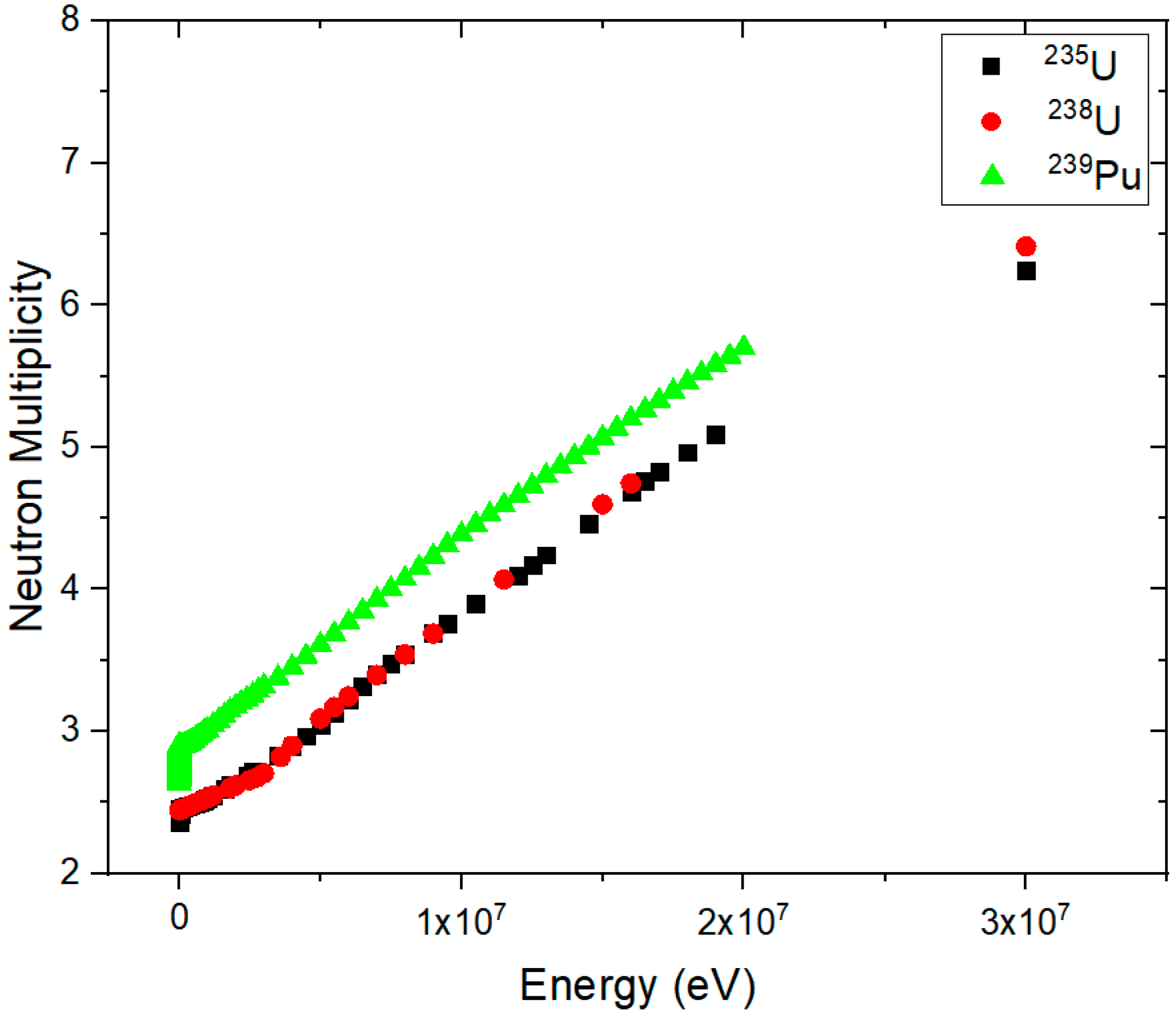
| Isotope | Neutron Number | Total Half-Life (years) | Average Spontaneous Fission Multiplicity |
|---|---|---|---|
| 242Cm | 146 | 0.447 | 2.528 |
| 249Bk | 152 | 0.877 | 3.4 |
| 252Cf | 154 | 2.645 | 3.768 |
| 248Cm | 148 | 3.84 | 3.161 |
| 240Pu | 146 | 6.56 | 2.151 |
| 238Pu | 144 | 87.7 | 2.21 |
| 238U | 143 | 4.47 × 10⁹ | 2.0 |
| 235U | 146 | 7.04 × 10⁸ | 1.87 |
| System Size Definition | Examples and Proposed Application in Literature | Detector/s | Industrial Designation |
|---|---|---|---|
| Fixed installation | Detection and localisation [62,63] | CsI(Na) | |
| Fixed installation | Detection, assessment and localisation [76] | HPGe & NaI | MISTI |
| Fixed installation | Detection, assessment and localisation [75] | CdZnTe | ORIGAMIX |
| Fixed installation/hand-held | Detection and localisation [77] | NaI | RMC |
| Fixed installation/hand-held | Detection and localisation [73] | CsI(Tl) | CARTOGAM |
| Fixed installation | Detection, assessment and localisation [78] | (GSO) | |
| Hand-held | Detection and localisation [71] | CdTe-Medpixi2 | |
| hand-held | Detection and localisation [74] | CsI(Na) | RADCAM |
| hand-held | Detection and localisation [79] | CsI(Tl) | |
| hand-held | Detection and localisation [72] | CdZnTe-Timepix | GAMPIX |
| Author, Year and Reference | Proposed Application | Collimation/Detection Technique | System Size Definition | Main Detection Materials | Approximate Intrinsic Efficiency (Thermal Neutrons/Fast Neutron 252Cf) (%) |
|---|---|---|---|---|---|
| Miller et al. (2003) [139] | Detection, assessment and localisation | Neutron scatter | Fixed installation | Plastic scintillator | NA/NA |
| Bravar et al. (2006) [140] | Detection and assessment | Neutron scatter | Fixed installation | BC-404 plastic scintillator | NA/NA |
| Vanier et al. (2007) [141] | Detection, assessment and localisation | Neutron scatter | Fixed installation | Plastic scintillator | NA/NA |
| Mascarenhas et al. (2009) [142] | Detection, assessment and localisation | Neutron scatter | Fixed installation | EJ-301 | NA/NA |
| Siegmund et al. (2009) [143] | Detection | Coded aperture and Stack of microchannel plates | Fixed installation | 10B doped microchannel plates | ~20%/NA |
| Herbach et al. (2010) [144] | Detection and assessment | Null/gamma from neutron capture | Fixed installation | BGO with Cd converter | 45%/NA |
| Ryzhikov et al. (2010) [145] | Detection and assessment | Null/gamma from neutron capture | Fixed installation | CdWO2 | 67%/42% |
| Marleau et al. (2010) [146] | Detection and assessment | Active coded aperture | Fixed installation | EJ-301 | NA/NA |
| Nakae et al. (2011) [147] | Detection and assessment | Null/Array of liquid scintillator | Fixed installation | Organic liquid scintillator (not specified) | NA/~6% (absolute) |
| Bellinger et al. (2012) [148] | Detection and assessment | Null/array of slabs | Hand-held | Si diodes with 6LiF | 6.8%/NA |
| Ide et al. (2012) [149] | Detection, assessment and localisation | Neutron scatter | Fixed scintillator | EJ-309 | NA/NA |
| Joyce et al. (2014) [150] | Detection and assessment | Null/Multiplicity assay | Fixed scintillator | EJ-309 | NA/ |
| Brennan et al. (2015) [151] | Detection, assessment and localisation | Coded aperture and Time-encoded imaging | Fixed installation | Organic liquid scintillator (not specified) | NA/NA |
| Fronk et al. (2015) [152] | Detection and assessment | Null/double sided Microstructure | Hand-held | Si diodes with 6LiF | ~29.48%/NA |
| Ianakiev et al. (2015) [153] | Detection and assessment | Null/6Li embedded in PVT | Fixed installation | 6Li and PVT | NA/NA |
| Hoshor et al. (2015) [154] | Detection and assessment | Null/array of slabs | Hand-held | Si diodes with 6LiF | ~22%/~4.5% |
| Goldsmith et al. (2016) [155] | Detection, assessment and localisation | Neutron scatter | Fixed installation | EJ-309 | NA/45% |
| Fulvio et al. (2017) [156] | Detection and assessment | Ring of multiplicity counters | Fixed installation | EJ-309 | NA/ |
| Cowles et al. (2018) [157] | Detection and assessment | Null/multiple panels | Fixed installation | LiF/ZnS | 36%/NA |
| Ochs et al. (2019) [158] | Detection | Microstructure semiconductor | Wearable device | Si diode with 6LiF | 30%/NA |
| Year | Author and Reference | Collimation | Main Detection Materials |
|---|---|---|---|
| 2004 | Aryaeinejad and Spencer [185] | None | 6Li and 7Li-loaded glass scintillators |
| 2007 | Baker et al. [186] | None | NaI(Tl) and LiI(Eu) |
| 2008 | Enqvist et al. [187] | None | Cross correlation BC-501A |
| 2009 | Runkle et al. [188] | None | NaI(Tl) and 3He |
| 2011 | Polack et al. [189] | Compton and neutron scattering | NaI(Tl) and EJ-309 |
| 2012 | Cester et al. [190] | None | LaBr(Ce), NaI(Tl), NE-213 and 3He |
| 2013 | Ayaz-Maierhafer et al. [191] | Coded aperture | CsI and EJ-309 |
| 2014 | Poitrasson-Rivière et al. [192] | Compton and neutron scattering | NaI(Tl) and EJ-309 |
| 2016 | Cester et al. [193] | Null | EJ-420, EJ-560 and EJ-299-33A |
© 2019 by the authors. Licensee MDPI, Basel, Switzerland. This article is an open access article distributed under the terms and conditions of the Creative Commons Attribution (CC BY) license (http://creativecommons.org/licenses/by/4.0/).
Share and Cite
Al Hamrashdi, H.; Monk, S.D.; Cheneler, D. Passive Gamma-Ray and Neutron Imaging Systems for National Security and Nuclear Non-Proliferation in Controlled and Uncontrolled Detection Areas: Review of Past and Current Status. Sensors 2019, 19, 2638. https://doi.org/10.3390/s19112638
Al Hamrashdi H, Monk SD, Cheneler D. Passive Gamma-Ray and Neutron Imaging Systems for National Security and Nuclear Non-Proliferation in Controlled and Uncontrolled Detection Areas: Review of Past and Current Status. Sensors. 2019; 19(11):2638. https://doi.org/10.3390/s19112638
Chicago/Turabian StyleAl Hamrashdi, Hajir, Stephen D. Monk, and David Cheneler. 2019. "Passive Gamma-Ray and Neutron Imaging Systems for National Security and Nuclear Non-Proliferation in Controlled and Uncontrolled Detection Areas: Review of Past and Current Status" Sensors 19, no. 11: 2638. https://doi.org/10.3390/s19112638
APA StyleAl Hamrashdi, H., Monk, S. D., & Cheneler, D. (2019). Passive Gamma-Ray and Neutron Imaging Systems for National Security and Nuclear Non-Proliferation in Controlled and Uncontrolled Detection Areas: Review of Past and Current Status. Sensors, 19(11), 2638. https://doi.org/10.3390/s19112638