Article

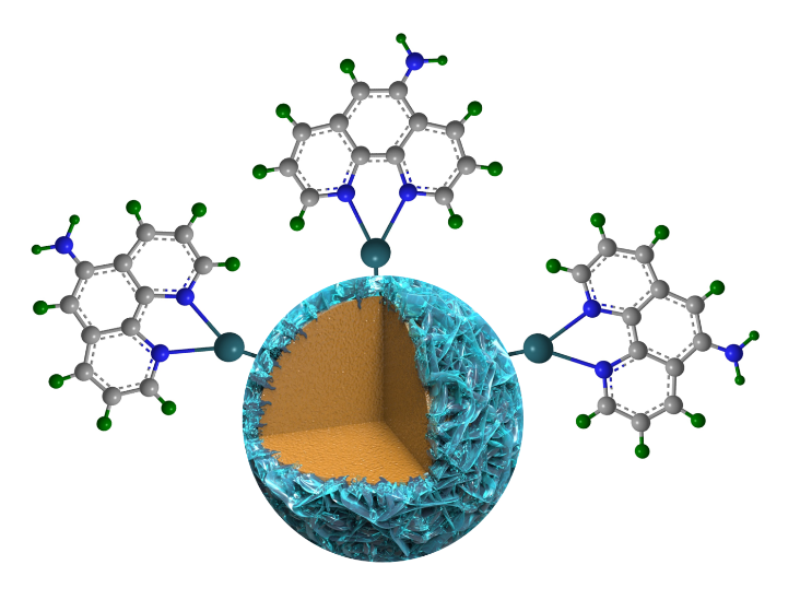
Fingerprinting of Nitroaromatic Explosives Realized by Aphen-functionalized Titanium Dioxide

Qinghai Provincial Key Laboratory of New Light Alloys, Qinghai Provincial Engineering Research Center of High Performance Light Metal Alloys and Forming, Qinghai University, Xining 810016, China
*
Author to whom correspondence should be addressed.
Sensors 2019, 19(10), 2407; https://doi.org/10.3390/s19102407
Submission received: 6 May 2019 / Revised: 20 May 2019 / Accepted: 23 May 2019 / Published: 27 May 2019
(This article belongs to the Special Issue Gas Sensors and Smart Sensing Systems)

Abstract

:
Developing sensing materials for military explosives and improvised explosive precursors is of great significance to maintaining homeland security. 5-Nitro-1,10-phenanthroline (Aphen)-modified TiO2 nanospheres are prepared though coordination interactions, which broaden the absorption band edge of TiO2 and shift it to the visible region. A sensor array based on an individual TiO2/Aphen sensor is constructed by regulating the excitation wavelength (365 nm, 450 nm, 550 nm). TiO2/Aphen shows significant response to nitroaromatic explosives since the Aphen capped on the surface of TiO2 can chemically recognize and absorb nitroaromatic explosives by the formation of the corresponding Meisenheimer complex. The photocatalytic mechanism is proved to be the primary sensing mechanism after anchoring nitroaromatic explosives to TiO2. The fingerprint patterns obtained by combining kinetics and thermodynamics validated that the single TiO2/Aphen sensor can identify at least six nitroaromatic explosives and improvised explosives within 8 s and the biggest response reaches 80%. Furthermore, the TiO2/Aphen may allow the contactless detection of various explosives, which is of great significance to maintaining homeland security.

Graphical Abstract

1. Introduction

Common explosives could be classified into military explosives (e.g., nitroaromatic explosives like 2,4,6-trinitrotoleune (TNT), 2,4-dinitrotoleune (DNT), 2,4,6-trinitrophenol (PA), etc.), and improvised explosives such as ammonium nitrate (NH4NO3), sulfur (S) and triacetone peroxide (TATP), etc. Developing sensors against military explosives or improvised explosive precursors has important significance for maintaining global security. At present, there are many methods for detecting these explosives, of which, the most traditional method is the use of sniffer dogs. However, sniffer dogs have lower working efficiency than sensors since they need a rest for half an hour after working for 20 min. In addition, existing explosive detection methods that are more mature include X-ray inspection technology, neutron analysis [1,2], fluorescence detection [3,4], electrochemical detection [5], mass spectrometry [6], ion migration method [7], and chromatography [8], etc. However, neutron analysis and X-ray detection instrument are cumbersome, expensive, and radiation hazard. Chromatographic methods are generally only suitable for laboratory sampling and testing and cannot meet the purpose of rapid detection on site. In addition, these methods are mostly contact-based detection methods. Furthermore, there are relatively few studies on detection of improvised explosives, limiting their application on the actual detection of explosives. Therefore, there is an urgent need for rapid vapor phase detection of conventional and improvised explosives with a small, low energy consumption, high sensitivity, rapid response, and contactless photoelectronic sensor.
Construction of a sensory array that is similar to an artificial olfactory system and utilizing the cross-sensitivity of different sensors towards complex component gases, and combining data analysis methods can achieve the identification of different explosive vapors [9,10]. However, the stability of the sensor array [11,12] is dependent on the synergistic effects of the different sensor units, and failure of a single unit can affect the performance of the sensor array. To this end, our previous work constructed a sensor array using a single sensor unit by illumination of different excitation wavelengths [13].
Titanium dioxide (TiO2) is a photoelectronic sensor candidate due to its chemical stability, environment friendliness and excellent optical response [14,15,16]. However, pure TiO2 can only absorb ultraviolet light due to its relatively wide band gap (3.2 eV) [17]. Moreover, the high recombination rate of photoinduced electron-hole pairs hinders the practical application of TiO2 in the field of sensors. 5-Amino-1,10-phenanthroline (Aphen) [18,19,20] is an excellent electron acceptor that can chelate with most metal ions to form stable complexes via coordination interactions. In this study, Aphen is employed as a functional ligand to modify TiO2 nanocrystals to promote the photoinduced charge carrier separation efficiency of the nanocrystals. Moreover, it is well known that the electron-rich amino group of Aphen can link with electron-deficient aromatic rings of nitroaromatic explosives to formation of Meisenheimer complexes. Thus, the Aphen-modified TiO2 nanocrystal is promising for discrimination of nitroaromatic explosives.

2. Materials and Methods

2.1. Experiment Reagents and Equipment

Tetrabutyltitanate (99%, Aladdin, Shanghai, China), concentrated sulfuric acid (AR, Wokai, Shanghai, China), hydrofluoric acid (AR, Aladdin), anhydrous ethanol (99.5%, Hushi, Shanghai, China), phenanthroline monohydrate (99%, Aladdin), and deionized water were used.
Field emission scanning electron microscopy (SEM, JSM-6610LV, JEOL, Ltd, Tokyo, Japan) and transmission electron microscopy (TEM, JEM-2100F microscope, JEOL Ltd, Tokyo, Japan) experiments were performed to observe microstructure. X-ray diffraction (XRD) patterns were measured using a D8 Advance X-ray diffractometer (Bruker Ltd, Bremen, Germany) at a scanning rate of 6° min−1 with 2θ ranging from 20° to 80°, using CuKα radiation (λ = 1.5418 Å). FTIR spectra were recorded on a Magna 560 FT-IR spectrometer (Nicolet Ltd, Green Bay, WI, USA). UV/Vis absorption spectra were recorded on a UV-2550 UV/Vis spectrometer (Shimadzu, Japan) in the range 200–800 nm. An X-ray photoelectron spectrometer (XPS, PHI5000 ESCA, Perkin Elmer, Waltham, MA, USA) equipped with an Al Kα source (1486.6 eV photons) was used to characterize the modifying of Aphen on TiO2. A Keithley 4200A-SCS parameter analyzer (Tektronix Ltd, Beaverton, OR, USA) was used to record the photocurrent of sensor chip in different explosive vapors.

2.2. Preparation of Aphen-functionalized Titanium Dioxide Nanosphere

2.2.1. Preparation of 5-amino-1,10-phenanthroline (Aphen)

Phenanthroline monohydrate (50.4 mmol) was mixed with concentrated sulfuric acid (7.5 mL), and heated before a mixture of concentrated sulfuric acid and concentrated nitric acid (1:1, 30 mL) was slowly added drop by drop. The mixture was refluxed for 3 h under 160 °C to obtain 5-nitro-1,10-phenanthroline. The synthesized 5-nitro-1,10-phenanthroline is rapidly added to an ethanol solution containing 5% Pd/C catalyst (0.01 g) and hydrazine hydrate (45 mmol) at 70 °C and refluxed for 2 h. The solution was cooled, filtered, and crystalized for subsequent experiments to obtain 5-amino-1,10-phenanthroline [21].

2.2.2. Preparation of TiO2 Nanocrastals

Tetrabutyltitanate (25 mL) and concentrated sulfuric acid (3 mL) were added to a reaction kettle and then hydrofluoric acid (0.17 mol) was added. The mixture was allowed to react at 180 °C for 24 h. After the reaction kettle has cooled to room temperature, then the product was washed by deionized water and ethanol repeatedly and dried in a vacuum drying oven.

2.2.3. Preparation of Aphen-functionalized TiO2 Nanocrystals (TiO2/Aphen)

A mass ratio of 1:50, 1:100, 1:200 of Aphen and TiO2 nanocrystals was placed in a 1 mL round-bottomed flask and vigorously shaken for 10 h at 60 °C. Then the nanocrystals were repeatedly washed with deionized water and ethanol until the wash solution does not emit light under an UV lamp. The product was vacuum-dried.

2.3. Preparation of Sensor Chips

Electrochemical deposition was used to deposit a gold interdigitated electrode with a width and interval of 3 mm at the ceramic substrate (10 mm × 9.5 mm). The gold thickness of the surface layer of the electrode was 0.5 mm. The TiO2/Aphen ethanol dispersion was spin coated on the interdigitated electrode and air-dried. The coating was aged for 12 h at a voltage of 4 V before use.

2.4. Gas Sensing Properties Testing

2,4,6-Trinitrotoluene (TNT), 2,4-dinitrotoluene (DNT), picric acid (PA), nitrate (NH4NO3), sulfur (S) and triacetone peroxide (TATP, diluted to 600 ppb by air) vapor were evaporated in advance to saturate. The photoelectric and gas sensitivity response performance of the TiO2/Aphen sensor chip to these six conventional and improvised explosives were recorded by the Keithley 4200A-SCS parameter analyzer under 365 nm, 450 nm, 550 nm monochromatic light. After the gas sensor chip was stabilized for half an hour in air under a voltage of 4 V, it was placed into a test bottle that contained saturated explosive vapor. The chip was removed after the current was stable. This process was carried out three times.

3. Results and Discussion

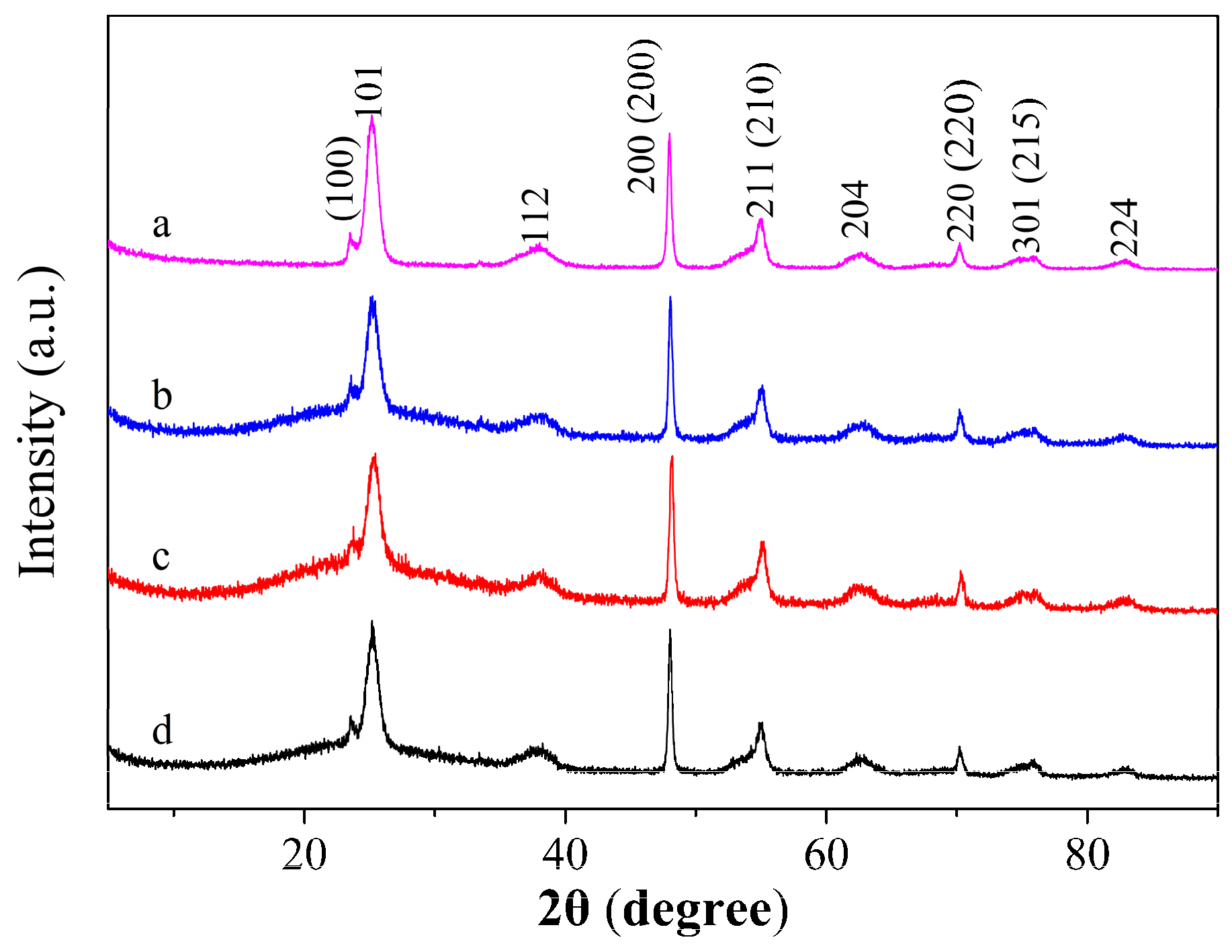
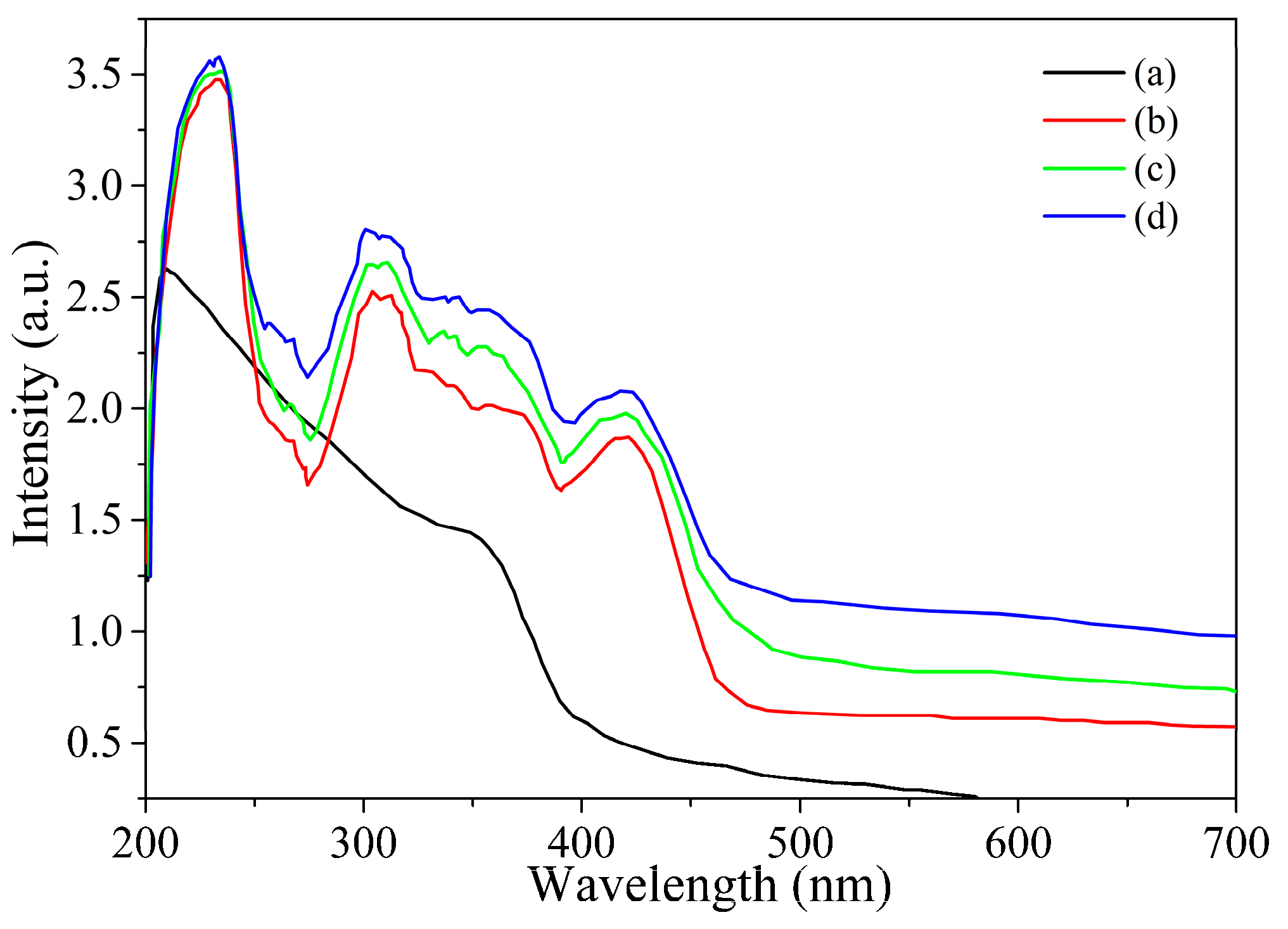
3.1. TiO2@Aphen Morphological and Structural Characterization

The TiO2 prepared by hydrothermal method was first modified by F ions and then doped with Aphen since the Aphen has high affinity to Ti ions dangling on the surface of TiO2 as depicted in Scheme 1. The TiO2 morphology was adjusted by using F ions. The tetrabutyltitanate underwent hydrolysis under acidic conditions to grow different TiO2 morphologies. As shown in Figure S1, when F ions were not present, the morphology of TiO2 appeared as aggregated nanosheet structures. When 0.03 mol of HF was added, the schistose titanium dioxide began to appear flocculent. When 0.06 mol of HF was added, the microstructure of titanium dioxide is changed to a cluster of many nanosheets. When 0.12 mol of HF was added, the morphology of TiO2 appeared as cubes with 1 µm sides.
Figure 1a shows that the litchi-like TiO2 nanospheres with streaks on the surface was regulated by F ions with a diameter ranging from 12 to 18 μm when 0.09 mol of HF was added, and these nanospheres aggregate each other. Then Aphen chelates with TiO2 nanospheres since the Aphen has high affinity to Ti ions overhung on the surface of TiO2. Obviously, Aphen modified TiO2 has favorable dispersibility as depicted in Figure 1b, which is roughly attributed to the steric effect of Aphen modified on the surface of TiO2 nanospheres. The enlarged SEM image (Figure 1b inset) clearly shows that the surface of litchi TiO2 nanosphere is a disordered TiO2 nanosheet growing vertically in the sphere. Figure 1c,d are the HRTEM diagrame of the edge of a nanosheet on a spherical shell of TiO2 and TiO2/Aphen. According to the measurement, both the lattice fringe spacings are 0.35 nm, corresponding to (101) planes of TiO2 nanocrystals. It is worth mentioning that the crystalline structure of the TiO2 remained intact after Aphen functionalization.
The crystallinity of the TiO2 nanospheres and TiO2/Aphen were confirmed by XRD. As shown in Figure 2, the XRD pattern (Line a) of TiO2 nanospheres shows sharp diffraction peaks at 25.0°, 38.2°, 47.9°, 55.1°, 62.8°, 70.2°, 75.6°, 82.9°, which are indicative of the (101), (112), (200), (211), (204), (220), (301), (224) characteristic planes of the anatase TiO2 crystal [22,23,24].
Moreover, characteristic planes of TiOF2 are also obtained at 23.6°, 48.1°, 54.2°, 70.5°, 75.2°, which are indicative planes of the (100), (200), (210), (220) and (215) [25,26,27] (PDF-2 Release 2015 RDB). It could be concluded that a small part of the precursor transformed into TiOF2 crystals after F ion modifcation and these TiOF2 dangling on the surface of TiO2 nanospheres, which supplies an adequate number of Ti ions to anchor Aphen through coordination interactions (Scheme 1). However, there are only the characteristic diffraction peaks of TiO2@TiOF2 phase when TiO2 was modified by Aphen at different ratios (Line b to d: 1:50, 1:100, 1:200), which is attributed to the very low content of the Aphen on the surface of TiO2. This result also indicates that the modification of Aphen does not change the crystal structure of TiO2@TiOF2.
Figure 3 shows the Fourier Transform infrared spectra (FTIR) of Aphen, TiO2, and TiO2/Aphen. The characteristic bands observed in as-synthesized Aphen at 3383 cm−1 are attributed to the stretching vibration peak of N=H, and 2970 cm−1, 3323 cm−1 and 3058 cm−1 bands are attributed to the stretching vibration peak of the aromatic ring skeleton. The broad characteristic band of TiO2 is observed at about 500 cm−1. Peaks at 3398 cm−1 and 3174 cm−1 are the stretching vibration peaks of -OH moieties on the surface of TiO2, which is attributed to the presence of water adsorbed on the surface of TiO2 and dangling hydroxyls on the surface of TiO2 [28,29,30,31]. When Aphen is anchored to the surface of TiO2, the obvious stretching vibration peaks of C=N on Aphen can be observed at 1468 cm−1 and 1544 cm−1. The peak of 3192 cm−1 is caused by the overlapping of the -OH vibration peak in TiO2 and the C=C vibration peak of the benzene ring in Aphen. These results indicate that the surface of TiO2 is is successfully modified with Aphen.
The Aphen-modified TiO2 was also analyzed by X-ray photoelectron spectroscopy (XPS). As shown in Figure 4, the binding energy (BE) of Ti 2p from Ti 2p1/2 and Ti 2p2/3 can be detected at 458.72 eV and 464.45 eV. The XPS spectra of N 1s in Aphen shows the BE at 339.85 eV. This is further proof for the adsorption of Aphen onto surface of the TiO2. Moreover, the XPS measurements were performed to reveal the atomic content. Based on the peak areas of Ti and N, it can be concluded that approximately 3.5% of the N on the surface of the TiO2.
UV-visible absorption spectra of TiO2/Aphen were recorded to prove the effect of Aphen on the photoelectric properties of TiO2. As shown in Figure 5, the anatase TiO2 naospheres shows a broad absorption wavelength in the ultraviolet region from 200 nm to 410 nm. Interestingly, the absorption of Aphen-modified TiO2 broaden the absorption band edge into the visible region. Therefore, the results show that the ultraviolet absorption wavelength of Aphen-functionalized TiO2 is greatly extended, which can avoid the risk of explosive explosions caused by high-energy electron beam as much as possible when detecting explosives by the photoelectric effect [32].

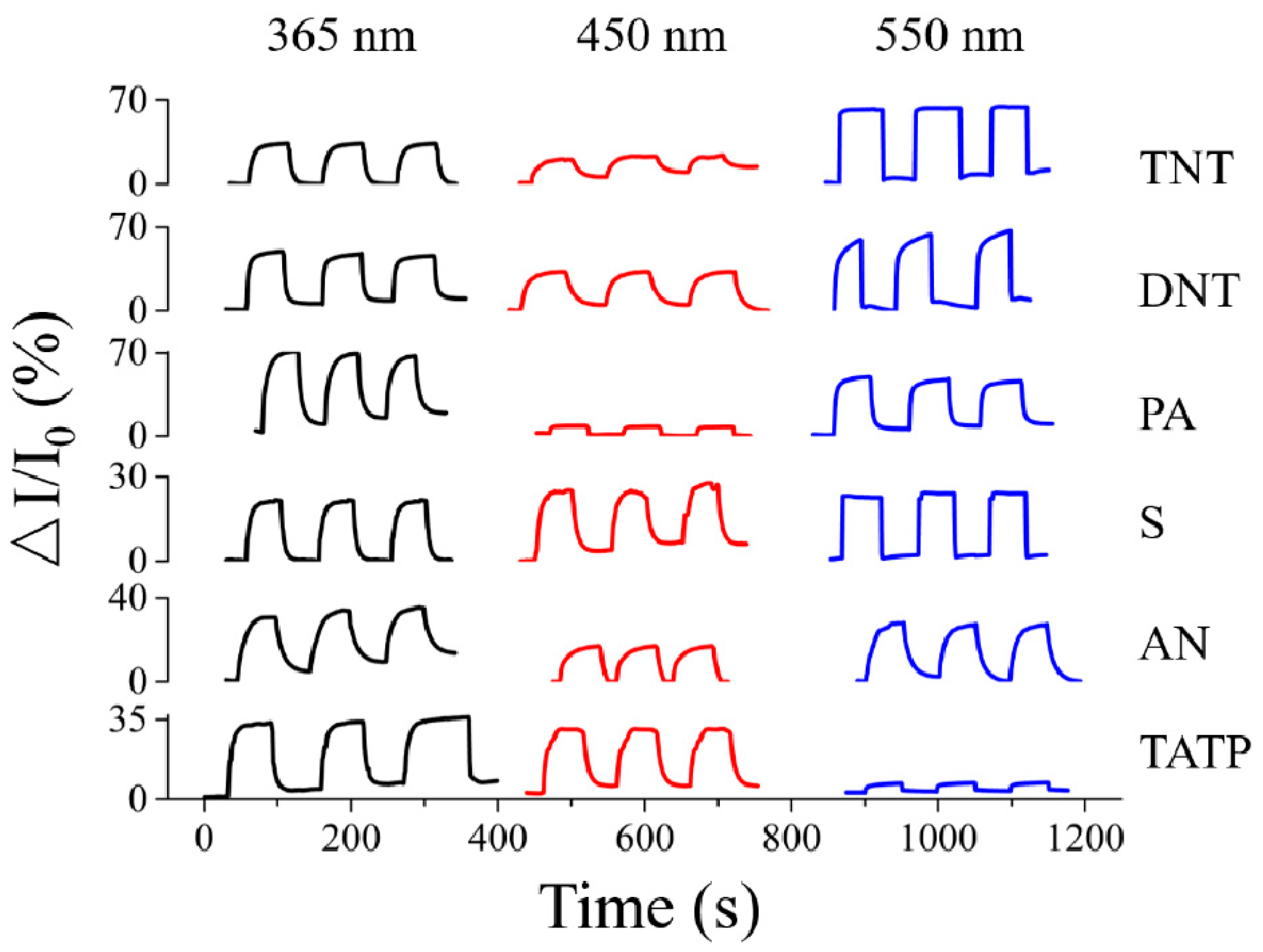
3.2. Gas Sensitivity Performance of TiO2@Aphen

As mentioned above, Aphen functionalization is an effective strategy to enhance the light absorption range of TiO2. Thus, the sensor array can be constructed by only one TiO2/Aphen chip used under various illumination wavelengths. To demonstrate this concept, the sensing properties of the TiO2/Aphen to TNT, DNT, PA, S, AN and TATP were evaluated under 365 nm, 450 nm and 550 nm as depicted in Figure 6. The response (defined as (It − I0)/I0) of TiO2/Aphen to TNT, DNT, PA, S, AN and TATP vapor were 33.8%, 43.5%, 61.5%, 21.2%, 30.5% and 29% under 365 nm illumination, 6.5%, 27.4%, 8%, 21.1%, 17.1%, and 24.5% under 450 nm illumination, and 58.1%, 58.5%, 81.1%, 21.9%, 25.5% and 3.6% under 550 nm illumination, respectively.
Thus, the TiO2/Aphen shows distinctive responses to the tested explosives indicating that tailoring the excitation wavelength has an obvious effect on the sensing performance of TiO2/Aphen. For comparison, the sensing performance of the pure TiO2 to explosives’ vapor was characterized under the same conditions. As shown in Figure S2, the response of pure TiO2 to TNT, DNT, PA, S, AN and TATP vapor were 15%, 22.3%, 32.5%, 11.6%, 7.9% and 4% under 365 nm illumination. Interestingly, the pure TiO2 has almost no sensitivity to these explosives under illuminations of 450 nm and 550 nm.
Therefore, by spinning coated TiO2/Aphen on the interdigitated electrode, a sensory array can be realized by tailoring the illumination wavelength used (Figure 7a). To gain a comprehensive understanding of the overall sensing performance of the sensory array, the sensing responses, response times and recovery times are statistically analyzed in Figure 7b–d. As shown in Figure 7b, the responses of the TiO2/Aphen to nitroaromatic explosives (TNT, DNT, PA) are 33.8%, 43.5%, and 69.5% under 365 nm illumination, and 58.1%, 58.5%, and 61.1% under 550 nm illumination, respectively. However, the responses of the TiO2/Aphen to improvised explosives (S, AN and TATP) are 21.2%, 30.5%, and 29% under 365 nm illumination, and 23.9%, 25.5%, and 36% under 550 nm illumination, respectively. Additionally, as depicted in Figure 6c, the response times of TiO2/Aphen to nitroaromatic explosives and improvised explosives are all within 10 s under the illuminations of 365 nm, 450 nm and 550 nm. The response time of TiO2/Aphen to TNT, DNT, PA, S, AN and TATP vapor were 4.04 s, 2.8 s, 4.83 s, 4.83 s, 6.2 s and 4.5 s under 365 nm illumination, 7.4 s, 6 s, 5.1 s, 4.5 s, 4.8 s, and 3.8 s under 450 nm illumination, and 0.5 s, 2.1 s, 3 s, 0.7 s, 8.4 s and 0.9 s under 550 nm illumination, respectively. TNT requires the longest recovery time (about 22 s) under 450 nm illumination, while DNT shows the shortest recover time (about 1 s) under 550 nm illumination. However, as shown in Figure S3 the response time and recover time of pure TiO2 to different explosives are more than 5 s under 365 nm illumination.
The above experiments reveal that TiO2/Aphen has better sensing properties to nitroaromatic explosives than improvised explosives. This phenomenon may be attributed to the formation of a Meisenheimer complex between the electron-rich amino groups of Aphen and the electron-deficient aromatic rings of nitroaromatic explosives, coupled with strong charge transfer transitions. Thus, nitroaromatic explosives can anchor with TiO2 via Aphen bridge bonds and affected markedly the photocurrent of TiO2/Aphen. In addition, the photocatalytic mechanism may be the primary sensing mechanism after nitroaromatic explosives anchor to TiO2. To verify this hypothesis, the catalytic activity measurements of the TiO2/Aphen and pure TiO2 were based on the catalytic reduction of 4-nitrophenol aqueous solution as a substitute for TNT, DNT and PA vapors. As shown in Figure 8a, the absorption peak at 400 nm of 4-nitrophenol immediately decreased in presence of TiO2/Aphen under 550 nm illumination, owing to the photocatalytic reduction of 4-nitrophenolate to 4-aminophenol. The reduction reaction finished in 210 s with a reduction rate constant k = −0.004 s−1 which is determined from the plot of C/C0 vs. time. However, with addition of the pure TiO2, the absorption peak at 400 nm of 4-nitrophenol remains undiminished under 550 nm illumination for 28 min, attributed to the lake of visible light absorption of unmodified catalyst TiO2. Thus it can be seen that Aphen plays a key role in broadening the absorption band edge of TiO2 to the visible light region.
Generally, with a single sensor it is difficult to meet the actual needs of qualitative identification of different explosives. In other words, a single sensor can respond to different explosive vapors but cannot discriminate them from each other. Stated thus, the TiO2/Aphen shows a distinctive response, response time and recovery time to the tested explosives at different excitation wavelengths. Utilizing the cross-sensitivity and combining data analysis methods can imitate an artificial olfactory system and achieve the identification of different explosive vapors. Here, a sensor array based on the above single optoelectronic TiO2/Aphen sensor was constructed by regulating the excitation wavelength used. The response value at different response time was chosen as the eigenvalue. The fingerprint patterns were constructed by extracting every 1 s and 16 eigenvalues within 16 s in total (method A), or every 1 s and 8 eigenvalues within 8 s in total (method B) or every 2 s and 8 eigenvalues within 16 s in total (method C). As a result, the response to each kind of explosive at three wavelengths is different by method A and method C. However, method A process more data than method C. Thus, the response of the sensor is sampled every 2 seconds. Then these eigenvalues were grouped together to construct fingerprint patterns (Figure 9) of each explosive. It is found that each explosive has a unique fingerprint pattern, just like the loops and whorls on a human’s finger. The fingerprint pattern of TNT, for example, shows three whorls which are obtained from the response value at 2 s to 16 s under 365 nm (black line), 450 nm (red line) and 550 nm (green line). The whorls group of TNT is quite different from the that of DNT, PA and other improvised explosives, and so are those of DNT, PA, AN, S and TATP. Thus, one can identify explosives through their fingerprint patterns. Based on the fingerprint patterns, it promises to be a simple method to quickly identify these explosives from each other by machine identification.
The role of the moisture is also addressed in Figure S4. The response ratios of TNT are almost unchanged at 40% and 60 % of relative humidity (RH) and under the illumination of 365 nm. However, the response ratio of TNT decreased at 80% RH due to competitive adsorption of TNT and moisture on the surface of TiO2/Aphen. This result illustrates that the sensing response cannot be affected by ambient air below the 60% RH. These results have been added in the revised Supporting Information.

4. Conclusions

In conclusion, litchi-like TiO2 nanospheres with streaks on the surface were prepared by modifying the F ion content. Then Aphen-functionalized TiO2 nanospheres was successfully constructed by coordination interactions between Aphen and Ti ions on the surface of TiO2. Aphen plays a key role in broadening the absorption band edge of TiO2 into the visible light region. Thus a sensor array composed by only TiO2/Aphen sensors can be constructed by regulating the excitation wavelength in the visible region, which is significant for a low energy consumption, high selectivity and rapid response photoelectronic sensor. Moreover, a photocatalytic mechanism was proved the primary sensing mechanism of nitroaromatic explosives after nitroaromatic explosives anchor onto TiO2 by formation of a Meisenheimer complex. This work shines light on the realization of an artificial olfactory system to achieve the identification of different explosive vapors. The results demonstrate promising potential for low-cost practical applications as optoelectronic sensors for nitroaromatic explosive landmine detection and security checks. Undoubtedly, this approach also provide reference for the development of novel photocatalysts under visible light.

Supplementary Materials

The following are available online at https://www.mdpi.com/1424-8220/19/10/2407/s1, Figure S1: SEM images of TiO2 regulated by (a) 0 mol, (b) 0.03 mol, (c) 0.06 mol, and (d) 0.12 mol of F ions. Figure S2: Response of pure TiO2 to TNT (a), DNT (b), PA (c), S (d), AN (e) and TATP (f) under 365 nm illumination. Figure S3: Response time and recover time of pure TiO2 to different explosives under 365 nm illumination. Figure S4: Effect of the humidity of TiO2/Aphen to TNT under the illumination of 365 nm.

Author Contributions

Conceptualization, B.L. and G.X.; methodology, G.X.; validation, G.X.; formal analysis, G.X.; investigation, G.X.; resources, B.L.; data curation, G.X.; writing—original draft preparation, G.X.; writing—review and editing, B.L.; visualization, G.X.; supervision, B.L.; project administration, B.L.; funding acquisition, B.L.

Funding

This research was funded by the Natural Science Foundation of Qinghai Province (2017-ZJ-924Q), National Natural Science Foundation of China (21804078) and Thousand Talents Program of Qinghai Province.

Conflicts of Interest

The authors declare no conflicts of interest.

References

  1. Brown, K.E.; Greenfield, M.T.; McGrane, S.D.; Moore, D.S. Advances in explosives analysis---part II: Photon and neutron methods. Anal. Bioanal. Chem. 2016, 408, 49–65. [Google Scholar] [CrossRef]
  2. Whetstone, Z.D.; Kearfott, K.J. A review of conventional explosives detection using active neutron interrogation. J. Radioanal. Nucl. Chem. 2014, 301, 629–639. [Google Scholar] [CrossRef]
  3. Sun, X.; Wang, Y.; Lei, Y. Fluorescence based explosive detection: from mechanisms to sensory materials. Chem. Soc. Rev. 2015, 44, 8019–8061. [Google Scholar] [CrossRef] [Green Version]
  4. Zhang, S.-R.; Du, D.-Y.; Qin, J.-S.; Bao, S.-J.; Li, S.-L.; He, W.-W.; Lan, Y.-Q.; Shen, P.; Su, Z.-M. A Fluorescent Sensor for Highly Selective Detection of Nitroaromatic Explosives Based on a 2D, Extremely Stable, Metal–Organic Framework. Chem. A Eur. J. 2014, 20, 3589–3594. [Google Scholar] [CrossRef] [PubMed]
  5. Zhang, L.; Han, Y.; Zhu, J.; Zhai, Y.; Dong, S. Simple and Sensitive Fluorescent and Electrochemical Trinitrotoluene Sensors Based on Aqueous Carbon Dots. Anal. Chem. 2015, 87, 2033–2036. [Google Scholar] [CrossRef] [PubMed]
  6. Sisco, E.; Forbes, T.P. Rapid detection of sugar alcohol precursors and corresponding nitrate ester explosives using direct analysis in real time mass spectrometry. Analyst 2015, 140, 2785–2796. [Google Scholar] [CrossRef] [PubMed]
  7. Mäkinen, M.; Nousiainen, M.; Sillanpää, M. Ion spectrometric detection technologies for ultra-traces of explosives: A review. Mass Spectrom. Rev. 2011, 30, 940–973. [Google Scholar] [CrossRef]
  8. Schramm, S.; Léonço, D.; Hubert, C.; Tabet, J.-C.; Bridoux, M. Development and validation of an isotope dilution ultra-high performance liquid chromatography tandem mass spectrometry method for the reliable quantification of 1,3,5-Triamino-2,4,6-trinitrobenzene (TATB) and 14 other explosives and their degradation products in environmental water samples. Talanta 2015, 143, 271–278. [Google Scholar]
  9. Schnorr, J.M.; van der Zwaag, D.; Walish, J.J.; Weizmann, Y.; Swager, T.M. Sensory Arrays of Covalently Functionalized Single-Walled Carbon Nanotubes for Explosive Detection. Adv. Funct. Mater. 2013, 23, 5285–5291. [Google Scholar] [CrossRef] [Green Version]
  10. Zheng, Y.; Xincun, D.; Shengli, Z.; Linjuan, G.; Baiyi, Z.; Zhaofeng, W.; Haibo, Z. A High-Performance Nitro-Explosives Schottky Sensor Boosted by Interface Modulation. Adv. Funct. Mater. 2015, 25, 4039–4048. [Google Scholar]
  11. Qu, J.; Ge, Y.; Zu, B.; Li, Y.; Dou, X. Transition-Metal-Doped p-Type ZnO Nanoparticle-Based Sensory Array for Instant Discrimination of Explosive Vapors. Small 2016, 12, 1369–1377. [Google Scholar] [CrossRef]
  12. Zhao, J.; Li, N.; Yu, H.; Wei, Z.; Liao, M.; Chen, P.; Wang, S.; Shi, D.; Sun, Q.; Zhang, G. Highly Sensitive MoS2 Humidity Sensors Array for Noncontact Sensation. Adv. Mater. 2017, 29, 1702076. [Google Scholar] [CrossRef]
  13. Lü, X.; Hao, P.; Xie, G.; Duan, J.; Gao, L.; Liu, B. A Sensor Array Realized by a Single Flexible TiO2/POMs Film to Contactless Detection of Triacetone Triperoxide. Sensors 2019, 19, 915. [Google Scholar] [CrossRef]
  14. Wang, S.; Guan, B.Y.; Yu, L.; Lou, X.W. (David) Rational Design of Three-Layered TiO2@Carbon@MoS2 Hierarchical Nanotubes for Enhanced Lithium Storage. Adv. Mater. 2017, 29, 1702724. [Google Scholar] [CrossRef]
  15. Low, J.; Cheng, B.; Yu, J. Surface modification and enhanced photocatalytic CO2 reduction performance of TiO2: A review. Appl. Surf. Sci. 2017, 392, 658–686. [Google Scholar] [CrossRef]
  16. Chen, X.; Liu, L.; Huang, F. Correction: Black titanium dioxide (TiO2) nanomaterials. Chem. Soc. Rev. 2015, 44, 1861–1885. [Google Scholar] [CrossRef] [PubMed]
  17. Yan, X.; Xu, Y.; Tian, B.; Lei, J.; Zhang, J.; Wang, L. Operando SERS self-monitoring photocatalytic oxidation of aminophenol on TiO2 semiconductor. Appl. Catal. B Environ. 2018, 224, 305–309. [Google Scholar] [CrossRef]
  18. Yoshihara, T.; Murayama, S.; Masuda, T.; Kikuchi, T.; Yoshida, K.; Hosaka, M.; Tobita, S. Mitochondria-targeted oxygen probes based on cationic iridium complexes with a 5-amino-1, 10-phenanthroline ligand. J. Photochem. Photobiol. A Chem. 2015, 299, 172–182. [Google Scholar] [CrossRef] [Green Version]
  19. Shervedani, R.K.; Foroushani, M.S.; Dehaghi, S.B. Functionalization of gold mercaptopropionic acid self-assembled monolayer with 5-amino-1,10-phenanthroline: Interaction with iron(II) and application for selective recognition of guanine. Electrochim. Acta 2015, 164, 344–352. [Google Scholar] [CrossRef]
  20. Feist, B.; Pilch, M.; Nycz, J. Graphene oxide chemically modified with 5-amino-1,10-phenanthroline as sorbent for separation and preconcentration of trace amount of lead(II). Microchim. Acta 2019, 186, 91. [Google Scholar] [CrossRef]
  21. Smith, G.F.; Cagle, F.W. The improved synthesis of 5-nitro-1, 10-phenanthroline. J. Org. Chem. 1947, 12, 781–784. [Google Scholar] [CrossRef]
  22. Meng, A.; Zhang, J.; Xu, D.; Cheng, B.; Yu, J. Enhanced photocatalytic H2-production activity of anatase TiO2 nanosheet by selectively depositing dual-cocatalysts on {101} and {001} facets. Appl. Catal. B Environ. 2016, 198, 286–294. [Google Scholar] [CrossRef]
  23. Liu, X.; Dong, G.; Li, S.; Lu, G.; Bi, Y. Direct Observation of Charge Separation on Anatase TiO2 Crystals with Selectively Etched {001} Facets. J. Am. Chem. Soc. 2016, 138, 2917–2920. [Google Scholar] [CrossRef] [PubMed]
  24. Yuan, Y.-J.; Ye, Z.-J.; Lu, H.-W.; Hu, B.; Li, Y.-H.; Chen, D.-Q.; Zhong, J.-S.; Yu, Z.-T.; Zou, Z.-G. Constructing Anatase TiO2 Nanosheets with Exposed (001) Facets/Layered MoS2 Two-Dimensional Nanojunctions for Enhanced Solar Hydrogen Generation. ACS Catal. 2016, 6, 532–541. [Google Scholar] [CrossRef]
  25. Gnedenkov, S.V.; Opra, D.P.; Sinebryukhov, S.L.; Kuryavyi, V.G.; Ustinov, А.Y.; Sergienko, V.I. Structural and electrochemical investigation of nanostructured C: TiO2–TiOF2 composite synthesized in plasma by an original method of pulsed high-voltage discharge. J. Alloys Compd. 2015, 621, 364–370. [Google Scholar] [CrossRef]
  26. Zhao, X.; Wei, G.; Liu, J.; Wang, Z.; An, C.; Zhang, J. Synthesis of heterostructured Pd@TiO2/TiOF2 nanohybrids with enhanced photocatalytic performance. Mater. Res. Bull. 2016, 80, 337–343. [Google Scholar] [CrossRef]
  27. Jung, M.-J.; Kim, Y.; Lee, Y.-S. Enhancement of the electrochemical capacitance of TiOF2 obtained via control of the crystal structure. J. Ind. Eng. Chem. 2017, 47, 187–193. [Google Scholar] [CrossRef]
  28. Gao, J.; Lu, C.; Lu, X. APhen-functionalized nanoparticles—Polymer fluorescent nanocomposites via ligand exchange and in situ bulk polymerization. J. Mater. Chem. 2007, 17, 4591–4597. [Google Scholar] [CrossRef]
  29. Du, Y.; Lu, C.; Su, Z.; Gao, J.; Fu, Y.; Lu, X. Synthesis and properties of transparent luminescent nanocomposites with surface functionalized semiconductor nanocrystals. J. Solid State Chem. 2008, 181, 2279–2284. [Google Scholar]
  30. Govindaiah, P.; Lee, J.M.; Jung, Y.J.; Lee, S.J.; Kim, J.H. Tunable fluorescent poly (styrene-co-methacrylic acid)/Au-Aphen hybrid nanoparticles via surface immobilization. J. Mater. Chem 2009, 19, 3529–3537. [Google Scholar] [CrossRef]
  31. Isaacs, M.; Sykes, A.G.; Ronco, S. Synthesis, characterization and photophysical properties of mixed ligand tris (polypyridyl) chromium (III) complexes, [Cr(phen)2L]3+. Inorganica Chim. Acta 2006, 359, 3847–3854. [Google Scholar] [CrossRef]
  32. Yang, Y.; Yin, L.-C.; Gong, Y.; Niu, P.; Wang, J.-Q.; Gu, L.; Chen, X.; Liu, G.; Wang, L.; Cheng, H.-M. An Unusual Strong Visible-Light Absorption Band in Red Anatase TiO2 Photocatalyst Induced by Atomic Hydrogen-Occupied Oxygen Vacancies. Adv. Mater. 2018, 30, 1704479. [Google Scholar] [CrossRef] [PubMed]
Scheme 1. Schematic illustration for the Aphen-functionalized TiO2 nanospheres through coordination interactions between Aphen and Ti ions dangling on the surface of TiO2.
Sensors 19 02407 sch001
Figure 1. SEM images (a,b) and HRTEM (c and d) images of TiO2 nanospheres before (a,c) and after (b,d) Aphen modifying. Inset: SEM images of single Aphen modified TiO2 nanocrystals.
Sensors 19 02407 g001
Figure 2. XRD pattern of TiO2 nanospheres before (a) and after modifying Aphen with Aphen/ TiO2 ratio of 1:50 (b); 1:100 (c) and 1:200 (d).
Sensors 19 02407 g002
Figure 3. FTIR spectra of Aphen, TiO2, and TiO2/Aphen (1:200).
Sensors 19 02407 g003
Figure 4. XPS spectra of Ti (a) and N (b) of TiO2/Aphen.
Sensors 19 02407 g004
Figure 5. UV-vis absorption of TiO2 nanospheres before (a) and after modifying Aphen with Aphen/ TiO2 ratio of 1:50 (b); 1:100 (c) and 1:200 (d).
Sensors 19 02407 g005
Figure 6. Response of TiO2/Aphen to different explosives under 365 nm, 450 nm and 550 nm illumination.
Sensors 19 02407 g006
Figure 7. (a) Schematic diagram of sensor array realized by single TiO2/Aphen chip under various illumination wavelengths. Response (b), response time (c) and recover time (d) of TiO2/Aphen to different explosives under 365 nm, 450 nm and 550 nm illumination.
Sensors 19 02407 g007
Figure 8. Successive UV-vis absorption spectra of the NaBH4 reduction of 4-nitrophenol catalyzed by TiO2/Aphen (a) and pure TiO2 (b).
Sensors 19 02407 g008
Figure 9. Fingerprint patterns collection of (a) TNT; (b) DNT; (c) PA; (d) S; (e) AN and (f) TATP by the response value at corresponding response time under 365 nm (black line), 450 nm (red line) and 550 nm (green line).
Sensors 19 02407 g009

