Abstract
A new colorimetric detection of methylmercury (CH3Hg+) was developed, which was based on the surface deposition of Hg enhancing the catalytic activity of gold nanoparticles (AuNPs). The AuNPs were functionalized with a specific DNA strand (HT7) recognizing CH3Hg+, which was used to capture and separate CH3Hg+ by centrifugation. It was found that the CH3Hg+ reduction resulted in the deposition of Hg onto the surface of AuNPs. As a result, the catalytic activity of the AuNPs toward the chromogenic reaction of 3,3,5,5-tetramethylbenzidine (TMB)-H2O2 was remarkably enhanced. Under optimal conditions, a limit of detection of 5.0 nM was obtained for CH3Hg+ with a linear range of 10–200 nM. We demonstrated that the colorimetric method was fairly simple with a low cost and can be conveniently applied to CH3Hg+ detection in environmental samples.
1. Introduction
Mercuric ions widely exist in the environment and have distinct toxic effects on human beings. Organic forms of mercury (Hg) demonstrate much higher toxicity than inorganic Hg due to their higher lipophilicity and easier bioaccumulation through the food chain, such as in the tissue of fish [1,2]. The main organic species of mercury, methylmercury (CH3Hg+), has been recognized as a potent neurotoxin that causes damage to the brain and nervous system [1,3]. Due to the severe effects of mercury, the U.S. Environmental Protection Agency has set a maximum level (10 nM, 2 ppb) for mercury species in drinking water [1]. Usually, complex hyphenated techniques, such as high performance liquid chromatography (HPLC) or gas chromatography (GC), coupled to specific detectors, such as mass spectrometry (MS), inductively coupled plasma mass spectrometry (ICP-MS) or atomic fluorescence spectrometry (AFS), are required for methylmercury detection [4,5,6]. However, these techniques generally require expensive instruments and are time-consuming and costly. To overcome the limitation of the above methods, recently, nanomaterial-based assays have been widely used for developing rapid and cost-effective methods for the detection of various heavy metal ions in environmental and biological samples [7,8,9]. Due to the strong metallophilic interactions between Hg2+ and some other metallic atoms, such as gold and silver, numerous metallic nanoparticle-based assays for Hg2+ have been developed [10,11,12,13,14,15,16]. However, there are fewer nanomaterial-based assays for CH3Hg+ compared to Hg2+ ions [17,18,19], which is probably due to the weak interactions between CH3Hg+ and metal nanomaterials.
Only a few studies have reported the development of nanomaterial-based detection methods for CH3Hg+. For example, Chen at el. developed a colorimetric nanosensor for mercury speciation, which was based on the analyte-induced aggregation of gold nanoparticles (Au NPs) with the assistance of a thiol-containing ligand of diethyldithiocarbamate (DDTC) [18]. Pandeeswar et al. presented a novel optoelectronic approach for detection of Hg2+ and CH3Hg+, which was based on nanoarchitectonics that consists of an adenine (A)-conjugated small organic semiconductor (BNA) and deoxyribo-oligothymidine (dTn) [20]. However, this device cannot distinguish Hg2+ from CH3Hg+. Recently, Deng et al. reported that a DNA strand, HT7, can bind to CH3Hg+ with a higher Kb value of (5.57 ± 0.47) × 106 M−1 compared to that of Hg2+ ((1.51 ± 0.18) × 106 M−1) [19]. Based on this, they were able to discriminate between CH3Hg+ and Hg2+ ions by forming Ag/Hg amalgam with a CH3Hg+-specific fluorophore-labeled DNA probe and fabricated a highly selective fluorescent assay for CH3Hg+. More recently, Yang et al. designed a specific visual detection method for CH3Hg+ and ethylmercury based on DNA-templated alloy Ag/Au NPs [21]. However, this visual detection method for methylmercury and ethylmercury requires subtle temperature adjustments and its sensitivity was above the micromolar level. Thus, developing a simple and selective colorimetric assay for CH3Hg+ is still an important and difficult task.
Recently, some methods for the detection of Hg2+ were reported, which were based on the peroxidase-like activity of the AuHg alloy NPs. For example, Long et al. [22] found that AuNPs possess excellent peroxidase-like activity after the deposition of Hg2+ onto the surface of AuNPs. The peroxidase-like activity enhancement of AuNPs, after Hg0 deposition onto the surface of AuNPs, was suggested to be the result of the accelerated decomposition of H2O2 and the stabilization of hydroxyl radicals on the surface of AuNPs. This phenomenon can be applied in the development of colorimetric and fluorescent assays for Hg2+ [22,23,24]. Our group also reported that catalytic DNA-AuNPs and DNA-Ag/Pt nanoclusters can be used to detect Hg2+ with high selectivity and sensitivity by stimulating or inhibiting their peroxidase-like activity [25,26]. Interestingly, compared with citrate stabilized AuNPs, AuNPs functionalized with a T-rich DNA strand can obviously improve the selectivity and can simplify the sample pretreatment for the colorimetric detection of Hg2+ [25]. However, to the best of our knowledge, there is no report on the application of nanomaterial enzyme mimics in CH3Hg+ detection. Herein, we found that CH3Hg+ captured by the AuNPs functionalized with CH3Hg+-specific DNA strands can be reduced by NaBH4. This results in Hg deposition onto the surface of AuNPs, thus stimulating the peroxidase-like activity of the AuNPs. Based on this finding, a highly sensitive and selective colorimetric assay for CH3Hg+ was developed.
2. Materials and Methods
2.1. Chemicals and Materials
HAuCl4, CH3Hg+Cl, NaBH4, 3,3,5,5-Tetramethylbenzidine (TMB) and H2O2 (30%) were purchased from Aladdin Reagent (Shanghai, China). The single-strand oligonucleotides were obtained from Sangon Biotech (Shanghai, China) and the sequences of these DNA strands were listed in Table 1. Hg(NO3)2 and all the other metal salts were purchased from the National Institute of Metrology (Beijing, China). All of the reagents used were of analytical grade. Ultra-pure water prepared with a Milli-Q Pure system was used for all experiments.
Table 1.
Oligonucleotide Sequences Used in This Work a.
2.2. Synthesis of AuNPs and the Modification by DNA Strands
The AuNPs were prepared through the citrate-mediated reduction of HAuCl4 [24]. Briefly, HAuCl4 (0.01%, 100 mL) was added to a flask, which had been washed with aqua regia and ultra-pure water. After the solution was heated to boiling, sodium citrate (1.0%, 2.0 mL) was quickly added with stirring. When we observed a color change in the mixture to wine red, the mixture was further boiled for another 5 min and cooled to room temperature. The diameter of AuNPs was about 15 nM and their concentration was estimated to be 3 nM.
The DNA modification of the AuNPs was achieved by directly incubating thiolated single-strand DNA (HT7) with the AuNPs. Briefly, the AuNPs (3 nM, 990 μL) and thiolated DNA (100 μM, 5 μL) were mixed together and incubated at an ambient temperature for 24 h. After this, the mixture was centrifuged for 15 min at 10,000× g rpm and excessive DNA strands were removed. After repeating the centrifugation once, the obtained DNA-AuNPs complex was resuspended in phosphate buffer (10 mM, pH of 7.0) and stored at 4 °C.
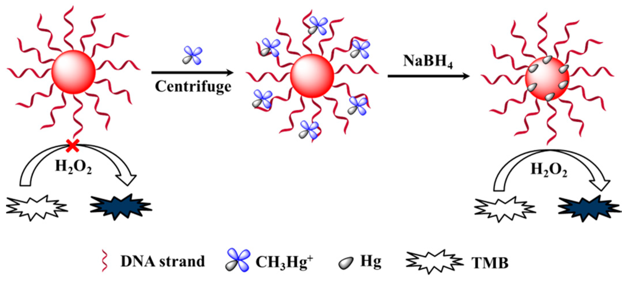
2.3. Colorimetric Detection of CH3Hg+
To 25 μL of DNA-AuNPs complex (0.6 nM), 175 μL of Tris-HNO3 buffer (5.0 mM, pH 7.0) and 500 μL of CH3Hg+ solution at different concentrations were added. After being incubated for 10 min, the mixtures were centrifuged at 10,000 rpm for 15 min and the supernatants were discarded. To the 50 μL of retained mixture, we added 50 μL of NaBH4 (1.0 mM). After being incubated for another 10 min, 90 μL of citrate buffer (100 mM, pH 4.5), 100 μL of TMB (1.5 mM) and 60 μL of H2O2 (1.5 M) were transferred into the solution. The catalytic reaction was subsequently recorded at 650 nm by a microplate reader (PowerWave XS2, Bio-Tek, Winooski, VT, USA) after 10 min. For detection of CH3Hg+ in lake water, the samples were filtered through microfiltration membranes and measured by the above method.
3. Results and Discussion
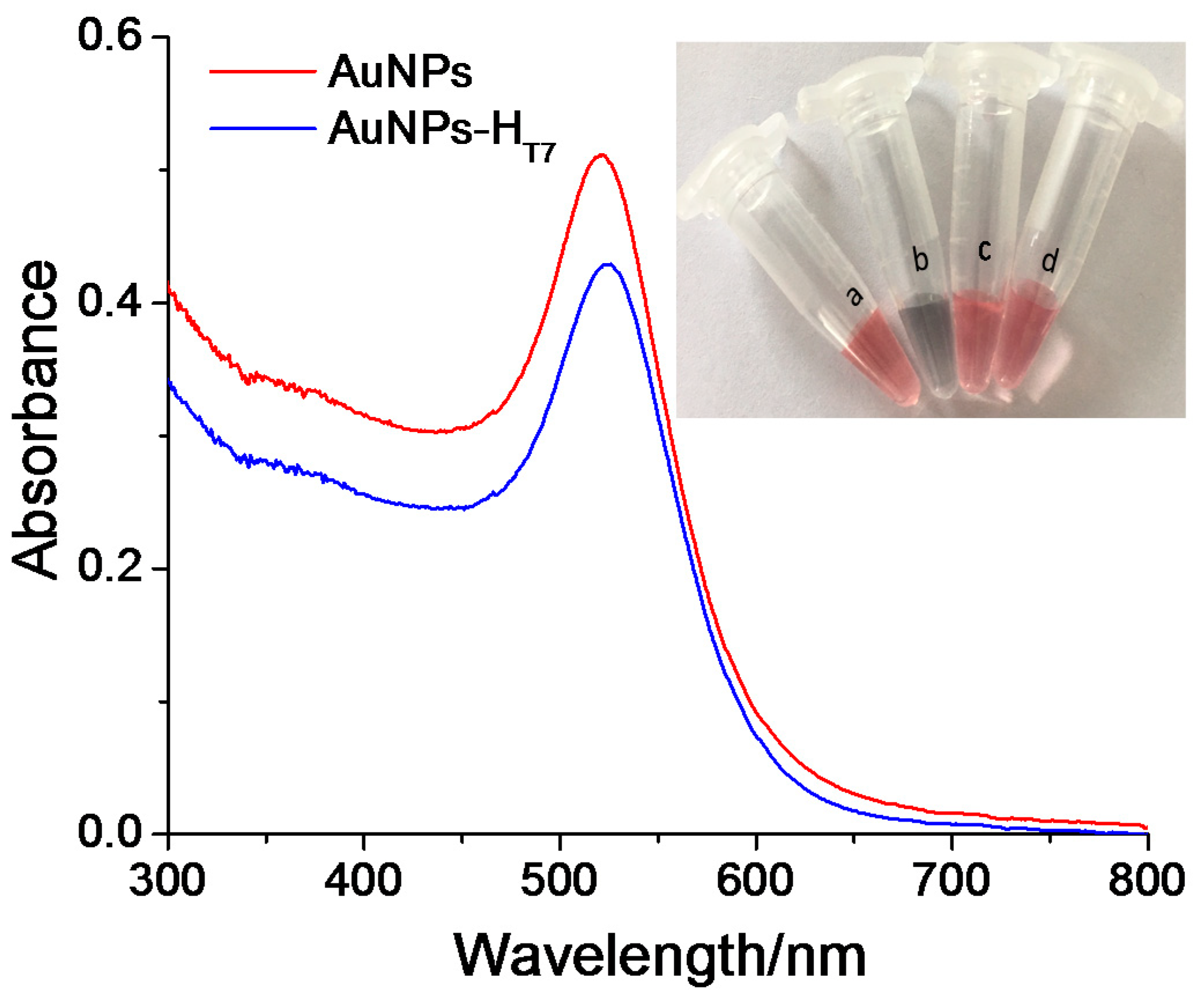
3.1. Characterization of AuNPs and DNA-AuNPs Complex
Figure 1 shows that the UV–vis absorption spectra of the AuNPs has a maximum absorption peak (λmax) at 520 nm. After the AuNPs were modified with HT7, which is a CH3Hg+ recognition DNA strand, the λmax of the AuNPs experienced a red shift to 522 nm. This result suggested that the DNA-AuNPs complex (HT7-AuNPs) was obtained. The HT7-AuNPs complex was stable in 0.15 M NaCl (the inset in Figure 1), which also indicated the successful preparation of the HT7-AuNPs.
Figure 1.
UV–vis spectra of AuNPs and AuNPs-HT7. The inset shows the photographs of (a) AuNPs, (b) AuNPs with 0.15 mol/L NaCl, (c) AuNPs-HT7 and (d) AuNPs-HT7 with 0.15 mol/L NaCl.
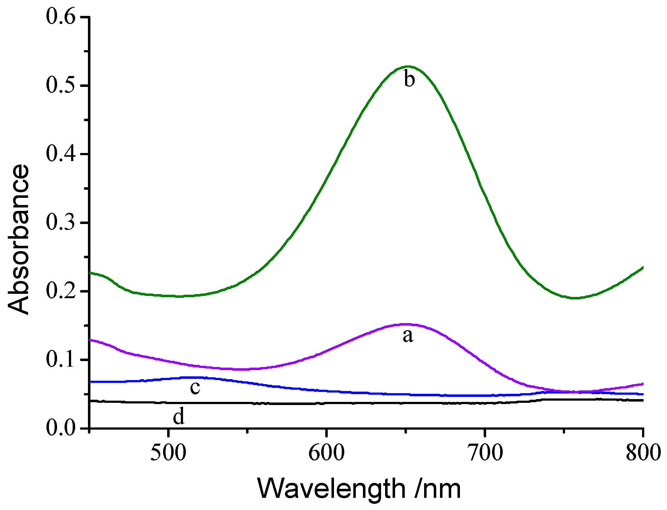
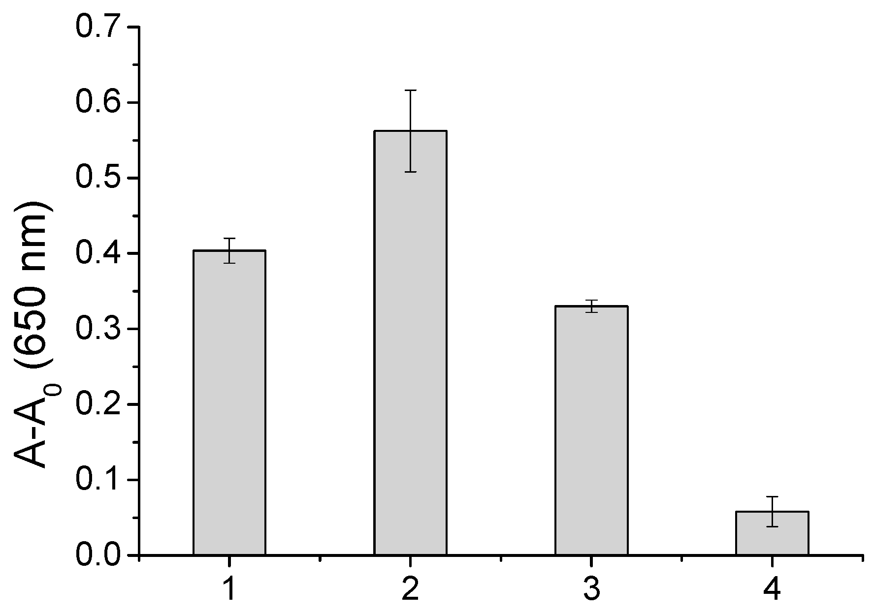
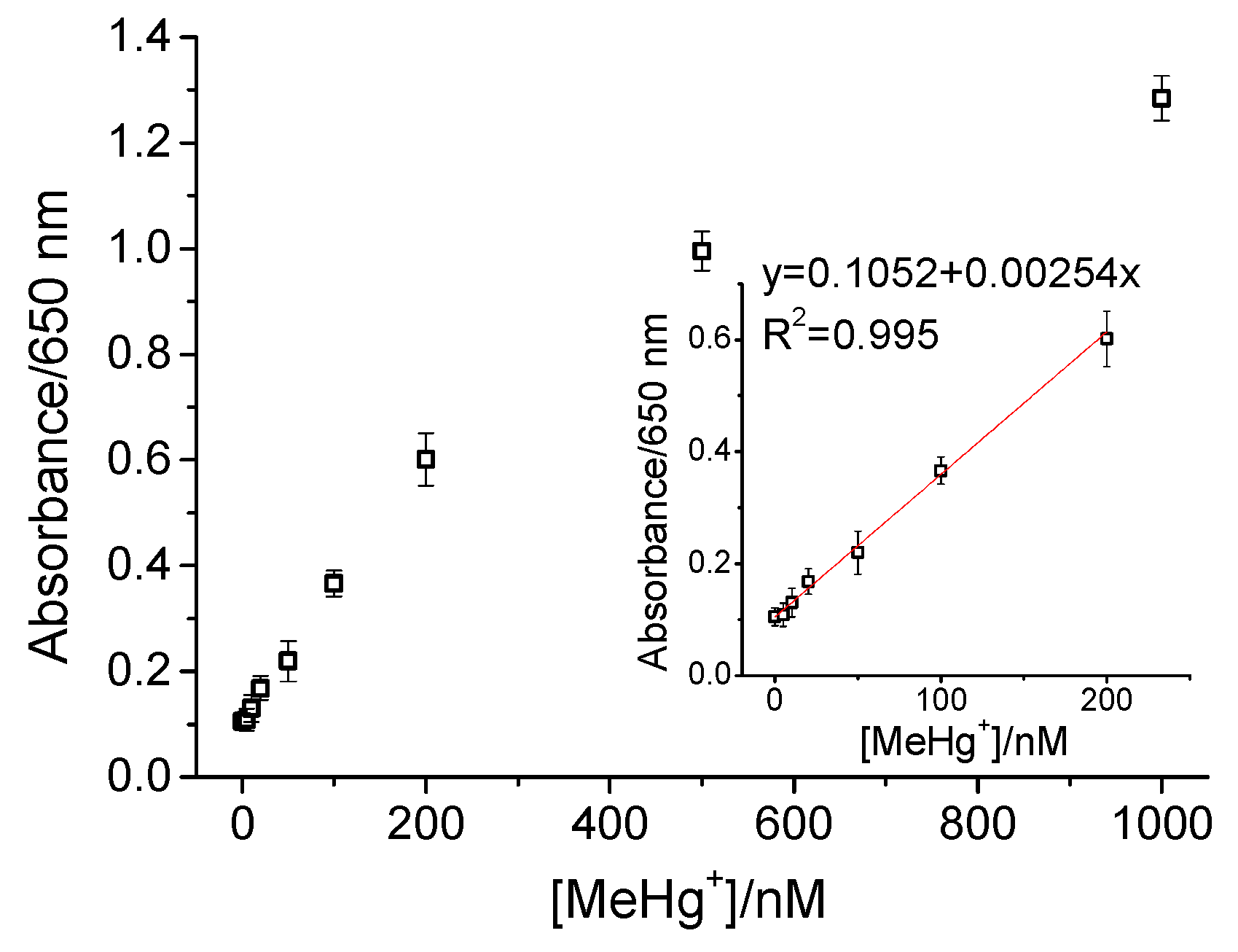
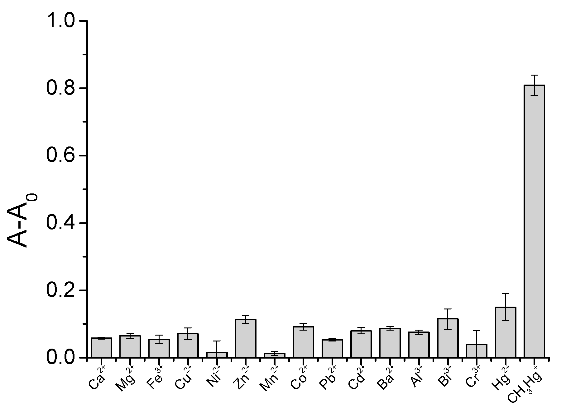
3.2. Colorimetric Detection of CH3Hg+
As shown in Figure 2, AuNPs-HT7 demonstrated weak catalytic activity and we only found a weak signal with a peak at 650 nm. After being captured by the HT7 strand, CH3Hg+ species can be deposited onto the surface of AuNPs through Au/Hg amalgamation since they can be reduced to Hg0 by NaBH4 [19,27,28]. In the above process, the catalytic activity of AuNPs-HT7 was obviously increased, which was supported by the appearance of a strong signal at 650 nm. This was due to the oxidation of TMB by the hydroxyl radical that is stabilized on the surface of AuNPs, which produces a blue one-electron oxidation product (i.e., cation free-radical, TMB+) [29]. The reaction is shown in Figure S1. This change in peroxidase-like activity of the AuNPs suggests the deposition of Hg onto the surface of AuNPs [22,25]. The CH3Hg+ sensing mechanism is depicted in Scheme 1.
Figure 2.
UV–vis spectra of AuNPs-HT7 + TMB-H2O2 reaction solution (a) before and (b) after capturing CH3Hg+ and Hg deposition, (c) AuNPs-HT7 solution and (d) TMB-H2O2 substrate.
Scheme 1.
CH3Hg+ sensing mechanism.
Since the number of T-T pairs may affect the response of the AuNPs-ssDNA complex to CH3Hg+, HT5, HT7 and HT9 strands were used to modify the AuNPs, respectively, before we carried out a comparison of these AuNPs-ssDNA complexes. These AuNPs-ssDNA complexes were also characterized by UV–vis spectra, which demonstrated the same change compared with the AuNPs. As shown in Figure 3, HT7 modified AuNPs demonstrated more sensitive responses to CH3Hg+ compared to HT5, HT9 or HR modified AuNPs. This result suggested that the higher affinity of DNA strand to CH3Hg+ over Hg2+ was still the main factor determining the selectivity of this probe [19].
Figure 3.
Effect of DNA sequence on the colorimetric detection for CH3Hg+. AuNPs, 0.6 nM; CH3Hg+, 100 nM. TMB, 1.0 × 10⁻³ M; H2O2, 1.5 M; pH, 4.5; and incubation time, 25 min.
The enhanced peroxidase-like activity of AuNPs caused by CH3Hg+ was further applied in the development of a colorimetric assay for CH3Hg+. As shown in Figure 4, the absorbance increased as the CH3Hg+ concentration increased in the range of 0–1000 nM. A good linear relationship between CH3Hg+ concentration and absorbance values can be obtained in the range of 10–200 nM. The limit of detection (3-fold signal to noise, S/N = 3) was evaluated to be 5.0 nM.
Figure 4.
Calibration curve for the detection of CH3Hg+. AuNPs, 6.0 × 10⁻¹⁰ M; TMB, 1.5 × 10⁻³ M; H2O2, 1.5 M; Hg2+, 4.0 × 10⁻⁷ M; pH, 4.4; and incubation time, 20 min.
Some common metal ions were tested in this colorimetric assay. As shown in Figure 5, most of common metal ions at a 20-fold higher concentration and the same concentration of Hg2+ showed very weak responses. On the contrary, when citrate-stabilized AuNPs were incubated with Hg2+ or CH3Hg+ ions, almost the same catalytic enhancement of AuNPs was observed (Figure S2). The above results clearly demonstrated the good selectivity of this colorimetric assay for CH3Hg+, which was mainly due to the two aspects: (1) CH3Hg+-specific DNA scaffold has much higher affinity to CH3Hg+ (a Kb value of (5.57 ± 0.47) × 106 M−1) compared to Hg2+ ((1.51 ± 0.18) × 106 M−1); and (2) the centrifugation and separation of AuNPs-HT7 enriched CH3Hg+ over Hg2+. It also should be pointed out that Hg2+ has good affinity with AuNPs. However, the DNA strand on the AuNPs will interact with Hg2+ and thus, will eventually affect the deposition of Hg0. In this case, the HT7 strands probably hindered the deposition of Hg2+.
Figure 5.
Selectivity of the colorimetric assay for CH3Hg+.
The sensitivity of the proposed method was higher than the two typical colorimetric methods [18,21] and comparable with some typical nanosensors or chemosensors (Table 2) [1,17,30,31]. However, the selectivity of this method needs to be further improved when compared with the established colorimetric [21] and fluorescent methods [19,30].
Table 2.
Summarize of some typical method of CH3Hg+.
The real water samples from the Li Lake in Wuxi, Jiangsu Province were obtained and spiked with different concentrations of CH3Hg+ (20 nM, 50 nM and 100 nM). As shown in Table 3, the recovery of the added CH3Hg+ with the colorimetric method was in the range of 93.6–102.1%, which demonstrated the reliability of this assay for the detection of CH3Hg+ in real samples.
Table 3.
Detection of CH3Hg+ in real water samples (n = 3).
4. Conclusions
In summary, we developed a highly sensitive and selective colorimetric method for the detection of CH3Hg+, which was based on the surface deposition of Hg enhancing the catalytic activity of AuNPs. The limit of detection was 5.0 nM with a linear range of 10–200 nM. This colorimetric method has potential in the detection of CH3Hg+ in environmental samples since it also demonstrated other advantages of being simple, rapid and cost-effective. However, this method needs to be further improved with respect to its selectivity to Hg2+. This probably can be further improved through adopting magnetic core gold shell nanocomposites due to their more convenient separation and enrichment capability.
Supplementary Materials
The following are available online at http://www.mdpi.com/1424-8220/18/8/2679/s1, Figure S1. Chromogenic reaction of TMB; Figure S2. UV–vis spectra of citrate-stabilized AuNPs + TMB-H2O2 reaction solution.
Author Contributions
Z.-J.X. mainly run the experiments, X.-Y.B. provided some reagents and participated in the draft writing, and C.-F.P. conceived and designed the experiments.
Funding
This research was funded by the Open Project Program of State Key Laboratory of Dairy Biotechnology, Bright Dairy & Food Co. Ltd. (SKLDB2017-00) and the Science and Technology Innovation Committee of Shenzhen (CXZZ20140419150802007), the National Natural Science Foundation of China (31371767).
Conflicts of Interest
The authors declare no conflict of interest.
References
- Lin, Y.-H.; Tseng, W.-L. Ultrasensitive Sensing of Hg2+ and CH3Hg+ Based on the Fluorescence Quenching of Lysozyme Type VI-Stabilized Gold Nanoclusters. Anal. Chem. 2010, 82, 9194–9200. [Google Scholar] [CrossRef] [PubMed]
- Liu, D.B.; Qu, W.S.; Chen, W.W.; Zhang, W.; Wang, Z.; Jiang, X.Y. Highly Sensitive, Colorimetric Detection of Mercury(II) in Aqueous Media by Quaternary Ammonium Group-Capped Gold Nanoparticles at Room Temperature. Anal. Chem. 2010, 82, 9606–9610. [Google Scholar] [CrossRef] [PubMed]
- Myers, G.J.; Marsh, D.O.; Davidson, P.W.; Cox, C.; Shamlaye, C.F.; Tanner, M.; Choi, A.; Cernichiari, E.; Choisy, O.; Clarkson, T.W. Main neurodevelopmental study of Seychellois children following in utero exposure to methylmercury from a maternal fish diet: Outcome at six months. Neurotoxicology 1995, 16, 653–664. [Google Scholar] [PubMed]
- Hight, S.C.; Cheng, J. Determination of methylmercury and estimation of total mercury in seafood using high performance liquid chromatography (HPLC) and inductively coupled plasma-mass spectrometry (ICP-MS): Method development and validation. Anal. Chim. Acta 2006, 567, 160–172. [Google Scholar] [CrossRef]
- Vallant, B.; Kadnar, R.; Goessler, W. Development of a new HPLC method for the determination of inorganic and methylmercury in biological samples with ICP-MS detection. J. Anal. At. Spectrom. 2007, 22, 322–325. [Google Scholar] [CrossRef]
- Gao, Y.; Galan, S.D.; Brauwere, A.D.; Baeyens, W.; Leermakers, M. Mercury speciation in hair by headspace injection–gas chromatography–atomic fluorescence spectrometry (methylmercury) and combustion-atomic absorption spectrometry (total Hg). Talanta 2010, 82, 1919–1923. [Google Scholar] [CrossRef] [PubMed]
- Priyadarshini, E.; Pradhan, N. Gold nanoparticles as efficient sensors in colorimetric detection of toxic metal ions: A review. Sens. Actuators B Chem. 2017, 238, 888–902. [Google Scholar] [CrossRef]
- Mao, S.; Chang, J.; Zhou, G.; Chen, J. Nanomaterial-enabled Rapid Detection of Water Contaminants. Small 2015, 11, 5336–5359. [Google Scholar] [CrossRef] [PubMed]
- Mehta, J.; Bhardwaj, S.K.; Bhardwaj, N.; Paul, A.K.; Kumar, P.; Kim, K.H.; Deep, A. Progress in the biosensing techniques for trace-level heavy metals. Biotechnol. Adv. 2016, 34, 47–60. [Google Scholar] [CrossRef] [PubMed]
- Chen, L.; Li, J.; Chen, L.X. Colorimetric Detection of Mercury Species Based on Functionalized Gold Nanoparticles. ACS Appl. Mater. Interfaces 2014, 6, 15897–15904. [Google Scholar] [CrossRef] [PubMed]
- Sener, G.; Uzun, L.; Denizli, A. Lysine-Promoted Colorimetric Response of Gold Nanoparticles: A Simple Assay for Ultrasensitive Mercury(II) Detection. Anal. Chem. 2014, 86, 514–520. [Google Scholar] [CrossRef] [PubMed]
- Jin, L.H.; Han, C.S. Eco-friendly colorimetric detection of mercury(II) ions using label-free anisotropic nanogolds in ascorbic acid solution. Sens. Actuators B Chem. 2014, 195, 239–245. [Google Scholar] [CrossRef]
- Liu, H.; Ma, L.; Ma, C.; Du, J.; Wang, M.; Wang, K. Quencher-Free Fluorescence Method for the Detection of Mercury(II) Based on Polymerase-Aided Photoinduced Electron Transfer Strategy. Sensors 2016, 16, 1945. [Google Scholar] [CrossRef] [PubMed]
- Xiao, W.; Xiao, M.; Fu, Q.; Yu, S.; Shen, H.; Bian, H.; Tang, Y. A Portable Smart-Phone Readout Device for the Detection of Mercury Contamination Based on an Aptamer-Assay Nanosensor. Sensors 2016, 16, 1871. [Google Scholar] [CrossRef] [PubMed]
- Kamaruddin, N.; Bakar, A.A.; Mobarak, N.; Zan, M.S.; Arsad, N. Binding Affinity of a Highly Sensitive Au/Ag/Au/Chitosan-Graphene Oxide Sensor Based on Direct Detection of Pb2+ and Hg2+ Ions. Sensors 2017, 17, 2277. [Google Scholar] [CrossRef] [PubMed]
- Wang, G.L.; Zhu, X.Y.; Jiao, H.J.; Dong, Y.M.; Li, Z.J. Ultrasensitive and dual functional colorimetric sensors for mercury (II) ions and hydrogen peroxide based on catalytic reduction property of silver nanoparticles. Biosens. Bioelectron. 2012, 31, 337–342. [Google Scholar] [CrossRef] [PubMed]
- Liu, Y.; Chen, M.; Cao, T.; Sun, Y.; Li, C.; Liu, Q.; Yang, T.; Yao, L.; Feng, W.; Li, F. A cyanine-modified nanosystem for in vivo upconversion luminescence bioimaging of methylmercury. J. Am. Chem. Soc. 2013, 135, 9869–9876. [Google Scholar] [CrossRef] [PubMed]
- Chen, L.; Li, J.; Chen, L. Colorimetric detection of mercury species based on functionalized gold nanoparticles. ACS Appl. Mater. Interfaces 2014, 6, 15897–15904. [Google Scholar] [CrossRef] [PubMed]
- Deng, L.; Li, Y.; Yan, X.; Xiao, J.; Ma, C.; Zheng, J.; Liu, S.; Yang, R. Ultrasensitive and highly selective detection of bioaccumulation of methyl-mercury in fish samples via Ag0/Hg0 amalgamation. Anal. Chem. 2015, 87, 2452–2458. [Google Scholar] [CrossRef] [PubMed]
- Pandeeswar, M.; Senanayak, S.P.; Govindaraju, T. Nanoarchitectonics of Small Molecule and DNA for Ultrasensitive Detection of Mercury. ACS Appl. Mater. Interfaces 2016, 8, 30362–30371. [Google Scholar] [CrossRef] [PubMed]
- Chen, Z.; Wang, X.; Cheng, X.; Yang, W.; Wu, Y.; Fu, F. Specifically and Visually Detect Methyl-Mercury and Ethyl-Mercury in Fish Sample Based on DNA-Templated Alloy Ag-Au Nanoparticles. Anal. Chem. 2018, 90, 5489–5495. [Google Scholar] [CrossRef] [PubMed]
- Long, Y.J.; Li, Y.F.; Liu, Y.; Zheng, J.J.; Tang, J.; Huang, C.Z. Visual observation of the mercury-stimulated peroxidase mimetic activity of gold nanoparticles. Chem. Commun. 2011, 47, 11939–11941. [Google Scholar] [CrossRef] [PubMed]
- Yan, L.; Chen, Z.P.; Zhang, Z.Y.; Qu, C.L.; Chen, L.X.; Shen, D.Z. Fluorescent sensing of mercury(II) based on formation of catalytic gold nanoparticles. Analyst 2013, 138, 4280–4283. [Google Scholar] [CrossRef] [PubMed]
- Peng, C.-F.; Pan, N.; Xie, Z.-J.; Wu, L.-L. Highly sensitive and selective colorimetric detection of Hg2+ based on the separation of Hg2+ and formation of catalytic DNA–gold nanoparticles. Anal. Methods 2016, 8, 1021–1025. [Google Scholar] [CrossRef]
- Wu, L.-L.; Wang, L.-Y.; Xie, Z.-J.; Xue, F.; Peng, C.-F. Colorimetric detection of Hg2+ based on inhibiting the peroxidase-like activity of DNA–Ag/Pt nanoclusters. RSC Adv. 2016, 6, 75384–75389. [Google Scholar] [CrossRef]
- Wang, C.I.; Huang, C.C.; Lin, Y.W.; Chen, W.T.; Chang, H.T. Catalytic gold nanoparticles for fluorescent detection of mercury(II) and lead(II) ions. Anal. Chim. Acta 2012, 745, 124–130. [Google Scholar] [CrossRef] [PubMed]
- Kenduzler, E.; Ates, M.; Arslan, Z.; McHenry, M.; Tchounwou, P.B. Determination of mercury in fish otoliths by cold vapor generation inductively coupled plasma mass spectrometry (CVG-ICP-MS). Talanta 2012, 93, 404–410. [Google Scholar] [CrossRef] [PubMed]
- Monteiro, A.d.C.P.; de Andrade, L.S.N.; Luna, A.S.; de Campos, R.C. Sequential quantification of methyl mercury in biological materials by selective reduction in the presence of mercury(II), using two gas–liquid separators. Spectrochim. Acta Part B At. Spectrosc. 2002, 57, 2103–2112. [Google Scholar] [CrossRef]
- Yin, J.; Cao, H.; Lu, Y. Self-assembly into magnetic Co3O4 complex nanostructures as peroxidase. J. Mater. Chem. 2012, 22, 527–534. [Google Scholar] [CrossRef]
- Costas-Mora, I.; Romero, V.; Lavilla, I.; Bendicho, C. In situ building of a nanoprobe based on fluorescent carbon dots for methylmercury detection. Anal. Chem. 2014, 86, 4536–4543. [Google Scholar] [CrossRef] [PubMed]
- Chatterjee, A.; Banerjee, M.; Khandare, D.G.; Gawas, R.U.; Mascarenhas, S.C.; Ganguly, A.; Gupta, R.; Joshi, H. Aggregation-Induced Emission-Based Chemodosimeter Approach for Selective Sensing and Imaging of Hg(II) and Methylmercury Species. Anal. Chem. 2017, 89, 12698–12704. [Google Scholar] [CrossRef] [PubMed]
© 2018 by the authors. Licensee MDPI, Basel, Switzerland. This article is an open access article distributed under the terms and conditions of the Creative Commons Attribution (CC BY) license (http://creativecommons.org/licenses/by/4.0/).