Article

Selective Functionalization of High-Resolution Cu2O Nanopatterns via Galvanic Replacement for Highly Enhanced Gas Sensing Performance

1
Department of Chemical and Biomolecular Engineering (BK-21 Plus), Korea Advanced Institute of Science and Technology (KAIST), 291 Daehak-ro, Yuseong-gu, Daejeon 34141, Korea
2
Korea Advanced Institute of Science and Technology (KAIST) Institute for NanoCentury, 291 Daehak-ro, Yuseong-gu, Daejeon 34141, Korea
*
Author to whom correspondence should be addressed.
These authors contributed equally to this work.
Sensors 2018, 18(12), 4438; https://doi.org/10.3390/s18124438
Submission received: 21 November 2018 / Revised: 10 December 2018 / Accepted: 13 December 2018 / Published: 15 December 2018
(This article belongs to the Special Issue Functional Materials for the Applications of Advanced Gas Sensors)

Abstract

:
Recently, high-resolution patterned metal oxide semiconductors (MOS) have gained considerable attention for enhanced gas sensing performance due to their polycrystalline nature, ultrasmall grain size (~5 nm), patternable properties, and high surface-to-volume ratio. Herein, we significantly enhanced the sensing performance of that patterned MOS by galvanic replacement, which allows for selective functionalization on ultrathin Cu2O nanopatterns. Based on the reduction potential energy difference between the base channel material (Cu2O) and the decorated metal ion (Pt2+), Pt could be selectively and precisely decorated onto the desired area of the Cu2O nanochannel array. Overall, the Pt-decorated Cu2O exhibited 11-fold higher NO2 (100 ppm) sensing sensitivity as compared to the non-decorated sensing channel, the while the channel device with excessive Pt doping showed complete loss of sensing properties.

1. Introduction

Metal oxide semiconductors (MOSs) (e.g., n-type [1,2,3,4,5] and p-type [6,7,8,9,10,11,12]) are widely used as gas sensing materials due to their high sensitivity, large specific surface area, applicability to various gases (NO2, CO2, H2, volatile organic compounds (VOCs), etc.), high electron mobility, and good chemical/thermal stability at high operating temperature [13,14,15,16,17,18,19,20]. Many approaches have been adopted to enhance the gas sensing performance of the MOS, including grain size refinement, increasing the surface area, reducing the interface area between the substrate and the device, and introduction of dopants and defects [1,18,19,20,21,22]. Doping with noble metals such as Pt [21,22], Pd [23], and Ag [2] has been widely used to enhance the sensing performance of the MOS by tuning the gas adsorption or diffusion properties. As sensitizers on the MOS surface, noble metals can induce electronic or chemical sensitization pathways [1,21]. Electronic sensitization occurs due to the electronic interaction between the semiconductor and the doped metal. When metal dopants undergo partial oxidation in air, their oxidation state of the metal additive can be changed into metallic state depending on the environment. This increases the depth of the electron-depleted charge layer due to electron extraction from the metal oxide, thereby changing the electronic state change of the MOS. The chemical sensitization represents a spill-over effect by activating target gas adsorption on the semiconductor surface [24]. The doped promoter facilitates target gas as active state, leading to increases gas concentration on the channel surface. During the sensing process, the concentration of the adsorbed target gas is effectively increased by the promoter.
Recently, a patterned p-type polycrystalline MOS with an ultrathin and a high aspect ratio showed 5-fold enhanced gas sensing performance due to its ultra-small grains (~5 nm) and large surface-to-volume ratio [6,25]. One of the promising ways to improve the sensing properties is noble metal decoration on the MOS sensing channel. The conventional methods for metal functionalization include sputtering [2,23], thermal evaporation [3], electrospinning [21], and wet impregnation [22] in an aqueous solution. Since the evaporation technique does not allow for selective functionalization of the MOS channel with noble metals, undesired decoration of noble metals on the electrode and substrate can hinder effective control of the doping concentration and cause non-uniform deposition. Moreover, non-directional decoration can produce another dopant-connected channel that reduces the sensitivity of the MOS channel and causes malfunction of the sensing device. Thus, for reduced channel dimensions and higher resolution, more selective and direct functionalization is required [26].
In this study, we selectively functionalized a high-resolution p-type Cu2O sensing channel with Pt nanoparticles by galvanic replacement in order to significantly enhance the gas sensing performance. High-resolution (10 nm scale) and high-aspect-ratio (~25) Cu2O nanopatterns are fabricated via a unique lithographic technique with low-energy plasma bombardments. The unique morphological characteristics (resolution, aspect ratio, grain composition, fully exposed structure) of the Cu2O nanopattern and the possibility of large-area fabrication make it the optimum nanostructure for the sensing channel of gas sensors. Galvanic replacement was employed to further enhance the gas sensing performance of the high-resolution Cu2O nanopattern. This facile decoration process involves a chemical reaction between two materials with different reduction potentials [27,28,29,30]. Hence, selective functionalization on the target site is possible, and the size and the amount of the state of Cu2O and to confirm the existence of Pt on the surface. Decorated dopants can be controlled by adjusting the precursor concentration and reaction conditions. The optimally functionalized nanochannel array (Cu2O/Pt) shows 11-fold higher sensitivity as compared to the pristine Cu2O array upon exposure to 100 ppm NO2 gas (300 °C), because of electronic and chemical sensitization effects of the decorated Pt nanoparticles.

2. Materials and Methods

2.1. Cu2O Nanopattern Fabrication

A polydimethylsiloxane (PDMS) mold was obtained by using a Si pre-patterned mask after curing a silicone elastomer mixture (Sylgard 184, 10:1 weight ratio of base to curing agent; Dow Corning, Midland, MI, USA). Polystyrene (18000 g/mol) solution in toluene was spin-coated (3000 rpm, 45 s) onto a SiO2-coated (200 nm) Si wafer to obtain a thin polystyrene (PS) film. The PS pre-pattern (500 nm width, 350 nm height) was created using capillary forces between PS and PDMS upon heating above the Tg (glass transition temperature, ~135 °C) in a vacuum oven. Then, the target metal (Cu, 25 nm thick) was uniformly deposited onto the PS pre-pattern by electron beam evaporation. By Ar ion bombardment at low energy (500 eV) with a wide angle distribution, the Cu thin layer was etched and emitted into the PS pre-patterned side wall to obtain a polycrystalline feature. After the PS residue was removed by oxygen reactive ion etching (RIE) under a low vacuum and O2 (100 standard cubic centimeters per minute (sccm)) plasma environment, thermal oxidation was performed in a tubular furnace at 450 °C for 3 h to obtain a Cu2O line channel.

2.2. Galvanic Reaction

To decorate Pt nanoparticles selectively onto the Cu2O line pattern, 1 mL of 1 mM potassium tetrachloroplatinate (K2PtCl6, Sigma Aldrich, Burlington, MA, USA), 1 mL of 1 mM hydrochloric acid (HCl, Sigma Aldrich), 5 mL of 20 mM sodium hydroxide powder (NaOH, Daejung), and 5 mL of 10 mM ascorbic acid (AA, Sigma Aldrich) were added to the Cu2O line pattern substrate. This reaction was performed at room temperature (RT) to control the reaction pace preventing over-loaded Pt. Then, the sample and solutions were injected into a 30 mL vial and agitated on a SK-300 benchtop shaker (90 rpm, Lab Companion, Daejeon, Korea). After each reaction, the substrate was cleaned with ethanol and DI water, and then dried under N2.

2.3. Characterization

The surface morphology of the fabricated Cu2O polycrystalline array and Pt-decorated line pattern was characterized by field-emission scanning electron microscopy (Magellan 400, Nova 230, ThermoFisher, Hillsboro, OR, USA) and energy-dispersive X-ray (EDS) spectroscopy (cmodel, manufacturer, city, xsttae abbrev if USA, country. The electron beam energy was 5 kV and 10 kV for the EDS analysis. For the line depth and width profiling of the metal oxide pattern array, atomic force microscopy (AFM; XE-100, Park Systems, Suwon, Korea) was used. X-ray photoelectron spectroscopy (XPS, K-alpha, ThermoFisher, Hillsboro, OR, USA) analysis was conducted to verify the oxidation state of Cu2O and to confirm the existence of Pt on the surface.

2.4. Sensor Fabrication and Measurement

To measure the resistance signal of the channel, 70-nm-thick Au electrodes with a predeposited 5-nm-thick Ti adhesion layer, as well as 100 μm spacing and width, were deposited the Cu2O-Pt line pattern by e-beam evaporation using a customized SERS mask [31]. For saturation of oxygen gas on the substrate, air (400 sccm) was injected over 3 h at 260 °C into the sealed gas reaction chamber where the prepared metal oxide channel device was located. The external sources are used to heat sensing substrate. Heater module is right below the chamber plate. The size of the sealed gas sensing chamber was approximately 10 cm (width) × 5 cm (height) × 8 mm (depth). The resistance signals were displayed directly on a computer via a customized data acquisition module (34970A, Agilent, Santa Clara, CA, USA) to verify the gas sensitivity. NO2 gas (100 ppm) was delivered into the sensing chamber every 10 min by a mass flow controller (MFC, 5850E, Brooks, Seattle, WA, USA). Air was used for observing the desorption properties.

3. Results and Discussion

3.1. Fabrication of Pt Decorated High-Resolution Cu2O Nanochannel

The overall fabrication of the Pt-functionalized Cu2O sensing channel (Cu2O/Pt) is illustrated in Figure 1. First, a PS prepattern with 500 nm width and 350 nm height was formed on a Si wafer substrate on which SiO2 was deposited via thermal pattern transfer using a PDMS mold (Figure 1a). By e-beam evaporation, a 25-nm-thick Cu nanofilm was uniformly deposited on the prepatterns (Figure 1b). The Cu layers were then etched such that they covered the side surfaces of the prepattern by using wide-angle distribution by a lower-energy plasma (500 eV, Ar+) bombardment process using ion-milling instruments. Bombardment of the Cu precursor layers with low-energy Ar+ plasma ruptures the Cu nanofilms to form 5-nm high-resolution grains that are sputtered over a wide area [32,33]. This leads to the formation of a polycrystalline Cu nanopattern with a high aspect ratio as a result of the attachment of the 5-nm Cu grains to the side wall of the PS prepatterns. After removing the polymer residue by oxygen RIE, ultra-high-resolution of the Cu line nanochannel remained (Figure 1d). To oxidize Cu into Cu2O, the fabricated nanopattern was subjected to thermal annealing in a tubular furnace at 450 °C for 3 h in air and then cooled to RT (Figure 1e).
Since the galvanic reaction occurs only between target materials that have different reduction energy potentials, selective doping on the Cu2O device channel can be possible without undesired deposition (e.g., on the substrate). Functionalized Pt can enhance the sensing performance of the Cu2O channel due to the spill-over effect, i.e., easy dissociation of the adsorbed gas on the metal surface and subsequent migration into the metal oxide surface. Therefore, interparticle doping via galvanic replacement not only leads to an accessible contact distance so that gases can easily adsorbed and desorbed, but also prevents the target gas from being captured by stray particles, which may hinder gas detection. Pt functionalization by the galvanic reaction was performed after synthesizing the Cu2O nanopattern (Figure 1f). Since galvanic replacement generally results in a porous interior morphology [34], changing the exterior surface of the original material using another precursor in a strongly acidic environment induces fast reduction, and the reaction pathway can be controlled by precise adjustment of the solution pH [35]. To control the reaction time slowly and prevent collapse of the nanopattern structure by reducing the power of galvanic reaction moderate, a certain amount of sodium hydroxide (NaOH) solution was added. Under harsh acidic conditions, the synthesized Cu2O channels were destroyed, resulting in rapid dissolution of Cu2O. Generally, reduction by a galvanic reaction will lead to the formation of a hollow structure via oxidation of the inner metal [34]. When the Cu2O sensing array is dispersed in an aqueous mixture solution which are Pt4+ precursor for functionalizing, H+ to initiate the galvanic reaction and NaOH salt for limiting the chemical reaction slowly maintaining the pH value around 5, electrons transfer from Cu2O to Pt4+ and reduction of Pt4+ itself on Cu2O surface occur at the same time. Since Pt has a higher reduction potential energy than does Cu2O, Pt is preferentially reduced, so that Cu2O is replaced with Pt on the surface [36]. It is well known that when the pH of the reaction solution increases, the reducing power of AA is enhanced [35].
Therefore, by controlling the pH of the reaction solution with NaOH, we can adjust the galvanic reaction slowly, thus allowing both replacement and reduction without destroying the line pattern structure. After several hours of galvanic reaction at room temperature on a shaker, Pt ions were reduced into Pt particles onto the Cu2O array surface, resulting in a Pt-decorated Cu2O nanopattern (Figure 1g). To induce complete replacement, the experiment was performed in the same manner except for the addition of NaOH in the case of Pt nanowire array fabrication (Figure 1h). Under strongly acidic conditions without the addition of NaOH, the galvanic reaction predominates, resulting in complete replacement of Cu2O with Pt. To understand the reaction mechanism, schematic illustrations and supporting experimental results are presented in Figures S1 and S2.

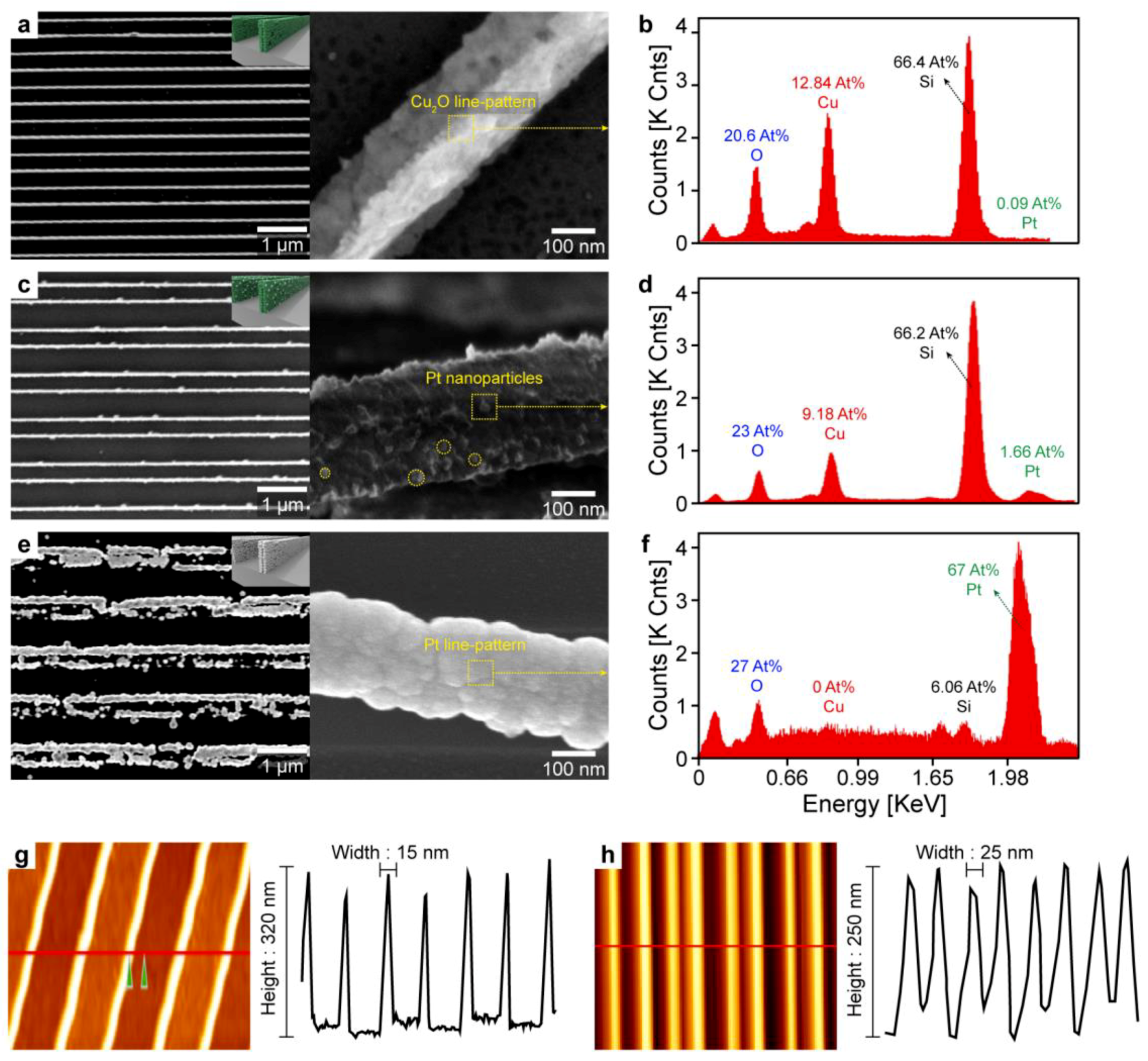
3.2. Morphology, Elements, and Dimension Characterizations

To verify that selective Pt functionalization occurred only on the desired spot, scanning electron microscopy (SEM), energy-dispersive X-ray spectroscopy (EDS), and atomic force microscopy (AFM) were used (Figure 2). The pristine Cu2O nanochannel with high resolution and high aspect ratio was well fabricated over a large area with a periodic spacing of 500 nm (Figure 2a). X-ray diffraction (XRD) shows that Cu nanopattern is successfully synthesized to Cu2O nanopattern via thermal oxidation process (Figure S3). Photographs of the devices show that the Cu2O nanopattern was fabricated over a large area (cm2 scale) with excellent pattern fidelity (Figure S4). After 4 h of galvanic reaction, a part of the Cu2O channel was successfully replaced with Pt nanoparticles without destruction or collapse of the high-aspect-ratio nanostructure. The yellow particles on the Cu2O surface in Figure 2c were confirmed to be Pt via point EDS analysis by comparison that of the pristine Cu2O. In addition, the overall distribution of the functionalized Pt was observed by TEM mapping analysis (Figure S5). Low-magnification SEM-EDS images clearly confirmed that the Pt nanoparticles existed only in the Cu2O nanopatterns and there was no Pt in the substrate area between the Cu2O channels (Figure 2c–f and Figure S6). These results revealed that the galvanic replacement selectively occurs at the target spots, i.e., the surface of the Cu2O nanopatterns. With the reaction was carried out for a sufficient time under acidic conditions, replacement of Cu2O with Pt was predominant, resulting in a pure Pt line pattern at the site of the Cu2O pattern. To ensure complete reaction, the reaction was performed without the addition of NaOH (pH ~2). After a few hours, a Pt line pattern could be located at the original Cu2O channel spots (Figure 2e), which supported the occurrence of the galvanic reaction, while EDS observations confirmed the presence of pure Pt wires (Figure 2f). AFM analysis revealed the feature dimensions of the Cu2O nanopattern channel. The periodic Cu2O lines were 15 nm in width and 320 nm in depth before the galvanic reaction; after Pt doping, the Cu2O line nanopattern maintained its original features within the error range of the synthesis, thus indicating that the structure did not collapse during doping (Figure 1g–h). Overall, we fabricated high-resolution (15 nm) and high-aspect-ratio nanopatterns uniformly on a large area and achieved selective functionalization of Pt nanoparticles on the patterns. As discussed in a previous study [6], these nanopattern arrays have distinct advantages as a gas sensor: (1) polycrystalline feature with small grains; (2) low channel resistance caused by the large surface-to-volume ratio, which is derived from the ultrahigh resolution and high aspect ratio (~25). In addition, the noble metal acting as a sensitizer to enhance the gas sensing performance could be selectively decorated onto the surface of the ultrahigh-resolution nanopattern array by the galvanic replacement method.

3.3. Chemical Binding States of Pt/Cu2O Nanochannel

To confirm the chemical binding state and oxidation state of the nanopattern channel, detailed surface elemental analysis was carried out by X-ray photoelectron spectroscopy (XPS). The obtained binding energy was calibrated with that of the C-C peak, 284.7 eV. Overlapping peaks were deconvoluted and identified using the Avantage Software program (Thermo ScientificTM). As shown in Figure 3a, the metallic Cu nanopattern was completely oxidized and converted to Cu2O upon thermal annealing. For Cu 2p photoelectron analysis, the binding energy range from 925 to 965 eV was scanned. The binding energy of metallic Cu was not detected in the Cu 2p spectrum, confirming that the Cu channel was fully oxidized into Cu2O under the mild oxidation conditions. The high-resolution Cu 2p spectrum showed two main peaks at 933.18 eV and 952. 98 eV, which could be assigned to the Cu2+ double peaks for Cu 2p3/2 and Cu 2p1/2 and three satellite peaks, respectively [37]. The two O 1 s peaks represented the O2- state (529.18 eV) from Cu2O and the OH- state (531.17 eV) from the adsorption of atmospheric oxygen on the surface [38]. The Cu 2p and O 1s peaks confirmed the formation of metal oxide (Cu2O). Figure 3c shows the electron states of the Pt atoms in the nanopattern channel. The doublet peaks at 74.24 eV and 71.19 eV corresponded to the binding energy of metallic Pt, and the binding energies at 78.2 eV and 75.3 eV were in agreement with Pt4+ which came from the binding between metal oxide and doped Pt [39]. During galvanic replacement, the Pt2+ ions penetrated the Cu2O environment to form Cu2O/Pt species, ascribed to Pt4+ in the X-ray photoelectron spectrum. This Pt species binds to other Pt ions by accepting electrons from Cu2O to form metallic Pt. From the Pt 4f spectral analysis, we can confirm that Pt2+ in the galvanic reaction solution (PtCl42−) is successfully reduced to Pt metal without remaining in the ionic state on the Cu2O surface.
The synthesized ultrathin Pt/Cu2O nanopattern exhibited 11-fold higher sensitivity than did the pristine Cu2O nanopattern. To determine the feasibility of using the Cu2O/Pt nanopattern as an electronic sensing channel, the baseline resistances and noise level of the channel were measured during 300 °C operation (Figure 4a). The channel resistance of the pristine Cu2O sensor was 24 MΩ, which increased, and approaching ~45 MΩ upon Pt nanoparticle decoration; a resistance level of tens of MΩ is within the optimal range for MOS-based gas sensors. The increase in the channel resistance was due to the formation of a depletion layer on the Cu2O channel because of electron transfer from the decorated Pt nanoparticles. In addition, the noise from the Cu2O/Pt channel increased from 0.88% to 4.59% of the baseline resistance. However, the Pt nanopattern obtained by the excessive galvanic reaction showed a significantly low resistance (tens of Ω) with an ultra-low noise level due to the metallic conductivity of the Pt nanopattern channel. To investigate the effects of Pt nanoparticle decoration on the sensing characteristics of the Cu2O nanopattern, a sensing device was fabricated with integration of a nanopattern channel (Cu2O, Cu2O/Pt, Pt) and two-terminal resistor type electrodes. A constant bias from 0.5 to 1.5 V was automatically applied to the two-probe electrode, and the electrical resistance of each channel was recorded as a sensing signal by a data acquisition module (Agilent 34970A). The sensing devices were simultaneously loaded on a home-made gas sensing chamber, and the sensing signals from each device were measured with multi-channel sensing systems.

3.4. NO2 Sensing Performances and Mechanism

Details of the in-house fabricates gas delivery system are provided in the Supporting Information (Figure S7) [40]. Figure 4b shows the responses of the Cu2O, Cu2O/Pt, and Pt nanopatterns toward 100 ppm NO2 at 300 °C. Gas response is defined as Ra/Rg, where Ra and Rg are the resistances of the sensor in air and in the target gas, respectively. The Cu2O/Pt nanopattern exhibited a markedly higher sensitivity upon exposure to NO2 (Ra/Rg = 11) than did the pristine Cu2O nanopattern (Ra/Rg = 1), which was attributed to the effect of the noble metal (Pt) nanoparticle decoration. For the Pt nanopattern obtained by the excessive galvanic replacement reaction, NO2 detection was not observed due to the perfect metallic properties of the nanopattern. To further investigate the high sensitivity of the Cu2O/Pt nanopattern sensor, real-time sensing response to single ppm of NO2 (1 to 4 ppm) is measured (Figure 4c). It is clearly seen that Cu2O/Pt nanopattern sensors show significant responses to single ppm NO2 with distinguishable signal resolutions. Figure 4d further demonstrates that the Cu2O/Pt nanopattern sensors show linear response variations to single ppm NO2 (1–4 ppm) with significant repose amplitudes ((ΔR/Rb)max) which was comparable value with other previous works (Table S1). Here, Rb and ΔR represent the baseline resistance of the sensor exposed to dry air and the change in resistance after exposure to NO2, respectively. Thus, selective functionalization of the Cu2O channel by galvanic replacement had a significant impact on the enhancement of the gas sensing performance. The response/recovery time (τ90%, time taken to reach the 90% of the minimum resistance level) of the sensors for NO2 is shown in Figure S8. Moreover, functionalized Cu2O/Pt doesn’t change the selectivity characterization comparing that of Cu2O (see the Figure S9 which shows similar response selectivity comparing that of Cu2O [6] among various gases). In addition, long-term stability test of the Cu2O/Pt nanopattern sensor was conducted showing high-stability of synthesized catalyst (Figure S10). The highly enhanced gas sensing performance of Cu2O with the Pt galvanic reaction could be explained via two mechanisms: electronic sensitization (ES) [24,41,42] and chemical sensitization (CS) [24,43,44]. The detailed enhancement mechanisms are shown in Figure 4e–g. First, the decorated Pt nanoparticles played an ES role during the sensing process. When exposed to air, each Pt nanoparticle consisting of Pt and Pt4+ forms a redox electrode Pt4+/Pt0. To achieve electronic equilibrium with the Pt nanoparticles, the Fermi level of Cu2O will be shifted and pinned at the Pt4+/Pt0 electrode potential, which produces an electron-depleted space charge region and decreases the charge (hole) accumulation layer (HAL) (Figure 4e). This eventually leads to an increase in the resistance of the pristine Cu2O/Pt in air. Second, it the functionalized Pt nanoparticles also play an important role via the CS mechanism because of the spillover effect. For the pristine Cu2O nanopattern, the adsorbed oxygen ions (O, O2−) localize the electrons, which leads to an increase in the concentration of holes in the surface layers. Upon exposure to NO2, the adsorbed NO2 ions (NO2−) further localize the electrons of Cu2O, thus increasing the HAL thickness and decreasing the resistance of the sensors (Figure 4f–g). In the case of Cu2O/Pt, significantly larger amounts of NO2 can be adsorbed onto Cu2O via the spill-over effect [43,44] due to the high catalytic activity of the Pt nanoparticles (Figure 4f). Thus, the high-resolution Cu2O nanopattern is selectively functionalized with Pt nanoparticles via the galvanic replacement reaction, and the gas sensing performance is significantly improved via both the ES and CS mechanisms.

4. Conclusions

In conclusion, an ultrahigh resolution array, i.e., a thin, polycrystalline, and high-aspect-ratio p-type sensing channel array (Cu2O) was selectively functionalized with Pt via galvanic replacement. Herein, by using a galvanic reaction which involves a chemical reaction between two materials, we achieved selective doping of Pt onto the surface of the ultrahigh-resolution nanopattern array. We successfully fabricated an ultrathin nanopattern array with a high surface-to-volume ratio over a large area by our unique lithography technique (low-energy plasma bombardment [23]) and showed enhancement of the sensing performance after selective Pt functionalization through the galvanic replacement reaction. The sensor with Pt-functionalized Cu2O showed a 11-fold increase in the gas response (Ra/Rg) to the bare Cu2O channel when exposed on 100 ppm NO2. The role of the functionalized Pt particles was suggested in terms of both ES and CS mechanisms. Using the selectively Pt-decorated high-resolution Cu2O array obtained via galvanic replacement, we could achieve significant enhancement of the NO2 sensing performance.

Supplementary Materials

The following are available online at https://www.mdpi.com/1424-8220/18/12/4438/s1, Figure S1: schematic illustrations of expected galvanic reaction process, Figure S2: scanning electron microscope (SEM) observations depending on various reaction condition, Figure S3: XRD spectrum of the Cu2O nanopattern before and after thermal oxidation, Figure S4: photo and SEM image of the Cu2O nanopattern sensors, Figure S5: elemental distribution analysis by transmission electron microscopy (TEM) - energy dispersive X-ray spectroscopy (EDS), Figure S6: the composition analysis of over-galvanic reacted sample to confirm a selective decoration property of galvanic reaction, Figure S7: schematic of the overall gas delivery system, Figure S8: response and recovery time of the Cu2O/Pt nanopattern sensors to 1 to 4 ppm NO2, Figure S9: selectivity characterization of the Cu2O/Pt nanopattern sensor, Figure S10: long-term stability of the Cu2O/Pt nanopattern sensor. Table S1: NO2 gas sensing performance comparison based on CuO or Cu2O materials.

Author Contributions

Conceptualization, validation, writing—review & editing, and writing—original draft preparation, funding acquisition, resources, project administration, J.Y.K. and S.-Y.C.; methodology, investigation, J.Y.K.; data curation, visualization, formal analysis, S.-Y.C.; supervision, and writing-review & editing, H.-T.J.

Funding

This research was supported by a National Research Foundation of Korea (NRF) grant funded by the Ministry of Science, ICT and Future Planning, Korea (NRF-2018R1A2B3008658) and a Global Frontier grant funded by the Center for Advanced Soft Electronics under the Global Frontier Research Program of the Ministry of Science, ICT and Future Planning, Korea (MSIP, NRF-2012M3A6A5055744).

Conflicts of Interest

The authors declare no conflict of interest.

References

  1. Velmathi, G.; Mohan, S.; Henry, R. Analysis of factors for improving functionality of tin oxide gas sensor. IETE Tech. Rev. 2016, 33, 122–129. [Google Scholar] [CrossRef]
  2. Hwang, I.-S.; Choi, J.-K.; Woo, H.-S.; Kim, S.-J.; Jung, S.-Y.; Seong, T.-Y.; Kim, I.-D.; Lee, J.-H. Facile control of C2H5OH sensing characteristics by decorating discrete Ag nanoclusters on SnO2 nanowire networks. ACS Appl. Mater. Interfaces 2011, 3, 3140–3145. [Google Scholar] [CrossRef] [PubMed]
  3. Shimizu, Y.; Matsunaga, N.; Hyodo, T.; Egashira, M. Improvement of SO2 sensing properties of WO3 by noble metal loading. Sens. Actuators B Chem. 2001, 77, 35–40. [Google Scholar] [CrossRef]
  4. Chang, Y.-E.; Youn, D.-Y.; Ankonina, G.; Yang, D.-J.; Kim, H.-G.; Rothschild, A.; Kim, I.-D. Fabrication and gas sensing properties of hollow SnO2 hemispheres. Chem. Commun. 2009, 27, 4019–4021. [Google Scholar] [CrossRef]
  5. Ahn, M.-W.; Park, K.-S.; Heo, J.-H.; Park, J.-G.; Kim, D.-W.; Choi, K.J.; Lee, J.-H.; Hong, S.-H. Gas sensing properties of defect-controlled ZnO-nanowire gas sensor. Appl. Phys. Lett. 2008, 93, 263103. [Google Scholar] [CrossRef] [Green Version]
  6. Cho, S.-Y.; Yoo, H.-W.; Kim, J.Y.; Jung, W.-B.; Jin, M.L.; Kim, J.-S.; Jeon, H.-J.; Jung, H.-T. High-resolution p-type metal oxide semiconductor nanowire array as an ultrasensitive sensor for volatile organic compounds. Nano Lett. 2016, 16, 4508–4515. [Google Scholar] [CrossRef]
  7. Kim, H.-J.; Lee, J.-H. Highly sensitive and selective gas sensors using p-type oxide semiconductors: Overview. Sens. Actuators B Chem. 2014, 192, 607–627. [Google Scholar] [CrossRef]
  8. Thirumalairajan, S.; Girija, K.; Mastelaro, V.R.; Ponpandian, N. Surface morphology-dependent room-temperature LaFeO3 nanostructure thin films as selective NO2 gas sensor prepared by radio frequency magnetron sputtering. ACS Appl. Mater. Interfaces 2014, 6, 13917–13927. [Google Scholar] [CrossRef]
  9. Joshi, N.; Silva, L.F.D.; Jadhav, H.; M’Peko, J.-C.; Torres, B.B.M.; Aguir, K.; Mastelaro, V.R., Jr.; Oliveira, N.O. One-step approach for preparing ozone gas sensors based on hierarchical NiCo2O4 structures. RSC Adv. 2016, 6, 92655–92662. [Google Scholar] [CrossRef]
  10. Joshi, N.; Silva, L.F.D.; Jadhav, H.S.; Shimizu, F.M.; Suman, P.H.; M’Peko, J.-C.; Orlandi, M.O.; Seo, J.G.; Mastelaro, V.R., Jr.; Oliveira, N.O. Yolk-shelled ZnCo2O4 microspheres: Surface properties and gas sensing application. Sens. Actuators B 2018, 257, 906–915. [Google Scholar] [CrossRef]
  11. Hu, J.; Zou, C.; Su, Y.; Li, M.; Han, Y.; Kong, E.S.-W.; Yang, Z.; Zhang, Y. An ultrasensitive NO2 gas sensor based on a hierarchical Cu2O/CuO mesocrystal nanoflower. J. Mater. Chem. A 2018, 6, 17120–17131. [Google Scholar] [CrossRef]
  12. Hübner, M.; Simion, C.E.; Tomescu-Stănoiu, A.; Pokhrel, S.; Bârsan, N.; Weimar, U. Influence of humidity on CO sensing with p-type CuO thick film gas sensors. Sens. Actuators B 2011, 153, 347–353. [Google Scholar] [CrossRef]
  13. Yoo, H.-W.; Cho, S.-Y.; Jeon, H.-J.; Jung, H.-T. Well-defined and high resolution Pt nanowire arrays for a high performance hydrogen sensor by a surface scattering phenomenon. Anal. Chem. 2015, 87, 1480–1484. [Google Scholar] [CrossRef] [PubMed]
  14. Zhang, J.; Qin, Z.; Zeng, D.; Xie, C. Metal-oxide-semiconductor based gas sensors: Screening, preparation, and integration. Phys. Chem. Chem. Phys. 2017, 19, 6313–6329. [Google Scholar] [CrossRef] [PubMed]
  15. Wang, C.; Yin, L.; Zhang, L.; Xiang, D.; Gao, R. Metal oxide gas sensors: Sensitivity and influencing factors. Sensors 2010, 10, 2088–2106. [Google Scholar] [CrossRef] [PubMed]
  16. Miller, D.R.; Akbar, S.A.; Morris, P.A. Nanoscale metal oxide-based heterojunctions for gas sensing: A review. Sens. Actuators B Chem. 2014, 204, 250–272. [Google Scholar] [CrossRef]
  17. Franke, M.E.; Koplin, T.J.; Simon, U. Metal and metal oxide nanoparticles in chemiresistors: Does the nanoscale matter? Small 2006, 2, 36–50. [Google Scholar] [CrossRef] [PubMed]
  18. Liu, X.; Ma, T.; Pinna, N.; Zhang, J. Two-dimensional nanostructured materials for gas sensing. Adv. Funct. Mater. 2017, 27, 1702168. [Google Scholar] [CrossRef]
  19. Joshi, N.; Hayasaka, T.; Liu, Y.; Liu, H., Jr.; Oliveira, N.O.; Lin, L. A review on chemiresistive room temperature gas sensors based on metal oxide nanostructures, graphene and 2D transition metal dichalcogenides. Microchim. Acta 2018, 185, 213. [Google Scholar] [CrossRef]
  20. Zhang, J.; Liu, X.; Neri, G.; Pinna, N. Nanostructured materials for room-temperature gas sensors. Adv. Mater. 2016, 28, 795–831. [Google Scholar] [CrossRef]
  21. Fu, J.; Zhao, C.; Zhang, J.; Peng, Y.; Xie, E. Enhanced gas sensing performance of electrospun Pt-functionalized NiO nanotubes with chemical and electronic sensitization. ACS Appl. Mater. Interfaces 2013, 5, 7410–7416. [Google Scholar] [CrossRef] [PubMed]
  22. Gou, X.; Wang, G.; Yang, J.; Park, J.; Wexler, D. Chemical synthesis, characterization and gas sensing performance of copper oxide nanoribbons. J. Mater. Chem. 2008, 18, 965–969. [Google Scholar] [CrossRef]
  23. Lu, Y.; Li, J.; Han, J.; Ng, H.-T.; Binder, C.; Partridge, C.; Meyyappan, M. Room temperature methane detection using palladium loaded single-walled carbon nanotube sensors. Chem. Phys. Lett. 2004, 391, 344–348. [Google Scholar] [CrossRef]
  24. Yamazoe, N. New Approaches for improving semiconductor gas sensors. Sens. Actuators B Chem. 1991, 5, 7–19. [Google Scholar] [CrossRef]
  25. Jeon, H.-J.; Kim, K.H.; Baek, Y.-K.; Kim, D.W.; Jung, H.-T. New top-down approach for fabricating high-aspect-ratio complex nanostructures with 10 nm scale features. Nano Lett. 2010, 10, 3604–3610. [Google Scholar] [CrossRef] [PubMed]
  26. Shen, G.; Chen, P.-C.; Ryu, K.; Zhou, C. Devices and chemical sensing applications of metal oxide nanowires. J. Mater. Chem. 2009, 19, 828–839. [Google Scholar] [CrossRef]
  27. Jang, H.-J.; Hong, S.; Ham, S.; Shuford, K.L.; Park, S. Site-specific growth of a Pt shell on Au nanoplates: Tailoring their surface plasmonic behavior. Nanoscale 2014, 6, 7339–7345. [Google Scholar] [CrossRef]
  28. Seo, D.; Song, H. Asymmetric hollow nanorod formation through a partial galvanic replacement reaction. J. Am. Chem. Soc. 2009, 131, 18210–18211. [Google Scholar] [CrossRef]
  29. Pradhan, M.; Chowdhury, J.; Sarkar, S.; Sinha, A.K.; Pal, T. Hierarchical gold flower with sharp tips from controlled galvanic replacement reaction for high surface enhanced Raman scattering Activity. J. Phys. Chem. C 2012, 116, 24301–24313. [Google Scholar] [CrossRef]
  30. Zhang, W.; Rahmani, M.; Niu, W.; Ravaine, S.; Hong, M.; Lu, X. Tuning interior nanogaps of double-shelled Au/Ag nanoboxes for surface-enhanced Raman scattering. Sci. Rep. 2015, 5, 8382. [Google Scholar] [CrossRef]
  31. Cho, S.-Y.; Kim, S.J.; Lee, Y.; Kim, J.-S.; Jung, W.-B.; Yoo, H.-W.; Kim, J.; Jung, H.-T. highly enhanced gas adsorption properties in vertically aligned MoS2 layers. ACS Nano 2015, 9, 9314–9321. [Google Scholar] [CrossRef] [PubMed]
  32. Cho, S.-Y.; Jeon, H.-J.; Yoo, H.-W.; Cho, K.M.; Jung, W.-B.; Kim, J.-S.; Jung, H.-T. Highly enhanced fluorescence signals of quantum dot–polymer composite arrays formed by hybridization of ultrathin plasmonic Au nanowalls. Nano Lett. 2015, 15, 7273–7280. [Google Scholar] [CrossRef] [PubMed]
  33. Cho, S.-Y.; Jeon, H.-J.; Kim, J.-S.; Ok, J.M.; Jung, H.-T. Hierarchical ordering of quantum dots and liquid with tunable super-periodicity into high aspect ratio moiré superlattice structure. Adv. Funct. Mater. 2014, 24, 6939–6947. [Google Scholar] [CrossRef]
  34. Zhang, W.; Yang, J.; Lu, X. Tailoring galvanic replacement reaction for the preparation of Pt/Ag bimetallic hollow nanostructures with controlled number of voids. ACS Nano 2012, 6, 7397–7405. [Google Scholar] [CrossRef] [PubMed]
  35. Yang, Y.; Liu, J.; Fu, Z.-W.; Qin, D. Galvanic replacement-free deposition of Au on Ag for core-shell nanocubes with enhanced chemical stability and SERS activity. J. Am. Chem. Soc. 2014, 136, 8153–8156. [Google Scholar] [CrossRef] [PubMed]
  36. Li, Q.; Xu, P.; Zhang, B.; Wu, G.; Zhao, H.; Fu, E.; Wang, H.-L. Self-supported Pt nanoclusters via galvanic replacement from Cu2O nanocubes as efficient electrocatalysts. Nanoscale 2013, 5, 7397. [Google Scholar] [CrossRef] [PubMed]
  37. Zhu, H.; Du, M.; Yu, D.L.; Wang, Y.; Wang, L.; Zou, M.; Zhang, M.; Fu, Y.Q. A new strategy for the surface-free-energy-distribution induced selective growth and controlled formation of Cu2O-Au hierarchical heterostructures with a series of morphological evolutions. J. Mater. Chem. A 2013, 1, 919–929. [Google Scholar] [CrossRef]
  38. Ji, R.; Sun, W.; Chu, Y. One-step hydrothermal synthesis of Ag/Cu2O heterogeneous nanostructure over Cu foil and their SERS applications. RSC Adv. 2014, 4, 6055–6059. [Google Scholar] [CrossRef]
  39. Murata, N.; Suzuki, T.; Kobayashi, M.; Togoh, F.; Asakura, K. Characterization of Pt-doped SnO2 catalyst for a high-performace micro gas sensor. Phys. Chem. Chem. Phys. 2013, 15, 17938–17946. [Google Scholar] [CrossRef]
  40. Cho, S.-Y.; Lee, Y.; Koh, H.-J.; Jung, H.; Kim, J.-S.; Yoo, H.-W.; Kim, J.; Jung, H.-T. Superior chemical sensing performance of black phosphorus: comparison with MoS2 and graphene. Adv. Mater. 2016, 28, 7020–7028. [Google Scholar] [CrossRef]
  41. Chen, D.; Liu, Z.; Guo, Z.; Yan, W.; Xin, Y. Enhancing light harvesting and charge separation of Cu2O photocathodes with spatially separated noble-metal cocatalysts towards highly efficient water splitting. J. Mater. Chem. A 2018, 6, 20393–20401. [Google Scholar] [CrossRef]
  42. Cao, D.; Wang, C.; Zheng, F.; Dong, W.; Fang, L.; Shen, M. High-efficiency ferroelectric-film solar cells with an n-type Cu2O cathode buffer layer. Nano Lett. 2012, 12, 2803–2809. [Google Scholar] [CrossRef] [PubMed]
  43. Wang, W.-J.; Liang, J.-R.; Li, C.-Q.; Yan, W.-J.; Hu, M. Room temperature NO2 gas sensing of Au-loaded tungsten oxide nanowires/porous silicon hybrid structure. Chin. Phys. B 2015, 25, 028102. [Google Scholar] [CrossRef]
  44. Mane, A.A.; Moholkar, A.V. Palladium (Pd) sensitized molybdenum trioxide (MoO3) nanobelts for nitrogen dioxide (NO2) gas detection. Solid-State Electron. 2018, 139, 21–30. [Google Scholar] [CrossRef]
Figure 1. Schematic illustration of fabrication of Pt-functionalized high-resolution Cu2O nanopattern. (a) The PS prepattern was transferred from the PDMS mold. (b) The target metal (Cu) was deposited onto the prepattern. (c) By low-energy plasma bombardments, the deposited metal was widely sputtered onto the side wall of the prepattern, resulting in polycrystalline Cu walls. (d) The prepattern residue was removed by RIE and (e) the ultra-thin Cu line pattern was oxidized by thermal annealing. (f) To functionalize the Cu2O surface with Pt, the prepared channel was immersed in a Pt precursor solution. (g) The electrons were transferred from Cu2O to Pt ions, resulting in the Pt-decorated Cu2O nanopattern. (h) In case of an excessive galvanic reaction, the Cu2O baseline channel was completely converted into Pt.
Sensors 18 04438 g001
Figure 2. Characterization of fabricated Cu2O, Pt/Cu2O, and Pt sensing channel. SEM image of the fabricated pattern array for (a) pristine Cu2O, (c) optimized Pt-doped Cu2O, and (e) completely reacted Pt. (bf) EDS point analysis of each channel. (gh) AFM depth and width profiles show ultrathin line channel and no disruption of the pattern after Pt doping.
Sensors 18 04438 g002
Figure 3. X-ray photoelectron spectra of Cu2O/Pt nanopattern. High-resolution elemental spectra of (a) Cu 2p, (b) O 1s, and (c) Pt 4f.
Sensors 18 04438 g003
Figure 4. Device characteristics and gas sensing performance of the Cu2O, Cu2O/Pt, and Pt nanopattern sensors. (a) Baseline resistance and noise of the Cu2O, Cu2O/Pt, and Pt nanopattern. (b) Real-time NO2 (100 ppm) sensing performance of the Cu2O, Cu2O/Pt, and Pt nanopattern sensors. (c) Real-time response behavior of the Cu2O/Pt nanopattern sensor to low concentration NO2 (1 to 4 ppm). (d) Maximum response amplitudes of the Cu2O/Pt nanopattern sensor (three sensors) to low concentration NO2. (e) Band diagram scheme of Cu2O/Pt to elucidate electronic sensitization (ES). Schematic illustrations of chemical sensing mechanism of (f) Cu2O and (g) Cu2O/Pt nanopattern with chemical sensitization (CS) of spill-over effect by Pt nanoparticles.
Sensors 18 04438 g004

