
Major Depression Detection from EEG Signals Using Kernel Eigen-Filter-Bank Common Spatial Patterns


Abstract
:1. Introduction
2. Materials and Methods
2.1. Participants
2.2. Apparatus, Settings, and Preprocessing
2.3. EEG Data Collection
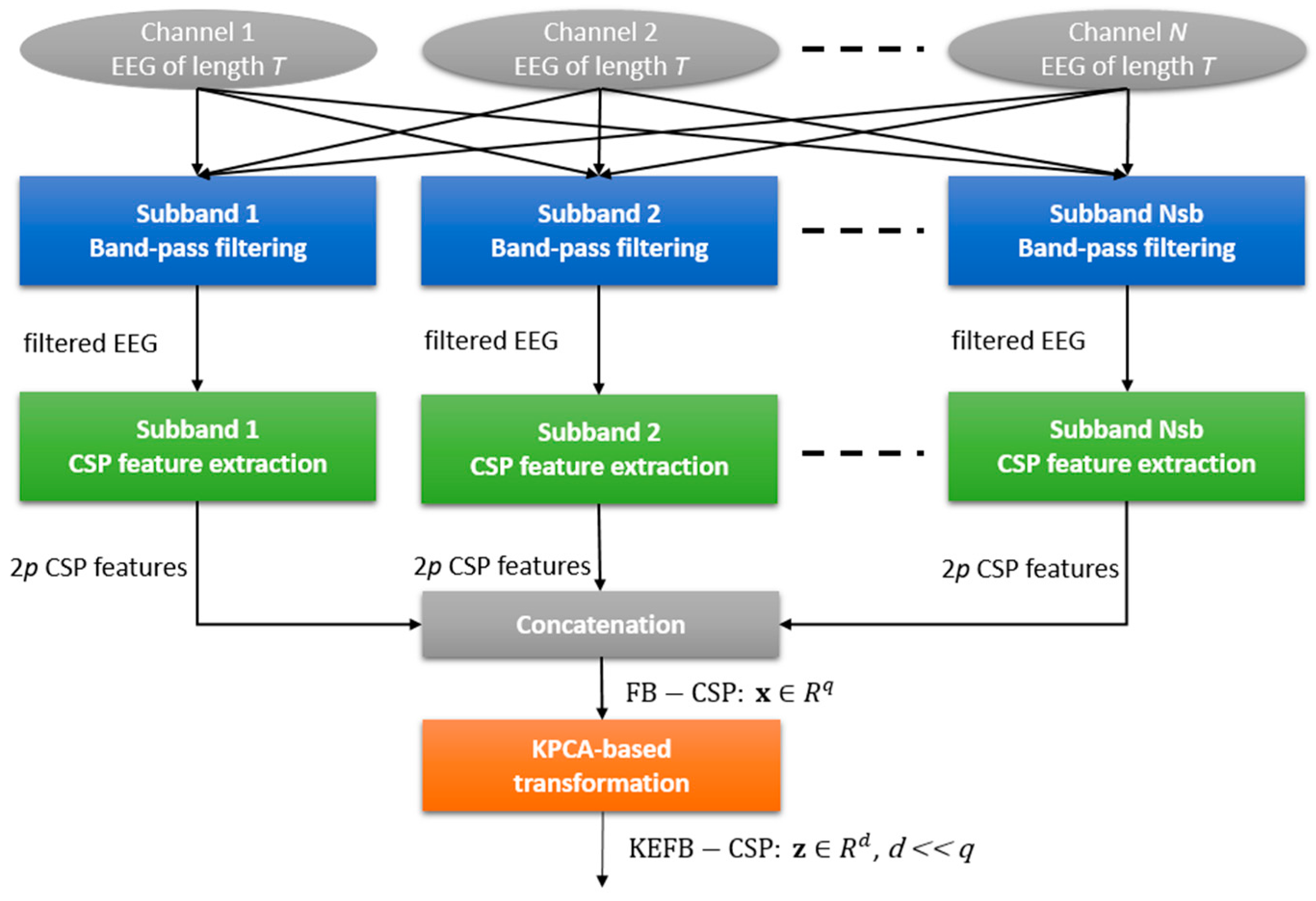
2.4. Feature Extraction
2.4.1. BP
2.4.2. GPFD
2.4.3. KEFB-CSP
- Step 1.
- Calculate the averaged spatial covariance matrices of the two classes and :where t denotes the transpose of a matrix, tr(.) denotes the trace of (.), is the ith training EEG data of class 1, is the ith training EEG of class 2, and and are the numbers of the training EEG data of the first and second classes, respectively. Notice here that an EEG training data is a signal matrix.
- Step 2.
- Diagonalize the composite covariance by , where , is the matrix in which the columns are the orthonormal eigenvectors of , and is an diagonal matrix in which the diagonal elements are the eigenvalues sorted in descending order.
- Step 3.
- Whiten the averaged spatial covariance metrics of the two classes by , I = 1,2, where is the whitening matrix.
- Step 4.
- Perform the simultaneous diagonalization of the whitened spatial covariance matrices:where and share the same eigenvectors, and equals an identity matrix , indicating that the eigenvectors having larger eigenvalues for have smaller ones for .
- Step 5.
- Projecting the whitened EEG of a trial onto the eigenvectors in yields new N-channel signals (an matrix):
2.5. Parameter Optimization and Participant-Independent Classification
3. Results and Discussion
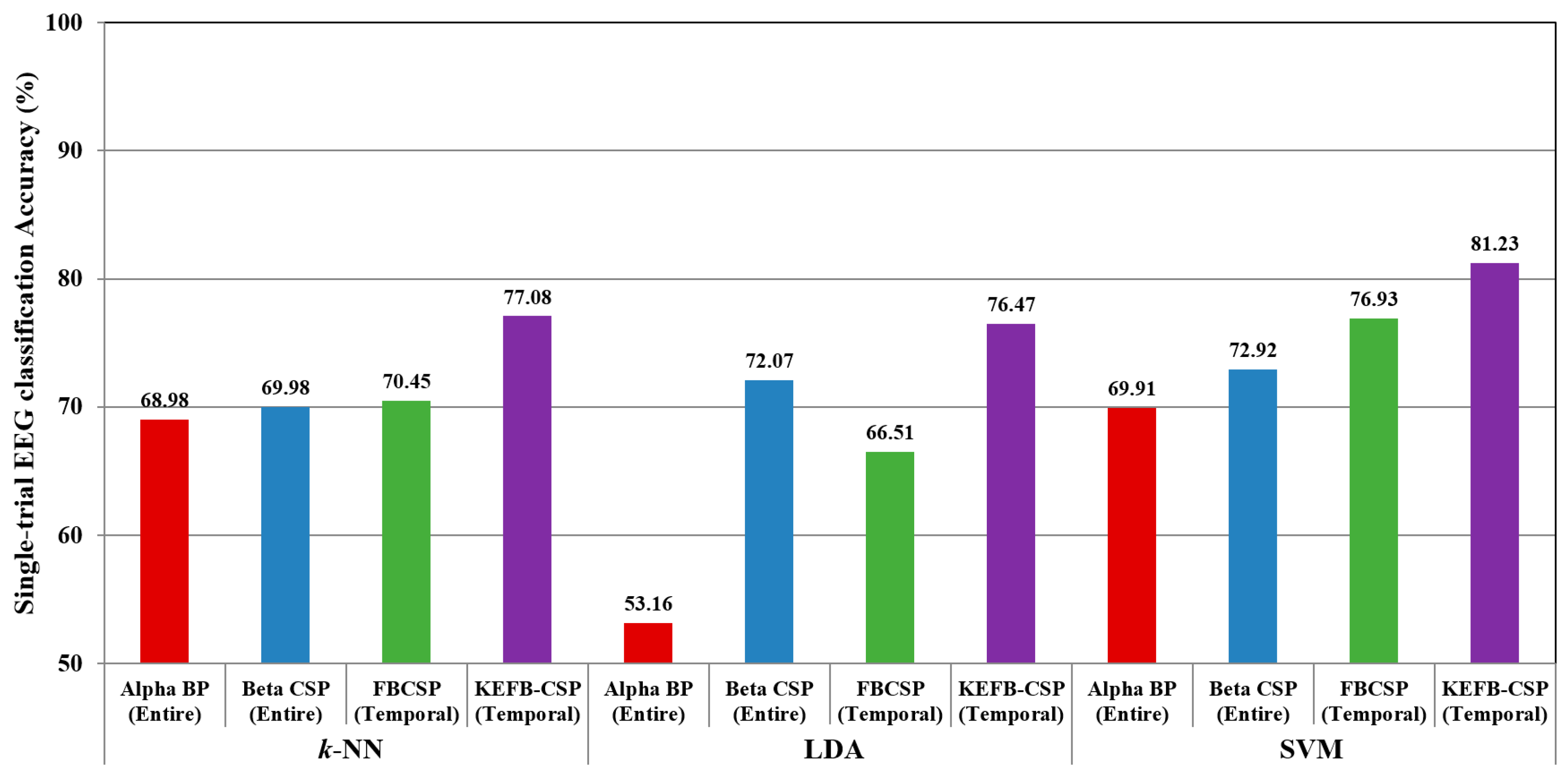
3.1. Comparison with Existing EEG Features for MDD Detection
3.2. Comparison with Different Classifiers in Single-Trial Analysis
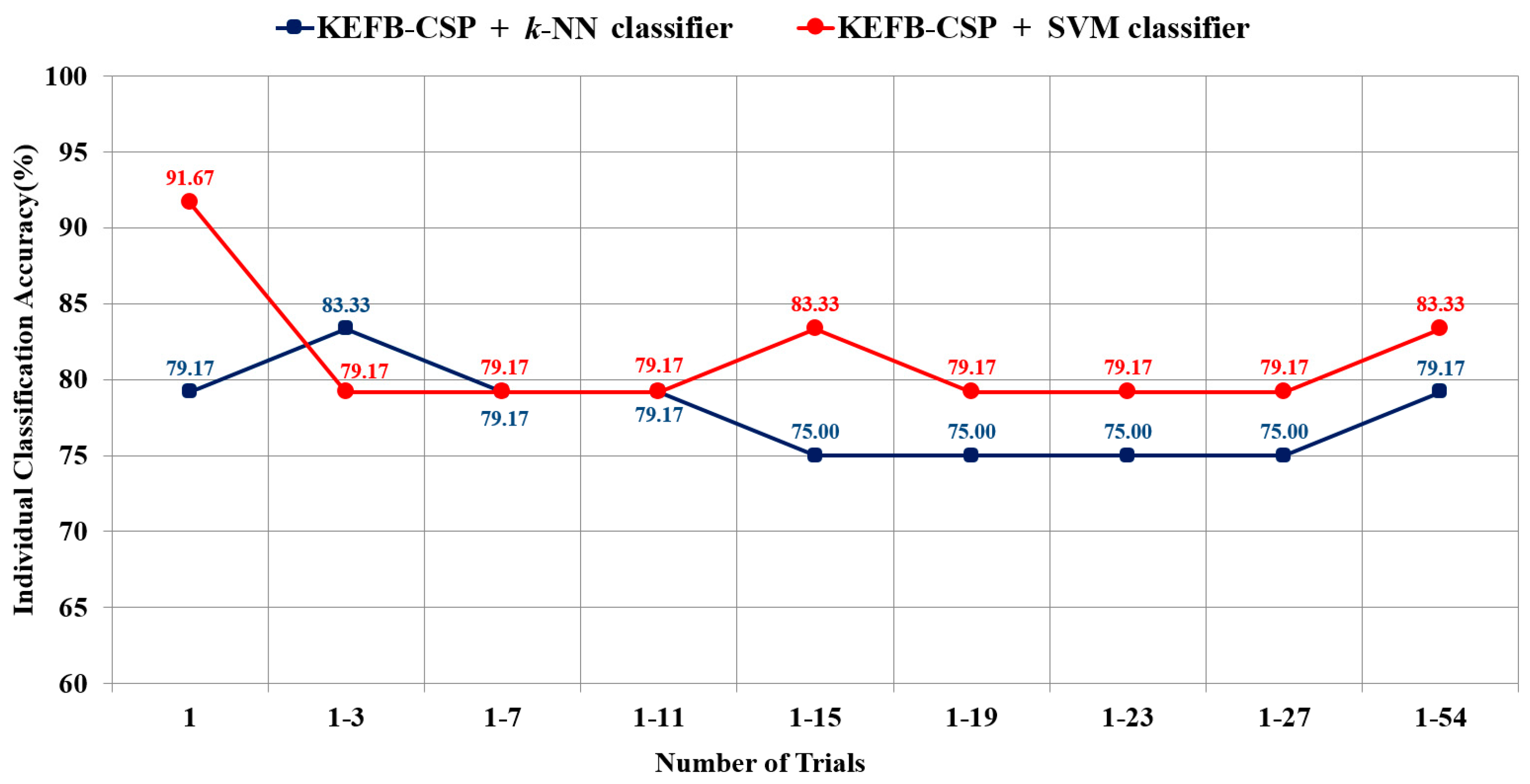
3.3. Individual Classification Using Majority Voting Strategy-Based LOPO-CV
| Question 1: | How many participants can be correctly classified based on the use of KEFB-CSP feature? |
| Question 2: | Can the individual classification accuracy be improved by using multiple single-trial EEG signals? |
3.4. Discussion
4. Conclusions
Acknowledgments
Author Contributions
Conflicts of Interest
References
- Kupfer, D.J.; Frank, E.; Phillips, M.L. Major depressive disorder: New clinical, neurobiological, and treatment perspectives. Lancet 2012, 379, 1045–1055. [Google Scholar] [CrossRef]
- American Psychiatric Association. Diagnostic and Statistical Manual of Mental Disorders DSM-IV-TR Washington 2000; American Psychiatric Association: Arlington, VA, USA, 2000. [Google Scholar]
- Murray, C.J.L.; Lopez, A.D. Alternative projections of mortality and disability by cause 1990–2020: Global burden of disease study. Lancet 1997, 349, 1498–1504. [Google Scholar] [CrossRef]
- Vos, T.; Flaxman, A.D.; Naghavi, M.; Lozano, R.; Michaud, C.; Ezzati, M.; Shibuya, K.; Salomon, J.A.; Abdalla, S.; Aboyans, V.; et al. Years lived with disability (YLDs) for 1160 sequelae of 289 diseases and injuries 1990–2010: A systematic analysis for the Global Burden of Disease Study 2010. Lancet 2012, 380, 2163–2196. [Google Scholar] [CrossRef]
- Papakostas, G.I.; Fava, M. Does the probability of receiving placebo influence clinical trial outcome? A meta-regression of double-blind, randomized clinical trials in MDD. Eur. Neuropsychopharmacol. 2009, 19, 34–40. [Google Scholar] [CrossRef] [PubMed]
- Cuijpers, P.; van Straten, A.; Schuurmans, J.; van Oppen, P.; Hollon, S.D.; Andersson, G. Psychotherapy for chronic major depression and dysthymia: A meta-analysis. Clin. Psychol. Rev. 2010, 30, 51–62. [Google Scholar] [CrossRef] [PubMed]
- Breitenstein, B.; Scheuer, S.; Holsboer, F. Are there meaningful biomarkers of treatment response for depression? Drug Discov. Today 2014, 19, 539–561. [Google Scholar] [CrossRef] [PubMed]
- Goldstein, B.L.; Klein, D.N. A review of selected candidate endophenotypes for depression. Clin. Psychol. Rev. 2014, 34, 417–427. [Google Scholar] [CrossRef] [PubMed]
- Sapolsky, R.M. Depression, antidepressants, and the shrinking hippocampus. Proc. Natl. Acad. Sci. USA 2001, 98, 12320–12322. [Google Scholar] [CrossRef] [PubMed]
- Schmaal, L.; Veltman, D.; van Erp, T.G.; Sämann, P.G.; Frodl, T.; Jahanshad, N.; Loehrer, E.; Tiemeier, H.; Hofman, A.; Niessen, W.J.; et al. Subcortical brain alterations in major depressive disorder: Findings from the ENIGMA Major Depressive Disorder working group. Mol. Psychiatry 2015, 21, 806–812. [Google Scholar] [CrossRef] [PubMed]
- Miller, M.C. Understanding Depression; Harvard Medical School Health Report: Boston, MA, USA, 2013. [Google Scholar]
- Drevets, W.C. Orbitofrontal cortex function and structure in depression. Ann. N. Y. Acad. Sci. 2007, 1121, 499–527. [Google Scholar] [CrossRef] [PubMed]
- Perlis, R.H. Abandoning personalization to get to precision in the pharmacotherapy of depression. World Psychiatry 2016, 15, 228–235. [Google Scholar] [CrossRef]
- Simon, G.E.; Perlis, R.H. Personalized medicine for depression: Can we match patients with treatments? Am. J. Psychiatry 2010, 167, 1445–1455. [Google Scholar] [CrossRef] [PubMed]
- Knott, V.; Mahoney, C.; Kennedy, S.; Evans, K. EEG power, frequency, asymmetry and coherence in male depression. Psychiatry Res. 2001, 106, 123–140. [Google Scholar] [CrossRef]
- Omel’chenko, V.P.; Zaika, V.G. Changes in the EEG-rhythms in endogenous depressive disorders and the effect of pharmacotherapy. J. Hum. Physiol. 2002, 28, 275–281. [Google Scholar] [CrossRef]
- Grin-Yatsenko, V.A.; Baas, I.; Ponomarev, V.A.; Kropotov, J.D. EEG power spectra at early stages of depressive disorders. Clin. Neurophysiol. 2009, 26, 401–406. [Google Scholar] [CrossRef] [PubMed]
- Grin-Yatsenko, V.A.; Baas, I.; Ponomarev, V.A.; Kropotov, J.D. Independent component approach to the analysis of EEG recordings at early stages of depressive disorders. Clin. Neurophysiol. 2010, 121, 281–289. [Google Scholar] [CrossRef] [PubMed]
- Lee, J.S.; Yang, B.H.; Lee, J.H.; Choi, J.H.; Choi, I.G.; Kim, S.B. Detrended fluctuation analysis of resting EEG in depressed outpatients and healthy controls. Clin. Neurophysiol. 2007, 118, 2486–2489. [Google Scholar] [CrossRef] [PubMed]
- Hosseinifard, B.; Moradi, M.H.; Rostami, R. Classifying depression patients and normal subjects using machine learning techniques and nonlinear features from EEG signal. Comput. Methods Programs Biomed. 2013, 109, 339–345. [Google Scholar] [CrossRef] [PubMed]
- Li, X.; Hu, B.; Shen, J.; Xu, T.; Retcliffe, M. Mild depression detection of college students: An EEG-based solution with free viewing tasks. J. Med. Syst. 2015, 39, 187. [Google Scholar] [CrossRef] [PubMed]
- Ahmadlou, M.; Adeli, H.; Adeli, A. Spatiotemporal analysis of relative convergence of EEGs reveals differences between brain dynamics of depressive women and men. Clin. EEG Neurosci. 2013, 44, 175–181. [Google Scholar] [CrossRef]
- Debener, S.; Beauducel, A.; Nessler, D.; Brocke, B.; Heilemann, H.; Kayser, J. Is resting anterior EEG alpha asymmetry a trait marker for depression? Findings for healthy adults and clinically depressed patients. Neuropsychobiology 2000, 41, 31–37. [Google Scholar] [CrossRef] [PubMed]
- Acharya, U.R.; Sudarshan, V.K.; Adeli, H.; Santhosh, J.; Koh, J.E.; Puthankatti, S.D.; Adeli, A. A Novel Depression Diagnosis Index Using Nonlinear Features in EEG Signals. Eur. Neurol. 2015, 74, 79–83. [Google Scholar] [CrossRef] [PubMed]
- Ahmadlou, M.; Adeli, H.; Adeli, A. Fractality analysis of frontal brain in major depressive disorder. Int. J. Psychophysiol. 2012, 85, 206–211. [Google Scholar] [CrossRef] [PubMed]
- Acharya, U.R.; Sudarshan, V.K.; Adeli, H.; Santhosh, J.; Koh, J.E.; Adeli, A. Computer-Aided Diagnosis of Depression Using EEG Signals. Eur. Neurol. 2015, 73, 329–336. [Google Scholar] [CrossRef] [PubMed]
- Subha, D.P.; Paul, J. Classification of EEG signals in normal and depression conditions by ANN using RWE and signal entropy. J. Mech. Med. Biol. 2012. [Google Scholar] [CrossRef]
- He, B.; Lian, J.; Li, G. EEG: A high-resolution new realistic geometry spline Laplacian estimation technique. Clin. Neurophysiol. 2001, 112, 845–852. [Google Scholar] [CrossRef]
- Müller-Gerking, J.; Pfurtscheller, G.; Flyvbjerg, H. Designing optimal spatial filters for single-trial EEG classification in a movement task. Clin. Neurophysiol. 1999, 110, 787–798. [Google Scholar] [CrossRef]
- Ramoser, H.; Müller-Gerking, J.; Pfurtscheller, G. Optimal spatial filtering of single trial EEG during imagined hand movement. IEEE Trans. Rehab. Eng. 2000, 8, 441–446. [Google Scholar] [CrossRef]
- Blankertz, B.; Tomioka, R.; Lemm, S.; Kawanabe, M.; Müller, K.-R. Optimizing spatial filters for robust EEG single-trial analysis. IEEE Signal Process Mag. 2008, 25, 41–56. [Google Scholar] [CrossRef]
- Rozado, D.; Duenser, A.; Howell, B. Improving the performance of an EEG-based motor imagery brain-computer interface using task evoked changes in pupil diameter. PLoS ONE 2016, 27, e0121262. [Google Scholar] [CrossRef] [PubMed]
- Yang, B.; Li, H.; Wang, Q.; Zhang, Y. Subject-based feature extraction by using fisher WPD-CSP in brain-computer interfaces. Comput. Methods Programs Biomed. 2016, 129, 21–28. [Google Scholar] [CrossRef] [PubMed]
- Hsu, W.C.; Lin, L.F.; Chou, C.W.; Hsiao, Y.T.; Liu, Y.H. EEG classification of imaginary lower limb stepping movements based on fuzzy support vector machine with kernel-induced membership function. Int. J. Fuzzy Syst. 2016. [Google Scholar] [CrossRef]
- Liu, Y.H.; Wu, C.T.; Cheng, W.T.; Hsiao, Y.T.; Chen, P.M.; Teng, J.T. Emotion recognition from single trial EEG based on kernel Fisher’s emotion pattern and imbalanced quasiconformal kernel support vector machine. Sensors 2014, 14, 13361–13388. [Google Scholar] [CrossRef] [PubMed]
- Schölkopf, B.; Smola, A.; Müller, K.R. Nonlinear component analysis as a kernel eigenvalue problem. Neural Comput. 1998, 10, 1299–1319. [Google Scholar] [CrossRef]
- Ma, X.; Zabaras, N. Kernel principal component analysis for stochastic input model generation. J. Comput. Phys. 2011, 230, 7311–7331. [Google Scholar] [CrossRef]
- Liu, Y.H.; Wang, S.H.; Hu, M.R. A self-paced P300 healthcare brain-computer interface system with SSVEP-based switching control and kernel FDA+SVM-based detector. Appl. Sci. 2016, 6, 142. [Google Scholar] [CrossRef]
- Liu, Y.H.; Liu, Y.C.; Chen, Y.J. Fast support vector data descriptions for novelty detection. IEEE Trans. Neural Netw. 2010, 21, 1296–1313. [Google Scholar] [PubMed]
- Lecrubier, Y.; Sheehan, D.V.; Weiller, E.; Amorim, P.; Bonora, I.; Harnett Sheehan, K.; Janavs, J.; Dunbar, G.C. The Mini International Neuropsychiatric Interview (MINI). A short diagnostic structured interview: Reliability and validity according to the CIDI. Eur. Psychiatry 1997, 12, 224–231. [Google Scholar] [CrossRef]
- Compumedics USA, Ltd. Neuroscan FAQs. Available online: http://compumedicsneuroscan.com/wp-content/uploads/3502C-Neuroscan-FAQs.pdf (accessed on 31 May 2017).
- Liu, S.I.; Yeh, Z.T.; Huang, H.C.; Sun, F.J.; Tjung, J.J.; Hwang, L.C.; Shih, Y.H.; Yeh, A.W. Validation of Patient Health Questionnaire for depression screening among primary care patients in Taiwan. Compr Psychiatry 2011, 52, 96–101. [Google Scholar] [CrossRef] [PubMed]

| Initialize Free Parameter(s) of the Chosen Method |
|---|
|
| Method | Descriptions | Values to Be Searched |
|---|---|---|
| BP | non | none |
| Coherence | non | none |
| GPFD | : time delay | |
| CSP | : number of chosen new signals | if is even; if is odd |
| FBCSP | : number of chosen new signals | if is even; if is odd |
| KEFB-CSP | : number of chosen new signals | if is even; if is odd |
| : number of chosen eigenvectors : width of the Gaussian kernel of KPCA | ; is searched within the range from 1 to 100. |
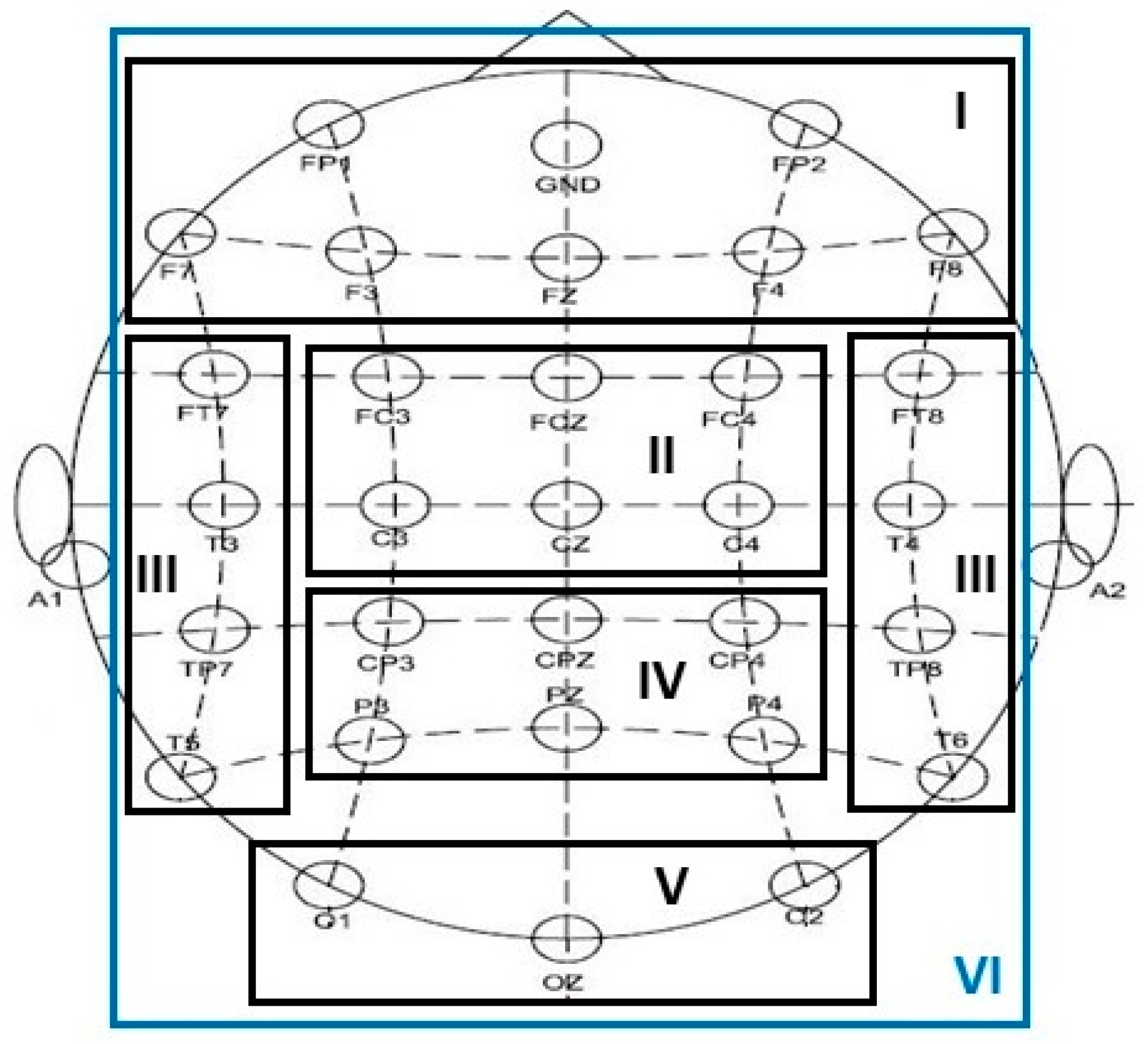
| Brian Area (Montage) | Frontal (I) | Central (II) | Temporal (III) | Parietal (IV) | Occipital (V) | Entire (VI) |
|---|---|---|---|---|---|---|
| Channels | FP1, FP2, Fz, F3, F4, F7, F8 | FCz, FC3, Cz, FC4, C3, C4 | FT7, T3, TP7, T5, FT8, T4, TP8, T6 | CP3, CPz, CP4, P3, Pz, P4 | O1, Oz, O2 | all channels |
| N | 7 | 6 | 8 | 6 | 3 | 30 |
| EEG Features | Frontal | Central | Temporal | Parietal | Occipital | Entire | |
|---|---|---|---|---|---|---|---|
| BP | Delta | 61.03 | 50.23 | 55.32 | 57.02 | 48.15 | 55.02 |
| Theta | 58.71 | 49.61 | 59.02 | 52.77 | 49.76 | 57.02 | |
| Alpha | 52.93 | 52.31 | 65.81 | 45.44 | 49.76 | 68.98 | |
| Beta | 48.68 | 53.08 | 59.49 | 54.55 | 41.97 | 50.30 | |
| Gamma | 43.59 | 43.67 | 60.33 | 54.24 | 52.16 | 44.29 | |
| Coherence | Delta | 46.22 | 50.93 | 43.83 | 46.84 | 44.06 | 48.69 |
| Theta | 47.22 | 49.69 | 42.67 | 48.07 | 46.76 | 51.93 | |
| Alpha | 52.01 | 51.39 | 47.45 | 46.53 | 49.07 | 57.10 | |
| Beta | 49.54 | 48.77 | 44.83 | 48.38 | 46.14 | 47.15 | |
| Gamma | 48.77 | 46.91 | 54.24 | 47.76 | 55.56 | 47.69 | |
| GPFD | 49.53 | 43.13 | 44.83 | 42.59 | 54.55 | 36.57 | |
| CSP | Delta | 49.31 | 52.16 | 55.02 | 57.64 | 47.22 | 53.01 |
| Theta | 55.63 | 56.17 | 56.32 | 57.33 | 41.74 | 59.56 | |
| Alpha | 58.33 | 63.11 | 60.26 | 60.33 | 52.62 | 64.58 | |
| Beta | 47.99 | 59.95 | 60.80 | 56.40 | 50.69 | 69.98 | |
| Gamma | 50.23 | 43.05 | 64.66 | 61.57 | 53.62 | 64.66 | |
| FBCSP | 4-Hz width | 56.71 | 60.65 | 70.45 | 64.43 | 50.08 | 65.35 |
| 2-Hz width | 60.65 | 60.19 | 69.44 | 60.42 | 52.31 | 67.67 | |
| FBCSP+PCA | 4-Hz width | 60.11 | 66.67 | 75.00 | 65.28 | 58.18 | 69.75 |
| KEFB-CSP | 4-Hz width | 62.73 | 69.29 | 77.08 | 67.90 | 57.06 | 72.37 |
| Initialize Initialize free parameter(s) of the chosen method Initialize ; |
|
| Participant | Classified as D | Classified as H | Correct Ratio | Participant | Classified as D | Classifier as H | Correct Ratio |
|---|---|---|---|---|---|---|---|
| 1 (D) | 15 | 0 | 1 | 13 (H) | 4 | 11 | 0.73 |
| 2 (D) | 15 | 0 | 1 | 14 (H) | 5 | 10 | 0.67 |
| 3 (D) | 15 | 0 | 1 | 15 (H) | 9 | 5 | 0.33 |
| 4 (D) | 0 | 15 | 0 | 16 (H) | 4 | 12 | 0.8 |
| 5 (D) | 15 | 0 | 1 | 17 (H) | 0 | 15 | 1 |
| 6 (D) | 6 | 9 | 0.4 | 18 (H) | 4 | 11 | 0.73 |
| 7 (D) | 11 | 4 | 0.73 | 19 (H) | 0 | 15 | 1 |
| 8 (D) | 15 | 0 | 1 | 20 (H) | 0 | 15 | 1 |
| 9 (D) | 15 | 0 | 1 | 21 (H) | 15 | 0 | 0 |
| 10 (D) | 15 | 0 | 1 | 22 (H) | 0 | 15 | 1 |
| 11 (D) | 9 | 6 | 0.6 | 23 (H) | 0 | 15 | 1 |
| 12 (D) | 15 | 0 | 1 | 24 (H) | 0 | 15 | 1 |
| Number of correctly classified patients | 10 | Number of correctly classified controls | 10 | ||||
| Sensitivity = 83.33 % (10/12) | Specificity = 83.33 % (10/12) | ||||||
| Individual classification accuracy = 83.33 % (20/24) | |||||||
| Participant | Classified as D | Classified as H | Correct Ratio | Participant | Classified as D | Classifier as H | Correct Ratio |
|---|---|---|---|---|---|---|---|
| 1 (D) | 1 | 0 | 1 | 13 (H) | 0 | 1 | 1 |
| 2 (D) | 1 | 0 | 1 | 14 (H) | 0 | 1 | 1 |
| 3 (D) | 1 | 0 | 1 | 15 (H) | 1 | 0 | 0 |
| 4 (D) | 1 | 0 | 1 | 16 (H) | 0 | 1 | 1 |
| 5 (D) | 1 | 0 | 1 | 17 (H) | 0 | 1 | 1 |
| 6 (D) | 1 | 0 | 1 | 18 (H) | 1 | 0 | 0 |
| 7 (D) | 1 | 0 | 1 | 19 (H) | 0 | 1 | 1 |
| 8 (D) | 1 | 0 | 1 | 20 (H) | 0 | 1 | 1 |
| 9 (D) | 1 | 0 | 1 | 21 (H) | 0 | 1 | 1 |
| 10 (D) | 1 | 0 | 1 | 22 (H) | 0 | 1 | 1 |
| 11 (D) | 1 | 0 | 1 | 23 (H) | 0 | 1 | 1 |
| 12 (D) | 1 | 0 | 1 | 24 (H) | 0 | 1 | 1 |
| Number of correctly classified patients | 12 | Number of correctly classified controls | 10 | ||||
| Sensitivity = 100% (12/12) | Specificity = 83.33 % (10/12) | ||||||
| Individual classification accuracy = 91.67 % (22/24) | |||||||
| Methods and Parameters | 1 | 3 | 7 | 11 | 15 | 19 | 23 | 27 | 54 | |
|---|---|---|---|---|---|---|---|---|---|---|
| KEFB-CSP | 3 | 3 | 3 | 3 | 3 | 3 | 3 | 3 | 3 | |
| 28 | 11 | 15 | 24 | 16 | 28 | 16 | 28 | 60 | ||
| d | 5 | 2 | 2 | 1 | 4 | 1 | 3 | 3 | 3 | |
| SVM | 0.4 | 0.1 | 21 | 3.5 | 1.5 | 2 | 4.5 | 5.3 | 4 | |
| C | 100 | 100 | 100 | 100 | 100 | 100 | 50 | 50 | 100 | |
© 2017 by the authors. Licensee MDPI, Basel, Switzerland. This article is an open access article distributed under the terms and conditions of the Creative Commons Attribution (CC BY) license (http://creativecommons.org/licenses/by/4.0/).
Share and Cite
Liao, S.-C.; Wu, C.-T.; Huang, H.-C.; Cheng, W.-T.; Liu, Y.-H. Major Depression Detection from EEG Signals Using Kernel Eigen-Filter-Bank Common Spatial Patterns. Sensors 2017, 17, 1385. https://doi.org/10.3390/s17061385
Liao S-C, Wu C-T, Huang H-C, Cheng W-T, Liu Y-H. Major Depression Detection from EEG Signals Using Kernel Eigen-Filter-Bank Common Spatial Patterns. Sensors. 2017; 17(6):1385. https://doi.org/10.3390/s17061385
Chicago/Turabian StyleLiao, Shih-Cheng, Chien-Te Wu, Hao-Chuan Huang, Wei-Teng Cheng, and Yi-Hung Liu. 2017. "Major Depression Detection from EEG Signals Using Kernel Eigen-Filter-Bank Common Spatial Patterns" Sensors 17, no. 6: 1385. https://doi.org/10.3390/s17061385
APA StyleLiao, S.-C., Wu, C.-T., Huang, H.-C., Cheng, W.-T., & Liu, Y.-H. (2017). Major Depression Detection from EEG Signals Using Kernel Eigen-Filter-Bank Common Spatial Patterns. Sensors, 17(6), 1385. https://doi.org/10.3390/s17061385