Review

Emerging Ranaviral Infectious Diseases and Amphibian Decline

Department of Microbiology and Immunology, University of Rochester Medical Center, Rochester, NY 14642, USA
Diversity 2010, 2(3), 314-330; https://doi.org/10.3390/d2030314
Submission received: 1 December 2009 / Accepted: 25 February 2010 / Published: 26 February 2010

Abstract

:
Infectious diseases caused by ranaviruses (RV, family Iridoviridae) not only affect wild amphibian populations but also agriculture and international animal trade. Although, the prevalence of RV infections and die offs has markedly increased over the last decade, it is still unclear whether these viruses are direct causal agents of extinction or rather are the resulting (secondary) consequences of weakened health of amphibian populations leading to increased susceptibility to viral pathogens. In either case, it is important to understand the critical role of host immune defense in controlling RV infections, pathogenicity, and transmission; this is the focus of this review.

Graphical Abstract

1. Introduction

Emerging infectious diseases (EIDs) are generally defined as diseases that are either newly recognized, novel in a population, or rapidly increasing in incidence, virulence, or geographic range [1]. While the direct impact of EIDs in human health is usually well appreciated, their threat to biodiversity is less well known. What is known, however, is that EIDs increasingly affect wildlife all over the world. In addition to playing an important ecological role, EIDs pose important problems for the conservation of endangered species, domestic and captive animals, and ultimately humans themselves [2].
With regard to amphibians, a dramatic worldwide decline of populations of multiple species was first noted about 30 years ago [3,4,5]. Since then, this decline has continued at an alarming rate. According to the most recent 2008 global assessment (globalamphibians.org), nearly one-third (32%), or 2,109 species of the world’s 6,593 amphibian species are threatened, compared to12% of avian and 23% of mammalian species. Some 122 amphibian species have become extinct since 1980, and the population size of at least 43% of species is declining. The causes of this declines are likely to be multiple and complex. They include already well recognized causes of extinction such as habitat destruction, overexploitation, and the introduction of exotic or predator species, as well as other less defined threats to amphibians, including climate change, increase of ultraviolet-B, chemical contaminants and EIDs [6,7,8,9,10]. In recent years, evidence for the critical involvement of EID in the decline of amphibian populations has grown and become more convincing, especially in the case of the chytrid fungus, Batrachochytrium dendrobatidis (Bd). Infections and mass die-offs closely associated with this pathogen have been reported for more than 350 amphibian species distributed all over the world [11,12,13].
Viral infections caused by ranaviruses (RVs) have also become more prevalent and are increasingly associated with mass amphibian dies-off of wild as well as captive populations [14,15]. In a recent epizootiology study from the US, RV infections were reported to be associated with nearly half (48%) of all amphibian mortality events recorded from 1996 to 2001 [16]. Therefore, despite the spotlight shinning on Bd, the importance of RV infections cannot be ignored. In fact, some type of synergy between these two pathogens should perhaps be considered more carefully, owing to the detection of both of these pathogens in one study on infected zoo animals [17]. As such, both Bd and RV have been added to the list of ‘notifiable’ diseases by the World Organization for Animal Health (OIE; http://www.oie.int/eng/maladies/en_classification2009.htm?e1d7). This means that international trade of live amphibians and related products now requires health certifications to be applied according to OIE standards, making it obligatory that both the public and the OIE be notified about the detection of either disease.
Despite the goodly amount of data supporting the important role played by EIDs, it remains unclear if these diseases are among the primary causes responsible for the extinction crisis faced by amphibians, or if their rapidly increasing prevalence is the consequences of environmental and host variables upon which are superimposed these two pathogens. In particular, it is unknown why some species are susceptible, whereas others are tolerant or even resistant to one or the other pathogen. More fundamentally, amphibian host immune defenses to these pathogens are still poorly defined and studied. The present review focuses on host immune responses against RVs for which characterization of host-pathogen interactions has made more progress than for Bd. The review also discusses recent evidence for the critical role of host immune defenses in the control of these viruses, and also perhaps in the progression and dissemination of RV infections.

2. Ranavirus Taxonomy, Life Cycle, Host Range

RVs are large double stranded DNA viruses that belong to the Iridoviridae family, which include 4 other genera, 2 that infect invertebrates (Iridovirus and Chloriridovirus) and 2 that infect ectothermic vertebrates (Lymphocystivirus and Megalocytivirus). There are several recent excellent reviews on iridoviruses [14,18]. Therefore, I will only succinctly point out several salient features of RV exemplified by Frog virus 3 (FV3), the virus type of the RV genus and one of the most extensively studied of the Iridoviridae family [19]. FV3 was originally isolated from the native North American leopard frog Rana pipiens. FV3 is a large (120 to 169 nm) dsDNA icosahedral virus that, like other iridoviruses, has the peculiarity of replicating both in the nucleus and in the cytoplasm of infected cells [20]. Another distinctive feature of FV3 and other iridoviruses is that a viral envelope added onto virions budding from the plasma membrane of infected cells is not required for infectivity. As such naked virus particles released during cell lysis are infectious. However, naked viral DNA is not infectious, which suggests that virion-associated proteins are required for infection. FV3 has a linear double-stranded genome of 106 kb in length that encodes at least 98 non-overlapping open reading frames with a G+C content of 55%. A last feature unique among animal viruses is that genomes of FV3 and other iridoviruses are both circularly permuted and terminally redundant [21]. As a result, terminal repeats account for between 5 to 50% of the full viral genome.
So far, three RV species infecting amphibians have been identified based on hosts range distributions, nucleotide sequences comparisons, and protein and RFLP profiles [19,22]. The Bohle virus (BHV), isolated from the native Australian frog, Limnodynastes ornatus, remains confined to Australia [19,23]. The Ambystoma trigrinum virus (ATV), initially isolated from salamanders in southern Arizona, infects ambystoid salamanders in the US and Canada [24,25]. In contrast to the relatively limited geographic distribution these two RV species, FV3 and several FV3-like diseases are widespread all over the world. The genomes of several ranavirus species and strains have been fully sequenced including FV3 (Genbank #AY548484), TVF (AF389451), ATV (AY150217), SGVI (AY521625), and GIV (AY666015) reviewed in [15].

3. Viral Immunity in Mammals

By and large, information concerning immunity against viruses comes from mammalian species, and the characteristics of immune responses vary depending on the virus considered (e.g., RNA versus DNA virus). A short introduction to the main features of the immune response against DNA virus such as poxvirus is provided here to help the uninitiated reader (for a general review see [26]). The host immune defenses following viral infection involve the integration and coordination of the innate and adaptive immune systems. Innate immunity is important as a first line of defense to limit the progression of the virus during the early course of infection, as well as to initiate a potent adaptive immune response. Among innate effector cells involved in viral immunity are natural killer (NK) cells. These large granulocytic leukocytes play an important role based on their ability to directly kill infected cells and by producing cytokines such as IFN-γ that has antiviral properties and activates other immune cells [27]. Infected cells also produce type I interferons that inhibit virus propagation by preventing protein synthesis as well as activating immune cells. Macrophages are another crucial cell effector involved in early stages of infection; they act as phagocytic cells that engulf and digest pathogens or infected dying cells in a stimulus-dependent but non-antigen-specific manner. In addition, macrophages recruit more phagocytic and effector cells to the area of infection by secreting chemokines such as interleukin-8 (IL-8) and proinflammatory cytokines such as interleukin-1β (IL-1β) and TNF-α (reviewed in [28,29]). Macrophages are also implicated in adaptive immune responses as professional antigen presenting cells (APCs) that can process viral antigens through Major Histocompatibility Complex (MHC) class I and class II presentation pathways that then activate CD8 and CD4 T cell effectors, respectively. APCs, which also include the very efficient dendritic cells, provide additional signals through co-stimulatory surface molecules (e.g., CD86 or B7) that are required for the proper activation of T cells.
The adaptive immune response starts by the expansion of the antigen-specific T cell clones and their differentiation into effectors. CD8 T cells give rise to cytotoxic T cells (CTL) that can kill virally infected cells by recognizing viral antigen peptide complex with MHC class I at their surface (reviewed in [30]). CTL produce also large amount of IFNγ and other cytokines such as TNF-α. CD4 T cells differentiate into various T helper effectors (Th1, Th2, etc.) that produce cytokines important for the production of CTLs (e.g., IL-2) and B cells. The second arm of the adaptive immune response is constituted of B cells that differentiate into plasma cells and produce antibodies that can directly neutralize the virus or promote antibody-dependent cell-mediated cytotoxicity. The peak of the adaptive response usually lead to the clearance of the virus and is followed by a contraction phase during which most of the T cell effectors are eliminated by programmed cell death, except a minor fraction of memory T cells that can survive for a long time and can respond faster to a second infection. There are also memory B cells and long-lived antibody-secreting plasma cells.
The relative importance and interaction of the different immune cells vary depending of the virus considered and is still the subject of active research by numerous scientists.

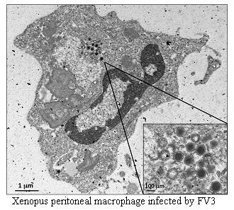
4. The Xenopus Laevis Immune System

Although the basic molecular elements of innate (e.g., Toll-like receptors) and adaptive immunity (Recombination Activating proteins [Rag], Immunoglobulin [Ig], T cell receptors [TCR] and MHC genes) are present in the entire Gnathostoma subphylum [31], the role of different lymphocyte subsets in anti-viral immunity of ectothermic vertebrates is still poorly characterized.
Most of the fundamental knowledge about the immune system in amphibian comes from the extensive studies in the South African clawed frog Xenopus laevis (reviewed in [32]). Studies with X. laevis over several decades have revealed the fundamental conservation of the immune system and its high degree of similarity to the mammalian immune system. In addition, the recent sequencing of the full genome of the sister species Silurana (Xenopus) tropicalis has highlighted the extent of gene similarity with mammals and allowed identification of many immunologically-relevant gene homologs [33].
Like mammals, X. laevis possesses lymphoid organs such as thymus and spleen, and displays a typical adaptive immune system mediated by B and T lymphocytes, as well as an innate immune system that includes NK cells and most other leukocyte subsets: monocyte, macrophages, neutrophils, eosinophils, and basophils. At the gene level, all the important innate immune genes (e.g., Toll-like receptors), cytokines, chemokines and their receptors are present in X. laevis and S. tropicalis. The somatic repertoire of TCRs and Ig receptors are generated in RAG-dependent manner, and B cells produce antibodies of IgM, IgD, IgY (IgG-equivalent) and IgX isotypes (reviewed in [34]). Finally, as in mammals, the development and function of the adaptive immune system depends on MHC molecules. MHC class I-restricted cytotoxic and MHC class-II helper T cell responses have been identified in X. laevis (reviewed in [32]).
A unique feature of X. laevis that is likely shared by all anuran species is the presence of distinct immune systems in the two developmental life stages, larval and adults, as well as the dramatic changes occurring during the metamorphosis. For example, X. laevis larvae and adults display distinct Ig repertoires. The thymus is first colonized by embryonic stem cells a few days after fertilization [35]. During metamorphosis, the thymus loses about 90% of its lymphocytes [36,37,38]. This loss is followed by a second wave of stem cell immigration [39]. The embryonic and larval periods of thymocyte differentiation take place in different environments during metamorphosis, since the whole organism is remodeled and many new proteins are expressed that could be considered antigenic by the larval immune system [40]. The emerging adult lymphocytes, therefore, are likely to be subjected to a new wave of negative selection by the adult “self”, resulting in a new balance of self-tolerance.
Although both larvae and adults are immunocompetent and have CD8 T cells, larvae lack significant expression of MHC class I [41,42] and LMP7 genes [43] until metamorphosis; this strongly suggests an absence of class I-restricted education during larval life. Perhaps related to the suboptimal expression of MHC class I, NK cells are not detected until late in larval stage at the time of metamorphosis. Furthermore, cell mediated cytotoxicity involving either CTLs or NK cells cannot be detected in larvae and becomes significant only several weeks after metamorphosis is completed. The relative weakness of the larval adaptive immune system extends to antibody production and T helper function, since the switch from IgM to IgY antibodies of higher affinity is poor in larvae [34,44]. Therefore, the existing data strongly suggest that the larva displays weak adaptive cell effectors, and most likely relies critically on its innate immune system (Table 1).
Table 1. Overview of the larval and adult immune system of X. laevis.
CharacteristicsLarvaAdult
MoleculesMHC class I
MHC class II
No to reduced expression
B cell only
Present
B and T cells
CellsCD8 T-cells
NK cells
Present
Present at late stages
Better
Yes
Immune
functions
Mixed lymphocyte reaction
Cytoxicity (CTL & NK cells)
IgM to IgY switch
Viral immunity
Poor
Not demonstrated
Incomplete
Weak
Present
Acute
Better
Potent

5. Immunity to FV3 in X. laevis and S. tropicalis

5.1. Adults

X. laevis has become an important laboratory model for studying immunity to, and pathogenesis of ranaviruses. Our original published study [45] revealed that FV3 infection of adult X. laevis is mildly pathogenic (~10−20% of adults infected with 107 pfu die within a month). Infected frogs that die exhibit both edema and hemorrhages. Frogs that survive the FV3 infection show only transitory signs of pathology (e.g., loss of appetite, cutaneous erythema of the legs, skin shedding). These symptoms disappear within a few weeks. Similar symptoms and resistance were observed using Ambystoma tigrinum virus (ATV; Robert, unpublished obs.). Interestingly, whereas viral DNA is detected by PCR in most tissues of infected moribund frogs, the kidney is the primary target of FV3 in X. laevis [46]. Histology of tissues from infected frogs has confirmed that the X. laevis kidney is the primary target of FV3. We were able to further localize FV3 in kidney tissue by immunohistology using an anti-FV3 monoclonal antibody [47]. Extensive necrosis of proximal tubules in parallel with accumulation of detectable viruses is typically observed during early stages of infection [46]. The kinetics of viral clearance, as measured by loss of FV3 DNA, correlates with the disappearance of pathological and behavioral symptoms and is consistent with the involvement of anti-viral immunity. We have observed similar resolution of symptoms of infection in a preliminary study with FV3 in S. tropicalis. Like X. laevis, adult S. tropicalis are relatively resistant to infection with a high dose of FV3 (i.p. injection of up to 107 pfu) and show transitory symptoms of infection, except a few individuals (~10%) that did die from acute infection. Like X. laevis, the kidney appears to be the main tissue infected in S. tropicalis. However, the virus clearance in this organ is significantly slower than it is in X. laevis (e.g., viral DNA still detected four weeks post-infection; Robert unpublished obs.).
With respect to adaptive immune responses, our studies with X. laevis clearly indicate that both antibodies [48,49] and CD8 T cell responses [50,51] play a critical role in adult resistance to FV3 infection. Both sub-lethal γ-irradiation and monoclonal antibody depletion of CD8 T cells markedly increases the susceptibility of adult X. laevis to FV3 infection. We have developed a flow cytometry assay using bromodeoxyuridine (BrdU) incorporation to assess lymphocyte proliferative responses in vivo, and have detected significant proliferation of CD8 T cells 6 days after FV3 infection [50]. Tissue infiltration of activated CD8 T cells was analyzed by immunohistology. Following primary infection, CD8 T cells significantly proliferate in the spleen and accumulate in infected kidneys from day 6 onward in parallel with virus clearance. Earlier proliferation and infiltration associated with faster viral clearance were observed during a secondary infection. However, there was a decrease of CD8 T cells proliferating in the spleen and infiltrating in the kidneys compared to the primary response. Therefore, although these results provide evidence of a protective CD8 T cell response in X. laevis against FV3, they also suggest the involvement of other effector mechanisms during a re-infection.
In this regard, it is noteworthy that we have only been able to detect anti-FV3 IgY antibodies during secondary viral infection [45,48]. No specific signal for anti-FV3 IgM or IgY antibodies can be detected (by ELISA) for up to a month in the sera of frogs infected for the first time. However, increased mRNA expression of IgY and Activation-induced Cytidine deaminase, an enzyme essential for the maturation, indicates that B cell are activated during primary FV3 infection [49]. Therefore, more study is needed to determine if a primary infection with FV3 affects production of antibodies at a too low titer or at a too low affinity to be detected by our assay.
Despite fundamental similarities of the immune systems of X. laevis and mammals, relative to mammals, affinity maturation in X. laevis is poor. For example, the affinity of X. laevis IgY antibody against dinitrophenol (DNP), a model antigen, increases less than 10 times during a humoral response in contrast to more than a 10,000 fold affinity increase in mammals (reviewed in [44]). Nevertheless, in this anuran, neutralizing antibodies are generated following a secondary infection with FV3, and immunological memory to FV3 lasts at least 15 months [48]. Therefore, when examined in a physiological context involving a natural viral pathogen, antibodies generated by X. laevis do appear to provide protective defenses against subsequent viral infection even though these antibodies are of a weaker affinity than their mammalian counterpart (Table 2).
Compared with adaptive immunity, much less in known about the role of innate immunity as a defense against FV3 infection. As a first step, we have investigated (using microscopy, flow cytometry and RT-PCR) the contribution of peritoneal leukocytes (PLs) in the immune response to FV3 by adult X. laevis (Morales et al., submitted). Besides the active involvement of NK cells during early stages of FV3 infection (i.e., before the onset of T cell responses), this study reveals that macrophage-like cells are likely to be crucial during the early stage of infection before the T cell response can be initiated (about 6 days after infection). The total number and the relative abundance of macrophage-like cells rapidly increases from days 1 to 6 post-infection, and these cells display an activated morphology including phagocytic vacuoles. FV3 infection also induces a rapid up-regulation of pro-inflammatory genes including Arginase 1, IL-1β and TNF-α that are likely to be produced in large part by macrophages.
In addition, this submitted study provides evidence of the particular permissiveness of certain PLs to FV3 infection. Notably, the persistence of transcriptionally inactive FV3 genomic DNA in PLs may explain the occurrence of asymptomatic infection and suggests that FV3 is capable of covert infection. Although some PLs are susceptible to FV3 infection as evidenced by apoptotic cells, active FV3 transcription and the detection of viral particles by electron microscopy, the infection is weaker (fewer infectious particles), more transitory and involves a lower fraction (less than 1%) of PLs than the kidney, the main site of infection. However, viral DNA remains detectable in PLs for at least 3 weeks post-infection; this is past the point of viral clearance observed in the kidneys. This suggests that although PLs are actively involved in anti-FV3 immune responses, some of these cells can be permissive and harbor quiescent, asymptomatic FV3.

5.2. Larvae

Whereas X. laevis adults resist FV3 infection even at high viral concentrations, larvae are considerably more susceptible and most of them (>90%) die within a month when infected by i.p. injection, with as few as 100 pfu [45]. Larvae are also very susceptible to infection by water-born FV3 [46]. So far, our attempts to detect any type of larval anti-FV3 immune response have had limited success. We have found that as in adults during primary infection, larval B cells up-regulated IgY and AID mRNA expression [49]. However, our attempt to detect specific anti-FV3 antibodies failed. Recently, we also detected up-regulation of several pro-inflammatory genes by RT-PCR such as IL-1β and TNF-α (Robert unpublished obs), but these preliminary data need to be substantiated. So far, attempts to detect secondary antibody responses in adult frogs that were exposed to FV3 at larval stages (either by infection or by immunization with heat-killed FV3 in the presence of adjuvant) have been inconclusive (Robert unpublished observation). Table 2 summarizes what is currently known about anti-FV3 immune responses in X. laevis.
Table 2. Overview of anti-FV3 immune response in X. laevis.
AdultsLarvae
Primary InfectionSecondary Infection
Symptoms disappearance2−3 wks3−5 daysLong lasting, >80% death
Innate Immune responses1 dpi* Macrophages
3 dpi NK cells
PLs possible APCsUp-regulation TNFα, IL-1β, Arg-1
Clearance FV31 month1 weekPoor
CD8 T cell proliferation (BrdU)6 dpi3 dpi but no increase in amplitudeIFNγ
Cell infiltration in the kidneys6 dpi CD8 T cells3 dpi but less CD8 T cellsIFNγ
Antibody responseUndetectableDetected 10−14 dpi memory >15 monthsNot detected so far
AID up-regulation9 dpi3 dpi6−7 dpi.

6. The Immunity to RVs in Other Amphibian Species

The few studies on other anuran species are consistent with those on X. laevis in that they suggest a better ability of adult frogs to develop a specific and protective adaptive immune response against RVs, than larvae or newly metamorphosed juveniles who are more vulnerable presumably because their immune systems are immature and/or less efficient. Notably, specific antibodies against RVs have been detected in the serum of the marine toad Bufo marinus from Australia and Venezuela [52]. Also a prior exposure of bullfrogs (Rana catesbeiana) to FV3 (relatively avirulent in this species) protects against a subsequent challenge with RCV-Z, a more virulent FV-3-like virus strain [53]. Importantly, results from these two studies imply the involvement of immunological memory, which means that as in mammals, the adaptive immune system of adult frogs provides a faster and more potent protection against a second RV infection. Directional selection of a particular MHC class I haplotype in a wild population of Rana temporaria in UK provides a genetic evidence of the important involvement of the adaptive immune system [54].
Although almost nothing is known about innate immune response to RVs in anuran species other than X. laevis, leukocyte accumulations at the site of infection reported on necropsies are consistent with the involvement of innate cell effectors [16,55]. Interestingly, several antimicrobial peptides produced at the surface of the skin of Rana pipiens and Rana catesbeiana have been shown to inactivate FV3 in vitro, which suggests that these compounds can contribute to innate defenses against RV infection [56,57].
Several studies are also consistent with a higher susceptibility of larvae and metamorphs to RVs. Unlike Rana pipiens adults that survive infection by injection of 106 pfu of FV3, embryos and tadpoles succumb to injections of doses as small as 900 pfu [58]. Several RV reported outbreaks appear to preferentially affect tadpoles [59,60,61,62,63]. The massive death reported in ranaculture, the practice of farm-raising bullfrogs (R. catesbeiana) for scientific and culinary purposes, also mainly targets tadpoles and individuals that have just metamorphosed [64,65].
In the case of urodele amphibians, some studies have been performed on host defenses to ATV using the tiger salamander, Ambystoma tigrinum as an animal model [25,66,67]. ATV is highly virulent to its salamander host, and infects both larvae and adults [68,69]. Symptoms of infection are typical for RV: lethargy, slow movement, red spots or swollen areas near the gills and hind limbs. Hemorrhages and ulceration of the skin, edema, swollen and pale livers, and fluid-filled intestines are also seen [70]. ATV virulence fluctuates in relation to temperature; animals kept at 26 ºC survive infection whereas lower temperatures (10 ºC) at which immune responses are likely to be inefficient, result in mortality of almost all infected animals [71]. Attempts to determine if antimicrobial peptides are involved in the immune response of tiger salamanders to ATV revealed inconsistent effects [72]. Whereas some natural mixtures of peptides from tiger salamanders inhibited reduced ATV-induced viral plaques, not all preparations of skin peptides were equally effective. However, some evidence of innate immune responses in axolotls (Ambystoma mexicanum) have been obtained using microarray technology [73]. These limited studies of Abystoma defense responses to ATV should be considered in the context of the finding that in vivo and in vitro immune responsiveness of urodeles are noticeably less "robust" than those of anurans (reviewed in [74,75]).
A last set of information implying effective amphibian anti-viral immune responses comes from the research on ranaviruses itself. Several potential immunoescape genes have been identified in the fully sequenced genome of several ranaviruses. The best studied so far is a viral gene homolog of the eukaryotic translational initiation factor 2α (eIF-2α) whose product is thought to maintain protein synthesis in virus-infected cells by binding a protein kinase activated by dsRNA (PKR) and blocking its interaction with eIF-2α [24,76]. Interestingly some strains of FV3 that have a truncated viral eIF-2α (vIF-2α) are less virulent for tadpoles than RCV-Z, a ranavirus that possesses a full-length vIF-2α [53]. Other potential imunoescape genes include: a CARD (caspase recruitment domain)-containing protein (designated vCARD), an hydroxysteroid dehydrogenase (vHSD), a viral homolog of the tumor necrosis factor receptor (vTNFR), the aforementioned IAP, and several ORFs encoding putative proteins containing immunoglobulin- or MHC-like domains [15,24]. More fundamental studies will be needed to elucidate the role of these genes in host-pathogen interactions, and X. laevis is likely to offer the best experimental model for these studies.

7. Role of Host Immune Defenses in Pathogenesis and Transmission of RV Infection

Studies on X. laevis and other anuran species are concordant and clearly indicate that host resistance to RV infection at adult stages is crucially dependent on an efficient adaptive immune system. In addition, the greater susceptibility of larvae is likely linked to the inefficiency or immaturity of their adaptive immune effector function. In this context, two potential issues are worthy of consideration. First, any factor with a negative influence on host imunocompetence may greatly impact the outcome of RV infection. Such factors include pollutants that can impair immune function (reviewed in [77,78]), as well as environmental “stressors” such as increased predation, species competition, habitat reduction, and climate change. Several experimental studies suggest that pollutants such as pesticides inhibit or compromise immune functions For example, chronic exposure to atrazine, a widely used herbicide that contaminates water, can alter expression of genes involved in immune function and development [79]. The concentration used is this study (400 parts per billion) is environmentally relevant since the Environmental Protection Agency’s “Recommended Water Quality Criterion” for atrazine is 350 parts per billion. Furthermore, atrazine and several other pesticides also alter cellularity and phagocytic activity of X. laevis and lymphocyte proliferation of R. pipiens [80]. To more directly investigate the relevance of immune status in viral susceptibility and dissemination, we developed a cross-infection model in which adults X. laevis were immunocompromised by sub-lethal γ-irradiation. Our data showed that immunocompromised adults as well as immature tadpoles are susceptible to FV3 infection by waterborne transmission from both immunocompromised and immunocompetent infected adults with whom they are cohoused.
At the individual level, impairment of immunity is likely to modify pathogenesis. In X. laevis, we have shown that FV3 localization in immunocompetent adults is mostly limited to kidneys, and that FV3 remains localized in discrete area that are rapidly cleared by the immune response [46]. By contrast, in immuncompromised adults, the infecting virus becomes rapidly systemic and spreads to other organs like the liver and intestine, and is accompanied by hemorrhages. Such infected animals release infectious particles in the water that can infect other interspecific and conspecific animals including tadpoles [46]. Although the available data emphasize adaptive immunity, defective components of the innate immune system may also contribute to increase host susceptibility to RV infection by weakening or delaying the initiation of the adaptive immune response. In this scenario, disease outbreaks and decline of amphibian populations associated with RVs are an indirect effect of environmental pressures (i.e., “stressors”) on the host immune system.
Another relevant issue for discussion concerns the potential ability of RV to take advantage of the host immune system to persist and increase its dissemination. The viral genes potentially involved in immune evasion mentioned at the end of the previous section gives a first hint of this possibility. Asymptomatic feral adults of different species including X. laevis have been reported to carry RVs [60,81]. RV infection in captive adult anurans may occur without clinical signs or consistent histopathologic lesions [82]. In addition, our study in X. laevis has shown that macrophage FV3 can infect macrophages and remain transcriptionally inactive for up to 3 weeks in these cells. This suggests that FV3 is capable of covert infection as are some other iridovirus in insects [18]. In turn, this type of infection may contribute to the dissemination of the disease. Asymptomatic carriers can serve as a viral reservoir that under immunocompromising conditions develop a systemic infection that rapidly spreads in a population that is subjected to the same immunocompromising conditions.
Although, the susceptibility to RV of various developmental stages in salamanders may differ from anurans, data obtained so far are also consistent with a tight dependence of efficient immunity and resistance to RVs. Larval salamanders become infectious soon after exposure to ATV [69]. Interestingly, atrazine exposure also increases susceptibility to ATV infection in larvae and decreases peripheral leukocyte counts in adults [67]. This is an indication that environmental contaminants may have immunosuppressive effects on tiger salamanders to ATV infection as it may in anuran species. A recent survey of ATV in natural salamander population indicates a relatively high prevalence of the virus in animals not associated with morbidity [83]. Therefore, it is possible that ATV may also be able of covert infection in salamanders.

4. Conclusions and Speculation

Experimental studies in X. laevis underscore the important role played by the host immune system in the protection from RV infectious diseases in amphibian populations. Convergent evidence from field and laboratory studies suggest that anuran amphibian adults have both innate immune response capabilities and the ability to generate efficient adaptive immune responses that involve T cells, neutralizing antibodies and immunological memory that persists through metamorphosis to improve protection at adult stages. On the other hand, fundamental studies indicate that the larval stage is the most susceptible to RV infections, which is consistent with reports from the field. Presently, it is unclear if exposed larvae escaping RV infection have sufficient long lasting immunological memory persisting through metamorphosis that may improve their protection at adult stage. One can infer from these studies, that the increased prevalence of RV diseases is intimately intertwined with the host immune defenses. As such, one can argue that the progression of RV diseases and the resulting massive deaths they cause in the wild or in captivity is, in large part, due to a degradation of the health status of amphibians, and more particularly to a weakening of their immune system.
Despite the risks and limitations of generalizations, it is tempting to apply what has been learned about anti-RV immune defenses to infection with the Bd fungus. It is usually assumed that Bd induces little to no host immune response and, therefore, that Bd is a primary cause of the decline of amphibian population [12,13,84,85]. Microarray analysis in S. tropicalis suggests that indeed Bd infection is not associated with strong increased expression of immune-relevant genes, which may suggest that Bd is not very immunostimulatory or perhaps even actively escapes or inhibits immune responses [86]. However, the conditions of infection (low temperature and high infectious dose) coupled with the early time points of analysis may have resulted in a failure to detect the development of an immune response. In addition, antimicrobial peptides (AMPs) released at the skin surface have potent inhibitory effect on Bd in vitro, and some correlations have been established between resistant species and production of active AMPs [87,88]. More importantly, some recent evidence suggests that despite an overall low level of immune activation, effective adaptive immune responses do occur following Bd infection in X. laevis. Antibodies from the three isotypes (IgM, IgX and IgY) are secreted in the mucus, and the Bd load is significantly increased in the skin of adults X. laevis that have been immunocompromised by sub-lethal γ-irradiation (Rollins-Smith, pers. comm.). Therefore, it is quite possible that the immune system (both innate and adaptive) is also critically involved in the outcome of Bd infection. This again raises the possibility that as may be the case with RV, the progression of Bd disease in adults is associated with an increased susceptibility of amphibian populations due, in large, part to an immune system weakened by environmental factors and stressors.
In conclusion, the recent progress in characterizing amphibian immune defense capacity to infection with RVs and Bd warns us about oversimplifying the role of EIDs in the worldwide decline of amphibian populations. Host factors, among which is immunocompetence, cannot be ignored. From the perspective of this review, a major component of the effect of EID on amphibian populations is an indirect consequence of stress, pollution, and environmental degradation that affect the state of host immune defenses, which, in turn, disrupts the balance between hosts and pathogens, on a global scale. More thorough investigation of immune responses to RVs and Bds using both laboratory model animals such as X. laevis, S. tropicalis and Ambystoma, as well as other species in the laboratory and in the field, therefore, is needed to fully understand and hence be able to modulate the cause(s) of amphibian extinctions.

Acknowledgements

The author would like to thank Nicholas Cohen for valuable discussion and for his critical review of the manuscript. Research support: 2 R24 Al 059830-06 from the NIH, and IOS-0923772 and IOS-0742711 from the NSF.

References

  1. Daszak, P.; Cunningham, A.A.; Hyatt, A.D. Emerging infectious diseases of wildlife—threats to biodiversity and human health. Science 2000, 287, 443–449. [Google Scholar] [CrossRef] [PubMed]
  2. Mathews, F. Zoonoses in wildlife integrating ecology into management. Adv. Parasitol. 2009, 68, 185–209. [Google Scholar] [PubMed]
  3. Daszak, P.; Berger, L.; Cunningham, A.A.; Hyatt, A.D.; Green, D.E.; Speare, R. Emerging infectious diseases and amphibian population declines. Emerg. Infect. Dis. 1999, 5, 735–748. [Google Scholar] [CrossRef] [PubMed]
  4. Stuart, S.N.; Chanson, J.S.; Cox, N.A.; Young, B.E.; Rodrigues, A.S.; Fischman, D.L.; Waller, R.W. Status and trends of amphibian declines and extinctions worldwide. Science 2004, 306, 1783–1786. [Google Scholar] [CrossRef] [PubMed]
  5. Gewin, V. Riders of a modern-day Ark. PLoS. Biol. 2008, 6, e24. [Google Scholar] [CrossRef] [PubMed]
  6. Weyrauch, S.L.; Grubb, T.C., Jr. Effects of the interaction between genetic diversity and UV-B radiation on wood frog fitness. Conserv. Biol. 2006, 20, 802–810. [Google Scholar] [CrossRef] [PubMed]
  7. Becker, C.G.; Fonseca, C.R.; Haddad, C.F.; Batista, R.F.; Prado, P.I. Habitat split and the global decline of amphibians. Science 2007, 318, 1775–1777. [Google Scholar] [CrossRef] [PubMed]
  8. Bancroft, B.A.; Baker, N.J.; Blaustein, A.R. A meta-analysis of the effects of ultraviolet B radiation and its synergistic interactions with pH, contaminants, and disease on amphibian survival. Conserv. Biol. 2008, 22, 987–996. [Google Scholar] [CrossRef] [PubMed]
  9. Lips, K.R.; Diffendorfer, J.; Mendelson, J.R.; Sears, M.W. Riding the wave: reconciling the roles of disease and climate change in amphibian declines. PLoS Biol. 2008, 6, e72. [Google Scholar] [CrossRef] [PubMed]
  10. Hayes, T.B.; Collins, A.; Lee, M.; Mendoza, M.; Noriega, N.; Stuart, A.A.; Vonk, A. Hermaphroditic, demasculinized frogs after exposure to the herbicide atrazine at low ecologically relevant doses. Proc. Natl. Acad. Sci. U.S.A. 2002, 99, 5476–5480. [Google Scholar] [CrossRef] [PubMed]
  11. Fisher, M.C.; Garner, T.W.; Walker, S.F. Global emergence of Batrachochytrium dendrobatidis and amphibian chytridiomycosis in space, time, and host. Annu. Rev. Microbiol. 2009, 63, 291–310. [Google Scholar] [CrossRef] [PubMed]
  12. James, T.Y.; Litvintseva, A.P.; Vilgalys, R.; Morgan, J.A.; Taylor, J.W.; Fisher, M.C.; Berger, L.; Weldon, C.; du Preez, L.; Longcore, J.E. Rapid global expansion of the fungal disease chytridiomycosis into declining and healthy amphibian populations. PLoS Pathog. 2009, 5, e1000458. [Google Scholar] [CrossRef] [PubMed] [Green Version]
  13. Kilpatrick, A.M.; Briggs, C.J.; Daszak, P. The ecology and impact of chytridiomycosis: an emerging disease of amphibians. Trends Ecol. Evol. 2010, 25, 109–118. [Google Scholar] [CrossRef] [PubMed]
  14. Chinchar, V.G. Ranaviruses (family Iridoviridae): emerging cold-blooded killers. Arch. Virol. 2002, 147, 447–470. [Google Scholar] [CrossRef] [PubMed]
  15. Chinchar, V.G.; Hyatt, A.; Miyazaki, T.; Williams, T. Family Iridoviridae: poor viral relations no longer. Curr. Top. Microbiol. Immunol. 2009, 328, 123–170. [Google Scholar] [PubMed]
  16. Green, D.E.; Converse, K.A.; Schrader, A.K. Epizootiology of sixty-four amphibian morbidity and mortality events in the USA, 1996−2001. Ann. N. Y. Acad. Sci. 2002, 969, 323–339. [Google Scholar] [CrossRef] [PubMed]
  17. Miller, D.L.; Rajeev, S.; Brookins, M.; Cook, J.; Whittington, L.; Baldwin, C.A. Concurrent infection with ranavirus, Batrachochytrium dendrobatidis, and Aeromonas in a captive anuran colony. J. Zoo Wildl. Med. 2008, 39, 445–449. [Google Scholar] [CrossRef] [PubMed]
  18. Williams, T.; Barbosa-Solomieu, V.; Chinchar, V.G. A decade of advances in iridovirus research. Adv. Virus Res. 2005, 65, 173–248. [Google Scholar] [PubMed]
  19. Hyatt, A.D.; Gould, A.R.; Zupanovic, Z.; Cunningham, A.A.; Hengstberger, S.; Whittington, R.J.; Kattenbelt, J.; Coupar, B.E. Comparative studies of piscine and amphibian iridoviruses. Arch. Virol. 2000, 145, 301–331. [Google Scholar] [CrossRef] [PubMed]
  20. Granoff, A. Frog virus 3: a DNA virus with an unusual life-style. Prog. Med. Virol. 1984, 30, 187–198. [Google Scholar] [PubMed]
  21. Goorha, R.; Murti, K.G. The genome of frog virus 3, an animal DNA virus, is circularly permuted and terminally redundant. Proc. Natl. Acad. Sci. U.S.A. 1982, 79, 248–252. [Google Scholar] [CrossRef] [PubMed]
  22. Mao, J.; Hedrick, R.P.; Chinchar, V.G. Molecular characterization, sequence analysis, and taxonomic position of newly isolated fish iridoviruses. Virology 1997, 229, 212–220. [Google Scholar] [CrossRef] [PubMed]
  23. Cullen, B.R.; Owens, L. Experimental challenge and clinical cases of Bohle iridovirus (BIV) in native Australian anurans. Dis. Aquat. Organ. 2002, 49, 83–92. [Google Scholar] [CrossRef] [PubMed]
  24. Jancovich, J.K.; Mao, J.; Chinchar, V.G.; Wyatt, C.; Case, S.T.; Kumar, S.; Valente, G.; Subramanian, S.; Davidson, E.W.; Collins, J.P.; Jacobs, B.L. Genomic sequence of a ranavirus (family Iridoviridae) associated with salamander mortalities in North America. Virology 2003, 316, 90–103. [Google Scholar] [CrossRef] [PubMed]
  25. Bollinger, T.K.; Mao, J.; Schock, D.; Brigham, R.M.; Chinchar, V.G. Pathology, isolation, and preliminary molecular characterization of a novel iridovirus from tiger salamanders in Saskatchewan. J. Wildl. Dis. 1999, 35, 413–429. [Google Scholar] [CrossRef] [PubMed]
  26. Murphy, K.M.; Travers, P; Walport, M. Janeway's Immunobiology, 7th ed.; Taylor and Francis Group: New York, NY, USA, 2008; p. 887. [Google Scholar]
  27. Lee, S.H.; Miyagi, T.; Biron, C.A. Keeping NK cells in highly regulated antiviral warfare. Trends Immunol. 2007, 28, 252–259. [Google Scholar] [CrossRef] [PubMed]
  28. Ellermann-Eriksen, S. Macrophages and cytokines in the early defence against herpes simplex virus. Virol. J. 2005, 2, 59. [Google Scholar] [CrossRef] [PubMed] [Green Version]
  29. Martinez, F.O.; Helming, L.; Gordon, S. Alternative activation of macrophages: an immunologic functional perspective. Annu. Rev. Immunol. 2009, 27, 451–483. [Google Scholar] [CrossRef] [PubMed]
  30. Doherty, P.C.; Christensen, J.P. Accessing complexity: the dynamics of virus-specific T cell responses. Annu. Rev. Immunol. 2000, 18, 561–592. [Google Scholar] [CrossRef] [PubMed]
  31. Du Pasquier, L.; Schwager, J.; Flajnik, M.F. The immune system of Xenopus. Annu. Rev. Immunol. 1989, 7, 251–275. [Google Scholar] [CrossRef] [PubMed]
  32. Robert, J.; Ohta, Y. Comparative and developmental study of the immune system in Xenopus. Dev. Dyn. 2009, 238, 1249–1270. [Google Scholar] [CrossRef] [PubMed]
  33. Hellsten, U.; Harland, R.M.; Gilchrist, M.J.; Hendrix, D.; Jurka, J. The genome of the western clawed frog Xenopus tropicalis. Science 2010, in press. [Google Scholar]
  34. Du Pasquier, L.; Robert, J.; Courtet, M.; Mussmann, R. B-cell development in the amphibian Xenopus. Immunol. Rev. 2000, 175, 201–213. [Google Scholar] [CrossRef] [PubMed]
  35. Kau, C.L.; Turpen, J.B. Dual contribution of embryonic ventral blood island and dorsal lateral plate mesoderm during ontogeny of hemopoietic cells in Xenopus laevis. J. Immunol. 1983, 131, 2262–2266. [Google Scholar] [PubMed]
  36. Du Pasquier, L.; Weiss, N. The thymus during the ontogeny of the toad Xenopus laevis: growth, membrane-bound immunoglobulins and mixed lymphocyte reaction. Eur. J. Immunol. 1973, 3, 773–777. [Google Scholar] [CrossRef] [PubMed]
  37. Rollins-Smith, L.A.; Parsons, S.C.; Cohen, N. During frog ontogeny, PHA and Con A responsiveness of splenocytes precedes that of thymocytes. Immunology 1984, 52, 491–500. [Google Scholar] [PubMed]
  38. Rollins-Smith, L.A.; Parsons, S.C.; Cohen, N. Effects of thyroxine-driven precocious metamorphosis on maturation of adult-type allograft rejection responses in early thyroidectomized frogs. Differentiation 1988, 37, 180–185. [Google Scholar] [CrossRef] [PubMed]
  39. Bechtold, T.E.; Smith, P.B.; Turpen, J.B. Differential stem cell contributions to thymocyte succession during development of Xenopus laevis. J. Immunol. 1992, 148, 2975–2982. [Google Scholar] [PubMed]
  40. Flajnik, M.F.; Du Pasquier, L.; Cohen, N. Immune responses of thymus/lymphocyte embryonic chimeras: studies on tolerance and major histocompatibility complex restriction in Xenopus. Eur. J. Immunol. 1985, 15, 540–547. [Google Scholar] [CrossRef] [PubMed]
  41. Flajnik, M.F.; Du Pasquier, L. MHC class I antigens as surface markers of adult erythrocytes during the metamorphosis of Xenopus. Dev. Biol. 1988, 128, 198–206. [Google Scholar] [CrossRef] [PubMed]
  42. Rollins-Smith, L.A.; Flajnik, M.F.; Blair, P.J.; Davis, A.T.; Green, W.F. Involvement of thyroid hormones in the expression of MHC class I antigens during ontogeny in Xenopus. Dev. Immunol. 1997, 5, 133–144. [Google Scholar] [CrossRef] [PubMed]
  43. Salter-Cid, L.; Nonaka, M.; Flajnik, M.F. Expression of MHC class Ia and class Ib during ontogeny: high expression in epithelia and coregulation of class Ia and lmp7 genes. J. Immunol. 1998, 160, 2853–2861. [Google Scholar] [PubMed]
  44. Hsu, E. Mutation, selection, and memory in B lymphocytes of exothermic vertebrates. Immunol. Rev. 1998, 162, 25–36. [Google Scholar] [CrossRef] [PubMed]
  45. Gantress, J.; Maniero, G.D.; Cohen, N.; Robert, J. Development and characterization of a model system to study amphibian immune responses to iridoviruses. Virology 2003, 311, 254–262. [Google Scholar] [CrossRef] [PubMed]
  46. Robert, J.; Morales, H.; Buck, W.; Cohen, N.; Marr, S.; Gantress, J. Adaptive immunity and histopathology in frog virus 3-infected Xenopus. Virology 2005, 332, 667–675. [Google Scholar] [CrossRef] [PubMed]
  47. Chinchar, V.G.; Metzger, D.W.; Granoff, A.; Goorha, R. Localization of frog virus 3 proteins using monoclonal antibodies. Virology 1984, 137, 211–216. [Google Scholar] [CrossRef] [PubMed]
  48. Maniero, G.D.; Morales, H.; Gantress, J.; Robert, J. Generation of a long-lasting, protective, and neutralizing antibody response to the ranavirus FV3 by the frog Xenopus. Dev. Comp. Immunol. 2006, 30, 649–657. [Google Scholar] [CrossRef] [PubMed]
  49. Marr, S.; Morales, H.; Bottaro, A.; Cooper, M.; Flajnik, M.; Robert, J. Localization and differential expression of activation-induced cytidine deaminase in the amphibian Xenopus upon antigen stimulation and during early development. J. Immunol. 2007, 179, 6783–6789. [Google Scholar] [CrossRef] [PubMed]
  50. Morales, H.D.; Robert, J. Characterization of primary and memory CD8 T-cell responses against ranavirus (FV3) in Xenopus laevis. J. Virol. 2007, 81, 2240–2248. [Google Scholar] [CrossRef] [PubMed]
  51. Morales, H.; Robert, J. In vivo and in vitro techniques for comparative study of antiviral T-cell responses in the amphibian Xenopus. Biol. Proced. Online 2008, 10, 1–8. [Google Scholar] [CrossRef] [PubMed]
  52. Zupanovic, Z.; Lopez, G.; Hyatt, A.D.; Green, B.; Bartran, G.; Parkes, H.; Whittington, R.J.; Speare, R. Giant toads Bufo marinus in Australia and Venezuela have antibodies against 'ranaviruses'. Dis. Aquat. Organ. 1998, 32, 1–8. [Google Scholar] [CrossRef] [PubMed]
  53. Majji, S.; LaPatra, S.; Long, S.M.; Sample, R.; Bryan, L.; Sinning, A.; Chinchar, V.G. Rana catesbeiana virus Z (RCV-Z): a novel pathogenic ranavirus. Dis. Aquat. Organ. 2006, 73, 1–11. [Google Scholar] [CrossRef] [PubMed]
  54. Teacher, A.G.; Garner, T.W.; Nichols, R.A. Evidence for directional selection at a novel major histocompatibility class I marker in wild common frogs (Rana temporaria) exposed to a viral pathogen (Ranavirus). PLoS One 2009, 4, e4616. [Google Scholar] [CrossRef] [PubMed] [Green Version]
  55. Cunningham, A.A.; Tems, C.A.; Russell, P.H. Immunohistochemical demonstration of Ranavirus Antigen in the tissues of infected frogs (Rana temporaria) with systemic haemorrhagic or cutaneous ulcerative disease. J. Comp. Pathol. 2008, 138, 3–11. [Google Scholar] [CrossRef] [PubMed]
  56. Chinchar, V.G.; Wang, J.; Murti, G.; Carey, C.; Rollins-Smith, L. Inactivation of frog virus 3 and channel catfish virus by esculentin-2P and ranatuerin-2P, two antimicrobial peptides isolated from frog skin. Virology 2001, 288, 351–357. [Google Scholar] [CrossRef] [PubMed]
  57. Chinchar, V.G.; Bryan, L.; Silphadaung, U.; Noga, E.; Wade, D.; Rollins-Smith, L. Inactivation of viruses infecting ectothermic animals by amphibian and piscine antimicrobial peptides. Virology 2004, 323, 268–275. [Google Scholar] [CrossRef] [PubMed]
  58. Tweedell, K.; Granoff, A. Viruses and renal carcinoma of Rana pipiens. V. Effect of frog virus 3 on developing frog embryos and larvae. J. Natl. Cancer Inst. 1968, 40, 407–410. [Google Scholar]
  59. Greer, A.L.; Berrill, M.; Wilson, P.J. Five amphibian mortality events associated with ranavirus infection in south central Ontario, Canada. Dis. Aquat. Organ. 2005, 67, 9–14. [Google Scholar] [CrossRef] [PubMed]
  60. Fox, S.F.; Greer, A.L.; Torres-Cervantes, R.; Collins, J.P. First case of ranavirus-associated morbidity and mortality in natural populations of the South American frog Atelognathus patagonicus. Dis. Aquat. Organ. 2006, 72, 87–92. [Google Scholar] [CrossRef] [PubMed]
  61. Balseiro, A.; Dalton, K.P.; Del Cerro, A.; Marquez, I.; Parra, F.; Prieto, J.M.; Casais, R. Outbreak of common midwife toad virus in alpine newts (Mesotriton alpestris cyreni) and common midwife toads (Alytes obstetricans) in Northern Spain: A comparative pathological study of an emerging ranavirus. Vet. J. 2009, in press. [Google Scholar]
  62. Gray, M.J.; Miller, D.L.; Schmutzer, A.C.; Baldwin, C.A. Frog virus 3 prevalence in tadpole populations inhabiting cattle-access and non-access wetlands in Tennessee, USA. Dis. Aquat. Organ. 2007, 77, 97–103. [Google Scholar] [CrossRef] [PubMed]
  63. Une, Y.; Sakuma, A.; Matsueda, H.; Nakai, K.; Murakami, M. Ranavirus outbreak in North American bullfrogs (Rana catesbeiana), Japan, 2008. Emerg. Infect. Dis. 2009, 15, 1146–1147. [Google Scholar] [CrossRef] [PubMed]
  64. Miller, D.L.; Rajeev, S.; Gray, M.J.; Baldwin, C.A. Frog virus 3 infection, cultured American bullfrogs. Emerg. Infect. Dis. 2007, 13, 342–343. [Google Scholar] [CrossRef] [PubMed]
  65. Mazzoni, R.; de Mesquita, A.; Fleury, L.; de Brito, W.; Nunes, I.; Robert, J.; Morales, H.; Coelho, A.; Barthasson, D.; Galli, L.; Catroxo, M. Mass mortality associated with a FV3-like ranavirus infection in farmed tadploels Rana catesbiana from Brazil. Dis. Aquat. Organ. 2009, 86, 181–191. [Google Scholar] [CrossRef] [PubMed]
  66. Brunner, J.L.; Richards, K.; Collins, J.P. Dose and host characteristics influence virulence of ranavirus infections. Oecologia 2005, 144, 399–406. [Google Scholar] [CrossRef] [PubMed]
  67. Forson, D.D.; Storfer, A. Atrazine increases ranavirus susceptibility in the tiger salamander, Ambystoma tigrinum. Ecol. Appl. 2006, 16, 2325–2332. [Google Scholar] [CrossRef] [PubMed]
  68. Jancovich, J.K.; Davids, E.W.; Seiler, A.; Jacobs, B.L.; Collins, J.P. Transmission of the Ambystoma tigrinum virus to alternative hosts. Dis. Aquat. Organ. 2001, 46, 159–163. [Google Scholar] [CrossRef] [PubMed]
  69. Brunner, J.L.; Schock, D.M.; Collins, J.P. Transmission dynamics of the amphibian ranavirus Ambystoma tigrinum virus. Dis. Aquat. Organ. 2007, 77, 87–95. [Google Scholar] [CrossRef] [PubMed]
  70. Docherty, D.E.; Meteyer, C.U.; Wang, J.; Mao, J.; Case, S.T.; Chinchar, V.G. Diagnostic and molecular evaluation of three iridovirus-associated salamander mortality events. J. Wildl. Dis. 2003, 39, 556–566. [Google Scholar] [CrossRef] [PubMed]
  71. Rojas, S.; Richards, K.; Jancovich, J.K.; Davidson, E.W. Influence of temperature on Ranavirus infection in larval salamanders Ambystoma tigrinum. Dis. Aquat. Organ. 2005, 63, 95–100. [Google Scholar] [CrossRef] [PubMed]
  72. Sheafor, B.; Davidson, E.W.; Parr, L.; Rollins-Smith, L. Antimicrobial peptide defenses in the salamander, Ambystoma tigrinum, against emerging amphibian pathogens. J. Wildl. Dis. 2008, 44, 226–236. [Google Scholar] [CrossRef] [PubMed]
  73. Cotter, J.D.; Storfer, A.; Page, R.B.; Beachy, C.K.; Voss, S.R. Transcriptional response of Mexican axolotls to Ambystoma tigrinum virus (ATV) infection. BMC Genomics 2008, 9, 493. [Google Scholar] [CrossRef] [PubMed]
  74. Tournefier, A.; Laurens, V.; Chapusot, C.; Ducoroy, P.; Padros, M.R.; Salvadori, F.; Sammut, B. Structure of MHC class I and class II cDNAs and possible immunodeficiency linked to class II expression in the Mexican axolotl. Immunol. Rev. 1998, 166, 259–277. [Google Scholar] [CrossRef] [PubMed]
  75. Cohen, N.; Koniski, A. Axolotl immunology: Lymphocytes, cytokines, and alloincompatibility reactions. Axolotl Newsletter 1994, 23, 24–33. [Google Scholar]
  76. Essbauer, S.; Bremont, M.; Ahne, W. Comparison of the eIF-2alpha homologous proteins of seven ranaviruses (Iridoviridae). Virus Genes 2001, 23, 347–359. [Google Scholar] [CrossRef] [PubMed]
  77. Carey, C.; Cohen, N.; Rollins-Smith, L. Amphibian declines: an immunological perspective. Dev. Comp. Immunol. 1999, 23, 459–472. [Google Scholar] [CrossRef] [PubMed]
  78. Fournier, M.; Robert, J.; Salo, H.; Dautremepuits, C.; Brousseau, P. Immunotoxicology of amphibians. Rev. Applied Herpetology 2005, 2, 297–309. [Google Scholar] [CrossRef]
  79. Langerveld, A.J.; Celestine, R.; Zaya, R.; Mihalko, D.; Ide, C.F. Chronic exposure to high levels of atrazine alters expression of genes that regulate immune and growth-related functions in developing Xenopus laevis tadpoles. Environ. Res. 2009, 109, 379–389. [Google Scholar] [CrossRef] [PubMed]
  80. Christin, M.S.; Menard, L.; Gendron, A.D.; Ruby, S.; Cyr, D.; Marcogliese, D.J.; Rollins-Smith, L.; Fournier, M. Effects of agricultural pesticides on the immune system of Xenopus laevis and Rana pipiens. Aquat. Toxicol. 2004, 67, 33–43. [Google Scholar] [CrossRef] [PubMed]
  81. Miller, D.L.; Gray, M.J.; Rajeev, S.; Schmutzer, A.C.; Burton, E.C.; Merrill, A.; Baldwin, C.A. Pathologic findings in larval and juvenile anurans inhabiting farm ponds in Tennessee, USA. J. Wildl. Dis. 2009, 45, 314–324. [Google Scholar] [CrossRef] [PubMed]
  82. Driskell, E.A.; Miller, D.L.; Swist, S.L.; Gyimesi, Z.S. PCR detection of ranavirus in adult anurans from the Louisville Zoological Garden. J. Zoo Wildl. Med. 2009, 40, 559–563. [Google Scholar] [CrossRef] [PubMed]
  83. Greer, A.L.; Brunner, J.L.; Collins, J.P. Spatial and temporal patterns of Ambystoma tigrinum virus (ATV) prevalence in tiger salamanders Ambystoma tigrinum nebulosum. Dis. Aquat. Organ. 2009, 85, 1–6. [Google Scholar] [CrossRef] [PubMed]
  84. Lips, K.R.; Brem, F.; Brenes, R.; Reeve, J.D.; Alford, R.A.; Voyles, J.; Carey, C.; Livo, L.; Pessier, A.P.; Collins, J.P. Emerging infectious disease and the loss of biodiversity in a Neotropical amphibian community. Proc. Natl. Acad. Sci. U.S.A. 2006, 103, 3165–3170. [Google Scholar] [CrossRef] [PubMed]
  85. Voyles, J.; Young, S.; Berger, L.; Campbell, C.; Voyles, W.F.; Dinudom, A.; Cook, D.; Webb, R.; Alford, R.A.; Skerratt, L.F.; Speare, R. Pathogenesis of chytridiomycosis, a cause of catastrophic amphibian declines. Science 2009, 326, 582–585. [Google Scholar] [CrossRef] [PubMed]
  86. Rosenblum, E.B.; Poorten, T.J.; Settles, M.; Murdoch, G.K.; Robert, J.; Maddox, N.; Eisen, M.B. Genome-wide transcriptional response of Silurana (Xenopus) tropicalis to infection with the deadly chytrid fungus. PLoS One 2009, 4, e6494. [Google Scholar] [CrossRef] [PubMed]
  87. Rollins-Smith, L.A. The role of amphibian antimicrobial peptides in protection of amphibians from pathogens linked to global amphibian declines. Biochim. Biophys. Acta 2009, 1788, 1593–1599. [Google Scholar]
  88. Tennessen, J.A.; Woodhams, D.C.; Chaurand, P.; Reinert, L.K.; Billheimer, D.; Shyr, Y.; Caprioli, R.M.; Blouin, M.S.; Rollins-Smith, L.A. Variations in the expressed antimicrobial peptide repertoire of northern leopard frog (Rana pipiens) populations suggest intraspecies differences in resistance to pathogens. Dev. Comp. Immunol. 2009, 33, 1247–1257. [Google Scholar] [CrossRef] [PubMed]

