Article

Molecular Identification of Cryptic Cysticercosis: Taenia spp. in Wild and Domestic Intermediate Hosts in Kazakhstan

1
Laboratory of Biodiversity and Genetic Resources, National Center for Biotechnology, Astana 010000, Kazakhstan
2
Scientific Center for Biological Research, Astana 010000, Kazakhstan
3
Institute of Parasitology, Justus Liebig University Giessen, 35392 Giessen, Germany
4
Department of Veterinary Medicine, S. Seifullin Kazakh Agrotechnical Research University, Astana 010000, Kazakhstan
*
Author to whom correspondence should be addressed.
Diversity 2025, 17(9), 655; https://doi.org/10.3390/d17090655
Submission received: 19 August 2025 / Revised: 16 September 2025 / Accepted: 17 September 2025 / Published: 18 September 2025

Abstract

Cysticercosis in wild and domestic ungulates, caused by the larval metacestode stages of Taenia hydatigena and Taenia multiceps (formerly known as Cysticercus tenuicollis and Coenurus cerebralis, respectively), is a widespread parasitic disease and poses a significant concern worldwide, particularly in endemic regions. Although Taenia species have been extensively studied globally, their epidemiology and genetic diversity in Kazakhstan remain poorly understood. In this study, wild (roe deer, red deer, moose) and domestic (cattle, sheep) ungulates, serving as intermediate hosts for Taenia spp., were examined for cysticerci in muscle tissues and internal organs. Phylogenetic analysis and pairwise nucleotide variation assessments of the cox1 and nad1 genes were conducted. An overall prevalence of 5.2% was recorded among 1370 ruminant carcasses (cattle = 773, sheep = 563, roe deer = 25, moose = 9), with infection rates of 0.6% in cattle, 1.1% in sheep, 8.0% in roe deer, and 11.1% in moose. Cattle, sheep, and moose were infected with T. hydatigena, while roe deer were infected with T. multiceps. DNA sequence analysis of all isolates revealed four nad1 gene haplotypes for T. hydatigena, with Hap_3 being the most common (10 isolates). Phylogenetic analysis showed that T. multiceps isolates from roe deer clustered within the clade defined by the reference sequences for this species. This study provides important baseline data on the prevalence and genetic variation in T. hydatigena and T. multiceps in Kazakhstan and lays the groundwork for future research on the epidemiology and population genetics of Taenia species in the region.

Graphical Abstract

1. Introduction

Cestodes of the genus Taenia, due to their complex life cycle, infect various species of intermediate hosts (wild and domestic herbivorous) [1,2,3,4,5,6] and definitive hosts (wild and domestic carnivores) [7,8], including humans [9]. In intermediate hosts, the larval metacestode stage causes cysticercosis or coenurosis [10,11], while in definitive hosts, the adult tapeworm develops [12,13].
A distinctive feature of the genus is that Taenia solium, T. saginata, and T. asiatica are zoonotic and cause taeniasis in humans [14], whereas T. hydatigena [15,16,17], T. multiceps [6,12,18], T. krabbei [19,20], T. serialis [21], and T. lynciscapreoli [22] are primarily of veterinary importance. Among the latter, T. hydatigena and T. multiceps are the most widespread in domestic and wild ruminants and are responsible for substantial economic losses [2,23,24,25].
The most common and well-studied Taenia species in both domestic and wild ungulates are T. hydatigena [5,15,16,17,19,20,26,27,28,29,30,31,32,33,34,35,36,37,38,39,40,41,42,43,44,45] and T. multiceps [6,12,18,46]. These parasites exhibit variable localization, with predominance in parenchymatous organs, the brain, and muscle tissue. In contrast, T. krabbei [19,20,37,41,47,48], T. serialis [21], and T. lynciscapreoli [22,49] are relatively rare and are found in wild ungulates. Cysts of these species are primarily localized in the liver, lungs, myocardium, and muscle tissue.
T. hydatigena is a ubiquitous tapeworm found in domestic ungulates worldwide. Dogs and other carnivores such as foxes, wolves, and cats serve as the definitive hosts of T. hydatigena. The larval metacestode stage of T. hydatigena, known as Cysticercus tenuicollis, primarily infects domestic and wild ungulates, as well as pigs and wild boars. This larval stage forms clear, fluid-filled cysts that are loosely attached to the liver, omentum, and mesentery, causing cysticercosis in the intermediate hosts [50,51]. T. hydatigena has a worldwide distribution with a prevalence between 0.1 and 32%, varying between different countries and hosts [23,26,52].
T. multiceps is a taeniid cestode that utilizes both wild and domestic carnivores (such as dogs, jackals, foxes, and coyotes) as definitive hosts, inhabiting their small intestines [53,54]. The larval stage, known as Coenurus cerebralis, typically infects the central nervous system (particularly the brain and spinal cord) of sheep, goats, cattle, buffaloes, yaks, horses, and pigs, as well as various other domestic and wild ruminants [9,55]. To a lesser extent, it can also affect the extracerebral tissues of sheep and goats [28]. The metacestode C. cerebralis manifests in infected tissue as a white, fluid-filled cyst enclosed within an adventitial membrane [56,57]. The cyst comprises a thin, transparent wall containing 400–500 protoscolices, which appear as white dots attached to the inner surface of the membrane [55,56,58,59]. T. multiceps has a nearly global distribution, with prevalence rates ranging from 0.35% to 42.1%, depending on the geographical region and host species [60,61,62,63,64,65].
The mitochondrial cox1 and nad1 gene regions are widely used and well-established molecular markers for assessing genetic diversity in Taenia species [9,15,50,59,66]. Numerous studies have revealed substantial genetic variability in T. multiceps, identifying three major haplotypes: Tm1, Tm2, and Tm3 [66,67,68,69,70]. However, analyses of genetic differentiation among various T. multiceps populations have not demonstrated any clear region- or host-specific patterns [12].
In contrast, the genetic diversity and epidemiological implications of cysticercosis caused by T. hydatigena remain poorly understood. Despite the relatively large number of studies investigating its genotypes, there is still no definitive understanding of the genetic variability or the presence of region- or host-specific genotypes [27,71,72,73]. Recent research has identified two major haplogroups of T. hydatigena based on mitochondrial nad1-nad5 gene sequences, highlighting the need for further investigations to determine whether these haplogroups represent distinct, separable major haplotypes/haplogroups [28].
Kazakhstan is a predominantly agricultural country, where traditional systems of livestock farming, particularly transhumance, nomadic, and semi-nomadic grazing, remain widespread. These practices involve extensive use of pasturelands, totaling over 70 million hectares, although only about 30% are currently utilized [74]. The country is actively developing its livestock sector, with annual increases in meat and milk production highlighting the strategic importance of this industry for Kazakhstan’s economy and food security [75]. In 2024, Kazakhstan reported 4.3 million head of cattle, 11.2 million sheep, 584,000 goats, 2.6 million horses, and over 160,000 camels [76]. These domestic ungulates often graze in areas overlapping with the habitats of wild ungulates and carnivores, creating ideal ecological conditions for the circulation of Taenia spp. To date, no molecular data on Taenia spp. in intermediate hosts have been reported from Kazakhstan. To address this gap, conducted a field investigation involving the collection and analysis of samples from wild and domestic ungulates to study cysticercosis and the genetic characteristics of Taenia spp. isolates. We examined the genetic characteristics of the recovered isolates and assessed their phylogenetic relationships by comparing them with reference sequences from GenBank. The results of this study provide baseline data for the development of future control and prevention strategies targeting cysticercosis caused by Taenia spp. in wild and domestic intermediate hosts.

2. Materials and Methods

2.1. Ethical Approval

The study protocol was approved by the Animal Ethics Committee of the National Center for Biotechnology (approval based on protocol extracts No. 1 dated 1 April 2022, and No. 10 dated 10 November 2022). All procedures were carried out in accordance with the ethical guidelines for animal research as outlined in the World Medical Association’s Declaration of Helsinki (http://ec.europa.eu/environment/chemicals/lab_animals/legal_en.htm, accessed on 16 September 2025).

2.2. Study Areas and Sampling

The sample collection was conducted over the period from August 2022 to December 2024. During this time, both domestic and wild ungulates were examined, including cattle (Bos taurus, n = 773), sheep (Ovis aries, n = 563), roe deer (Capreolus capreolus, n = 25), red deer (Cervus elaphus sibiricus = 2) and moose (Alces alces, n = 9).
For domestic ungulates, the main sampling criteria included: the presence of cystic lesions in internal organs; proper packaging in sealed and sterile plastic containers; clear labeling with sample ID, animal species, and collection date; transportation under refrigerated conditions (maintained at +4 °C); and storage in the laboratory at +4 °C filled with 70% ethanol until further analysis. Additionally, each sample was accompanied by basic supporting documentation indicating the collection location, date, and name of the person or organization that submitted the material. In the case of cattle and sheep, only organs exhibiting cystic lesions were selected for further analysis. These samples underwent molecular identification of the causative agent. Infected organs were collected from 14 regions of Kazakhstan, including Almaty (cattle: n = 47; sheep: n = 54), Zhambyl (cattle: n = 50; sheep: n = 51), West Kazakhstan (cattle: n = 55; sheep: n = 49), Kostanay (cattle: n = 50; sheep: n = 9), Karaganda (cattle: n = 50; sheep: n = 55), North Kazakhstan (cattle: n = 50; sheep: n = 46), Pavlodar (cattle: n = 56; sheep: n = 3), East Kazakhstan (cattle: n = 50; sheep: n = 50), Aktobe (cattle: n = 56; sheep: n = 34), Akmola (cattle: n = 56; sheep: n = 55), South Kazakhstan (cattle: n = 50; sheep: n = 65), Kyzylorda (cattle: n = 40; sheep: n = 50), Mangystau (cattle: n = 27; sheep: n = 41) and Atyrau (cattle: n = 39; sheep: n = 1).
Due to limitations on the number of wild ungulates that could be studied (regulated by scientific collection permits and hunting licenses) the following criteria were applied for sample selection: the presence of all parenchymatous organs and at least 0.5 kg of femoral muscle tissue; proper packaging (in a separate container or bag) and detailed labeling (including sample identification, location, date and time of collection, animal species, sampled organ, and contact information of the collector); transportation in a cooler bag; storage at 4–6 °C; and the inclusion of all required supporting documents. Wild ungulate specimens were examined for pathological changes in organs and muscle tissues associated with cysticercosis. Moose samples were collected from the Akmola (n = 3), Kostanay (n = 4), and East Kazakhstan (n = 2) regions. Roe deer were sampled in the Akmola (n = 3), Karaganda (n = 16) and East-Kazakhstan (n = 6) regions. Red deer samples were collected from the Akmola (n = 1) and East Kazakhstan (n = 1) regions (Figure 1).

2.3. Parasitological Methods

Sample processing was conducted at the Parasitology Laboratory of the Faculty of Veterinary Medicine, S. Seifullin Kazakh Agrotechnical Research University. The internal organs and muscle tissues of each animal were examined for the presence of helminths according to the method described by Skrjabin [77]. Larvae of Cysticercus spp. were isolated, washed in physiological saline, identified based on morphological characteristics [78,79], and preserved in 70% ethanol for further analysis.

2.4. DNA Extraction

The recovered larvae exhibited varying degrees of preservation, with some showing signs of calcification, while others contained a visible, intact scolex. Accordingly, all 1372 collected samples were used for subsequent molecular analysis, including 287 samples with a calcified consistency and 1085 cystic samples were selected for subsequent molecular analysis. Genomic DNA was extracted using the GeneJET Genomic DNA Purification Kit (Thermo Fisher Scientific, Vilnius, Lithuania Catalog No. K0701), with minor modifications to the manufacturer’s protocol. Briefly, individual cysts were homogenized in Eppendorf tubes using sterile disposable pestles in the presence of lysis buffer and Proteinase K, following their isolation from the host organs.

2.5. PCR and Sequencing

Molecular genetics and phylogenetic analyses were conducted using partial sequences of the mitochondrial cox1 and nad1 genes to assess the genetic diversity and evolutionary relationships among Taenia isolates. For the initial identification of Taenia spp., primers targeting the mitochondrial cox1 gene region were used, yielding a PCR product of approximately ~450 bp. To further assess the phylogenetic relationships among the studied isolates, a partial sequence of the nad1 gene (~550 bp) was amplified using specific primers and subjected to phylogenetic analysis.
Two primer pairs were used to amplify fragments of the mitochondrial cytochrome c oxidase subunit 1 (cox1) and NADH dehydrogenase subunit 1 (nad1) genes. The cox1 primers were: forward 5′-TTTTTTGGGCATCCTGAGGTTTAT-3′ and reverse 5′-TAAAGAAAGAACATAATGAAAATG-3′ [80,81]; the nad1 primers were: forward 5′-AGATTCGTAAGGGGCCTAATA-3′ and reverse 5′-ACCACTAACTAATTCACTTTC-3′ [82,83].
PCR was carried out in a 25 μL reaction mixture containing 2× DreamTaq PCR Master Mix (Thermo Fisher Scientific, Carlsbad, CA, USA), nuclease-free water, 10 pmol of each primer, and 20 ng of template genomic DNA. Amplification of cox1 and nad1 gene fragments was performed under the following thermal cycling conditions: for cox1 gene initial denaturation 94 °C 5 min, followed by 35 cycles of denaturation at 94 °C for 30 s, annealing at 55 °C for 30 s, and extension at 72 °C for 30 s and final extension step at 72 °C 7 min; for nad1 gene initial denaturation 95 °C 3 min, followed by 35 cycles of denaturation at 95 °C for 60 s, annealing at 50 °C for 60 s, and extension at 72 °C for 60 s and final extension step at 72 °C 5 min. Amplified products were separated by electrophoresis in a 1.5% agarose gel prepared in 1× TBE buffer, stained with ethidium bromide (8 ng/μL), and visualized under UV light.
All PCR-positive products were subjected to sequencing and genotyping. Amplicons were purified using the Quick PCR Purification Kit (QIAGEN, Germantown, MD, USA) in accordance with the manufacturer’s instructions. Sequencing was performed on a 3730xl DNA Analyzer 96-Capillary Array (Thermo Fisher Scientific, Applied Biosystems, Foster City, CA, USA). The nucleotide sequences obtained in this study have been deposited in the GenBank database. The accession numbers are as follows: T. multiceps from roe deer—PV526270 and PV526272; T. hydatigena from sheep—PV526264, PV526265, PV526266, PV526267, PV526268, and PV604265; T. hydatigena from cattle—PV526269; and T. hydatigena from moose—PV526271. The resulting nucleotide sequences were manually edited and compared with reference sequences in the GenBank database using the BLAST algorithm (https://www.ncbi.nlm.nih.gov/ accessed on 16 September 2025).

2.6. Bioinformatics and Statistic Analysis

The obtained sequences were manually edited, and sequence similarity searches were performed using the BLAST algorithm (https://blast.ncbi.nlm.nih.gov accessed on 16 September 2025) to compare them with the Genbank reference sequences (T. saginata: MN432861, MT553753, MIH675892, OL422142; T. solium: AY395068, PV232939,EF076753; T. krabbei: EU544625, EU544630, EU544631, EU544629, EU544632; T. multiceps: MZ713181, KR604806, KX505144, MZ713182, LC271787, LC271788; T. crassiceps: EU544602, OM992098, LC644712, JN849400, OP829304; T. hydatigena: MT776567, MT776569, JN831270). Nucleotide sequences were aligned using the MUSCLE multiple sequence alignment for the partial nad1 gene. Phylograms were constructed using a nad1 gene dataset with MEGA11 software (version 11.0) [84], employing the Maximum Likelihood (ML) method. Mesocestoides sp. (MH998121) was used as outgroup. The haplotype data file was analyzed using DnaSP software (version 6.0). Statistical parsimony networks, using TCS implemented in the PopART software (version 1.7) [85], were performed to analyze the haplotype genealogy in the nad1 datasets. The networks were constructed with a 95% probability limit. Nucleotide sequence translation was carried out using the DnaSP v.6 software (version 6.0) to distinguish between synonymous and non-synonymous mutations.
The observed prevalence, mean intensity and abundance of each Taenia species were calculated as described by Bush et al. [86]. The chi-square tests were conducted to evaluate differences in infection rates [87].

3. Results

3.1. Morphological Description and Identification

Of the 36 wild ungulates examined, 3 animals were found to be infected: 2 of 25 roe deer tested positive, with cysts localized in the thigh muscle (Figure 2A), and 1 of 9 moose showed hepatic localization of cysts (Figure 2B). All 2 red deer examined during the study were free from cysticercosis. Of 1336 domestic ungulates examined (773 cattle and 563 sheep), 11 were infected (5 sheep and 6 cattle), with cysts localized in the liver in all cases (Figure 2C,D, Table 1).
Fluid-filled cyst of irregular shape, approximately 0.7 cm in size, filled with a transparent liquid were found in the femoral muscle of the roe deer (Figure 2A). On the inner wall of the vesicle, there was one large scolex in the form of a white tubercle, which, based on the description, corresponds to the pathogen Coenurus cerebralis (Taenia multiceps cysts) [12].
Upon examining the organs of one of moose, we found a thin-walled cyst located on the liver (Figure 2B). The cyst was oval in shape, approximately 2.5 cm in size, and filled with a milky-white liquid. On the inner wall of the cyst, there was one large scolex in the form of a white tubercle, which allows us to conclude that the pathogen is Cysticercus tenuicollis (Taenia hydatigena cysts) [88].
The affected cattle liver appeared mildly edematous, with fibrous, hemorrhagic white lesions measuring 5–7 cm on the surface. Cysts were located beneath the liver capsule. Tissue degeneration followed by calcification, resembling a “cauliflower”-like structure, was observed. The cysts were predominantly superficial and subserosal in location (Figure 2C) [89].
The affected liver of the sheep was slightly enlarged and edematous. Translucent, white, round to oval parasitic cysts, measuring 2–3 mm in diameter, were observed attached to the liver capsule (Figure 2D). Within the liver parenchyma, dark red veins and channels measuring 1.0–3.0 cm or more in length and 0.1–0.2 cm in width were noted, representing subcapsular and intraparenchymal migration pathways [17,90].
A chi-square test was conducted to assess whether there were significant differences in the prevalence of cysticercosis among the studied host species. The results revealed a statistically significant difference in prevalence rates across host species (χ2 = 22.23, df = 4, p-value = 0.0001), indicating that certain species were more likely to be infected than others.

3.2. Molecular and Phylogenetic Analysis of Taenia spp.

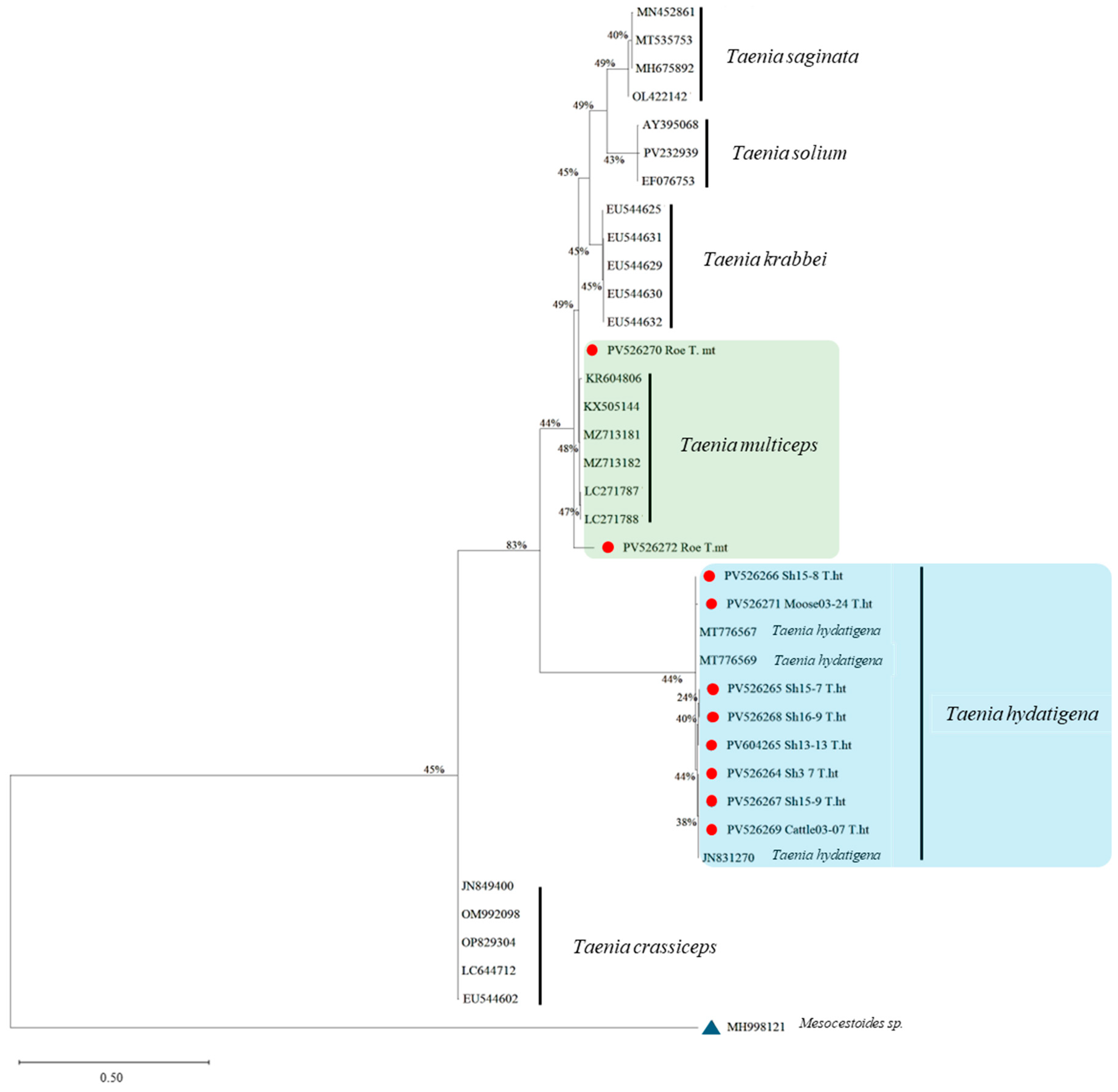
The resulting sequences allowed for species-level identification and were included in the construction of the phylogenetic tree (Figure 3), which illustrates the evolutionary relationships among the studied Taenia isolates and reference sequences from GenBank.
As can be seen in Figure 3, separate clades were formed according to the species. The external group for rooting the tree is Mesocestoides sp. (MH998121). The isolates studied from T. saginata, T. krabbei, T. solium, and T. crassiceps were assigned to their corresponding clades according to the species. In the case of T. multiceps and T. hydatigena species, they did not form monophyletic clusters in their respective branches and showed low support values. The percentage of identity from the last ancestor for each species varied from 24 to 83%.
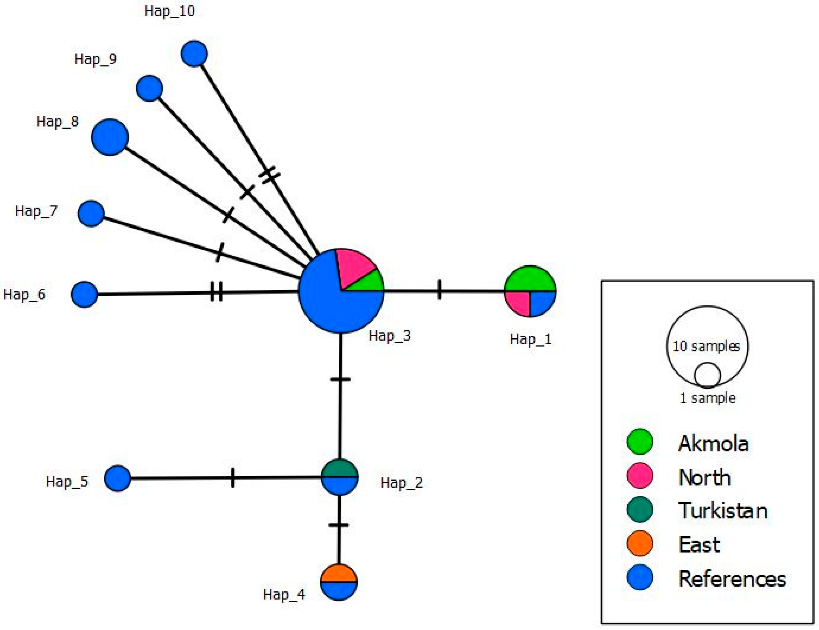
Analysis of haplotyping based on the mitochondrial gene nad1 formed four unique haplotypes for the isolates studied in this work, and together with the reference samples, 10 haplotypes were formed (Figure 4, Table 2).
The obtained samples were analyzed with reference samples from the GenBank source, which included haplotypes from China, Sudan, Nigeria, and Italy. As can be seen from the analysis, Hap_3 is the most common, including 10 isolates. It can also be noted that isolates from the North Kazakhstan region and the Akmola region share two haplotypes, which may be because these are neighboring regions.

4. Discussion

Despite the significant impact of cysticercosis caused by Taenia spp. on livestock production in endemic regions worldwide [26,52,59,60,61,62,63,64,65], this issue remains poorly understood in Kazakhstan. The combination of vast territories, transhumant livestock farming, and unregulated private yards slaughtering creates favorable conditions for the circulation of Taenia spp. in the country, while also posing major challenges to the comprehensive study of taeniasis. To date, no studies on the infection rates and intensity of cysticercosis in domestic and wild ungulates in Kazakhstan. Furthermore, data on species composition, as well as the genetic characteristics of circulating genotypes and haplotypes of Taenia spp., are lacking.
To identify cysticercosis in cattle and sheep, we examined only organs with visible cystic lesions, primarily the lungs and liver, collected from domestic ungulates in 14 regions across Kazakhstan. In total, 773 samples from cattle and 563 samples from sheep were analyzed. All samples underwent genetic analysis to differentiate cysticercosis from echinococcal infection. As a result, the prevalence of cysticercosis was found to be 0.6% in cattle and 1.1% in sheep. This suggests possible variation in exposure risk, ecological factors, or susceptibility to cysticercosis among the different ungulate hosts.
Analysis of the prevalence of cysticercosis in ungulates across Central Asian countries revealed a limited number of published studies. In Uzbekistan, the presence of cestode infections in both domestic and wild artiodactyls has been reported, with infection rates reaching up to 27.3%. Identified pathogens included T. hydatigena, Taeniarhynchus saginatus, and Echinococcus granulosus [91,92]. In the Republic of Tajikistan, brief reports describe possible causes for the spread of T. saginata (the larval stage of C. bovis) in cattle; however, specific data on infection rates in domestic ungulates are lacking [93,94]. Regarding Kyrgyzstan, available data are also scarce, with only one report indicating the presence of T. hydatigena in cattle, with a prevalence of 8.4% [95]. Currently, no data are available on the infection of ungulates with cysticerci in Turkmenistan.
To investigate parasitic infections in wild ungulates, including wild ruminants, we are conducting systematic fieldwork in collaboration with veterinary research institutions and private hunters [13,96,97]. Over the past three years, biological samples were collected and examined from 25 roe deer (Capreolus capreolus), 9 moose (Alces alces), and 2 red deer (Cervus elaphus sibiricus). In this study, the prevalence of cysticercosis was found to be 8% in roe deer and 11% in moose, indicating active circulation of cysticercosis pathogens among wild ungulates in Kazakhstan. These results are consistent with our previous reports confirming the presence of T. hydatigena in definitive wild hosts, specifically wolves and foxes [13,98]. Although our study only provides data on infection rates in intermediate hosts, the findings are in line with previous studies suggesting that the circulation of larval T. hydatigena in wild ungulates, such as moose, may be influenced by the presence of definitive hosts like wolves in the region [8,88].
Based on our observations, there was no clear pattern indicating that the anatomical localization of Taenia spp. cysts varied by host species. Cysts may develop in the thoracic and abdominal cavities, as well as in various organs, muscle tissues, and even the brain [5,6,12,15,16,17,18,19,20,21,22,26,27,28,29,30,31,32,33,34,35,36,37,38,39,40,41,42,43,44,45,46,47,48,49]. The only consistent distinguishing feature is the structural morphology of the cysts, which allows differentiation from other cyst-forming parasitic infections, such as echinococcosis.
In our study, Taenia cysts in domestic ungulates were found in sheep (n = 18) and cattle (n = 24), with localization in the liver. These lesions were characterized by fibrous, hemorrhagic, white-colored foci of varying sizes on the liver surface, often accompanied by internal degenerative tissue and signs of calcification, which is consistent with previous reports in the literature [17,88,90]. In roe deer, we observed cystic lesions (n = 15) in the femoral muscle tissue. The cysts were small (up to 1 cm in diameter), filled with clear fluid, and typically contained a single large scolex [12]. In moose, we identified a cyst (n = 1) on the liver surface that exhibited structural features rarely observed in intermediate ruminant hosts. The cyst had a thin wall, an oval shape, was filled with milky-white fluid, and contained a single cysticercoid measuring approximately 3 cm. The morphological characteristics of the observed cyst closely resembled those of a cyst previously described in the lung of a roe deer infected with T. lynciscapreoli [49].
Bioinformatic analysis of the mitochondrial nad1 gene from cysticercosis samples revealed that the studied isolates exhibited high similarity to reference sequences available in GenBank. In the phylogenetic tree, the T. multiceps isolates obtained from roe deer (accession numbers PV526270 and PV526272) clustered within the same clade as reference [32,99,100] sequences of the species, confirming their genetic identity.
The phylogenetic analysis shows that the Kazakhstan isolates cluster within established Taenia lineages, indicating their close genetic relationship with reference sequences from other regions, rather than forming distinct divergent clades. Overlap in parasite species between domestic and wild hosts (especially for T. hydatigena) implies environmental contamination and transmission overlap, likely due to shared pastures or predator-prey cycles involving canids [32,99,100]. Roe deer and moose act as important sentinels for sylvatic cycles, particularly for T. multiceps and T. hydatigena. T. saginata, T. solium, T. krabbei, and T. crassiceps were not detected in this study but were included as a comparison to define distinct evolutionary lineages among Taeniids.
To gain a more comprehensive understanding of the genetic diversity of T. multiceps, further studies employing both cox1 and nad1 markers are required, including a larger number of isolates from different geographic regions of Kazakhstan and host species. Further studies should also consider that the mitochondrial genes cox1 and nad1 are particularly useful for the identification of T. skrjabini, which was previously regarded as the causative agent of non-cerebral cysticercosis in sheep, but is now considered a genetic variant of T. multiceps [18].
The same can be seen with samples of T. hydatigena from sheep (PV526265, PV526268, PV604265, PV526264, PV526267, PV526266), from cattle (PV526269) and moose (PV526271). Haplotyping of the T. hydatigena sequences revealed the presence of four haplotypes among the studied samples, with Hap_3 being the most widespread. Haplotype diversity in populations was 0.82. In accordance with the present study, high haplotype diversity values have been reported in other haplotypes of T. hydatigena, including the China, Sudan, Nigeria, and Italy haplotypes. The nucleotide diversity was relatively low; p = 0.00397 for all studied isolates. This finding, together with the observed pattern of high haplotype diversity and low nucleotide diversity, may indicate a recent population bottleneck followed by rapid expansion [101]. Applied Tajima’s D test [102] for the allele frequency distribution of discrete nucleotide sites. Fu’s FS test [103] is based on the distribution of alleles or haplotypes. In our study, the Tajima D test and the Fu FS test revealed negative values for all populations, which are indicative of recent population expansion.

5. Conclusions

This study provides the first documented evidence of T. multiceps infection in roe deer and T. hydatigena infection in moose, sheep, and cattle in Kazakhstan. It also provides the first molecular data on Taenia spp. from the country. These can be findings helps identify infection sources and transmission routes, assess regional risks, and improve diagnostics. Molecular analysis is crucial due to the morphological similarity of larval Taenia species, which complicates identification by traditional methods. Understanding population structure supports timely preventive measures—from public awareness to adjustments in national parasite control programs in Kazakhstan.

Author Contributions

Conceptualization, V.K. and C.H.; methodology, V.K., L.L. and R.U.; software, N.M. and R.U.; validation, A.S., N.M. and K.J.; formal analysis, V.K. and C.H.; investigation, R.U. and A.S.; resources, V.K.; data curation, V.K. and L.L.; writing—original draft preparation, V.K., C.H. and R.U.; writing—review and editing, V.K. and R.U.; visualization, A.S. and N.M.; supervision, V.K.; project administration, V.K.; funding acquisition, V.K. All authors have read and agreed to the published version of the manuscript.

Funding

The research was carried out with financial support from the Committee of Science of the Ministry of Science and Higher Education of the Republic of Kazakhstan within the framework of project AP19576421 for 2023–2025.

Institutional Review Board Statement

The animal study protocol was approved by the Animal Ethics Committee of the National Center for Biotechnology (approval based on protocol extracts No. 10 dated 10 November 2022) for studies involving animals. All procedures were performed in compliance with ethical guidelines for animal research, in line with the principles of the World Medical Association’s Declaration of Helsinki (http://ec.europa.eu/environment/chemicals/lab_animals/legal_en.htm, accessed on 16 September 2025).

Informed Consent Statement

Not applicable.

Data Availability Statement

The data are contained within this article.

Acknowledgments

The authors would like to express their sincere gratitude to Alexandr Lyalchenko and Sergey Leontyev for their assistance in collecting the samples.

Conflicts of Interest

The authors declare that they have no conflicts of interest to disclose.

Abbreviations

The following abbreviations are used in this manuscript:
Nnumber
bpbase pairs
CIconfidence interval
cox1cytochrome c oxidase subunit 1
DNAdeoxyribonucleic acid
MLMaximum Likelihood
nad1NADH dehydrogenase subunit 1
OROdds Ratio
SDstandard deviation
TBETris-borate-EDTA buffer

References

  1. Samuel, W.; Zewde, G.G. Prevalence, risk factors, and distribution of Cysticercus tenuicollis in visceral organs of slaughtered sheep and goats in central Ethiopia. Trop. Anim. Health Prod. 2010, 42, 1049–1051. [Google Scholar] [CrossRef]
  2. Scala, A.; Urrai, G.; Varcasia, A.; Nicolussi, P.; Mulas, M.; Goddi, L.; Pipia, A.P.; Sanna, G.; Genchi, M.; Bandino, E. Acute visceral cysticercosis by Taenia hydatigena in lambs and treatment with praziquantel. J. Helminthol. 2016, 90, 113–116. [Google Scholar] [CrossRef]
  3. Oryan, A.; Goorgipour, S.; Moazeni, M.; Shirian, S. Abattoir prevalence, organ distribution, public health and economic importance of major metacestodes in sheep, goats and cattle in Fars, southern Iran. Trop. Biomed. 2012, 29, 349–359. [Google Scholar]
  4. Mekuria, E.; Shimelis, S.; Bekele, J.; Sheferaw, D. Sheep and goats Cysticercus tenuicollis prevalence and associated risk factors. Afr. J. Agric. Res. 2013, 8, 3121–3125. [Google Scholar] [CrossRef]
  5. Bariselli, S.; Maioli, G.; Pupillo, G.; Calzolari, M.; Torri, D.; Cirasella, L.; Luppi, A.; Torreggiani, C.; Garbarino, C.; Barsi, F.; et al. Identification and phylogenetic analysis of Taenia spp. parasites found in wildlife in the Emilia-Romagna region, northern Italy (2017–2022). Int. J. Parasitol. Parasites Wildl. 2023, 22, 20–27. [Google Scholar] [CrossRef] [PubMed]
  6. Kibona, T.; Buza, J.; Shirima, G.; Lankester, F.; Ngongolo, K.; Hughes, E.; Cleaveland, S.; Allan, K.J. The Prevalence and Determinants of Taenia multiceps Infection (Cerebral Coenurosis) in Small Ruminants in Africa: A Systematic Review. Parasitologia 2022, 2, 137–146. [Google Scholar] [CrossRef]
  7. Jenkins, D.J.; Urwin, N.A.R.; Williams, T.M.; Mitchell, K.L.; Lievaart, J.J.; Armua-Fernandez, M.T. Red foxes (Vulpes vulpes) and wild dogs (dingoes (Canis lupus dingo) and dingo/domestic dog hybrids), as sylvatic hosts for Australian Taenia hydatigena and Taenia ovis. Int. J. Parasitol. Pazasites Wildl. 2014, 3, 75–80. [Google Scholar] [CrossRef]
  8. Lavikainen, A.; Laaksonen, S.; Beckmen, K.; Oksanen, A.; Isomursu, M.; Meri, S. Molecular identification of Taenia spp. in wolves (Canis lupus), brown bears (Ursus arctos) and cervids from North Europe and Alaska. Parasitol. Int. 2011, 60, 289–295. [Google Scholar] [CrossRef]
  9. Deplazes, P.; Eichenberger, R.M.; Grimm, F. Wildlife-transmitted Taenia and Versteria cysticercosis and coenurosis in humans and other primates. Int. J. Parasitol. Parasites Wildl. 2019, 9, 342–358. [Google Scholar] [CrossRef]
  10. Dorbek-Kolin, E.; Ahlberg, T.; Tummeleht, L.; Tappe, D.; Johansen, M.V.; Lassen, B. Prevalence of cysticercosis in Estonian pigs and cattle. Parasitol. Res. 2018, 117, 591–595. [Google Scholar] [CrossRef]
  11. Trevisan, C.; Sotiraki, S.; Laranjo-González, M.; Dermauw, V.; Wang, Z.; Kärssin, A.; Cvetkovikj, A.; Winkler, A.S.; Abraham, A.; Bobić, B.; et al. Epidemiology of taeniosis/cysticercosis in Europe, a systematic review: Eastern Europe. Parasites Vectors 2018, 11, 569. [Google Scholar] [CrossRef]
  12. Varcasia, A.; Tamponi, C.; Ahmed, F.; Cappai, M.G.; Porcu, F.; Mehmood, N.; Dessì, G.; Scala, A. Taenia multiceps coenurosis: A review. Parasites Vectors 2022, 15, 84. [Google Scholar] [CrossRef]
  13. Uakhit, R.; Smagulova, A.; Lider, L.; Leontyev, S.; Kiyan, V. Epizootiological monitoring of wolf helminths in Northern and Central Kazakhstan. Vet. World 2024, 17, 1648–1654. [Google Scholar] [CrossRef]
  14. Flisser, A.; Craig, P.S.; Ito, A. Cysticercosis and Taeniosis: Taenia solium, Taenia saginata and Taenia asiatica; Palmer, S.R., Soulsby, L., Torgerson, P.R., Brown, D.W.G., Eds.; Oxford Textbook of Zoonoses Biology, Clinical Practice, and Public Health Control; Oxford University Press: Oxford, UK, 2011; pp. 625–649. [Google Scholar]
  15. Muku, R.J.; Yan, H.B.; Ohiolei, J.A.; Saaid, A.A.; Ahmed, S.; Jia, W.Z.; Fu, B.Q. Molecular Identification of Taenia hydatigena from Sheep in Khartoum, Sudan. Korean J. Parasitol. 2020, 58, 93–97. [Google Scholar] [CrossRef] [PubMed]
  16. Filip, K.J.; Pyziel, A.M.; Jeżewski, W.; Myczka, A.W.; Demiaszkiewicz, A.W.; Laskowski, Z. First Molecular Identification of Taenia hydatigena in Wild Ungulates in Poland. EcoHealth 2019, 16, 161–170. [Google Scholar] [CrossRef] [PubMed]
  17. Corda, A.; Dessì, G.; Varcasia, A.; Carta, S.; Tamponi, C.; Sedda, G.; Scala, M.; Marchi, B.; Salis, F.; Scala, A.; et al. Acute visceral cysticercosis caused by Taenia hydatigena in lambs: Ultrasonographic findings. Parasites Vectors 2020, 13, 568. [Google Scholar] [CrossRef] [PubMed]
  18. Alvi, M.A.; Ohiolei, J.A.; Saqib, M.; Tayyab, M.H.; Zafar Khan, M.U.; Li, L.; Aqib, A.I.; Hassan, A.; Alvi, A.A.; Qamar, W.; et al. First Report on Molecular Characterization of Taenia multiceps Isolates from Sheep and Goats in Faisalabad, Pakistan. Front. Vet. Sci. 2020, 7, 594599. [Google Scholar] [CrossRef]
  19. Pouchet, C.; Fernandez-Prada, C.; Dussault, C.; Leclerc, M.; Tremblay, J.P.; Côté, S.D. Variation in prevalence and intensity of macroparasites in moose and their interactions with winter tick load in eastern Canada. Wildl. Biol. 2024, e01205. [Google Scholar] [CrossRef]
  20. Žele Vengušt, D.; Kuhar, U.; Jerina, K.; Vengušt, G. Twenty Years of Passive Disease Surveillance of Roe Deer (Capreolus capreolus) in Slovenia. Animals 2021, 11, 407. [Google Scholar] [CrossRef]
  21. Morandi, B.; Bazzucchi, A.; Gambini, S.; Crotti, S.; Cruciani, D.; Morandi, F.; Napoleoni, M.; Piseddu, T.; Di Donato, A.; Gavaudan, S. A novel intermediate host for Taenia serialis (Gervais, 1847): The European roe deer (Capreolus capreolus L. 1758) from the Monti Sibillini National Park (MSNP), Italy. Int. J. Parasitol. Parasites Wildl. 2021, 17, 110–113. [Google Scholar] [CrossRef]
  22. Kautto, A.H.; Grandi, G.; Höglund, J. Taenia lynciscapreoli in semi-domesticated reindeer (Rangifer tarandus tarandus, L.) in Sweden. Int. J. Parasitol. Parasites Wildl. 2022, 18, 148–151. [Google Scholar] [CrossRef] [PubMed]
  23. Scala, A.; Pipia, A.P.; Dore, F.; Sanna, G.; Tamponi, C.; Marrosu, R.; Bandino, E.; Carmona, C.; Boufana, B.; Varcasia, A. Epidemiological updates and economic losses due to Taenia hydatigena in sheep from Sardinia, Italy. Parasitol. Res. 2015, 114, 3137–3143. [Google Scholar] [CrossRef]
  24. Deressa, A.; Tilahun, T.; Tadesse, A.; Beyene, M.; Gebrewold, G.; Pal, M. Assessment of Coenurus cerebralis and its economic impact in sheep brain harvested at Ethiopian Health and Nutrition Research Institute, Ethiopia. Int. J. Livest. Res. 2012, 2, 217–226. [Google Scholar]
  25. Shiferaw, A.; Abdela, N. Public health and economic significance of cerebral coenurosis in sheep and goat: A review. Acta Parasitol. Glob. 2016, 7, 54–65. [Google Scholar] [CrossRef]
  26. Braae, U.C.; Kabululu, M.; Nørmark, M.E.; Nejsum, P.; Ngowi, H.A.; Johansen, M.V. Taenia hydatigena cysticercosis in slaughtered pigs, goats, and sheep in Tanzania. Trop. Anim. Health Prod. 2015, 47, 1523–1530. [Google Scholar] [CrossRef]
  27. Ohiolei, J.A.; Luka, J.; Zhu, G.Q.; Yan, H.B.; Li, L.; Magaji, A.A.; Alvi, M.A.; Wu, Y.T.; Li, J.Q.; Fu, B.Q.; et al. First molecular description, phylogeny and genetic variation of Taenia hydatigena from Nigerian sheep and goats based on three mitochondrial genes. Parasites Vectors 2019, 12, 520. [Google Scholar] [CrossRef]
  28. Ohiolei, J.A.; Yan, H.B.; Li, L.; Li, W.H.; Wu, Y.D.; Alvi, M.A.; Zhang, N.Z.; Fu, B.Q.; Wang, X.L.; Jia, W.Z. A new molecular nomenclature for Taenia hydatigena: Mitochondrial DNA sequences reveal sufficient diversity suggesting the assignment of major haplotype divisions. Parasitology 2021, 148, 311–326. [Google Scholar] [CrossRef]
  29. Jarošová, J.; Antolová, D.; Iglodyová, A.; Königová, A.; Dolinská, M.U.; Víchová, B. Molecular identification of Taenia hydatigena from domestic and free-living animals in Slovakia, Central Europe. Parasitol. Res. 2022, 121, 1345–1354. [Google Scholar] [CrossRef]
  30. Moudgil, A.D.; Nehra, A.K.; Vohra, S.; Thakur, S.D.; Sharma, D. Prevalence and phylogeography of Taenia hydatigena metacestodes from goats of India. Parasitology 2022, 149, 1193–1198. [Google Scholar] [CrossRef]
  31. Muñoz-Caro, T.; González, M.F.; Villalobos, R.; Hidalgo, A. Parasitic findings on threatened pudu deer from Central Chile accounts first genetic characterization of lice parasitizing P. puda in Chile and the first molecular report of Taenia hydatigena metacestodes in this species. Vet. Q. 2024, 44, 1–8. [Google Scholar] [CrossRef]
  32. Abbas, I.; El-Alfy, E.S.; Janecek-Erfurth, E.; Strube, C. Molecular characterization of Cysticercus tenuicollis isolates from sheep in the Nile Delta, Egypt and a review on Taenia hydatigena infections worldwide. Parasitology 2021, 148, 913–933. [Google Scholar] [CrossRef]
  33. Ng-Nguyen, D.; Van Nguyen, T.; Van Nguyen, T.; Nguyen, H.Q.; Nguyen, V.T. Prevalence and risk factors of Taenia hydatigena in dogs, pigs, and cattle in the Central Highlands of Vietnam. Parasitol. Res. 2021, 120, 3245–3253. [Google Scholar] [CrossRef]
  34. Polaz, S.V. Helminths of wild ungulates living in different regions of Belarus. Russ. J. Parasitol. 2022, 16, 33–49. [Google Scholar] [CrossRef]
  35. Kirillova, N.Y.; Ruchin, A.B.; Kirillov, A.A.; Chikhlyaev, I.V.; Alpeev, M.A. Overview of Helminths in Land Vertebrates from the Mordovia Nature Reserve, European Russia. Nat. Environ. Pollut. Technol. 2023, 22, 1667–1690. [Google Scholar] [CrossRef]
  36. Alvi, M.A.; Ohiolei, J.A.; Saqib, M.; Li, L.; Muhammad, N.; Tayyab, M.H.; Qamar, W.; Alvi, A.A.; Wu, Y.D.; Li, X.R.; et al. Preliminary information on the prevalence and molecular description of Taenia hydatigena isolates in Pakistan based on mitochondrial cox1 gene. Infect. Genet. Evol. 2020, 85, 104481. [Google Scholar] [CrossRef] [PubMed]
  37. Wakid, M.H.; Alsulami, M.N. Genotyping of Taenia hydatigena isolated from sheep and goats in KSA based on Cox1 gene. Saudi J. Biol. Sci. 2022, 29, 1270–1275. [Google Scholar] [CrossRef]
  38. Maslennikova, O.V. Elk parasites and their danger to humans. In Biodiagnostics of the State of Natural and Natural-Technogenic Systems, Proceedings of the XII All-Russian Scientific-Practical Conference with International Participation, Kirov, Russia, 2–3 December 2014; Vesi Publishing House: Kirov, Russia, 2014; pp. 255–257. (In Russian) [Google Scholar]
  39. Gavrilova, N.A.; Belova, L.M.; Zabrovskaya, A.V. Larval cestodiasis of elks in the Leningrad region. Curr. Issues Vet. Biol. 2024, 1, 15–18. (In Russian) [Google Scholar] [CrossRef]
  40. Pelgunov, A.N. Helminthofaunal complex of wild ungulates in biocenoses contaminated with radionuclides. Russ. Parasitol. J. 2010, 2, 11–15. (In Russian) [Google Scholar]
  41. Baitursinov, K.K. Factors of formation of community of helminth fauna of wild and domestic ungulates of Kazakhstan. Russ. Parasitol. J. 2008, 4, 5–12. (In Russian) [Google Scholar]
  42. Bulgakova, N.F. Larval cestodiasis of animals of Belarus. Vet. Sci. Abstr. J. 2005, 1, 315. (In Russian) [Google Scholar]
  43. Kiriltsov, E.V. Population studies of the main parasitic systems of wild animals in the south of the Transbaikal Region. Perm Agrar. Bull. 2024, 2, 110–118. (In Russian) [Google Scholar] [CrossRef]
  44. Baitursinov, K.K. Ecological bases of prevention of helminthiasis of wild ungulates in conditions of Kazakhstan. Russ. Parasitol. J. 2008, 1, 47–53. (In Russian) [Google Scholar]
  45. Shikhalieva, M.A.; Golubev, A.A.; Sarbasheva, M.M.; Bittirov, A.M. Epizootological assessment of helminthiasis of chamois, deer and roe deer in the Kabardino-Balkarian Republic. Actual Issues Vet. Biol. 2012, 4, 36–38. (In Russian) [Google Scholar]
  46. Abdybekova, A.M.; Zhaksylykova, A.A.; Kushaliyev, K.Z.; Kidiraliyev, E.Z.; Kozhayeva, A.R.; Kuzhebayeva, U.Z.; Grachev, A.; Shevtsov, A.; Budke, C.M. A survey of the parasites of Ural saiga antelopes and Turkmenian kulans of Kazakhstan. Int. J. Parasitol. Parasites Wildl. 2023, 21, 232–236. [Google Scholar] [CrossRef]
  47. Mørk, T.; Eira, H.I.; Rødven, R.; Nymo, I.H.; Blomstrand, B.M.; Guttormsen, S.; Davidson, R.K. Necropsy findings and causes of loss in semi-domesticated reindeer (Rangifer tarandus tarandus) in Northern Norway. Acta Vet. Scand. 2023, 66, 1. [Google Scholar] [CrossRef]
  48. Maslennikova, O.V.; Kotelnikov, S.A. Taranda cysticercosis of elk in the Udmurt Republic. In Ecology of the Native Land: Problems and Solutions, Proceedings of the XIII All-Russian Scientific and Practical Conference with International Participation, Kirov, Russia, 23–24 April 2018; Vyatka State University Publishing House: Kirov, Russia, 2018; pp. 225–229. (In Russian) [Google Scholar]
  49. Myczka, A.W.; Jeżewski, W.; Filip-Hutsch, K.J.; Pyziel, A.M.; Kowal, J.; Demiaszkiewicz, A.W.; Laskowski, Z. The morphological and molecular identification of the tapeworm, Taenia lynciscapreoli, in intermediate and definitive hosts in Poland. Int. J. Parasitol. Parasites Wildl. 2020, 11, 213–220. [Google Scholar] [CrossRef]
  50. Scala, A.; Varcasia, A. Updates on morphobiology, epidemiology and molecular characterization of coenurosis in sheep. Parassitologia 2006, 48, 61–63. [Google Scholar]
  51. Luo, H.; Zhang, H.; Li, K.; Rehman, M.U.; Mehmood, K.; Lan, Y.; Huang, S.; Li, J. Epidemiological survey and phylogenetic characterization of Cysticercus tenuicollis isolated from Tibetan pigs in Tibet, China. Biomed. Res. Int. 2017, 2017, 7857253. [Google Scholar] [CrossRef]
  52. Nguyen, M.T.; Gabriël, S.; Abatih, E.N.; Dorny, P. A systematic review on the global occurrence of Taenia hydatigena in pigs and cattle. Vet. Parasitol. 2016, 226, 97–103. [Google Scholar] [CrossRef]
  53. Akbari, M.; Moazeni, M.; Oryan, A.; Sharifiyazdi, H.; Amrabadi, O. Experimen tal cerebral and non-cerebral coenurosis in goats: A comparative study on the morphological and molecular characteristics of the parasite. Vet. Parasitol. 2015, 211, 201–207. [Google Scholar] [CrossRef]
  54. Varcasia, A.; Tamponi, C.; Tosciri, G.; Pipia, A.P.; Dore, F.; Schuster, R.K.; Kandil, O.M.; Manunta, M.L.; Scala, A. Is the red fox (Vulpes vulpes) a competent definitive host for Taenia multiceps? Parasit. Vectors 2015, 8, 491. [Google Scholar] [CrossRef]
  55. Varcasia, A.; Pipia, A.P.; Dessì, G.; Zidda, A.; Tamponi, C.; Pau, M.; Scala, A.; Boufana, B. Morphology and genetic variability within Taenia multiceps in ruminants from Italy. Vet. Parasitol. 2016, 223, 181–185. [Google Scholar] [CrossRef]
  56. Scott, P.R. Diagnosis and treatment of coenurosis in sheep. Vet. parasitol. 2012, 189, 75–78. [Google Scholar] [CrossRef] [PubMed]
  57. Varcasia, A.; Tosciri, G.; Coccone, G.N.S.; Pipia, A.P.; Garippa, G.; Scala, A.; Damien, V.; Vural, G.; Gauci, C.G.; Lightowlers, M.W. Preliminary field trial of a vaccine against coenurosis caused by Taenia multiceps. Vet. Parasitol. 2009, 162, 285–289. [Google Scholar] [CrossRef] [PubMed]
  58. Oryan, A.; Akbari, M.; Moazeni, M.; Amrabadi, O.R. Cerebral and non-cerebral coenurosis in small ruminants. Trop. Biomed. 2014, 31, 1–16. [Google Scholar] [PubMed]
  59. Rostami, S.; Salavati, R.; Beech, R.N.; Sharbatkhori, M.; Babaei, Z.; Saedi, S.; Harandi, M.F. Cytochrome c oxidase subunit 1 and 12S ribosomal RNA characterization of Coenurus cerebralis from sheep in Iran. Vet. Parasitol. 2013, 197, 141–151. [Google Scholar] [CrossRef]
  60. Scala, A.; Cancedda, G.M.; Varcasia, A.; Ligios, C.; Garippa, G.; Genchi, C. A survey of Taenia multiceps coenurosis in Sardinian sheep. Vet. Parasitol. 2007, 143, 294–298. [Google Scholar] [CrossRef]
  61. Varma, T.K.; Malviya, H.C. Prevalence of coenuriosis in sheep, goat and pigs in Bareilly, Utar Pradesh. J. Vet. Parasitol. 1989, 3, 69–71. [Google Scholar]
  62. Tavassoli, M.; Malekifard, F.; Soleimanzadeh, A.; Tajik, H. Prevalence of Coenurus cerebralis in sheep in northwest Iran. Vet. Res. Forum 2011, 2, 274–276. [Google Scholar]
  63. Nooruddin, M.; Dey, A.; Ali, M.A. Coenurosis in Bengal goats of Bangladesh. Small Rumin. Res. 1996, 19, 77–81. [Google Scholar] [CrossRef]
  64. Ozmen, O.; Sahinduran, S.; Haligur, M.; Sezer, K. Clinicopathologic observa tions on Coenurus cerebralis in naturally infected sheep. Schweiz. Arch. Tierheilkd. 2005, 147, 129–134. [Google Scholar] [CrossRef]
  65. Miran, M.B.; Nzalawahe, J.; Kassuku, A.A.; Swai, E.S. Prevalence of coenurosis in sheep and goats at three slaughter slabs in Ngorongoro District, Tanzania. Trop. Anim. Health Prod. 2015, 47, 1591–1597. [Google Scholar] [CrossRef] [PubMed]
  66. Gunyakti Kilinc, S.; Celik, F.; Kesik, H.K.; Simsek, S. In silico analysis of the biodiversity and conservation status of mitochondrial cytochrome C oxidase subunit 1 (CO1) gene of Taenia multiceps. Acta Parasitol. 2020, 65, 852–858. [Google Scholar] [CrossRef] [PubMed]
  67. Varcasia, A.; Lightowlers, M.W.; Cattoli, G.; Cancedda, G.M.; Canu, S.; Garippa, G.; Scala, A. Genetic variation within Taenia multiceps in Sardinia, western Mediterranean (Italy). Parasitol. Res. 2006, 99, 622626. [Google Scholar] [CrossRef] [PubMed]
  68. Li, W.H.; Jia, W.Z.; Qu, Z.G.; Xie, Z.Z.; Luo, J.X.; Yin, H.; Sun, X.L.; Blaga, R.; Fu, B.Q. Molecular characterization of Taenia multiceps isolates from Gansu Province, China by sequencing of mitochondrial cytochrome C oxidase subunit 1. Korean J. Parasitol. 2013, 51, 197–201. [Google Scholar] [CrossRef]
  69. Sonmez, B.; Koroglu, E.; Simsek, S. Molecular characterization and detection of variants of Taenia multiceps in sheep in Turkey. Parasitology 2017, 144, 220–225. [Google Scholar] [CrossRef]
  70. Al-Riyami, S.; Ioannidou, E.; Koehler, A.V.; Hussain, M.H.; Al-Rawahi, A.H.; Giadinis, N.D.; Lafi, S.Q.; Papadopoulos, E.; Jabbar, A. Genetic characterisation of Taenia multiceps cysts from ruminants in Greece. Infect. Genet. Evol. 2016, 38, 110–116. [Google Scholar] [CrossRef]
  71. Boufana, B.; Scala, A.; Lahmar, S.; Pointing, S.; Craig, P.S.; Dessì, G.; Zidda, A.; Pipia, A.P.; Varcasia, A. A preliminary investigation into the genetic variation and population structure of Taenia hydatigena from Sardinia, Italy. Vet. Parasitol. 2015, 214, 67–74. [Google Scholar] [CrossRef]
  72. Adwan, K.; Jayousi, A.; Abuseir, S.; Abbasi, I.; Adwan, G.; Jarrar, N. Genetic diversity of Taenia hydatigena in the northern part of the West Bank, Palestine as determined by mitochondrial DNA sequences. Acta Parasitol. 2018, 63, 299–303. [Google Scholar] [CrossRef]
  73. Kilinc, S.G.; Kesik, H.K.; Simsek, S. Molecular characterization and haplotypes of sheep and goat isolates of Cysticercus tenuicollis in Turkey. Parasitology 2019, 146, 1047–1054. [Google Scholar] [CrossRef]
  74. Kazakhstan: Sustainable Livestock Development Program for Results. World Bank Group. Available online: https://www.worldbank.org/en/country/kazakhstan/brief/sustainable-livestock-development-program-for-results?utm_source=chatgpt.com (accessed on 10 September 2025).
  75. Skokov, R.; Dyussegalieva, B. Current state of livestock economy in Kazakhstan. In Proceedings of the International Scientific Conference “Fundamental and Applied Scientific Research in the Development of Agriculture in the Far East” (AFE–2023), Tashkent, Uzbekistan, 12 December 2023; Volume 462, p. e01023. [Google Scholar] [CrossRef]
  76. Key Indicators of Livestock Development in the Republic of Kazakhstan (January–December 2024). Available online: https://stat.gov.kz/ru/industries/business-statistics/stat-forrest-village-hunt-fish/publications/287928/ (accessed on 10 September 2025).
  77. Skrjabin, K.I. Method of Complete Helminthological Dissection of Vertebrates, Including Humans; Publishing House of 1st Moscow State University: Moscow, Russia, 1928; pp. 40–48. (In Russian) [Google Scholar]
  78. Deplazes, P.; Eckert, J.; Mathis, A.; Von Samson-Himmelstjerna, G.; Zahner, H. Parasitology in Veterinary Medicine, 3rd ed.; Wageningen Academic Publishers: Wageningen, The Netherlands, 2013; pp. 224–239. [Google Scholar]
  79. Taylor, M.A.; Coop, R.L.; Wall, R.L. Veterinary Parasitology. In Parasites of Sheep and Goats, 4th ed.; John Wiley & Sons: Hoboken, NJ, USA, 2015; pp. 436–523. [Google Scholar] [CrossRef]
  80. Bowles, J.; Blair, D.; McManus, D.P. Genetic variants within the genus Echinococcus identified by mitochondrial DNA sequencing. Mol. Biochem. Parasitol. 1992, 54, 165–173. [Google Scholar] [CrossRef] [PubMed]
  81. Al Malki, J.S.; Hussien, N.A. Molecular characterization and phylogenetic studies of Echinococcus granulosus and Taenia multiceps coenurus cysts in slaughtered sheep in Saudi Arabia. Open Life Sci. 2021, 16, 1252–1260. [Google Scholar] [CrossRef] [PubMed]
  82. Samorek-Pieróg, M.; Karamon, J.; Brzana, A.; Bilska-Zajac, E.; Zdybel, J.; Cencek, T. Molecular Confirmation of Massive Taenia pisiformis Cysticercosis in One Rabbit in Poland. Pathogens 2021, 10, 1029. [Google Scholar] [CrossRef] [PubMed]
  83. Abbas, I.; Elbeskawy, M. Molecular and phylogenetic status of Coenurus cerebralis infecting sheep from Dakahlia province, Egypt. J. Adv. Parasitol. 2016, 3, 117–124. [Google Scholar] [CrossRef]
  84. Tamura, K.; Stecher, G.; Kumar, S. MEGA11: Molecular Evolutionary Genetics Analysis Version 11. Mol. Biol. Evol. 2021, 38, 3022–3027. [Google Scholar] [CrossRef]
  85. Clement, M.; Posada, D.; Crandall, K.A. TCS: A computer program to estimate gene genealogies. Mol. Ecol. 2000, 9, 1657–1659. [Google Scholar] [CrossRef]
  86. Bush, A.O.; Lafferty, K.D.; Lotz, J.M.; Shostak, A.W. Parasitology meets ecology on its own terms: Margolis et al. revisited. J. Parasitol. 1997, 83, 575–583. [Google Scholar] [CrossRef]
  87. McHugh, M.L. The chi-square test of independence. Biochem. Med. 2013, 23, 143–149. [Google Scholar] [CrossRef]
  88. Pybus, M.J. Survey of hepatic and pulmonary helminths of wild cervids in Alberta, Canada. J. Wildl. Dis. 1990, 26, 453–459. [Google Scholar] [CrossRef]
  89. Office International des Epizooties (OIE). Chapter 3.9.5 Cysticercosis. OIE Manual of Diagnostic Tests and Vaccines for Terrestrial Animals (Mammals, Birds and Bees); Office International des Epizooties (OIE): Paris, France, 2018; pp. 1693–1704. Available online: https://www.woah.org/fileadmin/Home/eng/Health_standards/tahm/3.09.05_CYSTICERCOSIS.pdf (accessed on 2 June 2025).
  90. Perl, S.; Edery, N.; Bouznach, A.; Abdalla, H.; Markovics, A. Acute Severe Visceral Cysticercosis in Lambs and Kids in Israel. Isr. J. Vet. Med. 2015, 70, 49–53. [Google Scholar]
  91. Akramova, F.; Shakarbaev, U.; Mirzayeva, A.; Saidova, S.; Akbarova, M.; Uralova, F.; Hamrokulova, Z.; Ubbiniyazova, J.; Toremuratov, M.; Saparov, K.; et al. Helminths of domestic and wild artiodactyls (Mammalia, Artiodactyla) in Uzbekistan. Biosyst. Divers. 2025, 33, e2514. [Google Scholar] [CrossRef]
  92. Gaipova, M.E.; Akramova, F.D.; Saparov, K.A.; Azimov, D.A.; Shakarbaev, U.A. Fauna and ecology of helminths in cattle (Bos taurus Dom.) of Central Uzbekistan. Russ. J. Parasitol. 2016, 38, 447–453. (In Russian) [Google Scholar] [CrossRef]
  93. Razikov, S.S.; Sherkhonov, T. Reasons of prevalence of Cysticercus bovis in the zones of yak husbandry in the Republic of Tadjikistan. Theory Pract. Parasit. Dis. Anim. 2011, 12, 414–416. (In Russian) [Google Scholar]
  94. Murtazoev, D.M.; Valiev, H.G.; Pulotov, M.B.; Faizullaev, U.F. Epidemiological situation and problems of combating taeniasis in Tajikistan. Theory Pract. Combat. Parasit. Dis. 2015, 16, 276–278. (In Russian) [Google Scholar]
  95. Shakirov, A.B. Helminths and Helminth Diseases of Cattle in the Kyrgyz Republic and Methods for their Control. Ph.D. Dissertation, Dyishyeyeva Scientific Research Institute of Livestock, Veterinary and Pastures, Bishkek, Kyrgyzstan, 2004. (In Russian). [Google Scholar]
  96. Uakhit, R.; Bauer, C.; Smagulova, A.; Kiyan, V. First Reported Case of Accidental Gastric Myiasis Caused by Gasterophilus Larvae in a Gray Wolf. Acta Parasitol. 2025, 70, 1–5. [Google Scholar] [CrossRef]
  97. Bauer, C.; Uakhit, R.; Smagulova, A.; Jazina, K.; Lyalchenko, A.; Kiyan, V. Where there are moose (Alces alces) in Eurasia, there are moose nose botflies: First morphological and molecular identification of Cephenemyia ulrichii (Brauer, 1862) in Kazakhstan. Int. J. Parasitol. Parasites Wildl. 2025, 27, 101086. [Google Scholar] [CrossRef]
  98. Zhaxylykov, A.; Uakhit, R.; Kiyan, V. Study of prevalence and morphological features of cestodes in foxes (Vulpes vulpes) in the territory of Kazakhstan. J. Biol. Res. 2024, 1, 31–38. [Google Scholar] [CrossRef]
  99. Morais, D.F.; Vilela, V.L.R.; Feitosa, T.F.; Santos, V.M.D.; Gouveia, V.R.; Athayde, A.C.R.; Azevêdo, S.S. Prevalence and risk factors for Cysticercus tenuicollis in goats and sheep in Paraíba, northeastern Brazil. Rev. Bras. Parasitol. Vet. 2017, 26, 235–238. [Google Scholar] [CrossRef]
  100. Bhat, R.A.; Tak, H.; Bhat, B.A.; Dar, J.A.; Ahmad, R. Gastrointestinal helminth parasites of wild ungulates in Hirpora Wildlife Sanctuary, Kashmir, India. J. Parasit. Dis. 2022, 46, 804–810. [Google Scholar] [CrossRef]
  101. Avise, J.C.P. Phylogeography: The History and Formation of Species; Harvard University Press: Cambridge, MA, USA, 2001; p. 447. [Google Scholar]
  102. Tajima, F. Statistical method for testing the neutral mutation hypothesis by DNA polymorphism. Genetics 1989, 123, 585–595. [Google Scholar] [CrossRef]
  103. Fu, Y.X. Statistical tests of neutrality of mutations against population growth, hitchhiking and background selection. Genetics 1997, 147, 915–925. [Google Scholar] [CrossRef]
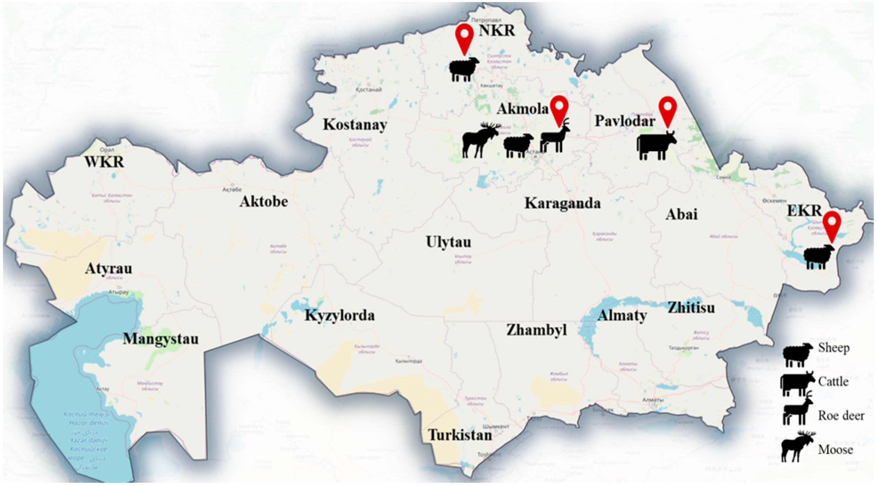
Figure 1. Map of Kazakhstan to show sampling region and locations of domestic and wild ungulates with cysticercosis. NKR—North Kazakhstan region; EKR—East Kazakhstan region; WRK—West Kazakhstan region.
Diversity 17 00655 g001
Figure 2. Cysticerci (indicated by arrows) found in the animals studied: (A) in the thigh muscle of a roe deer; (B) in the liver of a moose; (C) in the liver of cattle; (D) in the liver of sheep.
Diversity 17 00655 g002
Figure 3. Maximum Likelihood phylogenetic tree of the partial nad1 gene showing relationships between the examined Kazakhstan isolates and GenBank retrieved related sequences of Taenia spp. Red circle—isolates from this study, blue triangle—outgroup.
Diversity 17 00655 g003
Figure 4. The haplotype network for nad1 gene of cysticercosis of T. hydatigena. The size of the circles is proportional to the frequency of each haplotype. The color of each circle corresponds to the region of origin of the sample. The number of mutations separating haplotypes is indicated by dash marks. Hap: haplotype.
Diversity 17 00655 g004
Table 1. Prevalence and intensity of cysticerci in domestic and wild ungulates.
HostN infected/N Examined% Prevalence (95% CI)N Cyst FoundRange of
Intensity
Mean (SD)
Intensity
Cysticercosis
Species Identified
cattle5/7730.6 (0.2–1.5)182–53.6 (1.14)T. hydatigena
sheep6/5631.1 (0.3–2.3)242–64 (1.41)T. hydatigena
roe deer2/258.0 (0.9–2.6)156–97.5 (2.12)T. multiceps
moose1/911.11 (0.2–48.2)11-T. hydatigena
red deer0/2-- --
Chi-square test χ2 = 22.23,
df = 4,
p-value = 0.0001
95% CI: 95% confidence interval; SD: standard deviation; df = Degrees of freedom.
Table 2. Results from DNA sequence analysis of isolates using the DnaSP program.
nad1 Gene (894)
Number of mutations 11
Haplotypes number 10
Haplotype diversity 0.820
Nucleotide diversity 0.00397
Tajima’s test −1.65005
Fu’s test−5.282
Disclaimer/Publisher’s Note: The statements, opinions and data contained in all publications are solely those of the individual author(s) and contributor(s) and not of MDPI and/or the editor(s). MDPI and/or the editor(s) disclaim responsibility for any injury to people or property resulting from any ideas, methods, instructions or products referred to in the content.