Article

Fatty Acids and Fatty Acid Trophic Markers in Two Holothurian Species from the Central Mediterranean Sea

1
Council for Agricultural Research and Economics—Research Centre for Animal Production and Aquaculture, Via Salaria 31, 00015 Monterotondo, Italy
2
Department of Biology, University of Rome Tor Vergata, Via Cracovia 1, 00133 Rome, Italy
3
Department of Agricultural and Food Sciences—Division of Animal Sciences, University of Bologna, Viale G. Fanin 46, 40127 Bologna, Italy
4
Council for Agricultural Research and Economics—Research Centre for Agri-Food Engineering and Transformations, Via della Pascolare 16, 00015 Monterotondo, Italy
5
Institute of Aquaculture, University of Stirling, Stirling, FK9 4LA, UK
*
Author to whom correspondence should be addressed.
Diversity 2025, 17(8), 576; https://doi.org/10.3390/d17080576
Submission received: 17 July 2025 / Revised: 7 August 2025 / Accepted: 12 August 2025 / Published: 15 August 2025
(This article belongs to the Section Marine Diversity)

Abstract

Sea cucumbers, important members of the Echinoderm phylum, play a crucial role in sediment mixing and nutrient cycling on the seafloor. They also hold significant economic value, particularly in Asian food and pharmaceutical markets. In the Mediterranean Sea, the harvesting of sea cucumbers has recently intensified, often without regulation, threatening both species populations and benthic ecosystem health. This study investigated the potential of using fatty acid (FA) profiles as ecological biomarkers to trace the different origin and feeding ecology of two sea cucumber species, Holothuria polii and H. tubulosa, collected from ten coastal sites in Italy. A total of 285 individuals were analyzed by extracting and characterizing lipids from their body walls using gas chromatography (GC-FID and GC-MS). Key fatty acids identified included arachidonic acid, eicosapentaenoic acid, eicosenoic acid, palmitic acid, palmitoleic acid, stearic acid, and nervonic acid. Principal Component Analysis (PCA) revealed patterns consistent with geographic origin, suggesting that FA profiles can reflect site-specific trophic conditions. The analysis also indicated that sea cucumbers primarily feed on diatoms, bacteria, and blue-green algae, with notable regional variation. This study is the first to successfully apply FA-based trophic markers to differentiate Italian populations of these species, providing insights for ecological monitoring and fishery management.

1. Introduction

Holothurians, commonly known as sea cucumbers, are marine invertebrates belonging to the class Holothuroidea within the phylum Echinodermata, and are found exclusively in marine environments [1]. Characterized by their leathery skin and elongated bodies, they inhabit a variety of subtidal substrates, from rocky to fine silt sediments, and are most frequently observed on sandy bottoms in shallow coastal waters. Sea cucumbers serve important ecological functions: they are key grazers, integral links in benthic food webs, and dominant components of the abyssal megafauna [2,3]. As deposit feeders, holothurians ingest sediment and associated organic material, up to 82 kg per year, including plant and animal detritus, bacteria, protozoa, and diatoms [4]. Through this feeding activity, they contribute significantly to the processing of organic matter and sediment mineralization, thus playing a crucial role in nutrient cycling and energy flow within benthic ecosystems [5,6,7,8].
Sea cucumbers have been harvested for centuries in the central Indo-Pacific for human consumption and continue to be globally exploited. They are primarily consumed as trepang in Asia—especially in China and Japan—where they are considered luxury seafood [9,10,11,12]. Beyond their culinary value, they are also valued in traditional medicine for their nutraceutical properties [13] and are increasingly recognized for their pharmacological potential [4,5,14]. However, soaring global demand and intensified harvesting have raised serious concerns about the sustainability of many Asian sea cucumber populations, many of which face imminent risk of collapse [11,15]. As tropical stocks dwindle and international demand persists, new species are being targeted in non-traditional fishing regions, including the Mediterranean Sea [7,16,17,18]. Evidence of overexploitation includes declining abundances, loss of genetic diversity, and the disappearance of large individuals from populations, factors known to accelerate local stock collapse [19].
In the Mediterranean, intensive harvesting of holothurian species is relatively recent, with the exception of Turkey, where sea cucumbers are traditionally consumed [20]. In Italy, these invertebrates were historically collected only in small quantities, primarily used as bait in artisanal or recreational fishing, and limited to specific coastal areas. Recently, however, interest has surged, largely driven by the illegal trade targeting export markets in Asia, especially from regions such as Sardinia and Apulia [17,21]. In response, since 2018, the Italian Ministry of Agriculture, Food Sovereignty and Forests (MASAF) has enacted annual Ministerial Decrees (e.g., Decree No. 156 of 27/02/2018) banning the fishing, retention on board, and landing of sea cucumbers. These regulatory efforts underscore the urgent need for reliable biological data to guide future conservation strategies [22]. A total of 57 sea cucumber species have been reported in the Mediterranean Sea, of which 21 are likely endemic. Among these, at least six species, Holothuria forskali, H. mammata, H. polii, H. sanctori, H. tubulosa, and Parastichopus regalis, are currently harvested or are becoming increasingly attractive due to their accessibility and relative abundance [22].
Fatty acids (FAs) are fundamental lipid components present in all living organisms and are easily metabolized. In marine animals, they are essential for energy storage, growth, reproduction, buoyancy, and maintaining membrane integrity under environmental stress conditions [3,23,24,25]. Many essential fatty acids (EFAs), particularly polyunsaturated fatty acids (PUFAs), cannot be synthesized de novo by higher trophic organisms and must be obtained from dietary sources. These EFAs are incorporated into animal tissues with minimal modification, making their FA profiles largely diet-dependent [3,26,27,28,29]. Because of this, EFAs serve as fatty acid trophic markers (FATM), retaining the biochemical signatures of primary producers through the food web [25,27,30]. This allows researchers to infer feeding strategies, prey availability, and ecological interactions, as FA composition reflects the diet and habitat of organisms [26,31,32,33,34,35]. Specific FAs have been linked to particular groups, including bacteria [36,37], diatoms and dinoflagellates [25], blue-green algae [38], zooplankton [39], macroalgae [40,41], and aquatic vascular plants [42], making them effective in tracing the origins of organic matter in ecosystems. When combined with stable isotope analysis, FA profiles can also help determine the geographic origin of marine products [35,43,44,45]. Since the 1990s, FA-based biomarkers have significantly improved understanding of food web structures and trophic dynamics [46,47,48]. However, FA profiles can be altered by metabolism, environmental conditions, reproductive stages, and diet, and only relative proportions can usually be measured [3,23]. Despite this, many studies confirm the dietary accumulation of FAs in aquatic species, providing insights into ecological roles and fine-scale variation across seasons, locations, and life stages [30,49,50,51], offering long-term dietary data beyond the limits of gut content analysis [30].
This study aims to provide the first insights into the trophic ecology of two of the most common Mediterranean Sea cucumbers, Holothuria tubulosa (Gmelin, 1788) and Holothuria polii (Delle Chiaje, 1823), by analyzing their FA profiles across ten sampling locations spanning several regional Mediterranean basins bordering the Italian peninsula. We characterized their lipid composition and evaluated the utility of selected FATMs, previously validated in other taxa but not yet investigated in these two species. The primary objective was to determine whether lipid signatures correspond to the geographic distribution of the different groups, and to assess the potential for identifying trophic markers specifically associated with common dietary groups.

2. Materials and Methods

2.1. Sample Collection

Sample collection of wild H. polii and H. tubulosa (Prot. No. 0005606 of 3 September 2020) was carried out in the summer months, from August 2021 to June 2023, from ten sampling sites (NA: Northern Adriatic Sea, SA: Southern Adriatic Sea, NI: Northern Ionian Sea, WI: Western Ionian Sea, SA: Sicilian Channel, ST: Southern Tyrrhenian Sea, SS: Sea of Sardinia, WT: Western Tyrrhenian Sea, NT: Northern Tyrrhenian Sea, LS: Ligurian Sea) distributed along the coast of Italy (Figure 1). Such sampling sites represented different basins and covered the maximum spatial extension along the national coasts. Fifteen H. polii and fifteen H. tubulosa were manually sampled by scuba divers from each site, totaling 285 individuals (H. tubulosa was not found in one site), in a bathymetric range between 10 and 22 m. All samples were adult organisms with an average eviscerated weight (±SD) of 69.07 ± 18.07 g for H. polii and 128.6 ± 45.8 g for H. tubulosa. All collected samples were gutted immediately after collection in the field, and the body wall was washed with seawater and stored in food-grade plastic bags in a field refrigerator (−20 °C) for transportation to the laboratory. The animals were identified following the criteria defined by Rakaj & Fianchini [22] for the Mediterranean Sea cucumber species. In case of doubtful assignment, ossicles were extracted from the body wall for further confirmation [52].

2.2. Sample Preparation

The samples collected were weighed in the laboratory and stored at −20 °C. Completely frozen samples were freeze-dried at −50 °C until total dehydration was achieved (about 48 h). The central portion of the dried body wall was pulverized by a grater to a fine powder and used for all the analyses described in the following sections.

2.3. Total Lipid Extraction and GC-FID Analysis

The extraction and transesterification of fatty acid methyl esters (FAMEs) were performed using the method described by Lang et al. [53], selected for its efficiency in conserving reagents and minimizing laboratory procedures. For the extraction phase, 0.4 g of lyophilized sea cucumber powder was transferred into a 15 mL tube and 5 mL of methanol/toluol 2:1 (v/v) was added to the samples, followed by homogenization with an RX3 vortex mixer (Velp Scientifica, Usmate, Italy) for 30 s. As an internal standard to better identify the retention times of samples analyzed weeks and months apart, 150 μL of methylnonadecanoate C19:0 (10 mg/mL diluted in hexane) was used, chosen as this FA is present only in trace amounts in Echinodermata. Transesterification of lipid-bound FAs to their corresponding FAMEs was accomplished by adding 250 μL 2N methanolic potassium hydroxide [54] after 30 min shaking at 40 °C on an Orbital Mixing Chilling/Heating Plate (Torrey Pines Scientific Instruments, Carlsbad, CA, USA). The FAMEs were finally extracted two times with 1 mL hexane and 1 mL 1 M NaCl using a mixer. The hexane phases (supernatants) were transferred into a 10 mL graduated tube and reduced to 500 μL under streaming nitrogen. The samples were maintained at −20 °C before being analyzed by gas chromatography (GC). GC analysis was performed on a GC 6890 N (Agilent Inc., Santa Clara, CA, USA) instrument. A CP-Sil88 capillary column (Supelco Inc., Bellefonte, PA, USA) 100 m × 0.25 mm, 0.20 μm film thickness) was used to evaluate the methylated fatty acid content. Operating conditions were a helium flow rate of 1 mL min−1, flame ionization detector (FID) (Agilent, Santa Clara, CA, USA) at 300 °C, split/splitless injector at 250 °C, and 1 μL injection volume. The column temperature was increased with the following ramps: from 120 °C to 160 °C at a rate of 5 °C min−1, held for 5 min, from 160 °C to 190 °C at a rate of 3 °C min−1, held for 20 min, from 190 °C to 200 °C at a rate of 2 °C min−1, held for 10 min, and from 200 °C to 220 °C at a rate of 2 °C min−1, held for 10 min. The FID was set to 300 °C with a hydrogen flow of 60 mL min−1. Tentative peak identification was based on a comparison of retention times with those obtained for standards (FAME mix37; PUFA-1 Marine Source; PUFA-3, Menhaden Oil, Supelco Inc.—Bellefonte, PA, USA; Mixture BR2 and BR4, Larodan AB—Solna, Sweden) [55]. FAs were expressed as a percentage of the total FAMEs.

2.4. GC-MS Analysis

In order to confirm the identification of the FAs and to exclude potential sources of contamination by polluting organic substances (e.g., VOCs, PAHs, etc.) or other organic compounds such as steroids, sterols, terpenes, a more in-depth investigation was carried out using an Agilent 7890 gas chromatography-mass spectrometry (GC-MS) system (Agilent Inc, Santa Clara, CA, USA), using the same chromatographic operating conditions as for the FID-GC (including the same capillary column). The MS acquisition was performed in a full-scan mode in the m/z 50–450 range with an EI ion source (−70 eV) set at the same temperature as the transfer line (250 °C). By comparing the analytes in the reference standards with those identified in the samples analyzed, it was possible to determine and identify the unknown compounds in complex matrices such as those studied. The recognition of unknown analytes was carried out with Mass Hunter software 13.0 and using the NIST library (MS Search Software 3.0), limiting the results accepted to only the data with a match factor over 80%.

2.5. Fatty Acid Trophic Markers (FATM)

As part of the present study, potential primary sources of the sea cucumber diet were taken into consideration from the existing literature [3,7,16,17,56]. For the identified trophic categories (diatoms, bacteria, seagrasses, brown algae, red algae, blue-green algae), the used FATMs are shown in Table 1. The FA biomarkers (specific fatty acids and ratios of fatty acids) of major potential representative food sources were identified by comparison with the published literature.

2.6. Data Analysis

Principal Component Analysis (PCA) was performed to investigate potential variation in FA signatures between sites and to identify FAs most responsible for this variation. Data on fatty acid (expressed as a percentage) were normalized using an arcsine square root transformation [57]. Out of a total of 53 FAs identified as saturated fatty acids (SFA), monounsaturated fatty acids (MUFA), and PUFA, 42 were used for the elaborations; ten FAs were excluded, as they had an average share of <0.2% and peaked at <1.5% across all individuals, values too low to distinguish signal from background noise. Univariate differences between proximate composition contents and FAs of the two species were tested using the Mann–Whitney non-parametric test.
UPGMA (Unweighted Pair Group Method with Arithmetic Mean, with Bray-Curtis distance) clustering was employed to discern hierarchical relations within the various sampling sites. The dendrogram assists in visualizing similarities between sites, thereby revealing patterns of relationship among the sites based on the provided data (FA signatures). Statistical analyses were performed using PAST 4.13 software [58].

3. Results

3.1. Fatty Acid Characterization

In this study, 42 FAs were identified for H. polii and H. tubulosa, including 10 SFAs, 6 branched-chain fatty acids (BCFAs), 10 MUFAs, and 17 PUFAs. The FA profiles of both Holothuria polii and H. tubulosa, expressed as mean percentages (±SD) of total FAMEs (Table 2 and Table 3), indicate that polyunsaturated fatty acids (PUFAs) are the dominant group. The most abundant PUFAs were arachidonic acid (ARA, C20:4n-6), averaging 21.9 ± 3.6%, and eicosapentaenoic acid (EPA, C20:5n-3), at 11.8 ± 3.7%. Among monounsaturated fatty acids (MUFAs), eicosenoic acid (C20:1n-9) was most prevalent (8.9 ± 1.3%), followed by C23:1n-9 (6.9 ± 1.7%), palmitoleic acid (C16:1n-7; 5.5 ± 2.9%), and nervonic acid (C24:1n-9; 4.9 ± 0.77%). Saturated fatty acids (SFAs) were led by palmitic acid (C16:0; 6.7 ± 2.3%) and stearic acid (C18:0; 4.8 ± 1.1%). On average, H. polii presented 26.5 ± 5.0% SFAs, 33.0 ± 1.3% MUFAs, and 40.4 ± 5.7% PUFAs, while H. tubulosa showed similar values: 25.1 ± 3.8% SFAs, 31.5 ± 2.4% MUFAs, and 43.4 ± 5.4% PUFAs. Notably, individuals from the Northern Adriatic exhibited a reduced SFA content (16.9–20.6%) and an elevated PUFA fraction (up to 52.5% in H. polii and 50.2% in H. tubulosa), indicating regional variation in lipid composition.
The PCA performed on each species separately revealed high percentages of total variance explained by the first two principal components (67.4% and 72.7%, respectively for H. polii and H. tubulosa).

3.1.1. Holothuria polii

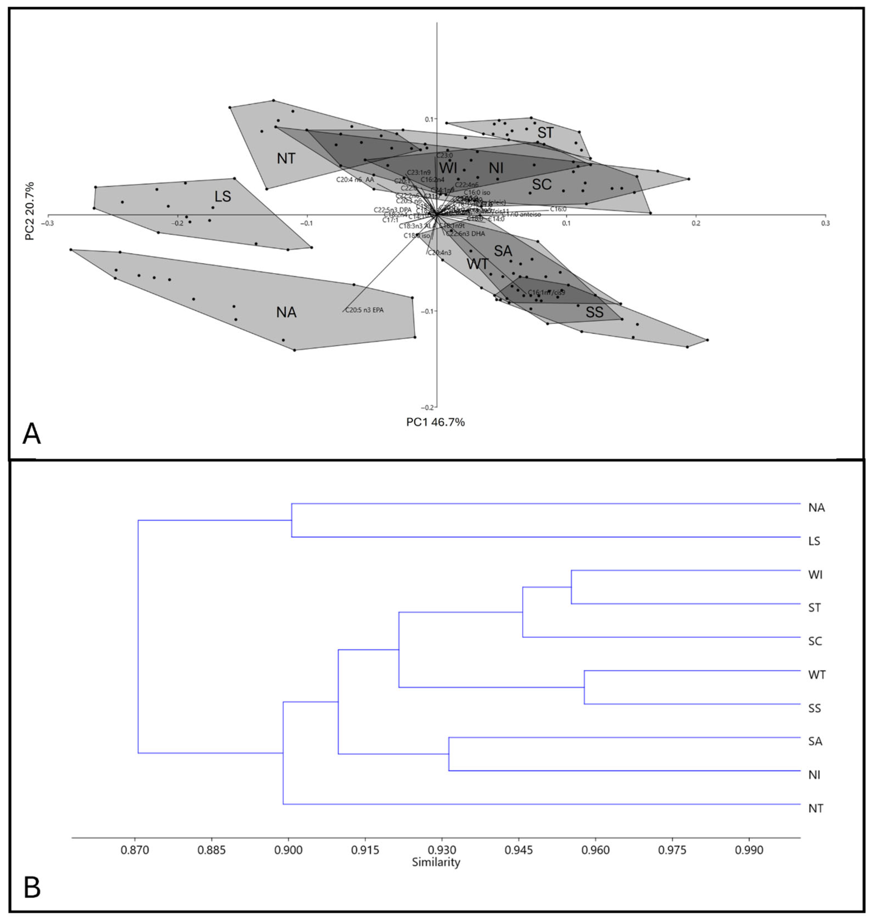
In H. polii, PC1 accounted for 46.7% of the variance, with palmitic acid C16:0 (loading plot value: 0.47), eicosapentaenoic acid (EPA), C20:5n-3 (loading plot value: 0.40), and palmitoleic acid C16:1n-7 (loading plot value: 0.38) making the most substantial contributions. PC2 explained 20.7% of the variance, with C20:5n-3 (loading plot value: 0.55) and C16:1n-7 (loading plot value: 0.45) displaying significant loadings. Consequently, the distribution in the first and third quadrants was predominantly influenced by high relative values of the EPA (C20:5n-3). The palmitoleic acid (C16:1n-7) mostly determined the distribution of the second quadrant, whereas the arrangement of the observations in the fourth quadrant was dictated mainly by elevated values of the arachidonic acid C20:4n-6 (Figure 2A).
Certain groups of sea cucumbers exhibited distinct characteristics from others. Sea cucumbers from the Ligurian Sea were notably isolated in the negative extreme of the PC1 axis, without any overlaps with other sites. Similarly, the Northern Adriatic group showed clear isolation, occupying the third quadrant. Sea cucumbers from the Southern Adriatic Sea tended to populate the second quadrant of the graph, partially overlapping with the two groups from the Sardinian sites (WT and SS). The remaining groups (NT, NI, WI, ST, and SC) exhibited significant overlap in the first and fourth quadrants, with a scattered dispersion. The UPGMA dendrogram, employing the intra-group averages of the 42 FAs loadings considered in the PCA (Figure 2B), underlined the geographic distribution of the samples collected. Notably, the clustering confirmed the highest similarity between the samples from WT and SS (similarity: 0.959), collected along Sardinia Island. Sea cucumbers from SA and NI, which are geographically close (collected in the Apulia region), exhibited a relatively high similarity (0.932). It is noteworthy that Sicilian samples (SC, WI, and ST) clustered together with a high similarity index (0.945 and 0.957). Finally, the lowest similarity indexes were recorded for samples collected from the LS and NA sites, and the ones from NT, which formed an isolated branch.

3.1.2. Holothuria tubulosa

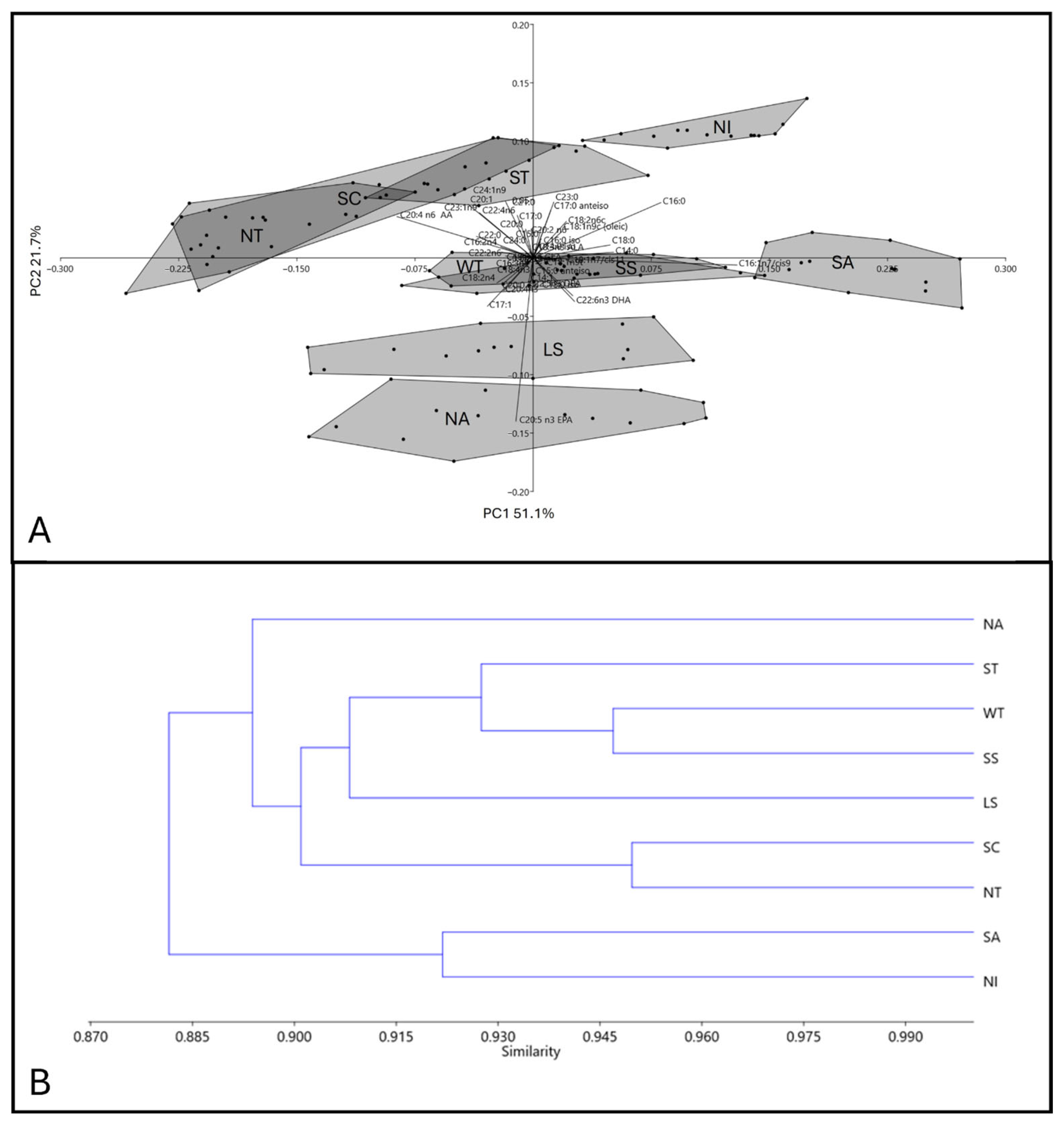
In H. tubulosa, PC1 explained 51.1% of the variance, with heavy loading from palmitoleic acid C16:1n-7 (loading plot value: 0.59), arachidonic acid C20:4n-6 (loading plot value: 0.40), and palmitic acid C16:0 (loading plot value: 0.37); PC2 explained 21.7% of the variance, with mainly the contribution from EPA C20:5n-3 (loading plot value: 0.64) driving the distribution, followed by C24:1n-9 (loading plot value: 0.22). Palmitic acid (C16:0) and C22:6n3 (DHA) drove the variance in the first and second quadrants, respectively (Figure 3A). Sea cucumbers from NA are distinct from others, occupying the negative portion of the PC2 axis across the second and third quadrants, as are the Ligurian and two Sardinian sites (SS and WT), the latter largely overlapping with each other. All the remaining groupings (except SA, isolated at the positive portion of PC1, and NI secluded in the first quadrant) are partially overlapped, with a scattered dispersion within the polygons.
The UPGMA dendrogram for this species also underlined the geographic pattern of the samples collected (Figure 3B). The high similarity between the cucumbers from the sampling sites in Sardinia (WT and SS) is evident (0.947). Sea cucumbers from the SA and NI sites clustered together (similarity 0.92). It is noteworthy that the Sicilian SC samples were more clustered with NT (0.95) than the other Sicilian site (ST). Finally, the lowest similarity index (0.890) was recorded for samples from the Northern Adriatic Sea.

3.2. Trophic Biomarkers

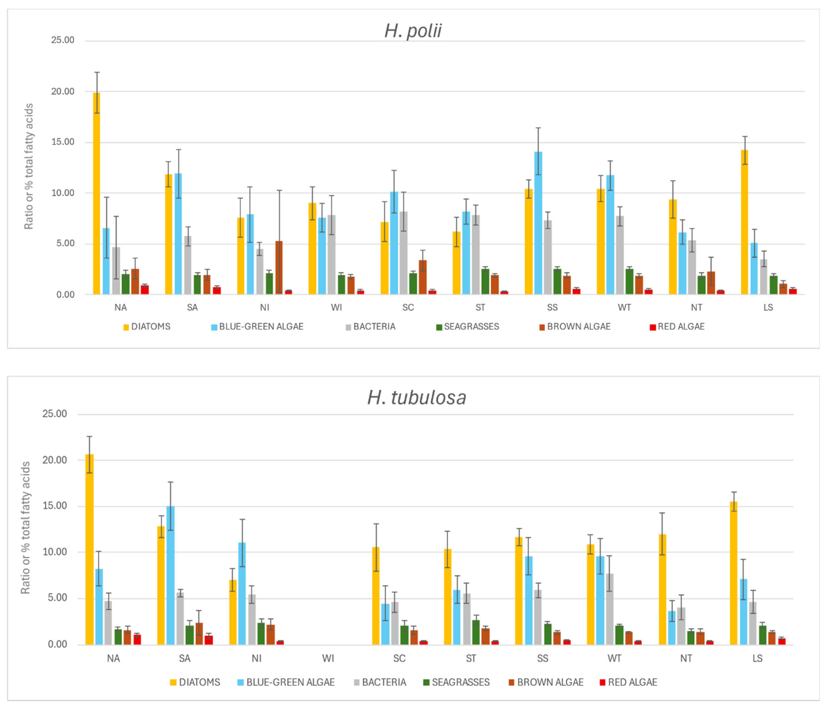
Figure 4 presents histograms illustrating the composition of six fatty acid trophic markers (FATMs), representing potential food sources, in the body walls of two sea cucumber species (15 specimens each) across all sampling sites. These visualizations highlight the potential dominance of specific dietary components within their respective trophic niches.
The analysis of the relative abundances of FATM revealed both differences and similarities between the two sea cucumber species (Holothuria polii and Holothuria tubulosa) across the various sampling sites. Diatoms emerged as one of the main trophic sources for both species, with particularly high values recorded at the NA, LS, and NT sites. In H. polii, NA and LS showed a dominant and consistent contribution of diatom-derived markers, while a similar trend was observed in H. tubulosa, especially at the same locations, with percentages in some cases exceeding 20%. Blue-green algae generally represented the second most abundant group. In H. polii, however, sites SC and SS showed higher contributions from blue-green algae compared to other sources, making them the dominant dietary component in these cases. In H. tubulosa, site NI displayed a strong dominance of this source, suggesting spatial variation in the trophic role of this group. Bacterial markers represented the third most important group overall. In H. polii, at sites WI, SC, and ST, the bacterial contribution was comparable to that of diatoms and cyanobacteria, indicating potential trophic plasticity in response to locally available resources. In contrast, H. tubulosa showed a consistently lower bacterial contribution across all sites, never reaching the levels observed for the other two major trophic groups. Seagrasses and brown algae were less represented in the diet of both species, suggesting a secondary trophic role. However, at site NI in H. polii, the contribution of brown algae was noticeably higher, representing the third most important trophic source. Red algae showed a mean value of less than 10 for both species and at all sites, reflecting the negligible importance of this dietary source.

4. Discussion

This study investigated the fatty acid (FA) profiles of two Holothuria species (H. tubulosa and H. polii) collected from ten sampling sites along the Italian coastline, encompassing a total of 42 fatty acids. The two species exhibited remarkably similar FA profile patterns, likely reflecting their co-occurrence in overlapping geographical areas and suggesting that they occupy comparable trophic niches [7,59,60]. Principal Component Analysis (PCA) revealed clearer clustering of individuals from certain sites, specifically NA, LS, NT and ST for H. polii and NA, LS, SA, and NI for H. tubulosa, based on their FA compositions. While this clustering partially corresponded with geographical proximity, it is more likely attributed to shared dietary resources across these locations. The PCA, an unsupervised multivariate statistical analysis, explained a high total variance for both species (67.4% for H. polii and 72.7% for H. tubulosa), which is notable given the complexity of the ecological context. The loadings of individual fatty acids highlighted those that most strongly contribute to either a similar trophic ecology (cluster overlap) or divergent trophic patterns (cluster separation). To further understand their biological significance, these fatty acids were analyzed using FATMs, which group them according to established studies in marine ecology. Although all sampling sites were selected from sandy substrates within seagrass-dominated habitats (mainly Posidonia oceanica and/or Cymodocea nodosa biocoenoses), site-specific differences in FA profiles were primarily driven by specific fatty acids or FA groups, especially polyunsaturated fatty acids (PUFAs), which are not synthesized by primary consumers [45,50]. Environmental variability, differences in local food availability, and ecological conditions likely contributed to the observed spatial patterns in FA composition. Most holothurians are epibenthic surface deposit feeders that consume fresh benthic microalgae and detritus from the upper sediment layers [22,61]. They accumulate dietary PUFAs, particularly when benthic microalgae and fresh phytodetritus are available, which are essential for key physiological processes including reproduction and growth [61]. Accordingly, the high proportions of PUFAs found in both species reflect their dependence on such high-quality dietary inputs. Lipid biomarker analysis revealed that diatoms and blue-green algae were the primary dietary sources for both sea cucumber species, followed by smaller contributions from bacteria and minimal inputs from seagrasses and brown macroalgae [6,56,62]. Considering the limitations of the index defined by Khotimchenko and Vaskovsky [41] as a FATM for red algae, the results suggest that this group does not represent a trophic source for the studied species at the investigated sites. This finding is further supported by the low abundance of this phylum observed during the sampling phase. These dietary signatures exhibited some spatial variability, likely reflecting differences in the composition and structure of local food webs. Diatoms, in particular, were consistently prominent dietary components, which is likely attributable to their ecological ubiquity and high nutritional value. This pattern corroborates previous research demonstrating the dominance of diatoms within epiphytic assemblages associated with Posidonia australis [63,64,65]. Although sea cucumbers are known to consume detritus composed of decomposing seagrass leaves and rhizomes, the fatty acid (FA) profiles suggest a stronger assimilation of diatom-derived biomarkers than of compounds originating directly from seagrasses. This supports the hypothesis that selective assimilation occurs during digestion, whereby sea cucumbers preferentially retain more nutritious or bioavailable components such as diatoms [66,67]. Blue-green algae biomarkers, while generally lower in abundance than diatom markers, were consistently detected across both species and sites, suggesting that cyanobacteria also contribute meaningfully to their diet. Their persistence across samples highlights their potential role as a supplementary but reliable dietary component, especially in areas where cyanobacterial biofilms are abundant. Moreover, the consistently elevated levels of bacterial lipid markers in both species indicate that bacteria from decaying sedimentary organic matter are also a vital dietary resource. These findings reinforce the important ecological role of holothurians in benthic nutrient recycling and sediment bioturbation [67]. Additional factors likely contributing to variability in dietary FA availability across sites include sedimentological and geochemical characteristics, as well as spatial differences in periphyton communities. These variables can influence both the type and abundance of available food sources, ultimately shaping the FA composition in sea cucumber tissues at a local scale. However, further explanations for these variations can be found in seasonal dietary shifts documented in other regions [3,20]. Studies conducted in China on FA biomarkers have demonstrated that the primary food sources of the sea cucumber Apostichopus japonicus vary markedly throughout the year [68,69]. In January, the diet is mainly composed of diatoms, dinoflagellates or protozoans, brown algae, and bacteria. By March, diatoms, flagellates or protozoans, and green microalgae become more prominent, with green microalgae dominating in June. During July, bacteria and chlorophytes constitute the primary food sources, while in August and September bacteria become the dominant component. From October through November, brown algae and bacteria together represent a significant part of the dietary intake [3]. These findings highlight the importance of sampling period, as temporal shifts in diet can strongly influence FA profiles. In the present study, sampling was conducted during the summer months, from June to September, a time window that likely encompasses seasonal subtle yet detectable dietary shifts in sea cucumbers. This underscores the importance of accounting for temporal variations when analyzing FA profiles and understanding ecological dynamics. Among sea cucumber species, PUFAs consistently emerge as the most dominant FA class, typically accounting for 20% to 60% of total fatty acids [3,70]. This trend is supported by the present study, in which PUFA concentrations ranged from 33.0 ± 4.5% to 52.5 ± 4.4%, indicating substantial variability across samples. Previous studies have suggested a link between PUFA variation and environmental salinity, with PUFA levels reportedly increasing under high salinity conditions while saturated fatty acid (SFA) concentrations decline. This shift has been interpreted as a cellular adaptation aimed at preserving membrane bilayer fluidity and enhancing cell motility in response to osmotic stress [71]. However, the relevance of this mechanism to our findings is limited. Despite observing site-specific variation in PUFA content across our sampling locations, salinity levels remained relatively stable during the sampling period, ranging around 38.0 ± 1.0 ppt in both the North and South Adriatic Sea, based on Practical Salinity Scale values from Copernicus data [72]. Therefore, the observed PUFA variability in this study cannot be attributed solely to salinity differences, as environmental fluctuations were minimal.
Fatty acid composition is shaped by a complex interplay of endogenous and exogenous factors. Endogenous factors include genetically regulated physiological processes such as growth, ontogenetic development, and annual reproductive cycles. In contrast, exogenous influences encompass environmental variables (such as temperature, light, salinity, and depth) as well as dietary components like food availability, feeding strategies, and diet quality [30,73,74]. The interaction of these internal and external factors makes it difficult to attribute inter- or intraspecific differences in FA profiles to a single cause [3]. Nonetheless, certain biochemical patterns appear to be conserved across holothurian species. These include consistently high levels of key FAs such as arachidonic acid (C20:4n-6, ARA), eicosapentaenoic acid (C20:5n-3, EPA), docosahexaenoic acid (C22:6n-3, DHA), C23:1n-9, palmitic acid (C16:0), oleic acid (C18:1), and palmitoleic acid (C16:1n-7), along with notable amounts of branched-chain fatty acids including iso-C15:0, iso-C17:0, and anteiso-C17:0 [3]. Our results align with this general profile, confirming the abundant presence of ARA, EPA, C16:0, C16:1n-7, and C23:1n-9. The latter, in particular, is recognized as a chemotaxonomic marker specific to holothurians [2,3]. Additionally, our analysis revealed relatively high levels of C18:0, C24:1n-9, and C20:1n-9, supporting findings from previous studies [66,75]. Conversely, docosahexaenoic acid (DHA) was found in relatively low concentrations compared to values reported in the literature. Given that DHA is a well-established fatty acid trophic marker (FATM) for dinoflagellates [23,30], this finding may reflect a limited presence or reduced dietary contribution of dinoflagellates during the summer sampling period, when primary productivity and species composition in the water column may differ from other seasons.

5. Conclusions

This study introduces the use of fatty acids to clarify the trophic ecology of H. polii and H. tubulosa along the Italian coasts, revealing their primary dependence on diatoms, cyanobacteria, and bacteria, with clear ecological and geographic differentiation. Through robust sampling and GC-FID/MS profiling, supported by both qualitative and quantitative analyses, we identified distinct fatty acid signatures. While not exhaustive, these findings establish fatty acids as effective markers for population-level dietary ecology and provide valuable insights for conservation and management. Fatty acid biomarkers have the potential to serve as powerful tools for linking feeding ecology with sea cucumber conservation efforts, through the identification and protection of vital habitats, enhancement of restocking success, and development of ecosystem-based management plans aimed at maintaining healthy benthic ecosystems. To strengthen and expand this work, future research should incorporate compound-specific stable isotope analysis (CSIA) of fatty acids, an increasingly refined tool for tracing nutrient and energy flow in aquatic ecosystems, alongside controlled feeding experiments to quantify trophic discrimination and better understand sea cucumber metabolism. Additionally, broader temporal and spatial monitoring (including multiple seasons, life stages, and sampling sites) as well as focusing on different classes and molecular species level of lipids, will help capture ontogenetic and environmental variability. Together, these approaches will deepen our understanding of trophic dynamics in sea cucumbers, among the most relevant species of the benthic coastal communities.

Author Contributions

N.T.: Conceptualization, Methodology, Data curation, Investigation, Validation, Visualization, Formal Analysis, Writing—Original Draft, Writing—Review and Editing. M.C.: Methodology. M.M.: Methodology, Writing—Review and Editing. A.M.: Writing—Review and Editing. A.F.: Resources. L.F.: Funding Acquisition. F.G.: Methodology. E.P.: Methodology, Writing—Review and Editing. D.P.: Conceptualization, Methodology, Funding Acquisition, Writing—Review and Editing. A.R.: Methodology, Resources, Writing—Review and Editing. R.N.: Writing—Review and Editing. F.C.: Conceptualization, Methodology, Writing—Review and Editing, Funding Acquisition, Supervision, Project Administration. All authors have read and agreed to the published version of the manuscript.

Funding

This research was supported by the Holothuria Research Project (N. J56J20000150001) from the Italian Ministry of Agriculture, Food Sovereignty and Forests (MASAF).

Institutional Review Board Statement

Capture, handling, and killing were done under permits of the Italian Ministry of Agriculture, Food Sovereignty and Forests (MASAF) (permit n. 0005606 released on 3 September 2020).

Data Availability Statement

The authors declare that the data supporting the findings of this study are available within the paper. Should any raw data files be needed in another format, they are available from the corresponding author upon reasonable request.

Acknowledgments

We would like to thank Failla for her guidance in the world of fatty acids.

Conflicts of Interest

The authors declare no conflicts of interest.

References

  1. Brusca, R.C.; Brusca, G.J. Invertebrates; Sinauer Associates: Sunderland, MA, USA, 2003; Volume 347. [Google Scholar]
  2. Drazen, J.C.; Phleger, C.F.; Guest, M.A.; Nichols, P.D. Lipid, sterols and fatty acid composition of abyssal holothurians and ophiuroids from the North-East Pacific Ocean: Food web implications. Comp. Biochem. Physiol.-B Biochem. Mol. Biol. 2008, 151, 79–87. [Google Scholar] [CrossRef]
  3. Zhukova, N.V. Fatty Acids of Echinoderms: Diversity, Current Applications and Future Opportunities. Mar. Drugs 2023, 21, 21. [Google Scholar] [CrossRef] [PubMed]
  4. Pasquini, V.; Giglioli, A.A.; Pusceddu, A.; Addis, P. Biology, ecology and management perspectives of overexploited deposit-feeders sea cucumbers, with focus on Holothuria tubulosa (Gmelin, 1788). Adv. Oceanogr. Limnol. 2021, 12. [Google Scholar] [CrossRef]
  5. Karapanagiotidis, I.T.; Gkalogianni, E.Z.; Apostologamvrou, C.; Voulgaris, K.; Varkoulis, A.; Vafidis, D. Proximate Compositions and Fatty Acid Profiles of Raw and Processed Holothuria polii and Holothuria tubulosa from the Aegean Sea. Sustainability 2024, 16, 6048. [Google Scholar] [CrossRef]
  6. Costa, V.; Mazzola, A.; Vizzini, S. Holothuria tubulosa Gmelin 1791 (Holothuroidea, Echinodermata) enhances organic matter recycling in Posidonia oceanica meadows. J. Exp. Mar. Biol. Ecol. 2014, 461, 226–232. [Google Scholar] [CrossRef]
  7. Boncagni, P.; Rakaj, A.; Fianchini, A.; Vizzini, S. Preferential assimilation of seagrass detritus by two coexisting Mediterranean sea cucumbers: Holothuria polii and Holothuria tubulosa. Estuar. Coast. Shelf Sci. 2019, 231, 106464. [Google Scholar] [CrossRef]
  8. Uthicke, S.; Karez, R. Sediment patch selectivity in tropical sea cucumbers (Holothurioidea: Aspidochirotida) analysed with multiple choice experiments. J. Exp. Mar. Bio. Ecol. 1999, 236, 69–87. [Google Scholar] [CrossRef]
  9. Kinch, J. Importance of Sea cucumber fisheries and trade for small island communities: A case study in Papua New Guinea. In The World of Sea Cucumbers Challenges, Advances, and Innovations; Academic Press: Cambridge, MA, USA, 2024; pp. 133–148. [Google Scholar] [CrossRef]
  10. Kinch, J.; Purcell, S.W.; Uthicke, S.; Friedman, K. Population status, fisheries and trade of sea cucumbers in the Western Central Pacific. In Sea Cucumbers. A Global Review of Fisheries and Trade; FAO Fisheries and Aquaculture Technical Paper No. 516; FAO: Rome, Italy, 2008; pp. 7–55. [Google Scholar]
  11. Purcell, S.W.; Mercier, A.; Conand, C.; Hamel, J.F.; Toral-Granda, M.V.; Lovatelli, A.; Uthicke, S. Sea cucumber fisheries: Global analysis of stocks, management measures and drivers of overfishing. Fish Fish. 2013, 14, 34–59. [Google Scholar] [CrossRef]
  12. Toral-Granda, V. Population status, fisheries and trade of sea cucumbers in Latin America and the Caribbean. In Sea cucumbers. A Global Review of Fisheries and Trade; FAO Fisheries and Aquaculture Technical Paper; FAO: Rome, Italy, 2008; pp. 213–229. [Google Scholar]
  13. Bordbar, S.; Anwar, F.; Saari, N. High-Value Components and Bioactives from Sea Cucumbers for Functional Foods—A Review. Mar. Drugs 2011, 9, 1761–1805. [Google Scholar] [CrossRef]
  14. Dakrory, A.I.; Fahmy, S.R.; Soliman, A.M.; Mohamed, A.S.; Amer, S.A.M. Protective and Curative Effects of the Sea Cucumber Holothuria atra Extract against DMBA-Induced Hepatorenal Diseases in Rats. BioMed Res. Int. 2015, 2015, 563652. [Google Scholar] [CrossRef]
  15. Anderson, S.C.; Flemming, J.M.; Watson, R.; Lotze, H.K. Serial exploitation of global sea cucumber fisheries. Fish Fish. 2011, 12, 317–339. [Google Scholar] [CrossRef]
  16. David, F.; Hubas, C.; Laguerre, H.; Badou, A.; Herault, G.; Bordelet, T.; Ameziane, N. Food sources, digestive efficiency and resource allocation in the sea cucumber Holothuria forskali (Echinodermata: Holothuroidea): Insights from pigments and fatty acids. Aquac. Nutr. 2020, 26, 1568–1583. [Google Scholar] [CrossRef]
  17. Sadoul, B.; Caprioli, J.P.; Barrier-Loiseau, C.; Cimiterra, N.; Laugier, T.; Lagarde, F.; Chary, K.; Callier, M.D.; Guillermard, M.O.; Roque d’Orbcastel, E. Is Holothuria tubulosa the golden goose of ecological aquaculture in the Mediterranean Sea? Aquaculture 2022, 554, 738149. [Google Scholar] [CrossRef]
  18. Azevedo and Silva, F.; Brito, A.C.; Pombo, A.; Simões, T.; Marques, T.A.; Rocha, C.; Madruga, A.S.; Sousa, J.; Venâncio, E.; Félix, P.M. Spatiotemporal Distribution Patterns of the Sea Cucumber Holothuria arguinensis on a Rocky-Reef Coast (Northeast Atlantic). Estuaries Coasts 2023, 46, 1035–1045. [Google Scholar] [CrossRef]
  19. González-Wangüemert, M.; Roggatz, C.C.; Rodrigues, M.J.; Barreira, L.; da Silva, M.M.; Custódio, L. A new insight into the influence of habitat on the biochemical properties of three commercial sea cucumber species. Int. Aquat. Res. 2018, 10, 361–373. [Google Scholar] [CrossRef]
  20. Sicuro, B.; Manuela, P.; Francesco, G.; Abete, M.C.; Antonio, D.; Franco, D.; Mioletti, S. Food quality and Safety of Mediterranean Sea Cucumbers Holothuria tubulosa and Holothuria polii in Southern Adriatic Sea. Asian J. Anim. Vet. Adv. 2012, 7, 851–859. [Google Scholar] [CrossRef]
  21. Meloni, D.; Esposito, G. Hygienic and commercial issues related to the illegal fishing and processing of sea cucumbers in the Mediterranean: A case study on over-exploitation in Italy between 2015 and 2017. Reg. Stud. Mar. Sci. 2018, 19, 43–46. [Google Scholar] [CrossRef]
  22. Rakaj, A.; Fianchini, A. Mediterranean sea cucumbers—Biology, ecology, and exploitation. In The World of Sea Cucumbers Challenges, Advances, and Innovations; Academic Press: Cambridge, MA, USA, 2024; pp. 753–773. [Google Scholar] [CrossRef]
  23. Alfaro, A.C.; Thomas, F.; Sergent, L.; Duxbury, M. Identification of trophic interactions within an estuarine food web (northern New Zealand) using fatty acid biomarkers and stable isotopes. Estuar. Coast. Shelf Sci. 2006, 70, 271–286. [Google Scholar] [CrossRef]
  24. Hazel, J.R.; Williams, E.E.; Livermore, R.; Mozingo, N. Thermal adaptation in biological membranes: Functional significance of changes in phospholipid molecular species composition. Lipids 1991, 26, 277–282. [Google Scholar] [CrossRef]
  25. Parrish, C.C.; Abrajano, T.A.; Budge, S.M.; Helleur, R.J.; Hudson, E.D.; Pulchan, K.; Ramos, C. Lipid and Phenolic Biomarkers in Marine Ecosystems: Analysis and Applications. In Marine Chemistry; Springer: Berlin/Heidelberg, Germany, 2000; pp. 193–223. [Google Scholar]
  26. Stowasser, G.; Pond, D.W.; Collins, M.A. Fatty acid trophic markers elucidate resource partitioning within the demersal fish community of South Georgia and Shag Rocks (Southern Ocean). Mar. Biol. 2012, 159, 2299–2310. [Google Scholar] [CrossRef]
  27. Hiltunen, M.; Strandberg, U.; Brett, M.T.; Winans, A.K.; Beauchamp, D.A.; Kotila, M.; Keister, J.E. Taxonomic, Temporal, and Spatial Variations in Zooplankton Fatty Acid Composition in Puget Sound, WA, USA. Estuaries Coasts 2022, 45, 567–581. [Google Scholar] [CrossRef]
  28. Kelly, J.R.; Scheibling, R.E. Fatty acids as dietary tracers in benthic food webs. Mar. Ecol. Prog. Ser. 2012, 446, 1–22. [Google Scholar] [CrossRef]
  29. Salant, C.D.; Shanks, A.L.; Schram, J.B.; Galloway, A.W.E. Trophic Biomarkers Indicate Coastal Surf Zone Hydrodynamics Affect Resource Assimilation by Mytilus californianus Mussels. Estuaries Coasts 2021, 44, 2212–2221. [Google Scholar] [CrossRef]
  30. Dalsgaard, J.; St John, M.; Kattner, G.; Müller-Navarra, D.; Hagen, W. Fatty acid trophic markers in the pelagic marine environment. Adv. Mar. Biol. 2003, 46, 225–340. [Google Scholar] [CrossRef]
  31. Coelho, H.; Lopes da Silva, T.; Reis, A.; Queiroga, H.; Serôdio, J.; Calado, R. Fatty acid profiles indicate the habitat of mud snails Hydrobia ulvae within the same estuary: Mudflats vs. seagrass meadows. Estuar. Coast. Shelf Sci. 2011, 92, 181–187. [Google Scholar] [CrossRef]
  32. Iverson, S.J. Tracing aquatic food webs using fatty acids: From qualitative indicators to quantitative determination. In Lipids in Aquatic Ecosystems; Springer: New York, NY, USA, 2009; pp. 281–308. [Google Scholar]
  33. Nerot, C.; Meziane, T.; Schaal, G.; Grall, J.; Lorrain, A.; Paulet, Y.M.; Kraffe, E. Spatial changes in fatty acids signatures of the great scallop Pecten maximus across the Bay of Biscay continental shelf. Cont. Shelf Res. 2015, 109, 1–9. [Google Scholar] [CrossRef]
  34. Penha-Lopes, G.; Torres, P.; Narciso, L.; Cannicci, S.; Paula, J. Comparison of fecundity, embryo loss and fatty acid composition of mangrove crab species in sewage contaminated and pristine mangrove habitats in Mozambique. J. Exp. Mar. Biol. Ecol. 2009, 381, 25–32. [Google Scholar] [CrossRef]
  35. Liu, Y.; Zhang, X.; Li, Y.; Wang, H. The application of compound-specific isotope analysis of fatty acids for traceability of sea cucumber (Apostichopus japonicus) in the coastal areas of China. J. Sci. Food Agric. 2017, 97, 4912–4921. [Google Scholar] [CrossRef]
  36. Kharlamenko, V.I.; Zhukova, N.V.; Khotimchenko, S.V.; Svetashev, V.I.; Kamenev, G.M. Fatty acids as markers of food sources in a shallow-water hydrothermal ecosystem (Kraternaya Bight, Yankich Island, Kurile Islands). Mar. Ecol. Prog. Ser. 1995, 120, 231–242. [Google Scholar] [CrossRef]
  37. Rajendran, N.; Suwa, Y.; Urushigawa, Y. Distribution of phospholipid ester-linked fatty acid biomarkers for bacteria in the sediment of Ise Bay, Japan. Mar. Chem. 1993, 42, 39–56. [Google Scholar] [CrossRef]
  38. Napolitano, G.E. Fatty acids as chemical and trophic markers in freshwater ecosystems. In Lipids in Freshwater Ecosystems; Springer: New York, NY, USA, 1999; pp. 21–44. [Google Scholar]
  39. Falk-Petersen, S.; Dahl, T.M.; Scott, C.L.; Sargent, J.R.; Gulliksen, B.; Kwasniewski, S.; Hop, H.; Millar, R.M. Lipid biomarkers and trophic linkages between ctenophores and copepods in Svalbard waters. Mar. Ecol. Prog. Ser. 2002, 227, 187–194. [Google Scholar] [CrossRef]
  40. Johns, R.B.; Nichols, P.D.; Perry, G.J. Fatty acid composition of ten marine algae from Australian waters. Phytochemistry 1979, 18, 799–802. [Google Scholar] [CrossRef]
  41. Khotimchenko, S.V.; Vaskovsky, V.E. Distribution of C 20 Polyenoic Fatty Acids in Red Macrophytic Algae. Bot. Mar. 1990, 33, 525–528. [Google Scholar] [CrossRef]
  42. Wannigama, G.P.; Volkman, J.K.; Gillan, F.T.; Nichols, P.D.; Johns, R.B. A comparison of lipid components of the fresh and dead leaves and pneumatophores of the mangrove Avicennia marina. Phytochemistry 1981, 20, 659–666. [Google Scholar] [CrossRef]
  43. Derrien, M.; Yang, L.; Hur, J. Lipid biomarkers and spectroscopic indices for identifying organic matter sources in aquatic environments: A review. Water Res. 2017, 112, 58–71. [Google Scholar] [CrossRef]
  44. Gopi, K.; Mazumder, D.; Sammut, J.; Saintilan, N. Determining the provenance and authenticity of seafood: A review of current methodologies. Trends Food Sci. Technol. 2019, 91, 294–304. [Google Scholar] [CrossRef]
  45. Zhang, X.; Liu, Y. Stable carbon isotope fractionation of fatty acid in sea cucumber (Apostichopus japonicus): Insights from an experimental study. N. Z. J. Mar. Freshw. Res. 2022, 56, 234–246. [Google Scholar] [CrossRef]
  46. Iverson, S.J.; Field, C.; Don Bowen, W.; Blanchard, W. Quantitative fatty acid signature analysis: A new method of estimating predator diets. Ecol. Monogr. 2004, 74, 211–235. [Google Scholar] [CrossRef]
  47. Budge, S.M.; Iverson, S.J.; Koopman, H.N. Studying Trophic Ecology in Marine Ecosystems Using Fatty Acids: A Primer on Analysis and Interpretation. Mar. Mammal. Sci. 2006, 22, 759–801. [Google Scholar] [CrossRef]
  48. Parrish, C.C. Lipids in Marine Ecosystems. ISRN Oceanogr. 2013, 2013, 1–16. [Google Scholar] [CrossRef]
  49. Owen, J.M.; Adron, J.W.; Sargent, J.R.; Cowey, C.B. Studies on the nutrition of marine flatfish. The effect of dietary fatty acids on the tissue fatty-acids of the plaice Pleuronectes platessa. Mar. Biol. 1972, 13, 160–166. [Google Scholar] [CrossRef]
  50. Guil-Guerrero, J.L.; Venegas-Venegas, E.; Rincón-Cervera, M.Á.; Suárez, M.D. Fatty acid profiles of livers from selected marine fish species. J. Food Compos. Anal. 2011, 24, 217–222. [Google Scholar] [CrossRef]
  51. Budge, S.M.; Iverson, S.J.; Bowen, W.D.; Ackman, R.G. Among- and within-species variability in fatty acid signatures of marine fish and invertebrates on the Scotian Shelf, Georges Bank, and Southern Gulf of St. Lawrence. Can. J. Fish. Aquat. Sci. 2002, 59, 886–898. [Google Scholar] [CrossRef]
  52. Tortonese, E. Fauna d’Italia Vol. VI—Echinodermata; Calderini: Bologna, Italy, 1965. [Google Scholar]
  53. Lang, I.; Hodac, L.; Friedl, T.; Feussner, I. Fatty acid profiles and their distribution patterns in microalgae: A comprehensive analysis of more than 2000 strains from the SAG culture collection. BMC Plant Biol. 2011, 11, 124. [Google Scholar] [CrossRef]
  54. IUPAC. Standard Methods for the Analysis of Oils, Fats and Derivatives, 7th ed.; Blackwell Jevent: Oxford, UK, 1992. [Google Scholar]
  55. Ackman, R.G. Gas-liquid chromatography of fatty acids and esters. Methods Enzymol. 1969, 14, 329–381. [Google Scholar]
  56. Purcell, S.W.; Conand, C.; Uthicke, S.; Byrne, M. Ecological Roles of Exploited Sea Cucumbers. Oceanogr. Mar. Biol. Annu. Rev. 2016, 54, 173–191. [Google Scholar] [CrossRef]
  57. Capoccioni, F.; Contò, M.; Failla, S.; Cataudella, S.; Ciccotti, E. Fatty acid profiles of migrating female silver eel from Mediterranean coastal lagoons as integrative descriptors of spawners biological quality. Estuar. Coast. Shelf Sci. 2018, 210, 87–97. [Google Scholar] [CrossRef]
  58. Hammer, Ø.; Harper, D.A.T.; Ryan, P.D. Past: Paleontological Statistics Software Package for Education and Data Analysis. Palaeontol. Electron. 2001, 4, 4–9. [Google Scholar]
  59. Massin, C.; Jangoux, M. Les observations écologiques se rapportant aux Holothuries des mers d’ Europe et, plus particulièrement, aux Aspidochirotes sont fort rares. Les renseignements donnés par les ouvrages généraux (Ludwig, 1889–1891). Cah. Biol. Mar. 1976, 17, 45–59. [Google Scholar]
  60. Mezali, K.; Zupo, V.; Francour, P. Population Dynamics of Holothuria (Holothuria) tubulosa and Holothuria (Lessonothuria) polii of an Algerian Posidonia oceanica meadow. Biol. Mar. Medit. 2006, 13, 158–161. [Google Scholar]
  61. Hudson, I.; Pond, D.; Billett, D.; Tyler, P.; Lampitt, R.; Wolff, G. Temporal variations in fatty acid composition of deep-sea holothurians: Evidence of bentho-pelagic coupling. Mar. Ecol. Prog. Ser. 2004, 281, 109–120. [Google Scholar] [CrossRef]
  62. Belbachir, N.-E.; Mezali, K. Food preferences of four aspidochirotid holothurians species (Holothuroidea: Echinodermata) inhabiting the Posidonia oceanica meadow of Mostaganem area (Algeria). SPC Beche-De-Mer. Inf. Bull. 2018, 36, 55–59. [Google Scholar]
  63. Nichols, P.D.; Klumpp, D.W.; Johns, R.B. Lipid components of the epiphyte material, suspended particulate matter and cultured bacteria from a seagrass, Posidonia australis, community as indicators of carbon source. Comp. Biochem. Physiol. Part B Comp. Biochem. 1985, 80, 315–325. [Google Scholar] [CrossRef]
  64. Kanjer, L.; Mucko, M.; Car, A.; Bosak, S. Epiphytic diatoms on Posidonia oceanica (L.) Delile leaves from eastern Adriatic Sea. Nat. Croat. 2019, 28, 1–20. [Google Scholar]
  65. Mabrouk, L.; Ben Brahim, M.; Hamza, A.; Mahfoudhi, M.; Bradai, M.N. A comparison of abundance and diversity of epiphytic microalgal assemblages on the leaves of the seagrasses Posidonia oceanica (L.) and Cymodocea nodosa (Ucria) asch in Eastern Tunisia. J. Mar. Biol. 2014, 2014, 275305. [Google Scholar] [CrossRef]
  66. Cutajar, K.; Falconer, L.; Massa-Gallucci, A.; Cox, R.E.; Schenke, L.; Bardócz, T.; Andolina, C.; Signa, G.; Vizzini, S.; Sprague, M.; et al. Stable isotope and fatty acid analysis reveal the ability of sea cucumbers to use fish farm waste in integrated multi-trophic aquaculture. J. Environ. Manag. 2022, 318, 115511. [Google Scholar] [CrossRef]
  67. Mfilinge, P.L.; Tsuchiya, M. Changes in Sediment Fatty Acid Composition during Passage through the Gut of Deposit Feeding Holothurians: Holothuria atra (Jaeger, 1883) and Holothuria leucospilota (Brandt, 1835). J. Lipids 2016, 2016, 4579794. [Google Scholar] [CrossRef]
  68. Gao, F.; Xu, Q.; Yang, H. Seasonal variations of food sources in Apostichopus japonicus indicated by fatty acid biomarkers analysis. J. Fish. China 2010, 34, 760–767. [Google Scholar] [CrossRef]
  69. Feng, J.; Zhang, L.; Tang, X.; Xia, X.; Hu, W.; Zhou, P. Season and geography induced variation in sea cucumber (Stichopus japonicus) nutritional composition and gut microbiota. J. Food Compos. Anal. 2021, 101, 103838. [Google Scholar] [CrossRef]
  70. Zhang, X.; Liu, Y.; Li, Y.; Zhao, X. Identification of the geographical origins of sea cucumber Population status, fisheries and trade of sea cucumbers in Latin America and the Caribbean. Sea cucumbers. A global review of fisheries and trade (Apostichopus japonicus) in northern China. Food Chem. 2017, 218, 269–276. [Google Scholar] [CrossRef]
  71. Nemova, N.N.; Fokina, N.N.; Nefedova, Z.A.; Ruokolainen, T.R.; Bakhmet, I.N. Modifications of gill lipid composition in littoral and cultured blue mussels Mytilus edulis L. under the influence of ambient salinity. Polar Rec. 2013, 49, 272–277. [Google Scholar] [CrossRef]
  72. CMEMS. Marine Data Store (MDS). Mediterranean Sea Physics Reanalysis; E.U. Copernicus Marine Service Information, Marine Data Store (MDS): Lisbon, Portugal, 2024. [Google Scholar]
  73. Silina, A.V.; Zhukova, N.V. Growth variability and feeding of scallop Patinopecten yessoensis on different bottom sediments: Evidence from fatty acid analysis. J. Exp. Mar. Biol. Ecol. 2007, 348, 46–59. [Google Scholar] [CrossRef]
  74. Hill, W.R.; Rinchard, J.; Czesny, S. Light, nutrients and the fatty acid composition of stream periphyton. Freshw. Biol. 2011, 56, 1825–1836. [Google Scholar] [CrossRef]
  75. Wen, J.; Hu, C.; Fan, S. Chemical composition and nutritional quality of sea cucumbers. J. Sci. Food Agric. 2010, 90, 2469–2474. [Google Scholar] [CrossRef]
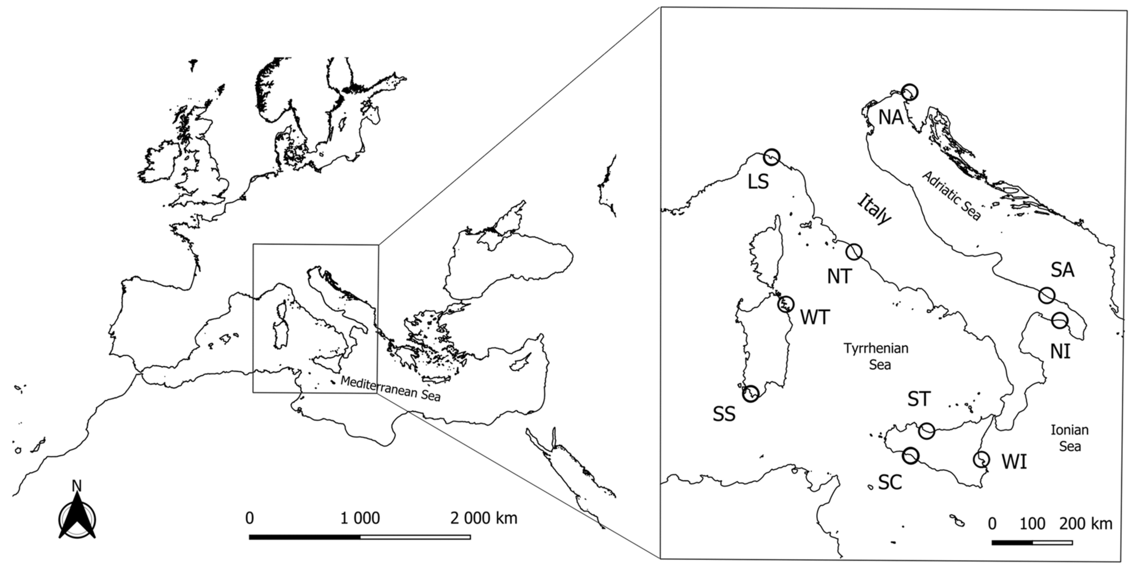
Figure 1. Map of the 10 sampled stations of H. polii and H. tubulosa along the Italian coast. NA: Northern Adriatic Sea, SA: Southern Adriatic Sea, NI: Northern Ionian Sea, WI: Western Ionian Sea, SC: Sicilian Channel, ST: Southern Tyrrhenian Sea, SS: Sea of Sardinia, WT: Western Tyrrhenian Sea, NT: Northern Tyrrhenian Sea, LS: Ligurian Sea.
Diversity 17 00576 g001
Figure 2. (A) Principal Component Analysis of the fatty acid composition of Holothuria polii from the ten sampling sites of the study. (B) Dendrogram distance of groups, calculated on the average value of the FAs that contribute the most to the PCA variance (NA: Northern Adriatic Sea, SA: Southern Adriatic Sea, NI: Northern Ionian Sea, WI: Western Ionian Sea, SC: Sicilian Channel, ST: Southern Tyrrhenian Sea, SS: Sea of Sardinia, WT: Western Tyrrhenian Sea, NT: Northern Tyrrhenian Sea, LS: Ligurian Sea).
Diversity 17 00576 g002
Figure 3. (A) Principal Component Analysis of the fatty acid composition of Holothuria tubulosa from nine of the ten sampling sites of the study. (B) The dendrogram distance of groups, calculated on the average value of the FAs that contribute the most to the PCA variance. (NA: Northern Adriatic Sea, SA: Southern Adriatic Sea, NI: Northern Ionian Sea, SC: Sicilian Channel, ST: Southern Tyrrhenian Sea, SS: Sea of Sardinia, WT: Western Tyrrhenian Sea, NT: Northern Tyrrhenian Sea, LS: Ligurian Sea).
Diversity 17 00576 g003
Figure 4. Composition of biomarkers (ratio or percent ± SD) for potential food sources in H. polii (above) and H. tubulosa (below) samples divided by sampling sites (NA: Northern Adriatic Sea, SA: Southern Adriatic Sea, NI: Northern Ionian Sea, WI: Western Ionian Sea (available for H. polii only), SA: Sicilian Channel, ST: Southern Tyrrhenian Sea, SS: Sea of Sardinia, WT: Western Tyrrhenian Sea, NT: Northern Tyrrhenian Sea, LS: Ligurian Sea).
Diversity 17 00576 g004
Table 1. FA ratios used by several authors as markers in the aquatic organisms for different food sources.
SourceBiomarker
Diatoms 1C20:5n-3
Bacteria 2∑15:0 + ∑17:0 + C18:1n-7
Seagrasses 3C18:2n-6 + C18:3n-3
Brown algae 4C18:1n-9
Red algae 5C20:5n-3/C20:4n-6 > 10
Blue-green algae 6C16:1n-7 + C18:1n-7
1 Parrish et al., 2000 [25], Derrien et al., 2017 [43]; 2 Rajendran et al., 1993 [37] Kharlamenko et al., 1995 [36]; 3 Wannigama et al., 1981 [42], Alfaro et al., 2006 [23]; 4 Johns et al., 1979 [40], Alfaro et al., 2006 [23]; 5 Khotimchenko and Vaskovsky, 1990 [41]; 6 Napolitano, 1999 [38]; Derrien et al., 2017 [43].
Table 2. Fatty acid composition (% on total FAMEs) of the body wall of H. polii (n = 15 for each site) from the study sites along the coast of Italy. SFAs: saturated fatty acids; MUFAs: monounsaturated fatty acids; PUFAs: polyunsaturated fatty acids.
Fatty AcidNorthern Adriatic Sea (NA)Southern Adriatic Sea (SA)Northern Ionian Sea (NI)Western Ionian Sea (WI)Sicilian Channel (SC)Southern Tyrrhenian Sea (ST)Sea of Sardinia (SS)Western Tyrrhenian Sea (WT)Northern Tyrrhenian Sea (NT)Ligurian Sea (LS)
iso-C14:0 0.3 ± 0.20.5 ± 0.10.7 ± 0.20.9 ± 0.40.9 ± 0.40.9 ± 0.10.6 ± 0.10.7 ± 0.10.7 ± 0.20.4 ± 0.1
C14:0 1.0 ± 0.62.8 ± 0.51.6 ± 0.52.0 ± 0.52.6 ± 0.52.2 ± 0.42.8 ± 0.42.5 ± 0.41.6 ± 0.51.1 ± 0.4
anteiso-C15:0 0.8 ± 0.70.9 ± 0.30.9 ± 0.31.3 ± 0.51.3 ± 0.41.0 ± 0.10.9 ± 0.21.2 ± 0.30.9 ± 0.30.8 ± 0.2
C15:00.4 ± 0.20.6 ± 0.10.8 ± 0.21.1 ± 0.41.5 ± 0.51.0 ± 0.21.2 ± 0.31.2 ± 0.30.8 ± 0.20.4 ± 0.2
iso-C16:00.4 ± 0.40.8 ± 0.20.8 ± 0.21.2 ± 0.31.2 ± 0.41.1 ± 0.10.8 ± 0.10.9 ± 0.20.8 ± 0.20.5 ± 0.1
C16:0 3.4 ± 1.68.4 ± 1.15.2 ± 1.27.6 ± 1.68.9 ± 1.79.4 ± 1.610.6 ± 1.88.7 ± 1.45.2 ± 1.23.6 ± 1.0
anteiso-C17:0 0.2 ± 0.20.6 ± 0.10.0 ± 0.00.8 ± 0.20.8 ± 0.20.7 ± 0.10.5 ± 0.10.5 ± 0.10.0 ± 0.00.0 ± 0.0
C17:0 0.6 ± 0.20.9 ± 0.20.8 ± 0.21.0 ± 0.21.1 ± 0.21.2 ± 0.21.1 ± 0.11.1 ± 0.10.8 ± 0.20.2 ± 0.1
iso-C18:0 0.3 ± 0.10.4 ± 00.1 ± 0.10.2 ± 0.10.2 ± 0.00.1 ± 0.00.3 ± 0.00.4 ± 0.00.1 ± 0.10.5 ± 0.1
C18:0 3.1 ± 0.86.4 ± 0.93.9 ± 0.44.4 ± 0.54.9 ± 0.55.0 ± 0.65.9 ± 0.65.6 ± 0.73.9 ± 0.44.1 ± 0.5
iso-C20:00.4 ± 0.10.3 ± 0.20.6 ± 0.10.7 ± 0.20.6 ± 0.20.7 ± 0.10.4 ± 0.10.4 ± 0.10.6 ± 0.10.1 ± 0.2
C20:02.3 ± 0.32.7 ± 0.22.4 ± 0.62.7 ± 0.22.5 ± 0.22.6 ± 0.12.2 ± 0.32.3 ± 0.22.4 ± 0.62.4 ± 0.1
C21:02.2 ± 0.32.9 ± 0.12.6 ± 0.22.6 ± 0.32.2 ± 0.41.8 ± 0.31.3 ± 0.21.5 ± 0.22.6 ± 0.22.2 ± 0.3
C22:01.6 ± 0.31.2 ± 0.22.4 ± 0.42.1 ± 0.41.8 ± 0.31.8 ± 0.31.1 ± 0.21.6 ± 0.32.4 ± 0.42.3 ± 0.2
C23:00.0 ± 0.00.1 ± 0.20.2 ± 0.20.5 ± 0.20.6 ± 0.10.5 ± 0.00.0 ± 0.00.0 ± 0.00.2 ± 0.20.6 ± 0.3
C24:00.1 ± 0.10.0 ± 0.10.0 ± 0.00.1 ± 0.10.3 ± 0.20.2 ± 0.20.0 ± 0.00.3 ± 0.00.0 ± 0.00.0 ± 0.0
∑SFA16.9 ± 3.629.4 ± 1.731.8 ± 2.329.4 ± 3.931.1 ± 4.230.3 ± 2.629.7 ± 2.628.8 ± 2.623.0 ± 2.419.1 ± 1.9
C14:1n-50.2 ± 0.10.1 ± 0.10.0 ± 0.10.2 ± 0.10.3 ± 0.10.1 ± 0.00.1 ± 0.00.2 ± 0.00.0 ± 0.10.6 ± 0.1
C15:1n-50.1 ± 0.00.3 ± 0.10.2 ± 0.10.3 ± 0.10.2 ± 0.00.2 ± 0.10.1 ± 0.10.1 ± 0.00.2 ± 0.10.3 ± 0.1
C16:1n-7 3.9 ± 1.69.2 ± 2.13.4 ± 0.84.0 ± 1.06.6 ± 1.74.2 ± 0.810.4 ± 2.28.0 ± 1.33.4 ± 0.83 ± 1.1
C17:1n-71.0 ± 0.40.1 ± 0.10.6 ± 0.10.6 ± 0.10.7 ± 0.10.5 ± 0.10.7 ± 0.10.8 ± 0.20.6 ± 0.11.5 ± 0.6
C18:1n-9t0.2 ± 0.10.6 ± 0.20.2 ± 0.20.3 ± 0.10.5 ± 0.10.4 ± 0.10.4 ± 0.10.4 ± 0.10.2 ± 0.20.4 ± 0.0
C18:1n-9c 2.6 ± 1.01.9 ± 0.62.3 ± 1.41.8 ± 0.23.4 ± 1.01.9 ± 0.21.9 ± 0.21.9 ± 0.22.3 ± 1.41.1 ± 0.3
C18:1n-72.7 ± 1.82.8 ± 0.42.8 ± 0.63.7 ± 0.83.6 ± 0.74.0 ± 0.63.7 ± 0.33.8 ± 0.32.8 ± 0.62.1 ± 0.3
C20:1n-9 7.8 ± 1.47.5 ± 0.99.7 ± 0.710.2 ± 1.57.6 ± 1.49.0 ± 1.17.9 ± 1.17.9 ± 1.09.7 ± 0.711.2 ± 0.8
C23:1n-97.2 ± 0.77.0 ± 2.19.2 ± 0.95.6 ± 0.67.8 ± 1.86.9 ± 0.74.6 ± 0.44.5 ± 0.69.2 ± 0.97.8 ± 0.9
C24:1n-94.9 ± 0.95.0 ± 0.85.6 ± 0.66.0 ± 0.75.3 ± 0.75.1 ± 0.54.8 ± 0.74.2 ± 0.95.6 ± 0.65.7 ± 0.3
∑MUFA30.5 ± 1.734.5 ± 1.833.6 ± 1.0132.5 ± 1.035.8 ± 1.832.3 ± 1.034.5 ± 1.331.8 ± 1.034.0 ± 1.633.7 ± 1.1
C16:2n-4 0.2 ± 0.10.1 ± 0.10.5 ± 0.10.4 ± 0.10.3 ± 0.00.3 ± 0.10.2 ± 0.00.1 ± 0.00.5 ± 0.10.5 ± 0.1
C16:3n-40.2 ± 0.10.1 ± 0.10.2 ± 0.10.2 ± 0.10.2 ± 0.10.3 ± 0.00.2 ± 0.00.2 ± 0.00.2 ± 0.10.1 ± 0.1
C18:2n-6c (LA)0.5 ± 0.30.7 ± 0.10.6 ± 0.20.6 ± 0.10.8 ± 0.10.9 ± 0.10.8 ± 0.10.9 ± 0.10.6 ± 0.20.3 ± 0.1
C18:2n-40.6 ± 0.20.0 ± 0.00.1 ± 0.10.1 ± 0.10.1 ± 00.2 ± 0.10.1 ± 0.10.2 ± 00.1 ± 0.10.3 ± 0.4
C18:3n-6 (GLA)0.2 ± 0.30.1 ± 0.10.6 ± 0.60.4 ± 0.10.3 ± 0.10.2 ± 0.10.4 ± 0.10.3 ± 0.10.6 ± 0.60.1 ± 0.1
C18:3n-3 (ALA)1.6 ± 0.21.3 ± 0.21.2 ± 0.21.3 ± 0.21.3 ± 0.21.6 ± 0.21.7 ± 0.21.6 ± 0.21.2 ± 0.21.6 ± 0.2
C18:4n-30.8 ± 0.20.6 ± 0.10.6 ± 0.10.6 ± 0.10.6 ± 0.10.7 ± 0.10.5 ± 0.10.6 ± 0.10.6 ± 0.10.6 ± 0.1
C20:2n-60.9 ± 0.11.0 ± 0.11.0 ± 0.11.1 ± 0.20.9 ± 0.11.1 ± 0.11.0 ± 0.11.0 ± 0.11.0 ± 0.10.8 ± 0.1
C20:3n-61.8 ± 0.21.5 ± 0.31.8 ± 0.11.8 ± 0.21.7 ± 0.21.7 ± 0.21.3 ± 0.21.4 ± 0.21.8 ± 0.11.9 ± 0.1
C20:4n-6 (ARA)23.0 ± 3.716.7 ± 1.624.7 ± 1.720.5 ± 2.517.3 ± 3.421.7 ± 1.817.1 ± 2.219.8 ± 2.124.7 ± 1.724.2 ± 2.5
C20:4n-30.7 ± 0.20.0 ± 0.00.0 ± 0.10.2 ± 0.20.3 ± 00.1 ± 0.10.4 ± 0.10.5 ± 0.10.0 ± 0.00.2 ± 0.1
C22:2n-60.3 ± 0.10.3 ± 0.10.7 ± 0.10.5 ± 0.20.5 ± 0.10.4 ± 0.10.3 ± 00.4 ± 0.10.7 ± 0.10.8 ± 0.2
C20:5n-3 (EPA)19.9 ± 2.011.9 ± 1.39.4 ± 1.99.0 ± 1.77.2 ± 2.06.2 ± 1.510.4 ± 0.910.4 ± 1.39.4 ± 1.914.2 ± 1.4
C22:4n-60.5 ± 0.10.3 ± 0.10.5 ± 0.10.5 ± 0.10.4 ± 0.10.8 ± 00.1 ± 0.20.5 ± 0.10.5 ± 0.10.0 ± 0.0
C22:5n-3 (DPA)0.4 ± 0.10.2 ± 0.10.4 ± 0.20.3 ± 0.10.2 ± 0.10.2 ± 0.10.2 ± 0.10.3 ± 0.10.4 ± 0.20.7 ± 0.1
C22:6n-3 (DHA)0.9 ± 0.31.2 ± 0.20.6 ± 0.20.6 ± 0.21.0 ± 0.20.8 ± 0.11.1 ± 0.21.0 ± 0.20.6 ± 0.20.9 ± 0.2
∑PUFA52.5 ± 4.436.0 ± 2.134.7 ± 2.538.1 ± 3.533.0 ± 4.537.4 ± 2.835.77 ± 2.739.4 ± 2.443.0 ± 2.947.19 ± 2.1
total n-627.2 ± 3.620.6 ± 1.818.0 ± 2.625.3 ± 2.621.8 ± 3.426.9 ± 1.821.0 ± 2.224.4 ± 2.329.9 ± 1.928.11 ± 2.6
total n-324.3 ± 1.815.1 ± 1.516.2 ± 1.311.9 ± 1.710.6 ± 1.79.6 ± 1.214.3 ± 0.714.5 ± 1.212.2 ± 1.818.13 ± 1.6
n3/n60.9 ± 0.10.7 ± 0.10.9 ± 0.20.5 ± 0.10.5 ± 0.10.4 ± 0.00.7 ± 0.00.6 ± 0.10.4 ± 0.10.65 ± 0.1
Table 3. Fatty acid composition (% on total FAMEs) of the body wall of H. tubulosa (n = 15 for each site) from the study sites along the coast of Italy. SFAs: saturated fatty acids; MUFAs: monounsaturated fatty acids; PUFAs: polyunsaturated fatty acids.
Fatty AcidNorthern Adriatic Sea (NA)Southern Adriatic Sea (SA)Northern Ionian Sea (NI)Sicilian Channel (SC)Southern Tyrrhenian Sea (ST)Sea of Sardinia (SS)Western Tyrrhenian Sea (WT)Northern Tyrrhenian Sea (NT)Ligurian Sea (LS)
iso-C14:0 0.3 ± 0.10.4 ± 0.10.5 ± 0.10.4 ± 0.20.5 ± 0.10.6 ± 0.10.8 ± 0.40.4 ± 0.20.4 ± 0.1
C14:0 2.2 ± 1.33.4 ± 0.42.7 ± 0.61.3 ± 0.71.8 ± 0.52.0 ± 0.52.3 ± 0.50.9 ± 0.41.8 ± 0.7
anteiso-C15:0 0.7 ± 0.20.8 ± 0.10.6 ± 0.20.6 ± 0.10.7 ± 0.20.7 ± 0.21.2 ± 0.40.6 ± 0.31.1 ± 0.3
C15:00.4 ± 0.20.6 ± 0.10.6 ± 0.10.6 ± 0.21.0 ± 0.40.7 ± 0.21.1 ± 0.30.5 ± 0.20.7 ± 0.2
iso-C16:00.5 ± 0.20.6 ± 0.10.7 ± 0.10.6 ± 0.20.7 ± 0.20.7 ± 0.11.0 ± 0.30.5 ± 0.20.5 ± 0.2
C16:0 4.6 ± 1.610.2 ± 1.38.3 ± 1.65.1 ± 2.87.2 ± 1.67.2 ± 2.27.6 ± 1.62.9 ± 1.05.5 ± 1.9
anteiso-C17:0 0.3 ± 0.10.6 ± 0.10.5 ± 0.10.4 ± 0.20.6 ± 0.10.4 ± 0.10.5 ± 0.30.4 ± 0.10.0 ± 0.0
C17:0 0.6 ± 0.10.8 ± 0.10.8 ± 0.10.8 ± 0.21.0 ± 0.10.9 ± 0.11.1 ± 0.10.7 ± 0.10.3 ± 0.2
iso-C18:0 0.3 ± 0.10.3 ± 0.10.2 ± 0.10.2 ± 0.30.1 ± 0.00.4 ± 0.10.4 ± 0.10.0 ± 0.00.4 ± 0.1
C18:0 4.2 ± 1.07.4 ± 1.06.0 ± 0.73.8 ± 0.64.4 ± 0.45.0 ± 0.64.9 ± 0.73.4 ± 0.35.3 ± 0.8
iso-C20:00.6 ± 0.20.2 ± 0.10.1 ± 0.10.6 ± 0.10.2 ± 0.10.2 ± 0.10.6 ± 0.20.5 ± 00.2 ± 0.1
C20:02.3 ± 0.22.5 ± 0.33.1 ± 0.32.9 ± 0.23.0 ± 0.22.7 ± 0.42.3 ± 0.72.8 ± 0.12.7 ± 0.2
C21:01.9 ± 0.22.7 ± 0.52.8 ± 0.32.6 ± 0.52.5 ± 0.52.0 ± 0.41.7 ± 0.32.7 ± 0.21.8 ± 0.3
C22:01.4 ± 0.41.0 ± 0.21.5 ± 0.22.4 ± 0.62.1 ± 0.31.8 ± 0.41.8 ± 0.32.7 ± 0.22.0 ± 0.5
C23:00.0 ± 0.10.3 ± 0.10.4 ± 0.10.2 ± 0.10.5 ± 0.10.1 ± 0.20.0 ± 0.00.1 ± 0.20.3 ± 0.2
C24:00.2 ± 0.10.0 ± 0.00.2 ± 0.10.1 ± 0.10.1 ± 0.10.1 ± 0.10.2 ± 0.10.1 ± 0.10.0 ± 0.0
∑SFA20.6 ± 4.231.8 ± 2.329.0 ± 2.822.7 ± 3.926.3 ± 2.725.3 ± 2.527.5 ± 3.619.32 ± 2.723.15 ± 3.4
C14:1n-50.3 ± 0.10.1 ± 0.00.2 ± 0.00.2 ± 0.10.2 ± 0.00.1 ± 0.00.2 ± 0.00.1 ± 0.10.6 ± 0.2
C15:1n-50.1 ± 0.00.2 ± 0.00.0 ± 0.00.2 ± 0.00.2 ± 0.00.2 ± 0.00.1 ± 0.00.2 ± 0.00.2 ± 0.1
C16:1n-7 5.5 ± 1.712.2 ± 2.68.2 ± 2.32.2 ± 1.53.6 ± 1.16.4 ± 1.85.7 ± 1.41.8 ± 0.74.5 ± 1.8
C17:1n-70.8 ± 0.20.1 ± 0.00.1 ± 00.8 ± 0.30.5 ± 0.10.7 ± 0.10.6 ± 0.10.5 ± 0.11.5 ± 0.5
C18:1n-9t0.4 ± 0.20.8 ± 0.10.4 ± 0.10.3 ± 0.10.2 ± 0.10.3 ± 0.10.3 ± 0.00.3 ± 0.40.3 ± 0.1
C18:1n-9c 1.6 ± 0.42.4 ± 1.32.2 ± 0.61.6 ± 0.41.8 ± 0.31.4 ± 0.21.4 ± 0.11.4 ± 0.31.4 ± 0.2
C18:1n-72.7 ± 0.42.9 ± 0.22.9 ± 0.62.3 ± 0.52.4 ± 0.43.2 ± 0.43.9 ± 1.01.9 ± 0.52.6 ± 0.6
C20:1n-9 6.7 ± 1.16.0 ± 1.08.7 ± 1.410.4 ± 1.59.7 ± 1.29.4 ± 1.18.1 ± 1.110.6 ± 1.19.1 ± 1.2
C23:1n-97.6 ± 0.85.2 ± 1.18.7 ± 1.87.8 ± 1.26.5 ± 0.65.1 ± 0.44.7 ± 0.59.9 ± 1.06.0 ± 1.4
C24:1n-93.6 ± 0.53.6 ± 0.55.6 ± 0.85.7 ± 1.14.9 ± 0.44.8 ± 1.03.4 ± 0.45.1 ± 0.54.3 ± 0.6
∑MUFA29.2 ± 1.433.6 ± 1.037.0 ± 1.231.5 ± 2.130.0 ± 1.031.6 ± 1.928.5 ± 1.131.8 ± 1.030.5 ± 1.2
C16:2n-4 0.1 ± 0.00.1 ± 0.00.2 ± 0.10.4 ± 0.20.4 ± 0.10.2 ± 0.10.2 ± 0.10.5 ± 0.10.6 ± 0.1
C16:3n-40.2 ± 0.10.1 ± 0.00.1 ± 0.00.2 ± 0.10.2 ± 00.2 ± 0.10.3 ± 0.10.2 ± 0.10.2 ± 0.1
C18:2n-6c (LA)0.5 ± 0.11.0 ± 0.51.0 ± 0.40.6 ± 0.51.2 ± 0.40.6 ± 0.20.7 ± 0.10.3 ± 0.10.5 ± 0.3
C18:2n-40.1 ± 0.10.0 ± 0.00 ± 0.00.2 ± 0.10.0 ± 0.00.6 ± 0.30.1 ± 0.10.2 ± 0.00.3 ± 0.2
C18:3n-6 (GLA)0.3 ± 0.10.1 ± 0.10.1 ± 00.3 ± 0.20.1 ± 0.10.3 ± 0.10.2 ± 0.10.4 ± 0.00.1 ± 0.1
C18:3n-3 (ALA)1.3 ± 0.11.1 ± 0.11.4 ± 0.31.4 ± 0.21.5 ± 0.21.6 ± 0.21.4 ± 0.11.1 ± 0.21.6 ± 0.1
C18:4n-31.0 ± 0.20.7 ± 0.10.7 ± 0.10.7 ± 0.10.6 ± 0.10.8 ± 0.20.9 ± 0.10.7 ± 0.10.8 ± 0.1
C20:2n-61.0 ± 0.11.1 ± 0.11.4 ± 0.21.3 ± 0.21.3 ± 0.11.3 ± 0.21.2 ± 0.11.2 ± 0.10.8 ± 0.2
C20:3n-61.2 ± 0.11 ± 0.11.2 ± 0.21.2 ± 0.21.1 ± 0.11.2 ± 0.21.1 ± 0.11.3 ± 0.11.5 ± 0.1
C20:4n-6 (ARA)20.2 ± 3.914.2 ± 2.818.8 ± 2.826.3 ± 2.224.6 ± 1.822.8 ± 2.824.5 ± 3.228.6 ± 2.521.9 ± 3.1
C20:4n-30.5 ± 0.20.0 ± 0.00.0 ± 0.00.3 ± 0.10.2 ± 0.10.3 ± 0.20.3 ± 0.10.1 ± 0.20.0 ± 0.0
C22:2n-60.4 ± 0.10.3 ± 0.10.3 ± 0.10.8 ± 0.30.5 ± 0.10.4 ± 0.10.5 ± 0.10.7 ± 0.10.6 ± 0.2
C20:5n-3 (EPA)20.6 ± 2.012.8 ± 1.27.0 ± 1.310.6 ± 2.610.3 ± 211.6 ± 0.910.9 ± 1.012.0 ± 2.215.5 ± 1.1
C22:4n-60.4 ± 0.10.3 ± 0.10.4 ± 0.20.5 ± 0.10.7 ± 0.10.0 ± 0.00.4 ± 0.10.5 ± 0.10.0 ± 0.0
C22:5n-3 (DPA)0.6 ± 0.30.3 ± 0.10.2 ± 0.10.2 ± 0.10.2 ± 0.10.2 ± 0.10.3 ± 0.10.4 ± 0.40.3 ± 0.2
C22:6n-3 (DHA)1.8 ± 0.91.3 ± 0.11.1 ± 0.30.5 ± 0.20.7 ± 0.10.9 ± 0.21.0 ± 0.20.5 ± 0.21.5 ± 0.4
∑PUFA50.2 ± 4.234.6 ± 2.533.9 ± 3.445.8 ± 4.043.7 ± 3.243.1 ± 2.944.0 ± 3.948.8 ± 3.546.4 ± 2.9
total n-623.9 ± 4.118.0 ± 2.623.2 ± 2.731.2 ± 2.329.6 ± 1.926.6 ± 2.628.7 ± 3.433.1 ± 2.5725.5 ± 2.8
total n-325.8 ± 2.216.2 ± 1.310.4 ± 0.913.7 ± 2.413.5 ± 1.715.1 ± 1.014.8 ± 1.014.8 ± 2.019.7 ± 1.2
n3/n61.1 ± 0.30.9 ± 0.20.4 ± 0.00.4 ± 0.10.5 ± 0.00.6 ± 0.10.5 ± 0.10.4 ± 0.10.8 ± 0.1
Disclaimer/Publisher’s Note: The statements, opinions and data contained in all publications are solely those of the individual author(s) and contributor(s) and not of MDPI and/or the editor(s). MDPI and/or the editor(s) disclaim responsibility for any injury to people or property resulting from any ideas, methods, instructions or products referred to in the content.