Article

New Insights in the Helicellini Ihering, 1909 with Description of Kherattolactea Gen. Nov. and the First Record of Orexana Chueca, Gómez-Moliner, Madeira & Pfenninger, 2018 from Algeria

by
Issaad Kawther Ezzine
1,2,*,
Houria Bouaziz-Yahiatene
3,
Willy De Mattia
4,5 and
Eike Neubert
2,6
1
LR Génétique, Biodiversité et Valorisation des Bio-Ressources, Institut Supérieur de Biotechnologie de Monastir, Avenue Taher Hadded (BP 74), Monastir 5000, Tunisia
2
Natural History Museum of the Burgergemeinde Bern, Bernastrasse 15, CH-3005 Bern, Switzerland
3
Laboratoire de Production, Sauvegarde des Espèces Menacées et des Récoltes, Influence des Variations Climatiques, Département de Biologie, Faculté des Sciences Biologiques et des Sciences Agronomiques, Université Mouloud, Mammeri de Tizi-Ouzou, Tizi Ouzou 15000, Algeria
4
Central Research Laboratories, Natural History Museum Vienna, Burgring 7, 1010 Vienna, Austria
5
Department of Evolutionary Biology, University of Vienna, Djerassiplatz 1, 1030 Vienna, Austria
6
Senckenberg Research Institute, Senckenberg Anlage 25, D-60325 Frankfurt, Germany
*
Author to whom correspondence should be addressed.
Diversity 2025, 17(8), 550; https://doi.org/10.3390/d17080550 (registering DOI)
Submission received: 4 July 2025 / Revised: 28 July 2025 / Accepted: 28 July 2025 / Published: 2 August 2025

Abstract

Recent sampling efforts in northern Algeria and the investigation of some collection material has revealed the presence of taxa with conchological and genital morphological similarities to the Candidula-like group. A detailed investigation of the anatomy of the genitalia combined with the analysis of two mitochondrial and two nuclear markers of representatives of the known genera as well as two Algerian taxa has been conducted. The result confirms the affiliation of the latter in the Candidula-like complex. Comparative anatomical analysis led to the description of a new genus, Kherattolactea gen. nov., endemic to northern Algeria with Xerophila keratae, Kobelt, 1892 as the type species. The anatomical distinction corroborates the result of the molecular analysis that reveals apomorphic nucleotide substitution. Moreover, the result of the combined analysis provides the first confirmed record of the genus Orexana Chueca, Gómez-Moliner, Madeira & Pfenninger, 2018, in north Africa, represented by the species Orexana lemoinei Kobelt, 1882 nov. comb. A new Xerogyra species, Xerogyra halgassi nov. sp., from the Apennine Mountain has been described based on the combination of morphoanatomical and molecular traits. For the first time, the morphoanatomical description of the known Xerogyra species has been presented.

1. Introduction

The relationship of the malacofauna between Northern Africa and Europe is manyfold but hampered by the insufficient knowledge of many groups and large areas in Africa. In general, the malacofauna of the area is of palearctic offspring [1]. Due to the geological events which shaped the Western Mediterranean basin [1], several mountain ranges have been transported from the north to the south, became stuck during the formation of the back-arc spreading islands, or rewelded with the African plate. Another important geological feature was the long-term existence of a land bridge between Sicily and Tunisia, which enabled dispersion of molluscs [1]. Through these pathways, the present-day north African malacofauna passively migrated to the continent resulting in an intriguing distribution pattern of species and genera. A well-documented example is the genus Tudorella, Fischer, 1885 [2], where the disjunct distribution of species could be explained by these geological events [3].
Historically, malacology was mainly conchology, and thus likewise boon and bane. Usually, most of the species are known by their shells; however, their respective position in modern taxonomy remains unclear due to the lack of live-collected animals. From a historical point of view, allocation of taxa in the system was based on similarities or discrepancies in shell characters. This approach suffers from the enormous overlap of such traits usually spanning the families Geomitridae and Hygromiidae: particularly in arid landscapes in the palaearctic regions, representatives of both families are sometimes almost indistinguishable [4]. In these families, shell features are obviously subject to environmental conditions. Similar climatic and edaphic factors induce formation of similar shells. A taxonomy based on such misunderstandings based on symplesiomorphic character states leads to incorrect results [5]. For classical authors, this was no challenge at all, since they usually placed their new species in “dumping bin” genera like Helix, Linnaeus, 1758 [6] or, as in this case, Xerophila Held, 1838 [7], just to name some of these playgrounds [4,7]. During the last years, increasing efforts were stepped up to close this enormous gap of knowledge and open the view to the actual relationships of the molluscan fauna of northern Africa to the rest of the palaearctic realm [8,9,10,11].
Currently, the tribe Helicellini, Ihering, 1909 contains 10 genera [12]. This increase mainly resulted from a recent re-appraisal of Candidula, Kobelt, 1871 [13], which has been recognised to be a polyphyletic composition of six genera, all of which exhibit similar external genital morphology [14]. This group shares the plesiomorphic Candidula of outer genital morphology [14]. It consists of a dart sac, an accessory sac and vaginal glands, with the single dart sac representing an autapomorphic trait contrasting the basic condition of a double sac system in the Helicellinae [5]. However, more traits can probably be found in the inner morphology of these organs.
For this reason, a detailed and standardized description of the internal elements of the male and female genital system of all known species of Xerogyra, Monterosato, 1892 [15] and of a single species of Orexana, Chueca, Gómez-Moliner, Madeira & Pfenninger, 2018 [14] deemed necessary.
Recently, an enigmatic species of the Kabylian (La Kabylie = Northeastern part of Algeria) malacofauna, which shares morphological characters with the known Candidula group, became available by the sampling efforts of Bouaziz-Yahiatene. Hence, we can place another enigmatic species of this fauna into the modern taxonomic context. The species, Xerophila keratae Kobelt, 1892 [16], could be sampled alive at its type locality and at a second site in the vicinity.
The aims of this study are (1) to investigate the shell morphology as well as the anatomy of the genital organs of the Xerophila keratae in order to confirm its affiliation to the Candidula-like group; (2) present a detailed description of the anatomy of the genital organs and the shell morphology of the known Xerogyra species; (3) describe a new Italian taxon of Xerogyra; (4) evaluate a taxon collected by Ton de Winter from Western Algeria, and (5) establish relative phylogenetic relationships within the Candidula-like group.

2. Material and Methods

2.1. Molecular Analyses

Two Algerian (X. keratae and H. lemoinei) taxa and one Italian taxon were investigated in order to confirm their relative positions within the Helicellini: the two Algerian taxa are represented by four and two specimens, respectively. The Italian taxon is represented by three specimens collected from the distribution area of the genus Xerogyra. To confirm the position of the investigated taxa, we have included sequences of representatives of all Candidula and Candidula-like species described by Chueca et al. [14]. Helicella itala (Linnaeus, 1758) [6] was chosen as an outgroup. All samples used in the study are listed in the Supplementary Table S1.

2.2. DNA Extraction, PCR Amplification, and Sequencing

DNA was extracted from a piece of foot muscle tissue using the Qiagen Blood and Tissue Kit (Qiagen cat nr. 69506) and the QIAcube extraction robot (Protocol 430, DNeasy Blood Tissue and Rodent tails Standard). Two mitochondrial (COI, 16S) and one nuclear marker (partial fragment of the 5.8S, complete ITS2 fragment, and a partial fragment of the 28S) have been investigated to establish the relative phylogenetic relationships between the taxa used in the study. The primer pairs used for mitochondrial markers could amplify fragments of ~690 bp and ~450 bp for COI and 16S, respectively. The 5.8S-ITS2-28S fragment was amplified and sequenced using two different primer pairs: the primer LSU1/LSU3 to amplify the partial fragment of the 5.8S (~100 bp), the complete ITS2 (~550 bp) and partial sequence of the 28S (~150 bp) and the primer LSU2/LSU4 to amplify a partial fragment of the 28S (550 bp). PCR-mixtures contained 10 µL of the GoTaq G2 HotStart Green Master Mix (Promega M7423 Dübendorf, Zürich, CH), 12 µL of nuclease-free H2O (Sigma-Aldrich, W4502, Buchs, CH) 1 µL of each primer, and 2 µL template DNA. The PCR reactions were conducted using the following protocols. For COI, the cycling protocol starts with 3 min at 95 °C, followed by 35 cycles of 1 min at 95 °C, 1 min at 40 °C, and 1 min at 72 °C, and ending with 5 min at 72 °C. The 16S was amplified starting with 3 min at 95 °C, followed by 35 cycles of 1 min at 95 °C, 1 min at 55 °C, and 1 min at 72 °C, terminating with 5 min at 72 °C. The cycling protocols of 28S and ITS2 were almost identical: 1 min at 96 °C was followed by 35 cycles of 30 s at 94 °C, 30 s at 44 °C (ITS2) or 50 °C (28S), and 1 min at 72 °C, followed by 10 min at 72 °C.
The PCR product purification and sequencing was performed by LGC (LGC Genomics, Berlin, Gemany). The primers used in the current study are listed in Table 1. All sequences obtained are submitted to GenBank under the accession numbers OR545714–OR553083.

2.3. Sequences Alignment and Diversity

Forward and reverse sequences were assembled, checked for ambiguities, and aligned using default settings of “Clustal W” implemented in Bioedit V 7.2.5 [20] and trimmed for 655 bp and 458 bp for COI and 16S, respectively. For the nuclear marker 5.8S-ITS2-28S, two primers (LSU1/LSU3; LSU2/LSU4) were used to amplify two fragments with an overlap of ~100 bp. The two fragments obtained were then combined using BioEdit V 7.2.5 [20] to get one sequence of ~1377 bp. All 5.8S-ITS2-28S were aligned and trimmed for 1382 bp.

2.4. Phylogenetic Analyses

The phylogenetic signal for each gene fragment was accessed using the entropy-based method of Xia [21,22], implemented in DAMBE v6.4.79 [23,24].
The concatenated data set of the two mitochondrial markers COI and 16S and the nuclear partial fragment 5.8S-ITS2-28S was initially partitioned into seven partitions: three partitions corresponding to the three codon positions of COI, one partition for 16S, one partition for the 5.8S fragment, one partition for the complete ITS2, and one partition for the partial fragment of the 28S. An evolutionary model for each partition was inferred (Supplementary Table S2) based on Akaike Information Criterion (AIC) using PartitionFinder v1.1.1 [25].
The ML analyses were conducted using the online version of IQTREE [26] under the GTRGAMMA model, with 1000 nonparametric bootstrap replicates to estimate node support. Bayesian Inference (BI) was performed using Mr Bayes v3.2.2 x64 [27,28,29] calculated through the UBELIx, (accessed on 1 August 2023) (http://www.id.unibe.ch/hpc) HPC cluster at the University of Bern. The nucleotide model was set to 4by4, and a mixed evolution model with G+I rate was chosen, considering this to be the model best-suited for the data of the concatenated sequences of the different regions (COI, 16S, 5.8S, ITS2, and 28S). The Monte Carlo Markov Chain (MCMC) parameter was set as follows: starting with four chains and four separate runs for 20 × 106 generations with a tree sampling frequency of 1000 and a burn-in of 25%. Convergence between runs was assessed by comparing the traces using Tracer v1.5 [30]. The topologies obtained and the support values for each node were displayed with Figtree V1.4.0 [31].

2.5. Description of New Taxa

The description of new taxa should be based on diagnostic differences from previously known taxa [32]. This approach ensures that the monophyly of higher taxa is supported by phylogenetic analyses that also allow the distinction between synapomorphic and symplesiomorphic character states.
The first molecular phylogeny of Candidula and Candidula-like taxa was performed by Chueca et al. [14], based on many markers. Among the markers, we use here the alignment for two mitochondrial markers (COI and 16S) and one nuclear (5.8S-ITS2-28S) marker to determine synapomorphic characters confirming the relative position of the Algerian taxon X. keratae as well as the new Italian taxon within the Candidula-like group. Nucleotide substitutions were estimated by parsimony mapping using the command “describestrees 1\/apolist = yes” in PAUP [33]. To assess the weight of homoplasy within the tested markers, the Consistency Index (CI) [34] and Retention Index (RI) [35] of the topology used were calculated using the same command. High CI and RI values are indicative of a low rate of homoplasy within the analysed data set, which could confirm low levels of convergent or parallel evolution.

2.6. Morphological Investigation

Preserved animals were removed from the shell; the genital organs were isolated and fixed on a wax-bed. Situs as well as interior detail were either drawn using a binocular lens with a drawing mirror system or photographed. The shells were photographed (in case these were not destroyed when extracting the animal). All shell photos were taken using a Leica M205 C microscope with the Leica DFC425 camera; picture stacking was performed using the Leica software LAS X (5.0.3.24880).
The map (Figure 1) of the localities of the taxa used in the current study was produced using the QGIS v3.10 [36] software.
Shells, animals, tissue samples, and DNA extracts are housed in the voucher collection of the Natural History Museum of the Burgergemeinde Bern in Berne, Switzerland.

3. Results

3.1. Phylogenetic Analysis

The results of substitution saturation test showed that all the markers used are suitable to infer the phylogenetic relationships among the taxa selected.
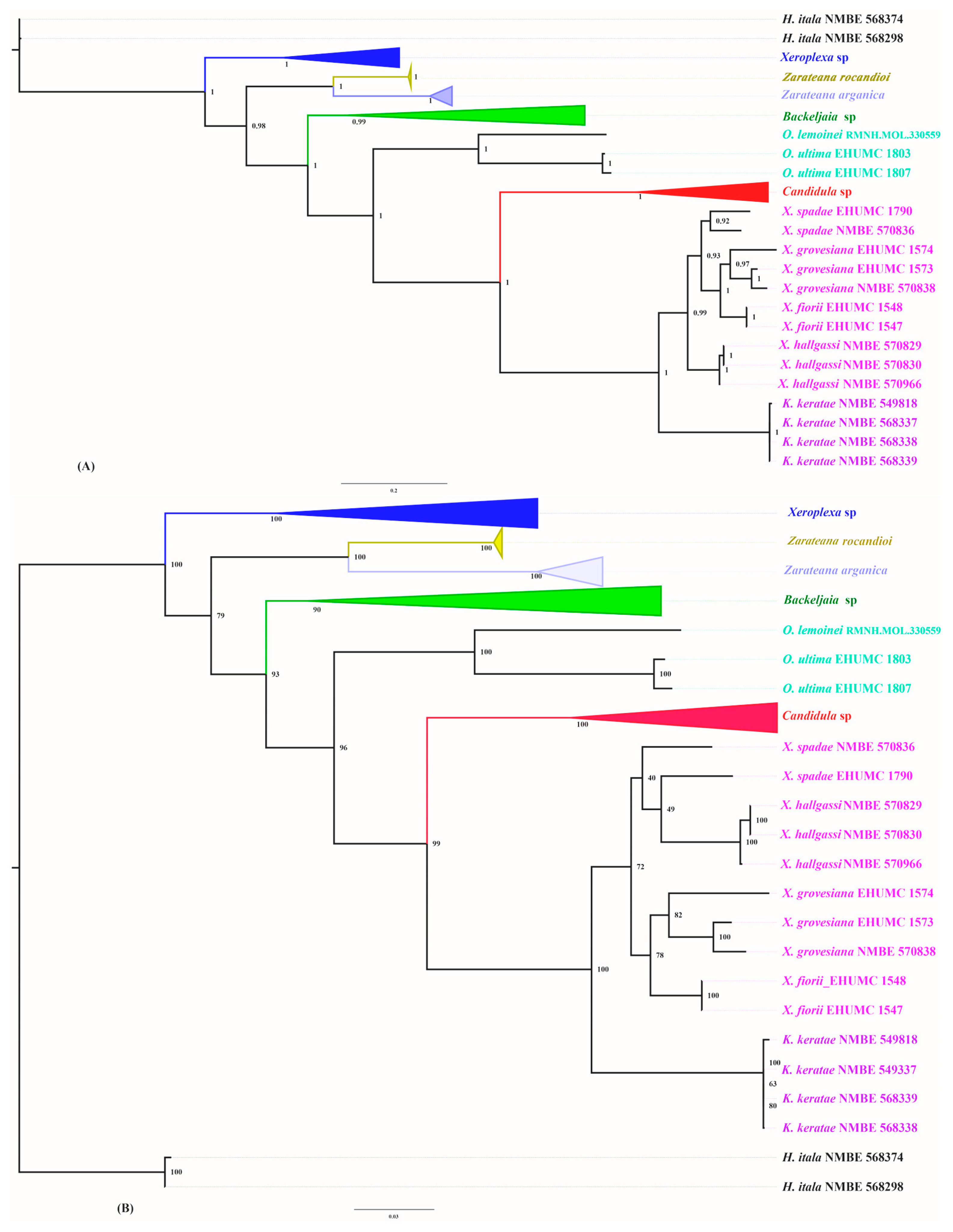
Both BI and ML trees were obtained based on a concatenated data set of the two mitochondrial markers (16S and COI) and one nuclear marker (5.8S-ITS2-28S) and rooted by H. itala showing similar topologies with some slight differences (Figure 2). The topologies obtained based on representative sequences of all the known Candidula-like species displayed six well supported clades: (1) a Xeroplexa, Monterosato, 1892 [15] clade (Blue) (PP = 1; BS = 100%) formed by a representative of seven Xeroplexa species; (2) a clade formed by Zarateana rocandioi, Ortiz de Zárate y López, 1950 [37] (Gold) and Zarateana arganica, Servain, 1880 [38] (Light Blue) (PP = 0.99; BS = 100%); (3) a Backeljaia, Chueca et al., 2018 [14] clade (Green) (PP = 1; BS = 90%) formed by representatives of the four Backeljaia species; (4) an Orexana clade (Turquoise) (PP = 1; BS = 100%) formed by sequences of the Spanish (Canarian) species O. ultima, Mousson, 1872 [39] and an Algerian taxon “Helix lemoinei”; (5) a Candidula clade (Red) (PP = 1; BS = 100%) composed of representatives of two Candidula species; (6) a Xerogyra clade (Pink) (PP = 0.98; BS = 72%) composed of a representative of the known Italian Xerogyra species and the new Italian taxon X. halgassi n. sp.; and (7) a clade consisting of sequences of the Algerian Xerophila keratae (Purple) (PP = 1; BS = 100%). The trees with uncollapsed clades are presented in the Supplementary documents S4 and S5. The phylogenetic relationships between and within the identified clades seem to be well supported in both phylogenetic construction methods reflecting reliable relative positions of the taxa used. However, the relationships between the Italian Xerogyra species seems to be unresolved. Although both the applied statistical methods could confirm the split of the Xerogyra clade from the rest of the Candidula type taxa, the relative relationships within the Italian clade depend on the used method. On the one hand, in the Bayesian analysis, the Italian clade splits into two well-supported subclades: (1) the first clade consists of the three known species Xerogyra spadae Calcara, 1845 [40], Xerogyra grovesiana, Paulucci, 1881 [41], and Xerogyra fiorii, Alzona & Alzona Bisacchi, 1938 [42], where each species forms a separate group, and (2) the second clade consists of sequences of X. halgassi n. sp. whereby the relationships between all species are well supported. On the other hand, the ML method splits the Italian Xerogyra into two moderately supported subclades: (1) the first subclade (BS = 40%) consists in X. halgassi and X. spadae where this later seems to be paraphyletic, and (2) the second subclade is composed of X. fiorii and X. grovesiana. The node connecting the two species is moderately supported (BS = 78%). The low support values might be the result of an under-sampling of the Italian radiation and the lack of the nuclear sequences in some samples.
The Algerian taxon X. keratae formed one fully supported clade (PP = 1; BI = 100%) with the Italian Xerogyra species, indicating its close affinity with the Italian Xerogyra compared to the rest of the studied genera. Additionally, the length of the branch relating the Algerian clade and the Italian Xerogyra indicates a high nucleotide divergence suggesting the separation between the Algerian taxon and the Italian genus.
The Algerian taxon H. lemoinei Kobelt, 1882 [43] seems to be more closely related to the species O. ultima from the Canary Islands than the rest of the investigated “Candidula-like” members, suggesting its identification as an Orexana species.

3.2. Molecular Description of New Taxa

The retention index test of the topology studied displayed a global RI value of 0.7511 suggesting a moderately low homoplasy. The test of the consistency index performed for each position of the concatenated data set of the COI, 16S and 5.8S-ITS2-28S displays 2925 nucleotide substitutions (Supplementary Table S6). Apomorphic nucleotides (after the arrow) as well as plesiomorphic characters (before arrow) are listed in Supplementary Table S6. Only characters with CI = 1 and with double arrow (non-ambiguous changes) are considered significant for the description of new taxa.
  • Kherattolactea nov. gen. Ezzine, Bouaziz, De Mattia & Neubert
  • Type species: Xerophila keratae Kobelt, 1892 [16].
Diagnosis: (1) “Candidula-type” genital anatomy with some internal differences than the known Xerogyra species; (2) nucleotide substitution: COI: T → C (22), T → A (331); 16S: T → C (777), A → C (788); 5.8S-ITS2-28S: T → G (1229), C → G (1411), C → T (1472), C → T (1621).
Zoobank ID: LSIDurn:lsid:zoobank.org:act:9627C69B-CE9D-4BEA-8DB2-15C28D04E290.
  • Xerogyra halgassi nov. sp. De Mattia, Ezzine & Neubert.
Diagnosis: “Xerogyra-type” genital anatomy; unkeeled shell; nucleotide substitutions: 16S: A → G (988); 5.8S-ITS2-28S: C → T (1334).
Zoobank ID: LSIDurn:lsid:zoobank.org:act:D804CBE1-F612-4FDE-BDBB-B789C059F642.
  • Orexana lemoinei (Kobelt, 1882) [43] nov. comb.
Diagnosis: COI: T → G (354), 16S: T → G (958), G → A (982), G → A (1165), T → C (1176); 5.8S-ITS2-28S: T → C (1229), G → T (1276), T → C (1229), G → T (1267), C → G (1269), T → C (1348), C → A (1352), G → A (1403), G → C (1446), A → C (1454), T → C (1476), T → C (1839), T → C (1914), G → T (1937).
Zoobank ID: LSIDurn:lsid:zoobank.org:act:7CB4E826-D3DC-4CCF-AE61-1AF88F3E2B9A.

3.3. Systematic Implications

Xerophila keratae and Helix lemoinei Kobelt, 1882 [43] turned out to be members of the Helicellini related to the Candidula-group. This is corroborated by genetic as well as anatomical data. Both taxa have not been mentioned for almost a century, so the opportunity is used to briefly characterize both taxa here, and their hitherto known distribution is shown (Figure 1).
  • Kherattolactea gen. nov. Ezzine, Bouaziz, De Mattia & Neubert
Zoobank registration: urn:lsid: zoobank.org:act:9627C69B-CE9D-4BEA-8DB2-15C28D04E290.
Type species: Xerophila keratae Kobelt, 1892 [16] [by original designation].
Etymology: The genus name has been chosen to refer to its type locality “Kheratta” and to refer the creamy white colour of the shell.
Diagnosis: The main diagnostic feature is the bipartite formation of the penial papilla with a spoon-like apical part differing from Xerogyra with a cone-shaped penial papilla. In case there are additional species of Kherattolactea detected in the area, the value of this trait must be confirmed as an autapomorphic character state of the genus.
  • Kherattolactea keratae Kobelt, 1892 [16]
Zoobank registration: urn:lsid:zoobank.org:act:825E72DD-7A76-4542-9C16-0341F0D7BA9F.
1892 Xerophila keratae Kobelt, Iconographie, (2) 6: 67, Taf. 167, fig. 1073. Type locality: “bei Kerata am Eingange der Schlucht Chabet-el-Akra zwischen Bougie und Setif”.
Type material: lectotype [hic!] SMF 10296; paralectotypes SMF 10471/12.
Specimens examined (all collected by H. Bouaziz): NMBE 549913/21, Chabet el Akra, Kherratta, 36.4987° N 5.2712° E, 676 m alt., 24.5.2015 (preserved specimens); NMBE 549818/1, same data (sequenced specimen); NMBE 542436/3, Irhi Emda, between Irhi Emda and Kherratta, Algeria 36.4917° N 5.2708° E, 553 m alt., 14.11.2015; NMBE 542435/17, same data as previous (preserved specimens); specimens from Draouate NMBE 568337/1, Draouate, 36.4584° N 5.1851° E, 21.09.2018 (Sequenced); NMBE 568338/1, Draouate, 36.4584° N 5.1851° E, 21.09.2018 (Sequenced); NMBE 568339/1, Draouate, 36.4584° N 5.1851° E, 21.09.2018 (Sequenced); NMBE 570374/12, same data as previous (preserved specimens); four lots in SMF, all from the type locality.
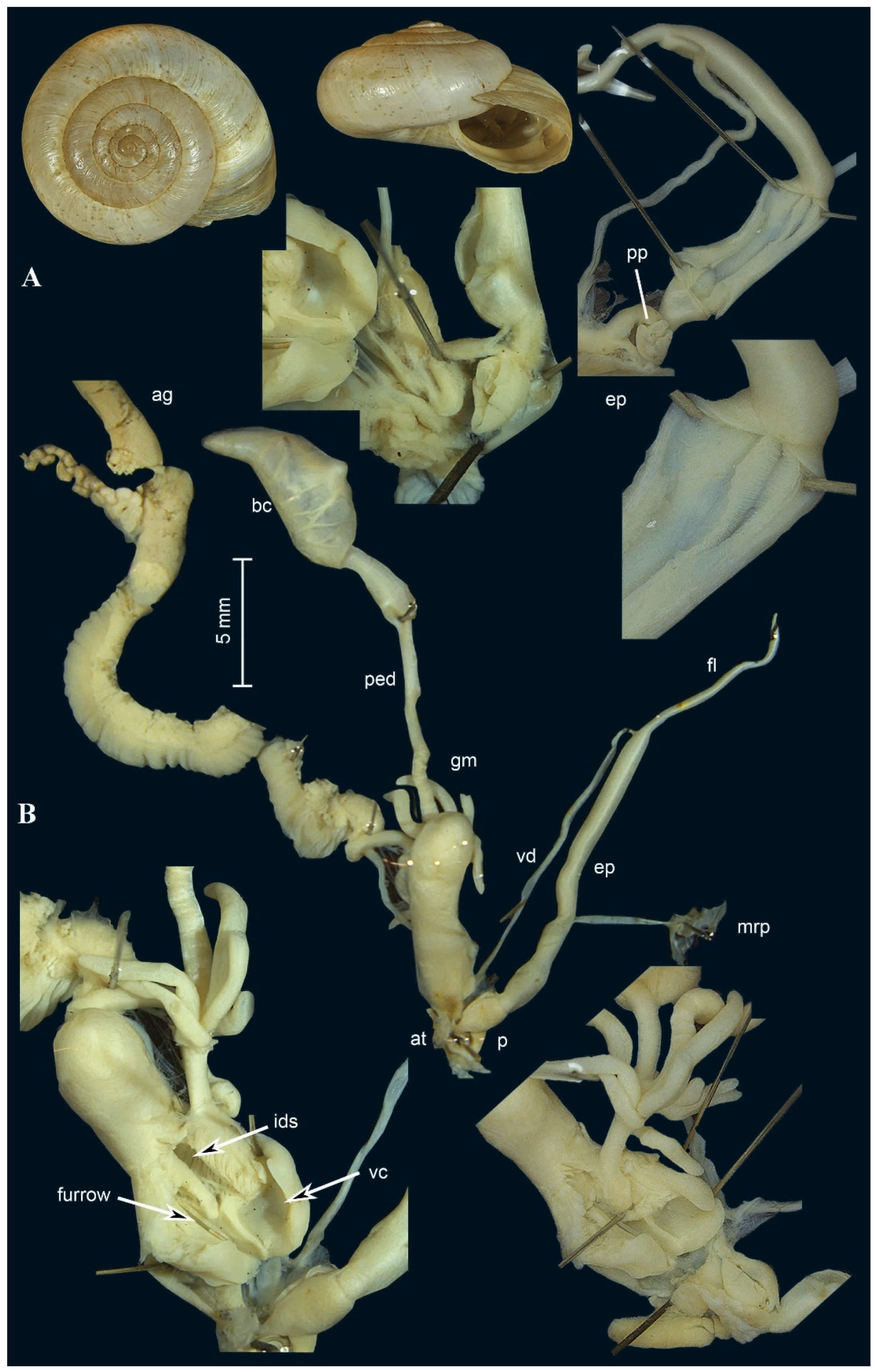
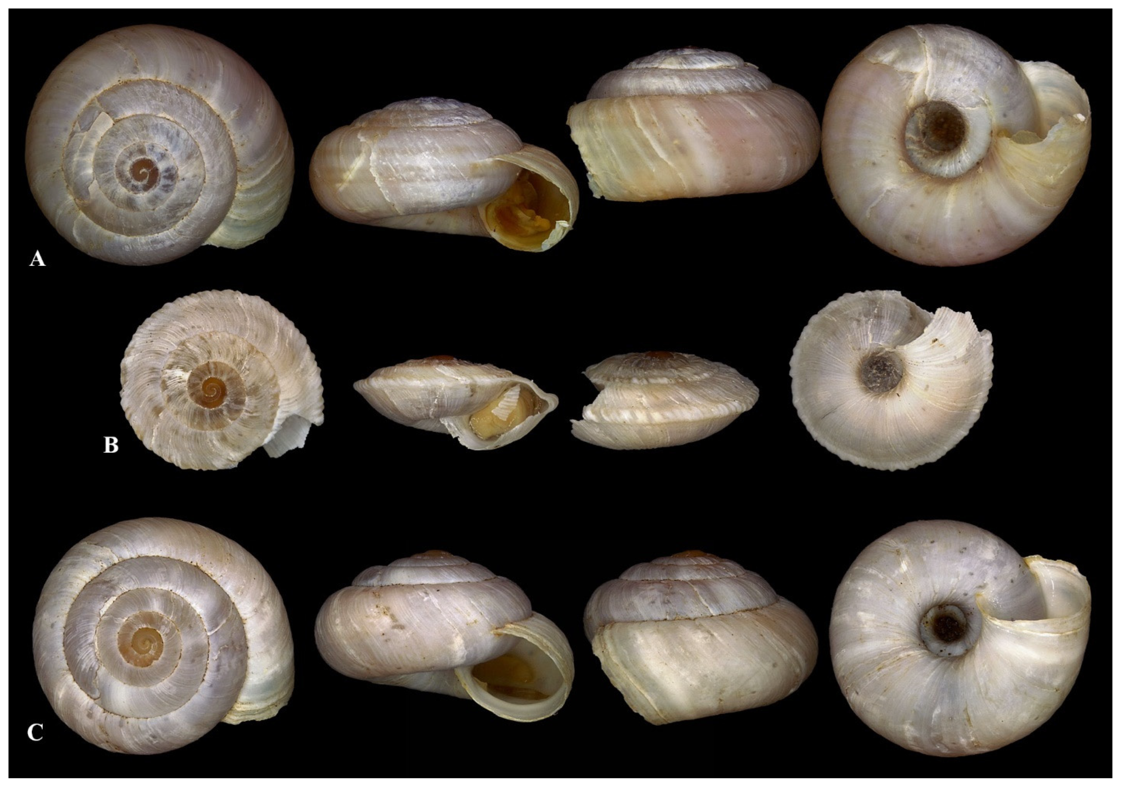
Description: shell relatively large, depressed to almost flat; protoconch corneous to bluish, smooth, polished, small; teleoconch of 5–6 regularly increasing whorls, basic shell colour white with irregularly dispersed groups of small bluish dots; teleoconch surface structured by irregularly arranged longitudinal groves and malleations, a few axial ribs occurring on the shell, with some more densely arranged ribs on the last whorl behind the aperture; teleoconch sometimes with faint brown spiral stripes; aperture broadly oval, wider than high, slightly descending to the periphery of the last whorl, apertural rim sharp, small internal lip white to reddish; umbilicus open, cylindrical, last whorl slightly eccentric reaching ca. ⅓ of the shell’s diameter (Figure 3 and Figure 4).
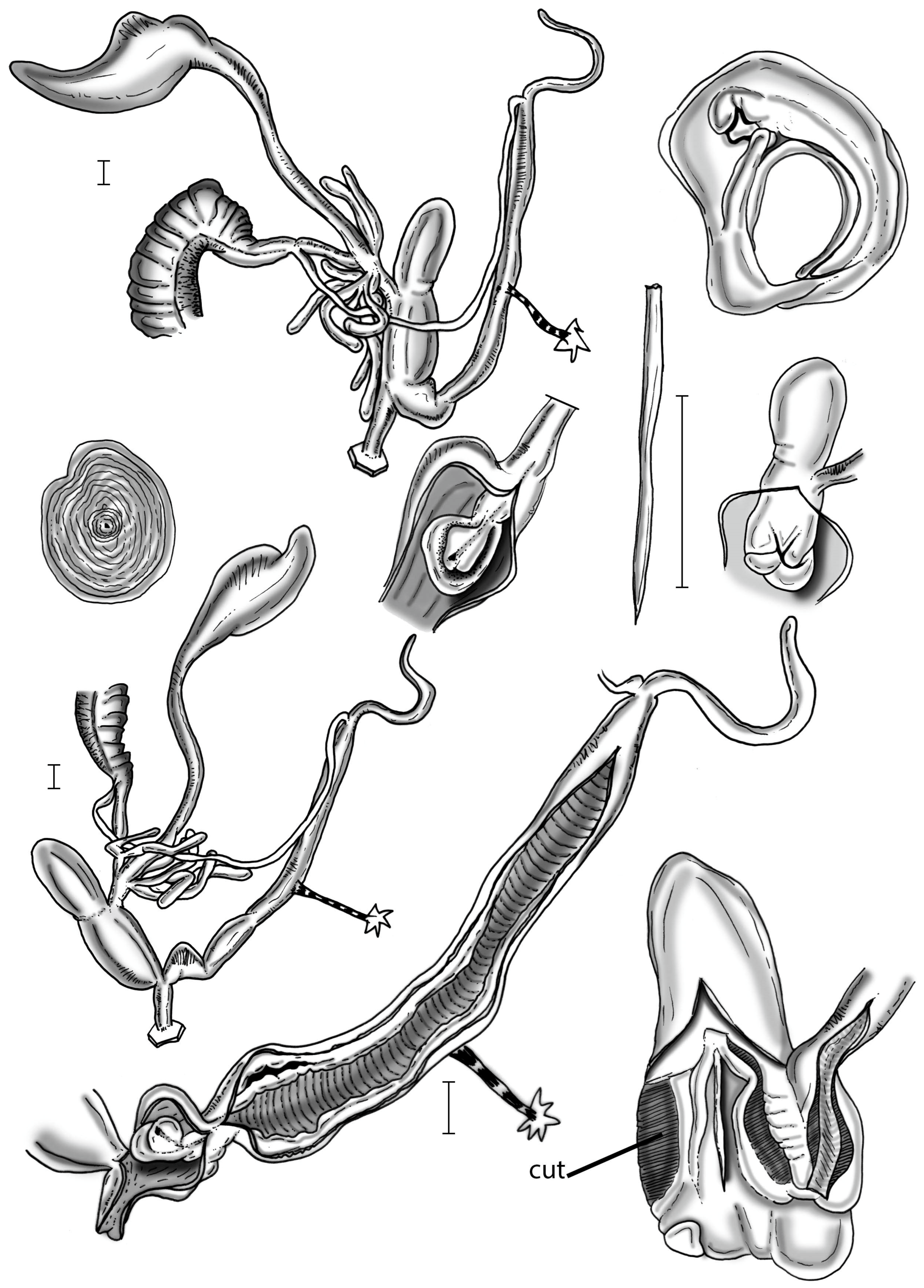
Genital organs: penis very short, conical, transgression zone to epiphallus indicated by a broad circular muscle tissue; epiphallus > 10× the length of the penis with the penial retractor muscle attaching approximately in the middle to distal third of the epiphallus; flagellum length equals length of epiphallus; penial papilla composed of two elements, with the first element formed like a deep table spoon, the proximal lumen filled with a butterfly-like bilobed lid covering the penial pore (Figure 4 and Figure 5).
Atrium filled with some longitudinal pilasters; single dart sac with complicated terminal openings: (1) a chamber-like cavity coated with a network of parallel lamellae, with an enormously strong muscular wall bordering the vaginal duct and opening into a second cup-like and completely smooth cavity (“vaginal chamber”) pointing into the atrium, and (2) a deep and narrow groove with the tip of the dart pointing into its lumen; vagina an elongate slender tube, glandulae mucosae consisting of a few non-ramifying tubules (Figure 3, Figure 4 and Figure 5).
Measurements: syntype: shell width (D) = 21 mm; aperture height (W) = 6.25 mm.
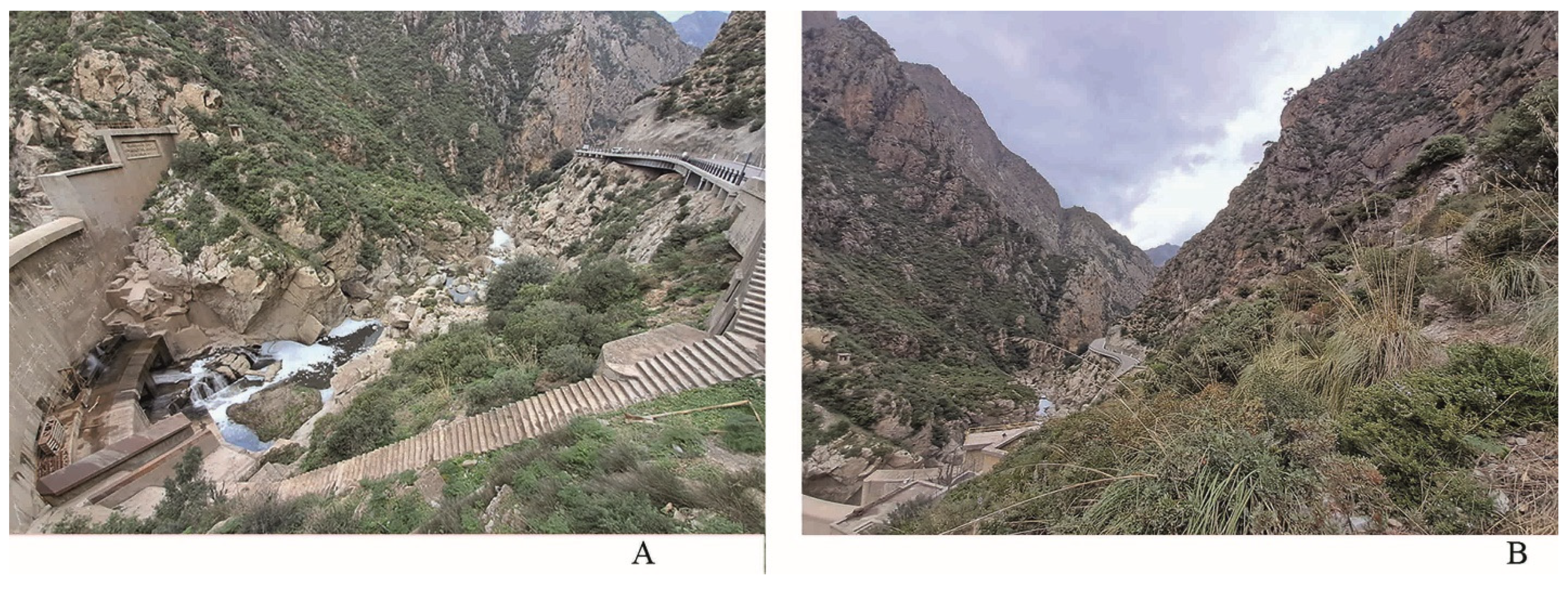
Distribution (Figure 1): So far, this species is only known from the type locality in the Kabylian Mountains at the southern entrance of the gorge of Chabet el-Akra (Figure 6). The second record originates from a locality a few kilometres towards west from the type locality.
Kherrata lies in a transition zone between Mediterranean steppes and semi-arid regions at an altitude between 500–600 m. This area benefits from a complex combination of edaphic and climatic factors, creating a rich and diverse habitat. The rugged terrain, characterized by steep slopes and deep valleys, significantly influences the characteristics of the soils. These soils are mainly clay-loam, well-drained, and often rich in limestone promoting the growth of Mediterranean vegetation and plants adapted to semi-arid conditions.
The climate of Kherrata is of the Mediterranean type with hot, dry summers and mild, rainy winters. In winter, temperatures are typically ranging around 10 °C. Days are cool, but nights can be colder. In summer, temperatures can reach up to 30 °C, with very hot days. However, the nights remain relatively cool due to the altitude and mountainous relief.
The vegetation is characteristic for Mediterranean areas and of the Macchia type, with dense plant cover on shaded slopes and more sparse vegetation on sun-exposed areas. The calcareous soils and moderate climatic conditions support forests of Aleppo pines, Atlas cedars, and Mediterranean scrubland. The wetter valleys host more luxuriant plant species, while the south-facing, drier slopes are home to plants resistant to heat and prolonged droughts. These microclimate variations create diverse ecological niches, providing a habitat for rich fauna.
Habitat: So far, the species is known from two localities in northern Algeria. Its habitat is a rocky calcareous area with a moderately dense plant cover. The area of Chabet El-Akra is also favourable for a great diversity of gastropods, including several species of land snails adapted to the specific conditions of dry, alkaline soils. Species like Eobania vermiculata (O. F. Müller, 1774) [44], Theba pisana (O. F. Müller, 1774) [44], Cornu aspersum (O. F. Müller, 1774), Xerosecta Monterosato, 1892 [15], Cernuella Schlüter; 1838 [45], and Ganula flava (Terver, 1839) [46] are present. Due to the variety of microhabitats, these snails find refuge under stones and in the cracks of stone walls, where the temperature remains low and stable.
  • Xerogyra Monterosato, 1892
1892 Xerogyra Monterosato [15], Atti della Reale Accademia di Scienze, Lettere e Belle Arti di Palermo. 3rd Series, 2: 24.
Type species: Helix spadae Calcara, 1845 [40] [Subsequent designation by Kobelt 1892 [16]].
  • Xerogyra fiorii Alzona & Alzona Bisacchi, 1938 [42]
Description of genital organs: a convoluted hermaphrodite duct arising from the slender multilobated gonad; albumen gland long and slender, connected to an approximately 2.5 times as long spermoviduct; prostatic part extends into a thin vas deferens approximately as long as the spermoviduct; free oviduct short, half as long as the vagina; duct of the bursa copulatrix large, approximately as long as the penis+epiphallus and uniform in diameter; bursa copulatrix symmetric to asymmetric in shape; transition area between duct and bursa itself weakly delimited with the duct gradually widening and turning into the bursa; spermatophore unknown; two tufts with two moderately long, large and not branched digitiform glands; no vaginal appendix; vagina merging into the dart sac’s side, partially sharing its distal path as far as the genital atrium; few little elevated pleats, irregularly spaced pleats running longitudinally along the inner surface of the proximal vagina; atrium short and thin with soft pleats running longitudinally as far the genital orifice; single big dart sac, blunt and cylindrical, with proximal strong muscular walls surrounding the dart channel, distally two fleshy thick pleats (two hemiplates) heading toward the atrium, fused together along the median side creating a spoon-like structure, each hemiplate showing two elongated longitudinal S-shaped slots; dart as long as half of the dart sac with an irregular tail and not very sharp apex; penial flagellum as long as half of the epiphallus; epiphallus long with internal walls showing four longitudinal smooth pleats; retractor muscle short and quite thin; penis lacking any muscular or glandular sheath, thick-walled and approximately 1/3 times as long as the epiphallus, slightly swollen along the penial papilla; inner walls of penis with irregular, spaced pleats, running longitudinally as far as the genital atrium; penial papilla blunt, cylindrical in shape, with smooth surface, central channel with elliptical cross-section and apical opening, measuring approximately ½ of the penial length, inner lumen of the penial papilla occupied by a spongy and sturdy tissue; longitudinal section of the penial papilla shows that its walls are the continuation of the penial walls that abruptly bend inward.
  • Xerogyra grovesiana Paulucci, 1881 [41]
Description of genital organs: a convoluted hermaphrodite duct arises from a slender multilobated gonad; albumen gland long and slender, connected to an approximately 1.5 times as long spermoviduct; prostatic part extends into a thin vas deferens approximately as long as the spermoviduct; free oviduct extremely short, 1/4 as long as the vagina; duct of the bursa copulatrix very large, approximately as long as the penis+epiphallus and uniform in diameter; bursa copulatrix elongated and symmetric in shape; transition area between duct and bursa itself somehow delimited with the duct rapidly widening and turning into the bursa; spermatophore unknown; two tufts with one or two moderately long, large and not branched digitiform glands; no vaginal appendix; vagina merging into the dart sac’s side, only briefly sharing its distal path as far as the genital atrium; few little elevated pleats, irregularly spaced pleats running longitudinally along the inner surface of the proximal vagina; atrium short and somehow large with soft pleats running longitudinally as far the genital orifice; single big dart sac, blunt and cylindrical, with proximal strong muscular walls surrounding the dart channel, distally two fleshy thick pleats (two hemiplates) heading toward the atrium, fused together along the median side creating a spoon-like structure, each hemiplate showing one elongated longitudinal S-shaped slots place in a proximal (upper) position; dart as long as half of the dart sac with an irregular tail and not very sharp apex; penial flagellum extremely long, almost three times as long as the epiphallus; epiphallus short with internal walls showing three longitudinal smooth pleats; retractor muscle short and quite strong; penis lacking any muscular or glandular sheath, thick-walled and approximately as long as the epiphallus, slightly swollen along the penial papilla; inner walls of penis almost smooth; penial papilla short and blunt, cylindrical in shape, with smooth surface, central channel with irregular cross-section and apical opening, measuring approximately 1/4 of the penial length, inner lumen of the penial papilla occupied by a spongy and sturdy tissue; longitudinal section of the penial papilla shows that its walls are the continuation of the penial walls that abruptly bend inward.
  • Xerogyra spadae Calcara, 1845 [40]
Description of genital organs: a convoluted hermaphrodite duct arises from a slender multilobated gonad; albumen gland short and bulky, connected to an approximately four times as long spermoviduct; prostatic part extends into a thin vas deferens approximately as long as the spermoviduct; free oviduct very short, about 1/4 as long as the vagina; duct of the bursa copulatrix very large, approximately as long as the penis+epiphallus and uniform in diameter; bursa copulatrix symmetric to asymmetric in shape; transition area between duct and bursa itself somehow well delimited with the duct widening and turning into a small round bursa; spermatophore unknown; two tufts of digitiform glands, each tuft with one or two long, large and not branched glands; no vaginal appendix; vagina merging into the dart sac’s side, partially sharing its distal path as far as the genital atrium; inner walls of distal vagina almost smooth; atrium large but short and almost smooth; single big dart sac, blunt and cylindrical, with proximal strong muscular walls surrounding the dart channel, distally two fleshy thick pleats (two hemiplates) reaching the atrium, fused together along the median side creating a spoon-like structure with swollen distal edges; each hemiplate’s surface is continuous, without slots or interruptions; dart as long as half of the dart sac with irregular tail and a very sharp apex; penial flagellum as long as half of the epiphallus; epiphallus moderately long with very smooth longitudinal pleats running along the internal walls; retractor muscle short and quite thin; penis lacking any muscular or glandular sheath, short and thick-walled and approximately 1/3 times as long as the epiphallus, swollen along the penial papilla; inner walls of penis with very weak longitudinal pleats; penial papilla short and blunt, cylindrical in shape, with smooth surface, central channel with irregular cross-section and apical opening, measuring approximately half of the penial length, inner lumen of the penial papilla occupied by a spongy and sturdy tissue; longitudinal section of the penial papilla shows that its walls are the continuation of the penial walls that abruptly bend inward.
  • Xerogyra halgassi De Mattia, Ezzine & Neubert n. sp.
Zoobank registration: urn:lsid: zoobank.org:act:D804CBE1-F612-4FDE-BDBB-B789C059F642
Type specimens: holotype NMBE 570829; leg. W. De Mattia & J. Macor, May 12, 2006; type locality: Sentiero della Rocchetta next to the Rocchetta spring, Gualdo Tadino, Province of Perugia, Umbria, Italy. 43°13′45.78″N 12°48′2.57″E, 590 m a.s.l.
Specimens examined: only known from the type locality.
Diagnosis: A big Xerogyra species, with a flat to slightly elevated shell; uniformly whitish with no dark bands; smooth only with fine growth striae; dart sac’s hemiplates partially fused, with a single big aperture; flagellum half as long as the epiphallus.
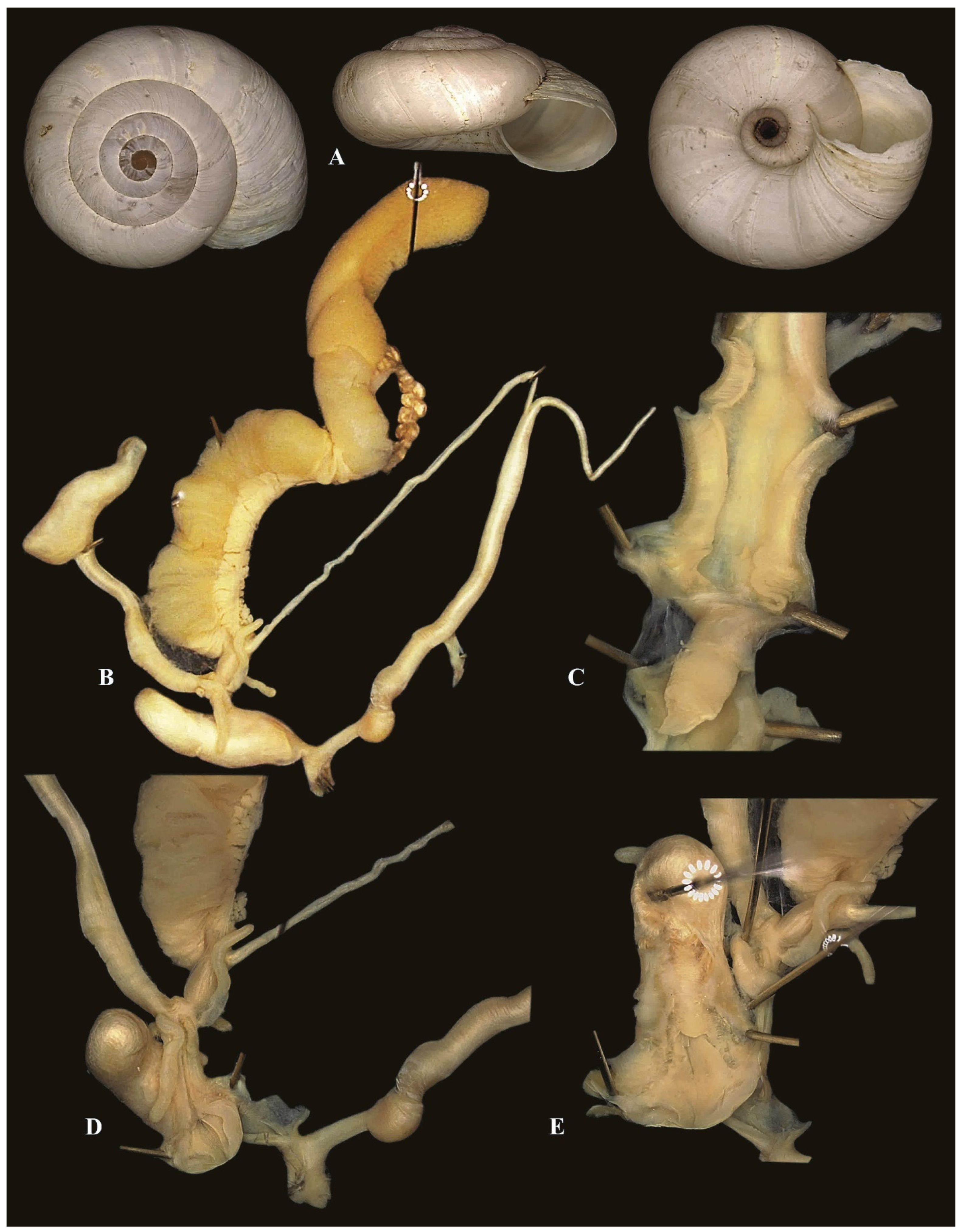
Description: shell dextral, hairless, relatively large for the genus, slightly elevated to almost flat; protoconch of 1 ½ smooth whorls, matte bluish; teleoconch of 4-4½ regularly and rapidly increasing whorls, shell shiny, whitish to blue-greyish without bands, dots or any other pattern; most shells present growth scars; teleoconch surface with irregular radial fine growth striae that are stronger toward the sutures, bottom surface of the last whorl smoother; body whorl’s height 80% of the total shell’s height; sutures deep and well visible; aperture interrupted, broadly oval, wider than high, slightly descending to the periphery of the last whorl, apertural rim sharp and not reflected, very mild internal white callous; umbilicus widely open, remarkably eccentric reaching more than ⅓ of the shell’s diameter.
Body: head and neck greyish; sides and posterior upper section of the foot whitish; pigmented ommatophoral retractor muscles visible through the skin of the back of the cephalic area; foot white to pinkish; sole longitudinally divided into three areas: central area smooth, two lateral areas with bands of muscles that are roughly arranged in a chevron pattern; mantle border greyish to pink-greyish, with five more or less developed lobes; walls of the pallial cavity colourless without stripes or spots; strong pulmonary vein is visible; jaw odonthognatous, variable in shape from almost straight to arched with many smooth transverse ridges; right ommatophore retractor independent from both penis and vagina.
Description of genital organs: general arrangement of the genital organs semidiaulic monotrematic; convoluted hermaphrodite duct arises from a multilobated gonad; albumen gland long and large connected to an approximately twice as long spermoviduct that consists of a prostatic and a uterine portion; prostatic part extends into a thin vas deferens approximately as long as the spermoviduct and terminates in the penial complex.
Distal portion of the uterine part extends into the free oviduct, turning into a vagina at the level of the duct of the bursa copulatrix; free oviduct very short, half as long as the vagina; duct of the bursa copulatrix large, approximately as long as the penis and uniform in diameter; bursa copulatrix asymmetric in shape; transition area between duct and bursa itself sharply delimited with the duct abruptly widening and turning into the bursa; spermatophore as long as the duct of the bursa despite the ends are unknown, quadrangular in section, horny and hyaline; two tufts of short, large and not branched digitiform glands arises from the proximal part of the vagina; no vaginal appendix; vagina merging into the dart sac’s side, partially sharing its path as far as the genital atrium; few little elevated pleats, irregularly spaced pleats running longitudinally along the inner surface of the proximal vagina.
Atrium short and thin with internal walls with soft pleats running longitudinally as far the genital orifice; single big dart sac, cylindrical, with proximal strong muscular walls surrounding the dart channel, distally a complex structure made of two fleshy thick pleats (hemiplates) heading toward the atrium along the vaginal chamber, fused together along the median side creating a spoon-like structure, each hemiplate showing an elongated longitudinal slot, proximally from the central hollow fleshy tongue surrounding the distal part (tip) of the dart.
Penial complex consists of a flagellum, an epiphallus (which extends from the insertion of the vas deferens to the penial retractor muscle) and a penis; thin penial flagellum as long as half of the epiphallus length; epiphallus long with internal walls showing three to four longitudinal, smooth pleats; retractor muscle short and robust; penis lacking any muscular or glandular sheath, thick-walled and approximately ⅔ times as long as the epiphallus, swollen along the penial papilla; inner walls of penis smooth or with irregular, spaced pleats, running longitudinally reaching the genital atrium; penial papilla blunt cylindrical to conical in shape, with smooth surface, central channel with irregular cross-section and apical opening, measuring approximately ½ of the penial length, inner lumen of the penial papilla occupied by a spongy and sturdy tissue, directly connecting with the walls of epiphallus; longitudinal section of the penial papilla shows that its walls are the continuation of the penial walls that abruptly bend inward.
Measurements: D = 16.8 ± 0.3 mm (range 16.5–17.4 mm); H = 8.6 ± 0.2 mm (range 8.3–8.9 mm); FW = 7.3 ± 0.2 mm (range 7.1–7.7 mm); DU 5.7 ± 0.1 mm (range 4.9–6.1 mm); (n = 11). Ratio D/H 2.
Etymology: named after Alessandro Halgass from Rome, Italy, in acknowledgement for his contribution to the knowledge of the malacofauna of the Apennines.
Distribution (Figure 1): Xerogyra halgassi n. sp. is known only from the locus typicus but probably is distributed along the Appennino Umbro-Marchigiano, from Monte Catria to Monte Cucco between Gualdo Tadino (Umbria, Italy) and Fabriano (Marche, Italy) (A. Hallgass, pers. comm. April 2021). More field and taxonomical research is needed to better define the range of this taxon.
Remarks: The main differences among Xerogyra species, concerning both shell morphology and genital morphology, are listed in Table 2.
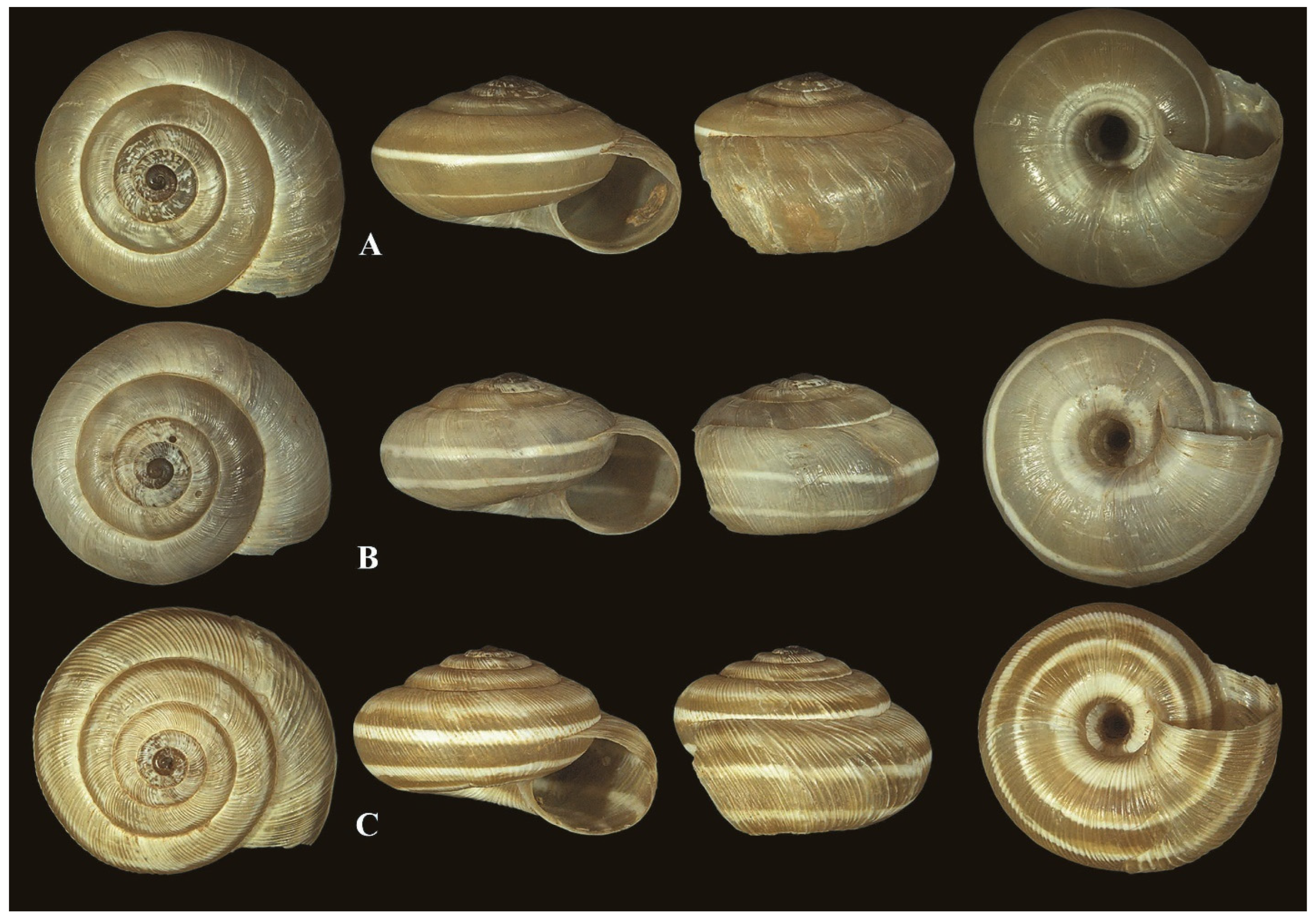
Xerogyra halgassi n. sp. can be confused with two Italian Xerogyra species, namely Xerogyra fiorii (Figure 10 and Figure 11) and Xerogyra spadae (Figure 8B) by virtue of the whitish smooth shell and the round and unkeeled last whorl. It is clearly distinguishable from the keeled and ribbed Xerogyra grovesiana (Figure 8A). Nonetheless, as a shell, Xerogyra halgassi n. sp. is distinguishable by virtue of the following: (1) a remarkably bigger shell; (2) a remarkably more eccentric, wide-open umbilicus; (3) a ratio D/U > of ⅓; (4) a broadly oval aperture; and (5) a very mild aperture’s internal callous.
Kherattolactea keratae mainly differs from Xerogyra halgassi n. sp. by its remarkably narrow umbilicus and the constant presence of strong colour bands along the last whorl.
Regarding genital organs, the overall external structure is stable throughout the genus, with minor differences mainly concerning the relative length of the flagellum (F/E), as appears in Table 2. The shape and ratios among the main anatomical traits reveal to be stable throughout species.
The main morphological differences among the Xerogyra species are found in the internal structure of the genital organs, primarily as regards the dart sac.
  • Orexana Chueca, Gómez-Moliner, Madeira & Pfenninger, 2018 [14]
2018 Orexana Chueca, Gómez-Moliner, Madeira & Pfenninger, Molecular Phylogenetics and Evolution 118: 366 [name not available from the online version published in October 2017].
Type species: Leucochroa ultima Mousson, 1872 [39] [by original designation].
  • Orexana lemoinei Kobelt, 1882 [43]
1882 Helix lemoinei Kobelt, Jahrbücher der Deutschen malakozoologischen Gesellschaft, 9: 69 [Tomadjeur a Nama im südlichen Oran].
1883 Helix lemoinei, Kobelt, Iconographie, (2) 1 (3/4): 44, Taf. 17, fig. 129.
Type material: lemoinei: lectotype SMF 10493 [hic!]; paralectotypes SMF 10131/2.
Additional specimens studied included the following: Algeria: RMNH-MOL 294533, Foot of Djebal Antar/Saida, 1150 m alt., 33.605706° N–0.254418° E, 07.04.1983, leg. T. de Winter; RMNH-MOL 294543, Djebal Souiga/Saida, 1400 m alt., 33.048839° N–0.413256° E, 02.05.1983, leg. T. de Winter; RMNH-MOL 329780 (dry shells), RMNH-MOL 330559 (preserved), Forthassa/Saida, 1420 m alt., 32.843391° N–1.233696° E, 06.04.1983, leg. T. de Winter; SMF 288259/4, Ain-ben-Khalil, coll. W. Kobelt ex O. Debeaux, 33.2884° N–0.7640° E; SMF 288260/1, between Djelfa and Lagouat, coll. O. v. Möllendorff, 34.2068° N 3.1518° E; SMF 98170/2, between Djelfa and Laghouat, coll. K.-L. Pfeiffer (3296b) ex Slg. Jetschin ex Joly; SMF 288261/2, T’iout, coll. O. v. Möllendorff, 32.7569° N–0.4224° E; SMF 98169/1, Ain-Safra, Sahara Oranais, coll. K.-L. Pfeiffer (3296a) ex Slg. Jetschin ex Monjo, 32.7591° N–0.5782° E; and SMF 288262/1, ex Oberwimmer ex Staudinger (received as astata Servain).
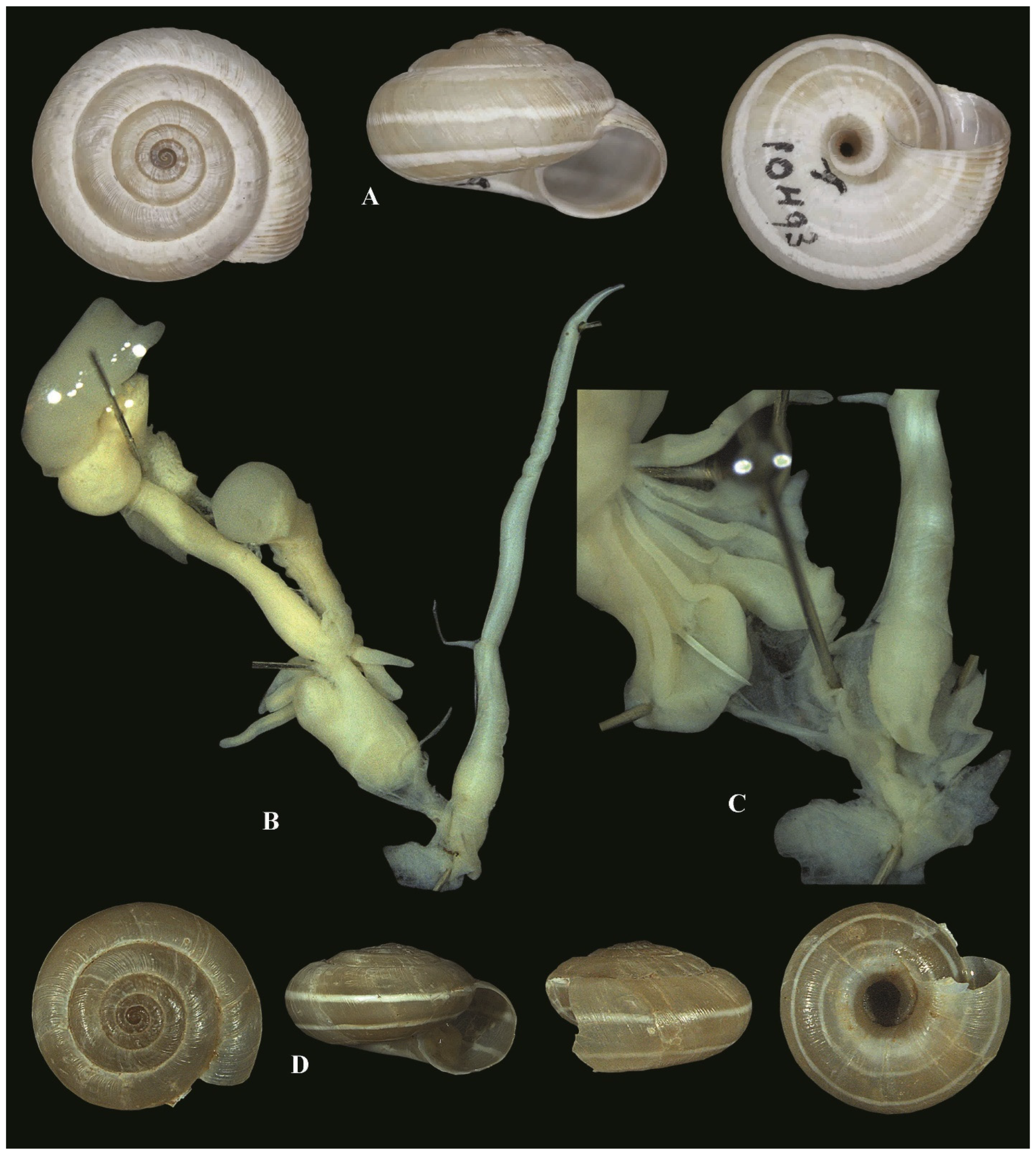
Diagnosis: shell flat with slightly elevated spire, teleochonch finely ribbed with last whorl with heavy ribbed pattern, basic colour brown with a pattern of white spiral bands: the spiral bands consist of a very prominent one on the periphery and a second broad white band around the umbilicus, last whorl only slightly descending, aperture small, umbilicus wide open, perspectively increasing in diameter. Genitalia with 1+0 dart sac, very short penis, very elongated epiphallus, flagellum ~1/5 epiphallus.
Description: shell flat with a slightly elevated dome-shaped spire; protoconch smooth, consisting of ca. two smooth whorls; teleoconch with 4–5 finely ribbed whorls, last whorl usually more heavily ribbed; suture deep; basic colour brown, with a pattern of white spiral bands; the white spiral band on the periphery very prominent and present in all specimens observed, with a second broad white band around the umbilicus; last whorl only slightly descending, aperture small, subcircular, labial callus weak or missing; umbilicus wide open, perspectively increasing in diameter.
Description of genital organs: penis very short, retractor muscle inserted at the first 3rd of the epiphallus, epiphallus >5 times longer than the penis; flagellum extremely short, penial papilla short, conical with an acute tip, with central opening covered with thick muscle at its distal part.
Atrium consisting in a simple cavity without any secondary structure.
Female part consisting of a single dart sac, simple with central opening that opens into the atrial chamber. Dart sac attached to a short vagina filled with longitudinal muscles forming a groove-like structure; few digitiform gland with simple ramifications.
Measurements: Syntype: D = 16 mm.
Distribution (Figure 1): This species is recorded from the southwestern Atlas in the border region between Algeria and Morocco. So far, no records for this taxon from Morocco are known; however, its presence in the area of Bel Mahdi and Iche is highly probable.
Remarks: A lectotype is selected here to determine the unambiguous use of the name.

4. Discussion

The current study allows a first insight into the Candidula-type radiation in Northern Africa. Representatives of two such genera were found, a relative of the Italian Xerogyra group, and the first species of the so far known, Canarian genus, Orexana.
Until recently, the genus Xerogyra was considered a synonym of Candidula [47]. Chueca et al. [14] resurrected Xerogyra as a monophyletic clade comprising three species from Italy, viz. X. fiorii, X. grovesiana and X. spadae, and claimed that the morphology of the genital organs was that of the Candidula type. Despite the promise of exploration of the value of the morphology of the genital organs for Candidula systematics, the authors have not yet provided a detailed investigation of these organs. The split between the different “Candidula-type” genera was accepted based on apomorphic nucleotide substitutions.
Major differences in the anatomy of the genital organs are widely used as discriminating character states between land snail genera [48,49]. For this reason, we carefully studied the genital anatomy of topotypic specimens of the known Italian Xerogyra species including a new species, X. halgassi n. sp. In Chueca et al. [14] “Candidula-type” obviously means “species with a single dart sac”, a character state that proved to be plesiomorphic as it appears in several independent lineages within Helicellinae. Additionally, there are fundamental morphological differences to the type species of Candidula, viz. C. unifasciata (Figure 2510 [50]). The morphological differences between some of these genera are described here supporting our molecular results and showing that X. keratae is more closely related to the Italian group than to the rest of the “Candidula-type” genera.
In the male system, the penial papilla in Candidula is a simple cone (Figure 2510) [50]; in Kherattolactea it is composed of a spoon-like structure detached from the penial walls, while, in Xerogyra, the papilla is cylindrical surrounded by some spongy tissue originating from the internal penial walls. In O. lemoinei, the penial papilla is short, conical with a thick muscular basis and an acute tip. In the female system, the dart sac of C. unifasciata terminates with a simple papilla-like structure to operate the dart. There is no trace of the “side cavity” (see Kherattolactea) with the internal lamellae, nor is there such a massive “vaginal chamber” pointing into the atrium. On the other hand, in K. keratae, it is composed of the spoon-like structure with a bilobed “cover”. In O. lemoinei, the dart sac is simple and thus similar to that in Candidula.
Chueca et al. [14] identified apomorphic nucleotide substitutions as a method to separate subclades as new genera; we did the same here and added unique traits of the genital organs to the analysis. Based on these results, the separation of Kherattolactea as sister group of Xerogyra, endemic to the northeastern Kabylie region of Algeria, is well supported.
The phylogenetic relationships within the Italian genus Xerogyra remain poorly resolved. Despite clear morpho-anatomical differences among the three known Xerogyra species and X. halgassi sp. n., phylogenetic analyses have yet to elucidate the relationships within the Italian radiation. Currently, Xerogyra species are known to occur in a fragmented distribution along the Apennine Mountain chain. Many studies have demonstrated the role of microhabitat variation in driving speciation and ecological adaptation in land snails within this region [51,52]. However, the limited sampling of Xerogyra lineages likely hinders a comprehensive understanding of their diversification. To gain deeper insights into the Xerogyra radiation and clarify the evolutionary relationships within the genus, it would be valuable to intensify sampling efforts across the Apennine region. Additionally, investigating morphogenetic variation in relation to ecological factors would provide a more integrated perspective on the evolutionary dynamics of this group.
Orexana ultima was originally described under the genus Leucochroa Beck, 1837 [53] (thus erroneously attributed to Fam. Sphincterochilidae) from the Canary Islands. Chueca et al. [14] finally reallocated L. ultima to the Fam. Geomitridae in their new monotypic genus Orexana based on molecular findings. Our study revealed that H. lemoinei is most closely related to O. ultima; it shares the same apomorphic nucleotide substitutions recorded by Chueca et al. [14] for the Orexana. Thus, H. lemoinei is herein considered to constitute another member of Orexana. The record of O. lemoinei from the southwestern Atlas Mountains in the border region between Algeria and Morocco represents the first continental record of the genus. Considering the large number of conchologically similar taxa in western Algeria and eastern Morocco, it is very likely that there are more species of Orexana out there.
The genus Candidula in its current form (https://www.molluscabase.org/aphia.php?p=taxdetails&id=426353) (accessed on 1 July 2025) comprises eight extant species, known from Portugal to Bulgaria. Hausdorf [54] revised two species from North Macedonia and Greece, viz. Candidula rhabdotoides Wagner, 1928 [55] and Candidula lernaea Hausdorf, 1991 [54]. Hausdorf (Figure 1 and Figure 2) [54] meticulously described the internal structures of the female genital organs, in particular the structure of the dart apparatus. For C. rhabdotoides, he explained that “after removing the upper tissue layer, a thick-walled “hull” appears, which is open towards the vagina” (translated from German). This feature is the same we found in K. keratae and called the “vaginal chamber”. Our “cavity coated with a network of parallel lamellae” corresponds well to the “rudimentary second dart sac” (“=Nebensack”) reported for both species, C. rhabdotoides and C. lernaea. The similarity of these structures is striking. However, there are also differences like the form of the penial papilla, which is a simple cylindrical tube without any additional differentiation. The folds found by Hausdorf ([54]: Figure 1B) are missing in K. keratae, and the separation of the functional outer dart sac from the internal rudimentary sac is more pronounced in K. keratae than in the two eastern Mediterranean species. These findings contrast the much simpler morphology of all dart sac-related features found in C. unifasciata. Thus, we hypothesize that these eastern Mediterranean species do not at all fall into Candidula, but rather, again represent a separate lineage of the so-called Candidula-like. Their relationship to Xerogyra needs more attention, and the exact position of Candidula castriota Soós, 1924 [56] needs to be investigated. So, the polyphyly of the former genus Candidula is still not fully resolved.

Supplementary Materials

The following supporting information can be downloaded at https://www.mdpi.com/article/10.3390/d17080550/s1. Supplementary S1: Taxa used in the molecular analysis of the current study: species, voucher numbers, localities, Geographic coordinates, and GenBank accession numbers for COI, 16S partial sequences and the 5.8S-ITS2-28S sequences. Supplementary S2: Summary of the sequence’s information, taxa used in the study and evolutionary model of each partition. Supplementary S3: Results of the phylogenetic signal analyses measuring substitution saturation. Results of Xia’s test are given based on simulations with 10.000 replicates and 32 OTUs. Iss = Index of substitution saturation; Iss.cSym and Iss.cAsym = critical substitution saturation index if the true tree is symmetrical or asymmetrical, respectively. Significant substitution saturation is present in the dataset if Iss > Iss.c.S3. Supplementary S4: Phylogram produced by Bayesian Inference (BI) constructed based on the concatenated data of two mitochondrial markers (COI, 16S) and one nuclear (5.8S-ITS2-28S) marker. The tree shows the details of the samples used in the study. Supplementary S5: Phylogram produced by Maximum Likelihood (ML) constructed based on the concatenated data of two mitochondrial markers (COI, 16S) and one nuclear (5.8S-ITS2-28S) marker. The tree shows the details of the samples used in the study. Supplementary S6: Consistency Index Test result showing the summary of the nucleotide changes within the concatenated dataset of two mitochondrial (COI, 16S) and one nuclear (5.8S-ITS2-28S) markers

Author Contributions

Conceptualization, I.K.E. and E.N.; Methodology, I.K.E.; Software, I.K.E.; Validation, I.K.E., W.D.M., H.B.-Y., and E.N.; Formal Analysis, I.K.E.; Investigation, I.K.E., W.D.M., H.B.-Y., and E.N.; Resources, I.K.E., W.D.M., H.B.-Y., and E.N.; Data Curation, I.K.E. and E.N.; Writing—Original Draft Preparation, I.K.E. and E.N.; Writing—Review and Editing, I.K.E., W.D.M., H.B.-Y., and E.N.; Visualization, I.K.E. and E.N.; Supervision, E.N.; Project Administration, I.K.E. and E.N.; Funding Acquisition, E.N. All authors have read and agreed to the published version of the manuscript.

Funding

This research received no external funding.

Data Availability Statement

The corresponding sequences of each analysed marker are submitted to the GenBank database and are available under the accession numbers: OR545714–OR553083. The supporting data of the described taxa and/or new taxonomic combination are submitted to the Zoobank database. Submitted data could be found under the following links: Kherattolactea: urn:lsid:zoobank.org:act:9627C69B-CE9D-4BEA-8DB2-15C28D04E290; Orexana lemoinei: urn:lsid:zoobank.org:act:7CB4E826-D3DC-4CCF-AE61-1AF88F3E2B9A; Xerogyra halgassi: urn:lsid:zoobank.org:act:D804CBE1-F612-4FDE-BDBB-B789C059F642; and Kherattolactea keratae: urn:lsid:zoobank.org:act:825E72DD-7A76-4542-9C16-0341F0D7BA9F.

Acknowledgments

This work would not have been possible without the input of many people. We are very grateful to Ton de Winter and the staff of the Museum of Natural History of Leiden, The Netherlands, for providing access to the Ton De Winter collection from Northern Africa. We would like to express our gratitude to Jessica Macor for her support of W. De Mattia in the collection of Italian samples. We thank J. Sigwart and S. Hof for the possibility to study the collections of the Senckenberg Research Institute, Frankfurt am Main, Germany. I.K. Ezzine was supported by the Swiss Government to stay at the Museum of Natural History of Bern. We are very grateful for linguistic revision by A. Jochum. The senior author and E. Neubert are very grateful to Eva Knop from the Unversity of Zurich for their support during Issaad’s postdoctoral scholarship. We are very grateful to Luis Javier Chueca for supporting I.K. Ezzine with the consistency index test. This work has been conducted in frame of a postdoctoral Swiss Government Excellence scholarship of I.K. Ezzine for the year 2020/2021 (ESKAS-Nr: 2020.0590).

Conflicts of Interest

The authors declare no conflicts of interest The funders had no role in the design of the study; in the collection, analyses, or interpretation of data; in the writing of the manuscript; or in the decision to publish the results.

Abbreviations of Museum’s Acronyms

MHNG-BGTMuseum d’Histoire Naturelle de Genève, coll. Bourguignat
NMBENaturhistorisches Museum der Burgergemeinde Bern
SMFResearch Institute Senckenberg, Frankfurt am Main, Germany
RMNH-MOLNaturalis, Leiden

References

  1. Rosenbaum, G.; Lister, G.S.; Duboz, C. Reconstruction of the tectonic evolution of the western Mediterranean since the Oligocene. Reconstructi+C2:C51on of the evolution of the Alpine-Himalayan Orogen. J. Virtual Explor. 2002, 8, 107–130. [Google Scholar] [CrossRef]
  2. Fischer, P. Manuel de Conchyliologie et de Paléontologie Conchyliologique, ou Histoire Naturelle des Mollusques Vivants et Fossiles Suivi d’un Appendice sur les Brachiopodes par D. P. Oehlert Avec 23 Planches Contenant 600 Figures Dessinées par S. P. Woodward; Published in 11 parts (fascicules), xxiv + 1369 pp., 23 pls. [21 September 1880, 1, 1–112; 16 March 1881, 2, 113–192; 28 July 1881, 3, 193–304; 5 May 1882, 4, 305–416; 21 February 1883, 5, 417–512; 20 December 1883, 6, 513–608; 30 June 1884, 7, 609–688; 29 January 1885, 8, 689–784; 31 August 1885, 9, 785–896; 30 April 1886, 10, 897–1008; 15 June 1887, 11, 1009–1369]; librairie F. Savy: Paris, France, 1880–1887. [Google Scholar]
  3. Pfenninger, M.; Véla, E.; Jesse, R.; Arantzazu-Elejalde, M.; Liberto, F.; Magnin, F.; Martínez-Ortí, A. Temporal speciation pattern in the western Mediterranean genus Tudorella P. Fischer, 1885 (Gastropoda, Pomatiidae) supports the Tyrrhenian vicariance hypothesis. Mol. Phylogenet. Evol. 2010, 54, 427–436. [Google Scholar] [CrossRef]
  4. Bourguignat, J.-R. Prodrome de la malacologie terrestre et fluviatile de la Tunisie. In Exploration scientifique de la Tunisie, Publiée Sous les Auspices du Ministère de l’Instruction Publique; Imprimerie Nationale: Paris, France, 1887; p. 166. [Google Scholar]
  5. Razkin, O.; Gómez-Moliner, B.J.; Prieto, C.E.; Martínez-Ortí, A.; Arrébola, J.R.; Muñoz, B.; Chueca, L.J.; Madeira, M.J. Molecular phylogeny of the western Palaearctic Helicoidea (Gastropoda, Stylommatophora). Mol. Phylogenet. Evol. 2015, 83, 99–117. [Google Scholar] [CrossRef]
  6. Linnaeus, C. Systema Naturae Per Regna Tria Naturae, Secundum Classes, Ordines, Genera, Species, Cum Characteribus, Differentiis, Synonymis, Locis; Tomus I. Editio decima, reformata; L. Salvii: Holmiae, Sweden, 1758; Volume 1, p. 824. [Google Scholar]
  7. Held, F. Notizen über die Weichthiere Bayerns; Isis (Oken); Leipzig, Germany, 1837–1838; 1837, 30, 303–309; 1838, 30, 901–919. Available online: https://biodiversitylibrary.org/page/27411580 (accessed on 1 June 2025).
  8. Bouaziz-Yahiatene, H.; Pfarrer, B.; Medjdoub-Bensaad, F.; Neubert, E. Revision of Massylaea Möllendorff, 1898 (Stylommatophora, Helicidae). ZooKeys 2017, 694, 109–133. [Google Scholar] [CrossRef]
  9. Ezzine, I.K.; Pfarrer, B.; Dimassi, N.; Said, K.; Neubert, E. At home at least: The taxonomic position of some north African Xerocrassa species (Pulmonata, Geomitridae). ZooKeys 2017, 712, 1–27. [Google Scholar] [CrossRef]
  10. Ezzine, I.K.; Dimassi, N.; Pfarrer, B.; Said, K.; Neubert, E. New records of the endemic Sicilian land snail species Marmorana (Murella) muralis (O. F. Müller, 1774) from the north of Tunisia (Pulmonata, Gastropoda). ZooKeys 2018, 775, 131–147. [Google Scholar] [CrossRef] [PubMed]
  11. Sadouk, G.; Ramdini, R.; Medjdoub-Bensaad, F.; Bouaziz-Yahiatene, H. Diversity and ecology of terrestrial gastropods of the Kabylia region (northernAlgeria). Ekológia 2023, 42, 248–256. [Google Scholar] [CrossRef]
  12. von Ihering, V. System und Verbreitung der Heliciden—Verhandlungen der Kaiserlich-Königlichen Zoologisch-Botanischen Gesellschaft in Wien. Holzhausen Wien 1909, 59, 420–455. [Google Scholar]
  13. Kobelt, W. Catalog der im Europäischen Faunengebiet Lebenden Binnenconchylien; Mit besonderer Berücksichtigung der in Rossmässler’s Sammlung enthaltenen Arten; TH. Fischer: Cassel, Germany, 1871; Volume 1–16, pp. 1–150. [Google Scholar]
  14. Chueca, L.J.; Gómez-Moliner, B.J.; Madeira, M.J.; Pfenninger, M. Molecular phylogeny of Candidula (Geomitridae) land snails inferred from mitochondrial and nuclear markers reveals the polyphyly of the genus. Mol. Phylogenet. Evol. 2018, 118, 357–368. [Google Scholar] [CrossRef] [PubMed]
  15. Monterosato, T.A. di. Molluschi terrestri delle isole adiacenti alla Sicilia. Atti della Reale Accademia di Scienze, Lettere e Belle Arti di Palermo. 1892, 2, 1–33. [Google Scholar]
  16. Kobelt, W.; Rossmässler, E.A. Iconographie der Land-Süßwasser-Mollusken mit vorzüglicher Berücksichtigung der europäischen noch nicht abgebildeten Arten. 1892–1893: 1892, (2) 6 (1/2), 1–48; 1892, (2) 6 (3/4), 49–80; 1892, 161–170; 1893, (2) 6 (5/6), 81–102, 171–180.
  17. Folmer, O.; Black, M.; Hoeh, W.; Lutz, R.; Vrijenhoek, R. DNA primers for amplification of mitochondrial cytochrome c oxidase subunit I from diverse metazoan invertebrates. Mol. Mar. Biol. Biotechnol. 1994, 3, 294–299. [Google Scholar]
  18. Palumbi, S.; Martin, A.; Romano, S.; McMillan, W.; Stine, O.; Grabowski, G. The Simple Fool’s Guide to PCR, 2nd ed.; University of Hawaii: Honolulu, HI, USA, 1991. [Google Scholar]
  19. Wade, C.M.; Mordan, P.B. Evolution within the gastropod molluscs; using the ribosomal RNA gene-cluster as an indicator of phylogenetic relationships. J. Molluscan Stud. 2000, 66, 565–570. [Google Scholar] [CrossRef]
  20. Hall, T.A. BioEdit: A user-friendly biological sequence alignment editor and analysis program for Windows 95/98/NT. Nucleic Acids Symp. Ser. 1999, 41, 95–98. [Google Scholar]
  21. Xia, X.; Xie, Z.; Salemi, M.; Chen, L.; Wang, Y. An index of substitution saturation and its application. Mol. Phylogenet. Evol. 2003, 7, 1–7. [Google Scholar] [CrossRef]
  22. Xia, X.; Lemey, P. Assessing substitution saturation with DAMBE. In The Phylogenetic Handbook; Lemey, P., Salemi, M., Vandamme, A.M., Eds.; Cambridge University Press: Cambridge, CA, USA, 2009; pp. 615–630. [Google Scholar] [CrossRef]
  23. Xia, X. DAMBE5: A comprehensive software package for data analysis in molecular biology and evolution. Mol. Biol. Evol. 2013, 30, 1720–1728. [Google Scholar] [CrossRef]
  24. Xia, X. DAMBE6: New tools for microbial genomics, phylogenetics, and molecular evolution. J. Hered. 2017, 108, 431–437. [Google Scholar] [CrossRef]
  25. Lanfear, R.; Calcott, B.; Ho, S.Y.W.; Guindon, S. PartitionFinder: Combined selection of partitioning schemes and substitution models for phylogenetic analyses. Mol. Biol. Evol. 2012, 29, 1695–1701. [Google Scholar] [CrossRef]
  26. Trifinopoulos, J.; Nguyen, L.T.; von Haeseler, A.; Minh, B.Q. W-IQ-TREE: A fast online phylogenetic tool for maximum likelihood analysis. Nucleic Acids Res. 2016, 44, W232–W235. [Google Scholar] [CrossRef]
  27. Huelsenbeck, J.P.; Ronquist, F. MRBAYES: Bayesian inference of phylogenetic trees. Bioinformatics 2001, 17, 754–755. [Google Scholar] [CrossRef] [PubMed]
  28. Ronquist, F.; Huelsenbeck, J.P. MrBayes 3: Bayesian phylogenetic inference under mixed models. Bioinformatics 2003, 19, 1572–1574. [Google Scholar] [CrossRef] [PubMed]
  29. Ronquist, F.; Teslenko, M.; Van der Mark, P.; Ayres, D.L.; Darling, A.; Höhna, S.; Larget, B.; Liu, L.; Suchard, M.A.; Huelsenbeck, J.P. MrBayes 3.2: Efficient Bayesian Phylogenetic Inference and Model Choice Across a Large Model Space. Syst. Biol. 2012, 61, 539–542. [Google Scholar] [CrossRef] [PubMed]
  30. Rambaut, A.; Drummond, A.J. Tracer v.1.5. 2007. Available online: http://tree.bio.ed.ac.uk/software/tracer/ (accessed on 31 July 2025).
  31. Rambaut, A. Fig. Tree: V 1.4.0. 2012. Available online: http://tree.bio.ed.ac.uk/software/figtree/ (accessed on 1 July 2025).
  32. Jörger, K.M.; Schrödl, M. How to describe a cryptic species? Practical challenges of molecular taxonomy. Front. Zool. 2013, 10, 59. [Google Scholar] [CrossRef]
  33. Swofford, D.L. PAUP: Phylogenetic Analysis Using Parsimony (and Other Methods); Version 4.0 Beta 10; Sinauer Associates: Sunderland, MA, USA, 2002. [Google Scholar]
  34. Kluge, A.G.; Farris, J.S. Quantitative Phyletics and the Evolution of Anurans. Syst. Zool. 1969, 18, 1–32. [Google Scholar] [CrossRef]
  35. Farris, J.S. The Retention Index and the Rescaled Consistency Index. Cladistics 1989, 5, 417–419. [Google Scholar] [CrossRef]
  36. QGIS Development Team. QGIS Geographic Information System. Open-Source Geospatial Foundation. 2020. Available online: http://qgis.org (accessed on 1 July 2025).
  37. Ortiz de Zárate, Y.; López, A. Observaciones anatómicas y posición sistemática de varios helícidos españoles. III. Bol. R. Soc. Esp. Hist. Nat. Bil. 1950, 48, 21–85. [Google Scholar]
  38. Servain, G. Etude sur les Mollusques Recueillis en Espagne et au Portugal; Saint-Germain, Imprimerie D. Bardin: France, 1880; pp. 1–172. Available online: https://www.biodiversitylibrary.org/page/46056557 (accessed on 30 June 2025).
  39. Mousson, A. Révision de la faune malacologique des Canaries. Neue Denkschr. Schweiz. Naturf. Ges. 1872, 25, 1–176. [Google Scholar]
  40. Calcara, P. Cenno sui molluschi viventi e fossili della Sicilia da servire da supplimento ed insieme di critiche osservazioni all’opera di R.A. Philippi 1845, 1–66, Tavola 1–4. [Google Scholar]
  41. Paulucci, M. Contributo alla fauna malacologica italiana. Specie raccolte dal Dott. G. Cavanna negli anni 1878, 1879, 1880, cpon elenco delle conchiglie Abruzzesi e descrizione di due nuove Succinea. Bull. Della Soc. Malacol. Ital. 1881, 7, 69–180. [Google Scholar]
  42. Alzona, C.; Alzona Bisacchi, J. Malacofauna italica. Genova. (Quinto al Mare), 1936–1940: 1936, I., Xx-xx; 1938, 93–128; 1939, 29–152; 1940, 153–170.
  43. Kobelt, W. Diagnosen neuer Arten. Jahrb. der Deutsch. Malakozool. Ges. 1882, 9, 68–72. [Google Scholar]
  44. Müller, O.F. Vermivm terrestrium et fluviatilium, seu animalium infusoriorum, helminthicorum, et testaceorum, non marinorum. In Succincta historia; Voluminis Imi pars altera. index [1–8]; Havniæ & Lipsiæ. (Heineck & Faber): 1774, 14–60.
  45. Schlüter, F. Kurzgefasstes Systematisches Verzeichniss Meiner Conchyliensammlung Nebst Andeutung Aller bis Jetzt von mir bei Halle Gefundenen Land-und Flussconchylien; Gebauersche Buchdruckerei: Halle, Germany, 1838; vii+40p, Available online: http://biodiversitylibrary.org/page/11544080 (accessed on 1 June 2025).
  46. Terver, A.P. Catalogue des Mollusques Terrestres et Fluviatiles, Observés Dans les Possessions Françaises au Nord de L’Afrique, Savy: Lyon, France, 1839; 1–39.
  47. Schileyko, A. Treatise on recent terrestrial pulmonate molluscs, part 13. Helicidae, Pleurodontidae, Polygyridae, Ammonitellidae, Oreohelicidae, Thysanophoridae. Ruthenica 2006, 13, 1765–1906. [Google Scholar]
  48. Schileyko, A.A. Taxonomic status, phylogenetic relations and system of the Helicoidea sensu lato (Pulmonata). Arch. Für Molluskenkd. Band 1991, 126, 187–236. [Google Scholar] [CrossRef]
  49. Manganelli, G.; Giusti, F. Notulae Malacologicae, XXXVIII. A new Hygromiidae from the Italian Apennines and notes on the genus Cernuella and related taxa (Pulmonata: Helicoidea). Boll. Malacol. Milano. 1987, 23, 327–380. [Google Scholar]
  50. Schileyko, A.A. Treatise on recent terrestrial pulmonate molluscs, part 14. Helicodontidae, Ciliellidae, Hygromiidae. Ruthenica 2006, 13 (Suppl. 2), 1907–2047. [Google Scholar]
  51. Fiorentino, V.; Manganelli, G.; Giusti, F.; Ketmaier, V. Recent expansion and relic survival: Phylogeography of the land snail genus Helix (Mollusca, Gastropoda) from south to north Europe. Mol. Phyl. Evol. 2016, 98, 358–372, ISSN 1055–7903. [Google Scholar] [CrossRef] [PubMed]
  52. Berrilli, E.; Biondi, M.; Garzia, M.; D’Alessandro, P.; Salvi, D. Apennine–Pyrenees disjunct distribution: An unusual biogeographic pattern revealed in flea beetles of the Longitarsus candidulus species-group (Coleoptera, Chrysomelidae). Curr. Zool. 2024, 70, 668–677. [Google Scholar] [CrossRef] [PubMed]
  53. Beck, H. Index Molluscorum Praesentis aevi Musei Principis Augustissimi Christiani Frederici. Hafniae, 1837, 124; 1838, 1–100; 1837, 101–124; Appendix: 1–8. Available online: https://biodiversitylibrary.org/page/32058074 (accessed on 1 June 2025).
  54. Hausdorf, B. Über zwei Candidula-Arten von der südlichen Balkanhalbinsel (Gastropoda: Hygromiidae). Arch. Molluskenkd. 1991, 120, 119–129. [Google Scholar] [CrossRef]
  55. Wagner, A.J. Studien zur Molluskenfauna der Balkanhalbinsel mit besonderer Berücksichtigung Bulgariens und Thraziens, nebst monographischer Bearbeitung einzelner Gruppen—Prace Zoologiczne Polskiego Panstwowego Muzeum Przyrod-niczego. Ann. Zool. Mus. Polon. Hist. Nat. 1928, 6, 263–399. [Google Scholar]
  56. Soós, L. Csiki Ernö állattani kutatásai Albániabán. XIII. Puhates tüek. Explorationes zoologicae ab E. Csiki in Albania peractae. XIII. Mollusca. A Magyar Tudományos Akadémia Balkán-kutatásainak Tudományos Eredményei. I. Kötet 1924, 13, 177–197. [Google Scholar]
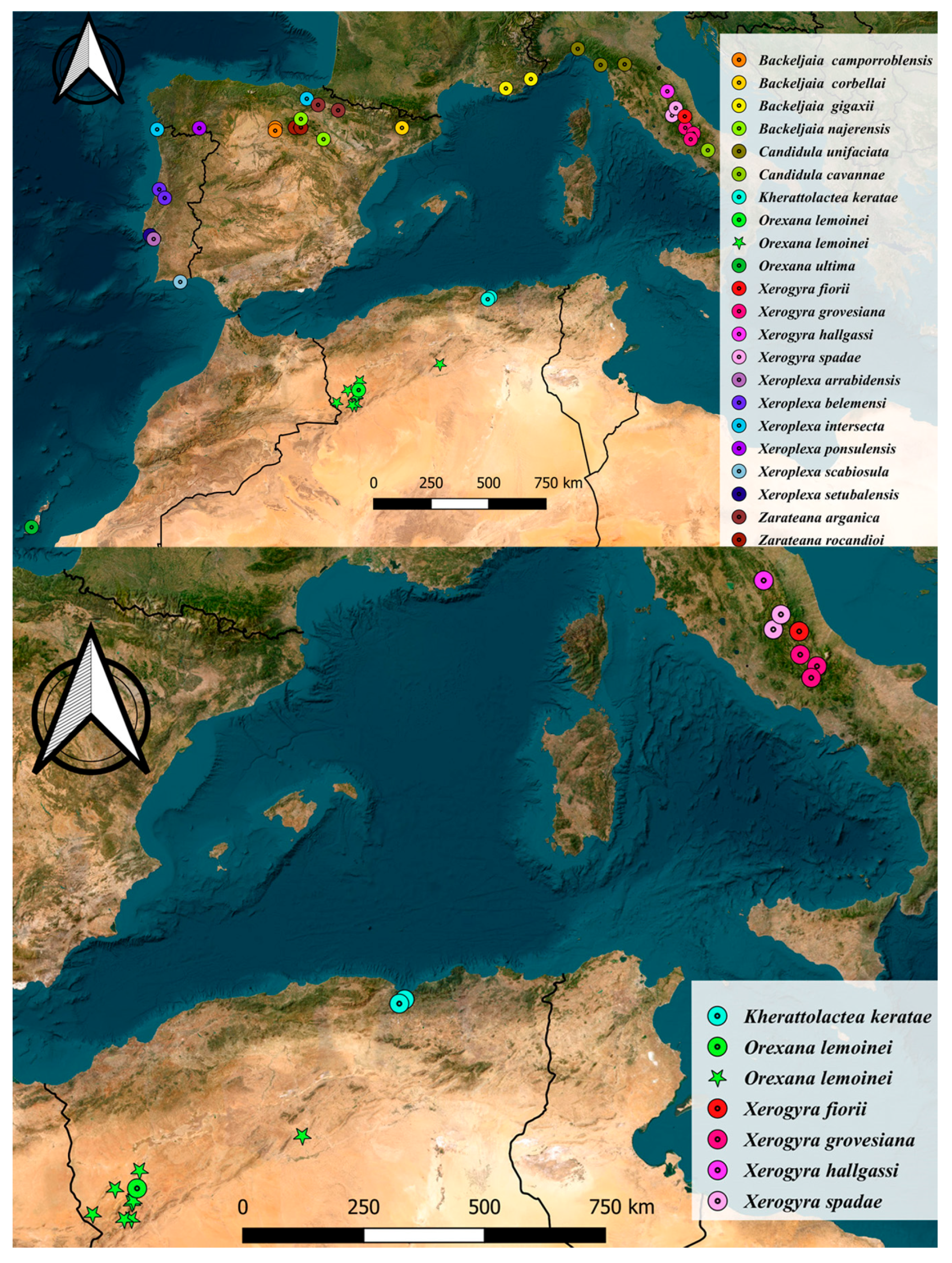
Figure 1. Maps showing the distribution of the taxa used in the study. (A) Records of the taxa used for molecular analysis. (B) Records of the Italian Xerogyra species and the two Algerian taxa; stars represent shell records only.
Diversity 17 00550 g001
Figure 2. (A) Phylogram produced by Bayesian Inference (BI). (B) Phylogram produced by Maximum likelihood (ML). Both trees constructed based on the concatenated data of two mitochondrial markers (COI, 16S) and one nuclear (5.8S-ITS2-28S) marker.
Diversity 17 00550 g002
Figure 3. Kherattolactea keratae NMBE 568338, Draouate. (A) Shell of sequenced specimen, D = 18.9 mm. (B) Situs and details of genital organs, situs = 27.2 mm, flagellum = 8.1 mm, epiphallus = 8.16 mm. All photos Neubert & Ezzine, shells ×2. Abbreviations used for the genital organs: ag = albumen gland; at = atrium; bc = bursa copulatrix; dgs = dart gun system; ep = epiphallus; fl = flagellum; gm = glandulae mucosae; ids = inner dart sac; mrp = musculus retractor penis; p = penis; ped = pedunculus; pp = penial papilla; spo = spermoviduct; vc = vaginal chamber; vd = vas deferens.
Diversity 17 00550 g003
Figure 4. Kherattolactea keratae. (A) Lectotype Xerophila keratae SMF 10296, Chabet-el-Akra, D = 20.96 mm. (B) NMBE 568339, Draouate, D = 18.2 mm. (C) NMBE 549913, situs of genital organs, Irhi Emda, Chabet el Akra, Kherratta, D = 19.3 mm. All photos Neubert & Ezzine, shells × 2.
Diversity 17 00550 g004
Figure 5. Genital organs of a paratype of Kherattolactea keratae. All drawings by De Mattia.
Diversity 17 00550 g005
Figure 6. Type locality of Kherattolactea keratae, Chabet el-Akra, Tizi-Ouzou, Algeria. (A) Gorge entrance; (B) Vegetation in the gorge.
Diversity 17 00550 g006
Figure 7. Sequenced specimens of Xerogyra species: (A) Xerogyra fiorii, NMBE 570837, Italy, Gran Sasso/Abruzzo, D = 10.1 mm. (B) Xerogyra grovesiana, NMBE 570838, Italy, Gole di Celano/L’Aquila, D = 7.7 mm. (C) Xerogyra spadae, NMBE 570836, Italy. Sella di Leonessa/Mt. Terminillo/Abruzzen, D = 9.9 mm.—All phot. Ezzine, ×4.
Diversity 17 00550 g007
Figure 8. Genital organs of Xerogyra fiorii, two specimens from Gran Sasso. (A) Specimen 1. (B) Specimen 2. All drawings by De Mattia.
Diversity 17 00550 g008
Figure 9. Genital organs of paratypes of Italian Xerogyra species. (A) Xerogyra grovesiana, Gole di Celano. (B) Xerogyra spadae, Sella Leonessa, Terminillo. All drawings by De Mattia.
Diversity 17 00550 g009
Figure 10. Shell and genital organs of Xerogyra halgassi n. sp., holotype NMBE 570829. (A) shell, D = 16.5 mm. (B) Situs of genital organs. (C) Penial papilla and interior of epiphallus. (D) Dart sac and glandulae mucosae. (E) Dart sac, internal details. All photos Neubert & Ezzine, shell ×2.
Diversity 17 00550 g010
Figure 11. Genital organs of paratypes of Xerogyra halgassi n. sp., Gualdo Tadino. (A) Specimen 2. (B) Specimen 1. All drawings by De Mattia.
Diversity 17 00550 g011
Figure 12. Orexana lemoinei. (A) Lectotype Helix lemoinei SMF 10493; D = 16.1 mm. (BD) RMNH-MOL 330559. Sequenced specimen from Forthassa, Saida, Algeria. (B) Situs genital organs. (C) Penis with penial papilla, dart sac with dart. (D) Shell, D = 12.4 mm. All photos Neubert & Ezzine, shells ×3.
Diversity 17 00550 g012
Figure 13. Orexana lemoinei. Variations of shell morphology. (A) RMNH-MOL 329780, Forthassa/Saida, D = 13.1 mm. (B) RMNH-MOL 294543, Djebal Souiga/Saida, D = 12.7 mm. (C) RMNH-MOL 294533, Foot of Djebal Antar/Saida, D = 12.9 mm. All photos Neubert & Ezzine, shells ×3.
Diversity 17 00550 g013
Table 1. A list of the markers and primers used in this study.
MarkerPrimer NamePrimer Sequence
COI [17]LCO14905′-GGTCAACAAATCATAAAGATATTGG-3′
HCO21985′-TAAACTTCAGGGTGACCAAAAAATCA-3′
16S [18]16S F5′-CGGCCGCCTGTTTATCAAAAACAT-3′
16S R5′-GGAGCTCCGGTTTGAACTCAGATC-3′
ITS2 [19]LSU15′-CTAGCTGCGAGAATTAATGTGA-3′
LSU35′-ACTTTCCCTCACGGTACTTG-3′
28S [19]LSU-25′-GGGTTGTTTGGGAATGCAGC-3′
LSU-45′-GTTAGACTCCTTGGTCCGTC-3′
Table 2. Conchological and anatomical differences among Xerogyra species and Kherattolactea keratae.
Xerogyra hallgassi n. sp.Xerogyra fioriiXerogyra spadaeXerogyra grovesianaKherattolactea keratae
Number of whorls5 ½ ± ½ (n = 11)5 ¼ ± ⅓ (n = 18)5 ⅓ ± ⅓ (n = 21)4 ¾ ± ½ (n = 15)5 ⅓ ± ⅓ (n = 6)
Average diameter16.8 ± 0.3 (n = 11)11.8 ± 0.4 (n = 18)9.2 ± 0.1 (n = 21)8.9 ± 0.2 (n = 15)18.5 ± 0.4 (n =4)
D/H2 (n = 11)1.8 (n = 18)1.5 (n = 21)2.2 (n = 15)1.8 (n = 4)
Shell’s surfaceradial fine growth striaeradial fine growth striaeradial fine growth striaeradial ribsradial fine growth striae
Bands along last whorlnonevery weak but visiblevery weak but visibleNoneweak to strong
Last whorlnot keelednot keelednot keeledKeelednot keeled
Umbilicus shaperemarkably eccentricslightly eccentricconcentricslightly eccentricconcentric
D/U>⅓¼¼
Aperture’s shapebroadly ovalroundishroundishsubquadrangularbroadly oval
Aperture’s internal callousvery mild to absentmildstrongabsentmild to strong
Flagellum/epiphallus0.74 (n = 4)0.72 (n = 3)0.91 (n = 3)2.8 (n = 3)0.8 (n = 2)
Disclaimer/Publisher’s Note: The statements, opinions and data contained in all publications are solely those of the individual author(s) and contributor(s) and not of MDPI and/or the editor(s). MDPI and/or the editor(s) disclaim responsibility for any injury to people or property resulting from any ideas, methods, instructions or products referred to in the content.