Article

Secondary Microplastics Disrupt Early Coral Development: Impacts on Brooding and Broadcast-Spawning Species

by
Camilo García-Galindo
1,*,
Luis A. Gómez-Lemos
2,
Sigmer Quiroga
1,* and
Rocío García-Urueña
1
1
Universidad del Magdalena, Santa Marta, Colombia
2
Universidad Nacional de Colombia–Sede de La Paz–Escuela de Pregrados, La Paz, Colombia
*
Authors to whom correspondence should be addressed.
Diversity 2025, 17(7), 468; https://doi.org/10.3390/d17070468
Submission received: 30 April 2025 / Revised: 24 May 2025 / Accepted: 2 June 2025 / Published: 8 July 2025

Abstract

Microplastics are pervasive in marine ecosystems and have been shown to affect a range of marine organisms, including corals. These particles can develop biofilms, facilitating the transport of pollutants and pathogenic microorganisms. Although the effects of primary microplastics on adult corals have been extensively studied, little is known about the impacts of secondary microplastics on early life stages. This study investigated (1) the effects of different abundances of secondary microplastics on the early development of Orbicella faveolata; (2) the influence of fibers and fragments on the larval settlement of Acropora palmata; and (3) the effects of the microplastic size, abundance, and biofilm presence on the settlement of Favia fragum. For O. faveolata, fertilization, embryogenesis, and settlement were all impaired by fragments, with embryonic development showing a 25.9% reduction in viability. Larval development was unaffected, but post-settlement survival declined by 35.8% with exposure to fibers and fragments. For A. palmata, settlement was reduced by fragments, while for F. fragum, only 2–4 mm fragments significantly decreased settlement rates. This study contributes to the knowledge of the effect of microplastics on the early development of corals, providing valuable information to improve strategies to control microplastic pollution.

1. Introduction

Microplastics are plastic pieces smaller than 5 mm but larger than 1 μm [1] and are classified into two types based on their origin: primary microplastics, intentionally manufactured at these sizes [2], and secondary microplastics, which mainly result from the progressive fragmentation of larger plastic items due to the mechanical force of waves and the effects of ultraviolet light over time [3]. Microplastic pollution in the ocean is estimated to range from 0.001 to 140 pieces/m3 and has been reported across various ecosystems, including coral reefs [4,5].
Due to their diverse densities, microplastics may be found throughout the ocean, interacting with a wide variety of marine organisms [6,7]. The most common interaction is ingestion, although the leaching of chemical additives, physical contact, and the presence of pollutants, such as heavy metals, also negatively affect organisms [6,8]. Most studies on the impact of microplastics on marine organisms focus on fish, annelids, crustaceans and mollusks [9,10]. Experiments on adult corals have shown that microplastic exposure can cause physiological stress, energy acquisition problems, and reduced growth, among other effects [11,12,13,14,15]. However, short-term exposure to these pollutants does not always harm coral health [16].
A noteworthy gap in the research is the limited knowledge about secondary microplastics, which account for 84.5–96.3% of all microplastics in the environment [17,18]. Moreover, the effects of microplastics on early coral development, a stage highly sensitive to environmental stressors due to the organisms’ small size and limited defense mechanisms, remain understudied [19,20].
The hydrophobic nature of microplastics makes them ideal substrates for biofilm formation, as they are composed of organic and inorganic material that facilitates microbial colonization [4,21]. Consequently, microplastics can act as vectors for pathogenic organisms [22]. Coral diseases are often linked to a microbial presence, particularly bacteria from the families Vibrionaceae, Rhodobacteraceae, and Flavobacteraceae, which are associated with tissue damage in stony corals and have been detected in microplastic biofilms in reef areas [23]. However, the biological, chemical, and physical interactions between biofilm-covered microplastics and marine organisms remain poorly understood [21].
Addressing these knowledge gaps is crucial for understanding how secondary microplastics and their associated biofilms interact with corals, particularly during early life stages, which are the most vulnerable phases in the coral life cycle. This understanding is essential for assessing the impact of microplastics on coral reef ecosystems [11,19,20]. In this context, our study assessed the effects of microplastics on the early development of the broadcast-spawning corals Orbicella faveolata and Acropora palmata and the brooding coral Favia fragum. For F. fragum, three experimental factors were evaluated: (1) the microplastic abundance, (2) microplastic size, and (3) the presence or absence of biofilms. For A. palmata, two factors were assessed: (1) the microplastic type and (2) microplastic abundance. For O. faveolata, only the microplastic abundance was assessed. We hypothesized that (1) increasing microplastic concentrations will have a dose-dependent negative effect on early developmental stages and (2) the impact of microplastics on early coral development will depend on both particle size and shape, with fragments being the most detrimental.

2. Materials and Methods

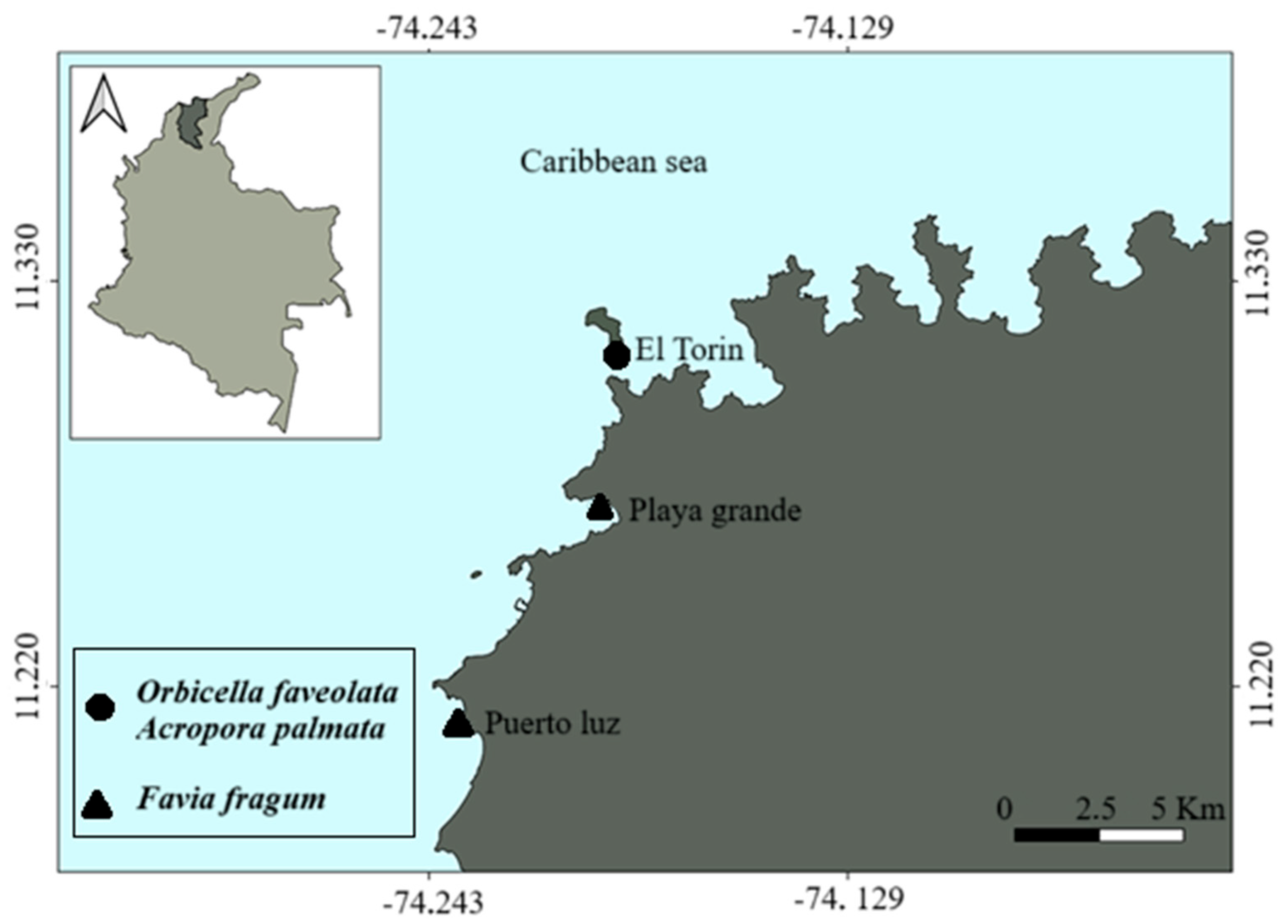
2.1. Gamete Collection of O. faveolata and A. palmata

Six colonies of O. faveolata and eight colonies of A. palmata were located and identified at Isla Aguja (11°18′36″ N, 74°11′27.6″ W) within Parque Nacional Natural Tayrona, at depths between 3 and 11 m (Figure 1). Colonies were monitored by SCUBA divers during the night until spawning occurred. O. faveolata spawned on 5 September 2023 and 23 September 2024, from 9 to 10:30 pm, and A. palmata spawned on 23 August 2024, from 9 to 10:30 pm. Gamete bundles were collected using conical nets made of woven polyester fabric (500 μm pore size) with 50 mL collection tubes (Supplementary Figure S1). The bundles were then transported to the laboratory for assisted fertilization, following the protocol of Alvarado-Chacón et al. [24].

2.2. Larvae Collection of F. fragum

Colonies of F. fragum were identified and collected in Taganga Bay (11°16′3.98″ N, 74°11′45.83″ W) and Gaira Bay (11°12′32.9″ N 74°14′01.9″ W), at depths between 1.5 and 2 m (Figure 1). Colonies were collected on 10 June and 8 August 2024, four days after the new moon, when planulation started [25]. Twenty colonies larger than 5 cm were collected to ensure they were reproductive [26]. Colonies were placed in 10 L plastic containers (dimensions: 11 × 11 × 7 cm) with 2 L of seawater and transported to the laboratory.
In the laboratory, colonies were transferred to 20 L glass containers (dimensions: 27 × 23 × 19 cm) with 15 L of seawater, natural light, and aeration. Seawater was changed daily, and the average temperature was 28.8 ± 0.3 °C. Each morning, larvae were collected using 3 mL Pasteur pipettes and transferred to 10 L containers (dimensions: 18 × 30 × 20 cm) with 8 L of filtered seawater (pore size 5 and 20 μm), treated with ultraviolet (UV) light [27,28]. This process was repeated for three days and larvae were kept in separate containers to avoid mixing larvae of different ages. To run the experiments, larvae of each age were added in equal proportion to each replicate to avoid a bias due to the differences in larval age.

2.3. Collection of Crustose Coralline Algae (CCA)

The CCA Porolithon antillarum was collected in September 2023 and 2024 for the settlement experiments of O. faveolata and in August 2024 for the experiments with A. palmata. Fifty fragments of P. antillarum were collected by SCUBA diving using a chisel and hammer. CCA fragments were transported to the laboratory and maintained in 10 L containers (dimensions: 18 × 30 × 20 cm) with filtered seawater (5–20 μm) treated with UV light. CCA were kept at 26.6 ± 0.2 °C with aeration and under artificial light.

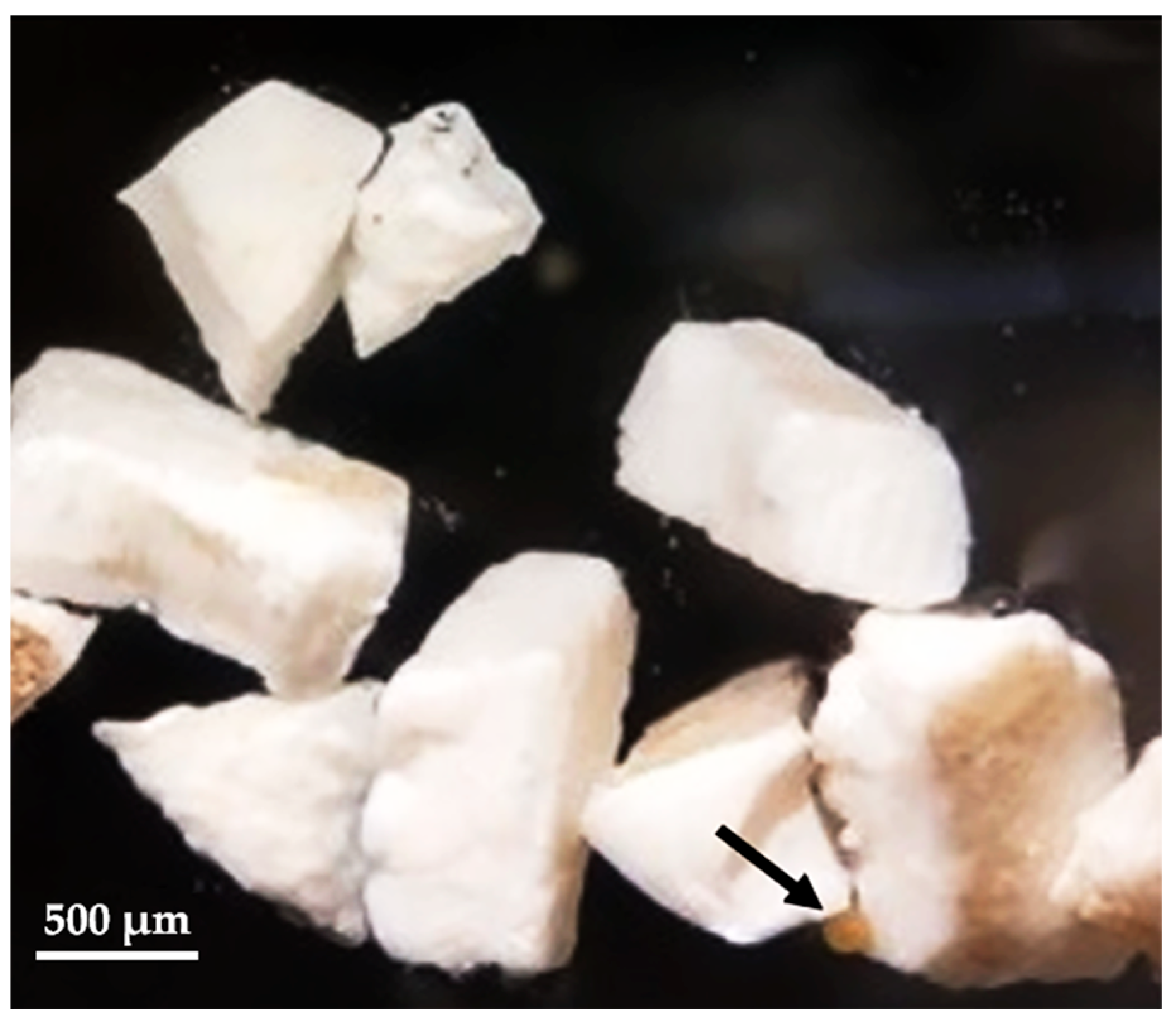
2.4. Microplastics Preparation

To test the effect of secondary microplastics, a piece of polypropylene was exposed to sunlight for ten months to simulate natural wearing. It was then crushed with metal tongs to obtain three fragment size ranges: 2–4 mm, 1–2 mm, and 110–450 µm. To test the effect of secondary microplastic fibers we used nylon bristles from a toothbrush (Oral-B pro-White 3D; Redwood City, CA, USA) as a proxy. The toothbrush was conditioned outdoors for one month to simulate natural weathering. Conditioned bristles were cut into pieces of 1 to 2 mm and 0.4 mm wide. To ensure that known abundances of microplastics were maintained in the treatments, fragments with sizes of 1–4 mm and fibers of 1–2 mm were counted manually, and fragments of 110–450 µm were counted using a dissecting microscope. We selected a maximum concentration of 200 pieces L−1, which matches the maximum amount of microplastics estimated to be present in the oceans by 2100 [29]. We also considered concentrations of microplastics used in similar experiments to choose our ranges [11,12].
To evaluate the effect of microplastic biofilms, we placed the experimental microplastics in conditioning chambers (Supplementary Figure S2). Chambers were maintained in 10 L containers (dimensions: 18 × 30 × 20 cm) with untreated seawater from Taganga Bay. During the 30-day conditioning period, complete water changes were performed twice a week.
Buoyancy varied according to the type and size of microplastics. Fragments with a size of 2–4 mm, 1–2 mm, and 110–450 µm remained at the water surface regardless of whether they had biofilm or not. The fibers tended to sink and settle on the bottom of the containers. Glass and metal materials were used throughout the experiments to avoid contamination with non-experimental microplastics.

2.5. Trace Metal Assessment

To assess the trace metals associated with coral tissue after microplastic exposure, we collected samples from each developmental stage (fertilization, embryonic development, larval development, and larval settlement) and stored them in 4% formalin until chemical analyses were performed. The trace metals assessed included mercury (Hg), lead (Pb), cadmium (Cd), vanadium (V), chromium (Cr), nickel (Ni), zinc (Zn), selenium (Se), arsenic (As), and boron (B). Mercury concentrations were measured with a Milestone Tricell DMA-80 direct mercury analyzer, based on atomic absorption spectroscopy (AAS) with thermal decomposition and amalgamation. A Zeeman graphite furnace atomic absorption spectrometer (model GFS35Z; Waltham, MA, USA) was used to determine concentrations of Pb, Cd, V, Cr, Ni, Se, B, Zn, and As. Analyses were performed with an ambient temperature of 22 °C and an average humidity of 53%.

2.6. Effect of Microplastics on O. faveolata Fertilization

In 2023, the assisted fertilization of O. faveolata was performed by mixing gametes from different colonies in 1 L fat separators (Norpro® Everett, WA, USA). We evaluated the effect of microplastics on fertilization through three treatments with concentrations of 70, 150, and 200 pieces L−1 (size range: 2–4 mm), with three replicates. A second experiment was conducted in 2024 with two factors: (1) microplastic abundance (70 and 200 pieces L−1) and (2) type of microplastic (fragments and fibers) with four replicates per treatment. Each experiment included a control in which embryogenesis occurred in the absence of microplastics. In both experiments 40 eggs were added per replicate in 80 mL glass vials with filtered seawater (pore size: 5 and 20 μm), sterilized with UV light. We counted the number of fertilized, unfertilized, and broken eggs after 2 h using a dissecting microscope.

2.7. Effect of Microplastics on O. faveolata Embryogenesis

In 2023, embryos were exposed to three treatments with concentrations of 70, 150, and 200 pieces L−1 (size range: 2–4 mm) and three replicates. In 2024 we carried out another experiment with two factors: (1) microplastic abundance (70 and 200 pieces L−1) and (2) type of microplastic (fragments and fibers) with four replicates. Each experiment had a control, where embryos developed in the absence of microplastics (Supplementary Figure S3). In both experiments 40 embryos (eight-cell stage embryos) were added per replicate in glass vials of 80 mL with filtered seawater (pore size: 5 and 20 μm), sterilized with UV light. O. faveolata embryonic development lasts 37 h [24]. After this period, embryos were classified and counted by developmental stage using a dissecting microscope.

2.8. Effect of Microplastics on Larval Development of O. faveolata

In 2023, larvae were exposed to three treatments with concentrations of 70, 150, and 200 pieces L−1 (size range: 2–4 mm) with six replicates. In 2024, a second experiment was performed, with two factors: (1) microplastic abundance (70 and 200 pieces L−1) and (2) type of microplastic (fragments and fibers), with four replicates. Each experiment had a control in which larvae developed in absence of microplastics (Supplementary Figure S3). In both experiments 40 larvae were added per replicate in 1 L containers (dimensions: 18 × 18 × 7 cm) with filtered water (pore size: 5 and 20 μm), sterilized with UV light. We counted the number of living larvae after 131 h [24] using a dissecting microscope.

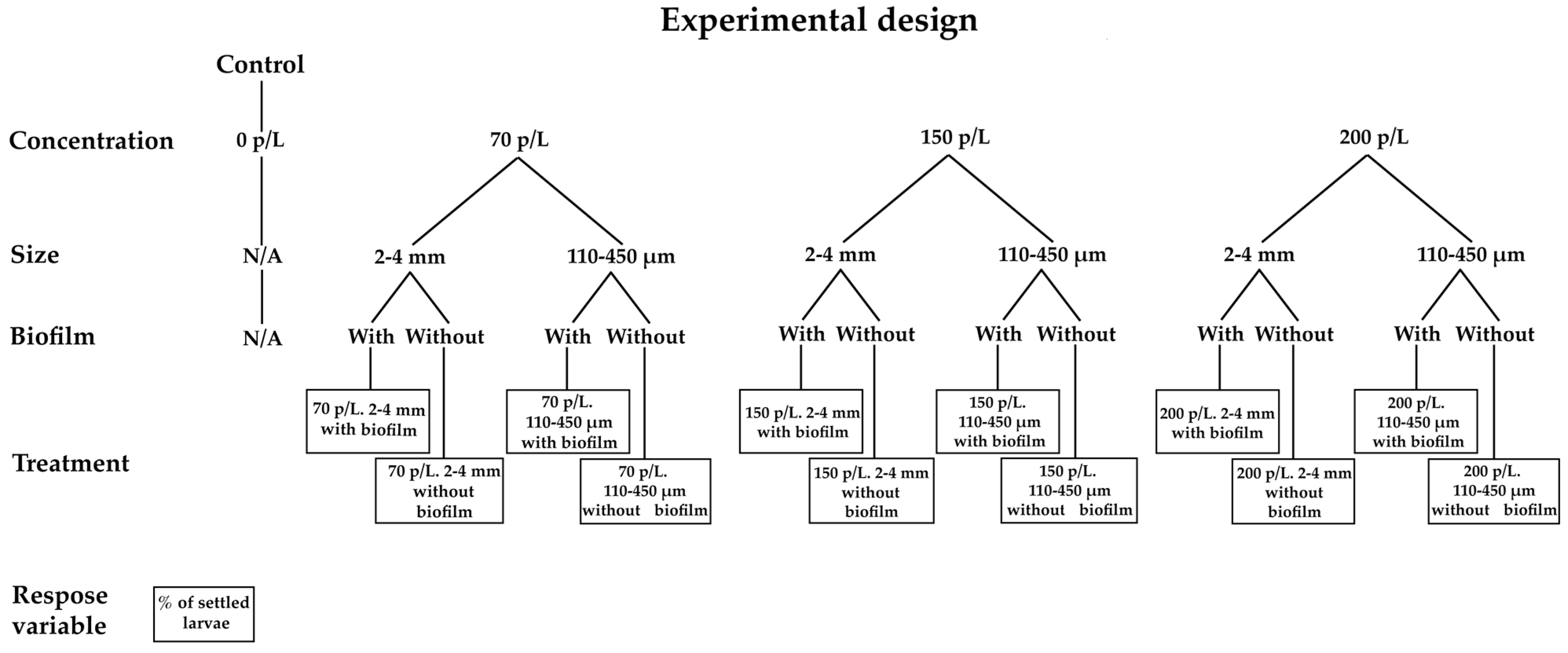
2.9. Effect of Microplastics on Larval Settlement of O. faveolata, A. palmata, and F. fragum

2.9.1. Orbicella faveolata

In 2023 experiments with O. faveolata were carried out six days after the assisted fertilization, once larvae were competent to settle. We used fragments of Porolithon antillarum (size 2 cm2) to induce larval settlement. There were three treatments with abundances of 70, 150, and 200 pieces L−1 (size range: 2–4 mm) and six replicates. In 2024 we performed another experiment with two factors: (1) microplastic concentration (70 and 200 pieces L−1) and (2) type of microplastic (fragments and fibers) with four replicates. Each experiment had a control, where larvae settled in the absence of microplastics. In both experiments 40 larvae were added per replicate in 1 L containers (dimensions: 18 × 18 × 7 cm) with filtered seawater (pore size: 5 and 20 μm), sterilized with UV light. At the end of the experiment, we counted the number of settled larvae using a dissecting microscope.

2.9.2. Acropora palmata

Experiments with A. palmata were carried out with four-day-old larvae. We used fragments of Porolithon antillarum with a size of 2 cm2 to induce larval settlement. This experiment had two factors: (1) microplastic abundance (70 and 200 pieces L−1) and (2) type of microplastic (fragments and fibers) with three replicates. In the control treatment larvae settled in the absence of microplastics (Supplementary Figure S4). Eighty larvae were added per replicate in 1 L containers (dimensions: 18 × 18 × 7 cm) with filtered seawater (pore size: 5 and 20 μm), sterilized with UV light. The number of settled larvae was counted after 48 h using a dissecting microscope.

2.9.3. Favia fragum

Settlement experiments with F. fragum were carried out in June and August 2024, using settlement tiles previously conditioned on the reef for 30 days (Supplementary Figure S5). The experimental design included three factors: (1) microplastic abundance (70, 150, and 200 pieces L−1), (2) microplastic size (2–4 mm and 110–450 μm), and (3) biofilm (presence or absence). In the control, larvae settled in the absence of microplastics (Figure 2). There were three replicates, and 30 larvae were added per replicate in 1 L containers (dimensions: 18 × 18 × 7 cm) with filtered seawater (pore size: 5 and 20 μm), sterilized with UV light. We counted the number of settled larvae after 48 h using a dissecting microscope.

2.9.4. Effect of Microplastics on O. faveolata Post-Settlement Survival

In 2023, the settlement of O. faveolata larvae was induced, using fragments (2 cm2) of P. antillarum for 48 h. Settled larvae were exposed to three treatments with concentrations of 70, 150, and 200 pieces L−1 (size range: 2 and 4 mm) with three replicates. In 2024, another experiment was carried out with two factors: (1) microplastic concentration (70- and 200-pieces L−1) and (2) type of microplastic: fragments (Fr) and fibers (Fi). Ceramic tiles previously conditioned during 30 days on the reef were used to induce larval settlement. In both experiments, eighty larvae were induced to settle on the ceramic tiles in 1 L containers (dimensions: 18 × 18 × 7 cm) with filtered seawater (pore size: 5 and 20 μm), sterilized with UV light. After four days, the number of successfully settled larvae was counted as the starting point (an average of 58, 39, 33, and 44 for treatments of 0, 70, 150, and 200 pieces L−1, respectively, in 2023 and 49, 14, 13, 17, and 17 for the treatments control, Fi70, Fi 200, Fr70, and Fr 200 pieces L−1, respectively, in 2024). Afterward, microplastics treatments were added to evaluate the effect on post-settlement survival. Surviving polyps were counted daily for seven days. Both experiments included a control group in which primary polyps were not exposed to microplastics.

2.10. Data Analysis

We performed separate statistical analyses for each experiment. Normality was assessed using a Shapiro–Wilk test and homogeneity of variances was tested using a Levene’s test. Data were transformed with arcsine when parametric assumptions were not met [30]. Data of O. faveolata fertilization, embryogenesis, larval development, and larval settlement in 2023 were analyzed using a one-way ANOVA followed by Tukey’s HSD test. In 2024, a two-way ANOVA was used to evaluate the effects of microplastic abundance and type on fertilization, embryogenesis, larval development, and larval settlement of O. faveolata. A two-way ANOVA was performed to test for the effects of microplastic concentration and type on larval settlement of A. palmata. The effects of microplastics’ abundance, microplastics’ size, and biofilm presence were analyzed using a three-way ANOVA. The Kaplan–Meier survival analysis, followed by the log-rank test (Mantel–Cox), was performed to assess differences in post-settlement survival of O. faveolata spats under different microplastic concentrations. Statistical analyses were performed using Statgraphics Centurion XVII and R version 4.4.1.

3. Results

3.1. Trace Metal Assessment

The results show that during fertilization the concentrations of Hg, Cd, and Zn did not exceed the detection limit, while V, Cr, Ni, Se, As, B, and Mo did not exceed the quantification limit. Pb exceeded the quantification limit, with an average of 1.55 μg/kg. Regarding the embryonic development, larval development, and larval settlement, the average levels of Hg and Zn did not reach the detection limit, whereas V, Cr, Ni, Se, As, B, and Mo remained below the quantification limit. On the other hand, Pb and Cd exceeded the quantification limit, with an average of 1.8 μg/kg and 0.14 μg/kg, respectively (Supplementary Table S1). However, these concentrations are relatively low [31,32].

3.2. Effect of Microplastics on O. faveolata Fertilization

In 2023 there were significant differences between the control and the treatments with 150 and 200 pieces L−1 (ANOVA; n = 6, F = 7,50, p = 0.001; Figure 3A). In 2024 we found significant differences between the control and the treatments of 70 and 200 pieces L−1 (two-way ANOVA; n = 3, F = 6, p = 0.009; Figure 3B; Supplementary Table S2).

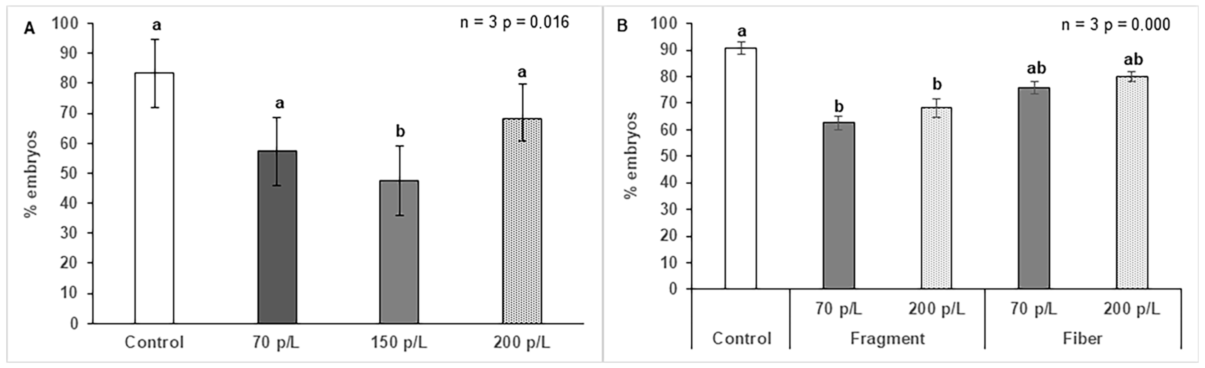
3.3. Effect of Microplastics on O. faveolata Embryogenesis

In 2023, we found a significant difference between the control and the treatment with 150 pieces L−1(ANOVA; n = 3, F = 6,31, p = 0.016; Figure 4A). In 2024, there was a significant difference between the control and the treatments of 70 and 200 pieces L−1 (two-way ANOVA; n = 3, F = 15, p < 0.001; Figure 4B; Supplementary Table S3).

3.4. Effect of Microplastics on Larval Development of O. faveolata

In 2023 we evaluated the larval development 120 h after fertilization, and there were no significant differences between the control and the experimental treatments (ANOVA; n = 6 F = 2,33 p = < 0.001; Figure 5A; Supplementary Table S4). In 2024, no significant differences were found between the treatments and the control (Two-way ANOVA; n = 3, F = 4,3, p = 0.573; Figure 5B).

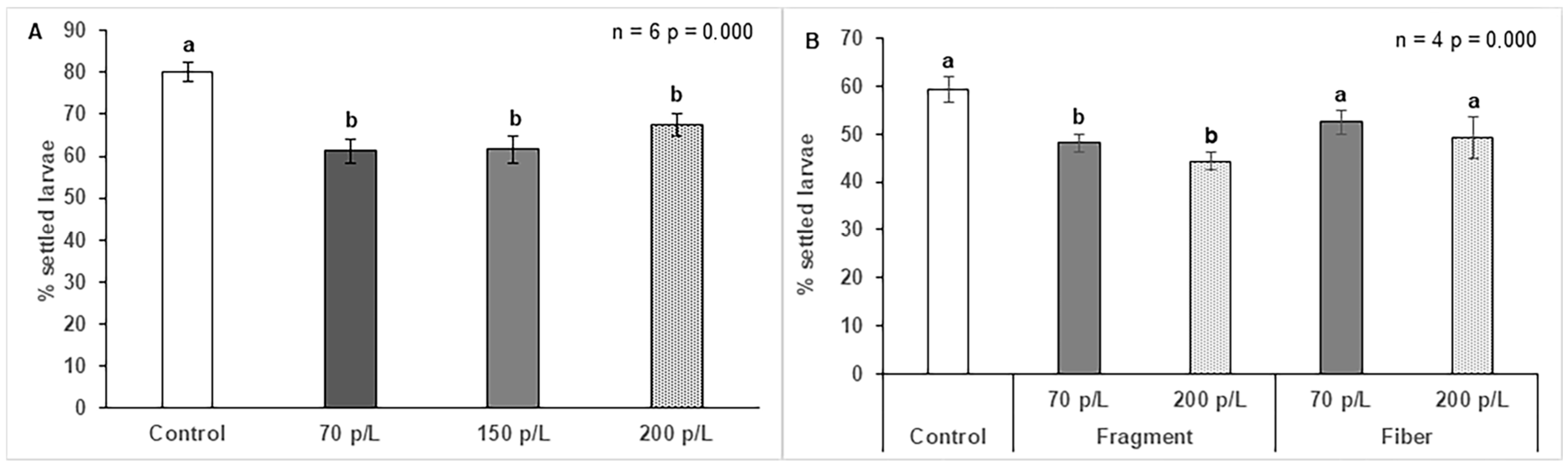
3.5. Effect of Microplastics on Larval Settlement of O. faveolata

In 2023 there was a significant decrease in settlement when comparing the control to the remaining treatments of microplastic concentrations (ANOVA; n = 6, F = 9,73, p < 0.001; Figure 6A). In 2024 there were significant differences between the control and the treatments of 70 and 200 fragments L−1 (Two-way ANOVA; n = 3, F = 15, p < 0.001; Figure 6B; Supplementary Table S5).

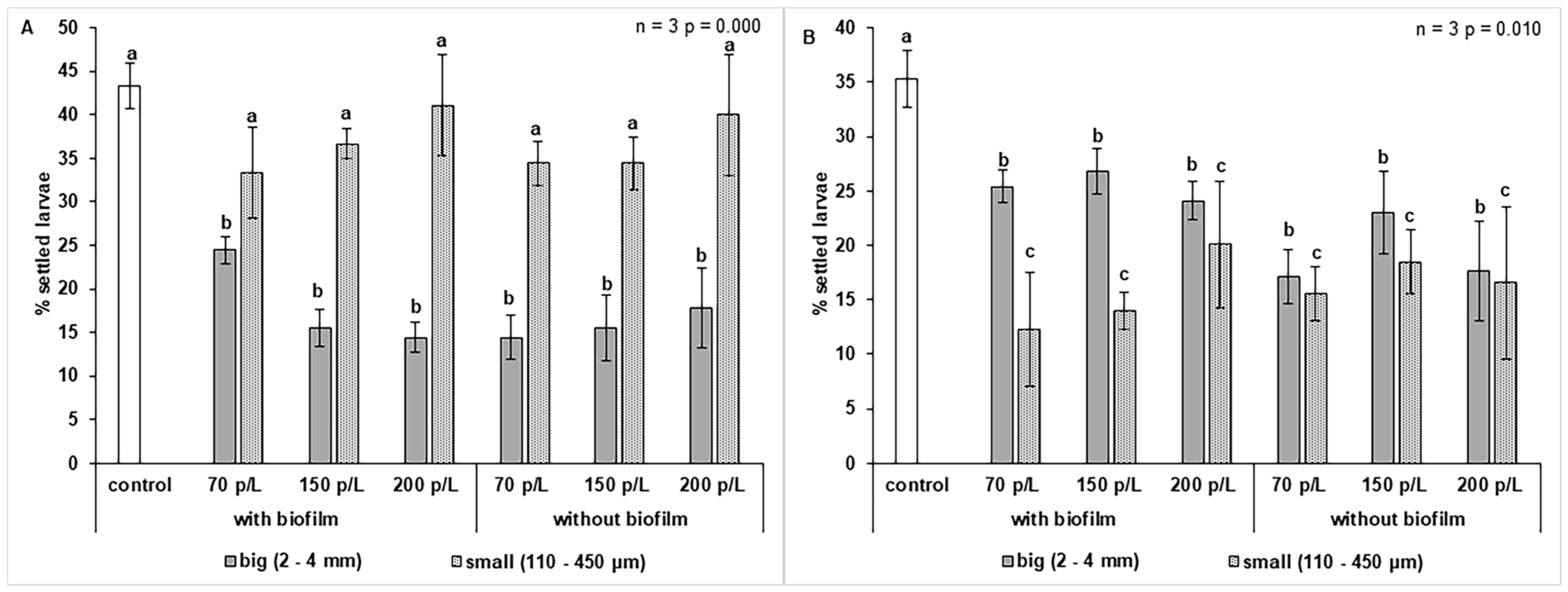
3.6. Effect of Microplastics on A. palmata Settlement

There was a significant decrease in the larval settlement in treatments with concentrations of 70 and 200 fragments L−1 compared to the control (ANOVA; n = 3, F = 6, p = 0.009; Figure 7; Supplementary Table S6).

3.7. Effect of Microplastics on Larval Settlement of F. fragum

In June, the size factor showed a significant negative effect on settlement (three-way ANOVA n = 3, F = 21.6, p < 0.001; Figure 8A; Supplementary Table S7). Settlement decreased from 43 ± 2.6% (SD) in the control to 26.8% due to the size factor, which is a reduction of 16.2%. In the “large” level of the size factor, there was an average settlement of 16.9%, a decrease of 26.1%. Although a decrease in settlement was also observed in the “small” size level, it was only 6.2% and was not statistically significant. In August, the size factor also had a negative effect on settlement (three-way ANOVA n = 3, F = 7.5, p = 0.010.; Figure 8B; Supplementary Table S8). Settlement decreased from 35 ± 2.1% (SD) in the control to 18.9%, due to the size factor, representing a reduction of 16.0%. In the “large” size level, the settlement was 22.6%, a drop of 12.4%. In the “small” size level, the settlement was 15.3%, corresponding to a decrease of 19.7%.

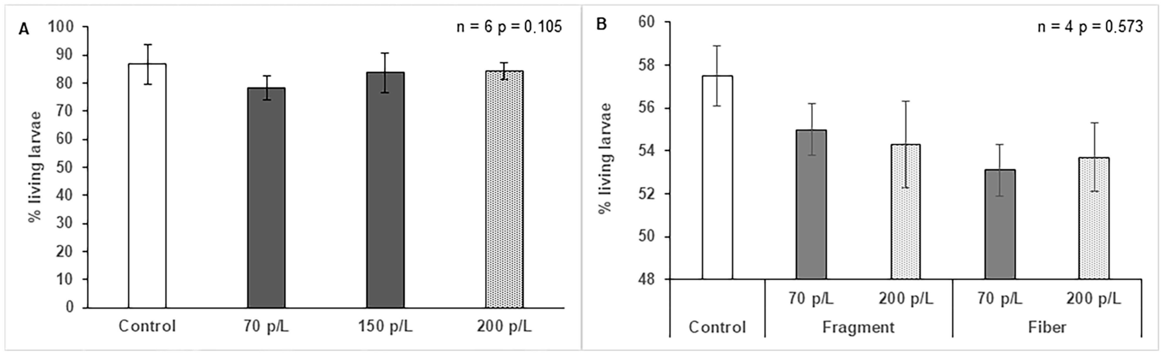
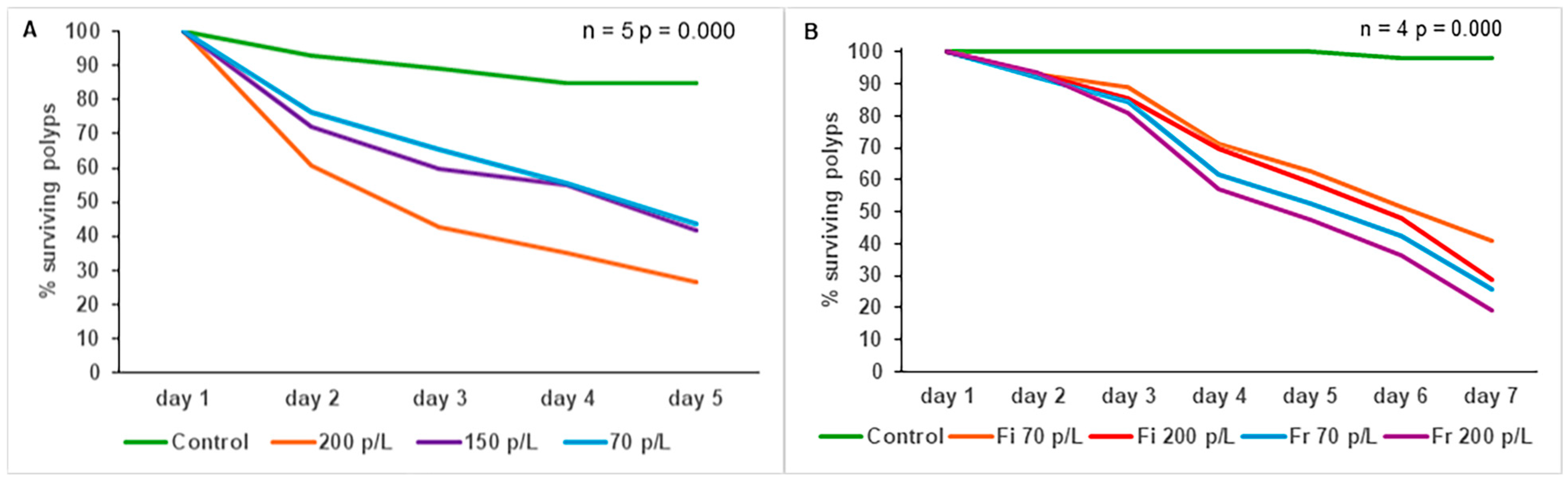
3.8. Effect of Microplastics on O. faveolata Post-Settlement Survival

In 2023 there was a significant difference in the post-settlement survival of O. faveolate between the control and all microplastic treatments, as well as between the treatment of 70 pieces L −1 and 150 and 200 pieces L −1 (Kaplan–Meier; n = 5, p < 0.001; Figure 9A; Supplementary Table S9). In 2024 there was also a significant decrease in post-settlement survival in all treatments compared to the control (Kaplan–Meier; n = 4, p < 0.001; Figure 9B; Supplementary Table S10).

4. Discussion

4.1. Trace Metal Assessment

It should be noted that the plastic fragments used in this study came from a paint container. We do not know the chemical composition of this paint, but these products usually contain polycyclic aromatic hydrocarbons (PAHs), such as anthracene, fluorene, and phenanthrene, and heavy metals (e.g., copper, arsenic, zinc, or mercury), which are toxic for corals affecting fertilization [33,34,35,36]. Additionally, microplastic fragments are capable of absorbing and releasing heavy metals, such as cobalt [37], which can alter the embryonic development of other cnidarians, such as Hydra magnipapillata [38]. There are heavy metals (such as copper, cadmium, and zinc) and PAHs that can cause a decrease in settlement in corals such as A. tenuis [31,36]. Microplastics can absorb and release heavy metals as leachates; this suggests that fragments may affect coral polyps by chemical interactions through the leaching of toxic substances [39]. As mentioned before, it is known that heavy metals, such as copper, cadmium, zinc, and lead, can negatively affect the early development of some coral species [32,34,36]. In our study, only lead (2.08 ± 0.020 µg/kg) and cadmium (0.18 ± 0.104 µg/kg) had concentrations above the quantification limit. These metals are capable of penetrating cell membranes through transport mechanisms and becoming trapped in the tissues [40]; however, these values were lower than those that can affect survival in the early development of marine invertebrates [31,32].

4.2. Effect of Microplastics on O. faveolata Fertilization

These results are similar to those of Berry et al. [11], who reported a decrease in fertilization of Acropora tenuis when it was exposed to propylene pieces (size: 2 mm²) at concentrations of 5, 15, and 50 pieces L−1. However, our results differ from Wilkins et al. [39], as they did not find a decrease in the fertilization of Montipora capitata after an exposure to microplastics from different sources, despite using similar concentrations.
It is known that the eggs of broadcast-spawning corals are poorly protected [41]. These eggs can suffer physical damage when interacting with particles in the environment, as they are vulnerable and cannot move to avoid damage [42]. Furthermore, microplastics can physically affect fertilization by acting as barriers between eggs and sperm, jeopardizing fertilization success [11,43,44]. In this context, the lack of a negative effect of nylon fibers in our study could be explained by the different densities of fragments (0.90 g/cm3) and fibers (1.14–1.04 g/cm3) [45,46]; the former remained on the water surface, where it is more likely to interact with the coral gametes causing a negative effect on them.

4.3. Effect of Microplastics on O. faveolata Embryogenesis

Our results are similar to the findings of Berry et al. [10] for A. tenuis, who reported a significant decrease in embryo quality when 2 mm2 polypropylene fragments were added at concentrations of 5, 15, and 50 pieces L−1. The negative effect observed in our study could be due to the physical action of sharp plastic fragments on embryos of O. faveolata; a similar result was reported for A. millepora [19,47,48]. The apparent harmlessness of experimental fibers in our study can be explained by their mostly smooth surfaces [49], which possibly does not pose a threat to embryos.

4.4. Effect of Microplastics on Larval Development of O. faveolata

Our results differ from those reported by e Silva et al. [10] for the sessile mollusk Perna perna, where the percentage of larvae with normal development decreased after an exposure to primary and secondary microplastics. Leachates released by primary microplastics can cause abnormal larval development in mollusks such as P. perna and echinoderms such as Lytechinus variegatus [10,50]. We found that O. faveolata larvae were resistant to microplastic exposure, and this can be explained since the larvae ectoderm acts as a barrier that prevents the entry of some pollutants [51]. Additionally, larvae may have some resistance to physical damage caused by suspended particles, as they produce a mucus mainly made of carbohydrates (such as d-arabinose, mannose, galactose and glucose), which covers larvae, preventing contaminants and suspended particles from coming into direct contact with them [52,53].

4.5. Effect of Microplastics on Larval Settlement of O. faveolata

Our findings differ from the results reported by Berry et al. [11], as they did not find negative effects on A. tenuis settlement after exposing larvae to 5, 15, and 50 pieces L−1. It is possible that the decrease in settlement resulted from the physical interaction between the microplastic fragments and the larvae, as we observed that fragments trapped the larvae, deterring the settlement process (Figure 10).

4.6. Effect of Microplastics on A. palmata Settlement

Our results diverge from the findings reported by Berry et al. [11], as they did not observe any effects on A. tenuis settlement after exposing larvae to 5, 15, and 50 pieces L−1. It is possible that the effect found in our study is derived from the physical mechanism previously described, where microplastics can trap larvae preventing settlement.

4.7. Effect of Microplastics on Larval Settlement of F. fragum

Our results differ from the findings described by Berry et al. [11], as they did not find any effect on A. tenuis settlement after exposing larvae to microplastics of 2, 1, and 0.5 mm2 and 1 and 6 μm. We observed that microplastics of 2–4 mm trapped larvae (Figure 10), and this may be the mechanism responsible for the decrease in larval settlement. It is possible that this mechanism can also occur for pieces of 110–450 μm, since Berry et al. [11] indicated that spherical microplastics of 1 and 6 μm surrounded larvae of A. tenuis under experimental conditions. Although in their experiment there was no negative impact on settlement, in our experiments the abrasive surfaces of fragments could irreparably damage larvae.
Biofilm formation decreases the buoyancy of microplastics and allows them to transport pathogenic microorganisms and pollutants such as heavy metals [16,54,55]. For these reasons we expected a negative effect of microplastic biofilms on larval settlement in our experiments. However, biofilms did not have any effect. This result supports the statement by Zettler et al. [54], who suggested that biofilms do not always increase the toxicity of microplastics. Similarly, the abundance of microplastics did not have a significant effect. This is similar to the findings reported by Berry et al. [11], as they did not find an effect of microplastic concentration on A. tenuis settlement using 5, 15, and 50 polypropylene pieces L−1. This trend can be explained as larvae may avoid the interaction with microplastics by swimming using their cilia and moving away from microplastics [56].

4.8. Effect of Microplastics on O. faveolata Post-Settlement Survival

Settlement and post-settlement survival are critical processes to maintain coral populations and reef perpetuity [41]. Under laboratory conditions, survival is largely influenced by the substrate chosen to induce larval settlement [57]. In our study the time span to evaluate survival was five days in 2023 and seven in 2024. This range corresponds to the phase in which the highest mortality rates of primary polyps occur under laboratory conditions [58]. At the end of the experiments, a marked difference was found in the survival of primary polyps for both years. Nylon fibers, due to their density (1.14–1.04 g/cm3) [45], sink, facilitating their contact with primary polyps, which may result in energy loss by the polyps [59]. On the other hand, the effect of polypropylene fragments cannot be explained by physical contact, since their density of 0.90 g/cm3 [46] prevents them from sinking (Figure 10).

5. Conclusions

We found a negative impact of microplastics on early developmental stages of the corals Favia fragum, Acropora palmata, and Orbicella faveolata. The fertilization, embryonic development, settlement, and post-settlement survival of these three species were affected by fragments in the 1–4 mm size range, while microplastics in the 110–450 µm range affected only the settlement of F. fragum. However, no negative effects on early development were observed in any of the studied species as a function of the microplastic concentration. Similarly, the presence of a biofilm did not appear to negatively affect the early development of Favia fragum. It is important to note that the specific effects of microplastics vary depending on the size, type, and concentration; therefore, comparisons and generalizations should be made with caution. Furthermore, the potential variability in species’ responses complicates the assessment of the magnitude of this pollution problem in coral reefs at a global scale. This fact highlights the need for more detailed studies that can provide a better understanding of the risks posed by microplastics for coral development.

Supplementary Materials

The following supporting information can be downloaded at https://www.mdpi.com/article/10.3390/d17070468/s1: Figure S1: (a) Nets used for the collection of Orbicella faveolata gametes, (b) gamete bundles stored in Falcon tubes (c), and assisted fertilization in the laboratory Figure S2: (a) Chambers used for conditioning microplastics with biofilm. (b) Assembly of the conditioning system to generate a biofilm on microplastics. Figure S3: Orthogonal experimental design to test for the effect of microplastic type and abundance on fertilization, embryogenesis, larval development, larval settlement, and post-settlement survival of Orbicella faveolata. Figure S4: Orthogonal experimental design to test for the effect of microplastic type and abundance on larval settlement of Acropora palmata. Figure S5: (a) Ceramic tiles used for larval settlement, unconditioned. (b) Ceramic tiles conditioned on the reef; the presence of crustose coralline algae can be observed on their surface. Table S1: Trace metal concentrations (µg/kg) in organisms exposed to microplastics. Mercury (Hg), cadmium (Cd), lead (Pb), vanadium (V), chromium (Cr), nickel (Ni), selenium (Se), zinc (Zn), arsenic (As), boron (B), and molybdenum (Mo). Below the limit of quantification (<). Table S2: Two-way ANOVA for the year 2024 to assess the effects of microplastic concentration and type on Orbicella faveolata coral fertilization. Table S3: Two-way ANOVA for the year 2024 to assess the effects of microplastic abundance and type on the embryogenesis of the coral Orbicella faveolata. Table S4: Two-way ANOVA for the year 2024 to assess the effects of microplastic concentration and type on larval development of the coral Orbicella faveolata. Table S5: Two-way ANOVA from year 2024 to assess the effects of microplastic concentration and type on larval settlement of Orbicella faveolata. Table S6: Two-way ANOVA to assess the effects of microplastic concentration and type on larval settlement of Acropora palmata coral. Table S7: Three-way ANOVA (14 June 2024) to assess the effects of microplastic concentration and size, as well as the absence or presence of a biofilm on larval settlement of the coral Favia fragum. Table S8: Three-way ANOVA (12 August 2024) to assess the effects of microplastic concentration and size, as well as the absence or presence of a biofilm on larval settlement of the coral Favia fragum. Table S9: Pairwise comparisons using Log-Rank test to determine the effect of microplastic concentration on post-settlement survival of the coral Orbicella faveolata. Table S10: Pairwise comparisons using Log-Rank test to determine the effect of microplastic concentration and type on post-settlement survival of the coral Orbicella faveolata.

Author Contributions

Conceptualization, C.G.-G. and L.A.G.-L.; formal analysis, C.G.-G.; investigation, C.G.-G.; methodology, C.G.-G., L.A.G.-L. and S.Q.; writing—original draft, C.G.-G.; funding acquisition, L.A.G.-L., S.Q. and R.G.-U.; writing—review and editing, L.A.G.-L. and S.Q.; project administration, S.Q. and R.G.-U.; supervision, R.G.-U. All authors have read and agreed to the published version of the manuscript.

Funding

This research was funded by the project “EFECTO DEL POLVILLO DE CARBÓN Y LOS MICROPLÁSTICOS EN EL DESARROLLO TEMPRANO DE ORGANISMOS ARRECIFALES”, under the contract CD 82648 CT ICETEX 2022-0733, from the Ministry of Science, Technology and Innovation of Colombia (Minciencias) and the Colombian Institute of Educational Credit and Technical Studies Abroad (ICETEX), and the Universidad del Magdalena.

Data Availability Statement

Data will be made available on request.

Acknowledgments

This study was carried out with the support of the University of Magdalena and the Research Group on Ecology and Diversity of Marine Algae and Coral Reefs (EDAMAC). We wish to thank Bonnie Lewis for providing insightful comments and edits to this manuscript.

Conflicts of Interest

The authors declare no conflicts of interest.

References

  1. Andrady, A.L. Microplastics in the marine environment. Mar. Pollut. Bull. 2011, 62, 1596–1605. [Google Scholar] [CrossRef] [PubMed]
  2. Auta, H.S.; Emenike, C.U.; Fauziah, S.H. Distribution and importance of microplastics in the marine environment: A review of the sources, fate, effects, and potential solutions. Environ. Int. 2017, 102, 165–176. [Google Scholar] [CrossRef] [PubMed]
  3. Sipe, J.M.; Bossa, N.; Berger, W.; von Windheim, N.; Gall, K.; Wiesner, M.R. From bottle to microplastics: Can we estimate how our plastic products are breaking down? Sci. Total Environ. 2022, 814, 152460. [Google Scholar] [CrossRef] [PubMed]
  4. Huang, W.; Chen, M.; Song, B.; Deng, J.; Shen, M.; Chen, Q.; Zeng, G.; Liang, J. Microplastics in the coral reefs and their potential impacts on corals: A mini-review. Sci. Total Environ. 2021, 762, 143112. [Google Scholar] [CrossRef]
  5. Albaseer, S.S.; Al-Hazmi, H.E.; Kurniawan, T.A.; Xu, X.; Abdulrahman, S.A.; Ezzati, P.; Habibzadeh, S.; Hollert, H.; Rabiee, N.; Lima, E.C.; et al. Micro-plastics in water resources: Global pollution circle, possible technological solutions, legislations, and future horizon. Sci. Total Environ. 2024, 946, 173963. [Google Scholar] [CrossRef]
  6. Cole, M.; Lindeque, P.; Halsband, C.; Galloway, T.S. Microplastics as contaminants in the marine environment: A review. Mar. Pollut. Bull. 2011, 62, 2588–2597. [Google Scholar] [CrossRef]
  7. Browne, M.A.; Niven, S.J.; Galloway, T.S.; Rowland, S.J.; Thompson, R.C. Microplastic moves pollutants and additives to worms, reducing functions linked to health and biodiversity. Curr. Biol. 2013, 23, 2388–2392. [Google Scholar] [CrossRef]
  8. Allen, A.S.; Seymour, A.C.; Rittschof, D. Chemoreception drives plastic consumption in a hard coral. Mar. Pollut. Bull. 2017, 124, 198–205. [Google Scholar] [CrossRef]
  9. de Sá, L.C.; Oliveira, M.; Ribeiro, F.; Rocha, T.L.; Futter, M.N. Studies of the effects of microplastics on aquatic organisms: What do we know and where should we focus our efforts in the future? Sci. Total Environ. 2018, 645, 1029–1039. [Google Scholar] [CrossRef]
  10. e Silva, P.P.G.; Nobre, C.R.; Resaffe, P.; Pereira, C.D.S.; Gusmão, F. Leachate from microplastics impairs larval development in brown mussels. Water Res. 2016, 106, 364–370. [Google Scholar] [CrossRef]
  11. Berry, K.L.; Epstein, H.E.; Lewis, P.J.; Hall, N.M.; Negri, A.P. Microplastic contamination has limited effects on coral fertilization and larvae. Diversity 2019, 11, 228. [Google Scholar] [CrossRef]
  12. Reichert, J.; Arnold, A.L.; Hoogenboom, M.O.; Schubert, P.; Wilke, T. Impacts of microplastics on growth and health of hermatypic corals are species-specific. Environ. Pollut. 2019, 254, 113074. [Google Scholar] [CrossRef] [PubMed]
  13. Rotjan, R.D.; Sharp, K.H.; Gauthier, A.E.; Yelton, R.; Lopez, E.M.B.; Carilli, J.; Kagan, J.C.; Urban-Rich, J. Patterns, dynamics and consequences of microplastic ingestion by the temperate coral, Astrangia poculata. Proc. R. Soc. B 2019, 286, 20190726. [Google Scholar] [CrossRef] [PubMed]
  14. Syakti, A.D.; Jaya, J.V.; Rahman, A.; Hidayati, N.V.; Raza’i, T.S.; Idris, F.; Trenggono, M.; Doumenq, P.; Chou, L.M. Bleaching and necrosis of staghorn coral (Acropora formosa) in laboratory assays: Immediate impact of LDPE micro-plastics. Chemosphere 2019, 228, 528–535. [Google Scholar] [CrossRef]
  15. Lanctôt, C.M.; Bednarz, V.N.; Melvin, S.; Jacob, H.; Oberhaensli, F.; Swarzenski, P.W.; Ferrier-Pagès, C.; Carroll, A.R.; Metian, M. Physiological stress response of the scleractinian coral Stylophora pistillata exposed to polyethylene microplastics. Environ. Pollut. 2020, 263, 114559. [Google Scholar] [CrossRef]
  16. Hankins, C.; Duffy, A.; Drisco, K. Scleractinian coral microplastic ingestion: Potential calcification effects, size limits, and retention. Mar. Pollut. Bull. 2018, 135, 587–593. [Google Scholar] [CrossRef]
  17. Sundt, P.; Schulze, P.E.; Syversen, F. Sources of Microplastic-Pollution to the Marine Environment; Report No. M-321-2015; Mepex for the Norwegian Environment Agency: Asker, Norway, 2014. [Google Scholar]
  18. Lee, S.; Kim, D.; Kang, K.K.; Sung, S.E.; Choi, J.H.; Sung, M.; Shin, C.H.; Jeon, E.; Kim, D.; Kim, D.; et al. Toxicity and biodistribution of fragmented polypropylene microplastics in ICR mice. Int. J. Mol. Sci. 2023, 24, 8463. [Google Scholar] [CrossRef]
  19. Jones, R.; Bessell-Browne, P.; Fisher, R.; Klonowski, W.; Slivkoff, M. Assessing the impacts of sediments from dredging on corals. Mar. Pollut. Bull. 2016, 102, 9–29. [Google Scholar] [CrossRef]
  20. Alderdice, R.; Pernice, M.; Cárdenas, A.; Hughes, D.J.; Harrison, P.L.; Boulotte, N.; Chartrand, K.; Kühl, M.; Suggett, D.J.; Voolstra, C.R. Hypoxia as a physiological cue and pathological stress for coral larvae. Mol. Ecol. 2022, 31, 571–586. [Google Scholar] [CrossRef]
  21. Rummel, C.D.; Jahnke, A.; Gorokhova, E.; Kühnel, D.; Schmitt-Jansen, M. Impacts of biofilm formation on the fate and potential effects of microplastic in the aquatic environment. Environ. Sci. Technol. Lett. 2017, 4, 258–267. [Google Scholar] [CrossRef]
  22. Oberbeckmann, S.; Löder, M.G.; Labrenz, M. Marine microplastic-associated biofilms–A review. Environ. Chem. 2015, 12, 551–562. [Google Scholar] [CrossRef]
  23. Feng, L.; He, L.; Jiang, S.; Chen, J.; Zhou, C.; Qian, Z.J.; Hong, P.; Sun, S.; Li, C. Investigating the composition and distribution of microplastics surface biofilms in coral areas. Chemosphere 2020, 252, 126565. [Google Scholar] [CrossRef] [PubMed]
  24. Alvarado-Chacon, E.M.; Gómez-Lemos, L.A.; Sierra-Sabalza, N.P.; Hernández-Chamorro, A.M.; Lozano-Peña, J.P.; Valcárcel-Castellanos, C.A.; Rojas, J.A. Early life history of the Caribbean coral Orbicella faveolata (Scleractinia: Merulinidae). Rev. Biol. Trop. 2020, 68, 1262–1274. [Google Scholar] [CrossRef]
  25. Szmant-Froelich, A.; Reutter, M.; Riggs, L. Sexual reproduction of Favia fragum (ESPER): Lunar patterns of gametogenesis, embryogenesis and planulation in Puerto Rico. Bull. Mar. Sci. 1985, 37, 880–892. [Google Scholar]
  26. Soong, K. Colony size as a species character in massive reef corals. Coral Reefs 1993, 12, 77–83. [Google Scholar] [CrossRef]
  27. Gómez-Lemos, L.A.; Doropoulos, C.; Bayraktarov, E.; Kennedy, E.V.; Diaz-Pulido, G. Coralline algal metabolites induce settlement and mediate the inductive effect of epiphytic microbes on coral larvae. Sci Rep. 2018, 8, 17557. [Google Scholar] [CrossRef]
  28. Aoki, N.; Weiss, B.; Jézéquel, Y.; Apprill, A.; Mooney, T.A. Replayed reef sounds induce settlement of Favia fragum coral larvae in aquaria and field environments. JASA Express Lett. 2024, 4, 107701. [Google Scholar] [CrossRef]
  29. Koelmans, A.A.; Kooi, M.; Law, K.L.; van Sebille, E. All is not lost: Deriving a top-down mass budget of plastic at sea. Environ Res Lett. 2017, 12, 114028. [Google Scholar] [CrossRef]
  30. Underwood, A.J. Experiments in Ecology: Their Logical Design and Interpretation Using Analysis of Variance; Cambridge University Press: Cambridge, UK, 1997. [Google Scholar]
  31. Reichelt-Brushett, A.J.; Harrison, P.L. The effect of copper on the settlement success of larvae from the scleractinian coral Acropora tenuis. Mar. Pollut. Bull. 2001, 41, 385–391. [Google Scholar] [CrossRef]
  32. Woods, R.M.; Baird, A.H.; Mizerek, T.L.; Madin, J.S. Environmental factors limiting fertilisation and larval success in corals. Coral Reefs 2016, 35, 1433–1440. [Google Scholar] [CrossRef]
  33. Negri, A.P.; Heyward, A.J. Inhibition of coral fertilization and larval metamorphosis by tributyltin and copper. Mar. Environ. Res. 2001, 51, 17–27. [Google Scholar] [CrossRef] [PubMed]
  34. Reichelt-Brushett, A.J.; Harrison, P.L. The effect of selected trace metals on the fertilization success of several scleractinian coral species. Coral Reefs 2005, 24, 524–534. [Google Scholar] [CrossRef]
  35. Oriji, O.G.; Nartey, V.K.; Brimah, A.K. Determination of the Polycyclic Aromatic Hydrocarbon and heavy metal contents of some Nigerian emulsion paints. Int. J. Appl. Chem. 2012, 8, 215–225. [Google Scholar]
  36. Nordborg, F.M.; Brinkman, D.L.; Ricardo, G.F.; Agustí, S.; Negri, A.P. Comparative sensitivity of the early life stages of a coral to heavy fuel oil and UV radiation. Sci. Total Environ. 2021, 781, 146676. [Google Scholar] [CrossRef]
  37. Squadrone, S.; Pederiva, S.; Bezzo, T.; Sartor, R.M.; Battuello, M.; Nurra, N.; Griglione, A.; Brizio, P.; Abete, M.C. Microplastics as vectors of metals contamination in Mediterranean Sea. Environ. Sci. Pollut. Res. 2021, 29, 29529–29534. [Google Scholar] [CrossRef]
  38. Zeeshan, M.; Murugadas, A.; Ghaskadbi, S.; Ramaswamy, B.R.; Akbarsha, M.A. Ecotoxicological assessment of cobalt using Hydra model: ROS, oxidative stress, DNA damage, cell cycle arrest, and apoptosis as mechanisms of toxicity. Environ. Pollut. 2017, 224, 54–69. [Google Scholar] [CrossRef]
  39. Wilkins, K.W.; Yew, J.Y.; Seeley, M.; Richmond, R.H. Microplastic Leachate Negatively Affects Fertilization in the Coral Montipora capitata. Integr. Comp. Biol. 2024, 64, 1131–1140. [Google Scholar] [CrossRef]
  40. Chiarelli, R.; Roccheri, M.C. Marine invertebrates as bioindicators of heavy metal pollution. Open J Metal. 2014, 4, 93–106. [Google Scholar] [CrossRef]
  41. Harrison, P.L.; Babcock, R.C.; Bull, G.D.; Oliver, J.K.; Wallace, C.C.; Willis, B.L. Mass spawning in tropical reef corals. Science 1984, 223, 1186–1189. [Google Scholar] [CrossRef]
  42. Heyward, A.J.; Negri, A.P. Turbulence, cleavage, and the naked embryo: A case for coral clones. Science 2012, 335, 1064. [Google Scholar] [CrossRef]
  43. Ricardo, G.F.; Jones, R.J.; Clode, P.L.; Humanes, A.; Negri, A.P. Suspended sediments limit coral sperm availability. Sci. Rep. 2016, 5, 18084. [Google Scholar] [CrossRef] [PubMed]
  44. Sussarellu, R.; Suquet, M.; Thomas, Y.; Lambert, C.; Fabioux, C.; Pernet, M.E.J.; Le Goïc, N.; Virgile, Q.; Mingant, C.; Epelboin, Y.; et al. Oyster reproduction is affected by exposure to polystyrene microplastics. Proc. Natl. Acad. Sci. USA 2016, 113, 2430–2435. [Google Scholar] [CrossRef] [PubMed]
  45. Estes, L.L.; Schweizer, M. Fibers, 4. Polyamide Fibers. In Ullmann’s Encyclopedia of Industrial Chemistry; Wiley-VCH: Weinheim, Germany, 2011. [Google Scholar]
  46. Sadiku, R.; Ibrahim, D.; Agboola, O.; Owonubi, S.J.; Fasiku, V.O.; Kupolati, W.K.; Jamiru, T.; Eze, A.A.; Adekomaya, O.S.; Varaprasad, K.; et al. Automotive Components Composed of Polyolefins. In Polyolefin Fibres; Woodhead Publishing: Cambridge, UK, 2017; pp. 449–496. [Google Scholar]
  47. Humphrey, C.; Weber, M.; Lott, C.; Cooper, T.; Fabricius, K.J. Effects of suspended sediments, dissolved inorganic nutrients and salinity on fertilization and embryo development in the coral Acropora millepora (Ehrenberg, 1834). Coral Reefs 2008, 27, 837–850. [Google Scholar] [CrossRef]
  48. Ricardo, G.F.; Jones, R.J.; Clode, P.L.; Humanes, A.; Giofre, N.; Negri, A.P. Sediment characteristics influence the fertilisation success of the corals Acropora tenuis and Acropora millepora. Mar. Pollut. Bull. 2018, 135, 941–953. [Google Scholar] [CrossRef]
  49. Qiao, R.; Deng, Y.; Zhang, S.; Wolosker, M.B.; Zhu, Q.; Ren, H.; Zhang, Y. Accumulation of different shapes of microplastics initiates intestinal injury and gut microbiota dysbiosis in the gut of zebrafish. Chemosphere 2019, 236, 124334. [Google Scholar] [CrossRef]
  50. Nobre, C.R.; Santana, M.F.M.; Maluf, A.; Cortez, F.S.; Cesar, A.; Pereira, C.D.S.; Turra, A. Assessment of microplastic toxicity to embryonic development of the sea urchin Lytechinus variegatus (Echinodermata: Echinoidea). Mar. Pollut. Bull. 2015, 92, 99–104. [Google Scholar] [CrossRef]
  51. Vandermeulen, J.H. Studies on reef corals. II. Fine structure of planktonic planula larva of Pocillopora damicornis, with emphasis on the aboral epidermis. Mar. Biol. 1974, 27, 239–247. [Google Scholar] [CrossRef]
  52. Wild, C.; Naumann, M.; Niggl, W.; Haas, A. Carbohydrate composition of mucus released by scleractinian warm- and cold-water reef corals. Aquat. Biol. 2010, 10, 41–45. [Google Scholar] [CrossRef]
  53. Ricardo, G.F.; Jones, R.J.; Clode, P.L.; Negri, A.P. Mucous Secretion and Cilia Beating Defend Developing Coral Larvae from Suspended Sediments. PLoS ONE 2016, 11, e0162743. [Google Scholar] [CrossRef]
  54. Zettler, E.R.; Mincer, T.J.; Amaral-Zettler, L.A. Life in the “plastisphere”: Microbial communities on plastic marine debris. Environ. Sci. Technol. 2013, 47, 7137–7146. [Google Scholar] [CrossRef]
  55. Pan, H.; Zhao, X.; Zhou, X.; Yan, H.; Han, X.; Wu, M.; Chen, F. Research progress on the role of biofilm in heavy metals adsorption-desorption characteristics of microplastics: A review. Environ. Pollut. 2023, 336, 122448. [Google Scholar] [CrossRef] [PubMed]
  56. Takeda-Sakazume, A.; Honjo, J.; Sasano, S.; Matsushima, K.; Baba, S.A.; Mogami, Y.; Hatta, M. Gravitactic Swimming of the planula larva of the coral Acropora: Characterization of straightforward vertical swimming. Zool. Sci. 2022, 40, 44–52. [Google Scholar] [CrossRef] [PubMed]
  57. Lei, X.; Jiang, L.; Zhang, Y.; Sun, Y.; Zhou, G.; Lian, J.; Huang, H. Coral larval settlement and post-settlement survival facilitated by crustose coralline algae with or without living tissue. Mar. Biol. 2021, 168, 128. [Google Scholar] [CrossRef]
  58. Martinez, S.; Abelson, A. Coral recruitment: The critical role of early post-settlement survival. ICES J. Mar. Sci. 2013, 70, 1294–1298. [Google Scholar] [CrossRef]
  59. Flores, F.; Hoogenboom, M.O.; Smith, L.D.; Cooper, T.F.; Abrego, D.; Negri, A.P. Chronic exposure of corals to fine sediments: Lethal and sub-lethal impacts. PLoS ONE 2012, 7, e37795. [Google Scholar] [CrossRef]
Figure 1. Collection site of Orbicella faveolata, Acropora palmata, and Favia fragum gametes. Map designed by Dayana Rada-Osorio.
Diversity 17 00468 g001
Figure 2. The orthogonal experimental design to test for the effect of the microplastics’ abundance, size, and biofilm in the larval settlement of Favia fragum.
Diversity 17 00468 g002
Figure 3. (A) The effect of different concentrations of microplastic fragments on the fertilization of Orbicella faveolata. (B). The effect of different types of secondary microplastics on the fertilization of Orbicella faveolata.
Diversity 17 00468 g003
Figure 4. (A) The percentage of Orbicella faveolata embryos in the gastrula stage 37 h after exposure to different concentrations of microplastic fragments. (B) Percentage of Orbicella faveolata embryos in the gastrula stage 37 h after exposure to the different concentrations and types of secondary microplastics.
Diversity 17 00468 g004
Figure 5. (A) Percentage of living Orbicella faveolata larvae after exposure to different concentrations of microplastic fragments; (B) Percentage of living Orbicella faveolata larvae after exposure to different concentrations and types of secondary microplastics.
Diversity 17 00468 g005
Figure 6. (A) Orbicella faveolata larval settlement after exposure to different concentrations of microplastic fragments. (B) Percentage of Orbicella faveolata larval settlement after exposure to different concentrations and types of secondary microplastics.
Diversity 17 00468 g006
Figure 7. Larval settlement of Acropora palmata after exposure to different concentrations and types of secondary microplastics.
Diversity 17 00468 g007
Figure 8. (A) The effect of microplastic concentration and size and the presence or absence of biofilms on larval settlement of Favia fragum in June 2024. (B) The effect of microplastic concentration and size and the presence or absence of biofilms on larval settlement of Favia fragum in August 2024. In both experiments, the control consisted of inducing settlement in the absence of microplastics.
Diversity 17 00468 g008
Figure 9. (A) The early post-settlement survival of Orbicella faveolata under different concentrations of microplastic fragments in 2023. (B) The early post-settlement survival of Orbicella faveolata under different concentrations and types of secondary microplastics in 2024. The acronyms correspond to Fi (fibers) and Fr (fragments).
Diversity 17 00468 g009
Figure 10. Favia fragum larvae exposed to microplastics fragments to assess their effect on settlement. The arrow indicates a larva trapped between fragments.
Diversity 17 00468 g010
Disclaimer/Publisher’s Note: The statements, opinions and data contained in all publications are solely those of the individual author(s) and contributor(s) and not of MDPI and/or the editor(s). MDPI and/or the editor(s) disclaim responsibility for any injury to people or property resulting from any ideas, methods, instructions or products referred to in the content.

