Article

Marine Bryozoans from the Northern Pacific Coast of Costa Rica

by
Beatriz Antillón-Obando
1,*,
Jorge Cortés
1,2 and
Jeffrey A. Sibaja-Cordero
1,2
1
Escuela de Biología, Universidad de Costa Rica, San José 11501-2060, Costa Rica
2
Centro de Investigaciones en Ciencias del Mar y Limnología (CIMAR), Universidad de Costa Rica, San José 11501-2060, Costa Rica
*
Author to whom correspondence should be addressed.
Diversity 2025, 17(7), 451; https://doi.org/10.3390/d17070451
Submission received: 8 May 2025 / Revised: 16 June 2025 / Accepted: 16 June 2025 / Published: 26 June 2025
(This article belongs to the Section Marine Diversity)

Abstract

Although diverse and abundant, the phylum Bryozoa has been the subject of few studies in Costa Rica. Nearly 50 years have passed since Banta and Carson identified, described, and published twenty-four bryozoan species, including scanning electron microscope (SEM) images. To expand the knowledge of bryozoan diversity in the region, we sampled shallow coastal waters along the northern Pacific coast of Costa Rica and extracted tissue for DNA barcoding using the mitochondrial COI marker. Photographs of living specimens and SEM images were taken for morphological identification. We identified fifteen individuals belonging to nine bryozoan species from the orders Cyclostomatida and Cheilostomatida. Five of these species represent new records for Costa Rica (Savignyella lafontii, Bugula neritina, Watersipora arcuata, Smittipora levinseni, and Biflustra tenuis), while the remaining four (Disporella sp., Parasmittina crosslandi, Cigclisula sp., and Biflustra sp.) had been previously reported.

1. Introduction

Bryozoa is a phylum of colonial, encrusting benthic animals [1,2,3]. The group exhibits a wide diversity of morphologies and growth strategies, ranging from laminar encrustations to erect, ramified colonies. Most commonly, bryozoans grow under rocks, on corals, or as epibionts [1,4,5,6]. These animals are suspension feeders [6,7] and serve as prey for a variety of specialized and generalist predators, including nudibranchs, pycnogonids, mollusks, fish, starfish, and other incidental consumers [6,8]. Most encrusting bryozoans form calcareous skeletons using calcium carbonate, making them ecologically and environmentally important. They can serve as nursery habitats, contribute to carbonate sediment formation, and act as potential indicator species due to their sensitivity to ocean acidification [1,9].
Because of their benthic lifestyle, bryozoans often compete with other organisms for space on the substrate. Adnate or encrusting bryozoans are among the fastest and most aggressive colonizers [10]. Cyclostomatida, a group composed mostly of adnate species, is one of the fastest to colonize rocky substrates [11]. However, Cheilostomatida tends to be the most diverse and dominant order on both the Pacific and Caribbean coasts of Central and South America, with the highest number of species and individuals recorded [3,12,13,14].
Accurate identification of bryozoan species is important for ecological studies due to their roles in benthic communities and their potential as fouling organisms [15,16]. Despite this, bryozoans remain understudied, particularly in tropical regions [7]. In Costa Rica, there are currently only three major publications focused on the richness of living bryozoan species, as summarized by Cortés et al. [2]. The first was by Raymond Carroll Osburn, who analyzed samples collected during the Allan Hancock expeditions in 1932 and 1935, reporting 40 marine species from Isla del Coco and the Pacific mainland of Costa Rica [17,18,19]. Later, William C. Banta and Renate J.M. Carson documented 25 species from both the Pacific and Caribbean coasts and included SEM images in their publication [13]. A third contribution, a book chapter by Jorge Cortés, Vanessa Nielsen, and Amalia Herrera-Cubilla, reported 61 bryozoan species for Costa Rica [2]. The most recent study, by Sibaja-Cordero et al. [20], recorded seven species from the subtidal benthos of Isla del Coco, including two new records for the island: one species of Membraniporidae and Reptadeonella tubulifera (Canu & Bassler, 1930).
Current efforts to inventory marine biodiversity on Costa Rica’s northern Pacific coast include the Área de Conservación Guanacaste (ACG), designated a UNESCO World Heritage site in 1999. Originally established in the 1980s to document and monitor terrestrial biodiversity, the ACG later expanded to include marine research through the BioMar-ACG project, initiated in 2015 [21]. This initiative, led by the CIMAR research center (Centro de Investigaciones en Ciencias del Mar y Limnología) of the University of Costa Rica, had reported 1479 species across 22 taxonomic groups by 2020, including nine bryozoan species previously documented by Osburn [22]. Since Osburn’s work [17,18,19], no further studies have focused on bryozoan species richness in this region.
To date, none of the existing publications have used genetic methods for species identification. Therefore, one of the primary objectives of this study is to initiate the application of genetic tools to assess bryozoan diversity in the region. Specifically, the BioMar-ACG project aims to generate data through DNA barcoding (COI markers) to identify species, particularly those not previously analyzed in the Eastern Tropical Pacific. In this study, we report COI barcodes for several bryozoan species collected from the Área de Conservación Guanacaste (ACG) for the first time, along with photographs of live specimens and SEM images for morphological reference, including microscope images for each taxon.

2. Materials and Methods

2.1. Study Site

The study was conducted in the Área de Conservación Guanacaste (ACG, Guanacaste Conservation Area; 10.6 to 11.0° N), located on the northern Pacific coast of Costa Rica, Central America. ACG spans 163,000 hectares in the northern region of Guanacaste Province and encompasses diverse marine habitats, including sandy beaches, soft bottoms, coral and rocky reefs, rocky intertidal zones, mangroves, islets, and islands [21,23,24]. The conservation area stretches from the Guanacaste Volcanic Mountain Range to the marine zone surrounding the Islas Murciélago archipelago. ACG’s protected land represents approximately 2% of Costa Rica’s total territory.
The northern Pacific coast is characterized by marked seasonality due to strong annual upwelling events from December to April. These upwellings lower seawater temperatures, increase nutrient availability, and decrease water acidity [22,25,26,27,28]. For the remainder of the year, the region experiences warmer marine waters and a rainy season. We collected samples from shallow waters within the Marine Sector of ACG and from the rocky shore of Junquillal Bay National Wildlife Refuge, visiting sites in both the Gulf and Bay of Santa Elena during fieldwork.

2.2. Live Colonies Processing

We took samples from rocky intertidal zones, reefs, subtidal rock walls, boulders on sandy bottoms, and the sublittoral zones of islets. At each site, we gathered living organisms of various morphologies and transported them to a field laboratory. Samples were stored in buckets containing seawater and aerated using portable air pumps until processing and preservation in 95% ethanol.
In intertidal zones, we searched tide pools on boulders and rocky substrates for bryozoan colonies. In shallow waters (<3 m depth), we conducted snorkeling surveys, and in subtidal zones (4–20 m depth), we performed SCUBA dives. During these dives, we examined substrates such as shells, pebbles, boulders, rock walls, and macroalgae. Rocks smaller than 25 cm in diameter were collected whole; for larger rocks, fragments were extracted using a hammer and chisel. For each site, we recorded the GPS coordinates, site name, maximum depth, substrate type, and associated fauna.
Underwater photographs of the colonies in situ were taken using a Lumix Panasonic 14 MP camera (Panasonic, Osaka, Japan) in an underwater housing with white light flash. Back in the field lab, live colonies were sorted morphologically. Close-up images of zooids were taken using an AmScope SM-4TZ-144A microscope (AmScope, Irvine, CA, USA) or an OMAX 18 MP camera (OMAX, Kent, WA, USA) with the software ToupLite (2010–2022, version macOS2.1.19627.20210925, ToupTek Photonics) (Touptek, Hangzhou, China). Macro photographs of entire colonies were taken with a Canon EOS Rebel SL3 (Canon Inc., Tokyo, Japan).
A fragment of each preserved colony was archived in the Bryozoa collection of the Museo de Zoología, Universidad de Costa Rica (MZUCR). Another small fragment (3 to 4 zooids) was used for DNA extraction and COI barcoding analysis. A third fragment was used for scanning electron microscopy (SEM) imaging.

2.3. Scanning Electron Microscope Images

To prepare specimens for SEM analysis, we removed soft tissue and sediment by soaking colony fragments in a 5% sodium hypochlorite (NaClO) solution for 24 h, refreshing the solution until bubbling ceased. The fragments were then rinsed five times with distilled water to eliminate residual bleach [3,29], followed by 5 min of sonication. Samples were dried in an oven at 40 °C for three days.
We followed the protocol described by Breedy & Guzmán [30] for SEM preparation. Dried fragments were mounted on aluminum stubs using double-sided carbon tape for optimal conductivity and then coated with a 30–60 nm layer of gold using an EMS 550X sputter coater (Electron Microscopy Sciences, Hatfield, PA, USA). SEM images were taken using a Hitachi S-3700n (Hitachi, Tokyo, Japan) and a ZEISS Sigma300VP electron microscope (ZEISS, Oberkochen, Germany) at the Centro de Investigación en Estructuras Microscópicas (CIEMic). Morphological identifications were made by comparing our SEM images with published references containing detailed images of zooidal structures [12,13].

2.4. DNA Tissue Extraction
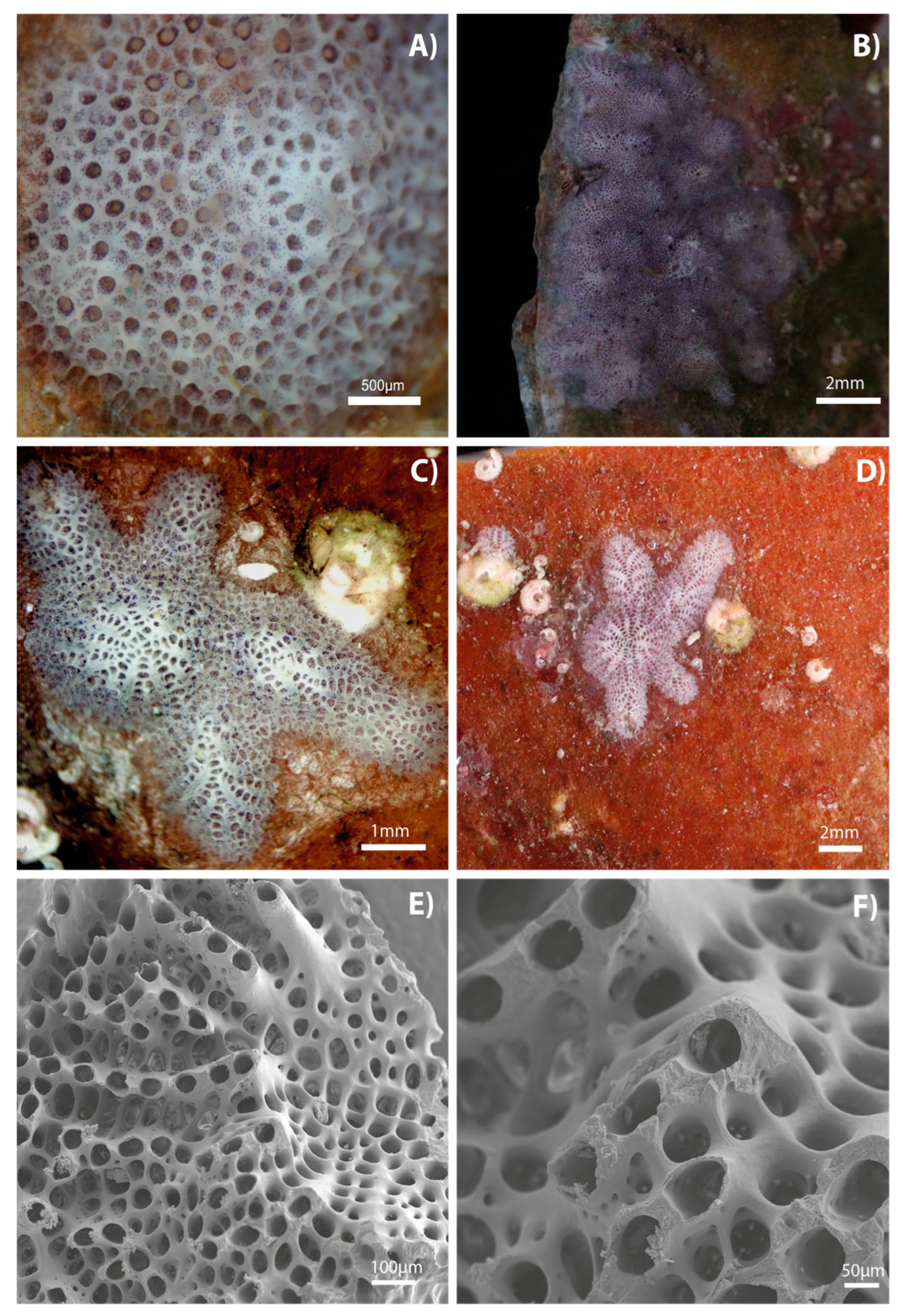
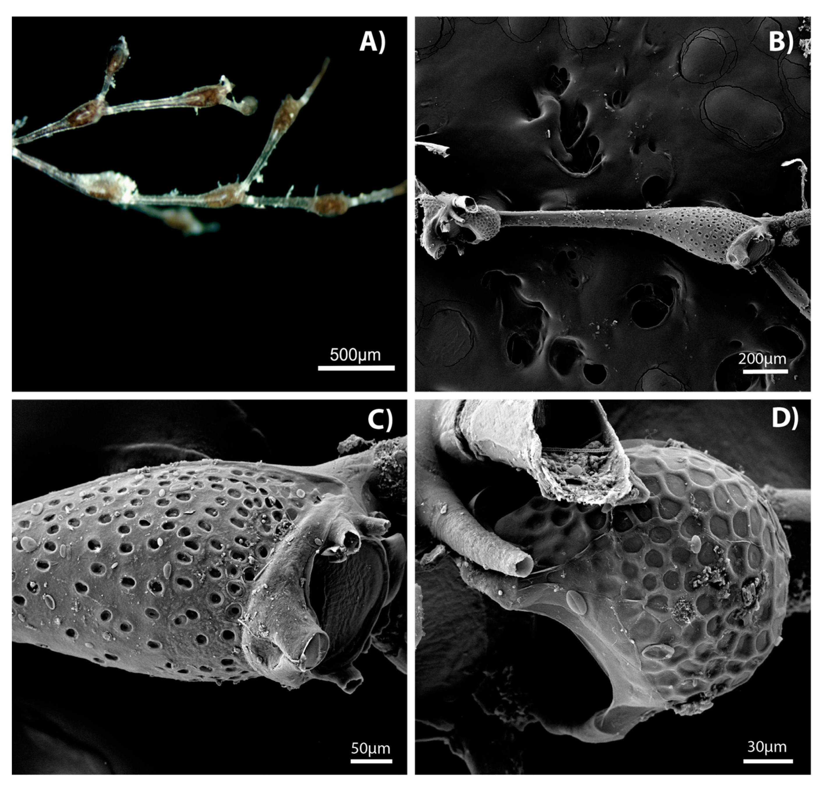
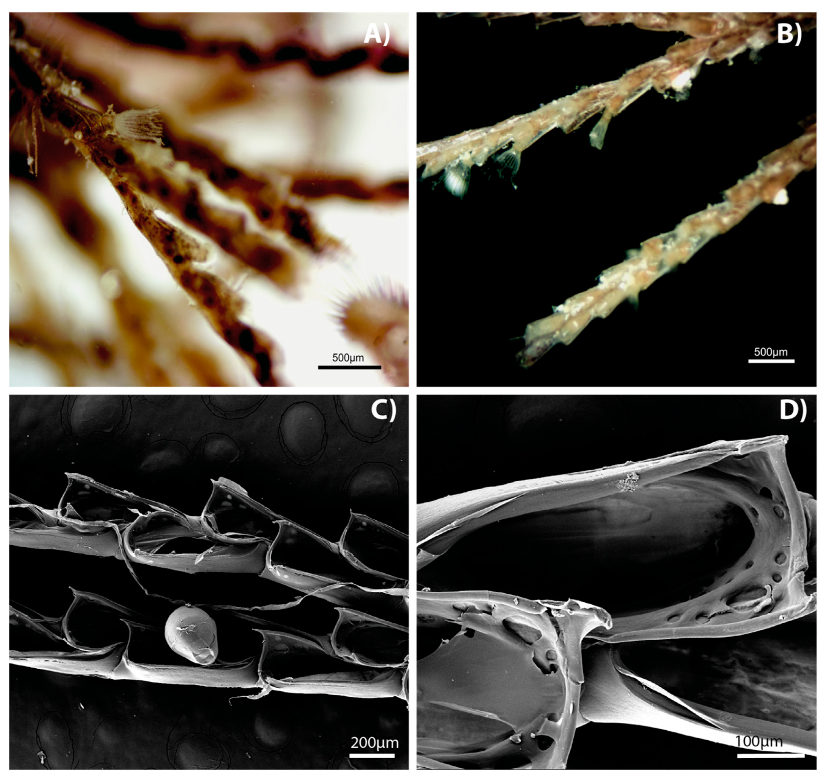
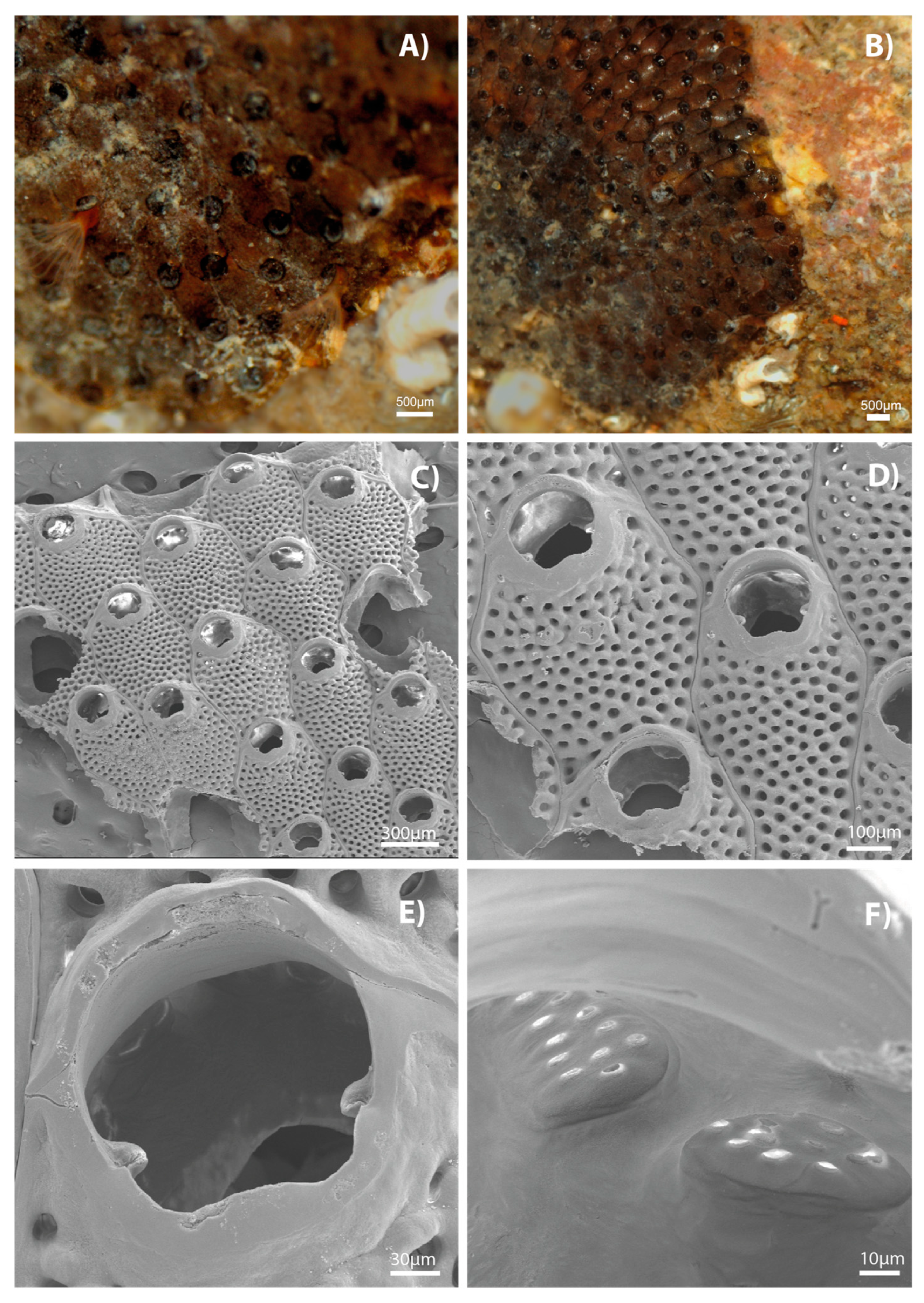
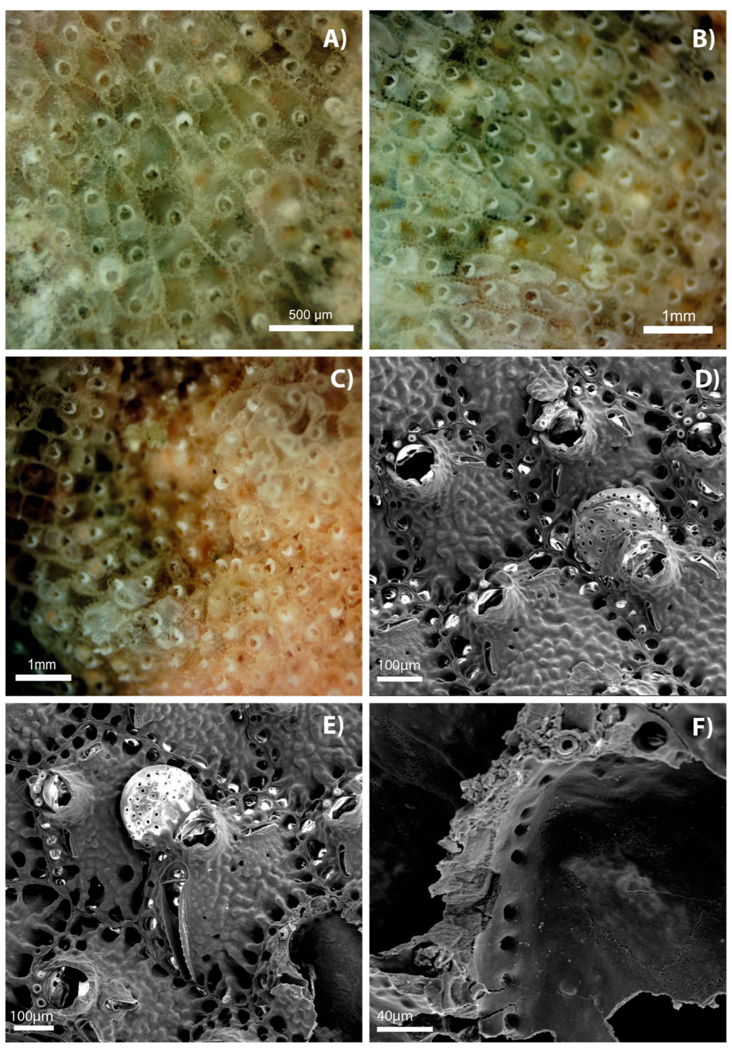
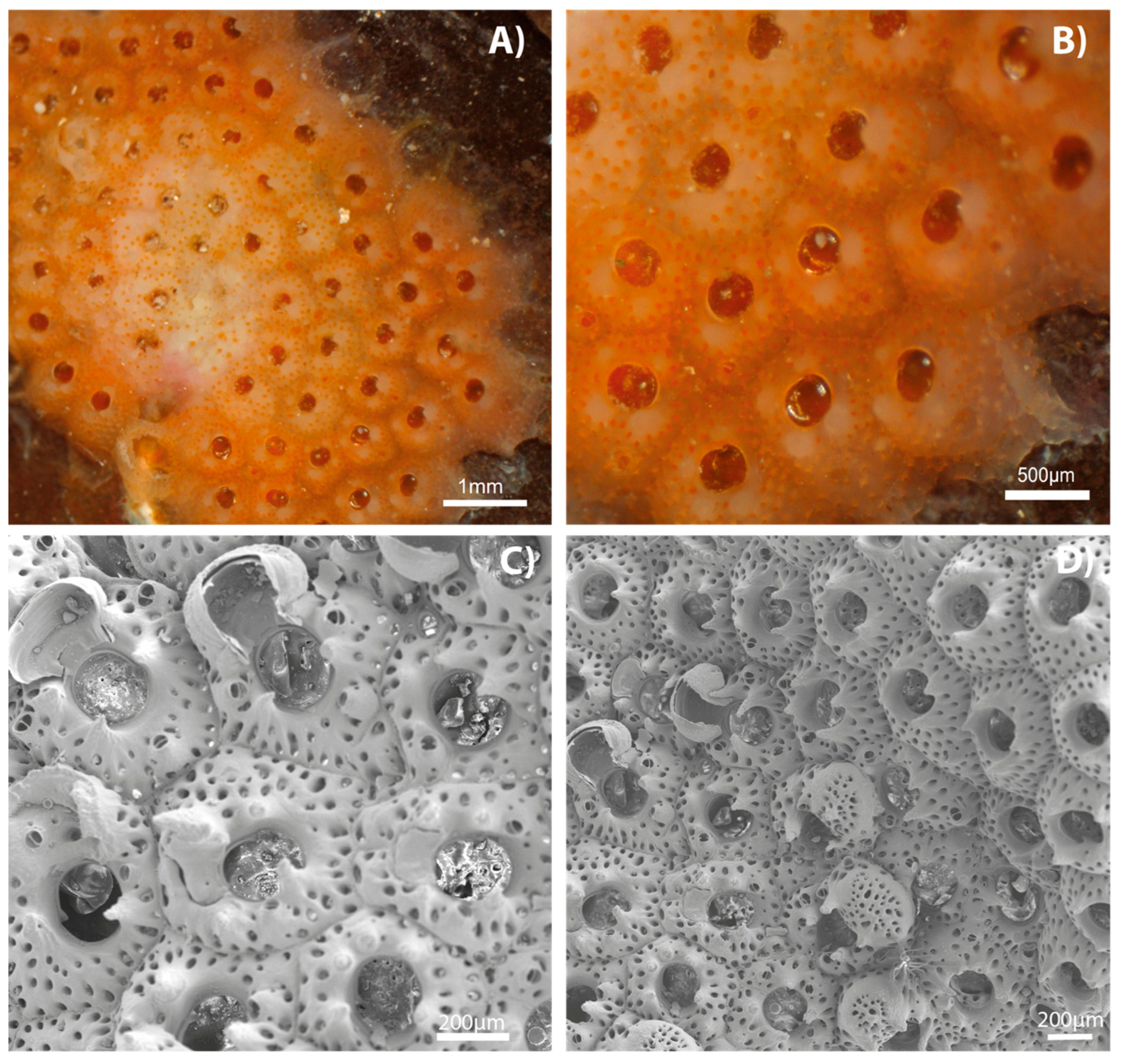
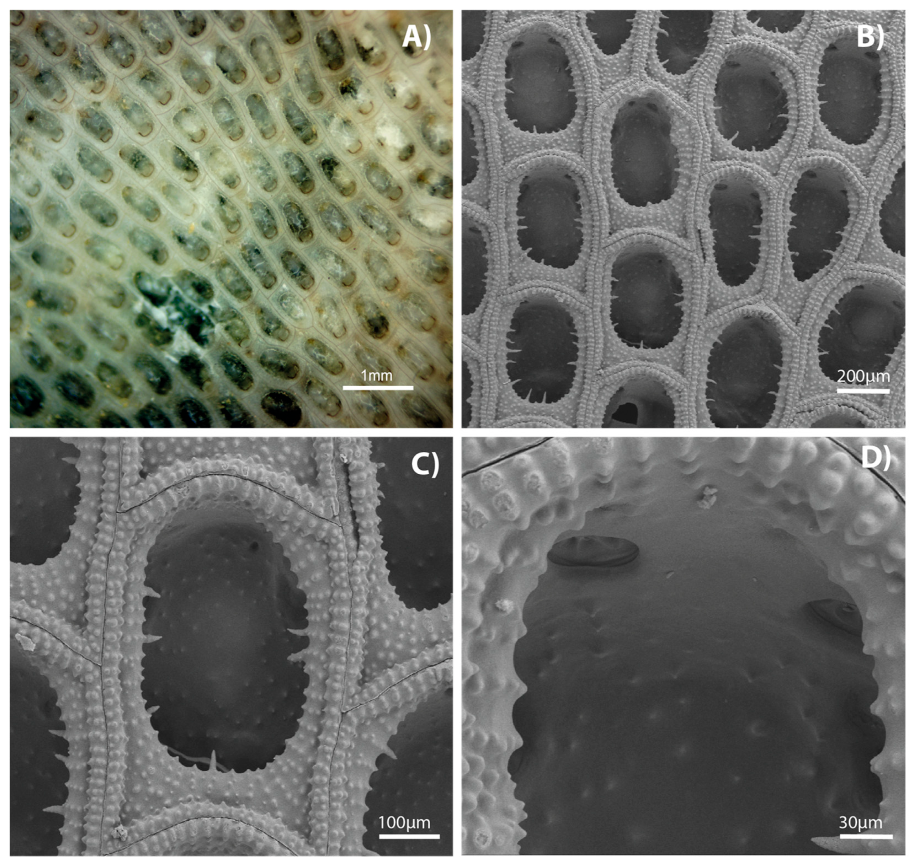
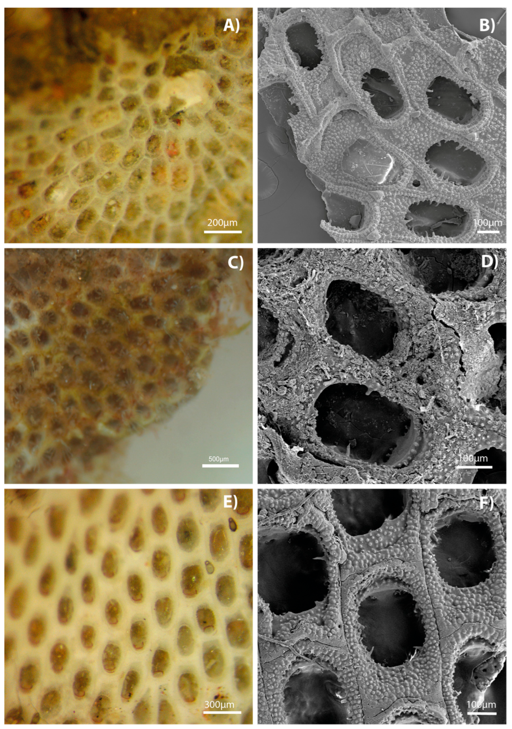
Tissue was scraped from preserved colony fragments for DNA extraction. Samples were stored in 95% ethanol in 95-well lysis plates and kept refrigerated until DNA barcoding was performed. Amplification targeted the mitochondrial cytochrome c oxidase I (COI) gene using primers C_LepFolF (5′-ATTCAACCAATCATAAAGATATTGG-3′) and C_LepFolR (3′-TAAACTTCTGGATGTCCAAAAAATCA-5′), following Folmer et al. [31] and the protocols of Ivanova et al. [32]. Barcoding analysis was conducted at the Centre for Biodiversity Genomics, University of Guelph, Canada.
The resulting nucleotide sequences were aligned using the MUSCLE algorithm (See Appendix A), and specimens were clustered using the Neighbor-Joining method in the BOLD system [33]. Sequence identities were confirmed by comparison against the BOLD Identification Engine [34] and GenBank via BLAST [35] (see Table 1). A Neighbor-Joining tree based on COI sequences was generated to visualize the genetic similarity among the bryozoan specimens collected in this study. However, no phylogenetic hypotheses were proposed based on this tree (See the end of Section 3 for details).

3. Results

SPECIES DESCRIPTIONS:
Order Cyclostomatida Busk, 1852
Family Lichenoporidae Smitt, 1867
Genus Disporella Gray, 1848
Disporella sp.
Collection data:
MZUCR-00068: Isla David (10.95790° N, 85.72384° W at 9 m depth). One colony, 3 August 2022. GenBank accession: PV053968. MZUCR-00087: Pitahaya (10.93759° N, 85.80171° W at 3.5–5.5 m depth). One colony, 29 June 2023. GenBank accession: PV010184. MZUCR-00217: Playa Pochote (10.94595° N, 85.81834° W at 8 m depth). One colony, 24 August 2023. GenBank accession: PV010183.
Description:
This colony is adnate, calcified and with a granulated irregularly mounded surface. Centers of the mounds have irregular brood chambers. When alive, it is a light purple color (Figure 1A–D), but pigmentation is lost when preserved [36]. Kenozoidal cavities contain palm-shaped-pinhead spinules (Figure 1F). The openings of the zoecia are subcircular and vary in different areas of the colony, which grows columns of fused peristomes (Figure 1E) with four or more autozooids [37].
Habitat: These colonies were found growing on shells and encrusting under rocks in the subtidal zone.
Distribution: This genus is globally distributed, with species present in tropical regions, polar regions and all oceans.
Figure 1. Disporella sp., live colonies of (A) MZUCR-00068, (B) MZUCR-00087, and (C) MZUCR-00217, and a close up of the colony (D) MZUCR-00217 at lower magnification showing it encrusting a shell and surrounded by other sessile animals (Vermetidae). SEM images of (E) a fraction of a representative colony and (F) details of the zoecia where small palm-shaped-pinhead spinules can be seen.
Diversity 17 00451 g001
Order Cheilostomatida Busk, 1852
Family Savignyellidae Levinsen, 1909
Genus Savignyella Levinsen, 1909
Savignyella lafontii (Audouin, 1826)
Collection data:
MZUCR-00188: Casa Verde/Bajo Chaca (10.98235° N, 85.70425° W, 6 to 4 m depth). One colony, 7 July 2023. GenBank accession: PV010181.
Description:
This colony belongs to a family of delicate erect bryozoans. They grow in uniserial chains of club shaped zooids (Figure 2A–D), separated by small joints [38]. Each zooid presents a pseudoporous frontal shield covered by a membranous outer layer (Figure 2C), giving them a porous appearance. The genus Savignyella is characterized for having non-articulated oral spines (Figure 2C), a suboral avicularium, and an evenly porous ovicell (Figure 2D).
Habitat: The colony was found growing on Padina spp. algae in shallow waters.
Distribution: The genus is widely distributed in tropical and subtropical zones globally. In some areas, they are invasive species introduced by vessels.
Figure 2. Savignyella lafontii, (A) live colonies of MZUCR-00188. SEM images taken by Fiorella Vargas of (B) a single zooid, (C) detail of the orifice with suboral avicularium, and (D) close up of the zooid showing an ovicell.
Diversity 17 00451 g002
Order Cheilostomatida Busk, 1852
Family Bugulidae Gray, 1848
Genus Bugula Oken, 1815
Bugula neritina (Linnaeus, 1758)
Collection data:
MZUCR-00179: Casa Verde/Bajo Chacas (10.98235° N, 85.70425° W at 6 to 4 m depth). One colony, 7 July 2023. GenBank accession: PV010190.
Description:
This species is erect and branching, with zooids opening on one side of the branches (Figure 3A,B). The zooids are lightly calcified, so the colony stays flexible and therefore thin-walled (Figure 3C,D). Bugula neritina is the type species of the genus, although it does not present the characteristic traits of its genus such as marginal spines and avicularia shaped like a bird’s head [38]. This species of bugulid lives in shallow waters and presents a rich burgundy color that tends to be retained after preservation in ethanol.
Habitat: This colony was found growing on algae in shallow waters (subtidal, 6 to 4 m).
Distribution: Gulf of Mexico, South Atlantic Ocean, coast of Brazil, North Pacific Ocean, South Pacific Ocean, Indian Ocean, Arabian Sea, and South Atlantic Ocean.
Figure 3. Bugula neritina, MZUCR-00188 (A,B) live colony. SEM images of (C) zooids with an ovicell present, and (D) detail of the zoecia.
Diversity 17 00451 g003
Order Cheilostomatida Busk, 1852
Family Watersiporidae Vigneaux, 1949
Genus Watersipora Neviani, 1896
Watersipora arcuata Banta, 1969
Collection data:
MZUCR-00059: Junquillal, Guanacaste, (10.97871° N, 85.89067° W, intertidal). One colony, 3 August 2022. GenBank accession: PV010188.
Description:
The species of this genus tend to have few to no distinctive features (no avicularium, oral spines, or ooecia). They form encrusting colonies with black or grey zooids and brick red borders and red polypides (Figure 4A,B). Autozooids are monomorphic (Figure 4C,D) and possess a concave border in the operculum (Figure 4E) and its opening [38,39]. Colonies reported from W. arcuata are usually large (up to 20 cm), unlike the one collected by us (2 to 3 cm).
Habitat: The colony was encrusting under a rock surface in the lower intertidal zone.
Distribution: North Pacific Ocean, South Pacific Ocean, and east coast of Australia.
Figure 4. Watersipora arcuata, (A) live colony showing red polypide, (B) live colony. SEM images of (C) multiple monomorphic zoecia, (D) close up of zoecia, (E) detail of the opesia, and (F) pore plates.
Diversity 17 00451 g004
Order Cheilostomatida Busk, 1852
Family Smittinidae Levinsen, 1909
Genus Parasmittina Osburn, 1952
Parasmittina crosslandi (Hastings, 1930)
Collection data:
MZUCR-00177: Isla Loros, lado Refugio (11.00261° N, 85.74739° W at 5 to 13 m depth). One colony, 7 July 2023. GenBank accession: PV010182. MZUCR-00227 and MZUCR-00233: Las Gemelas (10.93945° N, 85.78556° W at 10 m depth). Two colonies, 24 August 2023. GenBank accession: PV010183 and PV072687, respectively.
Description:
Colonies are adnate and unilaminar, although multilaminar colonies were observed by Banta & Carson [13]. Zooids are rectangular to pentagonal and follow a pentagonal array that may be disrupted due to irregularities in the substrate (Figure 5C). The frontal wall is thick and transparent (Figure 5A–C), with slight bumps or tubercles. Young zooids form spines that are lost early, leaving spine bases in the distal side of the orifice in older zooids. Areolae are present on all zooid edges but the distal side. The orifice is subcircular with a bordering peristome that is absent distally and forming a pseudosinus proximally. Adventitious avicularia are varied in size, usually around 70 µm or 200 µm (Figure 5D,E), with usually one or two per zooid, placed proximolaterally and with pointed mandibles [13]. The ovicells are raised and globose with pores scattered across its surface [37].
Habitat: These colonies were found encrusting under a rock growing on a pebble and encrusting a dead coral in shallow waters.
Distribution: Eastern Pacific, from the Gulf of California to Colombia.
Figure 5. Parasmittina crosslandi, live colonies of (A) MZUCR-00177, (B) MZUCR-00227, and (C) MZUCR-00233. SEM images of MZUCR-00177: (D) zoecia with porous ovicell and small adventitious avicularium, (E) zoecia with large adventitious avicularium and ovicell, and (F) septular pores.
Diversity 17 00451 g005
Order Cheilostomatida Busk, 1852
Family Colatooeciidae Winston, 2005
Genus Cigclisula Canu & Bassler, 1927
Cigclisula sp.
Collection data:
MZUCR-00085: Isla David (10.95751° N, 87.72021° W at 4 m depth). One specimen, 3 August 2022. GenBank accession: PV010193.
Description:
Colonies in this genus are unlaminar; zooids have a bright orange color (Figure 6A,B) that gets lost when they get preserved. The base of the zooids is hexagonal (Figure 6C). The walls are thick and opaque with a frontal aperture that is slightly oval. Each zooid presents pores near the junctures with other zooids. Adventitious avicularia can be present near the aperture (Figure 6C). Ovicells are prominent, smooth on its lateral sides, and are pierced by a band of pores (Figure 6C,D) [13,38].
Habitat: The colony was found encrusting on a mollusk shell in shallow waters.
Distribution: Gulf of Mexico, coastline of Australia, Indo-Pacific, and coast of Brazil.
Figure 6. Cigclisula sp., live colony of (A,B) MZUCR-00085. SEM images of a representative specimen, (C) detail of a few zooids with broken ovicells and small adventitious avicularia, and (D) zooids with intact porous ovicells visible.
Diversity 17 00451 g006
Order Cheilostomatida Busk, 1852
Family Onychocellidae Jullien, 1882
Genus Smittipora Jullien, 1882
Smittipora levinseni (Canu & Bassler, 1917)
Collection data:
MZUCR-00080: Isla David (10.95751° N, 87.72021° W at 4 m depth). One colony, 3 August 2022. GenBank accession: PV010191.
Description:
Adnate colony, uni- or multilaminar, light orange with dark brown mandibles (Figure 7A). Spines and zooidal gymnocysts absent. Autozooids are hexagonal, longer than wide, and irregularly arranged (Figure 7B). Bell shaped opesia with a straight proximal edge. Granulated cryptocysts are short and immediately slope downwards (Figure 7D). Small bumps give it an evenly granular texture with smaller and denser granules closer to the zoecial boundary. Vicarious avicularia are penta- or hexagonal, distally rounded, with a key-hole-shaped opesia that is also distally rounded and with serrated margins (Figure 7C). Mandibular abductor muscles leave two deep notches close to the mural rim. Multiporous septula on each wall for zooidal communication. Ovicells are inconspicuous; zooids with ovicells lack cryptocyst [40].
Habitat: The colony was found encrusting under a rock in shallow waters.
Distribution: This species is present in the Atlantic Ocean.
Figure 7. Smittipora levinseni, live colony of (A) MZUCR-00080 with detail of heterozooids and vicarious avicularium. SEM images of (B) heteromorphic zooids, (C) detail of vicarious avicularium with visible notches indicating mandibular muscle scarring, and (D) pore plates and cryptocyst.
Diversity 17 00451 g007
Order Cheilostomatida Busk, 1852
Family Membraniporidae Busk, 1852
Genus Biflustra d’Orbigny, 1852
Biflustra tenuis (Desor, 1848)
Collection data:
MZUCR-00200: Isla Muñeco (10.97939° N, 85.71512° W at 4 to 5 m depth). One colony, 7 July 2023. GenBank accession: PV010192.
Description:
Colony is adnate, unilaminar, white to transparent when alive and after preservation (Figure 8A). Autozooids are monomorph (Figure 8B) [38]. The opesia is oval and occupies most of the frontal wall. Small denticles protrude from the edge of the opesia on the proximolateral sides (Figure 8C). Mural rim is elevated, well developed, and crenulated [41]. Frontal wall and mural rim with small tubercles arranged in concentric rows (Figure 8C).
Habitat: The colony was found growing on an empty shell in shallow waters.
Distribution: North Sea, North Atlantic Ocean, Gulf of Mexico, and Pacific coast of Panama.
Figure 8. Biflustra tenuis, live colony of (A) MZUCR-00200. SEM images of (B) monomorph autozooids, (C) detail of zoecia with spines present on the opesia’s rim, and (D) inside of zooid showing pores.
Diversity 17 00451 g008
Order Cheilostomatida Busk, 1852
Family Membraniporidae Busk, 1852
Genus Biflustra d’Orbigny, 1852
Biflustra sp. (Desor, 1848)
Collection data:
MZUCR-00048 and MZUCR-00052: Junquillal, intertidal zone (10.97871° N, 85.89067° W). Two colonies, 3 August 2022. GenBank accession: PV010187 and PV010185, respectively. MZUCR-00090: Pitahaya (10.93759° N, 85.80171° W at 3.5 to 5.5 m depth). One colony specimen, 29 June 2023. GenBank accession: PV010189.
Description:
The genus Biflustra is contained in the family Membraniporidae, where all genera have simple autozooids with a wide opesia. The three colonies we observed were adnate and heavily calcified (Figure 9). The zooids were mostly rectangular with a round distal edge. Frontal wall is coarsely granular, as well as the small cryptocyst that goes downward almost instantly. Opesia is generally oval, with spinal processes on the rim and heavily calcified gymnocyst. Ovicells are absent; these colonies tend to develop cyphonautes larvae. Distal wall with two pore plates containing multiple pores and spinal processes [38].
Habitat: These colonies were found encrusting in intertidal rocks and growing on a shell in shallow waters.
Distribution: Gulf of Mexico, Sargasso Sea, coast of Brazil, North Sea, Indo-Pacific, and Eastern Pacific.
Figure 9. Biflustra sp. (A) MZUCR-00048, live colony and (B) SEM detail. (C) MZUCR-00052, live colony and (D) SEM detail. (E) MZUCR-00090, live colony and (F) SEM detail.
Diversity 17 00451 g009
Three well-supported clusters (bootstrap > 900) were identified, corresponding to the genera Disporella, Biflustra, and Parasmittina (Figure 10). The sequences of Biflustra sp. showed closest affinity to Biflustra tenuis. The remaining individual sequences were genetically distant from each other, supporting their identification as distinct species (Figure 10; Table 1).
Figure 10. (A) Taxon ID tree obtained from the BOLD engine for COI marker sequence similarity between the Bryozoa specimens in the present study. Distance 725 model = Jukes Cantor. Alignment = BOLD. (B) Taxon ID tree generated with the same sequences by Maximum likelihood (ML), including a Phoronid (accession number MK028621.1) as an external group, to obtain the bootstrap values out of 1000 iterations.
Diversity 17 00451 g010

4. Discussion

We identified nine species of bryozoans in the present study through a combination of genetic and morphological analyses. Two taxa—Disporella sp. and P. crosslandi—each formed a distinct cluster with three COI sequences in the genetic tree generated through the BOLD identification engine (Figure 10A). The match percentages for these sequences in both BOLD and BLAST were too low to confidently assign them to any known species (Table 1). Therefore, we referred to published SEM images to evaluate morphological similarities with known species. For Disporella sp., comparisons were made with descriptions provided by Dick et al. [37] and Taylor & Grischenko [36]; and for P. crosslandi, we consulted images from the Smithsonian Tropical Research Institute [42] and Soule & Soule [43]. This study presents the first published COI sequences for both Disporella sp. and P. crosslandi.
Although BOLD only confirmed the genus Parasmittina as the best match, we assigned the specimens to P. crosslandi based on morphological similarities and known distribution. This species has been previously reported in the Eastern Tropical Pacific and is included in the records and SEM imagery of the Smithsonian Tropical Research Institute [42], which supported our classification. Although P. crosslandi was also reported by Banta & Carson [13] in Playas del Coco, Costa Rica, their SEM images differ notably from our specimens and those in other reliable sources, suggesting that their material may belong to a different species.
As for Disporella sp., we propose that it may belong to one of several Eastern Pacific species, such as D. astraea, D. compta, D. densiporoides, D. julesi, D. minima, D. ovoidea, D. separata, or D. violacea. Osburn [19] documented one species, D. californica, in Santa Elena Bay (then referred to as Port Parker), but his description lacked sufficient detail, particularly in terms of zooid morphology and colony size, and did not match our specimens or those of contemporary researchers. Although our collection sites are geographically close to Osburn’s, all located within the ACG, the lack of definitive morphological or genetic evidence prevents a conclusive identification, and we therefore maintain this taxon as undetermined.
For the three Biflustra sp. specimens, the taxonomic decision was more complex due to the debated status of the group. While BOLD identified the specimens as belonging to the genus Membranipora sp., with matches above 90% to a specimen from Los Angeles, California (Table 1), our morphological analysis aligned them more closely with B. tenuis, as illustrated by the Smithsonian Tropical Research Institute [42]. This contrasts with the Membranipora sp. species described in Cook et al. [38]. Notably, Membranipora sp. has a wide, simple opesia and a small gymnocyst, whereas Biflustra sp. features a smaller opesia bordered by a granulated, calcified frontal wall and a more developed gymnocyst [38]. All three specimens shared these latter characteristics (Figure 9), strongly supporting their assignment to Biflustra sp. Moreover, the public BOLD record for Membranipora sp. lacked adequate photographic documentation, further undermining that identification. We believe the Los Angeles specimen was misidentified and is also likely a Biflustra sp. specimen.
One colony matched O. marioni Jullien, 1882, in BOLD, with a low similarity (84.33%) (Table 1). However, SEM analysis showed strong morphological similarity to S. levinseni, as described by Fransen [40] and the Smithsonian Tropical Research Institute [42]. Since S. levinseni had not previously been recorded in the BOLD database, this constitutes a new entry in the system.
For W. arcuata, B. neritina, and S. lafontii, we accepted the original identifications provided by BOLD, as these sequences had high match percentages (>90%) and morphological features consistent with published descriptions [38,39,42]. Cigclisula sp., although matched at a lower percentage (81.76%), was also retained based on comparison with SEM images and known distributions.
The documented capacity for dispersion and colonization by W. arcuata in other regions [44,45] raises the possibility that previously unrecorded species are now established in Costa Rica. While we cannot confirm an invasion due to the lack of long-term historical data, it remains plausible that bryozoans from distant regions may reach and colonize the ACG through various dispersal mechanisms, including biofouling and rafting on floating debris.

5. Conclusions

Five of the nine bryozoan species identified in this study represent new records for Costa Rica: S. lafontii, B. neritina, W. arcuata, S. levinseni, and B. tenuis. These species had not been previously reported by Osburn [17,18,19] or Banta & Carson [13]. Within the Área de Conservación Guanacaste (ACG), we documented seven new records, which include the five species mentioned above, along with P. crosslandi and Cigclisula sp. Of these, three species—S. lafontii, P. crosslandi, and S. levinseni—also represent new genetic sequence entries in public databases. Notably, S. lafontii and S. levinseni are new records for the Eastern Pacific.
This study aims to reinvigorate the study of bryozoans in Costa Rica and provides a foundational reference for future research on the phylum. Our documentation includes both live and SEM images of the specimens, as well as verified records based on morphological and molecular data. We hope this work encourages continued exploration and more comprehensive studies of bryozoans in the Eastern Pacific and the Central American region.

Author Contributions

Conceptualization, J.A.S.-C.; methodology, J.A.S.-C. and J.C.; validation, J.A.S.-C. and J.C.; formal analysis, J.A.S.-C. and B.A.-O.; investigation, B.A.-O. and J.A.S.-C.; resources, J.A.S.-C. and J.C.; data curation, B.A.-O. and J.A.S.-C.; writing—original draft preparation, B.A.-O. and J.A.S.-C.; writing—review and editing, J.A.S.-C. and J.C.; visualization, B.A.-O.; supervision, J.A.S.-C.; project administration, J.A.S.-C. and J.C.; funding acquisition, J.A.S.-C. and J.C. All authors have read and agreed to the published version of the manuscript.

Funding

Travel expenses for our investigation were covered by Project B9508 “Marine biodiversity of Guanacaste Conservation Area, Costa Rica” project BioMar-ACG, a cooperative project between Universidad de Costa Rica (UCR) and the Sistema Nacional de Áreas de Conservación (SINAC) R-CONV-004-2019. Terrestrial transportation was funded by the Centro de Investigación en Ciencias del Mar y Limnología (CIMAR). Aquatic transportation, diving activities, and scanning electron microscopy images were funded by the Guanacaste Dry Forest Conservation Fund. The study is part of the BioMarACG-project B9508 of Universidad de Costa Rica (UCR). We thank Alejandro Masis and Roger Blanco for the collection permission from the Ministry of Environment and Energy of Costa Rica (MINAE): R-SINAC-ACG-PI-026-2020/038-2021/051-2022/045-2023. The access to genetic material is permitted by resolution-047/CBio-68-2022 of the Comisión Institucional de Biodiversidad (UCR).

Institutional Review Board Statement

Not applicable.

Data Availability Statement

The original contributions presented in this study are included in the article. Further inquiries can be directed to the corresponding author.

Acknowledgments

We thank Frank Joyce for his support on this project. We also want to thank parataxonomists Yelba Vega and Gilberth Ampié, who collaborated with us and were involved in the specimen collection in the field and tissue extraction in the field laboratory. Without their efforts and involvement, this publication would not exist. All specimen collections were carried out by Beatriz Antillón-Obando, Gilberth Ampié, Yelba Vega, Anna Leal-Quirós, Priscilla Monge, and Jeffrey Sibaja-Cordero.

Conflicts of Interest

The authors declare no conflicts of interest. The funders had no role in the design of the study; in the collection, analyses, or interpretation of data; in the writing of the manuscript; or in the decision to publish the results.

Abbreviations

The following abbreviations are used in this manuscript:
SEMScanning electron microscope
ACGÁrea de Conservación Guanacaste

Appendix A. Sequences Generated

Order Cyclostomatida Busk, 1852
Family Lichenoporidae Smitt, 1867
Genus Disporella Gray, 1848
Disporella sp.
MZUCR-00068, BOLD data BMAR3600-24, BIN: AFW4124
GenBank accession: PV053968
Sequence:
GAATTGAACTTTAGTTCTCCTGGATCTTTTTTAATAAATGATCAATTATATAATAGGATTGTCACTGGACATGCTATAGTTATAATTTTCATATTTGTTATACCAGTAATAATTGGAGGTTTCGGAAATTGGCTAATTCCCTTAAAATTAGGAATGCCAGATATGCTTTTCCCTCGACTTAACAATTTAAGATTCTGACTTTTAATACCATCAATAGCATTATTAGTTTTTTCCCTAATTGTGGAAGCTGGATGTGGGACAGGGTGAACAATGTATCCTCCCTTATCAGATTGTATTGCTCATAGAGGTAAAAGAGTGGATTTAGTTATTTTTTCCCTACATCTTGCAGGTGCTTCATCTATTTTAAGAAGATTAAATTTTATTTCTACTGTGNTAATTGCCCTATTAGCTAAAAAATCACCCGATACATTAATACTATTTGTATGGACAATTTTAATTACAGTTATTTTACTACTTTTATCCCTTCCTGTACTAGCTGGGGGTATTACAATGTTATTAAGAGATCGAAATTTAAACACATCATTTTT
MZUCR-00087, BOLD data BMAR3620-24, BIN: AFW4124
GenBank accession: PV010184
Sequence:
AACTATTTATTTACTATTTGGAGTATGAAGTGGCTTAGTAGGAACATCTATGTCAGGTTTAATTCGAATTGAACTTAGTTCTCCTGGATCTTTTTTAATAAATGATCAATTATATAATAGGATTGTCACTGGACATGCTATAGTTATAATTTTCATATTTGTTATACCAGTAATAATTGGAGGTTTCGGAAATTGGTTAATTCCCTTAAAATTAGGAATGCCAGATATGCTTTTCCCTCGACTTAACAATTTAAGATTCTGACTTTTAATACCATCAATAGCATTATTAGTTTTTTCCCTAATTGTGGAAGCTGGATGTGGGACAGGGTGAACAATGTATCCTCCCTTATCAGATTGTATTGCTCATAGAGGTAAAAGAGTGGATTTAGTTATTTTTTCCCTACATCTTGCAGGTGCTTCATCTATTTTAAGAAGATTAAATTTTATTTCTACTGTGATAATTGCCCTATTAGCTAAAAAATCACCCGATACATTAATACTATTTGTATGGACAATTTTAATTACAGTTATTTTACTACTTTTATCCCTTCCTGTACTAGCTGGGGGTATTACAATGTTA
MZUCR-00217, BOLD data BMAR3845-24, BIN: AFW4124
Genbank accession: PV010183
Sequence:
TTAATTCGAATTGAACTTAGTTCTCCTGGATCTTTTTTAATAAATGATCAATTATATAATAGGATTGTCACTGGACATGCTATAGTTATAATTTTCATATTTGTTATACCAGTAATAATTGGAGGTTTCGGAAATTGGTTAATTCCCTTAAAATTAGGAATGCCAGATATGCTTTTCCCTCGACTTAACAATTTAAGATTCTGACTTTTAATACCATCAATAGCATTATTAGTTTTTTCCCTAATTGTGGAAGCTGGATGTGGGACAGGGTGAACAATGTATCCTCCCTTATCAGATTGTATTGCTCATAGAGGTAAAAGAGTGGATTTAGTTATTTTTTCCCTACATCTTGCAGGTGCTTCATCTATTTTAAGAAGATTAAATTTTATTTCTACTGTGATAATTGCCCTATTAGCTAAAAAATCACCCGATACATTAATACTATTTGTATGGACAATTTTAATTACAGTTATTTTACTACTTTTATCCCTTCCTGTACTAGCTGGGGGTATTACAATGTTATTAAGAGATCGAAATTTAAACACAT
Order Cheilostomatida Busk, 1852
Family Savignyellidae Levinsen, 1909
Genus Savignyella Levinsen, 1909
Savignyella lafontii (Audouin, 1826)
MZUCR-00188, BOLD data BMAR3816-24, BIN: ADJ3487
Genbank accession: PV010181
Sequence:
AACTATATAATGTAATTGTTACGGCTCATGCTTTCTTAATAATTTTCTTTATAGTAATACCTATTATAATTGGAGGATTTGGAAACTGACTAGTACCACTAATACTAGGAGTACCAGATATAGCATTCCCTCGACTAAACAACATAAGATTTTGATTACTACCTCCAGCAATCTTTCTGCTACTTATATCATCAATAGTAGAAAGAGGAGCAGGAACCGGTTGAACTGTATACCCTCCTTTATCATCAAACATTGCGCATAGAGGACCCTCAGTAGACCTAGCCATTTTCTCACTTCATTTAGCAGGTGTGTCTTCAATTCTAGGAGCTATTAATTTTATAACAACAATTATCAATATACGAAGAAAGCTAATAACATTCATACGAATTACACTTTTAACTTGAGCAGTATTTATTACAGCAATTCTTCTACTTCTCTCACTTCCCGTGCTAGCTGGAGCCATTACTATACTACTAACAGATCGAAACATTAATTCAT
Order Cheilostomatida Busk, 1852
Family Bugulidae Gray, 1848
Genus Bugula Oken, 1815
Bugula neritina (Linnaeus, 1758)
MZUCR-00179, BOLD data. BMAR3807-24, BIN: MN064606
Genbank accession: PV010190
Sequence:
CGACCAACTATATAATGAAATTGTCACAGCTCATGCATTTTTAATAATTTTTTTTATAGTAATACCAGTTATAATCGGGGGGTTTGGCAATTGATTGGTTCCATTAATACTGGGGGCCCCTGATATAGCATTTCCCCGCCTAAACAACATAAGATTTTGGCTTCTTCCCCCGGCATTATTACTTTTATTAATATCTTCTTTAGTAGAAAGAGGAGCTGGAACTGGATGGACCGTATACCCCCCCTTATCATCAGGATTAGGTCACAGAGGAGCAGCCGTAGATCTGGCCATTTTCTCTTTACACCTTGCCGGTGTATCTTCAATTTTAGGAGCAATTAACTTTATTACTACAACAATTAATATACGAAGAAACTCTATAAAAATAATTCAAATCCCTTTATTTATTTGGGCAGTTTTCATTACAGCCATTTTGTTGCTTTTAT
Order Cheilostomatida Busk, 1852
Family Watersiporidae Vigneaux, 1949
Genus Watersipora Neviani, 1896
Watersipora arcuata Banta, 1969
MZUCR-00059, BOLD data. BMAR3591-24, BIN: AFW8494
Genbank accession: PV010188
Sequence:
AACATTATATTTTATATTTGGATTATGATCAGCTATAACCGGTAGAGGATTGAGAATAGCTATCCGATCGGAATTAGGAAATCCAGGAAGATTAATAGGAAATGACCAAATTTACAACGTAGTAGTAACAGCTCACGCATTTTTCATGATTTTTTTTATAGTAATACCAATCATAATTGGGGGATTTGGAAACTGGTTAGTTCCTCTTATAATGGGAATCCCAGATATAGCATTCCCACGAATAAATAACATAAGATTTTGATTACTACCACCTGCAGCTATTTTACTAATAATATCAGCACTAGTAGAAAGAGGTGCAGGGACAGGGTGAACAGTATACCCTCCTTTGTCTTCAAACATTTCTCATAGAGGATCATCAGTAGATATAGCAATTTTCGCTCTTCACTTAGCAGGAGTCTCTTCAATTTTAGGAGCAATTAACTTTATAGTAACAATTATTAATATACGAGGAAAAATAATAGCATTCATAAACTTACCATTATTTGTATGAGCCGTATTCATCACTGCCCTCTTATTATTACTATCTCTCCCAGTTCTAGCAGGGGCTATTACTATACTACTAACAGACCGTAATTTTAACTCATCATTCTTTGACCCAGCT
Order Cheilostomatida Busk, 1852
Family Smittinidae Levinsen, 1909
Genus Parasmittina Osburn, 1952
Parasmittina crosslandi (Hastings, 1930)
MZUCR-00177, BOLD data. BMAR3805-24, BIN: AFW0950
Genbank accession: PV010182
Sequence:
AGAACTTGGACAGCCTGGGAGACTTATAGGAAATGATCAGCTTTACAATGTTATTGTTACAGCCCACGCATTCCTAATAATTTTCTTTATAGTTATGCCTATTATAATCGGAGGATTTGGGAATTGATTAGTTCCTTTAATATTAGGGGTCCCAGATATAGCTTTCCCACGACTAAATAATATAAGATTTTGGTTACTACCCCCTGCTATTATGTTACTACTTATATCATCGATAGTTGAAAGAGGAGCAGGTACAGGATGGACAGTGTACCCTCCCTTGTCATCCAACATCTCCCATAGAGGACCTTCAGTAGATTTAGCAATTTTTTCACTACATCTAGCAGGAGTCTCCTCTATCTTAGGAGCTATTAATTTCATAACGACAGTAATCAATATACGAAGAGATAAGATGACCTTTATACATATCACTTTAATAACATGGGCCGTCTTCATTACAGCACTCTTATTACTACTGTCGTTACCAGTATTAGCTGGTGCTATTACAATATTACTAACTGACCGTAATATTAATACTT
MZUCR-00227, BOLD data. BMAR3855-24, BIN: AFW0950
Genbank accession: PV010183
Sequence:
TGCACTAATTCGAGTAGAACTTGGACAGCCTGGGAGACTTATAGGAAATGATCAGCTTTACAATGTTATTGTTACAGCCCACGCATTCCTAATAATTTTCTTTATAGTTATGCCTATTATAATCGGAGGATTTGGGAATTGATTAGTTCCTTTAATATTAGGGGTCCCAGATATAGCTTTCCCACGACTAAATAATATAAGATTTTGGTTACTACCCCCTGCTATTATATTACTACTTATATCATCGATGGTTGAAAGAGGAGCAGGTACAGGATGGACAGTGTACCCTCCATTGTCATCCAACATCTCCCATAGAGGACCTTCAGTAGATTTAGCAATTTTTTCACTACATCTAGCAGGAGTCTCCTCTATCTTAGGAGCTATTAATTTCATAACGACAGTAATCAATATACGAAGAGATAAGATGACCTTTATACATATCACTTTAATAACATGGGCCGTCTTCATTACAGCACTCTTATTACTACTGTCGTTACCGGTATTAGCTGGTGCTATTACAATATTACTAACTGACCGTAATATTAATACTT
MZUCR-00233, BOLD data. BMAR3861-24, BIN: AFW0950
Genbank accession: PV072687
Sequence:
TAAGTGCACTAATTCGAGTAGAACTTGGACAGCCTGGGAGACTTATAGGAAATGATCAGCTTTACAATGTTATTGTTACAGCCCACGCATTCCTAATAATTTTCTTTATAGTTATGCCTATTATAATCGGAGGATTTGGGAATTGATTAGTTCCTTTAATATTAGGGGTCCCAGATATAGCTTTCCCACGACTAAATAATATAAGATTTTGGTTACTACCCCCTGCTATTATATTACTACTTATATCATCGATGGTTGAAAGAGGAGCAGGTACAGGATGGACAGTGTACCCTCCCTTGTCATCCAACATCTCCCATAGAGGACCTTCAGTAGATTTAGCAATTTTTTCACTACATCTAGCAGGAGTCTCCTCTATCTTAGGAGCTATTAATTTCATAACGACAGTAATCAATATACGAAGAGATAAGATGACCTTTATACATATCACTTTAATAACATGGGCCGTCTTCATTACAGCACTCTTATTACTACTGTCGTTACCGGTATTAGCTGGTGCTATTACAATATTACTAACTGACCGTAATATTAATACTTCCCTTTTTTTGACCCAGCAGGAGGAGGAGA
Order Cheilostomatida Busk, 1852
Family Colatooeciidae Winston, 2005
Genus Cigclisula Canu & Bassler, 1927
Cigclisula sp.
MZUCR-00085, BOLD data. BMAR3618-24, BIN: AFU9282
Genbank accession: PV010193
Sequence:
TACAACGTAGTAGTAACAGCCCATGCATTCATCATAATTTTTTTTATAGTTATACCAGTGATAATTGGAGGATTTGGAAACTGATTAGTACCTTTAATGCTAGGAGCACCAGACATAGCCTTCCCCCGTCTCAATAATATAAGATTCTGACTACTTCCCCCAGCTATCTTTTTACTACTCTCTTCATCCCTAGTTGAAAGAGGAGCAGGTACCGGATGAACTGTGTATCCCCCTTTATCATCTAACATTGCTCATAGAGGTCCTTCAGTAGATCTTGCAATTTTTTCACTACATTTAGCTGGTGCATCCTCTATTTTAGGTGCCATCAATTTCATAACCACTACCACAAACATACGACCAAAAATACTAGAAATAATACGAGTACCCCTTTTAGTTTGAGCCATCTTTATTACTGCAACTCTTCTTCTACTTTCCCTTCCTGTACTTGCAGGGGCAATCACCATACTATTAACAGACCGAAATGTTAATACCTCATTCTTTGACCCAGCAGG
Order Cheilostomatida Busk, 1852
Family Onychocellidae Jullien, 1882
Genus Smittipora Jullien, 1882
Smittipora levinseni (Canu & Bassler, 1917)
MZUCR-00080, BOLD data. BMAR3613-24
Genbank accession: PV010191
Sequence:
TTGATTCCTTTAATATTAGGTGTTCCTGATATAGCTTTTCcTcGtTTGAATAATATAAGGTTTTGATTATTACCGCCTGCTTTGTCGTTGTTGTTGATGTCTTCTATGGTAGAGAGGGGTGCGGGTACAGGTTGAACGGTTTATCCTCCTTTGGCTTCTAATTTGGCTCATGGAGGCTCTTCAGTTGACATGGCTATTTTTTCTTTGCATTTGGCAGGTGCTTCTTCTATTTTAGGGGCTATTAATTTTATGACTACGGTTATTAATATACGAAGTACTTCTATAGGTATAATAAAAGTTCCTTTATTTGTTTGAGCGGTTTTTATTACTGCTCTTCTTTTATTGCTTTCTTTACCTGTATTGGCTGGTGCTATTACGATGCTTTTAACCGATCGTAACTTAAA
Order Cheilostomatida Busk, 1852
Family Membraniporidae Busk, 1852
Genus Biflustra d’Orbigny, 1852
Biflustra tenuis (Desor, 1848)
MZUCR-00200 BOLD data. BMAR3828-24
Genbank accession: PV010192
Sequence:
CAGGACTAAGTGCCTTAATTCGAGTTGAACTAAGTCAACCAGGAAGGCTATTAGGAAATGATCAACTGTATAATGTTATCGTGACAAGACACGCTTTCGTGATAATTTTTTTTATAGTAATACCTGTAATAATTGGGGGGTTTGGAAACTGACTAGTCCCATTAATACTAGGAATTCCAGATATAGCTTTCCCTCGATTAAATAACATAAGATTTTGACTTTTACCCCCCGCTCTAACGCTTTTACTTATATCTGCCATAGTAGAGTCTGGAGCAGGAACAGGTTGAACAGTTTACCCGCCCTTATCTTCTAATATTGCTCATGGAGGGGGANCNNTAGATTTGGCTATTTTCTCCCTGCATTTAGCTGGTGTTTCCTCTAT
Order Cheilostomatida Busk, 1852
Family Membraniporidae Busk, 1852
Genus Biflustra d’Orbigny, 1852
Biflustra sp. (Desor, 1848)
MZUCR-00048, BOLD data. BMAR3580-24
Genbank accession: PV010187
Sequence:
GGAAACTGATTGGTACCTTTAATATTAGGGGTACCAGACATAGCCTTCCCCCGATTAAATAATATAAGATTTTGATTATTACCCCCTGCTCTTTTCTTACTTCTTGCTTCTTCTATAACTGAATCTGGTGCAGGCACCGGATGGACTGTATATCCTCCTCTCTCCGCCACTCATACGGGAGGGGCAGTAGATTTGGCTATTTTCTCTCTACATTTAGCTGGTGTATCATCAATTCTAGGTGCTATTAATTTCATTACTACTACTAACAATATGCGTAATAAGATAATACAATTTGAACGGATTACCTTATTCTGNTGATCTGTTCTAATTACTGCTATTNTACTATTACTGTCCTTACC
MZUCR-00052, BOLD data. BMAR3584-24
Genbank accession: PV010185
Sequence:
TTAGGGGTACCAGACATAGCCTTCCCCCGATTAAATAATATAAGATTTTGATTATTACCCCCTGCtctTTTCTTACTTcttGCTTCTTcTATAACTGAATCTGGTGCAGGCACCGGATGGACTGTATATCCTCCTCTCTCCGCCAcTCATACGGGAGGGGCAGTAGATTTGGCTATTTTCTCTCTACATTTAGCTGGTGTATCATCAATTCTAGGTGCTATTAATTTCATTACTACTACTAACAATATGCGTAATAAGATAATACAATTTGAACGGATTACCTTATTCTGTTGATCTGTTCTAATTACTGCTATTCTACTATTACTGTCCTTACCAGTACTAGCAGGAGCTATTACCATGCTCTTAACTGATCGAAACTTAAATAC
MZUCR-00090 BOLD data. BMAR3623-24
Genbank accession: PV010189
Sequence:
TAAGACAACCTGGAAGATTTTTAGGGAATGACCAATTATATAATGTAATTGTTACCGCCCATGCTTTTGTTATGATTTTCTTCATAGTTATGCCTGTAATAATTGGTGGATTTGGAAACTGATTGGTACCTTTAATATTAGGGGTACCAGACATGGCCTTCCCCCGATTAAATAATATAAGATTTTGATTATTACCCCCTGCTCTTTTCTTACTTCTTGCTTCTTCTATAACTGAATctGGTGCAGGCACCGGATGGACTGTATATCCTCCTCTCTCCGCCACTCATACGGGAGGGGCAGTAGATTTGGCTATTTTTTCTCTACATTTAGCTGGTGTATCATCAATTCTAGGTGCTATTAATTTCATTACTACTACTAACAATATGCGTAATAAGATAATACAGTTTGAACGGATTACCTTATTCTGTTGATCTGTTCTAATTACTGCTATTCTACTATTACTGTCCTTACCAGTACTAGCAGGAGCTATTACCATGCTTTTAACTGATCGAAACTTAAATAC

References

  1. Cook, P.L.; Bock, P.E.; Gordon, D.P.; Weaver, H.J. Australian BRYOZOA: Volume 1 Biology, Ecology and Natural History; CSIRO Publishing: Melbourne, Australia, 2018. [Google Scholar]
  2. Cortés, J.; Nielsen, V.; Herrera-Cubilla, A. Bryozoans. In Marine Biodiversity of Costa Rica, Central America; Wehrtmann, I.S., Cortés, J., Eds.; Springer: Berlin/Heidelberg, Germany, 2009; pp. 413–416. [Google Scholar]
  3. Flórez-Romero, P.; Montoya-Cadavid, E.; Reyes-Forero, J.; Santodomingo, N. Briozoos cheilostomados del Caribe colombiano. Boletín De Investig. Mar. Y Costeras—INVEMAR 2007, 36, 229–250. [Google Scholar] [CrossRef]
  4. Bock, P.E. Bryozoans (Phylum Bryozoa). In Marine Invertebrates of Southern Australia Part 1; Shepherd, S.A., Thomas, I.M., Eds.; South Australian Government: Adelaide, Australia, 1982; pp. 319–394. [Google Scholar]
  5. Key, M.M.; Jeffries, W.B.; Voris, H.K. Epizoic Bryozoans, Sea Snakes, and Other Nektonic Substrates. Bull. Mar. Sci. 1995, 56, 462–474. [Google Scholar]
  6. Ryland, J.S. Physiology and ecology of marine Bryozoans. Adv. Mar. Biol. 1976, 14, 285–443. [Google Scholar]
  7. Ciavatta, M.L.; Lefranc, F.; Vieira, L.M.; Kiss, R.; Carbone, M.; van Otterlo, W.A.L.; Lopanik, N.B.; Waeschenbach, A. The phylum Bryozoa: From biology to biomedical potential. Mar. Drugs 2020, 18, 200. [Google Scholar] [CrossRef] [PubMed]
  8. Lidgard, S. Predation on marine bryozoan colonies: Taxa, traits and trophic groups. Mar. Ecol. Prog. Ser. 2008, 359, 117–131. [Google Scholar] [CrossRef]
  9. Smith, A.M. Growth and Calcification of Marine Bryozoans in a Changing Ocean. Biol. Bull. 2014, 226, 203–210. [Google Scholar] [CrossRef]
  10. Winston, J.E.; Jackson, J.B.C. Ecology of cryptic coral reef communities. IV. Community development and life histories of encrusting Cheilostome Bryozoa. J. Exp. Mar. Biol. Ecol. 1984, 76, 1–21. [Google Scholar] [CrossRef]
  11. Labarbera, M. Mechanisms of spatial competition of Discinisca strigata (Inarticulata: Brachiopoda) in the intertidal of Panama. Biol. Bull. 1985, 168, 91–105. [Google Scholar] [CrossRef]
  12. Banta, W.C. The Bryozoa of the Galápagos. In Galapagos Marine Invertebrates; Springer: Berlin/Heidelberg, Germany, 1991; pp. 371–389. [Google Scholar]
  13. Banta, W.C.; Carson, R.J.M. Bryozoa from Costa Rica. Pac. Sci. 1977, 31, 381–424. [Google Scholar]
  14. Liuzzi, M.G.; López-Gappa, J.; Salgado, L. Bryozoa from the continental shelf off Tierra del Fuego (Argentina): Species richness, colonial growth-forms, and their relationship with water depth. Estuar. Coast. Shelf Sci. 2018, 214, 48–56. [Google Scholar] [CrossRef]
  15. Miranda, A.A.; Almeida, A.C.S.; Vieira, L.M. Non-native marine bryozoans (Bryozoa: Gymnolaemata) in Brazilian waters: Assessment, dispersal and impacts. Mar. Pollut. Bull. 2018, 130, 184–191. [Google Scholar] [CrossRef] [PubMed]
  16. Yan, S.; Huang, Z. Study of fouling organisms in daya bay, China. Biofouling 1990, 2, 229–237. [Google Scholar] [CrossRef]
  17. Osburn, R.C. Bryozoa of the Pacific Coast of America. Part 1, Cheilostomata-Anasca; University of Southern California Press: Los Angeles, CA, USA, 1950. [Google Scholar]
  18. Osburn, R.C. Bryozoa of the Pacific Coast of America. Part 2, Cheilostomata-Ascophora; University of Southern California Press: Los Angeles, CA, USA, 1952. [Google Scholar]
  19. Osburn, R.C. Bryozoa of the Pacific Coast of America. Part 3, Cyclostomata, Ctenostomata, Entoprocta, and Addenda; University of Southern California Press: Los Angeles, CA, USA, 1953. [Google Scholar]
  20. Sibaja-Cordero, J.A.; Troncoso, J.S.; Cortés, J.; Moreira, J.; Vargas, J.A.; Benavides-Varela, C. Biodiversity and density of subtidal benthos of an oceanic tropical island (a comparison within the Pacific Ocean). J. Sea Res. 2016, 115, 47–58. [Google Scholar] [CrossRef]
  21. Cortés, J. Marine biodiversity baseline for Área de conservación Guanacaste, Costa Rica: Published records. Zookeys 2017, 2017, 129–179. [Google Scholar] [CrossRef]
  22. Cortés, J.; Joyce, F. BioMar-ACG: A successful partnership to inventory and promulgate marine biodiversity. Biotropica 2020, 52, 1103–1106. [Google Scholar] [CrossRef]
  23. Blanco, R. ACG Sitio Patrimonio Mundial. 2012. Available online: https://www.acguanacaste.ac.cr/acg/designaciones-internacionales/acg-sitio-patrimonio-mundial (accessed on 7 May 2024).
  24. ACG. Área de Conservación Guanacaste: Sector Marino. Available online: https://www.acguanacaste.ac.cr/biodesarrollo/sector-marino2022 (accessed on 7 May 2024).
  25. Alfaro, E.J.; Cortés, J.; Alvarado, J.J.; Jiménez, C.; León, A.; Sánchez-Noguera, C.; Nivia-Ruiz, J.; Ruiz, E. Clima y temperatura sub-superficial del mar en Bahía Culebra, Golfo de Papagayo, Costa Rica. Rev. Biol. Trop. 2012, 60, 159–171. [Google Scholar] [CrossRef]
  26. Alfaro, E.J.; Cortés, J. Atmospheric forcing of cool subsurface water events in Bahía Culebra, Gulf of Papagayo, Costa Rica. Rev. Biol. Trop. 2012, 60, 173–186. [Google Scholar] [CrossRef]
  27. Cortés, J.; Samper-Villarreal, J.; Bernecker, A. Seasonal phenology of Sargassum liebmannii J. Agardh (Fucales, Heterokontophyta) in an upwelling area of the Eastern Tropical Pacific. Aquat. Bot. 2014, 119, 105–110. [Google Scholar] [CrossRef]
  28. Fernández-García, C.; Salas-Moya, C.; Mena, S.; Azofeifa-Solano, J.C.; Alvarado, J.J. Subtidal habitats diversity of Santa Elena Peninsula and Murciélago Islands, North Pacific, Costa Rica. Rev. Biol. Trop. 2021, 69, S160–S179. [Google Scholar] [CrossRef]
  29. Banta, W.C.; Soule, J.D.; Soule, D.F. Preparing Whole Mounts of Hard Parts of Recent Bryozoa. Source Chesap. Sci. 1973, 14, 62–64. [Google Scholar] [CrossRef]
  30. Breedy, O.; Guzmán, H. A revision of the genus Pacifigorgia (Coelenterata: Octocorallia: Gorgoniidae). Proc. Biol. Soc. Wash. 2002, 115, 782–839. [Google Scholar]
  31. Folmer, O.; Black, M.; Hoeh, W.; Lutz, R.; Vrijenhoek, R. DNA primers for amplification of mitochondrial cytochrome c oxidase subunit I from diverse metazoan invertebrates. Mol. Mar. Biol. Biotechnol. 1994, 3, 294–299. [Google Scholar] [PubMed]
  32. Ivanova, N.V.; Dewaard, J.R.; Hebert, P.D.N. An inexpensive, automation-friendly protocol for recovering high-quality DNA. Mol. Ecol. Notes 2006, 6, 998–1002. [Google Scholar] [CrossRef]
  33. Edgar, R.C. MUSCLE: A multiple sequence alignment method with reduced time and space complexity. BMC Bioinform. 2004, 5, 113. [Google Scholar] [CrossRef]
  34. Ratnasingham, S.; Wei, C.; Chan, D.; Agda, J.; Agda, J.; Ballesteros-Mejia, L.; Boutou, H.A.; El Bastami, Z.M.; Ma, E.; Manjunath, R.; et al. BOLD v4: A Centralized Bioinformatics Platform for DNA-Based Biodiversity Data. In DNA Barcoding: Methods and Protocols; Humana: New York, NY, USA, 2024; pp. 403–441. [Google Scholar] [CrossRef]
  35. Boratyn, G.M.; Camacho, C.; Cooper, P.S.; Coulouris, G.; Fong, A.; Ma, N.; Madden, T.L.; Matten, W.T.; McGinnis, S.D.; Merezhuk, Y.; et al. BLAST: A more efficient report with usability improvements. Nucleic Acids Res. 2013, 41, W29–W33. [Google Scholar] [CrossRef]
  36. Taylor, P.D.; Grischenko, A.V. Two new species of heavily calcified cyclostome bryozoans from the intertidal of Akkeshi Bay, Hokkaido, Japan. J. Nat. Hist. 2015, 49, 1763–1775. [Google Scholar] [CrossRef]
  37. Dick, M.H.; Tilbrook, K.J.; Mawatari, S.F. Diversity and taxonomy of rocky-intertidal Bryozoa on the Island of Hawaii, USA. J. Nat. Hist. 2006, 40, 2197–2257. [Google Scholar] [CrossRef]
  38. Cook, P.L.; Bock, P.E.; Gordon, D.P.; Weaver, H.J. Australian BRYOZOA Volume 2: Taxonomy of Australian Families; CSIRO Publishing: Melbourne, Australia, 2018. [Google Scholar]
  39. Banta, W.C. Watersipora arcuata, A new species in the Subovoidea-Cucullata-nigra complex (Bryozoa, Cheilostomata). Bull. South. Calif. Acad. Sci. 1969, 68, 96–102. [Google Scholar]
  40. Fransen, C. Caribbean Bryozoa: Anasca and Ascopora Imperfecta of the inner bays of Curaçao and Bonaire. Stud. Fauna Curaçao Other Caribb. Islands 1986, 68, 1–102. [Google Scholar]
  41. Badve, R.M.; Sonar, M.A. Bryozoa Cheilostomata from Holocene, West Coast of Maharashtra, India. Geobios 1995, 28, 317–335. [Google Scholar] [CrossRef]
  42. Herrera Cubilla, A. Bryozoos del Pacifico. Available online: https://biogeodb.stri.si.edu/briozoos/pages/acerca/2017 (accessed on 20 May 2024).
  43. Soule, D.F.; Soule, J.D.; Morris, P.A.; Chaney, H.W. Bryozoa. In The Light and Smith Manual: Intertidal Invertebrates from Central California to Oregon; Carlton, J.T., Ed.; University of California Press: Berkeley, CA, USA, 2007; pp. 866–904. [Google Scholar]
  44. Duncan, M.; Chow, B.; Myron, K.; Stone, J.; Hubbell, M.; Schriock, E.; Hunt, C.; Khtikian, W.K.; Cohen, C.S. First report of genetic data from two invasive Watersipora (Bryozoa) species in the central California coast rocky intertidal. Aquat. Invasions 2022, 17, 136–152. [Google Scholar] [CrossRef]
  45. Key, M.M.J.R.; Hendrickx, M.E. Biflustra irregulata (Cheilostomata: Membraniporidae): A tsunami debris rafted Indo-Pacific bryozoan found in the eastern Pacific Ocean. Zootaxa 2022, 5128, 340–354. [Google Scholar] [CrossRef] [PubMed]
Table 1. Data associated with genetic identifiers. We included codes for collection where specimens were deposited, and codes for matches with BOLD and Genbank and their respective match percentages can be found here. We also mentioned the original species identification given by BOLD (Nearest Neighbor ID) and the final identification we made. * Final species identifications were based on morphological characteristics and previous records in the region. Further details on identification decisions are provided in the Section 4.
Collection CodeBest Match BOLDOverlap (bp)%MatchBest Match GenBank%MatchNearest Neighbor IDFinal Species ID
MZUCR-00068BOLD:AEI541238787.67MW28480182.85Disporella sp.Disporella sp.
MZUCR-00087BOLD:AEI541239787.91MW28480182.64Disporella sp.Disporella sp.
MZUCR-00217BOLD:AEI541240587.91MW28480183.21Disporella sp.Disporella sp.
MZUCR-00048BOLD:ADQ248728793.92MW12483985.75Membranipora sp. *Biflustra sp.
MZUCR-00052BOLD:ADQ248729295.43MW12484484.68Membranipora sp. *Biflustra sp.
MZUCR-00090BOLD:ADQ248746296.12MW12483986.42Membranipora sp. *Biflustra sp.
MZUCR-00177BOLD:AAI261946582.55HM42558183.96Parasmittina sp. *Parasmittina crosslandi
MZUCR-00227BOLD:AAI261947884.07HM42558184.25Parasmittina sp. *Parasmittina crosslandi
MZUCR-00233BOLD:AAI261950184.11HM42558183.93Parasmittina sp. *Parasmittina crosslandi
MZUCR-00085BOLD:AFE908144481.76AFU9282 Cigclisula sp.Cigclisula sp.
MZUCR-00080BOLD:AFE457336684.33NA Onychocella marioni *Smittipora levinseni
MZUCR-00059BOLD:AAF482252398.02DQ41745998.02Watersipora arcuataWatersipora arcuata
MZUCR-00179BOLD:AAD248342393.61MN06460693.68Bugula neritinaBugula neritina
MZUCR-00188BOLD:ADJ348744799.8ADJ3487 Savignyella lafontiiSavignyella lafontii
MZUCR-00200BOLD:ACH221535182.66NA Biflustra tenuisBiflustra tenuis
Disclaimer/Publisher’s Note: The statements, opinions and data contained in all publications are solely those of the individual author(s) and contributor(s) and not of MDPI and/or the editor(s). MDPI and/or the editor(s) disclaim responsibility for any injury to people or property resulting from any ideas, methods, instructions or products referred to in the content.

