Article

Hidden Microbial Diversity in Mangrove Depths: New Cyanobacterial Species of Picosynechococcus and Two New Records of Sirenicapillaria and Allocoleopsis from the Andaman Coast of Thailand

by
Billy Lim Chun Ginn
1,
Faradina Merican
2,
Jantana Praiboon
1,
Sinchai Maneekat
1 and
Narongrit Muangmai
1,3,4,*
1
Department of Fishery Biology, Faculty of Fisheries, Kasetsart University, Bangkok 10900, Thailand
2
School of Biological Sciences, Universiti Sains Malaysia, Penang 11800, Malaysia
3
Biodiversity Center Kasetsart University (BDCKU), Chatuchak, Bangkok 10900, Thailand
4
Seto Inland Sea Carbon-Neutral Research Center, Hiroshima University (S-CNC), Hiroshima 739-0046, Japan
*
Author to whom correspondence should be addressed.
Diversity 2025, 17(5), 319; https://doi.org/10.3390/d17050319
Submission received: 7 April 2025 / Revised: 24 April 2025 / Accepted: 24 April 2025 / Published: 27 April 2025

Abstract

:
In Thailand, mangrove forests form a major component of the Andaman coastal ecosystems in the southern provinces. However, studies on their microbial assemblage largely revolved around groups of bacteria, fungi, and eukaryotic microalgae, while the diversity of cyanobacteria in these regions remains almost unknown. This taxonomic study applied the polyphasic approach to examine seven cyanobacterial strains collected from different mangrove environments (including soil crust, tree bark, wood, and rock surface) across Ranong, Phang-Nga, and Phuket provinces. The comprehensive analysis combining morphology, ecology, 16S rRNA phylogenetic relationships, genetic identity, ITS secondary structure, and ITS dissimilarity resulted in the first records of the genera Picosynechococcus, Allocoleopsis, and Sirenicapillaria in Thailand, and led to the description of a new species, Picosynechococcus mangrovensis sp. nov. This new species was differentiated from the type species P. fontinalis based on the distinct 16S rRNA gene phylogenetic position, low 16S rRNA genetic similarity, its slightly halophilic nature, and ability to form pseudo-filaments with up to 160 cells. Our research significantly expands the documented cyanobacterial diversity of Southeast Asian mangrove ecosystems, establishing a critical foundation for future ecological and biotechnological investigations in these understudied yet vital tropical habitats.

1. Introduction

Mangrove forests host diverse yet obscure communities of cyanobacteria with major ecological roles [1]. To date, at least 400 morphospecies of mangrove-associated cyanobacteria inhabiting niches such as mangrove soils, tree trunks, leaves, and pneumatophores, have been reported worldwide [1,2]. Identifying cyanobacteria is challenging due to their phenotypic plasticity and limited distinct morphological traits [3], requiring an integration of morphology, ecology, and genetic data for unambiguous delimitation [4,5,6]. This polyphasic approach has facilitated the discovery of new cyanobacterial taxa from Brazilian and Indian mangrove environments in the past decade [7,8,9].
In Thailand, mangrove forests are concentrated along the Andaman coast in the southern provinces of Ranong, Phang-Nga, Phuket, Krabi, Trang, and Satun, which constitutes 67.5% of the total mangrove coverage (277,923.2 ha) [10]. Nevertheless, research on microbial assemblages in Thailand’s mangroves has been restricted to groups of bacteria [11,12], fungi [13,14], and eukaryotic microalgae [15], with limited attention to cyanobacteria. Regarding this knowledge gap, the following question needs to be addressed: What taxa of cyanobacteria grow in the Andaman mangroves of Thailand?
Several cyanobacterial genera with distinctive morphological and ecological characteristics are of relevance to coastal ecosystems. The genus Picosynechococcus Komárek, Johansen, and Strunecký was established for unicellular picocyanobacteria inhabiting freshwater mineral springs and slightly saline water, and characterized by solitary, sheathless, rod-like cells occasionally forming few-celled pseudo-filaments [16]. The genus has been recently reclassified within the order Chroococcales and family Geminocystaceae, distinct from Aphanothecaceae and Synechococcaceae [17]. The little-known marine genus Sirenicapillaria Berthold, Lefler, and Laughinghouse currently comprises three species: S. glauca, S. rigida, and S. stauglerae, all originally isolated from the coasts of Florida, USA [18]. These benthic mats are composed of long, erect, net-like thalli with facultative and lamellated sheaths, therefore they are often justifiably misidentified as Microseira McGregor and Sendall or Lyngbya C. Agardh ex Gomont [18]. The monospecific genus Allocoleopsis Moreira-Fernandes, Giraldo-Silva, D. Roush, and Garcia-Pichel was notably established to accommodate a terrestrial species, A. franciscana Moreira-Fernandes, Giraldo-Silva, D. Roush, and Garcia-Pichel, previously regarded as Microcoleus steenstrupii J.B. Petersen, that commonly colonize biological soil crusts in semi-arid to arid regions [19].
In this study, we investigated cyanobacterial diversity from mangrove environments in the provinces of Ranong, Phuket, and Phang-Nga through a comprehensive polyphasic approach, incorporating morphological, molecular, ecological, and biogeographical information to expand our understanding of cyanobacterial distribution in Thailand’s Andaman mangroves. This research aims to contribute significantly to our knowledge of mangrove-associated cyanobacteria in Southeast Asia and provide insights into potentially novel taxa within this understudied ecosystem.

2. Materials and Methods

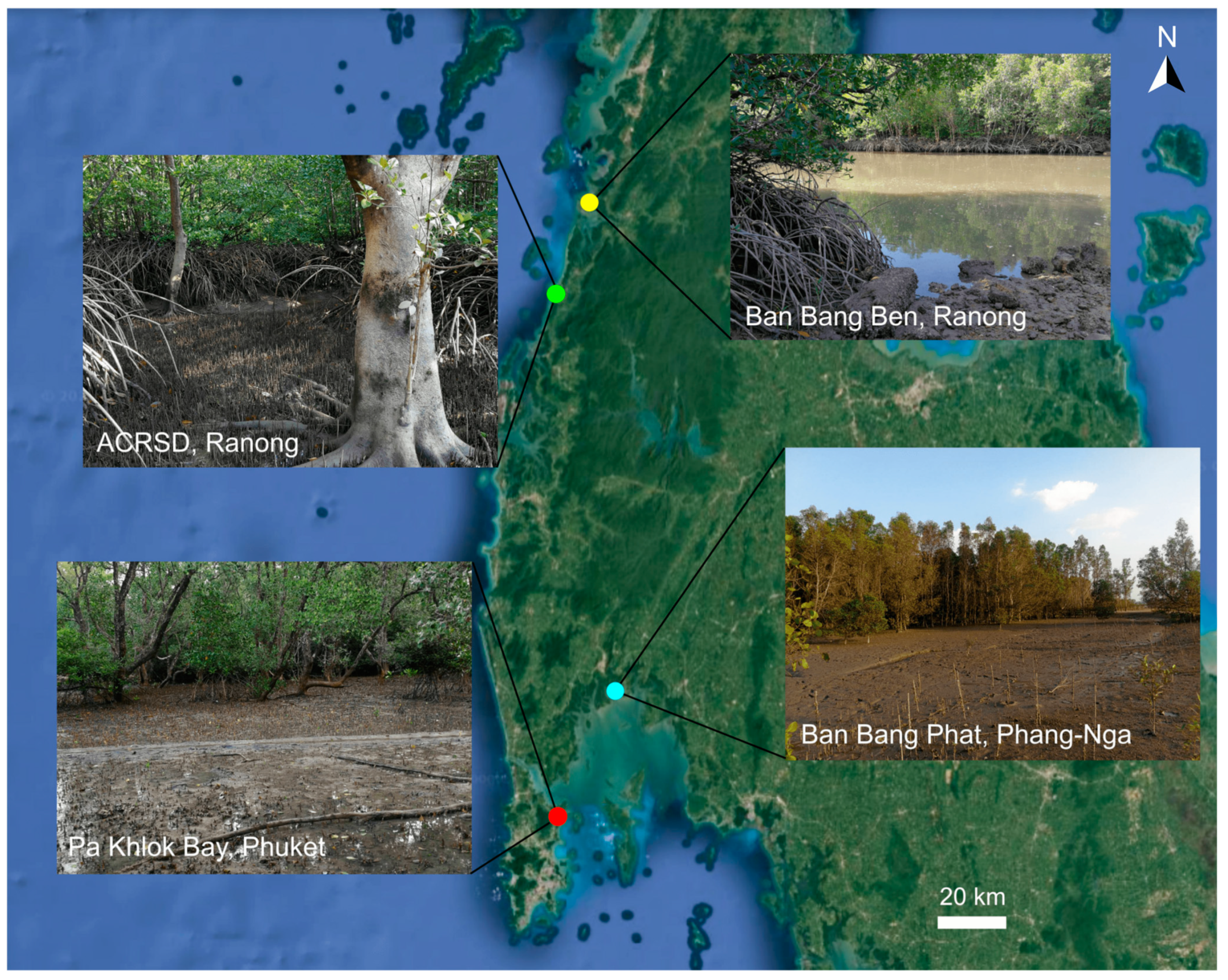
2.1. Sample Collection

The samples from mangrove environments of Ranong, Phang-Nga, and Phuket provinces were collected during low tide in February 2024 as part of a larger floristic survey of cyanobacterial diversity in southern Thailand. Samples were collected from various substrates including bark surfaces in Pa Khlok Bay, Phuket (08°02′11″ N, 98°24′54″ E), and the Andaman Coastal Research Station for Development (ACRSD), Ranong (09°22′38″ N, 98°24′11″ E), rock surfaces in Ban Bang Ben, Ranong (09°35′50″ N, 98°30′05″ E), and biological soil crusts in the intertidal zone of a mangrove forest in Ban Bang Phat, Phang-Nga (08°21′37″ N, 98°34′35″ E) (Figure 1). Collection was performed using a metal spatula and the samples were stored in 60 mL sterile polycarbonate screw-top containers and rinsed with sterilized seawater to remove any sand or sediment prior to further processing at the laboratory.

2.2. Isolation and Culture

Subsamples of the field specimens were enriched in f/2 liquid media [20], adjusted to a range of salinities (0, 15, 30, 45, 60, and 75 psu), and treated with 100 µg mL-1 cycloheximide to suppress any eukaryotic growth. Favorable growth was observed at a salinity of 30 psu for Picosynechococcus mangrovensis sp. nov. and Sirenicapillaria glauca, and at 15 psu for Allocoleopsis franciscana; therefore, cultures were maintained at these salinities thereafter. For filamentous cyanobacteria, isolation was performed by first placing the inoculum in the center of the agar medium, wherein small blocks of agar with single filaments that had migrated from the initial point of inoculation were then cut with sterile forceps and transferred to a fresh agar medium [21]. Additionally, filaments were serial-diluted and incubated in a 96-well plate containing f/2 medium for 2 weeks to achieve an unialgal status. The enriched cultures of unicellular cyanobacteria were dilution-plated and the formed single colonies were consecutively re-streaked on f/2 agar medium. Single colonies were subsequently re-streaked for culture maintenance. To achieve greater biomass for further analyses, the cells/filaments were cultured in 250 mL Erlenmeyer flasks containing 125 mL of f/2 liquid media, and incubated on an orbital shaker at 25 ± 3 °C under 12:12 h light:dark cycle with white cool fluorescent light of approximately 30 µmol m−2 s−1.

2.3. Morphological Observation

The morphology of natural samples and cultured, isolated strains was examined utilizing an Olympus CX31 microscope (Olympus, Tokyo, Japan) equipped with a DinoEye AM4031X USB eyepiece camera (Dino-Lite, Taiwan, China) for digital image acquisition via DinoCapture 2.0 software. Comprehensive morphological characterization included documentation of trichome or filament attributes, vegetative and apical cell morphology and dimensions, presence or absence of extracellular sheath material, and reproductive mechanisms. Quantitative morphometric measurements were conducted on 50 randomly selected specimens.

2.4. Herbarium Voucher Preparation

Cells/filaments were filtered, immobilized, and attached to herbarium paper. The herbarium paper was subsequently air-dried and enclosed in a glassine envelope and packed with archival acid-free paper. The herbaria were then deposited at the Thailand Natural History Museum (THNHM), Pathum Thani, Thailand with the numbers THNHM-P-2024-0078, THNHM-P-2024-0079, THNHM-P-2024-0080, THNHM-P-2024-0081, THNHM-P-2024-0082, THNHM-P-2024-0083, and THNHM-P-2024-0084. The studied strains were preserved in 2% glutaraldehyde and stored in glass vials. Additionally, living cultures are maintained at the Applied Plankton and Aquatic Plant Research Laboratory, Center for Advanced Studies for Agricultural and Food (CASAF), Kasetsart University, Bangkok Thailand.

2.5. Molecular and Phylogenetic Analyses

DNA was extracted from unialgal cultures using the NucleoSpin Tissue kit (Macherey-Nagel, Dueren, Germany) according to manufacturer protocols. The primers 16S27F1 (5′-AGA GTT TTG ATC CTG GCT CAG-3′) [22] and 23S30R (5′-CTT CGC CTC TGT GTG CCT AGG T-3′) [23] were used for the PCR amplification of the 16S rRNA gene and 16S-23S rRNA ITS region. The 20 µL PCR reaction consisted of 1X PCR master mix of i-Taq™ iNtRON kit (Biotechnology DR, Seongnam, South Korea), 1 µL each of forward and reverse primers (10 pmol μL−1), and 2 µL of DNA template. The PCR was performed using a Labcycler thermal cycler (SensoQuest GmbH, Göttingen, Germany) with the following conditions: initial denaturation at 95 °C for 3 min; 30 cycles of 94 °C for 1 min, 58 °C for 1 min, and 72 °C for 2 min; a final extension at 72 °C for 7 min; and a storage step at 10 °C. The PCR products were revealed using 1% agarose gel electrophoresis, later commercially purified and sequenced by U2Bio (Seoul, South Korea). Additionally, the amplicons of the 16S rRNA gene and associated ITS region were ligated into the pTG19-T cloning vector (Vivantis, Korea) and transformed into competent Escherichia coli DH5α cells, followed by selection of multiple positive transformants from each sample for sequencing using the primers M13F and M13R [24]. A total of one, four, and two clones of our strains A. franciscana, S. glauca, and P. mangrovensis were obtained, respectively.
Chromatograms were manually edited and assembled using Geneious Prime version 2025.0.2 (Biomatters, http://www.geneious.com, (accessed on 20 March 2025)). The 16S rRNA sequence alignment of 80 operational taxonomic units (OTUs) was constructed using the MUSCLE algorithm in Geneious Prime. The alignment incorporated publicly available sequences representing the genera Allocoleopsis, Sirenicapillaria, Picosynechococcus, along with sequences from closely related genera within the same respective families, all of which demonstrated high sequence identity based on BLAST+ 2.15.0 analyses (https://blast.ncbi.nlm.nih.gov/Blast.cgi, (accessed on 20 March 2025)). The phylogenetic tree was inferred by Maximum Likelihood (ML) and Bayesian Inference (BI) methods. ML analysis was performed in IQ-TREE 2 [25] with the best-fit model selection (TIM3 + F + I + G4) implemented in ModelFinder [26]. Nodal support values were estimated using standard nonparametric bootstraps (BSs) with 1000 replicates [27]. The BI method was implemented using MrBayes v3.2 [28], with the best-fit nucleotide model (GTR + G + I) selected by Kakusan 4 [29]. Two independent Markov Chain Monte Carlo (MCMC) runs were conducted, each for 2,000,000 generations, sampling every 1000 generations. The convergence of runs was confirmed by split frequency standard deviations (<0.01). Additionally, run convergence and parameter stability were assessed using Tracer 1.6 [30]. Branch support was estimated from the posterior distribution of trees, discarding the initial 25% of samples as burn-in, based on an assessment of convergence among runs. Both ML and BI trees were visualized using FigTree v1.4.4 [31].
The analysis of the ITS region was conducted separately for operons with both tRNAIle and tRNAAla genes, operons with only one tRNAIle gene, and operons lacking tRNA genes. The tRNA genes and the ITS domains were identified according to [32] and analyzed using Geneious Prime version 2025.0.2 (Biomatters, http://www.geneious.com, (accessed on 20 March 2025)). The secondary structures of the ITS domain were predicted using the Mfold program [33], with the draw mode set at untangle with loop fix and other parameters set to default. Additionally, the percent similarity of the 16S rRNA gene and percent dissimilarity of the 16S-23S rRNA ITS region were calculated using MEGA version 11 [34].

2.6. Salinity Tolerance and Growth

To facilitate the adaptation of cyanobacterial isolates to high salinity conditions, cultures were incubated in low light for 1 week. Subsequently, triplicates of P. mangrovensis were inoculated to a starting OD730 value of approximately 0.1 and cultured at salinities of 0, 15, 30, 45, 60, and 75 psu in 250 mL Erlenmeyer flasks containing 125 mL f/2 culture medium. Growth conditions were maintained as previously described. Cell growth was monitored daily for 2 weeks by measuring the optical density (OD) at 730 nm in a 1 cm light path with a UV-1800 spectrophotometer (Shimadzu, Kyoto, Japan). A. franciscana and S. glauca isolates were grown separately in 24 well-plates, in 2 mL culture volume, as the filaments tend to form clumps rather than a homogenous distribution in liquid medium, their growth was observed daily for 2 weeks.

3. Results

The identification of the isolated cyanobacterial strains in this study was predominantly based on the analyses of the 16S rRNA gene and ITS sequences, with morphological and ecological information used to complement our molecular findings.

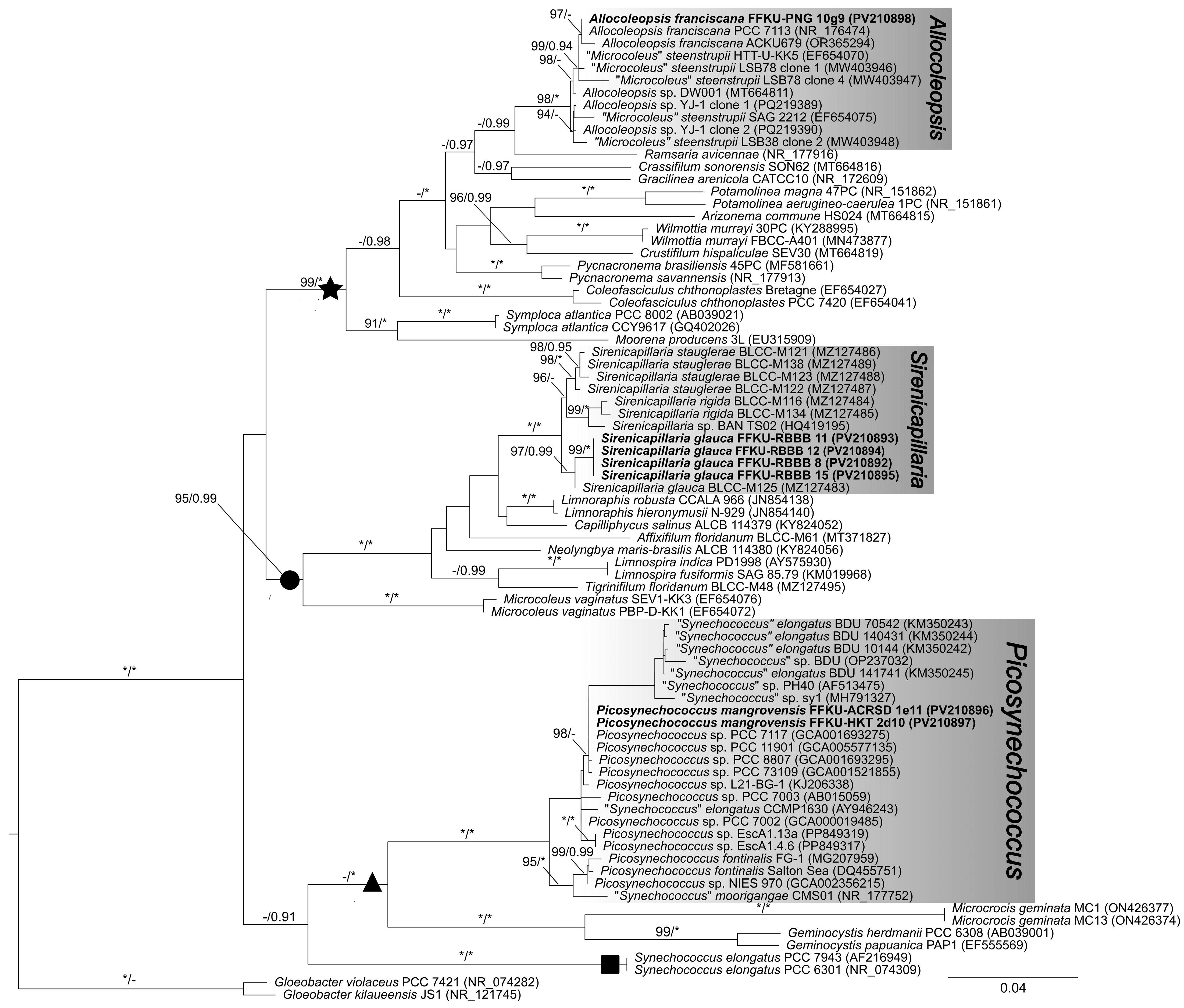
3.1. 16S rRNA Gene Phylogenetic and p-Distance Analyses

Both ML and BI analyses yielded congruent topologies, with the ML tree presented in Figure 2. Genetic distances (p-distance) are provided in Table S1. Our phylogenetic reconstruction revealed that the isolated cyanobacteria strains were placed into three distinct monophyletic clades, representing the genera Picosynechococcus, Sirenicapillaria, and Allocoleopsis (Figure 2).
Our strain FFKU-PNG 10g9 (PV210898) was identified as Allocoleopsis franciscana based on its well-supported monophyletic clade (ML = 97%) with the type strain A. franciscana PCC 7113 (NR_176474). The genetic identity between our strain and the type strain reached 100%, with 99.7% sequence similarity to strain ACKU679 (OR365294) (Table S1). Furthermore, our Sirenicapillaria isolates (FFKU-RBBB 8, FFKU-RBBB 11, FFKU-RBBB 12, and FFKU-RBBB 15) were identified as S. glauca based on their placement in a strongly supported monophyletic clade (ML = 97%, BI = 0.99) with the reference strain S. glauca BLCC-M125 (MZ127483). The genetic identity between our strains and the reference S. glauca strain was 99.6% (Table S1).
In contrast to the clear species-level identifications of our A. franciscana and S. glauca isolates, our Picosynechococcus strains FFKU-ACRSD 1e11 (PV210896) and FFKU-HKT 2d10 (PV210897) represented a taxonomically distinct lineage. These strains formed a moderately supported clade with several unnamed Picosynechococcus strains (e.g., PCC 7117 from low salinity brine pond, PCC 11901 from brackish water, PCC 73109 from seawater), exhibiting variable genetic identities ranging from 98.5% to 100% within this group (Table S1). More importantly, our Picosynechococcus isolates were phylogenetically distant from the type strain P. fontinalis FG-1 (MG207959), with this separation receiving strong statistical support (ML = 100%, BI = 1.00) and significantly lower percent similarities (97.3–98.1%, Table S1) below the genetic identity threshold of ≤98.7% [35], suggesting that our Picosynechococcus isolates likely represent a novel species within the genus.

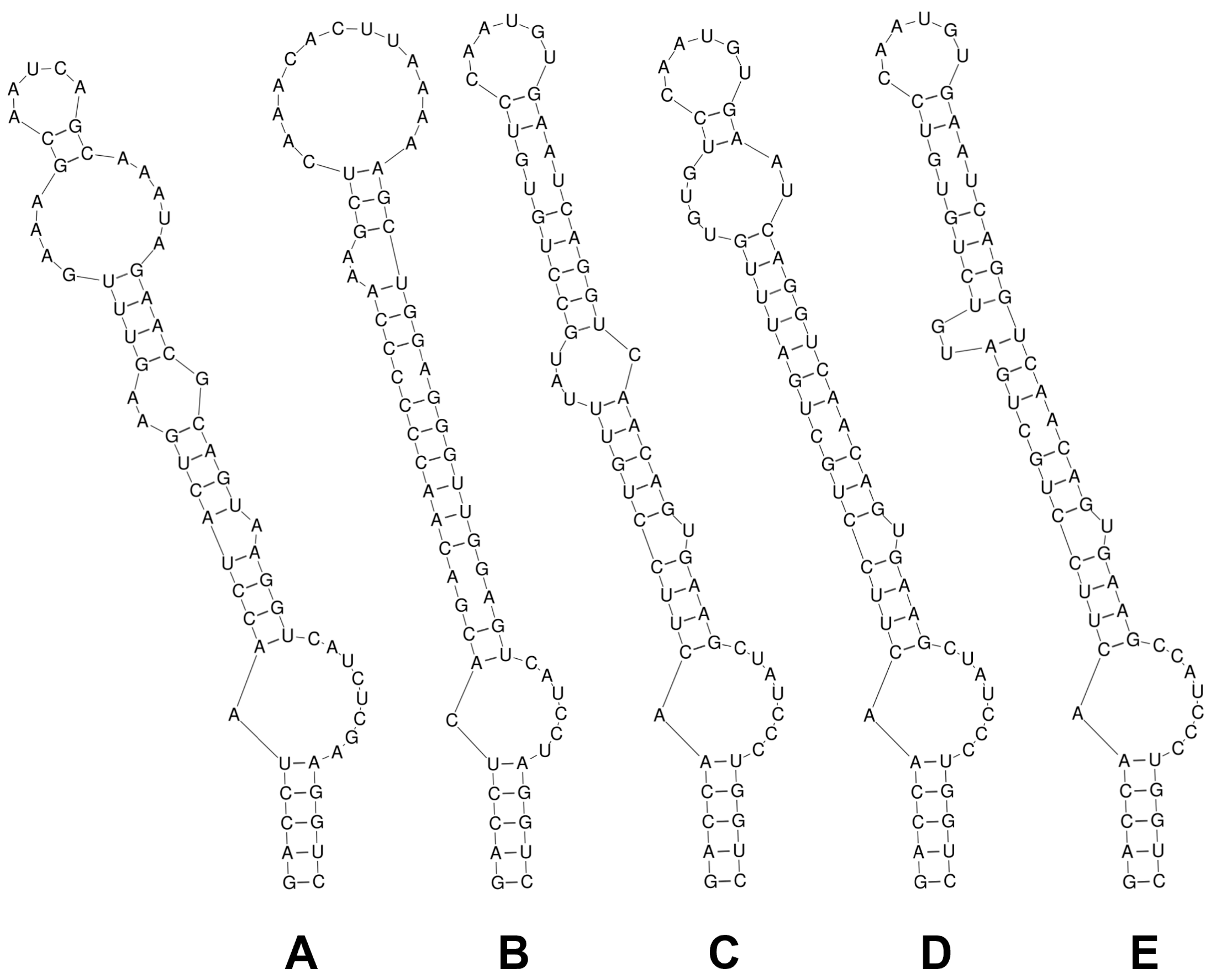
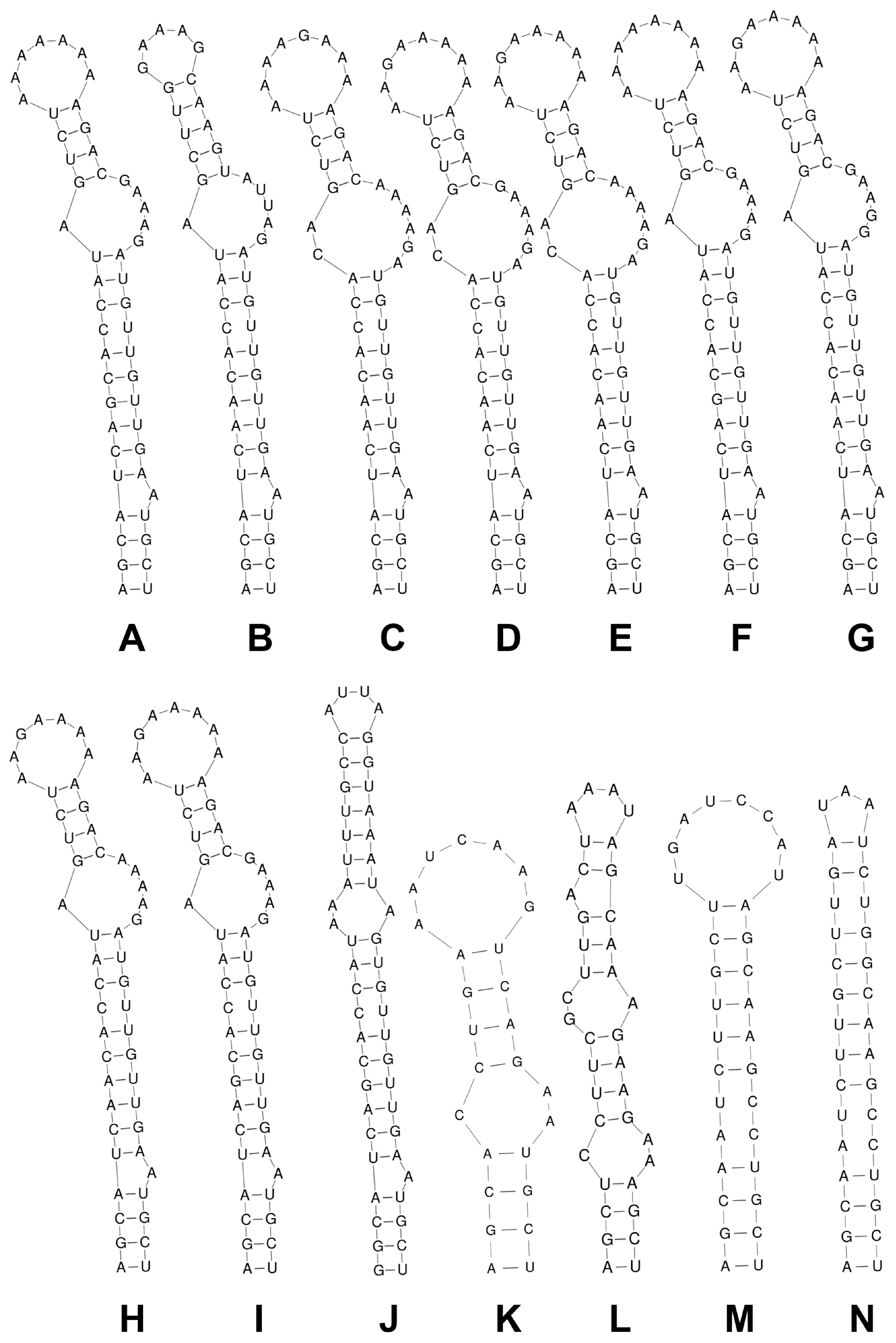
3.2. 16S–23S rRNA ITS Secondary Structure and p-Distance Analyses

Only one operon type containing both tRNA genes was identified in the Picosynechococcus strains analyzed. The ITS region of Picosynechococcus spp. (FFKU-ACRSD 1e11, FFKU-HKT 2d10, NIES 970, PCC 7002, PCC 7117, PCC 7003, PCC 73109, PCC 8807, PCC 11901, BDU 10144, BDU 70542, BDU 140431, BDU 141741) exhibited an identical D1-D1′ helical structure (Figure 3A). In contrast, the Box-B secondary structures varied across strains, particularly in the terminal loop and the bilateral bulge preceding it (Figure 4A–J). The Box-B folding pattern was nearly identical across strains, with minor differences in the nucleotide sequences of their terminal loops: AAAAAAA (FFKU-ACRSD 1e11 and FFKU-HKT 2d10) (Figure 4A), AAAAAAAA (PCC 73109, PCC 8807) (Figure 4F), and AAGAAAAA (BDU 141741) (Figure 4I). ITS sequences were not available for the type species, P. fontinalis. Our strains showed 0.9–2.0% dissimilarity in ITS sequences compared to certain strains (BDU 141741, BDU 10144, BDU 70542, BDU 140431) and 9.2–14.5% dissimilarity compared to others (NIES 970, PCC 73109, PCC 11901, PCC 7002, PCC 7003, PCC 8807) (Table 1).
For S. glauca strains, two operon types were observed: one containing both tRNA genes (FFKU-RBBB 11, FFKU-RBBB 12), and another lacking tRNAs (FFKU-RBBB 8, FFKU-RBBB 15, BLCC-M125). Interestingly, the D1-D1′ and Box-B secondary structures were identical for both operon types across all S. glauca strains, while the structures for both domains remained distinct from each other (Figure 3B and Figure 4K). ITS sequence analysis revealed 0–1.1% dissimilarity in tRNA-lacking operons of S. glauca, while the dissimilarity between operon types was 27.0–34.0% (Table 2). An ITS dissimilarity of >3% has been widely used as strong evidence for species separation in cyanobacterial taxonomy [36,37,38,39,40,41,42,43,44,45]. Overall, S. glauca was 8.6–19.1% dissimilar from S. rigida and S. stauglerae sequences lacking tRNAs, and 5.9–9.6% dissimilar from those with both tRNAs, thereby satisfying the threshold requirement (Table 2).
Similarly, two distinct operon types were observed in Allocoleopsis sequences: one containing a single tRNAIle gene (FFKU-PNG 10g9 and the type strain PCC 7113) and another lacking tRNAs (YJ-1, LSB38, LSB78) (Table 3). The D1-D1′ secondary structure was identical in strains FFKU-PNG 10g9 and PCC 7113 (Figure 3C) but differed from strains YJ-1, LSB38 (Figure 3D), and LSB78 (Figure 3E), particularly in the region between the first bilateral bulge and the terminal loop. The Box-B domain varied across operon types and among sequences lacking tRNAs, both in folding pattern (Figure 4L–N) and length (37 bp for FFKU-PNG 10g9 and PCC 7113; 32 bp for YJ-1 and LSB38; 33 bp for LSB78). The ITS dissimilarity between operon types reached 14.0–17.9% (Table 3). FFKU-PNG 10g9 and PCC 7113 were 0.4% dissimilar from each other, while ITS data were not available for ACKU679 (Table 3). LSB78 clones were 5.7–7.8% dissimilar from clones of YJ-1 and LSB38, while the dissimilarity among the latter sequences ranged from 0.2% to 3.0% (Table 3).

3.3. Taxonomic Description

Genetic analyses and comprehensive morphological examinations have led to the establishment of a new taxon within the genus Picosynechococcus, while simultaneously confirming the presence of Allocoleopsis franciscana and Sirenicapillaria glauca in the Andaman coastal waters of Thailand. The taxonomic characterization of each species is as follows:

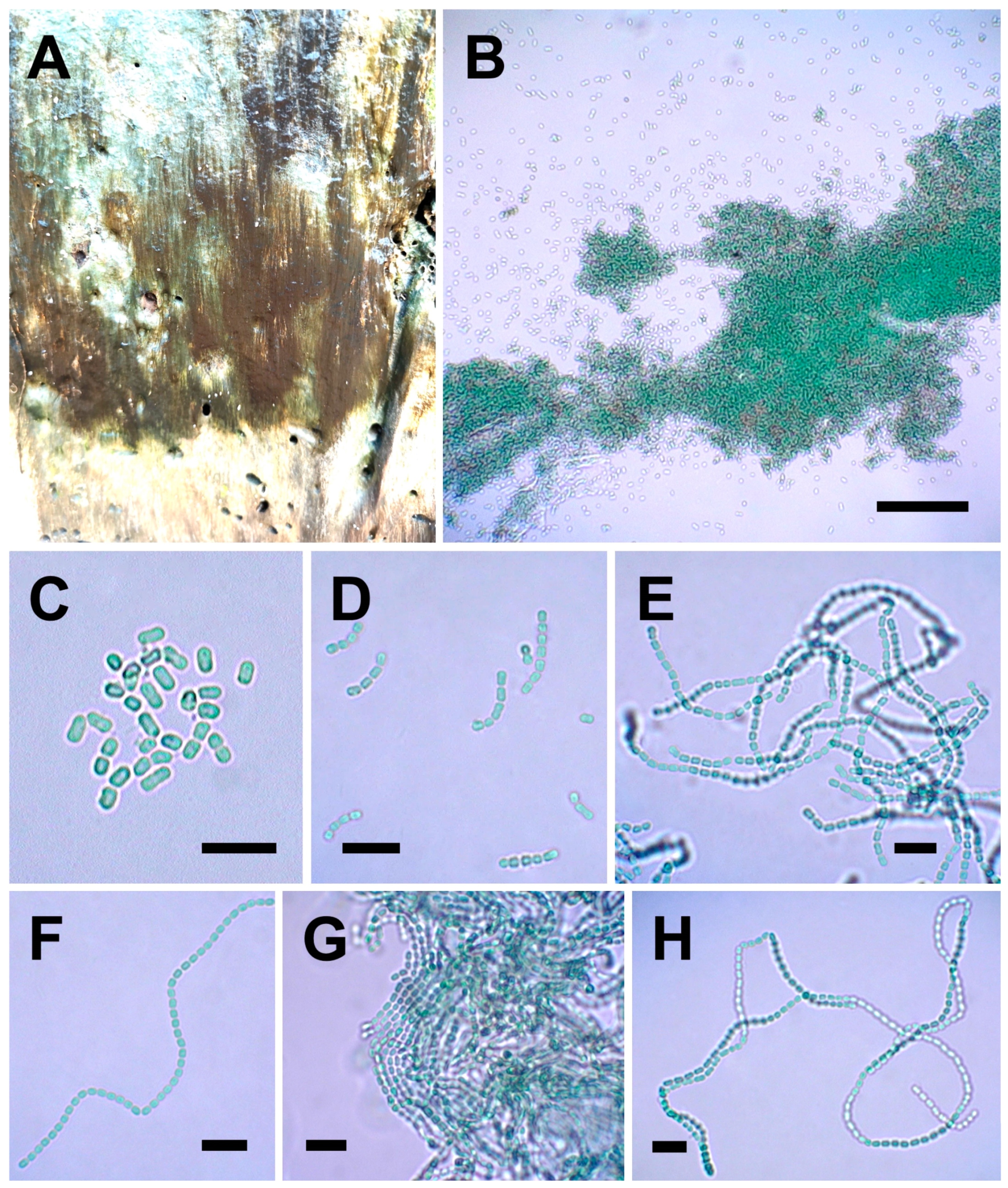
3.3.1. Picosynechococcus mangrovensis sp. nov. Lim and Muangmai (Figure 5A–H)

Description: Cells bright blue-green, with homogenous content, solitary (Figure 5C), or connected in pseudo-filamentous formations of up to 160 cells (Figure 5D–H), oval to cylindrical, 1.7–4.0 μm long, 1.3–2.5 μm wide, with gliding motility. Cells divide by binary fission in one plane perpendicular to the longitudinal axis. Slightly halophilic, grows well in salinities ranging from 0 to 45 psu, with optimal growth at 30 psu.
Diagnosis: Morphologically similar to P. fontinalis and other Picosynechococcus strains, overlapping in cell length and cell width. Distinguished by its ability to form very long pseudo-filaments of up to 160 cells, slightly halophilic nature, its phylogenetic position and lower similarity in 16S rRNA gene sequence to other Picosynechococcus species (Figure 2).
Habitat: This species grew on bark or wood surfaces (Figure 5A).
Etymology: The species epithet refers to its occurrence in mangrove environments.
Type locality: Isolated from the bark surface of a stump within the intertidal zone of a mangrove environment in Pa Khlok Bay, Phuket, Thailand (08°02′11″ N, 98°24′54″ E).
Holotype: Portion of a culture of the reference strain FFKU-HKT 2d10, preserved in a metabolically inactive form, placed on herbarium paper and deposited at the Thailand Natural History Museum (THNHM), Pathum Thani, Thailand with the number THNHM-P-2024-0084.
Materials examined: FFKU-HKT 2d10, FFKU-ACRSD 1e11.
GenBank accession number: PV210896, PV210897 (16S rRNA), PV210904, PV210905 (ITS).
Note: The strains FFKU-HKT 2d10 from Phuket and FFKU-ACRSD 1e11 from Ranong were not observed in environmental samples but bloomed in enrichment cultures and were subsequently isolated through serial dilution. The strains were isolated from an algal consortium of Lyngbya-like and Persinema sp. mats, respectively. A detailed morphological comparison of our P. mangrovensis and other closely related species was presented in Table 4.
Figure 5. Morphology of Picosynechococcus mangrovensis strains (A,CE) FFKU-HKT 2d10 and (B,FH) FFKU-ACRSD 1e11. (A) Mat consortium of Picosynechococcus and Lyngbya-like cyanobacteria in nature. (B) Solitary and clustered cells in enrichment culture. (C,D) Cells solitary or in short pseudo-filaments. (EH) Cells forming very long pseudo-filaments. Scale bar: (B) 70 µm, (CH) 10 µm.
Diversity 17 00319 g005

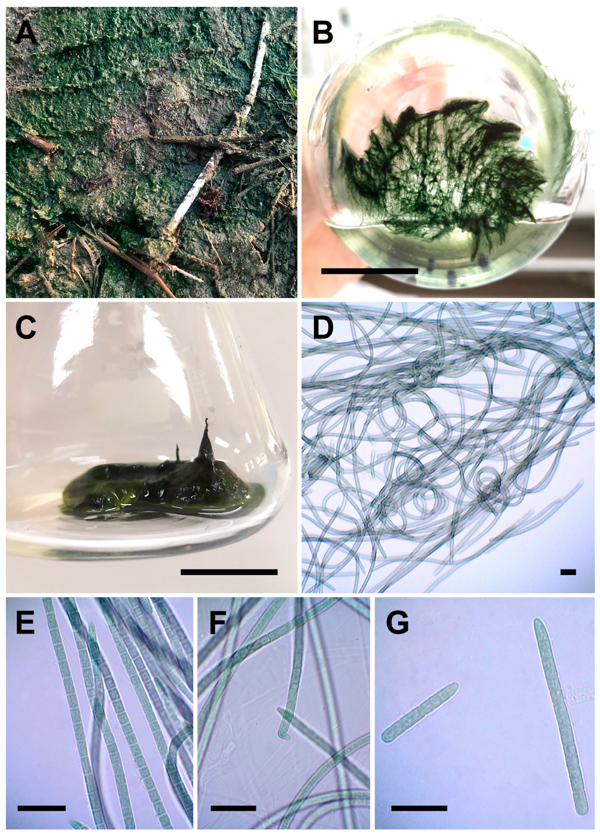
3.3.2. Allocoleopsis franciscana Moreira-Fernandes, Giraldo-Silva, D. Roush, and Garcia-Pichel (Figure 6A–G)

Description: Thallus-forming mat on biological soil crust in nature (Figure 6A), fasciculated (Figure 6B), or erect (Figure 6C) in culture. Solitary filaments, or forming rope-like bundles (Figure 6D). Trichomes dark olive green, heteropolar, slightly constricted at cross walls, with thin, hyaline sheath that open at the ends. Vegetative cells longer than wide to isodiametric (Figure 6E), 3.4–8.7 µm long and 3.4–5.0 µm wide. Apical cells rounded, elongated, conical or bent (Figure 6F). Reproduction by hormogonia formation with or without the help of necridia (Figure 6G).
Collection locality: Isolated from biological soil crusts in the intertidal zone of a mangrove forest in Ban Bang Phat, Phang-Nga (08°21′37″ N, 98°34′35″ E).
Herbarium deposition: Portion of a culture of the strain FFKU-PNG 10g9 was preserved in a metabolically inactive form, placed on herbarium paper and deposited at the Thailand Natural History Museum (THNHM), Pathum Thani, Thailand with the number THNHM-P-2024-0082.
GenBank accession number: PV210898 (16S rRNA), PV210899 (ITS).
Note: A detailed morphological comparison of our Allocoleopsis franciscana and other strains was presented in Table 5.
Figure 6. Morphology of Allocoleopsis franciscana FFKU-PNG 10g9. (A) Thallus on biological soil crust. (B) Fasciculated filaments in culture. (C) Thallus tightly aggregated and erect in culture. (D) Flexuous filaments. (E) Trichomes with distinct cross walls and conical apices. (F) Bent apical cell. (G) Hormogonia formation. Scale bar: (B,C) 4 cm, (DG) 20 µm.
Diversity 17 00319 g006

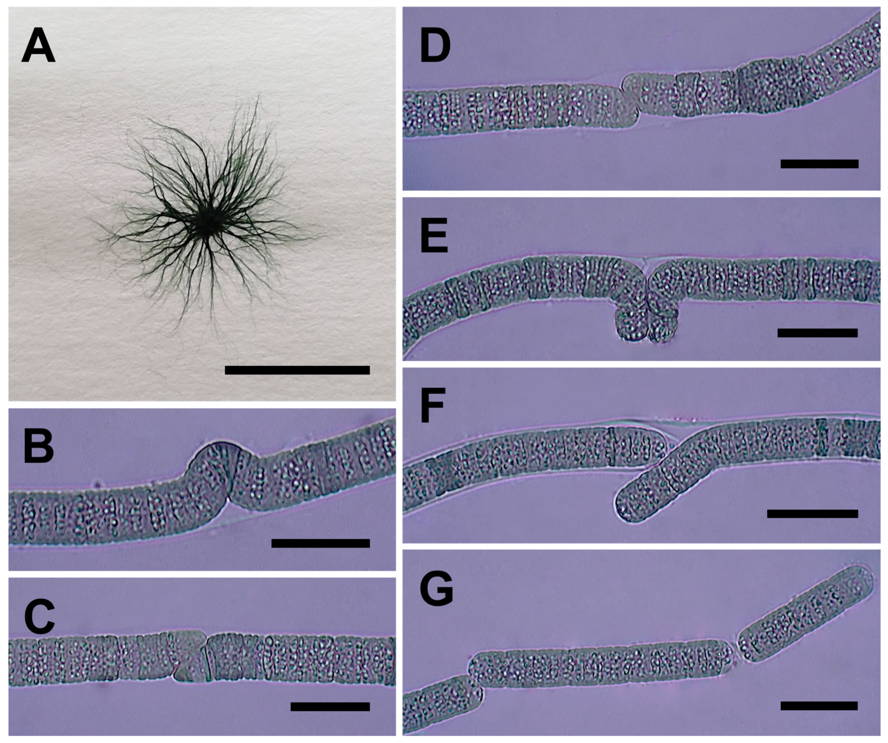
3.3.3. Sirenicapillaria glauca Berthold, Leftler, and Laughinghouse (Figure 7A–G)

Description: In liquid culture, thallus dark green, forming free-floating globose colony with hair-like filaments radiating from a common center (Figure 7A). Filaments straight, 10.9–18.0 µm in diameter. Sheath colorless, thin, firm, open at the ends. Trichomes pale to dark green, cylindrical, mostly not tapered towards the ends, rarely forming knot-like arrangements (Figure 7B) or having two trichomes occupying the same sheath. Vegetative cells discoid, shorter than wide, with cross walls slightly constricted and heavily granulated, 0.9–2.2 µm long, 8.9–11.8 (12.8) µm wide. Apical cell rounded, sometimes slightly narrowed, no calyptra. Reproduction by straight fragmentation with or without the help of necridic cells, diagonal fragmentation (Figure 7C), false branching (Figure 7D–F), hormogonia (Figure 7G).
Figure 7. Morphology of Sirenicapillaria glauca (A,B) FFKU-RBBB 8, (C) FFKU-RBBB 11, (D,E) FFKU-RBBB 12, and (F,G) FFKU-RBBB 15. (A) Hair-like filaments radiating from a common center. (B) Curved filament. (C) Diagonal fragmentation of filament. (D) Filament prior to false branching. (E,F) False branching. (G) Hormogonia. Scale bar: (A) 2 cm, (BG) 20 µm.
Diversity 17 00319 g007
Collection locality: Isolated from a rock surface in Ban Bang Ben, Ranong, Thailand (09°35′50″ N, 98°30′05″ E).
Herbarium deposition: Portion of a culture of the strains (FFKU-RBBB 8, FFKU-RBBB 11, FFKU-RBBB 12, FFKU-RBBB 15) was preserved in a metabolically inactive form, placed on herbarium paper and deposited at the Thailand Natural History Museum (THNHM), Pathum Thani, Thailand with the numbers THNHM-P-2024-0078, THNHM-P-2024-0079, THNHM-P-2024-0080, THNHM-P-2024-0081.
GenBank accession number: PV210892–PV210895 (16S rRNA), PV210900–PV210903 (ITS).
Note: The rock surface was primarily inhabited by colonies of cf. Kyrtuthrix sp., while S. glauca filaments were not directly observed from environmental samples, they proliferated in enrichment cultures and were subsequently isolated through serial dilution. A detailed morphological comparison of our Sirenicapillaria glauca and other strains was presented in Table 6.

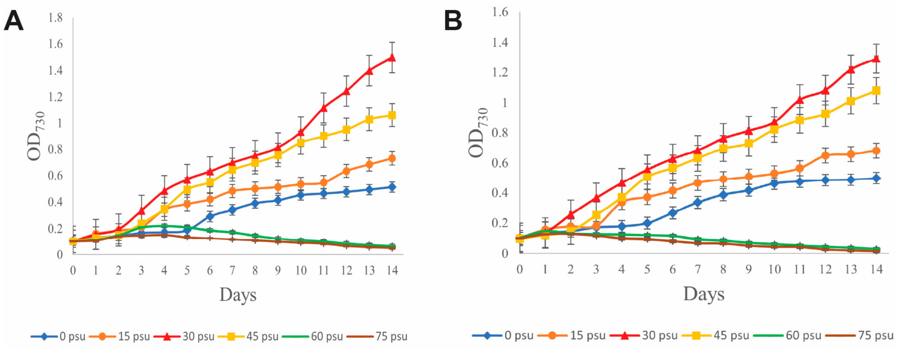
3.4. Growth at Different Salinities

Both strains of Picosynechococcus mangrovensis demonstrated similar growth trends (Figure 8A,B): exponential growth in salinities ranging from 0 to 45 psu but poor growth 60 and 75 psu. Optimal growth was observed at 30 psu. Comparatively, lower growth was exhibited in 45 psu, followed by 15 and 0 psu. Additionally, A. franciscana FFKU-PNG 10g9 grew in salinities 0–30 psu. Our S. glauca isolates grew well in salinities 0–30 psu but exhibited yellowish-green filaments and poor growth in 40–65 psu.

4. Discussion

4.1. Picosynechococcus mangrovensis sp. nov.

4.1.1. 16S rRNA Phylogeny and ITS Region Analyses

The 16S rRNA gene sequencing has been pivotal in confirming its phylogenetic placement and genetic distinctiveness from the type species P. fontinalis (Figure 2). As recommended by [35], the species threshold value of <98.7% serves as a marker for separating morphologically cryptic taxa. Interestingly, the strains of P. mangrovensis fall below the 98.7% limit in comparison to P. fontinalis (MG207959, DQ455751), Picosynechococcus sp. NIES 970, and “Synechococcusmoorigangae CMS01. Our analyses also revealed that “Synechococcusmoorigangae CMS01 is more closely related to the genus Picosynechococcus rather than Synechococcus. However, the lack of ITS data on P. fontinalis and “Synechococcusmoorigangae limits further taxonomic resolution. To address this, additional approaches from the polyphasic method were employed to support the novelty of P. mangrovensis.

4.1.2. Morphological Characterization

Picosynechococcus mangrovensis, though unicellular, exhibited pronounced filamentous formations of up to 160 cells in this study (Figure 5B–H). This morphological character was found to be consistent throughout the 6-month observation period. In both agar and liquid cultures, solitary cells and short pseudo-filaments of up to 10 cells were mostly present in salinities < 15 psu, whereas long pseudo-filaments of up to 160 cells were the predominant morphology at salinities > 15 psu. This salinity-dependent morphological plasticity stands in contrast to the behavior of Crocosphaera subtropica ATCC 51142, a unicellular cyanobacterium isolated from the intertidal area of Port Aransas, Texas, which formed short filaments of 4–16 cells under low salinity but reverted to its unicellular lifestyle as salinity or cell density increased [51]. In contrast, P. mangrovensis formed progressively longer filaments at higher salinities, suggesting a regulated, adaptive response to the dynamic salinity regimes due to tidal fluctuations, rather than a fixed morphological trait. Its preference for a higher salinity (Figure 8A,B) could indicate that its true ecological niche to be seaward rather than landward as found in the mangrove ecosystems. Furthermore, tidal movements likely facilitated cell/filament dispersal and its subsequent colonization of various niches beyond the core habitat of this species [52,53]. These observations underscore the broader ecological significance of morphological plasticity in cyanobacteria and the need to further investigate the links between environmental cues, morphological, and physiological responses among mangrove-associated cyanobacteria. On another note, we also consider this phenotype a unique trait to discriminate against P. fontinalis (few-celled) [16] and other strains including “Synechococcusmoorigangae CMS01 (4–6 cells) [46]. Additionally, thallus morphology of this new species in nature is unknown as they were not directly observed from the field. While morphological comparisons of natural and cultured populations should ideally be included in species identification [54], rare representatives of an algal consortium tend to be obscured by more dominant groups.

4.1.3. Ecological Adaptation and Geographical Distribution

Picosynechococcus presented a wide ecological range, such as those reported in Nodosilinea [41], Haloleptolyngbya [55], and Halomicronema [56]. The genus was originally documented inhabiting freshwater mineral springs (P. fontinalis), but their occurrences have since been reported in caves (EscA1.13a, EscA1.4.6), decaying wood (BDU 141741), sand (BDU 70542), low salinity brine pond (PCC 7117), hypersaline (L21-BG-1), marine (PCC 7002, PCC 7003, PCC 73109, BDU 140431, sy1), and brackish habitats (PCC 11901, BDU 10144, PH40, NIES 970, CMS01) [16,46,47]. In the case of P. mangrovensis, strains were isolated from tree bark and stump in the intertidal zone of mangrove environments. Interestingly, it exhibited a slight halophilic nature [9] with favorable growth ranging from 30 to 45 psu, while the growth was reduced with increasing salt concentrations. The genetics, morphology, and ecology of this taxon not only warrants its classification as a new Picosynechococcus species but also highlights the plasticity and ability of cyanobacteria to thrive and adapt to a variety of habitats. Moreover, Picosynechococcus predominantly occurred in India (KM350242–KM350245, OP237032, NR_177752), Europe (AY946243, PP849317, PP849319, MG207959), North America (AB015059, DQ455751), the Hawaiian archipelago (AF513475, GCA001521855), and South America (GCA001693275), with records also available from Kiribati (KJ206338), Gabon (GCA001693295), Western Australia (GCA000019485), East Asia (Japan, GCA002356215; China, MH791327), and Southeast Asia (Singapore, GCA005577135). Our new taxon represents the first record of Picosynechococcus in Thailand, and the second sighting of the genus in Southeast Asia.

4.2. Allocoleopsis franciscana

4.2.1. 16S rRNA Phylogeny and ITS Region Analyses

Our strain FFKU-PNG 10g9 was embedded in a subclade containing the type strain Allocoleopsis franciscana PCC 7113, with a 16S rRNA genetic similarity of 100%, ITS dissimilarity of 0.4%, and identical D1-D1′ and Box-B secondary structures, thereby confirming the identity of our strain as Allocoleopsis franciscana. However, we could not compare the ITS sequences of the former two taxa with other Allocoleopsis strains as they had different operon types [57]. Even so, based on the ITS dissimilarity analysis of the sequences lacking tRNAs, it is very likely that there are at least two cryptic species in the genus. Nevertheless, this would require additional polyphasic studies of Allocoleopsis and a greater taxon sampling to characterize and diagnose them.

4.2.2. Morphological Characterization

The key feature that differentiates our strain from other Allocoleopsis strains YJ-1, LSB78, and LSB38 is the cell width. FFKU-PNG 10g9 displayed a length range of 3.4–5.0 μm, narrower than YJ-1 (5–7 μm), LSB78 (5.3–7.3 μm), and LSB38 (4.4–8.4 μm) (despite a minor overlap) (Table 5). The cell width of Allocoleopsis franciscana FFKU-PNG 10g9 spanned 3.4–5.0 μm, significantly narrower from the type strain PCC 7113 (5–7 μm), while the cell length of our strain achieved a broader range of 3.4–8.7 μm, in contrast to the length range 6.0–7.5 μm of the latter strain (Table 5). These observations highlight the morphological plasticity of cyanobacteria in different environments [3]. Additionally, we also provided the thallus morphology of Allocoleopsis franciscana in nature (Figure 6A) and in cultures (Figure 6B,C), which was not reported in previous literature. We are hopeful that it will aid in the identification of other Allocoleopsis specimens in future floristic surveys.

4.2.3. Ecological Adaptation and Geographical Distribution

Whilst the terrestrial Allocoleopsis has been generally described in greenhouse soil crusts [19], urban soils [49], and desert soils [44,50], they also grew on gravel submerged in freshwater [48]. The occurrence of Allocoleopsis franciscana in Thailand mangroves highlights the ecological flexibility and its potential role in the ecological dynamics of mangrove ecosystems. The genus has also been predominantly documented in the tropical regions of the USA [19,44] and some temperate regions of South Korea [48], China [49], and Algeria [50]. Our study expands the previously known geographic distribution of the genus to the Southeast Asian region.

4.3. Sirenicapillaria glauca

4.3.1. 16S rRNA Phylogeny and ITS Region Analyses

The morphological resemblance of our Lyngbya-like strains to Sirenicapillaria species (S. glauca, S. rigida, and S. stauglerae) is problematic for taxonomical evaluation [18], in addition to their high 16S rRNA genetic similarity to S. stauglerae and S. glauca (>98.7%). However, they are genetically distant enough to assign our strains to S. glauca on the basis of 16S rRNA phylogeny (Figure 2), ITS secondary structures (Figure 3B and Figure 4K), ITS dissimilarity values (Table 6), as seen in other works on cryptic taxa [18,40,58]. This work also represents the first report of an ITS operon type containing both tRNA genes for S. glauca (FFKU-RBBB 11 and 12).

4.3.2. Morphological Characterization

A comparison between S. glauca collected from Thailand (in this study) and the USA [18] revealed notable differences in cell size and reproduction (Table 6). Although the filament width and cell width of the type strain BLCC-M125 ranged from 18.4 to 25.2(–28) µm and (14–)15–20(–21) µm, our strains demonstrated a narrower range of 10.9–18.0 µm and 8.9–11.8 (12.8) µm, respectively (Table 6). Furthermore, what caught our attention was the frequent occurrence of Scytonema-like (Figure 7E) and Tolypothrix-like (Figure 7F) false branching in cultured materials [59]. This particular morphological trait was stable and consistent throughout the 6-month long light microscopy observation on Sirenicapillaria glauca strains, which has ultimately expanded our knowledge of the morphological diversity in the genus.

4.3.3. Ecological Adaptation and Geographical Distribution

S. glauca was originally described as epipsammic, epiphytic, planktonic, and benthic organisms, living on seagrass beds in the marine coasts of the USA [18]. We consider our strains of the species to be epilithic, albeit not directly observed on the surface of the rock sample from which they were isolated. Notably, the occurrence of S. glauca in Thailand mangroves represents a notable geographic extension for the genus, as well as its first documentation in the Andaman Sea of Thailand.

4.4. Current Status and Future Surveys of Cyanobacterial Diversity in Thailand Mangroves

The world of mangrove-associated cyanobacteria provides vast room for new discoveries. Despite the prevalent mangrove conservation efforts in Thailand, the diversity of cyanobacteria in the Andaman region remains unknown. The first documentation of Picosynechococcus, Sirenicapillaria, and Allocoleopsis from this study highlights the importance of investigating cyanobacterial diversity in less explored mangrove habitats (e.g., subaerial, benthic, epilithic) of the southern regions of Thailand. Moreover, our findings from this polyphasic study serve as a baseline for establishing a reliable and comprehensive checklist based on updated identification keys from the tropical region.

Supplementary Materials

The following supporting information can be downloaded at: https://www.mdpi.com/article/10.3390/d17050319/s1, Table S1: Percent similarity of 16S rRNA gene sequences among strains of Picosynechococcus, Sirenicapillaria, Allocoleopsis, and other closely related genera.

Author Contributions

Conceptualization, N.M.; methodology, B.L.C.G., F.M., J.P., S.M. and N.M.; software, B.L.C.G. and N.M.; validation, B.L.C.G., F.M. and N.M.; formal analysis, B.L.C.G., F.M. and N.M.; investigation, B.L.C.G., J.P., S.M. and N.M.; resources, B.L.C.G., J.P., S.M. and N.M.; data curation, B.L.C.G. and S.M.; writing—original draft preparation, B.L.C.G. and N.M.; writing—review and editing, B.L.C.G. and N.M.; visualization, B.L.C.G.; supervision, J.P. and N.M.; project administration, N.M.; funding acquisition, N.M. All authors have read and agreed to the published version of the manuscript.

Funding

This research was funded by Kasetsart University through the Graduate School Fellowship Program and by Faculty of Fisheries, Kasetsart University.

Institutional Review Board Statement

Not applicable.

Data Availability Statement

All nucleotide sequences used in this study are available from NCBI, with their accession numbers provided in the main text.

Acknowledgments

We thank Panjapol Pleebut for their support during the field trip and laboratory work. We are also grateful to Chatcharee Kaewsuralikhit for providing the laboratory facilities for the isolation and culture of cyanobacteria.

Conflicts of Interest

The authors declare no conflicts of interest. The funders had no role in the design of the study; in the collection, analyses, or interpretation of data; in the writing of the manuscript; or in the decision to publish the results.

References

  1. Alvarenga, D.O.; Rigonato, J.; Branco, L.H.Z.; Fiore, M.F. Cyanobacteria in mangrove ecosystems. Biodivers. Conserv. 2015, 24, 799–817. [Google Scholar] [CrossRef]
  2. Dvořák, P.; Hašler, P.; Casamatta, D.A.; Poulíčková, A. Underestimated cyanobacterial diversity: Trends and perspectives of research in tropical environments. Fottea 2021, 21, 110–127. [Google Scholar] [CrossRef]
  3. Dvořák, P.; Jahodářová, E.; Stanojković, A.; Skoupý, S.; Casamatta, D.A. Population genomics meets the taxonomy of cyanobacteria. Algal Res. 2023, 72, 103128. [Google Scholar] [CrossRef]
  4. Alvarenga, D.O.; Rigonato, J.; Branco, L.H.Z.; Melo, I.S.; Fiore, M.F. Phyllonema aviceniicola gen. nov., sp. nov. and Foliisarcina bertiogensis gen. nov., sp. nov., epiphyllic cyanobacteria associated with Avicennia schaueriana leaves. Int. J. Syst. Evol. Microbiol. 2016, 66, 689–700. [Google Scholar] [CrossRef]
  5. Alvarenga, D.O.; Andreote, A.P.D.; Branco, L.H.Z.; Fiore, M.F. Kryptousia macronema gen. nov., sp. nov. and Kryptousia microlepis sp. nov., nostocalean cyanobacteria isolated from phyllospheres. Int. J. Syst. Evol. Microbiol. 2017, 67, 3301–3309. [Google Scholar] [CrossRef]
  6. Komárek, J. A polyphasic approach for the taxonomy of cyanobacteria: Principles and applications. Eur. J. Phycol. 2016, 51, 346–353. [Google Scholar] [CrossRef]
  7. Chakraborty, S.; Maruthanayagam, V.; Achari, A.; Mahansaria, R.; Pramanik, A.; Jaisankar, P.; Mukherjee, J. Oxynema aestuarii sp. nov. (Microcoleaceae) isolated from an Indian mangrove forest. Phytotaxa 2018, 374, 24–40. [Google Scholar] [CrossRef]
  8. Chakraborty, S.; Maruthanayagam, V.; Achari, A.; Pramanik, A.; Jaisankar, P.; Mukherjee, J. Aerofilum fasciculatum gen. nov., sp. nov. (Oculatellaceae) and Euryhalinema pallustris sp. nov. (Prochlorotrichaceae) isolated from an Indian mangrove forest. Phytotaxa 2021, 522, 165–186. [Google Scholar] [CrossRef]
  9. Genuário, D.B.; Vaz, M.G.M.V.; Hentschke, G.S.; Sant’Anna, C.L.; Fiore, M.F. Halotia gen. nov., a phylogenetically and physiologically coherent cyanobacterial genus isolated from marine coastal environments. Int. J. Syst. Evol. Microbiol. 2015, 65, 663–675. [Google Scholar] [CrossRef]
  10. Mangrove Forest Conservation Division. Mangrove Knowledge Guide, Department of Marine and Coastal Resources (DMCR); Ploy Media Co., Ltd.: Bangkok, Thailand, 2020; pp. 1–68. Available online: https://www.dmcr.go.th/detailLib/5720 (accessed on 2 April 2025).
  11. Muangham, S.; Suksaard, P.; Mingma, R.; Matsumoto, A.; Takahashi, Y.; Duangmal, K. Nocardiopsis sediminis sp. nov., isolated from mangrove sediment. Int. J. Syst. Evol. Microbiol. 2016, 66, 3835–3840. [Google Scholar] [CrossRef]
  12. Nimnoi, P.; Pongsilp, N. Insights into bacterial communities and diversity of mangrove forest soils along the Upper Gulf of Thailand in response to environmental factors. Biology 2022, 11, 1787. [Google Scholar] [CrossRef] [PubMed]
  13. Am-In, S.; Limtong, S.; Yongmanitchai, W.; Jindamorakot, S. Candida andamanensis sp. nov., Candida laemsonensis sp. nov. and Candida ranongensis sp. nov., anamorphic yeast species isolated from estuarine waters in a Thai mangrove forest. Int. J. Syst. Evol. Microbiol. 2011, 61, 454–461. [Google Scholar] [CrossRef]
  14. Limtong, S.; Yongmanitchai, W.; Kawasaki, H.; Seki, T. Candida phangngensis sp. nov., an anamorphic yeast species in the Yarrowia clade, isolated from water in mangrove forests in Phang-Nga Province, Thailand. Int. J. Syst. Evol. Microbiol. 2008, 58, 515–519. [Google Scholar] [CrossRef] [PubMed]
  15. Kunthiphun, S.; Endoh, R.; Takashima, M.; Ohkuma, M.; Tanasupawat, S.; Savarajara, A. Prototheca paracutis sp. nov., a novel oleaginous achlorophyllous microalga isolated from a mangrove forest. Mycoscience 2019, 60, 165–169. [Google Scholar] [CrossRef]
  16. Komárek, J.; Johansen, J.R.; Šmarda, J.; Strunecký, O. Phylogeny and taxonomy of Synechococcus-like cyanobacteria. Fottea 2020, 20, 171–191. [Google Scholar] [CrossRef]
  17. Strunecký, O.; Ivanova, A.P.; Mareš, J. An updated classification of cyanobacterial orders and families based on phylogenomic and polyphasic analysis. J. Phycol. 2023, 59, 12–51. [Google Scholar] [CrossRef]
  18. Berthold, D.E.; Lefler, F.W.; Laughinghouse, H.D., IV. Recognizing novel cyanobacterial diversity in marine benthic mats, with the description of Sirenicapillariaceae fam. nov., two genera Sirenicapillaria gen. nov. and Tigrinifilum gen. nov., and seven new species. Phycologia 2022, 61, 146–165. [Google Scholar] [CrossRef]
  19. Fernandes, V.M.C.; Giraldo-Silva, A.; Roush, D.; Garcia-Pichel, F. Coleofasciculaceae, a monophyletic home for the Microcoleus steenstrupii complex and other desiccation-tolerant filamentous cyanobacteria. J. Phycol. 2021, 57, 1563–1579. [Google Scholar] [CrossRef]
  20. Guillard, R.R.L.; Ryther, J.H. Studies of marine planktonic diatoms. I. Cyclotella nana Hustedt, and Detonula confervacea (cleve) Gran. Can. J. Microbiol. 1962, 8, 229–239. [Google Scholar] [CrossRef]
  21. Waterbury, J.B. The cyanobacteria—Isolation, purification and identification. Prokaryotes 2006, 4, 10531073. [Google Scholar]
  22. Neilan, B.A.; Jacobs, D.; Del Dot, T.; Blackall, L.L.; Hawkins, P.R.; Cox, P.T.; Goodman, A.E. rRNA sequences and evolutionary relationships among toxic and nontoxic cyanobacteria of the genus Microcystis. Int. J. Syst. Bacteriol. 1997, 47, 693–697. [Google Scholar] [CrossRef] [PubMed]
  23. Lèpere, C.; Wilmotte, A.; Meyer, B. Molecular diversity of Microcystis strains (Cyanophyceae, Chroococcales) based on 16S rDNA sequences. Syst. Geogr. Plants 2000, 70, 275–283. [Google Scholar] [CrossRef]
  24. Boyer, S.L.; Flechtner, V.R.; Johansen, J.R. Is the 16S-23S rRNA internal transcribed spacer region a good tool for use in molecular systematics and population genetics? A case study in cyanobacteria. Mol. Biol. Evol. 2001, 18, 1057–1069. [Google Scholar] [CrossRef] [PubMed]
  25. Minh, B.Q.; Schmidt, H.A.; Chernomor, O.; Schrempf, D.; Woodhams, M.D.; von Haeseler, A.; Lanfear, R. IQ-TREE 2: New models and efficient methods for phylogenetic inference in the genomic era. Mol. Biol. Evol. 2020, 37, 1530–1534. [Google Scholar] [CrossRef]
  26. Kalyaanamoorthy, S.; Minh, B.Q.; Wong, T.K.F.; von Haeseler, A.; Jermiin, L.S. ModelFinder: Fast model selection for accurate phylogenetic estimates. Nat. Methods 2017, 14, 587–589. [Google Scholar] [CrossRef]
  27. Felsenstein, J. Confidence limits on phylogenies: An approach using the bootstrap. Evolution 1985, 39, 783–791. [Google Scholar] [CrossRef]
  28. Ronquist, F.; Teslenko, M.; van der Mark, P.; Ayres, D.L.; Darling, A.; Höhna, S.; Larget, B.; Liu, L.; Suchard, M.A.; Huelsenbeck, J.P. MrBayes 3.2: Efficient Bayesian phylogenetic inference and model choice across a large model space. Syst. Biol. 2012, 61, 539–542. [Google Scholar] [CrossRef]
  29. Tanabe, A.S. Kakusan4 and Aminosan: Two programs for comparing nonpartitioned proportional and separate models for combined molecular phylogenetic analyses of multilocus sequence data. Mol. Ecol. Resour 2011, 11, 914–921. [Google Scholar] [CrossRef]
  30. Tracer v1.6. Available online: http://beast.bio.ed.ac.uk/Tracer (accessed on 27 February 2025).
  31. FigTree v1.4.4. Available online: http://tree.bio.ed.ac.uk/software/figtree/ (accessed on 27 February 2025).
  32. Iteman, I.; Rippka, R.; de Marsac, T.N.; Herdman, M. Comparison of conserved structural and regulatory domains within divergent 16S rRNA-23S rRNA spacer sequences of cyanobacteria. Microbiology 2000, 146, 1275–1286. [Google Scholar] [CrossRef]
  33. Zuker, M. Mfold web server for nucleic acid folding and hybridization prediction. Nuc. Acids Res. 2003, 31, 3406–3415. [Google Scholar] [CrossRef]
  34. Tamura, K.; Stecher, G.; Kumar, S. MEGA11: Molecular Evolutionary Genetics Analysis version 11. Mol. Biol. Evol. 2021, 38, 3022–3027. [Google Scholar] [CrossRef] [PubMed]
  35. Yarza, P.; Yilmaz, P.; Pruesse, E.; Glockner, F.O.; Ludwig, W.; Schleifer, K.H.; Whitman, W.B.; Euzeby, J.; Amann, R.; Rossello-Mora, R. Uniting the classification of cultured and uncultured bacteria and archaea using 16S rRNA gene sequences. Nat. Rev. Microbiol. 2014, 12, 635–645. [Google Scholar] [CrossRef]
  36. Erwin, P.M.; Thacker, R.W. Cryptic diversity of the symbiotic cyanobacterium Synechococcus spongiarum among sponge hosts. Mol. Ecol. 2008, 17, 2937–2947. [Google Scholar] [CrossRef] [PubMed]
  37. González-Resendiz, L.; Johansen, J.R.; Alba-Lois, L.; Segal-Kischinevzky, C.; Escobar-Sánchez, V.; Jimenez Garcia, L.F.; Hauer, T.; León-Tejera, H. Nunduva, a new marine genus of Rivulariaceae (Nostocales, Cyanobacteria) from marine rocky shores. Fottea 2018, 18, 86–105. [Google Scholar] [CrossRef]
  38. Johansen, J.R.; Casamatta, D.A. Recognizing cyanobacterial diversity through adoption of a new species paradigm. Algol. Stud. 2005, 116, 71–93. [Google Scholar] [CrossRef]
  39. Johansen, J.; Kovácik, L.; Casamatta, D.A.; Fucikova, K.; Kaštovský, J. Utility of 16S-23S ITS sequence and secondary structure for recognition of intrageneric and intergeneric limits within cyanobacterial taxa: Leptolyngbya corticola sp. nov. (Pseudanabaenaceae, Cyanobacteria). Nova Hedwig. 2011, 92, 283–302. [Google Scholar] [CrossRef]
  40. Osorio-Santos, K.; Pietrasiak, N.; Bohunická, M.; Miscoe, L.H.; Kováčik, L.; Martin, M.P.; Johansen, J.R. Seven new species of Oculatella (Pseudanabaenales, Cyanobacteria): Taxonomically recognizing cryptic diversification. Eur. J. Phycol. 2014, 49, 450–470. [Google Scholar] [CrossRef]
  41. Perkerson III, R.B.; Johansen, J.R.; Kovácik, L.; Brand, J.; Kaštovský, J.; Casamatta, D.A. A unique Pseudanabaenalean (Cyanobacteria) genus Nodosilinea gen. nov. based on morphological and molecular data. J. Phycol. 2011, 47, 1397–1412. [Google Scholar] [CrossRef]
  42. Pietrasiak, N.; Mühlsteinová, R.; Siegesmund, M.A.; Johansen, J.R. Phylogenetic placement of Symplocastrum (Phormidiaceae, Cyanophyceae) with a new combination S. californicum and two new species: S. flechtnerae and S. torsivum. Phycologia 2014, 53, 529–541. [Google Scholar] [CrossRef]
  43. Řeháková, K.; Johansen, J.R.; Casamatta, D.A.; Li, X.; Vincent, J. Morphological and molecular characterization of selected desert soil cyanobacteria: Three species new to science including Mojavia pulchra gen. et sp. nov. Phycologia 2007, 46, 481–502. [Google Scholar] [CrossRef]
  44. Siegesmund, M.A.; Johansen, J.R.; Karsten, U.; Friedl, T. Coleofasciculus gen. nov. (Cyanobacteria): Morphological and molecular criteria for revision of the genus Microcoleus Gomont. J. Phycol. 2008, 44, 1572–1585. [Google Scholar] [CrossRef]
  45. Vázquez-Martínez, J.; Gutierrez-Villagomez, J.M.; Fonseca-Garc, C.; Ramírez-Chávez, E.; Mondragón-Sánchez, M.L.; Partida-Martínez, L.; Johansen, J.R.; Molina-Torres, J. Nodosilinea chupicuarensis sp. nov. (Leptolyngbyaceae, Synechococcales) a subaerial cyanobacterium isolated from a stone monument in central Mexico. Phytotaxa 2018, 334, 167–182. [Google Scholar] [CrossRef]
  46. Singh, T.; Bhadury, P. Description of a new marine planktonic cyanobacterial species Synechococcus moorigangaii (Order Chroococcales) from Sundarbans mangrove ecosystem. Phytotaxa 2019, 393, 263–277. [Google Scholar] [CrossRef]
  47. Włodarczyk, A.; Selão, T.T.; Norling, B.; Nixon, P.J. Newly discovered Synechococcus sp. PCC 11901 is a robust cyanobacterial strain for high biomass production. Commun. Biol. 2020, 3, 215. [Google Scholar] [CrossRef]
  48. Wang, H.R.; Song, J.H.; Lee, N.J.; Kim, D.H.; Kim, S.W.; Lee, O.M. Six newly recorded species of epilithic cyanobacteria isolated in Korea. J. Species Res. 2024, 13, 10–31. [Google Scholar] [CrossRef]
  49. Chen, J.X.; Cai, F.F. Allocoleopsis (Coleofasciculales, Cyanobacteria), a new record genus of terrestrial cyanobacteria in China. Acta Hydrobiol. Sin. 2024, 49, 1–9. [Google Scholar] [CrossRef]
  50. Mehda, S.; Muñoz-Martín, M.A.; Oustani, M.; Hamdi-Aïssa, B.; Perona, E.; Mateo, P. Microenvironmental conditions drive the differential cyanobacterial community composition of biocrusts from the Sahara Desert. Microorganisms 2021, 9, 487. [Google Scholar] [CrossRef]
  51. Tang, S.; Pichugin, Y.; Hammerschmidt, K. An environmentally induced multicellular life cycle of a unicellular cyanobacterium. Curr. Biol. 2023, 33, 764–769. [Google Scholar] [CrossRef]
  52. Wang, Y.J.; Jiao, M.; Zhao, Z.; Wang, Y.H.; Li, T.; Wei, Y.; Li, R.; Yang, F. Insight into the role of niche concept in deciphering the ecological drivers of MPs-associated bacterial communities in mangrove forest. Water Res. 2024, 249, 120995. [Google Scholar] [CrossRef]
  53. Hasan, J.; Shaha, D.C.; Kundu, S.R.; Yusoff, F.M.; Cho, Y.; Haque, F.; Salam, M.A.; Ahmed, S.; Wahab, M.A.; Ahmed, M.; et al. Phytoplankton community in relation to environmental variables in the tidal mangrove creeks of the Pasur River Estuary, Bangladesh. Conservation 2022, 2, 587–612. [Google Scholar] [CrossRef]
  54. Hentschke, G.S.; Sant’Anna, C.L. Current trends and prospects for cyanobacterial taxonomy—Are only cultured populations enough? Algol. Stud. 2015, 147, 3–6. [Google Scholar] [CrossRef]
  55. Cordeiro, R.; Luz, R.; Lage, S.; Menezes, C.; Dias, E.; Flores, C.; Fonseca, A.; Gonçalves, V. Metabolomic and taxonomic characterization of Haloleptolyngbya lsitanica sp. nov. (Cyanobacteria, Synechococcales). Phycologia 2024, 63, 225–234. [Google Scholar] [CrossRef]
  56. Abed, R.M.M.; Garcia-Pichel, F.; Hernández-Mariné, M. Polyphasic characterization of benthic, moderately halophilic, moderately thermophilic cyanobacteria with very thin trichomes and the proposal of Halomicronema excentricum gen. nov., sp. nov. Arch. Microbiol. 2002, 177, 361–370. [Google Scholar] [CrossRef] [PubMed]
  57. Villanueva, C.D.; Bohunická, M.; Johansen, J.R. We are doing it wrong: Putting homology before phylogeny in cyanobacterial taxonomy. J. Phycol. 2024, 60, 1071–1089. [Google Scholar] [CrossRef]
  58. Engene, N.; Rottacker, E.C.; Kaštovský, J.; Byrum, T.; Choi, H.; Ellisman, M.H.; Komárek, J.; Gerwick, W.H. Moorea producens gen. nov., sp. nov. and Moorea bouillonii comb. nov., tropical marine cyanobacteria rich in bioactive secondary metabolites. Int. J. Syst. Evol. Microbiol. 2012, 62, 1171–1178. [Google Scholar] [CrossRef]
  59. Komárek, J. Cyanoprokaryota. 3. Teil/3rd Part: Heterocytous Genera. In Süßwasserflora von Mitteleuropa, Bd. 19/3; Büdel, B., Gärtner, G., Krienitz, L., Schagerl, M., Eds.; Springer Spektrum: Heidelberg, Germany, 2013; pp. 67–1131. [Google Scholar]
Figure 1. Map showing the sampling sites of the mangrove environments in the southern provinces of Thailand.
Diversity 17 00319 g001
Figure 2. Maximum likelihood tree based on the 16S rRNA gene sequence dataset of 1517 base pairs (bp), including gaps. ML bootstrap support values (≥90%) (left) and BI posterior probabilities (≥90%) (right) are shown on the branches. Asterisks (*) indicate 100% bootstrap support and/or 1.00 posterior probabilities. ML bootstrap values below 90, BI posterior probabilities below 0.90, or topology without support are labelled with “-”. Strains isolated from this study are bolded. Taxonomic groups are indicated by symbols, with each symbol marking the branch where the respective taxonomic group is delimited: star for Coleofasciculales (including Coleofasciculaceae and Wilmottiaceae), circle for Microcoleaceae, triangle for Geminocystaceae, and square for Synechococcaceae.
Diversity 17 00319 g002
Figure 3. Secondary structures of the D1-D1′ helices of the 16S–23S rRNA ITS region. (A) Picosynechococcus mangrovensis FFKU-ACRSD 1e11 and FFKU-HKT 2d10. (B) Sirenicapillaria glauca BLCC-M125 clone B, FFKU-RBBB 8, FFKU-RBBB 11, FFKU-RBBB 12, FFKU-RBBB 15. (C) Allocoleopsis franciscana FFKU-PNG 10g9 and PCC7113. (D) Allocoleopsis sp. YJ-1 clone 1, clone 2, and LSB38 clone 2. (E) Allocoleopsis sp. LSB78 clone 1 and clone 4.
Diversity 17 00319 g003
Figure 4. Secondary structures of the Box-B helices in the 16S–23S rRNA ITS region. (A) Picosynechococcus mangrovensis FFKU-ACRSD 1e11 and FFKU-HKT 2d10. (B) Picosynechococcus sp. NIES 970. (C) Picosynechococcus sp. PCC 7002. (D) Picosynechococcus sp. PCC 7117. (E) Picosynechococcus sp. BDU 140431. (F) Picosynechococcus sp. PCC 73109 and PCC 8807. (G) Picosynechococcus sp. BDU 10144. (H) Picosynechococcus sp. BDU 70542 and PCC 11901. (I) Picosynechococcus sp. BDU 141741. (J) Picosynechococcus sp. PCC 7003. (K) Sirenicapillaria glauca BLCC-M125 clone B, FFKU-RBBB 8, FFKU-RBBB 11, FFKU-RBBB 12, FFKU-RBBB 15. (L) Allocoleopsis franciscana FFKU-PNG 10g9 and PCC7113. (M) Allocoleopsis sp. YJ-1 clone 1, clone 2, and LSB38 clone 2. (N) Allocoleopsis sp. LSB78 clone 1 and clone 4.
Diversity 17 00319 g004
Figure 8. Growth curve of Picosynechococcus mangrovensis (A) FFKU-ACRSD 1e11 and (B) FFKU-HKT 2d10 in salinities 0–75 psu.
Diversity 17 00319 g008
Table 1. Percent dissimilarity based on 16S–23S rRNA ITS region of Picosynechococcus mangrovensis sp. nov. and its allied strains. Strains of this study are bolded.
Taxa123456789101112
1P. mangrovensis FFKU-ACRSD 1e11
2P. mangrovensis FFKU-HKT 2d100
3Synechococcuselongatus BDU 1417410.90.9
4Synechococcuselongatus BDU 101440.90.91.1
5Synechococcuselongatus BDU 705421.11.11.30.9
6Synechococcuselongatus BDU 1404311.31.31.71.50.9
7Picosynechococcus sp. NIES 97014.514.55.25.44.95.4
8Picosynechococcus sp. PCC 731099.49.40.90.91.11.35.1
9Picosynechococcus sp. PCC 1190110.410.41.51.10.21.14.91.2
10Picosynechococcus sp. PCC 71172.02.01.51.51.31.50.61.61.5
11Picosynechococcus sp. PCC 700210.810.81.71.71.11.35.51.31.01.3
12Picosynechococcus sp. PCC 700310.910.95.45.45.45.75.75.15.16.15.3
13Picosynechococcus sp. PCC 88079.29.20.90.91.11.35.60.21.11.61.35.1
Table 2. Percent dissimilarity based on 16S–23S rRNA ITS region of Sirenicapillaria species. Sequences with both tRNAs are labelled with double asterisks (**) and those lacking tRNAs are not labelled. Strains of this study are bolded.
Taxa12345678
Sirenicapillaria glauca FFKU-RBBB 15
S. glauca FFKU-RBBB 80
S. glauca FFKU-RBBB 12 **29.934.0
S. glauca FFKU-RBBB 11 **29.934.00
S. glauca BLCC-M125 clone B1.11.127.027.0
S. rigida BLCC-M116 clone A8.68.627.427.48.8
S. rigida BLCC-M116 clone B **34.739.05.95.928.424.4
S. stauglerae BLCC-M121 clone C19.119.138.438.418.918.939.2
S. stauglerae BLCC-M121 clone A **44.849.69.69.637.938.69.723.9
Table 3. Percent dissimilarity based on 16S-23S rRNA ITS region of Allocoleopsis strains. Sequences with a single tRNA are labelled with an asterisk (*) and those lacking tRNAs are not labelled. Strains of this study are bolded.
Taxa123456
1Allocoleopsis franciscana FFKU-PNG 10g9 *
2A. franciscana PCC 7113 *0.4
3Allocoleopsis sp. YJ-1 clone 114.014.1
4Allocoleopsis sp. YJ-1 clone 214.314.40.2
5Microcoleussteenstrupii LSB38 clone 214.214.42.73.0
6Microcoleussteenstrupii LSB78 clone 417.317.57.27.55.7
7Microcoleussteenstrupii LSB78 clone 117.617.97.57.85.80.2
Table 4. Morphological comparison of Picosynechococcus mangrovensis sp. nov. and closely related taxa.
TaxonP. mangrovensisP. fontinalisSynechococcusmoorigangaePicosynechococcus sp.
StrainsFFKU-ACRSD 1e11, FFKU-HKT 2d10FG-1CMS01PCC 11901
Cell morphologysolitary, oval to cylindrical, connected in short to very long filamentous formations up to 160 cellssolitary, slightly ovalsolitary, oval to cylindrical, form short chain filaments of 4 to 16 cellssolitary, elongated, form short filaments of 2 to 6 cells regardless of growth phase
Cell length (μm)1.7–4.01.2–3.01.2–3.01.5–3.5
Cell width (μm)1.3–2.50.8–2.00.8–2.01–1.5
Habitat/localityon tree bark, Ranong, Thailand; on stump, Phuket, Thailandplanktic, periphytic, or metaphytic in freshwater pool, BulgariaSundurban mangrove, IndiaPlanktonic, Johor Strait, Singapore
ReferenceThis study[16][46][47]
Table 5. Morphological comparison of Allocoleopsis strains.
StrainsFFKU-PNG 10g9PCC 7113ACKU 679YJ-1LSB78LSB38
Filament morphology in cultureerect or fasciculate, solitary or forming rope-like bundlessolitary, forming rope-like bundlesfasciculatesolitary, forming rope-like bundlesforming rope-like bundles
Cell length (μm)3.4–8.76–7.54.1–6.84–73.4–6.74–9.4
Cell width (μm)3.4–5.05–64.6–5.45–75.3–7.74.4–8.4
Trichomeheteropolar, cross-walls slightly constrictedheteropolar, cross-walls slightly constrictedheteropolar, cross-walls slightly constrictedheteropolar, cross-walls slightly constricted--
Apical cellrounded, elongated, conical or bentrounded, slightly conicalrounded to conicalrounded to conical--
Reproductionhormogonia, necridia-hormogonia, necridiahormogonia--
Habitat/localitymangrove soil, Phang-Nga, Thailandorchid greenhouse soil and biocrust, USAfreshwater gravel, South Koreamoist soil nearby an IKEA building, Wuhan, Chinadesert soil crust, Taleb Larbi, Algeria
ReferencesThis study[19][48][49][50]
Table 6. Morphological comparison of Sirenicapillaria glauca strains.
StrainsFFKU-RBBB 8, 11, 12, 15BLCC-M125
Thallus morphology in culturepale to dark green, free-floating globose colony with filaments radiating from a common centerlight to dark blue-green, erect, loose
Filament width (µm)10.9–18.018.4–25.2(–28)
Cell length (μm)0.9–2.21.3–2.8
Cell width (μm)8.9–11.8 (12.8)(14–)15–20(–21)
Trichomecylindrical, rarely attenuated towards the ends, forming knot-like arrangement or two trichomes in the same sheathcylindrical, attenuated towards the ends, cross-walls slightly constricted
Apical cellrounded, slightly conical, no calyptrarounded, conical, occasional calyptra
Reproductionsingle cell release, straight or diagonal trichome fragmentation, hormogonia, necridia, false branchingsingle cell release, straight or diagonal trichome fragmentation, hormogonia, necridia
Habitat/localityepilithic, mangrove forest, Ban Bang Ben, Ranongbenthic, growing on seagrass, Florida, USA
ReferenceThis study[18]
Disclaimer/Publisher’s Note: The statements, opinions and data contained in all publications are solely those of the individual author(s) and contributor(s) and not of MDPI and/or the editor(s). MDPI and/or the editor(s) disclaim responsibility for any injury to people or property resulting from any ideas, methods, instructions or products referred to in the content.

