Article

New Information on the Morphology and Tooth Replacement of Xenodens calminechari (Squamata: Mosasauridae), a Unique Mosasaurid from the Maastrichtian Phosphates of Morocco

by
Nicholas R. Longrich
1,*,
Nathalie Bardet
2,*,
Nour-Eddine Jalil
2,3,
Xabier Pereda-Suberbiola
4,
Anne Schulp
5,6 and
Mohamed Ghamizi
3,*
1
Department of Biology and Biochemistry, University of Bath, Bath BA1 5BG, UK
2
CR2P, Centre de Recherche en Paléontologie—Paris, Muséum National d’Histoire Naturelle, 75005 Paris, France
3
Muséum d’Histoire Naturelle de Marrakech, Faculté des Sciences Semlalia, Université Cadi Ayyad, Marrakech 40000, Morocco
4
Departamento de Geología, Facultad de Ciencia y Tecnología, Universidad del País Vasco/Euskal Herriko Unibertsitatea, 48080 Bilbao, Spain
5
Naturalis Biodiversity Center, Darwinweg 2, 2333 CR Leiden, The Netherlands
6
Department of Earth Sciences, Universiteit Utrecht, Princetonlaan 8A, 3584 CB Utrecht, The Netherlands
*
Authors to whom correspondence should be addressed.
Diversity 2025, 17(12), 819; https://doi.org/10.3390/d17120819
Submission received: 9 October 2025 / Revised: 17 November 2025 / Accepted: 19 November 2025 / Published: 27 November 2025
(This article belongs to the Section Animal Diversity)

Abstract

Xenodens calminechari is a highly derived mosasaurid from the latest Maastrichtian Phosphates of the Oulad Abdoun Basin, Morocco. Originally described based on a single maxilla, Xenodens differs from all known squamates in its closely packed, bladelike marginal teeth and modified tooth implantation and replacement. Xenodens’ relationships and anatomy remain poorly understood, and a recent study suggested that the holotype represents a composite, and furthermore that the animal might represent a juvenile of Carinodens. Evidence from a new referred specimen of Xenodens and CT scans corroborate the original description of Xenodens. Scans of the holotype and referred specimen of Xenodens reveal highly derived tooth implantation; interdental ridges are reduced in the posterior part of the jaw and teeth implant in a groove, with adjacent roots contacting and fusing. Tooth roots bear large, deep replacement pits, as is typical of derived mosasaurids, but in posterior teeth the replacement pits merge lingually to create a single large pit for two teeth. We provide an updated diagnosis of Xenodens, detailing unusual features of its tooth anatomy, implantation and replacement. Differences between Xenodens and Carinodens are numerous and no intermediate morphologies exist; furthermore, the size overlap between Carinodens and Xenodens indicates that Xenodens cannot represent a juvenile Carinodens. Xenodens highlights the remarkable diversity of mosasaurids, as well as the exceptional range of ecological niches occupied by this highly successful group of marine reptiles before their extinction.

1. Introduction

The final 25 million years of the Cretaceous saw a remarkable adaptive radiation of the Mosasauridae, giant marine lizards related to modern snakes, iguanas, and monitor lizards [1,2,3]. Although mosasaurids have been known for more than two centuries, recent work, particularly in Morocco, has revealed extraordinary and previously unknown diversity and ecological disparity [4]. Here, mosasaurids occupied a huge range of ecological niches in the Late Maastrichtian, as durophages [5,6,7], piscivores [8,9,10], and apex predators [11,12], and evolved complex and specialized teeth and jaws [4,13,14].
Among the most extraordinary mosasaurids to emerge is Xenodens calminechari, a small species characterized by a highly modified dentition [9]. Xenodens is unique in that the jaws bear a series of small, bladelike teeth packed edge to edge to create a sawlike cutting surface. No similar anatomy occurs in any other squamate, or any other tetrapod, although a similar arrangement occurs in some sharks [9]. At the time of original description, Xenodens was known from just a single specimen, a left maxilla with teeth.
Recently, a paper has questioned the validity of Xenodens calminechari [15]. They suggested that the holotype specimen was a composite, with tooth crowns from one mosasaurid added to a jaw of another (p. 2160: “the articulated tooth crowns of this taxon were artificially placed in the maxilla”) and that the species was not valid.
This conclusion hinged on the following claims:
(1)
The assertion that the tooth arrangement is unnatural, because the tooth crowns supposedly do not exhibit a one-to-one correspondence with replacement pits (p. 2166: “However, the four “articulated” tooth crowns only show two resorption pits proximally, strongly suggesting that only two teeth should be present here”) as is typical of most, but not all mosasaurids: see, e.g., Pluridens serpentis [13], Russellosaurus coheni [16] and Dallasaurus turneri [17];
(2)
Supposed ‘gummy’ material on the fossil as evidence of alteration (p. 2166: “…in lateral view, the tooth crowns appear to be joined to the maxilla by a gummy, paste-like material that is smeared to the ventrolateral surface of the maxilla”);
(3)
The specimen was proposed to represent a juvenile of Carinodens (p. 2170: “the constituent elements are possibly juvenile and cannot be adequately distinguished from Carinodens spp.”) while providing no evidence for immaturity.
Here, we provide additional data on Xenodens from a second, referred specimen and CT scans of both specimens and: (1) show the authenticity of the holotype and the validity of the species Xenodens calminechari; (2) verify our interpretations of its anatomy; (3) provide a revised diagnosis for the species; (4) challenge the assertion that Xenodens represents a juvenile Carinodens.
Abbreviations: MHNM, Muséum d’Histoire naturelle de Marrakech, University Cadi Ayyad, Marrakech (Morocco); MNHN, Muséum national d’Histoire naturelle, Paris (France).

2. Materials and Methods

Specimens were scanned using a micro-CT scanner at the AST-RX platform—MNHN (Paris), at 130 kV and 230 μA, with a 0.35 mm copper filter to reduce artifacts. Each scan used three rotations and captured 1280 projections for the scan of the holotype and 1248 for the scan of the referred specimen, at a framerate of 4 frames/second, resulting in voxel dimensions of 20 µm. Data were processed into TIFF slices and then resliced using ImageJ 1.53 [18]. Raw CT data are available online (see Supplementary Materials).

3. Results

3.1. Systematic Paleontology

Squamata Oppel, 1811 [19]
Mosasauridae Gervais, 1852 [20]
Mosasaurinae Gervais, 1852 [20]
Xenodens calminechari Longrich, Bardet, Schulp & Jalil, 2021 [9]
Holotype: MHNM.KHG 331, left maxilla (Figure 1 and Figure 2), Phosphates, upper Couche III, uppermost Maastrichtian; Sidi Chennane, Khouribga Province, Morocco [9].
Referred specimen: MHNM.KHG.1117, left maxilla (Figure 3, Figure 4, Figure 5 and Figure 6), Phosphates, upper Couche III, uppermost Maastrichtian; Sidi Daoui, Khouribga Province, Morocco.
Revised diagnosis: Small mosasaurid, with the following unique combination of characters (* = autapomorphy). Maxilla long and low in lateral view. Premaxillary-maxillary suture straight and extending 6–7 tooth positions, forming a low angle with the ventral margin. At least 14 and likely 17 or more maxillary teeth. Marginal tooth crowns low and broad mesiodistally, and compressed labiolingually to form subrectangular blades*, with crowns bearing strongly convex anterior surface and concave posterior one. Carinae sharp but unserrated. Enamel smooth (no surface ornamentation), bearing a low apicobasal ridge along the anterior half of the crown, defining a shallow groove or gutter on lingual and labial surfaces. Tooth apex strongly hooked posteriorly. Crown bases oriented obliquely relative to dentigerous margin of jaw in lateral view. Reduced neck between crown and root. Teeth closely packed to form a saw-like cutting edge*, in an en echelon arrangement. Anterior teeth implanted into discrete alveoli (i.e., interdental ridges present), posterior teeth implanted into a continuous groove (i.e., interdental ridges reduced)*. Replacement pits of posterior teeth elongated and spanning two tooth bases*. Tooth replacement alternating anteriorly, but teeth possibly replaced and shed in pairs posteriorly.

3.2. Authenticity of the Xenodens Holotype

The Xenodens calminechari holotype (MHNM.KHG.331, Figure 1 and Figure 2) was acquired unprepared in a block of original matrix in the city of Ouled Bou Ali (Oulad Abdoun Basin, Morocco) from a local farmer. The fossil comes from an active phosphate strip mine and so visiting the holotype locality was not possible. However, the pale grey color and coarse consistency of the matrix, as well as the abundant bony fish remains in the block are typical of fossils coming from the upper beds of Couche III (latest Maastrichtian) at Sidi Chennane, as is the characteristic pale white preservation of the bone in this layer [21,22], consistent with the provenance data reported by the collector.
The specimen was prepared from the original matrix using a combination of knife, tungsten carbide needle, paintbrush, and Gypsona support jacket under a dissecting scope. Usually, alteration to fossils to embellish them leaves telltale inconsistencies in the matrix, which will be either poorly consolidated where matrix has been added, or heavily consolidated by glues, and such alteration leaves telltale gaps between the tooth roots and bases that are revealed during preparation. Here, the matrix of the block showed no variation in consistency or hardness, and the crown-root junction is continuous, proving as such that the specimen was not altered before being recovered by our team.
Here we provide additional evidence for the authenticity of the Xenodens holotype from CT scans. As in other mosasaurids, the teeth are formed by a crown, made of enamel and dentine, borne on a massive, cylindrical root formed of osteocementum sensu LeBlanc et al. [23], which fuses to the tooth-bearing elements of the jaws.
CT scans indeed reveal a continuous connection between the four tooth crowns preserved in the holotype (Figure 2) and the underlying tooth roots. The gap between the first tooth crown and its associated root results from breakage during preparation; scans show however that a continuity between the two exists.

3.3. Further Description of the Xenodens Holotype

Tooth morphology and implantation in Xenodens broadly resemble those of other Mosasaurinae, but with a number of unique features which have not previously been reported in any other mosasaurid.
As in other squamates, the crown is formed by enamel and dentine; the dentine extends below the crown to form a root. In contrast to other squamates, but as in other Mosasauridae [3], this dentine root is relatively short, and is surrounded by a tall and massive pillar formed of osteocementum (Figure 2A) (e.g., [23]) which forms the bulk of the root.
The crown’s hollow pulp cavity connects to the replacement pit formed in the osteocementum (Figure 2A). In the first two teeth, this pulp cavity extends far into the root and connects with the replacement pit of the tooth bases. The connection of the pulp cavity with the replacement pit has allowed matrix to infill the teeth (Figure 2B). The next two teeth are not infilled with matrix, resulting in cracking and crushing of the hollow crowns.
The replacement pits of the first and second preserved crowns are unusual in being anteroposteriorly extended, so that a single large replacement pit extends across two tooth bases (Figure 2B). Remarkably, CT scans show that both the first and second tooth bear replacement pits at the bases of their crowns; these pits then expand and merge medially to create a single, anteroposteriorly elongate replacement pit (Figure 2B). This appears to explain the supposed lack of 1:1 correspondence between replacement pits and tooth crowns [15]: each elongate pit actually represents the lingually merged replacement pits of two adjacent teeth. No replacement teeth are visible in the holotype.
Examination of the holotype and CT scans of the type also suggest that the space anterior to the four marginal tooth crowns (Figure 1D) represents two adjacent tooth positions, not a single large alveolus as previously hypothesized [9]. The more posterior of these two alveoli is empty; the second contains part of an incompletely resorbed root. The maxilla thus appears to preserve 14 alveoli, rather than 13 as previously thought. Therefore, the teeth are more uniform in size and less heterodont than originally reconstructed. Assuming several more teeth are present posteriorly, the maxillary tooth count would have been around 17 teeth, higher than in Carinodens, which has 13 maxillary teeth [24].
No evidence of alteration of any kind is visible, except for the repair of the first tooth, which detached during preparation. The matrix infilling the replacement pits and pulp cavities of the first and second teeth is original (Figure 2A). No adhesives, fillers, plaster, or alteration to the original phosphatic matrix is visible.
A structure that Sharpe et al. [15] identified as “possible adhesive” (p. 2166) shows the typical structure of the root osteocementum, with a coarse, spongy texture formed by longitudinally oriented canals running through cellular cementum as in other mosasaurids (Figure 2) [23,25]. This structure consists of four coalesced tooth roots, as originally suggested [9], with no visible interdental ridges or transition in microanatomy suggesting presence of a soft-tissue ligament separating them. The “gummy material” identified and “possible adhesive” claimed by Sharpe et al. [15] is shown in cross-section to be rugose, coalesced osteocementum covered with a thin layer of Paraloid B-72 consolidant, as is standard in paleontological preparation and conservation.
CT data therefore fully corroborates the original, firsthand observations of the specimen [9]. Scans clearly and unambiguously show that the Xenodens calminechari holotype is original fossil material with no alteration to the fossil beyond preparation and standard use of consolidants (Figure 2). The supposed lack of correspondence between tooth roots and crowns results from an unusual condition in which adjacent dental replacement pits merge lingually (Figure 2B), creating the anteroposteriorly elongated pits described by Longrich et al. [9].

3.4. Anatomy of Xenodens Referred Specimen

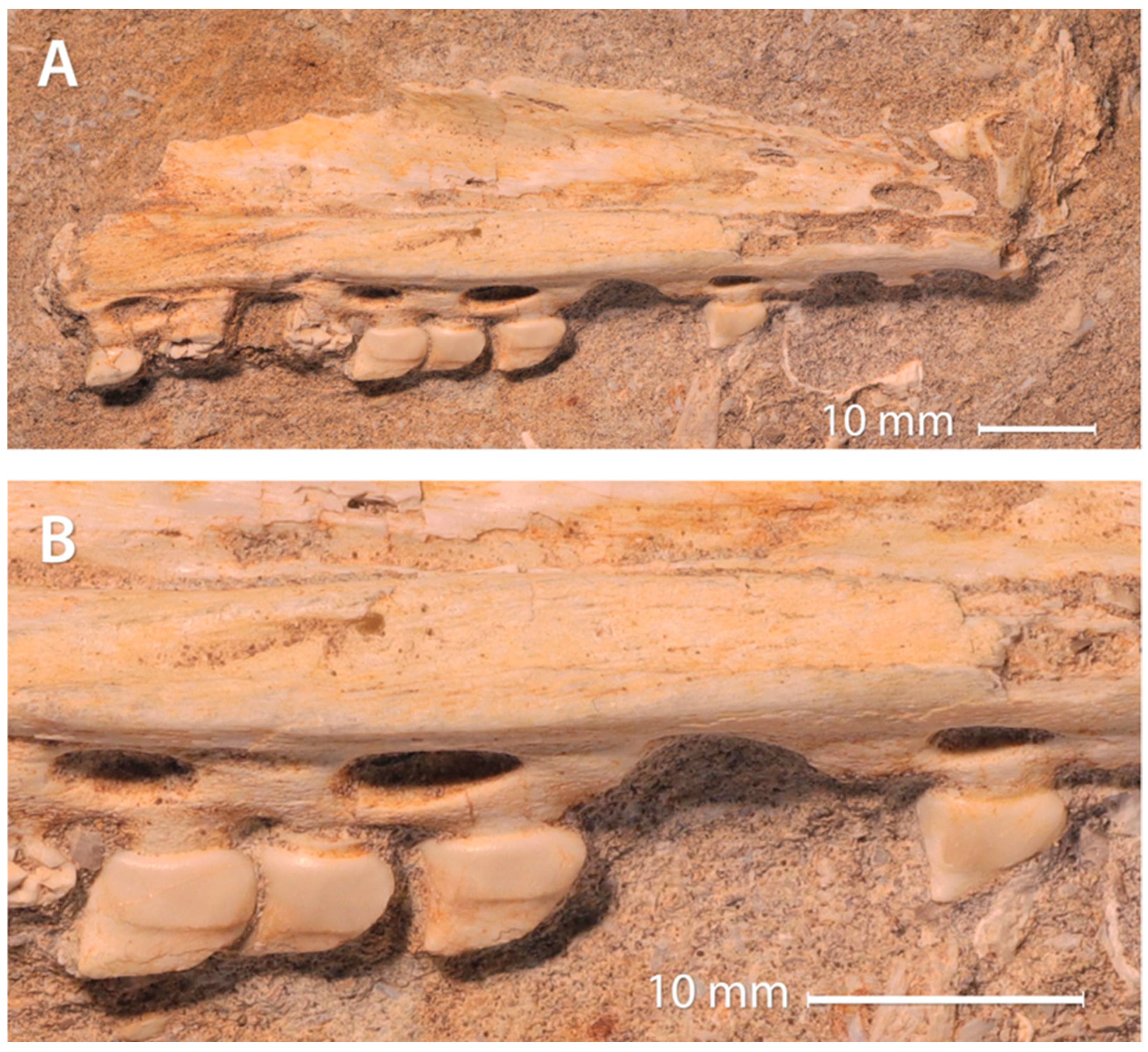
In the process of preparing this paper, we reexamined MHNM.KHG.1117, a small maxilla (Figure 3 and Figure 4) which was prepared and initially studied but not included in the original Xenodens paper; it comes from the same stratigraphical level and phosphatic basin of Morocco as the holotype, but from a different locality.
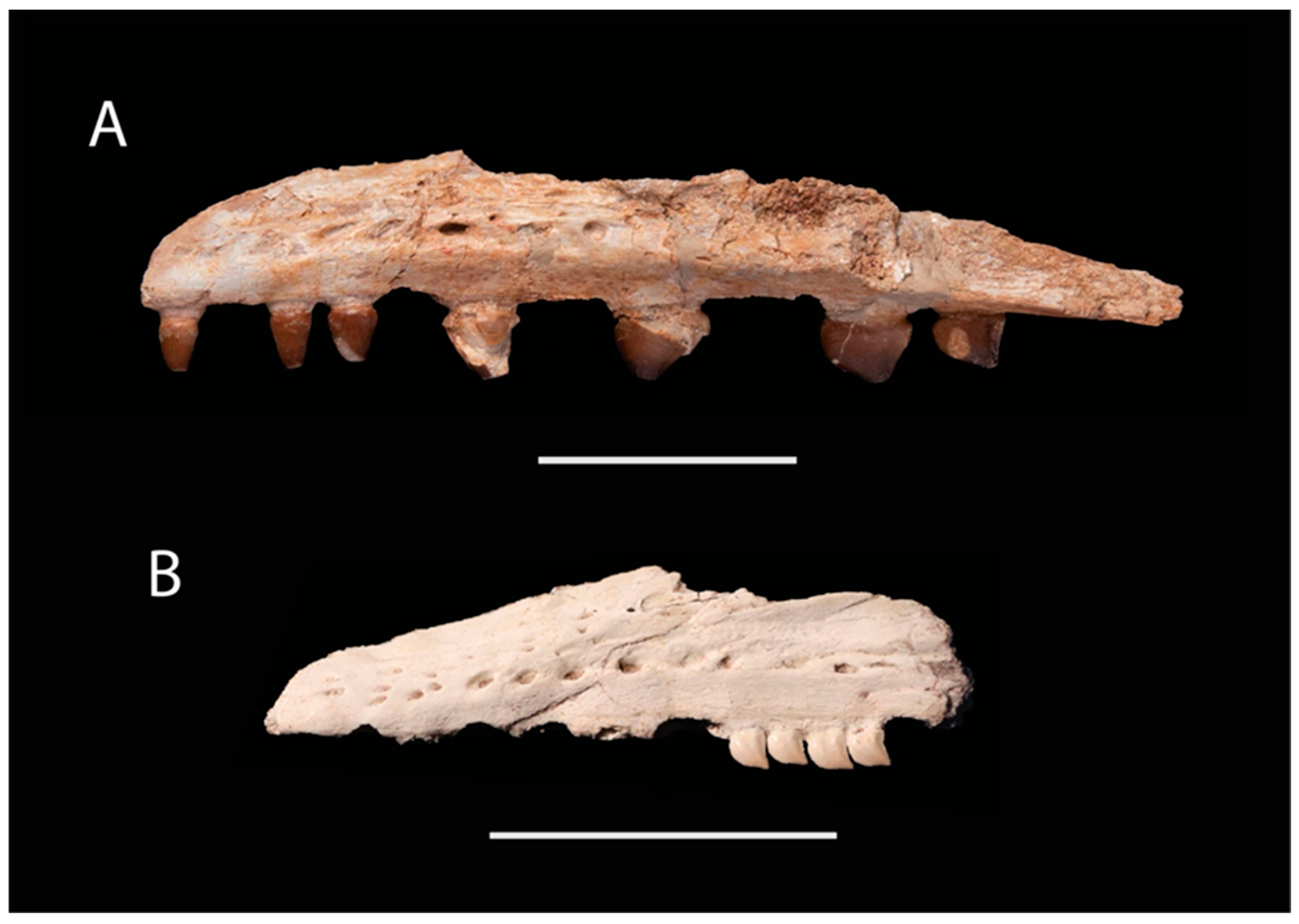
The apices of the crowns in MHNM.KHG.1117 are heavily worn (Figure 3B) and so it was originally uncertain if the specimen represented a Xenodens or a small Carinodens. Closer examination and comparisons of anatomy to the Xenodens holotype, and now CT scans, as well as new material of Carinodens [24] allow the definitive assignment of MHNM.KHG.1117 to Xenodens using the autapomorphies that characterize the genus.
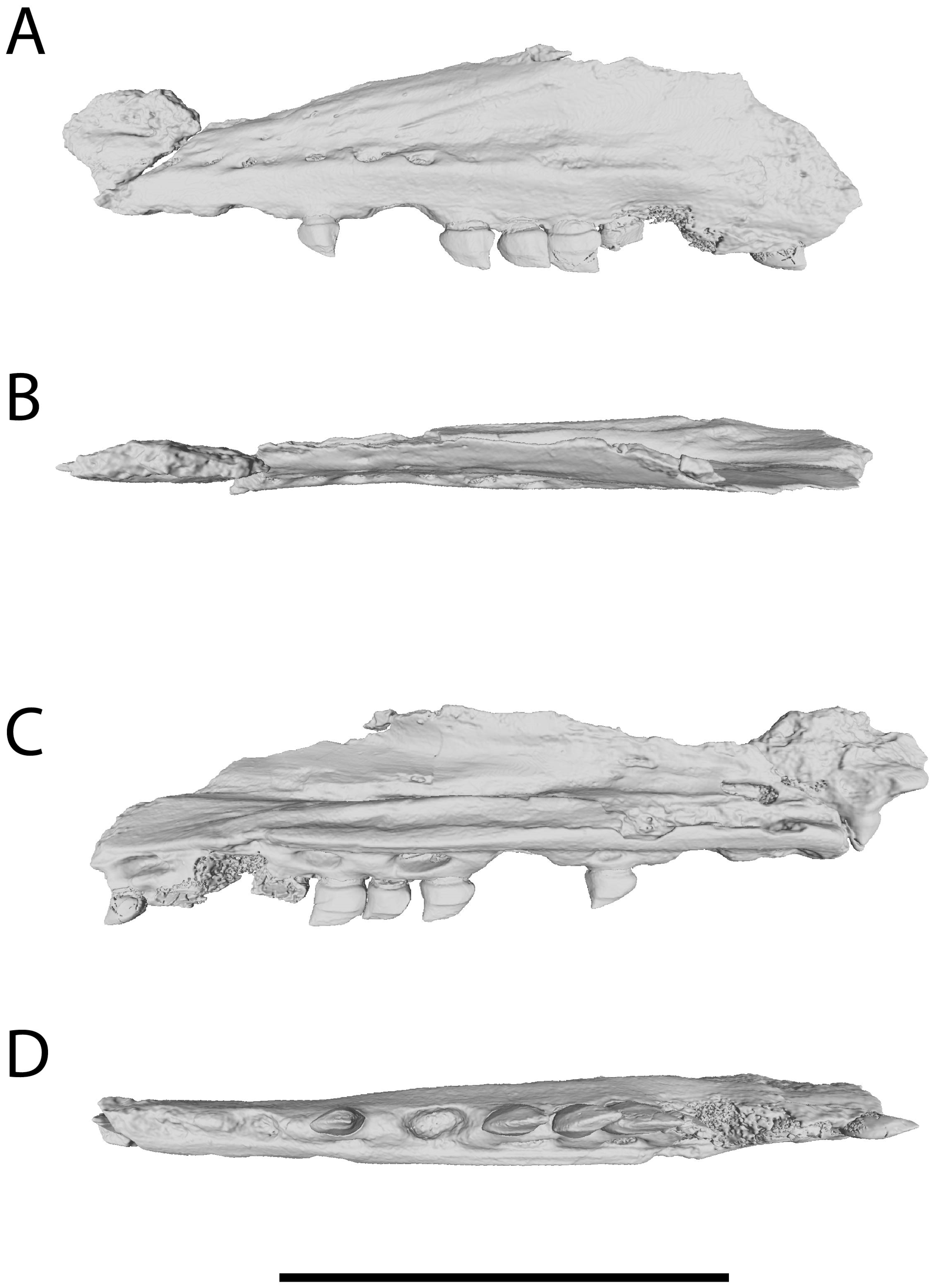
MHNM.KHG.1117 belongs to a relatively small mosasaurid, comparable in size to the holotype, again suggesting that Xenodens is characterized by small adult size. It also has a long and low maxilla and, though damaged anteriorly, it appears to have a long maxilla-premaxilla contact. At least thirteen alveoli are present, eight bearing teeth, but the maxilla is broken posteriorly and slightly anteriorly, so that the total tooth count was presumably higher.
Tooth crowns are long, low and laterally compressed as in the holotype (Figure 3, Figure 4 and Figure 6). In contrast to the holotype, however, the enamel of the tooth apices is eroded away to expose the dentine. This damage appears to represent wear facets as opposed to taphonomic alteration (e.g., digestion by other mosasaurids [11]) because the wear forms facets, the enamel of the tooth base is unaltered, and the rest of the bone is well-preserved. Tooth wear extends along the apex and onto the labial and lingual surfaces. This wear pattern supports the hypothesis that the teeth were used to pierce into and cut prey [4,9]. The absence of such wear facets in the holotype may reflect different feeding habits in the two individuals, perhaps due to variation in prey availability through the phosphate series or different feeding strategies; similar variation in tooth wear is also seen in Thalassotitan, with some individuals showing extensive and heavy tooth wear and others more limited tooth wear [11]; killer whale subpopulations also show variation in tooth wear as a result of different diets [26]. Crowns appear to be lower, but this may be because the tips are worn. The unworn bases of the crowns are smooth, and without ornament, fluting, or facets, again as in the holotype.
As in the holotype, crowns are arranged in a tightly packed series (Figure 3, Figure 4 and Figure 6). They again lie in an en echelon arrangement, with anterior carina deflected medially and posterior carina deflected laterally relative to adjacent teeth. Teeth are connected to roots by a short, narrow neck.
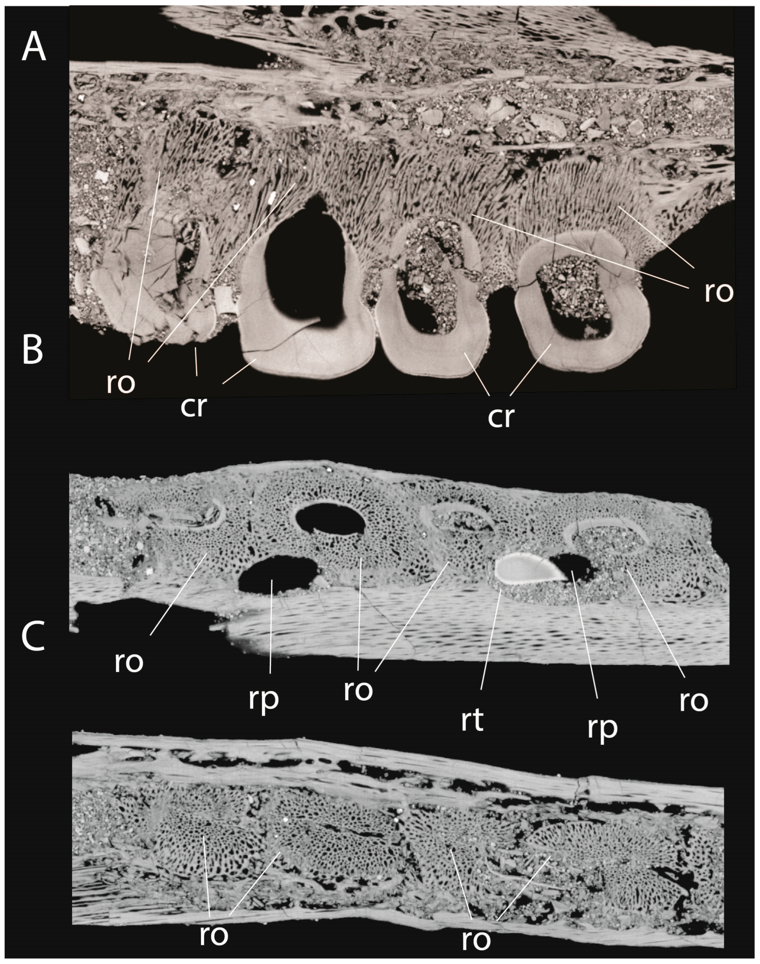
Roots of posterior teeth are anteroposteriorly expanded and coalesce to create a continuous ridge formed of osteocementum sensu LeBlanc et al. [23] without gaps or septa between them (Figure 5), again as in the holotype. Large anteroposteriorly expanded replacement pits are visible as in the holotype, with each external pit spanning the bases of two teeth.
CT scans show that the dentine of the tooth crowns extends into the osteocementum of roots. Interdental septa are visible anteriorly, but are not visible posteriorly. Instead, as in the holotype, the posterior teeth implant into an alveolar groove, with the roots of adjacent teeth expanded and coalesced to create a ridge of cementum.
One of the replacement pits contains a single replacement tooth. The lack of correspondence between the single replacement tooth and the pair of teeth being replaced could be explained if two teeth developed adjacently in the same replacement pit. However, further finds of Xenodens are needed to clarify the species’ apparently unique patterns of tooth replacement.

3.5. Tooth Implantation and Replacement of Xenodens

The dentition, implantation, and replacement of Xenodens are highly distinctive compared to those of other mosasaurids (Figure 1, Figure 2, Figure 3, Figure 4, Figure 5 and Figure 6). As previously described [9], tooth crowns are short, mesiodistally expanded with a prominent mesial carina, and strongly laterally compressed, with a bladelike shape (Figure 1, Figure 3, Figure 4 and Figure 6). Furthermore, individual crowns are packed to create a saw-like cutting blade: each tooth has unserrated carinae, but the close packing and en echelon arrangement of all the teeth creates a coarsely serrated cutting surface. Although a similar arrangement is seen in certain sharks, this arrangement was described as being unique among mosasaurids and even tetrapods [9] and we are still unaware of any other tetrapod with a similar dentition.
Another peculiar feature observed in both specimens is that, probably for the greatest efficiency of this “saw”, teeth have a staggered or alternating arrangement of taller and shorter tooth crowns (i.e., tall-short-tall-short), enhancing the “serrated” aspect. Furthermore, multiple teeth in series are ankylosed; in mosasaurids in general, every other tooth is typically fully ankylosed (NB pers. obs.).
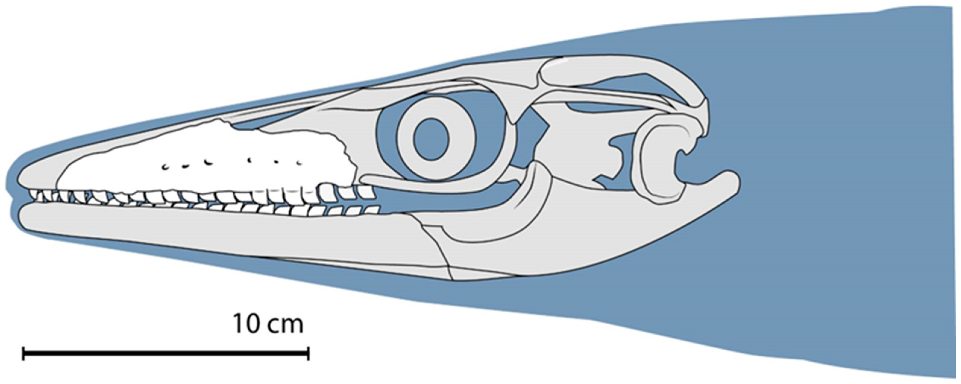
The original description [9] of the maxilla erred, however, in interpreting a pair of alveoli as a single, large replacement alveolus, due to the absence of interdental ridges delimiting alveoli. As described above, what appears to be one large tooth socket instead represents two smaller alveoli; the dentition is therefore more homodont than originally interpreted. An updated reconstruction is presented in Figure 7.
Perhaps unsurprisingly, given how modified the teeth are, tooth implantation and replacement are also highly distinctive. As in other Mosasauridae, the tooth crowns are borne on large, pillar-shaped roots, which are formed of osteocementum around a fibrous matrix. In mosasaurids, there is typically a gap between adjacent tooth bases. Interdental ridges partially separate the teeth [23].
In Tethysaurinae [27] and Halisaurinae [8,13,28], the tooth roots are broadly exposed medially, whereas in most other mosasaurids, including Tylosaurinae, Plioplatecarpinae, and Mosasaurinae, the jaw’s medial parapet extends as high as the lateral parapet [29]. Together, the lateral parapet, interdental ridges, and medial parapet define a socket or alveolus for the roots, creating a functionally “thecodont” arrangement convergent on that of archosaurs and mammals [23].
Xenodens exhibits this distinctive thecodont-like tooth implantation in the anterior part of the maxilla. Here, the tall medial parapet and interdental ridges form a discrete alveolus for each tooth. However, in the back of the maxilla, interdental ridges are reduced, and teeth implant into a groove. Curiously, a similar arrangement is seen in certain dinosaurs, with troodontid theropods exhibiting reduced interdental septa and alveoli that coalesce to form an alveolar groove receiving the tooth roots [30].
CT scans also provide new information on tooth replacement in Xenodens. As described above, the first two teeth in the holotype have replacement pits excavating their bases (Figure 2B). These connect to the pulp cavity of the tooth labially, and merge to form a single large replacement pit lingually. In the subsequent tooth pair, the replacement pits merge lingually, but do not invade the pulp cavity. The preceding pair of teeth are absent; they were presumably in the process of being replaced. A similar pattern— 2 shed teeth, 2 teeth in the process of replacement, and 2 coalesced teeth— is seen in the referred specimen (Figure 3, Figure 4 and Figure 5).

3.6. Ontogeny of Xenodens

Sharpe et al. [15] argued that Xenodens might represent a juvenile of Carinodens. However, this hypothesis can be rejected based on several lines of evidence.
First, a newly described specimen of Carinodens [24], which for the first time includes the maxilla of this genus (previously known only from dentaries and isolated teeth), demonstrates that the maxilla and associated dentition of Carinodens differ radically in shape from those of Xenodens (Figure 8). Notably, Carinodens has a taller anterior maxilla with a convex rather than straight anterodorsal margin, similar to the condition in Mosasaurus [3]. The maxilla also bears a massive lateral ridge which is absent in Xenodens. The Carinodens maxilla has fewer teeth (13 versus ~17), and middle teeth are massively enlarged relative to anterior and posterior teeth, in contrast to the relatively homodont dentition of Xenodens. Tooth roots also show strong transverse expansion. All these features are absent in Xenodens.
Tooth implantation and replacement are also very different; Carinodens appears to lack coalescence of adjacent tooth roots [24] and instead appears to retain interdental ridges. Carinodens also lacks the unique condition seen in Xenodens where adjacent tooth replacement pits coalesce, and in which adjacent tooth pairs are shed together; instead, it exhibits the typical mosasaurine pattern in which each tooth bears a replacement pit.

4. Discussion

4.1. Authenticity of Xenodens

The holotype of Xenodens calminechari is shown by CT scans to be original:
  • Each tooth crown is associated with a single tooth root.
  • Each tooth shows a continuous connection between the tooth crown and the tissues of the associated root.
  • Each tooth root is associated with a replacement pit, as in Mosasaurinae and other derived mosasaurids.
  • Original phosphate sand matrix infills the pulp cavities of teeth and replacement pits.
  • No evidence of glue, filler or plaster is visible. Adjacent teeth are similar in size, shape, color, and preservation.
This clearly demonstrates that the holotype specimen is both original and unaltered beyond the preparation of the specimen, corroborating what is readily visible from preparation and first-hand study of the fossil [9].
Further evidence of the authenticity of Xenodens comes from the referred specimen MHNM.KHG.1117—a second maxilla. This specimen was also prepared by our team and no alteration to teeth or matrix was observed. CT scanning also confirms it is original. Finally, it exhibits the diagnostic features of Xenodens [9], which include: long, low posterior tooth crowns closely packed into an en echelon arrangement, posterior tooth bases being coalesced to create a ridge of cementum supporting the crowns, enlarged replacement pits formed by the merging of two tooth replacement pits, and teeth shed in pairs. It furthermore exhibits the same small size as the holotype, consistent with the idea that small adult size is one of the features diagnosing this taxon.

4.2. Synonymy with Carinodens

New Carinodens material has, for the first time, enabled comparison of the maxillae and maxillary dentition of the two genera, demonstrating that they are very distinct.
For Xenodens to represent a juvenile Carinodens, it would require major changes in the shape of the maxilla, tooth morphology, implantation, and replacement during a short period in development (Table 1). Though changes of tooth size and shape do occur during the ontogenetic development of some extant squamates including certain Teiidae [31] and the Nile Monitor [32], we are unaware of any mosasaurid that undergoes such radical and global metamorphosis as it grows, especially as far as tooth replacement patterns are concerned.
Moreover, the smallest known Carinodens specimen (whether juvenile or not) C. palestinicus [33], though comparable in size to Xenodens, still displays the characteristic tooth morphology and ornamentation seen in larger Carinodens. The maxillary tooth row length for C. palestinicus is around 105 mm. The preserved maxillary toothrow is 97 mm for the Xenodens holotype; accounting for the teeth missing from the back of the jaw, the tooth row is ~110 mm. Neither are major differences in tooth shape, tooth count, or implantation seen between larger and smaller Carinodens in the phosphates [6]. The available evidence indicates that the tooth morphology, ornamentation and implantation of Carinodens are probably not strongly size-dependent.
Furthermore, in mosasaurids generally, the ornamentation patterns (prisms, ridges, striae, etc.) that characterize species, though less pronounced in juveniles, are already present in the juvenile stages (NB pers. obs.). Likewise, no major changes in tooth implantation or replacement are known to occur over the course of ontogeny.
The presence or absence of transitional morphologies is another way to test “synonymy through ontogeny” [34,35]. While Carinodens individuals span a range of sizes with a relatively stable anatomy [6,24], small individuals do not resemble the morphology seen in Xenodens as would be expected if the one developed into the other.
While jaws of Xenodens and Carinodens are both rare, Carinodens teeth are relatively common around the world [6,24]; Carinodens occurs in 10 countries and five continents (see, e.g., Paleobiology Database, reference [33]). If Xenodens transformed into Carinodens, then Xenodens teeth should be widely distributed too; this is not the case. The distinct biogeography of the two morphologies is inconsistent with synonymy.
To summarize the arguments against Xenodens representing a juvenile Carinodens:
  • Small Carinodens specimens, which overlap in size with Xenodens, lack Xenodens-like morphology. This argues against size-related changes in morphology of the jaws, teeth and tooth implantation;
  • Absence of intermediate morphologies of jaws or teeth morphologies (no growth series connecting the two genera) argues against synonymy;
  • Extensive differences in the maxilla structure, tooth count, tooth morphology, and tooth implantation exceed the range of intraspecific or ontogenetic variation known in any Mosasauridae.
  • Distinct geographic distribution (Carinodens very broadly distributed, Xenodens only in Morocco).

4.3. Tooth Implantation and Replacement in Xenodens

Xenodens has an unusual pattern of tooth implantation relative to other mosasaurids; the teeth are not separated by interdental ridges but instead roots are borne in an alveolar groove posteriorly, with adjacent roots abutting and apparently ankylosed to each other. Tooth implantation also seems to follow an unusual pattern; the holotype and referred specimen have a 2-2-2 sequence of tooth replacement in the jaws, i.e., two shed teeth, two teeth in the process of replacement, and two fused teeth. This suggests that tooth replacement proceeded in waves down the jaw, as is typical of Tetrapoda [36,37], but that replacement in the back of the jaw may have occurred in adjacent pairs rather than in alternation, as is more typical of reptiles [36,37]. In addition to its unique dentition, Xenodens may also have had a specialized strategy of tooth replacement, but further study and additional specimens are needed to better understand its pattern of replacement. Mosasaurids therefore show not just diversity in tooth crown morphology, but also in how crowns implant and are replaced.

5. Conclusions

  • Reexamination of the holotype specimen of Xenodens calminechari, combined with the study of a referred specimen, and CT scans reveals its highly derived and unique tooth implantation and inferred replacement pattern.
  • Newly identified features of Xenodens allow the recognition of new autapomorphies (tooth replacement pits extending across two teeth, paired tooth replacement in posterior teeth) which further underscore its unique morphology.
  • New specimens of Carinodens show that Xenodens differs from Carinodens in terms of the maxillary morphology, maxillary tooth count, weak heterodonty, and its unique mode of tooth implantation and ornamentation. These differences, together with the absence of intermediate morphologies contradict the hypothesis that Xenodens is a juvenile of Carinodens.
The evolution of Mosasauridae saw huge changes in tooth anatomy, development, replacement and implantation, not only for the clade as a whole, but also within mosasaurid subclades such as Mosasaurinae, and genera such as Xenodens. Such major shifts in morphology and development, which reflect a great plasticity of mosasaurid teeth, may have been driven by adaptation to new ecological niches, may have facilitated adaptations to new niches, or both.

Supplementary Materials

The following supporting information can be downloaded at: https://www.morphosource.org/, accessed on 18 November 2025.

Author Contributions

N.R.L., N.B., X.P.-S., A.S., N.-E.J. and M.G. contributed to the conceptualization, investigation and writing of the paper. All authors have read and agreed to the published version of the manuscript.

Funding

Research of XPS is financed by the Basque Government (research group IT1485-22).

Data Availability Statement

Both the holotype and the referred specimen are in the MHNM collections and are available on request. CT scan slices will be made available via MorphoSource.

Acknowledgments

Thanks to Mustapha Meharich for assistance in Morocco and to the Université Cadi Ayyad in Marrakech. Thanks to Marta Bellato (AST-RX platform, MNHN, Paris) for providing the scans of both specimens of Xenodens. Thanks to Andrey Atuchin for assistance with rendering and segmentation of CT scans. Thanks also to the three anonymous reviewers whose valuable comments and suggestions have improved this manuscript.

Conflicts of Interest

The authors declare no conflicts of interest.

References

  1. Bardet, N.; Falconnet, J.; Fischer, V.; Houssaye, A.; Jouve, S.; Pereda-Suberbiola, X.; Perez-García, A.; Rage, J.-C.; Vincent, P. Mesozoic marine reptile palaeobiogeography in response to drifting plates. Gondwana Res. 2014, 26, 869–887. [Google Scholar] [CrossRef]
  2. Polcyn, M.J.; Jacobs, L.L.; Araújo, R.; Schulp, A.S.; Mateus, O. Physical drivers of mosasaur evolution. Palaeogeogr. Palaeoclimatol. Palaeoecol. 2014, 400, 17–27. [Google Scholar] [CrossRef]
  3. Russell, D.A. Systematics and morphology of American mosasaurs. In Bulletin of the Peabody Museum of Natural History; Yale University: New Haven, CT, USA, 1967; Volume 23, pp. 1–240. [Google Scholar]
  4. Bardet, N.; Fischer, V.; Jalil, N.-E.; Khaldoune, F.; Yazami, O.K.; Pereda-Suberbiola, X.; Longrich, N. Mosasaurids Bare the Teeth: An Extraordinary Ecological Disparity in the Phosphates of Morocco Just Prior to the K/Pg Crisis. Diversity 2025, 17, 114. [Google Scholar] [CrossRef]
  5. Bardet, N.; Pereda-Suberbiola, X.; Iarochène, M.; Amalik, M.; Bouya, B. Durophagous Mosasauridae (Squamata) from the Upper Cretaceous phosphates of Morocco, with description of a new species of Globidens. Neth. J. Geosci. 2005, 84, 167–175. [Google Scholar] [CrossRef]
  6. Schulp, A.S.; Bardet, N.; Bouya, B. A new species of the durophagous mosasaur Carinodens (Squamata, Mosasauridae) and additional material of Carinodens belgicus from the Maastrichtian phosphates of Morocco. Neth. J. Geosci. 2009, 88, 161–167. [Google Scholar] [CrossRef]
  7. LeBlanc, A.R.; Mohr, S.R.; Caldwell, M.W. Insights into the anatomy and functional morphology of durophagous mosasaurines (Squamata: Mosasauridae) from a new species of Globidens from Morocco. Zool. J. Linn. Soc. 2019, 186, 1026–1052. [Google Scholar] [CrossRef]
  8. Bardet, N.; Pereda-Suberbiola, X.; Iarochène, M.; Bouya, B.; Amaghzaz, M. A new species of Halisaurus from the Late Cretaceous phosphates of Morocco, and the phylogenetical relationships of the Halisaurinae (Squamata: Mosasauridae). Zool. J. Linn. Soc. 2005, 143, 447–472. [Google Scholar] [CrossRef]
  9. Longrich, N.R.; Bardet, N.; Schulp, A.S.; Jalil, N.-E. Xenodens calminechari gen. et sp. nov., a bizarre mosasaurid (Mosasauridae, Squamata) with shark-like cutting teeth from the upper Maastrichtian of Morocco, North Africa. Cretac. Res. 2021, 123, 104764. [Google Scholar] [CrossRef]
  10. Strong, C.R.; Caldwell, M.W.; Konishi, T.; Palci, A. A new species of longirostrine plioplatecarpine mosasaur (Squamata: Mosasauridae) from the Late Cretaceous of Morocco, with a re-evaluation of the problematic taxon ‘Platecarpusptychodon. J. Syst. Palaeontol. 2020, 18, 1769–1804. [Google Scholar] [CrossRef]
  11. Longrich, N.R.; Jalil, N.-E.; Khaldoune, F.; Yazami, O.K.; Pereda-Suberbiola, X.; Bardet, N. Thalassotitan atrox, a giant predatory mosasaurid (Squamata) from the Upper Maastrichtian Phosphates of Morocco. Cretac. Res. 2022, 140, 105315. [Google Scholar] [CrossRef]
  12. Bardet, N.; Pereda-Suberbiola, X.; Iarochène, M.; Bouyahyaoui, F.; Bouya, B.; Amaghzaz, M. Mosasaurus beaugei Arambourg, 1952 (Squamata, Mosasauridae) from the Late Cretaceous phosphates of Morocco. Geobios 2004, 37, 315–324. [Google Scholar] [CrossRef]
  13. Longrich, N.R.; Bardet, N.; Khaldoune, F.; Yazami, O.K.; Jalil, N.-E. Pluridens serpentis, a new mosasaurid (Mosasauridae: Halisaurinae) from the Maastrichtian of Morocco and implications for mosasaur diversity. Cretac. Res. 2021, 126, 104882. [Google Scholar] [CrossRef]
  14. Longrich, N.R.; Jalil, N.-E.; Pereda-Suberbiola, X.; Bardet, N. Stelladens mysteriosus: A Strange New Mosasaurid (Squamata) from the Maastrichtian (Late Cretaceous) of Morocco. Fossils 2023, 1, 2–14. [Google Scholar] [CrossRef]
  15. Sharpe, H.S.; Powers, M.J.; Caldwell, M.W. Reassessment of Xenodens calminechari with a discussion of tooth morphology in mosasaurs. Anat. Rec. 2024, 308, 2160–2172. [Google Scholar] [CrossRef]
  16. Polcyn, M.J.; Bell, G.L. Russellosaurus coheni n. gen., n. sp., a 92 million-year-old mosasaur from Texas (USA), and the definition of the parafamily Russellosaurina. Neth. J. Geosci. 2005, 84, 321–333. [Google Scholar] [CrossRef]
  17. Bell, G.; Polcyn, M. Dallasaurus turneri, a new primitive mosasauroid from the Middle Turonian of Texas and comments on the phylogeny of Mosasauridae (Squamata). Neth. J. Geosci. 2005, 84, 177. [Google Scholar] [CrossRef]
  18. Collins, T.J. ImageJ for microscopy. Biotechniques 2007, 43, S25–S30. [Google Scholar] [CrossRef]
  19. Oppel, M. Die Ordnungen, Familien und Gattungen der Reptilien als Prodrom Einer Naturgeschichte Derselben; Lindauer: München, Germany, 1811; p. 87. [Google Scholar]
  20. Gervais, P. Zoologie et Paléontologie Françaises (Animaux Vertébrés): Nouvelles Recherches sur les Animaux Vivants de la France; Arthus Bertrand: Paris, France, 1852. [Google Scholar]
  21. Cappetta, H.; Bardet, N.; Pereda-Suberbiola, X.; Adnet, S.; Akkrim, D.; Amalik, M.; Benabdallah, A. Marine vertebrate faunas from the Maastrichtian phosphates of Benguérir (Ganntour Basin, Morocco): Biostratigraphy, palaeobiogeography and palaeoecology. Palaeogeogr. Palaeoclimatol. Palaeoecol. 2014, 409, 217–238. [Google Scholar] [CrossRef]
  22. Bardet, N.; Gheerbrant, E.; Noubhani, A.; Cappetta, H.; Jouve, S.; Bourdon, E.; Pereda-Suberbiola, X.; Jalil, N.-E.; Vincent, P.; Houssaye, A.; et al. Les Vertébrés des phosphates crétacés-paléogènes (72, 1-47, 8 Ma) du Maroc. In Mémoires de la Société Géologique de France N.S. 180; Société géologique de France: Paris, France, 2017; pp. 351–452. [Google Scholar]
  23. LeBlanc, A.R.; Lamoureux, D.O.; Caldwell, M.W. Mosasaurs and snakes have a periodontal ligament: Timing and extent of calcification, not tissue complexity, determines tooth attachment mode in reptiles. J. Anat. 2017, 231, 869–885. [Google Scholar] [CrossRef]
  24. Longrich, N.R.; Pereda-Suberbiola, X.; Jalil, N.-E.; Bardet, N. A New Species of the Durophagous Mosasaurid carinodens from the Late Maastrichtian Phosphates of Morocco and Implications for Maastrichtian Mosasaurid Diversity. Diversity 2024, 17, 25. [Google Scholar] [CrossRef]
  25. Luan, X.; Walker, C.; Dangaria, S.; Ito, Y.; Druzinsky, R.; Jarosius, K.; Lesot, H.; Rieppel, O. The mosasaur tooth attachment apparatus as paradigm for the evolution of the gnathostome periodontium. Evol. Dev. 2009, 11, 247–259. [Google Scholar] [CrossRef]
  26. Ford, J.K.; Ellis, G.M.; Matkin, C.O.; Wetklo, M.H.; Barrett-Lennard, L.G.; Withler, R.E. Shark predation and tooth wear in a population of northeastern Pacific killer whales. Aquat. Biol. 2011, 11, 213–224. [Google Scholar] [CrossRef]
  27. Bardet, N.; Pereda-Suberbiola, X.; Jalil, N.-E. A new mosasauroid (Squamata) from the Late Cretaceous (Turonian) of Morocco. Comptes Rendus Palevol 2003, 2, 607–616. [Google Scholar] [CrossRef]
  28. Longrich, N.R. A new species of Pluridens (Mosasauridae: Halisaurinae) from the upper Campanian of Southern Nigeria. Cretac. Res. 2016, 64, 36–44. [Google Scholar] [CrossRef]
  29. Bell, G.L., Jr. A phylogenetic revision of North American and Adriatic Mosasauroidea. In Ancient Marine Reptiles; Elsevier: Amsterdam, The Netherlands, 1997; pp. 293–332. [Google Scholar]
  30. Currie, P.J. Bird like characteristics of the jaws and teeth of troodontid theropods (Dinosauria: Saurischia). J. Vertebr. Paleontol. 1987, 7, 72–81. [Google Scholar] [CrossRef]
  31. Estes, R.; Williams, E.E. Ontogenetic variation in the molariform teeth of lizards. J. Vertebr. Paleontol. 1984, 4, 96–107. [Google Scholar] [CrossRef]
  32. D’Amore, D.C. Illustrating ontogenetic change in the dentition of the Nile monitor lizard, Varanus niloticus: A case study in the application of geometric morphometric methods for the quantification of shape–size heterodonty. J. Anat. 2015, 226, 403–419. [Google Scholar] [CrossRef]
  33. Kaddumi, H.F. The first and most complete Carinodens (Squamata: Mosasauridae) skeleton yet with a description of a new species from the Harrana Fauna, Fossils of the Harrana Fauna and the Adjacent Areas. In Fossils of the Harrana Fauna and the Adjacent Area; Areas, A., Ed.; Publications of the Eternal River Museum of Natural History: Amman, Jordan, 2009; pp. 49–64. [Google Scholar]
  34. Longrich, N.R.; Saitta, E.T. Taxonomic status of Nanotyrannus lancensis (Dinosauria: Tyrannosauroidea)—A distinct taxon of small-bodied tyrannosaur. Foss. Stud. 2024, 2, 1–65. [Google Scholar] [CrossRef]
  35. Longrich, N.R.; Field, D. Torosaurus is not Triceratops: Ontogeny in Chasmosaurine Ceratopsids as a Case Study in Dinosaur Taxonomy. PLoS ONE 2012, 7, e32623. [Google Scholar] [CrossRef]
  36. Whitlock, J.A.; Richman, J.M. Biology of tooth replacement in amniotes. Int. J. Oral Sci. 2013, 5, 66–70. [Google Scholar] [CrossRef]
  37. Berkovitz, B. 13 Tooth replacement patterns in non-mammalian vertebrates. In Development, Function and Evolution of Teeth; Mark, F., Teaford, M.M.S., Mark, W., Ferguson, J., Eds.; Cambridge University Press: Cambridge, UK, 2007; p. 186. [Google Scholar]
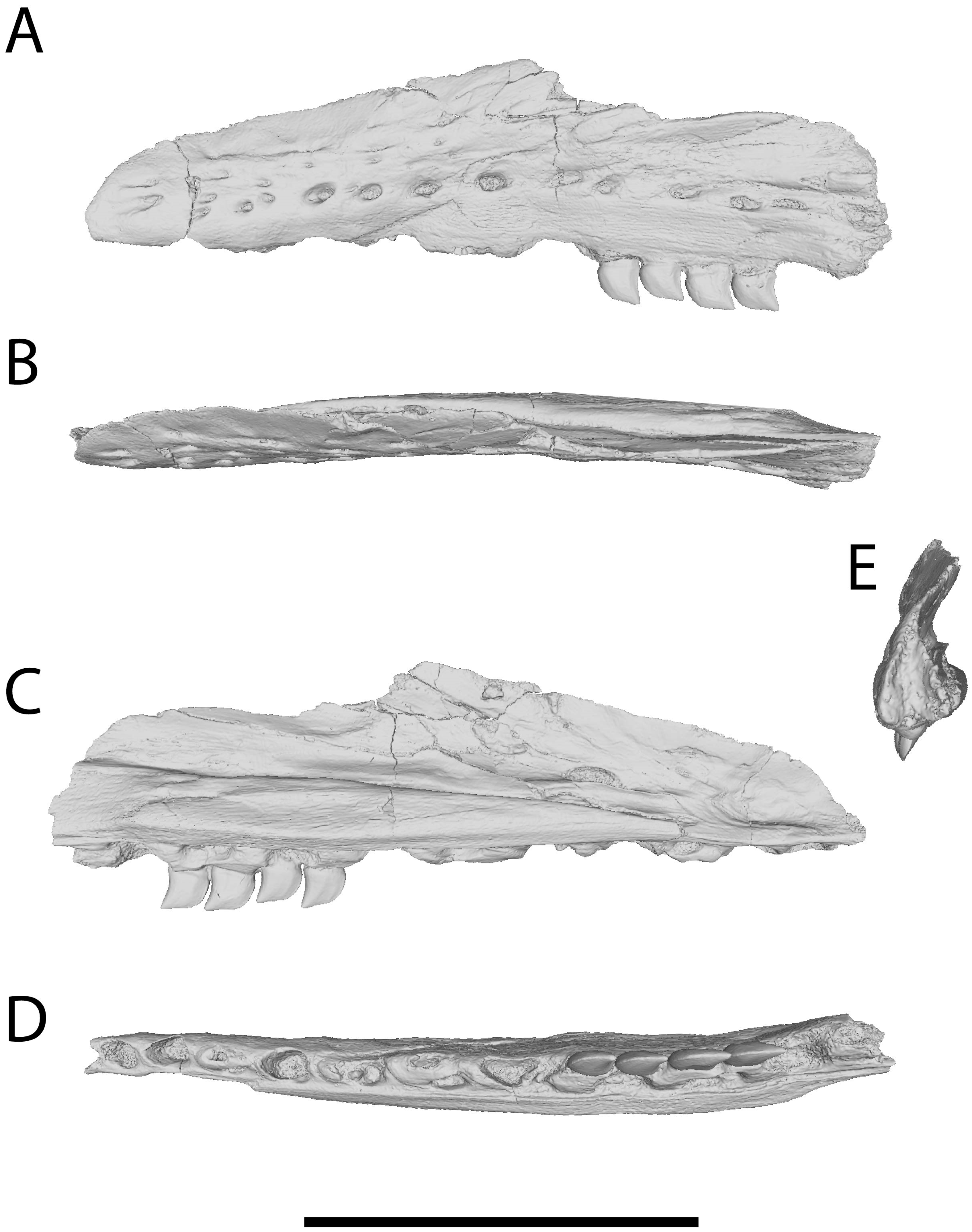
Figure 1. Rendering of CT scan segmentation of Xenodens calminechari, MHNM.KHG.331 (holotype), left maxilla; Sidi Chennane, Oulad Abdoun Basin, Morocco; Phosphates, Upper Couche III, uppermost Maastrichtian [9]. (A), lateral view; (B), dorsal view; (C), medial view; (D), ventral view; (E), anterior view. Scale = 50 mm.
Diversity 17 00819 g001
Figure 2. CT scan cross-sections of Xenodens calminechari, MHNM.KHG.331 (holotype), left maxilla; Sidi Chennane, Oulad Abdoun Basin, Morocco; Phosphates, Upper Couche III, uppermost Maastrichtian [9]. (A), sagittal section showing attachment of tooth crowns to tooth roots (=osteocementum pedicel) and implantation of roots into the maxilla; (B), coronal plane through the level of the tooth crown base showing resorption of tooth roots and merged replacement pits; (C), coronal plane through the level of the root showing expanded and coalesced roots and replacement pits. Abbreviations: cr, crown; mx, maxilla; re, resorbed tooth bases; ro, root; rp, replacement pit.
Diversity 17 00819 g002
Figure 3. Xenodens calminechari, MHNM.KHG.1117 (referred specimen), left maxilla; Sidi Daoui, Oulad Abdoun Basin, Morocco; Phosphates, Upper Couche III, uppermost Maastrichtian. (A), medial view; (B), closeup showing teeth and elongate tooth replacement pits.
Diversity 17 00819 g003
Figure 4. Rendering of CT scan segmentation of Xenodens calminechari, MHNM.KHG.1117 (referred specimen), left maxilla; Sidi Daoui, Oulad Abdoun Basin, Morocco; Phosphates, Upper Couche III, uppermost Maastrichtian. (A), lateral view; (B), dorsal view; (C), medial view; (D), ventral view. Scale = 50 mm.
Diversity 17 00819 g004
Figure 5. CT scan cross-sections of Xenodens calminechari, MHNM.KHG.1117 (referred specimen), left maxilla; Sidi Daoui, Oulad Abdoun Basin, Morocco; Phosphates, Upper Couche III, uppermost Maastrichtian. (A), sagittal plane in medial view; (B), coronal plane through the level of the tooth crowns and roots in dorsal view; (C), coronal plane through the level of the roots. Abbreviations: cr, crown; ro, root; rp, replacement pit; rt, replacement tooth.
Diversity 17 00819 g005
Figure 6. Rendering of CT scan segmentation of maxilla of Xenodens calminechari, MHNM.KHG.1117 (referred specimen), left maxilla; Sidi Daoui, Oulad Abdoun Basin, Morocco; Phosphates, Upper Couche III, uppermost Maastrichtian, showing tooth crowns and the dentine component of the roots. (A), medial view; (B), lateral view; (C), closeup of teeth in lateral view. Scales = 10 mm.
Diversity 17 00819 g006
Figure 7. Revised reconstruction of Xenodens calminechari.
Diversity 17 00819 g007
Figure 8. Carinodens acrodon holotype left maxilla (MHNM.KHG.1510) (A) compared to Xenodens calminechari holotype left maxilla (MHNM.KHG.331) (B), in lateral view; both from Sidi Chennane, Oulad Abdoun Basin, Morocco; Phosphates, Upper Couche III, uppermost Maastrichtian [9,24]. Scales = 50 mm.
Diversity 17 00819 g008
Table 1. Characters differentiating Carinodens and Xenodens.
CharacterCarinodensXenodens
MaxillaRounded and short premaxillary-nasal sutureStraight, posteriorly extended premaxillary-nasal suture
HeterodontyExtremeWeakly developed
Tooth morphology/
Ornamentation
Inflated/ornamentedBladelike/smooth
Tooth implantationTeeth separated by interdental ridgesInterdental ridges reduced in posterior teeth
Tooth replacement pitsModerately elongate, one per toothHyperelongate, extending across two teeth
Tooth root shapeTransversely expandedTransversely narrow
Tooth root fusionAbsentPresent
HeterodontyExtreme enlargement of
lateral teeth
Moderate heterodonty
Interdental ridgesPresentAbsent
Disclaimer/Publisher’s Note: The statements, opinions and data contained in all publications are solely those of the individual author(s) and contributor(s) and not of MDPI and/or the editor(s). MDPI and/or the editor(s) disclaim responsibility for any injury to people or property resulting from any ideas, methods, instructions or products referred to in the content.