Article

Comparison of Gut Bacterial Communities in the Freshwater Mussel Sinanodonta woodiana at Different Life Stages

1
Wuxi Fisheries College, Nanjing Agricultural University, Wuxi 214081, China
2
Freshwater Fisheries Research Center, Chinese Academy of Fishery Sciences, Wuxi 214081, China
3
College of Marine Science and Technology and Environment, Dalian Ocean University, Dalian 116023, China
4
Ministry of Fisheries and Marine Resources, Freetown 190, Sierra Leone
*
Author to whom correspondence should be addressed.
These authors contributed equally to this work.
Diversity 2025, 17(12), 814; https://doi.org/10.3390/d17120814
Submission received: 22 October 2025 / Revised: 20 November 2025 / Accepted: 24 November 2025 / Published: 25 November 2025
(This article belongs to the Special Issue Ecology and Conservation of Freshwater Bivalves)

Abstract

Freshwater mussels hold significant ecological and economic value. Gut bacterial communities can regulate the growth and immunity of freshwater mussels. However, the dynamics of gut bacterial communities in freshwater mussels at different life stages are still limited. This study used the globally widespread mussel, Sinanodonta woodiana, as a model animal and employed 16S rRNA sequencing technology to comparatively analyze the gut bacterial communities of early juveniles, late juveniles, and adults. Alpha diversity indices indicated a trend of increasing richness and diversity of the gut bacterial communities with the mussel growth. Beta diversity analysis revealed distinct stage-specific taxonomic profiles. At the phylum level, four dominant phyla were identified in the early juveniles, namely Fusobacteriota, Verrucomicrobiota, Pseudomonadota, and Cyanobacteriota; and seven dominant phyla were identified in both late juveniles and adults, namely Fusobacteriota, Pseudomonadota, Verrucomicrobiota, Cyanobacteriota, Bacillota, Bacteroidota, and Chloroflexota. Among them, the relative abundance of Fusobacteriota decreased with the mussel growth (p < 0.05), while the relative abundances of Pseudomonadota and Cyanobacteriota increased with the mussel growth (p < 0.05). At the genus level, four dominant genera were identified in the early juveniles: Cetobacterium, LD29, Cyanobium_PCC-6307, and Cupriavidus; seven dominant genera were identified in the late juveniles: Cetobacterium, Roseomonas, LD29, Cyanobium_PCC-6307, Limnolyngbya_CHAB4449, Terrimicrobium, Limnothrix; and nine dominant genera were detected in the adults: Cetobacterium, LD29, Roseomonas, Cyanobium_PCC-6307, Limnothrix, Limnolyngbya_CHAB4449, Sediminibacterium, Terrimicrobium, Acidibacter. Among these, the relative abundance of Cetobacterium decreased with the mussel growth (p < 0.05), while the relative abundance of Acidibacter increased with the mussel growth (p < 0.05). Functional prediction revealed that the gut bacterial communities were primarily involved in metabolic pathways, including the biosynthesis of ansamycins, biosynthesis of vancomycin group antibiotics, D-glutamine and D-glutamate metabolism, biotin metabolism, valine, leucine and isoleucine biosynthesis, and fatty acid biosynthesis. The findings provide insights for enhancing the nutrition and health of freshwater mussels.

1. Introduction

The freshwater mussel Sinanodonta woodiana originated in the Yangtze River basin of China [1]. Currently, this mussel has spread to 46 countries across Asia, Europe, North America, and Africa [1,2]. It holds significant ecological value. Through powerful water filtration (260 L/h/kg dry soft tissue weight) [3], it can effectively purify water quality [4]. Due to its wide distribution, benthic nature, high accumulation and low metabolism of pollutants, it has also been selected as an ideal bioindicator for monitoring environmental pollution dynamics [5]. Furthermore, S. woodiana has important economic value, including for food [6], pearl cultivation [7], and extraction of antitumor drugs [8].
The typical life stages of S. woodiana include glochidia, early juveniles (0–180 days), late juveniles (180–approximately 750 days), and adults (>750 days) [9]. Glochidia have not yet differentiated organs like the intestine and need to parasitize host fish, such as the yellow catfish (Pelteobagrus fulvidraco), absorbing nutrients from the host fish to metamorphose into independently viable early juveniles [9,10]. Compared to late juveniles and adults, early juveniles grow faster (absolute growth rate 0.02–0.65 mm/d) but also have a higher mortality rate (often experiencing “complete wipeout”) [9,11]. Differences in ingested food are considered one of the key factors leading to variations in growth and survival among different life stages of S. woodiana [11].
The food composition of freshwater mussels typically includes phytoplankton, zooplankton, bacteria, and sediment [12]. Our previous study identified the phytoplankton composition in the digestive tracts of early juveniles (2 and 5 months old) and late juveniles (8 and 11 months old) [11]. Although Chlorophyta predominated (54.5–92.3%), algal diversity was found to gradually increase with the growth of S. woodiana [11]. Liu et al. [13] analyzed the gut microbiota of S. woodiana (age or size not reported) collected from the Gan River in China and found that the dominant phylum in the gut microbiota was Bacillota (accounting for 21.84%), and the gut bacterial communities played dual roles in nutrition and immunity. In the freshwater mussel Lampsilis cardium, the gut bacterial communities also serve as an indicator of the contamination level by contaminants of emerging concern in agricultural watersheds [14]. Meanwhile, the gut bacterial communities of the marine green-lipped mussel Perna canaliculus demonstrates resilience to short-term starvation [15]. However, the dynamics of gut bacterial communities in mussels (including both freshwater and marine species) across different life stages are still limited.
This study employs S. woodiana as a model animal to characterize the diversity and predict the functions of its gut bacterial communities at different life stages, including early juveniles, late juveniles, and adults. The results provide insights for enhancing the nutrition and health of freshwater mussels.

2. Materials and Methods

2.1. Sample Collection

In July 2024, 100 early juveniles, 15 late juveniles, and 5 adults were collected from the Nanquan Base of the Freshwater Fisheries Research Center, Chinese Academy of Fishery Sciences. Their ages and sizes are shown in Table 1. These S. woodiana were artificially bred and cultured in the same pond (water area 333 m2, water depth 1.5 m). The mussels were fed on natural diets without supplementary feeding. Water quality parameters were measured using a combination of in situ and laboratory-based methods. Temperature and pH were determined on-site using a portable pH meter (Hanna Instruments, Villafranca, Italy). Dissolved oxygen was measured in situ with a dissolved oxygen probe (Mettler Toledo, Giessen, Germany), while conductivity and total dissolved solids were recorded using a conductivity meter (Mettler Toledo, Germany). For additional parameters, water samples were transported to the laboratory under cold chain conditions. Subsequently, total nitrogen and total phosphorus concentrations were quantified using a multiparameter water quality analyzer (Hach, Loveland, CO, USA), and chlorophyll-a content was determined spectrophotometrically (Mapada Instruments, Shanghai, China). The measured parameters were as follows: temperature 23.5 °C, pH 7.6, dissolved oxygen 9.3 mg/L, conductivity 265 μS/cm, total dissolved solids 137 mg/L, total nitrogen 0.68 mg/L, total phosphorus 0.05 mg/L, and chlorophyll-a 5.5 μg/L.
Owing to their small size and the minimal amount of intestinal content, neither early nor late juveniles provided sufficient material for analysis of gut bacterial communities per individual. To compensate, 100 early juveniles and 15 late juveniles were randomly pooled into five groups (n = 5), respectively. In contrast, as each adults yielded sufficient intestinal content, the five adults were processed as individual samples (n = 5) without pooling. Mussels were rinsed with 70% alcohol and sterile water. Subsequently, the intestinal contents were aseptically collected into sterile centrifuge tubes, immediately snap-frozen in liquid nitrogen, and stored until DNA extraction.

2.2. Genomic DNA Extraction, PCR Amplification, and Sequencing

Total genomic DNA was extracted from intestinal contents using the CTAB method [16]. DNA concentration and quality were detected using 1% agarose gel electrophoresis and NanoDrop 2000 spectrophotometer (Thermo Scientific, Waltham, MA, USA). The V3-V4 hypervariable region of the 16S rRNA gene was amplified by PCR using primers 341F (5′-CCTAYGGGRBGCASCAG-3′) and 806R (5′-GGACTACNNGGGTATCTAAT-3′), yielding an amplicon of approximately 460 bp. The reaction system contained 15 µL Phusion® High-Fidelity PCR Master Mix (New England Biolabs, Evry, France), 0.2 µM of each forward and reverse primer, and 10 ng template DNA. PCR products were detected by 2% agarose gel electrophoresis. Subsequently, amplification products from each sample were mixed in equal amounts and purified using the Agencourt AMPure XP nucleic acid purification kit (Beckman Coulter, Brea, CA, USA). The purified amplicon libraries were sequenced on an Illumina NovaSeq platform, and 250 bp paired-end reads were generated. To monitor for potential contamination, negative controls (using nuclease-free water instead of template DNA) were included during the DNA extraction and PCR amplification steps. No amplification products were detected in these controls via gel electrophoresis. All raw data can be found in Table S1.

2.3. Data Processing

The raw data obtained from sequencing were subjected to the following quality control and processing: First, Trimmomatic software (version 0.36) [17,18] was used for quality filtering (sliding window size: 50 bp, average quality threshold: 20), and sequences shorter than 120 bp were truncated. Then, FLASH software (version 1.20) [14,19] was used to merge paired-end reads (minimum overlap: 10 bp, maximum mismatch rate: 0.1) to obtain complete V3-V4 region sequences. The UCHIME algorithm [14,20] was used to identify and remove chimeric sequences, ultimately yielding high-quality sequences. At a 97% similarity level, high-quality sequences were clustered into Operational Taxonomic Units (OTUs) using the UPARSE method [21,22]. Representative sequences were annotated against the Silva database (Release 132 http://www.arb-silva.de/) [14,23].

2.4. Statistical Analysis

Subsequent analyses were performed using QIIME 1.8.0 software [24,25]. To ensure fair comparisons, the OTU table was rarefied to a consistent sampling depth prior to all diversity calculations. Based on the normalized OTU table, Chao1, Shannon, Simpson, and Good’s coverage indices were calculated to assess alpha diversity. To assess sequencing depth coverage and sample size adequacy, rarefaction curves [26,27] and species accumulation curves [28,29] were generated, respectively. A heatmap was employed to analyze the weighted UniFrac distances among samples. To visualize the compositional differences based on these distances, Principal Coordinate Analysis (PCoA) was applied. Furthermore, PERMANOVA was used to statistically test the observed differences between samples. Hierarchical clustering analysis was performed based on the beta diversity distance matrix to obtain a similarity tree for all samples. Phyla or genera with an average relative abundance above 1% were defined as dominant. The PICRUSt2 software (version 2.1.2-b) was used to predict the functional potential of the gut bacterial communities.
Additionally, differences in gut bacterial communities—including alpha diversity indices, relative abundances at the phylum and genus levels, and predicted functional profiles—among early juveniles, late juveniles, and adults were assessed using one-way ANOVA. Prior to ANOVA, the normality of the data distribution was examined. If the data met the normality assumption, post hoc comparisons were performed using the LSD method; otherwise, the Dunnett’s T3 method was applied. Data are presented as mean ± standard deviation.

3. Results

3.1. OTU Analysis and Alpha Diversity

After data preprocessing, 1,507,820 high-quality sequences were ultimately obtained from the 15 samples. The counts of clean reads and clean tags obtained for each sample are presented in Table S2. Both the sample rarefaction curves (Figure 1a) and species accumulation curves (Figure 1b) approached saturation plateaus, indicating that the sequencing depth and sample size were sufficient to cover most of the bacterial community. A total of 3448 OTUs were obtained from the clean tags. Of these, 42 OTUs (1.22%) were common to all 15 samples. All OTUs were annotated at different taxonomic levels, and 19 phylum-level taxa were identified in all samples to understand the species composition of the bacterial community.
Three alpha diversity indices, Chao1, Simpson, and Shannon, were calculated to assess the bacterial community richness and diversity of the early juveniles, late juveniles, and adults. The sequencing completeness of all samples was estimated using the Good’s coverage index, and all reached 100% (Table 2). The data showed that the Chao1 of the early juveniles was significantly lower than that of the late juveniles and adults (p < 0.05), while there was no significant difference in Chao1 between the late juveniles and adults (p > 0.05; Table 2). Both the Shannon and Simpson indices significantly increased with the mussel growth (p < 0.05; Table 2).

3.2. Beta Diversity

The heatmap revealed the most pronounced dissimilarity between the early juveniles and adults, followed by that between the early juveniles and late juveniles (Figure 2a). The PCoA effectively captured 91.1% of the total variation, clearly illustrating a distinct separation among the early juveniles, late juveniles, and adults (Figure 2b). Furthermore, PERMANOVA results confirmed that the pairwise differences between all three life stages were statistically significant (all p < 0.001). Clustering results showed that the early juveniles clustered separately in one branch, while the late juveniles and adults clustered in another branch (Figure 2c).

3.3. Taxonomic Composition

The dominant phyla of gut bacterial communities in the early juveniles, late juveniles, and adults are shown in Table 3. The early juveniles had four dominant phyla: Fusobacteriota, Verrucomicrobiota, Pseudomonadota, and Cyanobacteriota, accounting for 98.55% of the total abundance. Seven dominant phyla were identified in the late juveniles: Fusobacteriota, Pseudomonadota, Verrucomicrobiota, Cyanobacteriota, Bacillota, Bacteroidota, Chloroflexota, accounting for 98.86% of the total abundance. All dominant phyla in the adults were the same as those in the late juveniles, with the seven dominant phyla accounting for 98.43% of the total abundance. Among them, the relative abundance of Verrucomicrobiota remained stable during the mussel growth (p > 0.05) and the relative abundance of Fusobacteriota decreased with the mussel growth (p < 0.05), while the relative abundances of Pseudomonadota and Cyanobacteriota increased with the mussel growth (p < 0.05).
The dominant genera of gut bacterial communities in the early juveniles, late juveniles, and adults are shown in Table 4. Four dominant genera were detected in the early juveniles: Cetobacterium, LD29, Cyanobium_PCC-6307, Cupriavidus, accounting for 96.01% of the total abundance. Seven dominant genera were detected in the late juveniles: Cetobacterium, Roseomonas, LD29, Cyanobium_PCC-6307, Limnolyngbya_CHAB4449, Terrimicrobium, Limnothrix, accounting for 81.91% of the total abundance. Nine dominant genera were detected in the adults: Cetobacterium, LD29, Roseomonas, Cyanobium_PCC-6307, Limnothrix, Limnolyngbya_CHAB4449, Sediminibacterium, Terrimicrobium, Acidibacter, accounting for 79.17% of the total abundance. Among them, the relative abundances of LD29 and Terrimicrobium remained stable during the mussel growth (p > 0.05) and the relative abundance of Cetobacterium decreased with the mussel growth (p < 0.05), while the relative abundance of Acidibacter increased with the mussel growth (p < 0.05).

3.4. Functional Prediction

The critical functions at the genus level of the gut bacterial communities, predicted by PICRUSt2, are presented in Figure 3. Early juvenile communities were enriched in KEGG pathways including ansamycin biosynthesis, vancomycin group antibiotic biosynthesis, D-glutamine and D-glutamate metabolism, and biotin metabolism. Late juvenile communities were associated with ansamycin biosynthesis, vancomycin group antibiotic biosynthesis, D-glutamine and D-glutamate metabolism, and valine, leucine, and isoleucine biosynthesis. In adults, the communities were linked to ansamycin biosynthesis, vancomycin group antibiotic biosynthesis, valine, leucine, and isoleucine biosynthesis, and fatty acid biosynthesis. Among these, biosynthesis of ansamycins, biosynthesis of vancomycin group antibiotics, and biotin metabolism significantly decreased with the mussel growth (p < 0.05), whereas valine, leucine and isoleucine biosynthesis and fatty acid biosynthesis remained stable during the mussel growth (p > 0.05).

4. Discussion

4.1. Diversity of Gut Bacterial Communities

The gut bacterial communities of freshwater bivalves are co-influenced by environmental and biological factors [13,30]. For example, Weingarten et al. [30] conducted 16S rRNA sequencing on the gut microbiota of four freshwater mussel species (Cyclonaias asperata, Fusconaia cerina, Lampsilis ornata, Obovaria unicolor) and found that the gut microbiota significantly resembled the filtered seston in the water, and the host species had a selective effect on the finally retained microorganisms. This suggests that mussels “introduce” environmental microorganisms into their gut during feeding, which are subsequently retained through the host’s physiological selection. In this study, early juveniles, late juveniles, and adults of S. woodiana were collected from the same environment; therefore, the differences in their gut bacterial communities should be attributed to different life stages.
This study observed a trend of increasing richness and diversity of the gut bacterial communities in S. woodiana with mussel growth, and the gut bacterial composition of late juveniles and adults was more similar. This should be related to the growth and development characteristics of S. woodiana. Early juveniles must undergo the formation of organs such as incurrent/excurrent siphons, gills, visceral mass, intestine, and heart, whereas late juveniles and adults have fully developed organs [9]. Correspondingly, the feeding capacity of late juveniles and adults increases compared to early juveniles, with the size range of ingested food particles expanding from less than 10 µm to 30 µm [11]. This trend of increasing gut microbiota diversity during development is corroborated by studies on other aquatic species. For instance, in the giant freshwater prawn (Macrobrachium rosenbergii), the community richness (Chao1 index) significantly increased from one to two months of age [29]. Similarly, in the Tibetan fish Schizothorax o’connori, the Shannon diversity index showed a clear increasing trend from 6 to 15 years of age [31].

4.2. Composition of Gut Bacterial Communities

This study revealed that Fusobacteriota was the most important dominant phylum in the gut of early juveniles, late juveniles, and adults, accounting for 34.12–83.82% of the total abundance. This differs from the report by Liu et al. [13] where Bacillota (accounting for 21.84% of total abundance) was the most important dominant phylum in the gut microbiota of S. woodiana (age or size not reported). This could be attributed to the significant environmental differences between the two studies. Interestingly, the gut microbiota of healthy yellow catfish is also dominated by Fusobacteriota and the genus Cetobacterium as dominant microbiota [32]. Cetobacterium, as a beneficial bacterium, plays an important role in the nutrition and health of fish [33,34,35]. It can ferment peptide carbohydrates, produce vitamin B12 and butyrate, and inhibit the growth of other pathogenic strains [36,37]. This suggests that Cetobacterium may also play nutritional and immune roles during the growth of S. woodiana.
Cupriavidus and Roseomonas are two genera belonging to the Pseudomonadota. A previous study indicated that Cupriavidus is a beneficial bacterium that promotes the growth of improved crucian carp WR-II [38]. In this study, Cupriavidus was a dominant genus in the gut bacterial communities of early juveniles but not in late juveniles and adults. Furthermore, early juveniles exhibited significantly higher growth rates than late juveniles and adults [9], suggesting that Cupriavidus may have a growth-promoting effect in S. woodiana. Roseomonas is a heterotrophic bacterium capable of utilizing various organic compounds as carbon sources. Some Roseomonas strains also exhibit resistance to heavy metals, for example, Roseomonas corallii possesses resistance genes for copper, cadmium, lead, and arsenic [39]. The dominance of Roseomonas in late juveniles and adults may improve their survival rates [9] by enhancing resistance to environmental stressors like heavy metals.
While containing algal toxins [40], Cyanobacteriota are also rich in protein, carotenoids, vitamins, minerals, and essential fatty acids [41]. This analysis found that three genera of Cyanobacteriota (Cyanobium_PCC-6307, Limnothrix, Limnolyngbya_CHAB4449) were dominant genera in late juveniles and adults. This may be related to the feeding characteristics of S. woodiana. The dietary spectrum of S. woodiana shifts with ontogeny, with both the diversity of ingested algae and the capacity to consume and digest cyanobacteria increasing throughout development, particularly in late juveniles and adults [11,40].
The Bacillota/Bacteroidota ratio is closely related to host lipid synthesis and storage. An increase in Bacillota abundance is often accompanied by upregulation of fatty acid synthesis genes, leading to increased body fat [42]. In this study, the abundance of Bacillota was highest in the late juveniles, with no difference between early juveniles and adults; the abundance of Bacteroidota was highest in the adults, with no difference between early juveniles and late juveniles. This may be related to the high demand for energy and lipids for tissue construction in growing late juveniles, and the increased Bacillota abundance might provide more short-chain fatty acids (SCFAs) and fatty acid synthesis precursors, promoting lipid deposition. Bacteroidota can benefit the host by helping digest complex carbohydrates, biotransforming bile acids, synthesizing vitamins, and developing the immune system [43]. Furthermore, various genera within it can produce SCFAs, thereby regulating the intestinal barrier and energy metabolism [44]. For example, Sediminibacterium affects the thyroid status of the little yellow croaker Larimichthys polyactis by producing SCFAs and plays a key role in the amino acid and energy metabolism pathways of the croaker [45]. Additionally, Sediminibacterium possesses the genetic potential to decompose complex carbohydrates (such as alginate, cellulose) and produce various exoenzymes [46]. The enrichment of Sediminibacterium in adults is consistent with their access to a richer variety of algal species.
Verrucomicrobiota are recognized as important contributors to polysaccharide degradation, especially exhibiting significant decomposition of refractory sulfated polysaccharides [47]. Two genera of Verrucomicrobiota, LD29 and Terrimicrobium, were detected in the gut of S. woodiana and maintained stable relative abundances throughout its development. LD29 can degrade phytoplankton-derived organic matter and utilize complex polysaccharides like sulfated methyl pentoses (fucose, rhamnose) [48]. Given its capacity for degrading refractory polysaccharides, LD29 likely aids S. woodiana in decomposing algal cell walls and other complex carbohydrates into absorbable monosaccharides and SCFAs, aligning with the host’s feeding ecology. Meanwhile, Terrimicrobium can utilize various monosaccharides for fermentation under both aerobic and anaerobic conditions, producing SCFAs [49]. These metabolites positively impact host energy supply, intestinal pH regulation, and immune regulation. LD29 and Terrimicrobium appear to play complementary roles, forming a multi-level carbohydrate decomposition network. This partnership enables the stepwise processing of complex polysaccharides into simple sugars, thereby maximizing the host’s extraction of carbon and energy from its diet. The stable association of Verrucomicrobiota throughout the growth of S. woodiana suggests a specific and mutually beneficial symbiosis, which likely constitutes an integral component of the host’s survival strategy.
The relative abundance of Acidibacter (phylum Acidobacteriota), while low in early juveniles, increased progressively throughout the growth of S. woodiana. Acidibacter has only appeared in key bacterial communities at different time points [50], but studies on its association with the host or functional role are lacking. However, as an acidophilic bacterium, it may benefit the host by producing SCFAs like acetate and propionate through anaerobic fermentation.
Notably, our 16S rRNA sequencing did not detect common pathogenic bacterial genera (e.g., Aeromonas, Enterobacteriaceae, Pseudomonas) in the gut of S. woodiana. This contrasts with reports from other mussel species like Dreissena polymorpha [51] and indicates a low pathogen load under our controlled conditions, which is a positive signal for aquaculture safety. Although a single method was used and some data were unclassified, our analysis confirms the absence of these specific pathogens at detectable levels. To fully assess the pathogen status, future work using more comprehensive methods is recommended.

4.3. Physiological Functions of Gut Bacterial Communities

The functional prediction indicated a significant enrichment of biosynthetic pathways for ansamycins and vancomycin group antibiotics in the gut bacterial communities of early juveniles, which subsequently declined with the growth of S. woodiana. This pattern may be attributed to the high relative abundance of specific bacterial genera in early juveniles with known genetic potential for antibiotic synthesis. The genus Cetobacterium, as the dominant genus, is a potential contributor to these predicted antibiotic biosynthesis pathways. This potential is exemplified by Cetobacterium sp. C33, a strain isolated from Nile tilapia (Oreochromis niloticus), which demonstrated antibacterial activity in vitro [52]. Genomic evidence confirmed this strain possesses genes for bacteriocin synthesis, specifically encoding ribosomal sactipeptides (RiPPs) [52]. Concurrently, another dominant genus in early juveniles, Cupriavidus, has been reported to harbor nonribosomal peptide synthetase (NRPS) gene clusters in its genome [53,54], which are pivotal in the biosynthesis of glycopeptide antibiotics like vancomycin [55]. The co-dominance of these genera may collectively contribute to the predicted antibiotic biosynthesis pathways. Ecologically, this functional profile aligns with the imperative for early juveniles, which have a less mature immune system, to establish a microbial defense barrier against pathogens. As the mussel growth and the gut microbiota becomes more diverse, this putative strategy of chemical defense might be supplanted by more complex microbial interactions, such as competitive exclusion, potentially explaining the observed decrease in these specific metabolic pathways.
Beyond antibiotic synthesis, the predicted functional profiles of the gut microbiota in S. woodiana also encompass vitamin, amino acid, and fatty acid metabolism. The enrichment of the biotin metabolism pathway in early juveniles is consistent with the high relative abundance of Cetobacterium, a genus known for its capacity to produce vitamin B12 [36]. The subsequent parallel decline in both the abundance of Cetobacterium and the predicted biotin metabolism pathway with mussel growth further supports their potential association. Several dominant genera across life stages, including Cetobacterium, Acidibacter, LD29, and Terrimicrobium, are recognized for their ability to produce SCFAs. Moreover, the stability of the valine, leucine, and isoleucine biosynthesis and fatty acid biosynthesis pathways throughout development may be facilitated by the functional redundancy within the microbial community. This is supported by the reported prevalence of complete amino acid synthesis genomes and the retention of amino acid synthesis genes in a high proportion of gut microbial strains, even amidst shifts in community structure [56]. The sustained provision of these essential metabolites likely supports continuous mussel growth and vital activities.
These beneficial bacteria show potential for development as probiotics in freshwater mussel aquaculture. However, it should be noted that all functional profiles discussed here are predicted rather than being directly validated. Their accuracy may be limited for non-model organisms such as S. woodiana, due to the incomplete genomic representation in reference databases. Thus, while these results offer valuable insights into potential functional shifts, they still require further confirmation through targeted experimental studies

5. Conclusions

This study compared the gut bacterial communities of the freshwater mussel S. woodiana across three distinct life stages: early juveniles, late juveniles, and adults. Our results demonstrated that the richness and diversity of the gut bacterial communities increased significantly from the early juvenile to the adult stage. While the taxonomic composition was distinct at each life stage, the predicted potential functions were consistently centered on core processes such as nutritional metabolism and immunity. These findings could contribute to the development of probiotics (microecological agents) for freshwater mussels and to enhancing the health level of freshwater mussels. Future research should focus on validating the postulated functions of these dominant bacterial communities through controlled feeding experiments.

Supplementary Materials

The following supporting information can be downloaded at https://www.mdpi.com/article/10.3390/d17120814/s1, Table S1: Bacterial data generated in this study; Table S2: Summary of sequencing output and high-quality tags across all samples.

Author Contributions

Conceptualization, X.C.; methodology, X.C. and M.G.; investigation, M.G., T.J. and J.X.; data curation, H.W. and X.C.; writing—original draft preparation, M.G., M.W., I.B. and X.D.; Writing—review and editing, X.C. and H.W.; funding acquisition, X.C.; supervision, X.C. All authors have read and agreed to the published version of the manuscript.

Funding

This work was funded by Basic Research Program of Jiangsu (BK20231137), National Natural Science Foundation of China (32373140), and Central Public-interest Scientific Institution Basal Research Fund, CAFS (2023TD18).

Institutional Review Board Statement

The study protocol was approved by the Ethics Committee of the Freshwater Fisheries Research Center, Chinese Academy of Fisheries Sciences (protocol code LAECFFRC-2022-06-13).

Data Availability Statement

The original contributions presented in this study are included in the article/Supplementary Material. Further inquiries can be directed to the corresponding author.

Conflicts of Interest

The authors declare no conflicts of interest.

References

  1. Douda, K.; Zieritz, A.; Vodáková, B.; Urbanska, M.; Bolotov, I.N.; Marková, J.; Froufe, E.; Bogan, A.E.; Lopes-Lima, M. Review of the globally invasive freshwater mussels in the genus Sinanodonta modell, 1945. Hydrobiologia 2025, 852, 1243–1273. [Google Scholar] [CrossRef]
  2. Chen, X.B.; Jiang, T.; Xue, J.R.; Gu, M.Y.; Wang, M.Y.; Liu, K. Chromosome-level genome assembly of the freshwater bivalve Anodonta woodiana. Sci. Data 2025, 12, 731–740. [Google Scholar] [CrossRef]
  3. Zhou, C.Q.; Huang, J.C.; Liu, F.; He, S.B.; Zhou, W.L. Removal of selenium containing algae by the bivalve Sinanodonta woodiana and the potential risk to human health. Environ. Pollut. 2018, 242, 73–81. [Google Scholar] [CrossRef]
  4. Ningrum, R.A.; Adam, M.A.; Diniarti, N.; Anjani, Y.D.; Maulana, I. Strategies for effective plankton management in Kijing Taiwanese (Anodonta woodiana, Lea): A study on stocking density and aquaculture techniques. Indones. J. Limnol. 2024, 5, 1–10. [Google Scholar] [CrossRef]
  5. Yang, J.; Harino, H.; Liu, H.B.; Miyazaki, N. Monitoring the organotin contamination in the Taihu Lake of China by bivalve mussel Anodonta woodiana. Bull. Environ. Contam. Toxicol. 2008, 81, 164–168. [Google Scholar] [CrossRef]
  6. Konieczny, P.; Tomaszewska-Gras, J.; Andrzejewski, W.; Mikolajczak, B.; Urbanska, M.; Mazurkiewicz, J.; Stangierski, J. DSC and electrophoretic studies on protein denaturation of Anodonta woodiana (Lea, 1834). J. Therm. Anal. Calorim. 2016, 126, 69–75. [Google Scholar] [CrossRef]
  7. Rahayu, S.Y.S.; Solihin, D.D.; Manalu, W.; Rahayu, S. Nucleus pearl coating process of freshwater mussel Anodonta woodiana (Unionidae). Hayati J. Biosci. 2013, 20, 24–30. [Google Scholar] [CrossRef]
  8. Liu, J.; Gu, B.; Bian, J.; Hu, S.G.; Cheng, X.; Ke, Q.K.; Yan, H.F. Antitumor activities of liposome-incorporated aqueous extracts of Anodonta woodiana (Lea, 1834). Eur. Food Res. Technol. 2008, 227, 919–924. [Google Scholar] [CrossRef]
  9. Chen, X.B.; Liu, H.B.; Su, Y.P.; Yang, J. Morphological development and growth of the freshwater mussel Anodonta woodiana from early juvenile to adult. Invertebr. Reprod. Dev. 2015, 59, 131–140. [Google Scholar] [CrossRef]
  10. Douda, K.; Vrtílek, M.; Slavík, O.; Reichard, M. The role of host specificity in explaining the invasion success of the freshwater mussel Anodonta woodiana in Europe. Biol. Invasions 2012, 14, 127–137. [Google Scholar] [CrossRef]
  11. Su, Y.P.; Chen, X.B.; Liu, H.B.; Wu, X.G.; Yang, J. Characteristics of dietary algae selection for Anodonta woodiana at different growth stages from the aquaculture pond. Acta Hydrobiol. Sin. 2016, 40, 634–640. [Google Scholar]
  12. Christian, A.D.; Smith, B.N.; Berg, D.J.; Smoot, J.C.; Findlay, R.H. Trophic position and potential food sources of 2 species of unionid bivalves (Mollusca: Unionidae) in 2 small Ohio streams. J. N. Am. Benthol. Soc. 2004, 23, 101–113. [Google Scholar]
  13. Liu, X.J.; Cao, Y.L.; Ouyang, S.; Wu, X.P. Comparative analysis of gut microbiota diversity in endangered, economical, and common freshwater mussels using 16S rRNA gene sequencing. Ecol. Evol. 2020, 10, 12015–12023. [Google Scholar] [CrossRef] [PubMed]
  14. Gill, S.P.; Learman, D.R.; Annis, M.L.; Woolnough, D.A. Freshwater mussels and host fish gut microbe community composition shifts after agricultural contaminant exposure. J. Appl. Microbiol. 2022, 133, 3645–3658. [Google Scholar] [CrossRef]
  15. Li, S.; Young, T.; Archer, S.; Lee, K.; Alfaro, A.C. Gut microbiome resilience of green-lipped mussels, Perna canaliculus, to starvation. Int. Microbiol. 2024, 27, 571–580. [Google Scholar] [CrossRef]
  16. Chen, X.; Sun, C.F.; Dong, J.J.; Li, W.H.; Tian, Y.Y.; Hu, J.; Ye, X. Comparative analysis of the gut microbiota of mandarin fish (Siniperca chuatsi) feeding on compound diets and live baits. Front. Genet. 2022, 13, 797420. [Google Scholar] [CrossRef]
  17. Bolger, A.M.; Lohse, M.; Usadel, B. Trimmomatic: A flexible trimmer for Illumina sequence data. Bioinformatics 2014, 30, 2114–2120. [Google Scholar] [CrossRef]
  18. Zhou, X.; Liu, X.Y.; Liu, M.Y.; Liu, W.X.; Xu, J.Z.; Li, Y.W. Comparative evaluation of 16S rRNA primer pairs in identifying nitrifying guilds in soils under long-term organic fertilization and water management. Front. Microbiol. 2024, 15, 1424795. [Google Scholar] [CrossRef]
  19. Magoc, T.; Salzberg, S.L. FLASH: Fast length adjustment of short reads to improve genome assemblies. Bioinformatics 2011, 27, 2957–2963. [Google Scholar] [CrossRef]
  20. Edgar, R.C.; Haas, B.J.; Clemente, J.C.; Quince, C.; Knight, R. Uchime improves sensitivity and speed of chimera detection. Bioinformatics 2011, 27, 2194–2200. [Google Scholar] [CrossRef]
  21. Edgar, R.C. UPARSE: Highly accurate OTU sequences from microbial amplicon reads. Nat. Methods 2013, 10, 996–998. [Google Scholar] [CrossRef]
  22. Prodan, A.; Tremaroli, V.; Brolin, H.; Zwinderman, A.H.; Nieuwdorp, M.; Levin, E. Comparing bioinformatic pipelines for microbial 16S rRNA amplicon sequencing. PLoS ONE 2020, 15, e0227434. [Google Scholar] [CrossRef]
  23. Quast, C.; Pruesse, E.; Yilmaz, P.; Gerken, J.; Schweer, T.; Yarza, P.; Peplies, J.; Glöckner, F.O. The silva ribosomal RNA gene database project: Improved data processing and web-based tools. Nucleic Acids Res. 2013, 41, D590–D596. [Google Scholar] [CrossRef] [PubMed]
  24. Caporaso, J.G.; Kuczynski, J.; Stombaugh, J.; Bittinger, K.; Bushman, F.D.; Costello, E.K.; Fierer, N.; Peña, A.G.; Goodrich, J.K.; Gordon, J.I.; et al. QIIME allows analysis of high-throughput community sequencing data. Nat. Methods 2010, 7, 335–336. [Google Scholar] [CrossRef]
  25. Li, X.Q.; Wu, P.; Zeng, X.J.; Lang, Q.L.; Lin, Y.; Huang, H.; Qian, P.X. Protocol for correlation analysis of the murine gut microbiome and meta-metabolome using 16S rDNA sequencing and UPLC-MS. Star Protoc. 2022, 3, 101494. [Google Scholar] [CrossRef]
  26. Amato, K.R.; Yeoman, C.J.; Kent, A.; Righini, N.; Carbonero, F.; Estrada, A.; Gaskins, H.R.; Stumpf, R.M.; Yildirim, S.; Torralba, M.; et al. Habitat degradation impacts black howler monkey (Alouatta pigra) gastrointestinal microbiomes. ISME J. 2013, 7, 1344–1353. [Google Scholar] [CrossRef]
  27. Thormar, E.A.; Hansen, S.B.; Jorgensen, L.V.; Limborg, M.T. Sampling fish gut microbiota—A genome-resolved metagenomic approach. Ecol. Evol. 2024, 14, e70302. [Google Scholar] [CrossRef] [PubMed]
  28. Maughan, H.; Wang, P.W.; Caballero, J.D.; Fung, P.; Gong, Y.C.; Donaldson, S.L.; Yuan, L.J.; Keshavjee, S.; Zhang, Y.; Yau, Y.C.W.; et al. Analysis of the cystic fibrosis lung microbiota via serial Illumina sequencing of bacterial 16S rRNA hypervariable regions. PLoS ONE 2012, 7, e45791. [Google Scholar] [CrossRef]
  29. Xu, H.X.; Ren, J.D.; Xu, X.J.; Lou, B.; Zhang, D.M. The composition and diversity of intestinal microbes at different growth stages of giant freshwater prawns (Macrobrachium rosenbergii). Fishes 2023, 8, 473. [Google Scholar] [CrossRef]
  30. Weingarten, E.A.; Atkinson, C.L.; Jackson, C.R. The gut microbiome of freshwater Unionidae mussels is determined by host species and is selectively retained from filtered seston. PLoS ONE 2019, 14, e0224796. [Google Scholar] [CrossRef] [PubMed]
  31. Shang, Z.D.; Kong, Q.H.; Liu, S.Z.; Tan, Z.K.; Shang, P.; Wang, H.H. Dynamic distribution of intestinal microbes in Schizothorax o’connori at different growth stages. Fish. Sci. 2020, 86, 87–95. [Google Scholar] [CrossRef]
  32. Yang, J.C.; Lin, Y.Y.; Wei, Z.H.; Wu, Z.B.; Zhang, Q.Q.; Hao, J.W.; Wang, S.Y.; Li, A.H. Edwardsiella ictaluri almost completely occupies the gut microbiota of fish suffering from enteric septicemia of catfish (Esc). Fishes 2023, 8, 30. [Google Scholar] [CrossRef]
  33. Zhang, Z.; Ran, C.; Ding, Q.W.; Liu, H.L.; Xie, M.X.; Yang, Y.L.; Xie, Y.D.; Gao, C.C.; Zhang, H.L.; Zhou, Z.G. Ability of prebiotic polysaccharides to activate a HIF1α-antimicrobial peptide axis determines liver injury risk in zebrafish. Commun. Biol. 2019, 2, 274. [Google Scholar] [CrossRef]
  34. Wang, A.R.; Zhang, Z.; Ding, Q.W.; Yang, Y.L.; Bindelle, J.; Ran, C.; Zhou, Z.G. Intestinal Cetobacterium and acetate modify glucose homeostasis via parasympathetic activation in zebrafish. Gut Microbes 2021, 13, 1–15. [Google Scholar] [CrossRef] [PubMed]
  35. Chen, X.Y.; Liu, S.B.; Teame, T.; Luo, J.; Liu, Y.; Zhou, Q.W.; Ding, Q.W.; Yao, Y.Y.; Yang, Y.L.; Ran, C.; et al. Effect of Bacillus velezensis T23 solid-state fermentation product on growth, gut and liver health, and gut microbiota of common carp (Cyprinus carpio). Aquaculture 2025, 596, 741733. [Google Scholar] [CrossRef]
  36. Sugita, H.; Miyajima, C.; Deguchi, Y. The vitamin B12-producing ability of the intestinal microflora of freshwater fish. Aquaculture 1991, 92, 267–276. [Google Scholar] [CrossRef]
  37. Larsen, A.M.; Mohammed, H.H.; Arias, C.R. Characterization of the gut microbiota of three commercially valuable warmwater fish species. J. Appl. Microbiol. 2014, 116, 1396–1404. [Google Scholar] [CrossRef] [PubMed]
  38. Zhang, X.Y.; Liu, F.L.; Li, B.; Duan, L.J.; Yu, J.M.; Huang, Z.Y.; Huang, S.Y.; Liu, H.W.; Liu, Q.F.; Liu, S.J. Multi-omics reveals the molecular mechanisms of rapid growth in distant hybrid fish. Aquaculture 2025, 596, 741783. [Google Scholar] [CrossRef]
  39. Li, F.; Huang, Y.L.; Hu, W.J.; Li, Z.; Wang, Q.Z.; Huang, S.S.; Jiang, F.J.; Pan, X.L. Roseomonas coralli sp. nov., a heavy metal resistant bacterium isolated from coral. Int. J. Syst. Evol. Microbiol. 2021, 71, 004624. [Google Scholar] [CrossRef]
  40. Liu, Y.; Hao, A.; Iseri, Y.; Li, C.; Zhang, Z.; Kuba, T. Possible survival mechanisms of Sinanodonta woodiana exposed to microcystin. J. JSCE 2015, 3, 259–267. [Google Scholar] [CrossRef]
  41. Barone, G.D.; Cernava, T.; Ullmann, J.; Liu, J.; Lio, E.; Germann, A.T.; Nakielski, A.; Russo, D.A.; Chavkin, T.; Knufmann, K.; et al. Recent developments in the production and utilization of photosynthetic microorganisms for food applications. Heliyon 2023, 9, e14708. [Google Scholar] [CrossRef]
  42. Magne, F.; Gotteland, M.; Gauthier, L.; Zazueta, A.; Pesoa, S.; Navarrete, P.; Balamurugan, R. The firmicutes/bacteroidetes ratio: A relevant marker of gut dysbiosis in obese patients? Nutrients 2020, 12, 1474. [Google Scholar] [CrossRef]
  43. Spence, C.; Wells, W.G.; Smith, C.J. Characterization of the primary starch utilization operon in the obligate anaerobe Bacteroides fragilis: Regulation by carbon source and oxygen. J. Bacteriol. 2006, 188, 4663–4672. [Google Scholar] [CrossRef]
  44. Zheng, X.C.; Xu, X.D.; Liu, M.Y.; Yang, J.; Yuan, M.; Sun, C.X.; Zhou, Q.L.; Chen, J.M.; Liu, B. Bile acid and short chain fatty acid metabolism of gut microbiota mediate high-fat diet induced intestinal barrier damage in Macrobrachium rosenbergii. Fish Shellfish Immunol. 2024, 146, 109376. [Google Scholar] [CrossRef]
  45. Liang, X.; Zhang, Y.; Ye, T.; Liu, F.; Lou, B. Thyroid-active agents triiodothyronine, thyroxine and propylthiouracil differentially affect growth, intestinal short chain fatty acids and microbiota in little yellow croaker Larimichthys polyactis. Fishes 2025, 10, 69. [Google Scholar] [CrossRef]
  46. Sethuraman, A.; Stancheva, R.; Sanders, C.; Caceres, L.; Castro, D.; Hausknecht-Buss, H.; Henry, S.; Johansen, H.; Kasler, A.; Lastor, S.; et al. Genome of a novel Sediminibacterium discovered in association with two species of freshwater cyanobacteria from streams in Southern California. G3-Genes Genomes Genet. 2022, 12, jkac123. [Google Scholar] [CrossRef] [PubMed]
  47. Orellana, L.H.; Ben Francis, T.; Ferraro, M.; Hehemann, J.H.; Fuchs, B.M.; Amann, R. Verrucomicrobiota are specialist consumers of sulfated methyl pentoses during diatom blooms. ISME J. 2022, 16, 630–641. [Google Scholar] [CrossRef] [PubMed]
  48. Foucault, P.; Halary, S.; Duval, C.; Goto, M.; Marie, B.; Hamlaoui, S.; Jardillier, L.; Lamy, D.; Lance, E.; Raimbault, E.; et al. A summer in the greater Paris: Trophic status of peri-urban lakes shapes prokaryotic community structure and functional potential. Environ. Microbiome 2025, 20, 24. [Google Scholar] [CrossRef] [PubMed]
  49. Qiu, Y.L.; Tourlousse, D.M.; Matsuura, N.; Ohashi, A.; Sekiguchi, Y. Draft genome sequence of Terrimicrobium sacchariphilum NM-5T, a facultative anaerobic soil bacterium of the class spartobacteria. Genome Announc. 2017, 5, 10–1128. [Google Scholar] [CrossRef]
  50. Piloto-Sardiñas, E.; Abuin-Denis, L.; Maitre, A.; Foucault-Simonin, A.; Corona-González, B.; Díaz-Corona, C.; Roblejo-Arias, L.; Mateos-Hernández, L.; Marrero-Perera, R.; Obregon, D.; et al. Dynamic nesting of Anaplasma marginale in the microbial communities of Rhipicephalus microplus. Ecol. Evol. 2024, 14, e11228. [Google Scholar] [CrossRef]
  51. Mathai, P.P.; Magnone, P.; Dunn, H.M.; Sadowsky, M.J. Water and sediment act as reservoirs for microbial taxa associated with invasive dreissenid mussels. Sci. Total Environ. 2020, 703, 134915. [Google Scholar] [CrossRef] [PubMed]
  52. Gómez, M.A.C.; Melo-Bolívar, J.F.; Pardo, R.Y.R.; Rodriguez, J.A.; Villamil, L.M. Unveiling the probiotic potential of the anaerobic bacterium Cetobacterium sp. nov. C33 for enhancing Nile tilapia (Oreochromis niloticus) cultures. Microorganisms 2023, 11, 2922. [Google Scholar] [CrossRef]
  53. Kreutzer, M.F.; Kage, H.; Nett, M. Structure and biosynthetic assembly of cupriachelin, a photoreactive siderophore from the bioplastic producer Cupriavidus necator H16. J. Am. Chem. Soc. 2012, 134, 5415–5422. [Google Scholar] [CrossRef]
  54. Kreutzer, M.F.; Nett, M. Genomics-driven discovery of taiwachelin, a lipopeptide siderophore from Cupriavidus taiwanensis. Org. Biomol. Chem. 2012, 10, 9338–9343. [Google Scholar] [CrossRef]
  55. Huang, Z.K.; Peng, Z.J.; Zhang, M.L.; Li, X.H.; Qiu, X.T. Structure, function and engineering of the nonribosomal peptide synthetase condensation domain. Int. J. Mol. Sci. 2024, 25, 11774. [Google Scholar] [CrossRef] [PubMed]
  56. Ashniev, G.A.; Petrov, S.N.; Iablokov, S.N.; Rodionov, D.A. Genomics-based reconstruction and predictive profiling of amino acid biosynthesis in the human gut microbiome. Microorganisms 2022, 10, 740. [Google Scholar] [CrossRef] [PubMed]
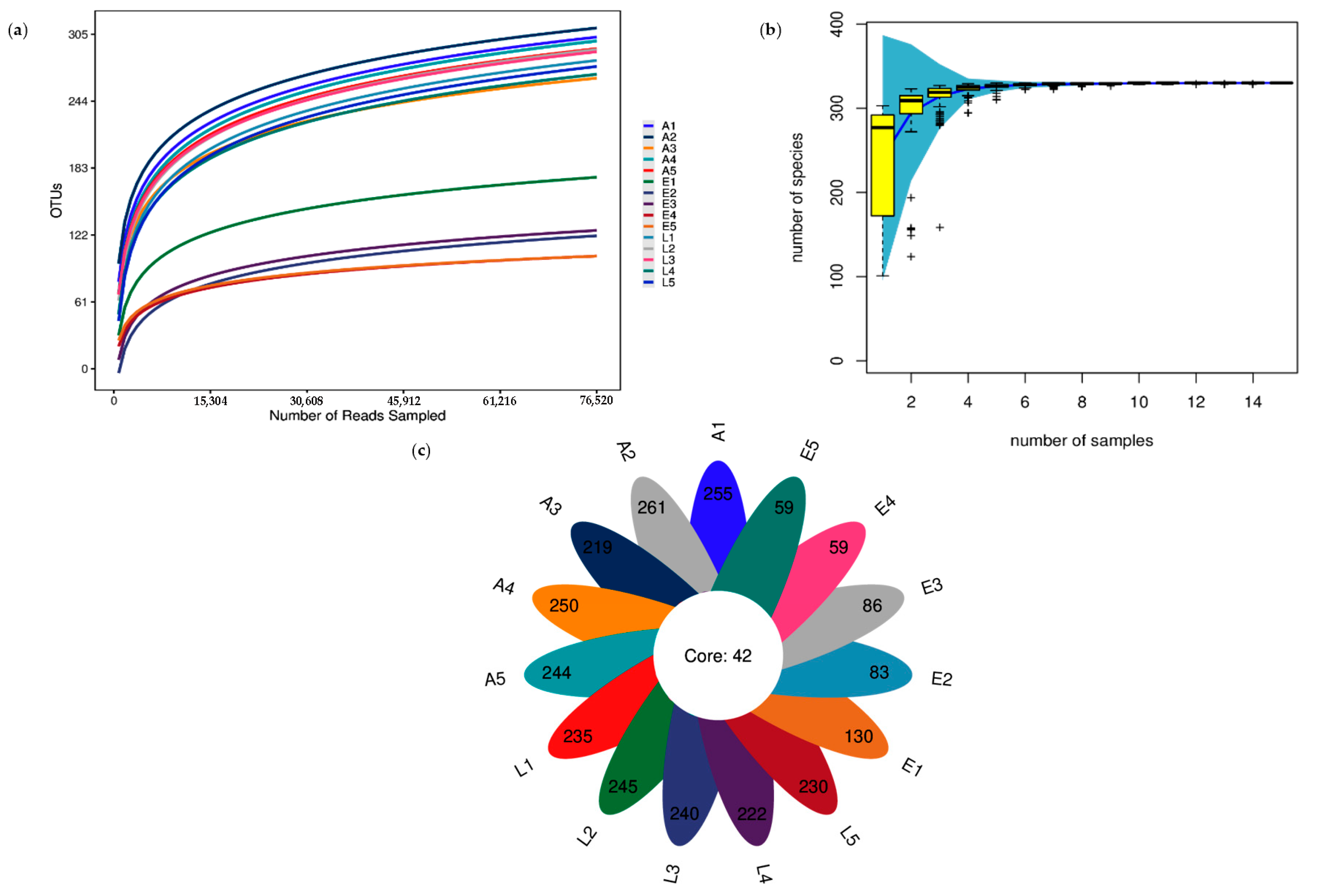
Figure 1. Bacterial diversity analysis for early juveniles (E), late juveniles (L), and adults (A): (a) OTU rarefaction curves; (b) species accumulation curves; (c) OTU distribution petal diagram.
Diversity 17 00814 g001
Figure 2. Beta diversity analysis of gut bacterial communities in early juveniles (E), late juveniles (L), and adults (A). (a) Heatmap analysis based on weighted UniFrac distance; (b) PCoA analysis based on weighted UniFrac distance; (c) Similarity tree for all samples.
Diversity 17 00814 g002
Figure 3. Predicted critical functions of gut bacterial communities across life stages of Sinanodonta woodiana.
Diversity 17 00814 g003
Table 1. Biological parameters of Sinanodonta woodiana samples.
GroupAge (Month)Shell Length (cm)Shell Width (cm)Shell Height (cm)Weight (g)
Early juveniles32.98 ± 2.610.81 ± 1.182.23 ± 2.172.25 ± 0.68
Late juveniles127.59 ± 3.043.21 ± 1.975.05 ± 3.4546.33 ± 6.59
Adults3615.48 ± 5.745.78 ± 4.3410.98 ± 8.24384.44 ± 38.98
Table 2. Statistics of bacterial community richness and diversity indices in the samples.
Diversity IndicesEarly JuvenilesLate JuvenilesAdults
Chao1142.40 ± 33.10 b293.94 ± 10.47 a308.98 ± 15.48 a
Shannon0.83 ± 0.26 c2.33 ± 0.26 b2.95 ± 0.32 a
Simpson0.30 ± 0.12 c0.71 ± 0.07 b0.86 ± 0.06 a
goods_coverage1.00 ± 0.00 a1.00 ± 0.00 a1.00 ± 0.00 a
Note: Different superscript letters in the same row indicate significant differences (p < 0.05).
Table 3. Relative abundance (%) of dominant phyla in the gut bacterial communities of early juveniles, late juveniles, and adults.
Phyla NameEarly JuvenilesLate JuvenilesAdults
Fusobacteriota83.82 ± 8.34 a56.85 ± 5.15 b34.12 ± 7.68 c
Verrucomicrobiota9.65 ± 6.85 a9.77 ± 4.38 a15.24 ± 8.71 a
Pseudomonadota2.61 ± 1.39 c12.69 ± 2.87 b22.04 ± 10.70 a
Cyanobacteriota2.47 ± 0.70 c9.24 ± 2.00 b16.84 ± 6.24 a
Bacillota0.94 ± 1.05 *,b7.05 ± 1.60 a2.72 ± 3.00 b
Bacteroidota0.11 ± 0.09 *,b2.20 ± 0.84 b5.38 ± 3.54 a
Chloroflexota0.09 ± 0.04 *,b1.06 ± 0.34 ab2.09 ± 1.27 a
Note: Non-dominant phyla in each group are marked with *; Different superscript letters in the same row indicate significant differences (p < 0.05).
Table 4. Relative abundance (%) of dominant genera in the gut bacterial communities of early juveniles, late juveniles, and adults.
Genus NameEarly JuvenilesLate JuvenilesAdults
Cetobacterium83.81 ± 8.34 a56.79 ± 5.16 b34.09 ± 7.68 c
LD298.77 ± 6.83 a7.35 ± 4.12 a13.80 ± 8.25 a
Cyanobium_PCC-63072.35 ± 0.65 b5.81 ± 0.90 b9.84 ± 4.58 a
Cupriavidus1.08 ± 0.90 a0.26 ± 0.19 *,b0.19 ± 0.10 *,b
Roseomonas0.25 ± 0.28 *,b7.82 ± 2.30 a11.56 ± 7.36 a
Limnolyngbya_CHAB44490.06 ± 0.04 *,b1.42 ± 0.79 ab1.77 ± 1.70 a
Terrimicrobium0.86 ± 0.69 *,a1.39 ± 0.75 a1.29 ± 0.71 a
Limnothrix0 *,b1.33 ± 0.90 b4.13 ± 2.49 a
Sediminibacterium0 *,b0.61 ± 0.30 *,b1.54 ± 0.93 a
Acidibacter0.02 ± 0.03 *,c0.36 ± 0.06 *,b1.15 ± 0.34 a
Note: Non-dominant genera in each group are marked with *; Different superscript letters in the same row indicate significant differences (p < 0.05).
Disclaimer/Publisher’s Note: The statements, opinions and data contained in all publications are solely those of the individual author(s) and contributor(s) and not of MDPI and/or the editor(s). MDPI and/or the editor(s) disclaim responsibility for any injury to people or property resulting from any ideas, methods, instructions or products referred to in the content.