Article

Metabolites Fingerprinting Variations and Chemotaxonomy of Related South African Hypoxis Species

Department of Pharmaceutical Sciences, School of Pharmacy, Sefako Makgatho Health Sciences University, Molotlegi Street, Ga-Rankuwa, Pretoria 0204, South Africa
Diversity 2025, 17(10), 729; https://doi.org/10.3390/d17100729
Submission received: 21 August 2025 / Revised: 8 October 2025 / Accepted: 8 October 2025 / Published: 17 October 2025
(This article belongs to the Section Chemical Diversity and Chemical Ecology)

Abstract

Hypoxis hemerocallidea (Hypoxidaece) is thoroughly researched and well documented for its plethora of anecdotal and scientifically backed pharmacological potentials. Its anecdotal uses and pharmacological activities are attributed to its extract’s inherent bioactive compounds like hypoxoside, rooperol, and β-sitosterol. This study aimed at conducting a targeted and holistic phytochemical profiling of variations in Hypoxis hemerocallidea (H. hemerocallidea) and related species. The chemotaxonomic classifications of H. hemerocallidea and seven other related species were also carried out to avert the possibility of over harvesting H. hemerocallidea and the encouragement of species inter-change. The plant extracts were analysed with reverse phase ultra-pure liquid chromatography quadrupole time-of-flight mass spectrometry and gas chromatography, as well as high-performance thin-layer chromatography. The generated chromatographic data were made compatible for chemometric computation using Principal Component Analysis (PCA) and Orthogonal Partial Least Squares Discriminant Analysis (OPLS-DA) models. The results obtained unveil orcinol glycoside, curculigoside C, hypoxoside, dehydroxyhypoxoside, bisdehydroxy hypoxoside, hemerocalloside, galpinoside, cholchicoside, geraniol glycoside, β-sitosterol, oleic acid, and 2-hydroxyethyl linoleate as target phytochemicals that define the profiles of the Hypoxis species. In addition, three distinct chemotypes defined by hemerocalloside, galpinoside, and colchicoside, respectively, were observed, as well as holistic variations in all secondary metabolites. Due to similarities in the phytochemical constituents of selected species, species inter-change seems imminent if further research confirms the findings of this study.

1. Introduction

Plants contain complex mixtures of secondary metabolites with diverse chemical properties and variations in their concentrations [1]. Geographic locations, genetic traits, harvesting time, processing methods, and other biotic factors largely influence the chemical variation in plant metabolites [2]. These factors necessitate the classification of plants into different chemotypes based on their phytochemical content using a variety of methods. It can be assumed that plants with different chemistries will also have different efficacies as medicines, since the individual or synergistic actions of molecules are responsible for the biological activities of these plants [3]. The complexity of plant metabolites makes it unfeasible to use a single technique to classify these plants [4]. Quality assurance and standardisation protocols aimed at providing consumers with safe, high-quality products and enhancing profit for the manufacturer are critical. One of the methods of standardising plant extracts is the use of chemical fingerprinting, since this provides a unique pattern representing the presence of a particular chemical composition that defines the plant [5]. Chemical fingerprints can be linked to the efficacy of plant extracts towards a specific biological activity, thereby taking synergism into account [6]. The quantitative analysis of many indigenous Hypoxis species to determine several polar metabolites and β-sitosterol indicated similar phytochemical profiles for H. obtusa and H. hemerocallidea, on the one hand, and for H. galpinii and H. rigidula var. rigidula, on the other. The quantitative data corroborate morphological similarities described for these Hypoxis species [7]. Since many samples were available in this study, multivariate analysis of the chromatographic data was used to provide a holistic view of both intra- and interspecies phytochemical variations within the indigenous members of the genus.
Metabolomics has become a useful and powerful tool for the analysis of plants and related natural products [8]. The Plant Kingdom alone is estimated to produce 100,000–200,000 metabolites [9]. This technique is comprehensive for the simultaneous and systematic evaluation of metabolite levels in the metabolome and their periodic changes [10]. Metabolomics is a valuable tool for comparing variations in metabolites in biological systems and has found application in human, plant, and microbial systems [11]. A metabolome is a complete set of small-molecule metabolites which are either intermediates or the products of metabolism. Primary metabolites, such as carbohydrates, fatty acids, and amino acids, are associated with plant growth and reproduction. However, secondary metabolites including terpenes, alkaloids, polyphenols, and flavonoids are considered to be an expression of the individuality of the plant species [12]. Secondary metabolites serve a variety of functions in plants, including protection from predators, disease pathogens, and the attraction of insect pollinators [13]. Metabolomics has been successfully used for the standardisation of herbal materials and related products. Data from several analytical methods, including Nuclear Magnetic Resonance (NMR) spectroscopy, Liquid Chromatography (LC) and Gas chromatography (GC), are suitable for the metabolomic assessment of phytomedicines and related raw materials [14,15].
The principle underlining metabolomics is either a succinct targeted approach or a random non-targeted or holistic perspective [16]. The targeted approach involves the identification and determination of plant or other natural extracts that belong to a specific class of compounds or have desired properties, using known standard compounds. Such compounds, if present in the extracts, can be used as biomarkers in the standardisation of traditional medicines [14]. On the contrary, the untargeted approach measures the “whole” metabolite regardless of the pharmacological value of the constituents. In the latter approach, the identification of all the metabolites is not mandatory prior to data processing. In both metabolomic approaches, many samples are investigated, and the data generated often contain multiple variables [17]. This type of data is difficult to analyse with classical statistical techniques [18], because, in Multivariate data (MVD, the number of samples analysed are usually smaller than the number of variables generated [19]. To circumvent this challenge with classical statistical analysis, chemometric computation of MVD is almost indispensable. Chemometrics are suitable for summarising, visualising, and interpreting complex data in a robust and statistically appropriate way [17]. The unsupervised principal component analysis (PCA) is applied to identify trends, variables, clusters, and outliers within a dataset [20]. Without the introduction of bias to the data, PCA allows the researcher to project and extract systematic variation in MVD to reveal clustering and allow for the recognition of metabolite variation [17]. This type of modelling does not rely on class information for the discrimination of samples. In contrast, Orthogonal Partial Least Squares Discriminant Analysis (OPLS-DA) is a supervised technique that is able to account for differences between classes (each species in a genus could be seen as a class). In addition, the marker compounds responsible for these differences are revealed by the model. Once an OPLS-DA model has been constructed, it can be used as a prediction model to allocate unknown samples to a specific class [19].
Although many reports detail the phytochemistry of a particular Hypoxis species [20], only a few comparative studies have been performed [21,22]. Considering the wide range of distribution of the species in South Africa [23], variations in the chemical profiles of specimens harvested from different regions can be expected because of differences in genetic traits, climate, soil, and precipitation. These differences could drastically impact the medicinal efficacy of that specimen for a particular purpose. Boukes et al. [21] determined the levels of β-sitosterol and other sterols and hypoxoside in corms of H. hemerocallidea, H. stellipilis and H. sobolifera var. sobolifera from the eastern and southern Cape regions of South Africa. Hypoxis sobolifera var. sobolifera and H. hemerocallidea were found to contain the most β-sitosterol and hypoxoside, respectively. Since the plants, regrown from the purchased and collected corms, were exposed to the same conditions for six months prior to analysis, and the analysed samples representing each individual species originated from pooled corms, any phytochemical variations between specimens that may have been present were voided. A later study by [24] using extracts from the same three species indicated differences in their cytotoxicity towards various cell lines, suggesting phytochemical differences. The need for fundamental studies that contribute to knowledge of the biosystematics of the genus was mentioned by Van Wyk as far back as 1997 [25]. Singh [26] has already made a valuable contribution towards this knowledge by revising the taxonomy of the southern African species. Phytochemical variation studies are vital to achieve successful and sustainable commercialisation, since initial plant selection, quality control, and proof of efficacy are intrinsically linked to the chemical composition of the medicinal product [27].
The objective of the manuscript is reporting on ten identified metabolites and their variations in eight Hypoxis species, as well as classifying the eight Hypoxis species into three distinct chemotypes.

2. Materials and Methods

2.1. Plants Collection and Comminution

The location, altitude of each site where Hypoxis corms were collected by a licenced collector, and their voucher specimen numbers post-identification are displayed in Table 1. Each fresh corm was separated from the Hypoxis plant and the tap roots removed from it. After washing, the corms were chopped into small pieces and oven dried at 30 °C for 36 h prior to extraction. The resulting dried plant material was then pulverised using a Retsch1MM 400 ball milling frequency of 30.0 Hz for 120 s to yield fine brown powders. These powders were sieved using a 500 mm mesh size (Endcotts Filters Ltd., London, UK) to ensure uniform particle size.

2.2. Plant Extraction

Approximately 5 g mass of one specimen of each of the eight species (H. hemerocallidea, H. colchicifolia, H. obtusa, H. rigidula var. rigidula, H. rigidula var. polissisima, H. galpinii, H. argentea, and H. multiceps) was accurately weighed in duplicate into 50.00 mL Erlenmeyer flasks. A 10.0 mL aliquot of Analytical Reagent (AR) grade methanol (MeOH) was added, and the mixture was sonicated at 45 °C for 30 min. After filtering through a filter paper (No 4, Whatman Ltd., Maidstone, UK), the filtrate was kept aside, and the residue was returned to the flask. This process was repeated twice, whereafter the filtrates were combined, and the solvent was evaporated until dryness. The dried extracts were weighed and stored at 25 °C for analysis. Thereafter, the same procedure was followed to prepare chloroform (CHCl3) extracts of a second set of the weighed powdered plant material. The MeOH extracts were used for the determination of the polar analytes by reverse phase ultra-pure liquid chromatography quadrupole time-of-flight photodiode array detector/mass spectrometry (RP-UPLC-QTOF-PDA/MS) and high-performance thin layer chromatography (HPTLC), while the chloroform extracts were used for the determination of non-polar extracts GC-MS.

2.3. RP-UPLC-QTOF-MS Analysis

The RP-UPLC-QTO-MS method and conditions used in this study have previously been reported [28].

2.4. GC-MS Analysis

An Agilent 7890A gas chromatograph fitted with a mass selective detector (5975C GC/MSD) was used for the analysis of the CHCl3 extracts of the wild South African Hypoxis. The GC system was equipped with an autosampler (Model 7693) and a split–splitless injector. Samples were introduced via split injection with a split ratio of 1:15, using an injector temperature of 300 °C. Separation was achieved on a DB-5MS-fused silica capillary column (30 m × 0.32 mm id., 0.25 µm film thickness; J & W Scientific, Folsom, CA, USA). The thermal conditions for the oven were 80 °C for 2 min, 10 °C min−1 to 300 °C (held for 14 min), while the helium carrier gas flow was maintained at 1 mL min−1. A transfer temperature of 300 °C, a scan range of m/z 50–600, a sampling rate of 1.1 scans−1, and an ionisation potential of 70 eV were applied. A detector temperature of 300 °C was maintained over the total run time of 30 min.

2.5. HPTLC Analysis

The HTLC analysis reported by K. Bassey et al. [28] was used in this study.

2.6. Chemometric Data Analysis

Chemometric analysis of the data was performed using Simca-P 13.0 software (Umetrics AB, Malmo, Sweden). The H. argentea and H. multiceps samples were excluded from the chemometric analysis due to the too small sample size (n = 6) that would generate zero components, a statistically insignificant parameter. Removing small samples in chemometrics analysis could lead to bias in overfitting a model, imprecise estimates, reduced statistical power, and limited generalizability. Such biases were prevented through the construction of a five-component PCA model and using the first two components for the data analysis.
After alignment of the RP-UHPLC-Q-TOF-MS and GC-MS chromatographic data, PCA models were constructed and used to analyse the data obtained for the individual Hypoxis specimens. Data were pareto scaled, but no data pre-processing was performed. The presence of outliers was investigated, and the effects of excluding these on model statistics were ascertained. The PCA model was constructed to indicate trends (similarities and/or differences) within the dataset. Thereafter, the samples were assigned to one of six separate classes (Classes 1–6) based on the Hypoxis species under investigation. Each separate class was tentatively assumed to contain a unique set of phytochemicals. An OPLS-DA model was constructed and used for analysis of the class-assigned dataset. This model is the most suitable for the discrimination of data that show similarities from an unsupervised PCA plot [29]. This is because samples that are unrelated (orthogonal) to those that are most similar are excluded by the OPLS-DA model. External validation of all the models was performed by randomly removing a subset of the data (test set), constructing a new model with the remaining observations (training set), and subsequently using the new model to predict the class to which each member of the test sets belongs [30]. The accuracy of prediction was then determined.

3. Results

3.1. Chemical Fingerprinting of Wild Hypoxis by HPTLC

The mobile phase used was suitable for the resolution of all the phytocomponents in the Hypoxis extracts investigated. Even compounds with very close Rf-values such as colchicoside and hypoxoside (Rf 0.28 and 0.29) and galpinoside and curculigoside C (Rf 0.48 and 0.49) were resolved by the eluent (Figure 1).
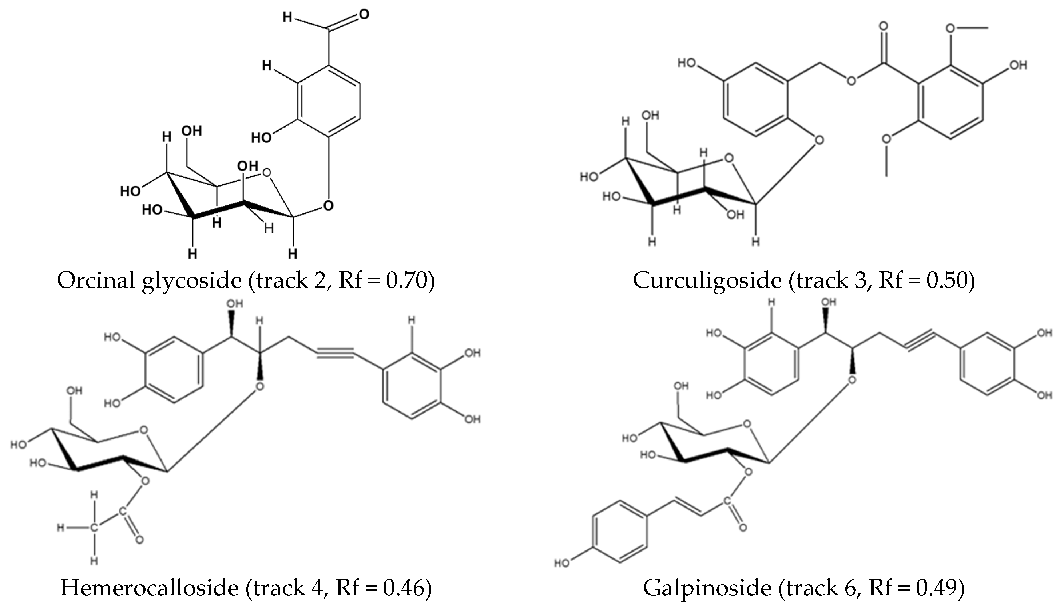
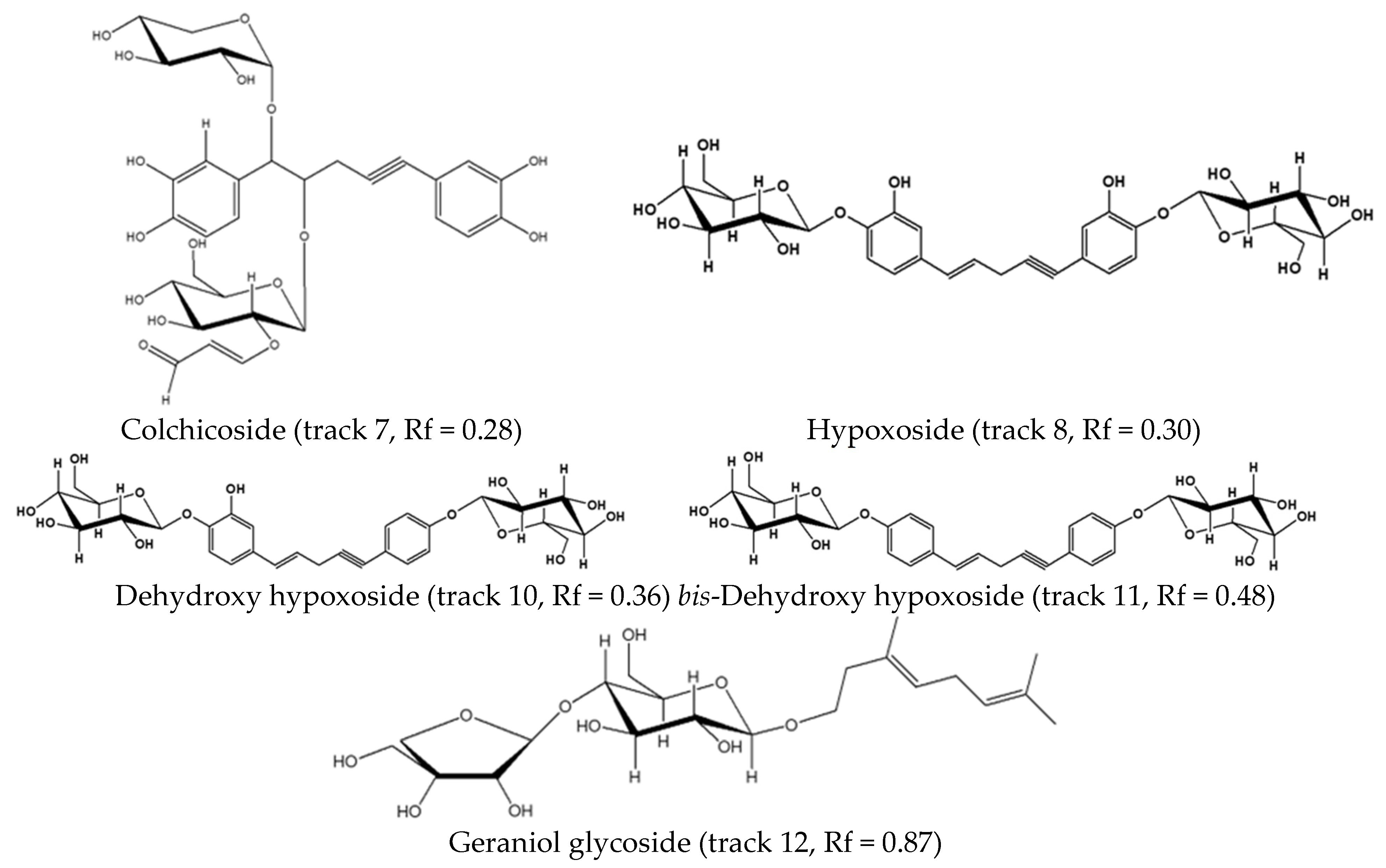
After the analysis of the extracts against the standard compounds (Figure 2) using the same mobile phase, spots that were common to all the extracts were identified as hypoxoside, dehydroxyhypoxoside, bis-dehydroxyhypoxoside, and geraniol glycoside.
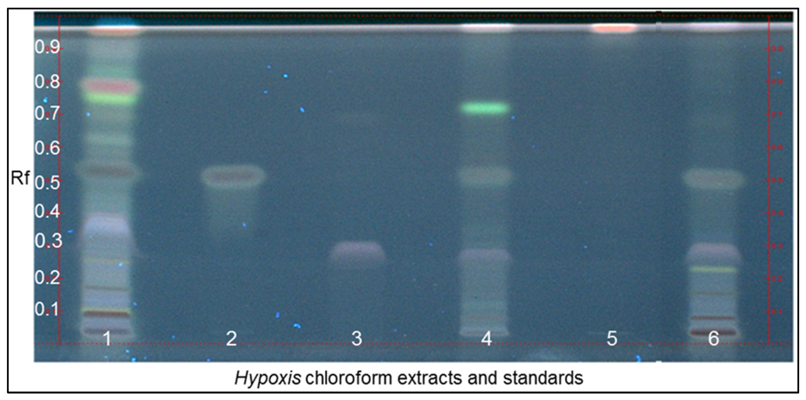
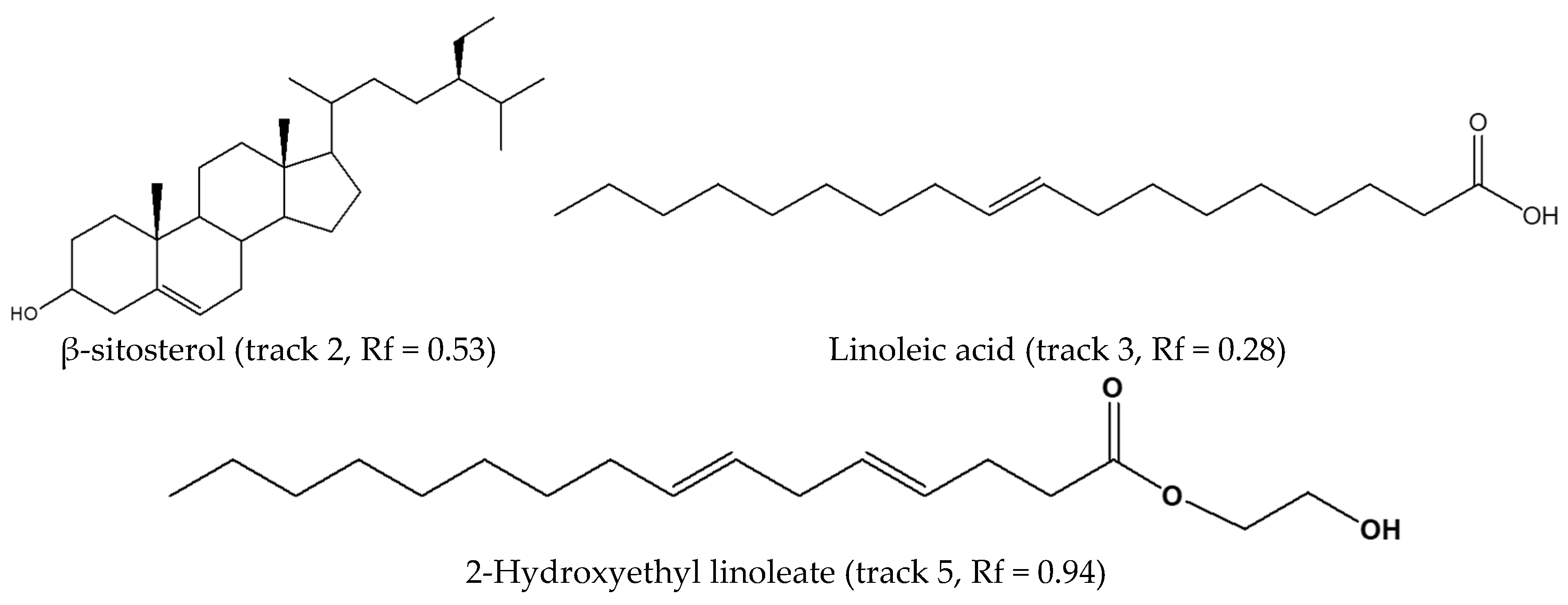
The hydrophobic nature of β-sitosterol, the other active metabolite in Hypoxis raw materials, motivated our investigation of the Hypoxis chloroform extracts. Even though this extract is not popular commercially due to the carcinogenicity of the solvent, ref. [21] determined β-sitosterol, stigmasterol, demosterols, and stigmastenolin in CHCl3 extracts of H. hemerocallidea, H. sobolifera, and H. stellipilis. In the current study, using optimised HPTLC conditions, an attempt was made to overcome the obstacle of co-migration of the sterols, which was encountered when using manual thin layer chromatography (TLC) [21]. Using the semi-automated HPTLC system, the Hypoxis CHCl3 extracts were applied to glass plates, which were developed in CHCl3-EtOAc-HCOOH (5-4-1 v/v/v), as reported by Boukes et al. [21]. The Rf value (0.95) for β-sitosterol was found to be higher than that reported by them. Most of the compounds migrated with the solvent front. It was decided that the eluent was not appropriate for good resolution of the CHCl3 extract of Hypoxis. So, this study applied chloroform as the HPTLC mobile phase and a better result, displayed in Figure 3 using the standard whose structures are detailed in Figure 4, was the outcome.

3.2. Chemical Fingerprinting of Wild Hypoxis by RP-UPLC-QTOF-MS Using a Targeted Approach

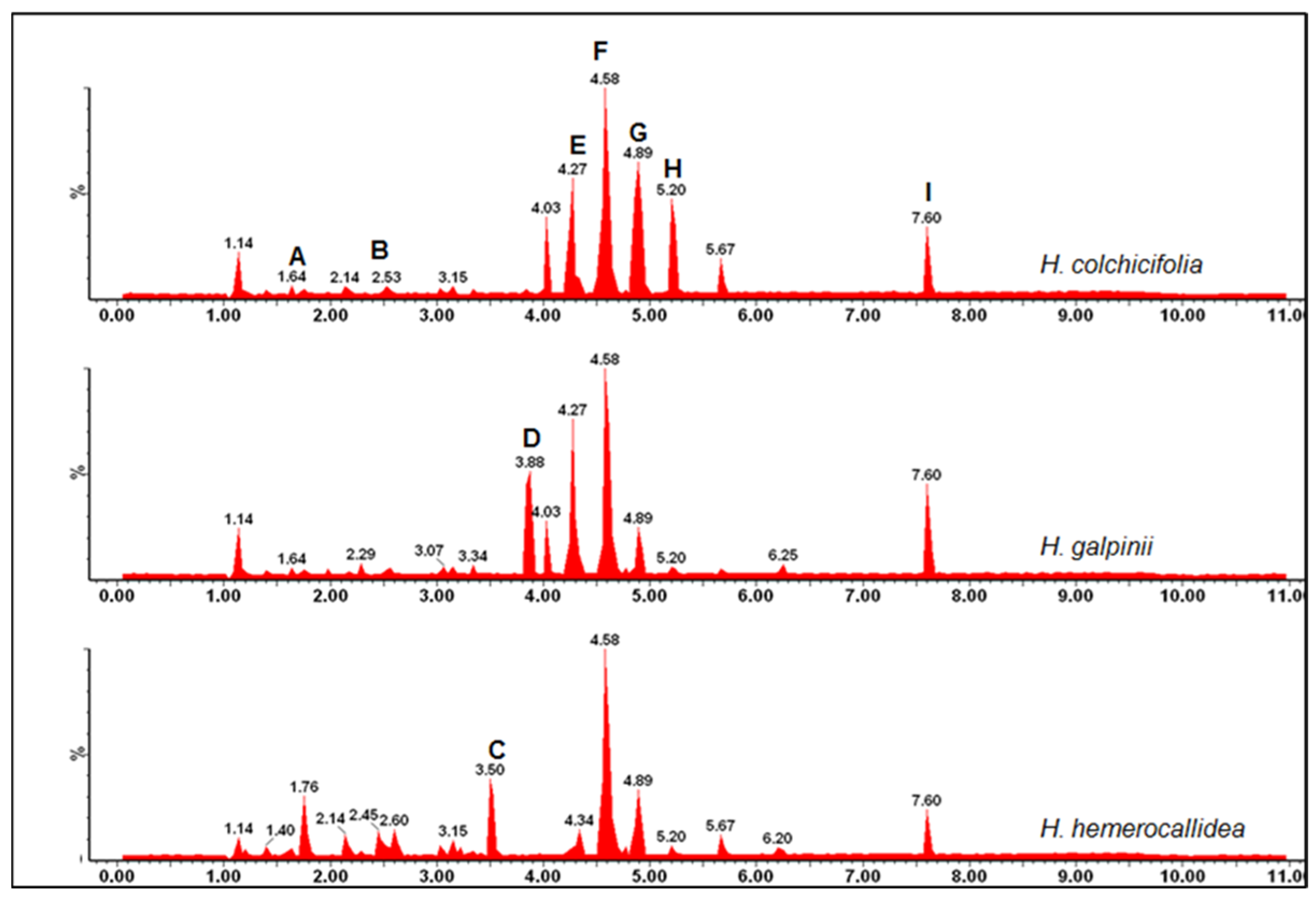
Contrary to previous reports that Hypoxis MeOH extracts contain only hypoxoside, dehydroxyhypoxoside, and bis-dehydroxyhypoxoside polar compounds [31], we anticipated that with our RP-UPLC-QTOF-MS, other previously unresolved and undetected secondary metabolites, would be determined from our investigation. Analysis of the MeOH extracts of 105 Hypoxis samples in negative ion mode by RP-UPLC-QTOF-MS yielded well resolved peaks. As was observed by HPTLC, some peaks were common to all the species, while other peaks were species specific. By comparing the pseudo-molecular ion mass to charge (m/z) ratios, mass fragmentation patterns, and UV absorbance and retention times (Rt) of the standard compounds (Table 2), as well as the literature references to the Hypoxis extracts, nine typical peaks in the Hypoxis MeOH extracts were annotated (Figure 5). These peaks are herein reported as the chemical signature of MeOH extracts of the South African Hypoxis species.

3.3. Chemical Fingerprinting of Wild Hypoxis by GC-MS Using a Targeted Approach

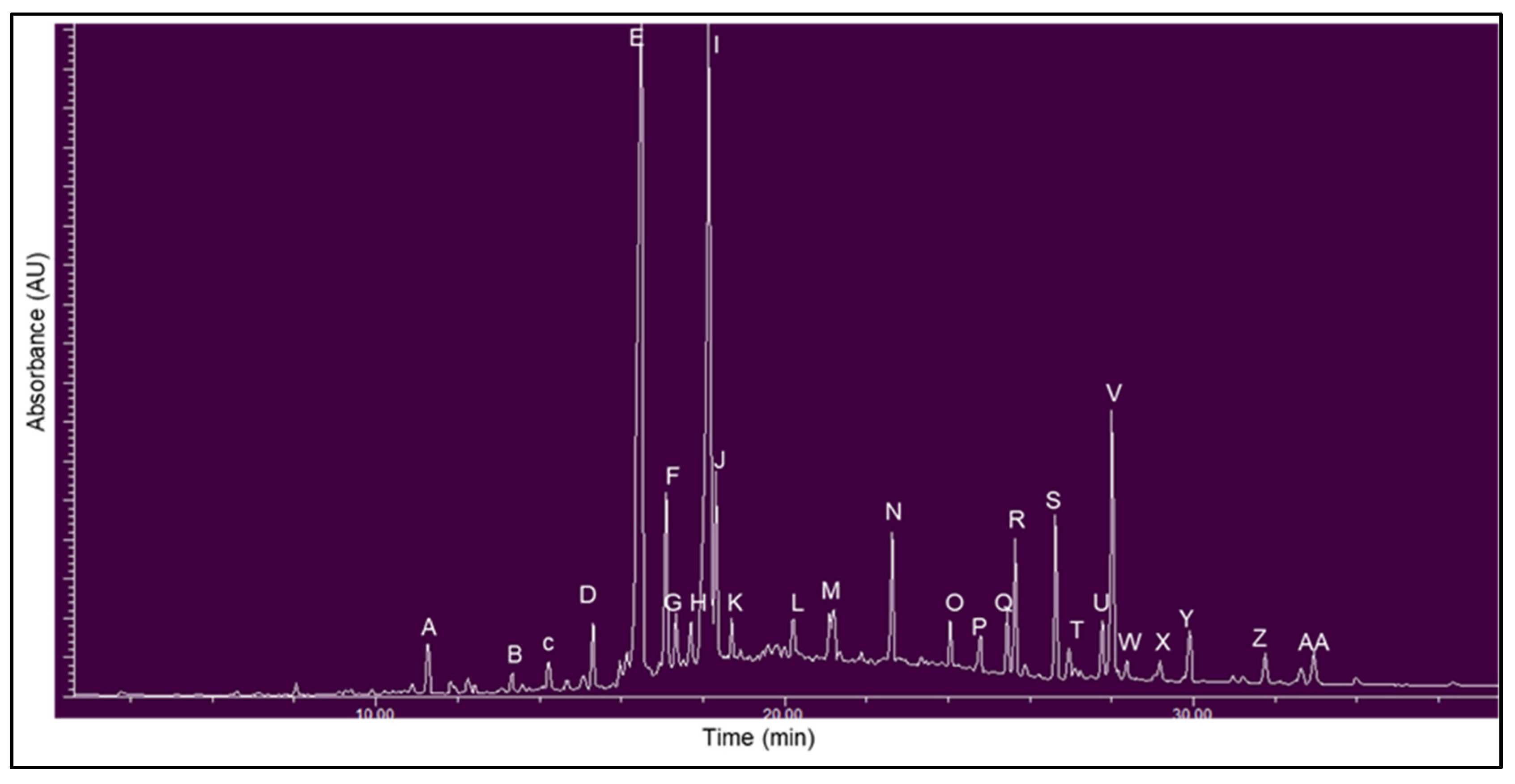
Analysis of CHCl3 extracts (10 mg mL−1) of the wild Hypoxis by GC-MS yielded a satisfactory resolution and sensitivity for the separation of the different phytocomponents from the Hypoxis corms. By comparing the pseudo-molecular ion mass to charge (m/z) ratios, ion fragmentation patterns, and retention times (Rt) to the literature data [21], the known compounds (campesterol, and β-sitosterol) were identified. (Figure 6)
Table 3. Relative retention times, fragmentation ions, and mass to charge ratios used for the identification of the phytoconstituents in the CHCl3 extracts of H. hemerocallidea, H. obtusa, H. rigidula var. rigidula, H. rigidula var. polissisima, H. galpinii, H. colchicifolia, and H. multiceps.
PeaksRetention
Time (min)
Mass to Charge Ratio
[M]+ (m/z)
Mass Fragmentation
Pattern
Compound
A11.29224224, 111, 97, 69, 572-hexadecanol
B13.33248248, 242, 225, 185, 143, 129, 102 isopropyl-propyl 12-methyltetradecanoate
C14.68288199, 185, 171, 166, 143, 129, 115tetradecanoic acid
D15.32 242242, 213, 199, 157, 143, 129, 97pentadecanoic acid
E16.50 256239, 227, 213, 199, 185, 171, 157n-hexadecanoic (palmitic) acid
F17.11 312283, 270, 253, 241, 227, 205, 199propyl-14-methylhexadecanoate
G17.34 270241, 227, 213, 171, 143, 129, 115heptadecanoic acid
H17.71 282264, 222, 180, 137, 125, 111, 97cis-13-octadecanoic oleic acid
I18.16 264246, 235, 222, 207, 193, 180, 165oleic acid
J18.33 284 284, 255, 241, 227, 199, 185, 171octadecanoic acid
K18.72 310 259, 241, 121, 199, 173, 157, 147cis-13-eicosenoic acid
L20.21 268 341, 313, 272, 241, 199, 147, 129hexanedioc acid-bis(2-ethylhexyl) ester
M21.20 341 380, 351, 323, 295, 267, 239, 211heptacosane
N22.64 365341, 322, 281, 264, 250, 207, 183oleic acid-3-(octadecyloxy) propyl ester
O24.05 490462, 418, 392, 348, 320, 292, 26717-pentatriacontene
P24.79 380337, 309, 281, 253, 225, 197, 169heptacosane
Q25.45 400367, 253, 213, 185, 159, 145, 121ethylisoallocholate
R25.65 484424, 365, 330, 304, 287, 244, 2277,8-epoxylanostan-11-ol
S26.65 450421, 393, 365, 337, 309, 281, 253dotriacontane
T26.95 400382, 367, 340, 315, 289, 273, 255campesterol
U27.77 364337, 295, 281, 251, 225, 197, 183octadecane-3-ethyl-5-(2-ethylbutyl)
V28.02 414396, 354, 329, 303, 255, 231, 213β-sitosterol
W28.49 490462, 418, 391, 348, 320, 292, 26717-pentatriacontene
X29.54410395, 368, 296, 241, 229, 187, 174stigmasta-3,5-dien-7-one
Y29.91 484424, 365, 330, 304, 287, 244, 2278-epoxylanostan-11-ol
Z31.73 490460, 432, 390, 362, 334, 306, 285 pentatricontanol
AA32.95 428413, 400, 328, 285, 269, 227, 213citrost-7-en-3-ol

3.4. Investigation of Phytochemical Variation by RP-UPLC-QTOF-MS Using a Targeted Approach

Eight Hypoxis samples were initially studied using UPLC-QTOF-MS to obtain an overview of the qualitative metabolite variations. The UPLC-QTOF-MS profiles of the eight Hypoxis species investigated were used to summarise the variation in the phytoconstituents within the samples. Hypoxoside (4) and geraniol glycoside (7) were present in all the Hypoxis species (Table 4).

3.5. Qualitative Variation in the Methanol and Dichloromethane Extracts of the Hypoxis Samples

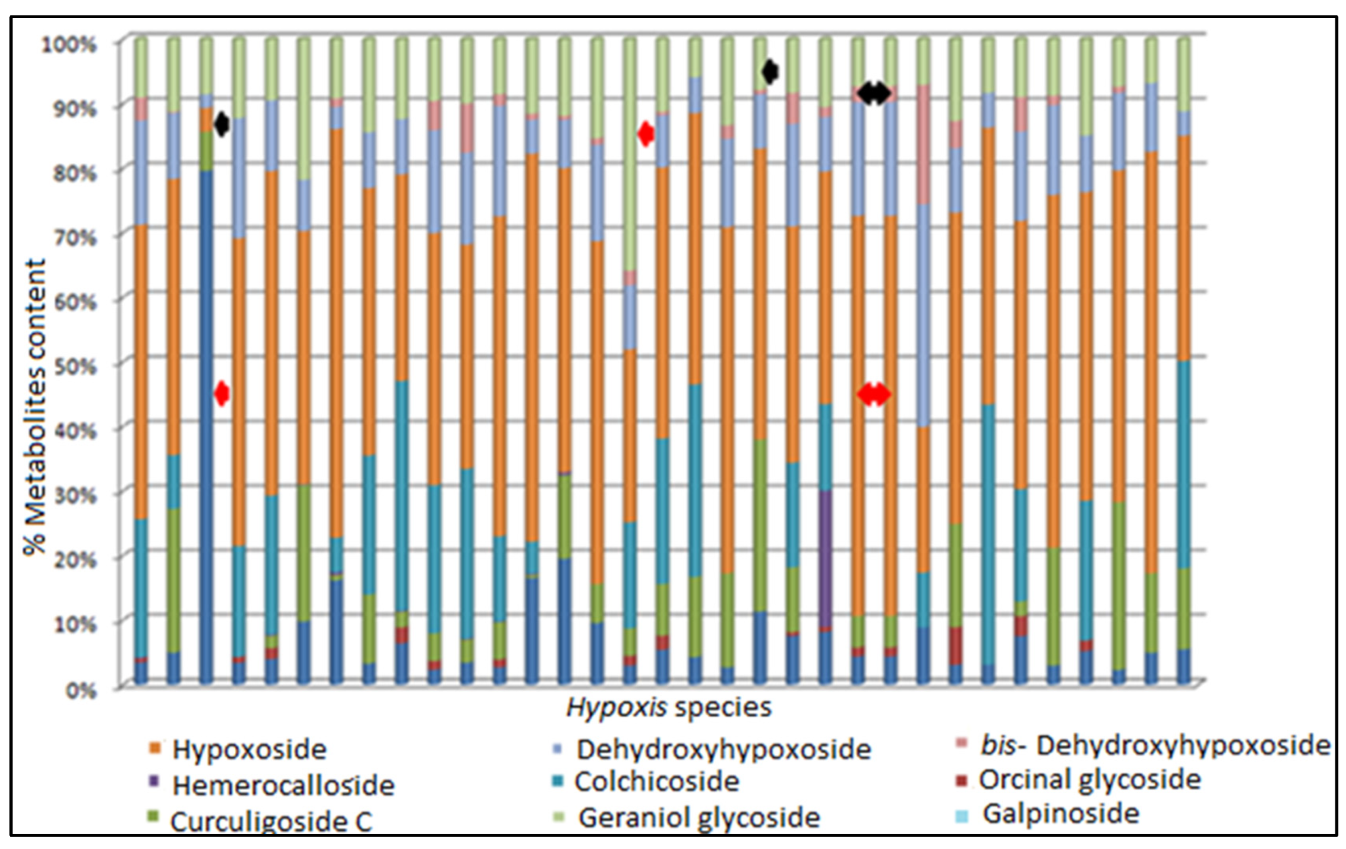
The qualitative variation study of the MeOH extracts of the 105 Hypoxis species encompassing 33 populations in South Africa was conducted using the area under the curve (AUC) of the identified peaks obtained from UPLC-QTOF-MS. The relative amounts of the marker compounds, namely, hypoxoside, dehydroxyhypoxoside, bis-dehydroxyhypoxoside, colchicoside, hemerocalloside, galpinoside, and orcinal glycoside, could be visualised using a column plot (Figure 7).
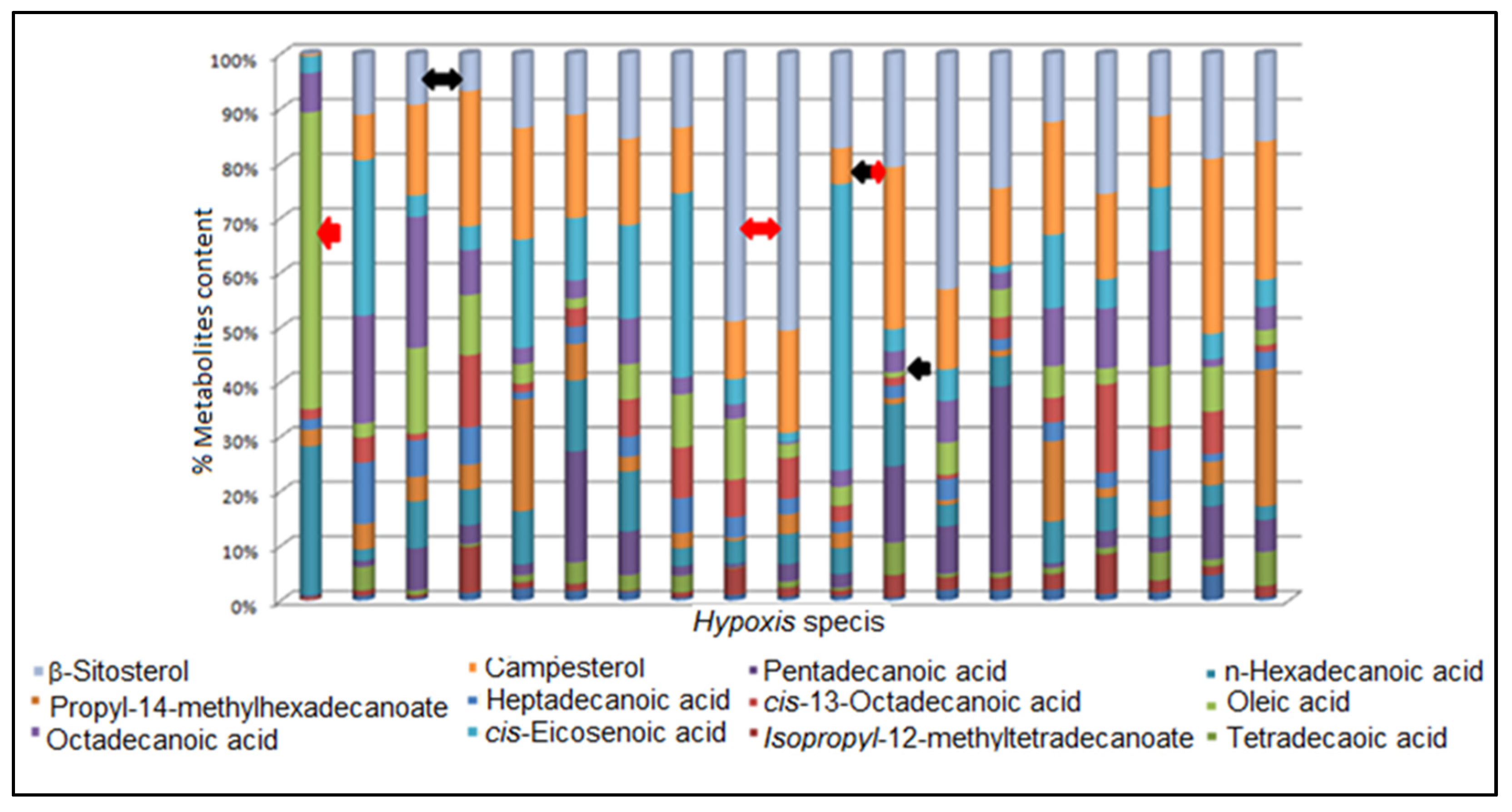
Similarly, the qualitative variations in β-sitosterol, campesterol, and some of the fatty acids, expressed as area under the curve (AUC), were investigated in the non-polar extracts of Hypoxis samples, and Figure 8 depicts the results.

3.6. Chemotype Identification and Variations as Determined by RP-UPLC-QTOF-MS and Metabolomic Analysis

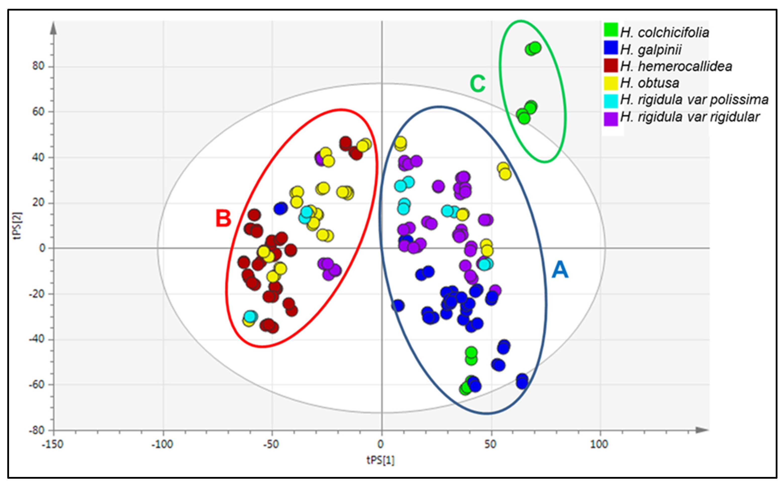
After converting the RP-UPLC-QTOF-MS data to Excel and exporting to Simca-14®, chemometric analysis was performed. Two of the species (H. argentea and H. multiceps) were excluded from the dataset since the number of samples (three corms each) was too few to provide reliable statistics. A five-component PCA model of the unsupervised UPLC-QTOF-MS data grouped the samples based on their chemical composition into three clusters (A, B, and C; Figure 9), which are not well defined.
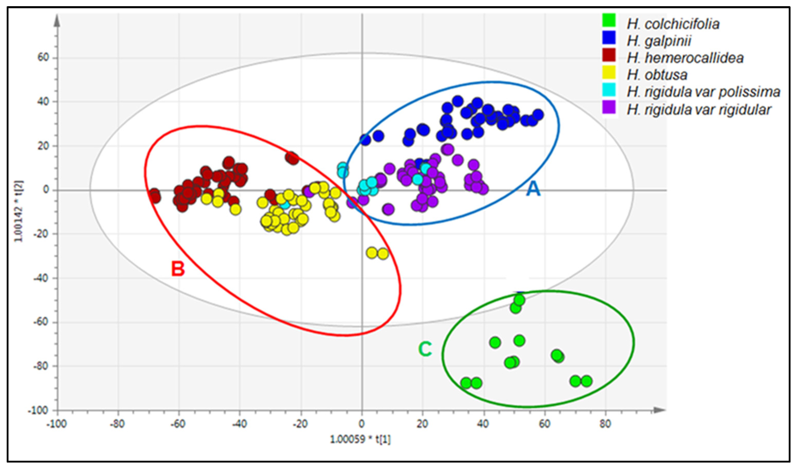
The scores plot of the OPLS-DA model revealed clear differences between some species (Figure 10), although others were clustered together. This plot allowed for the identification of distinct groupings that can be viewed as having distinct chemical profiles and therefore as chemotypes. The OPLS-DA model was able to separate the H. galpinii, H. rigidula var. rigidula (except one), and H. rigidula var. polissisima (Figure 10) samples (Cluster A) from those of H. hemerocallidea and H. obtusa (Cluster B) in the first component. The H. colchicifolia samples were grouped in Cluster C, which was separated from most of the Cluster A samples by the second component. The colchicifolia had a higher intensity or concentration of dehydroxyhypoxoside (peak G) and bis-dehydroxyhypoxoside (peak H) than all the other Hypoxis species. This factor could be responsible for the colchicifolia samples being placed outside the 95% confidence level of the analysis to underscore a possible difference in the species chemistry and morphology compared with all the other species analysed.
H. rigidula var. rigidula and H. rigidula var. polissisima samples from location L25 and L27 and two samples from L16 were grouped into chemotype B and not in chemotype A like the rest. The reasons for the slight variation in the chemotype of these samples were thought to be related to the Global Positioning System. This is because these samples were harvested from Impendle (−29.5996955, 29.8670563), Ukahlamba (−29.3767, 29.5377), and Wagendrift (−23.467, 29.650), all in the mountainous Drakensberg’s Area. This locality is characterised by an identical longitude of twenty-nine degrees and a similar altitude. These factors can influence the chemistry of these samples away from the other H. rigidula var. rigidula and H. rigidula var. polissisima samples that were placed in chemotype A.
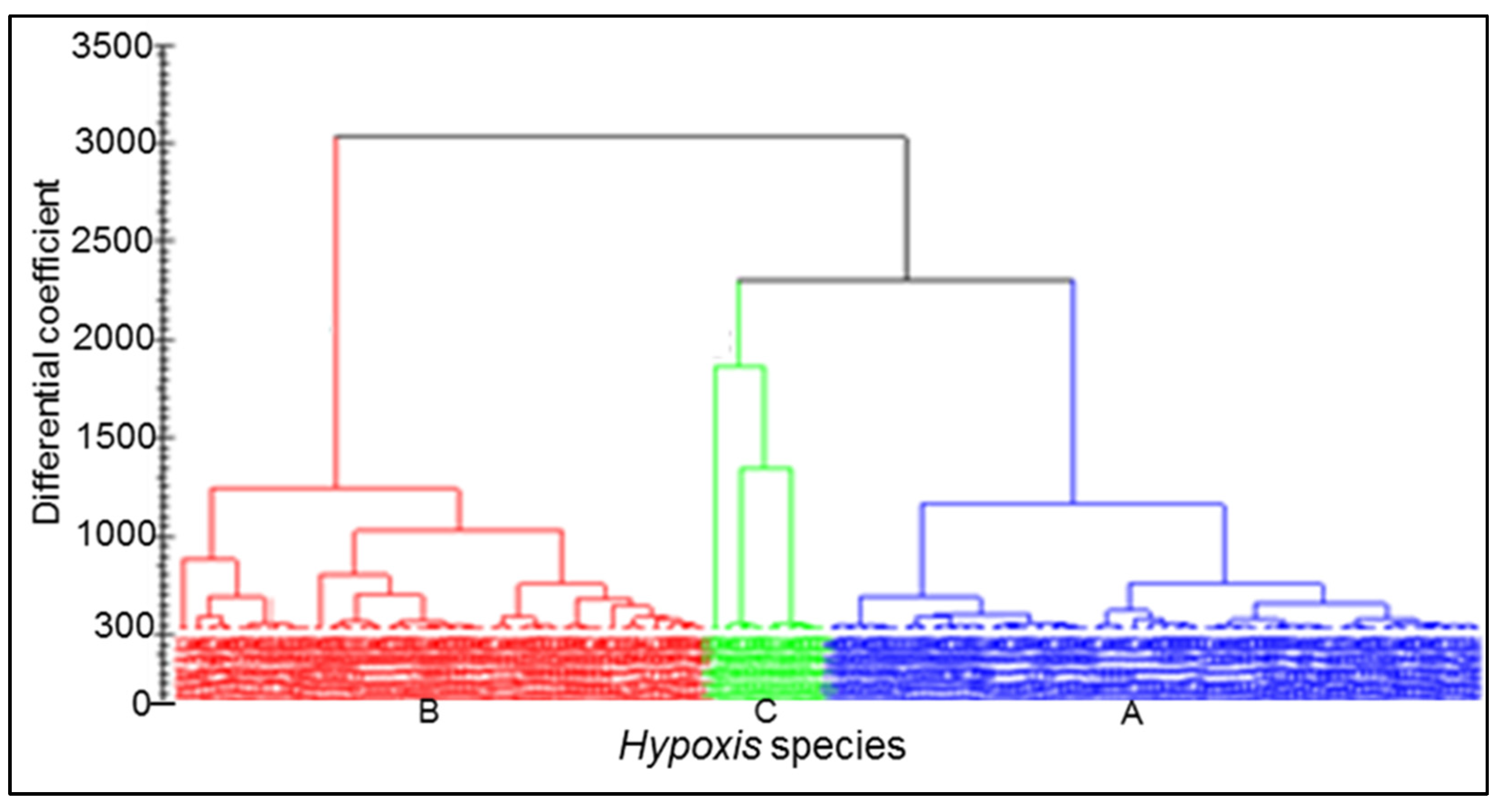
Further confirmation of the three defined chemovars was performed via a hierarchical analysis (HCA), which was performed with no predefined classes of the dataset. The HCA of the non-predefined data results agreed with the predefined OPLS-DA analysis, with all the observations grouped into the three distinctly defined chemotypes A, B, and C (Figure 11), with each cluster consisting of species with similar (A, B) or unique (C) phytochemicals.
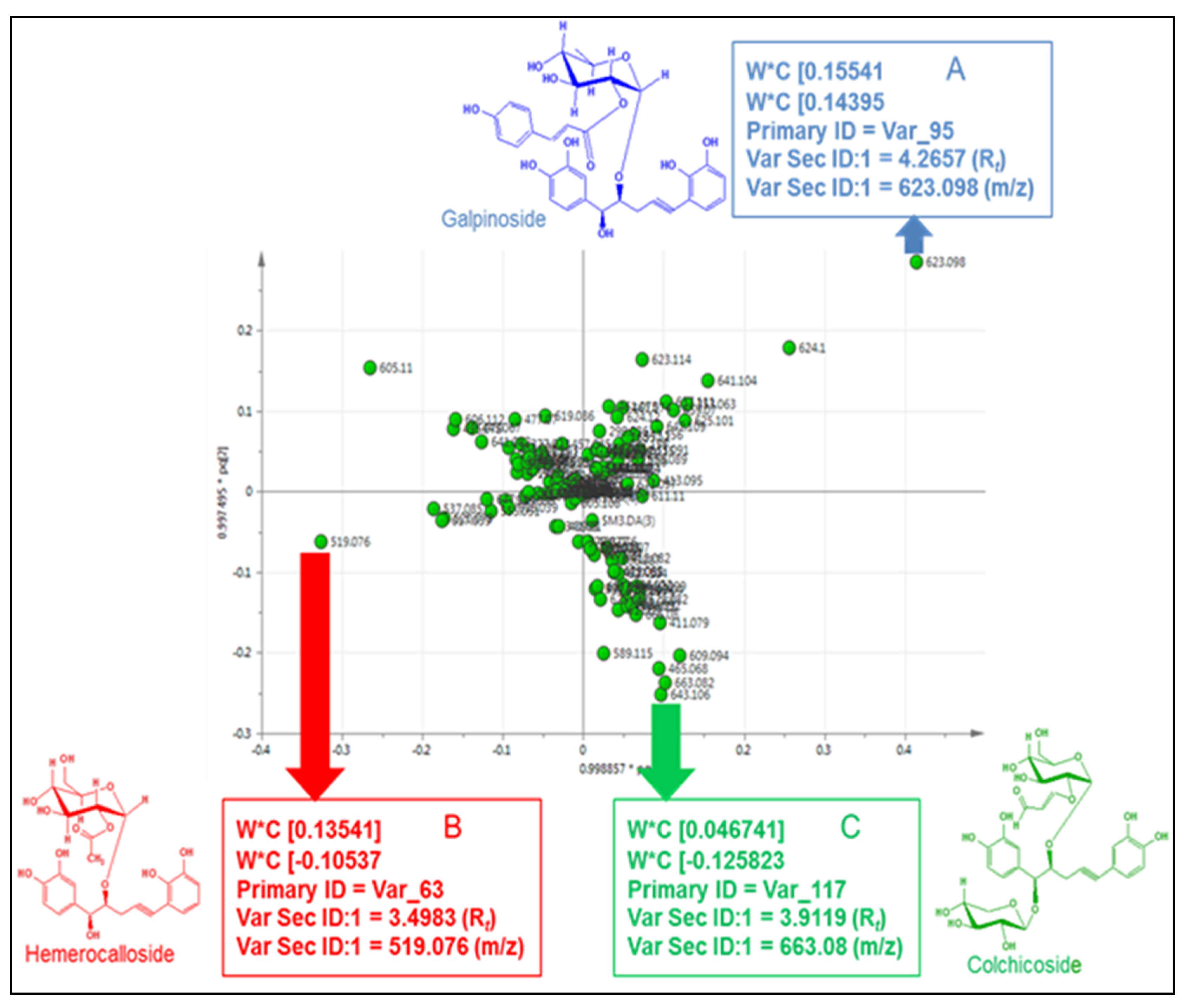
Considering the premise that clustering was based on similarities or differences in the structures of the compounds in the different Hypoxis species per chemotype, an OPLS-DA loading scores plot (Figure 12) was constructed using the same data. This plot was able to single out the compound that has the largest influence on the separation of clusters (chemotypes). This compound or chemical marker compounds could therefore be used to define each chemotype. From the loadings plot, the three clusters were annotated with a mass number 623 (for Cluster A), 519 (for Cluster B), and 663 (for Cluster C) by the model as compounds responsible for the separation of each chemotype. The assigned mass numbers matched the molecular weight of galpinoside (m/z 623), hemerocalloside (m/z 519), and colchicoside (m/z 663) at Rt of 4.26 min, 3.49 min, and 3.91 min, respectively.
A thorough scrutiny of each chemotype revealed that chemotype A predominantly consists of H. galpinii, H. rigidula var. rigidula, and H. rigidula var. polissisima; chemotype B encompasses H. hemerocallidea and H. obtusa, while chemotype C encompasses only H. colchicifolia. Comparing the RP-UPLC-QTOF-MS fingerprints of Hypoxis species within each chemotype (Table 5) clearly indicated close similarities between the chemical profiles. The chemical similarities correspond to morphological similarities between the species [6] within each of the three chemotypes. Table 5 shows the classification of the 105 Hypoxis samples into the three chemotypes.

4. Discussions

The HPTLC profiles of the methanol extract could be grouped into H. hemerocallidea-type, H. galpinii-type, and H. colchicifolia-type, with common bands of 0.29, 0.35, 0.39, and 0.74 at Rf values and species-specific uncommon bands. Bands at a Rf below 0.2 and above 0.85 could not be identified due to a lack of standards. The chloroform extract, on the other hand, mainly indicated the presence of β-sitosterol [35], with trace amounts of campesterol and stigmasterol [21], oleic acid, and 2-hydroxyethyl linoleate.
Chemical profiling of the Hypoxis methanol extract using RP-UPLC-ESI-Q-TOF-MS, due to its high sensitivity, throughput capacity, and high resolution [36], revealed the common peaks as hypoxoside, dehydroxyhypoxoside, bis-dehydroxyhypoxoside, and geraniol glycoside with retention times of 4.59, 4.90, 5.21, and 7.60 min, respectively. On the other hand, the species-specific peaks were identified as orcinal glycoside, galpinoside, hemerocalloside, colchicoside, and curculigoside C with corresponding retention times of 1.64, 3.47, 3.49, 3.91, and 4.96 min. The unidentified peaks by HPTLC resolved at 2.60 (m/z 621), 4.03 (m/z 643), 5.67 (m/z 611), and 6.25 min (m/z 773). Using the mass fragmentation patterns of these peaks, a thorough search of LC-MS databases, including the National Institute of Health (NIH), Pubchem, Scifinder, and Chemspider, provided no matching information for possible compounds. All the unidentified peaks had a large molecular weight of m/z = 621, 611, 773, and 643. However, three of the peaks had very minor intensities and one (m/z 643) had a moderate one. None of the peaks were nominated by the PCA and OPLS-DA chemometric modals as the marker compound responsible for the three Hypoxis clusters. Hence, none of the four compounds could influenced the holistic classification of the Hypoxis chemotype.
From the GC-MS analysis, twenty-four volatile compounds, mostly C-18 fatty acids (saturated and unsaturated) and their derivatives, were identified by using the NIST 8 spectral library. Several of these additional 24 metabolites were not identified in previous studies on Hypoxis plants.
Palmitic acid and oleic acid were dominant over campesterol and β-sitosterol. Sterolins were not detected in either the MeOH or CHCl3 extracts of South African Hypoxis, which is contrary to previous reports that claimed that 100 g of the enriched aqueous extract of Hypoxis contained 9 mg [37] and less than 6 mg [38] of sterolins.
Hypoxoside is the principle active component in the Hypoxis genus. Therefore, its availability in all the species investigated is of immense clinical importance as the presence or absence of the compound is likely to affect the efficacy of the products formulated from Hypoxis. Due to the clinical importance of hypoxoside, H. hemerocallidea was identified as containing the highest levels of hypoxoside (60–65%). In contrast, H. colchicifolia contained 3% of hypoxoside and the highest 70% of colchicoside.
The other recorded clinically important β-Sitosterol was detected in all of the samples, with the exception of the H. hemerocallidea sample from the Gauteng province, H. rigidula var. rigidula from the KwaZulu-Natal Province, and H. obtusa from the Mpumalanga Province. It was of significant interest that H. hemerocallidea also afforded the largest amount of β-sitosterol (53% of the total), while that of H. rigidula var. polissisima was found to contain only 8.0% of this compound. The relative amounts of campesterol, stigmasterol, heptadecanoic acid, hexadecanoic acid, oleic acid, and other fatty acids esters varied between 16 and 31%.
Principal component analysis models and OPLS-DA [39] were used to reveal clustering of all the Hypoxis samples arising from their chemical similarities or differences thereof without any interference [18]. Such clustering of the Hypoxis species was expected, because morphological analysis revealed such patterns with the Hypoxis samples analysed [7]. Whereas H. galpinii, H rigidula var. rigidula, and H. rigidula var. polissisima formed Cluster A, H. hemerocallidea and H. obtusa formed Cluster B, and H. colchicifolia constituted Cluster C. The clustering was not based on geographic origin, because each cluster contains samples from all the geographic localities. Although bias can easily be introduced into OPLS-DA models because of class assignment, the clustering evident on the PCA plot indicated the presence of clear chemical composition differences between the samples, thereby ruling out bias.
Hence, the South African Hypoxis species were classified into three chemotypes, namely, chemotype, chemotype B, and chemotype C. The assigned m/z ratio at the specified retention time of the marker compounds was interpolated to the RP-UPLC-QTOF-MS chromatograms of the Hypoxis samples to further confirm the information of the marker compounds that defined each Hypoxis chemotype. Hence chemotypes A, B, and C were defined by galpinoside, hemerocalloside and colchicoside, respectively. This study, like similar studies, has underpinned the importance of using metabolomics in the identification of plant secondary metabolites or markers that define plant chemotypes or mitigate oxidative stresses, cancer, and diabetes [40,41,42].

5. Conclusions

The following conclusions could be made after completing this study: the six Hypoxis species investigated can be grouped into three chemotypes. Chemotype A, consisting of H. galpinii, H. rigidula var. rigidula, and H. rigidula var. polissisima; chemotype B, comprising H. hemerocallidea and H. obtuse; and chemotype C, made up of H. colchicifolia. Chemotype A is characterised by galpinoside as a marker compound and chemotype B by hemerocalloside, while colchicoside serves as the biomarker for chemotype C. All studies on the biological activities of Hypoxis raw material and products have hitherto associated the activity with the presence of rooperol, the aglycone formed from hypoxoside in the gastrointestinal tract, and β-sterol or sterolins. However, it is necessary that the chemotype marker compounds (galpinoside, hemerocalloside, and colchicoside) be fully explored for their biological activities. The presence of a variety of bioactive compounds could explain the plethora of uses of Hypoxis for traditional medicine and as a health supplement. For the purpose of the standardisation of Hypoxis raw material, this study recommends that H. obtusa and H. hemerocallidea, classified as chemotype B, be cultivated. This will ensure the presence of hypoxoside and other metabolites in optimum yields. The chemical similarities between H. hemerocallidea and H. obtusa (chemotype B) and between H. galpinii and H. rigidula var. rigidula (chemotype A) mirror the morphological similarities and justifies their potential interchangeable use of H. hemerocallidea and H. obtusa.

Funding

This research received no external funding, and the APC was funded by Sefako Makgatho Health Sciences University, Moletlegi street, Pretoria South Africa.

Data Availability Statement

The data generated in this study is part of this report.

Acknowledgments

The author gratefully thanks Alvaro Viljoen and Sandara Combrinck who mentored me when this study was conducted.

Conflicts of Interest

The author declares no conflicts of interest.

Abbreviations

The following abbreviations are used in this manuscript:
PCA Principal Component Analysis
OPLS-DAOrthogonal to Partial Least Square-Discriminant Analysis
LCLiquid Chromatography
GCGass Chromatography
NMRNuclear Magnetic Resonance
MVDMultivariate Data Analysis
CHCl3 Chloroform
MeOH Methanol
RP-UPLC-QTOF-PDA/MS Reverse Phase Ultra Performance
QTOFQuadrupole Time of Flight
PDAPhotodiode Array Detector
MSMass Spectrometry
HPTLCHigh Performance Thin Layer Chromatography
GC-MS.Gas-Chromatography -Mass Spectrometry
MSDMass Selective Detector
EtOAcEthyl Acetate
H2SO4Sulphuric Acid
UVUltra-Violet
AUCArea Under the Curve

References

  1. Hu, L.F.; Li, S.P.; Cao, H.; Liu, J.J.; Gao, J.L.; Yang, F.Q.; Wang, Y.T. GC-MS fingerprint of Pogostemon cablin in China. J. Pharm. Biomed. Anal. 2006, 42, 200–206. [Google Scholar] [PubMed]
  2. Jayanthy, A.; Prakash, K.U.; Remashree, A.B. Seasonal and geographical variations in cellular characters and chemical contents in Desmodium gangeticum (L.) DC: An Ayurvedic medicinal plant. Int. J. Herb. Med. 2013, 1, 34–37. [Google Scholar]
  3. Liu, R.H. Potential synergy of phytochemicals in cancer prevention: Mechanism of action. J. Nutr. 2004, 134, 3479–3485. [Google Scholar] [CrossRef] [PubMed]
  4. Fraser, P.D.; Pinto, M.E.S.; Holloway, D.E.; Bramley, P.M. Application of high-performance liquid chromatography with photodiode array detection to the metabolic profiling of plant isoprenoids. Plant J. 2000, 24, 551–558. [Google Scholar] [CrossRef] [PubMed]
  5. Tadmor, Y.; Jefthas, E.; Goliath, J.; Smith, M.; Langenhoven, P.; Acquaye, D.; Juliani, R.; Letchamo, W.; Renaud, E.; Zimba, N.; et al. Quality assurance and quality control for African natural plant products. In Trends in New Crops and New Uses; Janick, J., Whipkey, A., Eds.; ASHS Press: Alexandria, VA, USA, 2002. [Google Scholar]
  6. Joshi, D.D. Herbal Drugs and Fingerprints: Evidence Based Herbal Drugs; Springer Science & Business Media: Delhi, India, 2012. [Google Scholar]
  7. Singh, Y. Systematics of Hypoxis (Hypoxidaceae) in South Africa. Ph.D. Thesis, University of Pretoria, Pretoria, South Africa, 2009. [Google Scholar]
  8. Sumner, L.W.; Mendes, P.; Dixon, R.A. Plant metabolomics: Large-scale phytochemistry in the functional genomics era. Phytochemistry 2003, 62, 817–836. [Google Scholar] [CrossRef]
  9. Oksaman-Caldentey, K.-M.; Inze, D. Plant cell factories in the post genomic era: New ways to produce designer secondary metabolites. Trends Plant Sci. 2004, 9, 433–440. [Google Scholar] [CrossRef]
  10. Villas-Boas, S.G.; Moxley, J.F.; Akesson, M.; Stephanopoulos, G.; Nielsen, J. High-throughput metabolic state analysis: The missing link in integrated functional genomics of yeasts. Biochem. J. 2005, 388, 669–677. [Google Scholar] [CrossRef]
  11. De Vos, R.C.H.; Moco, S.; Lomen, A.; Keurrentjes, J.J.B.; Bino, R.J.; Hall, R.D. Untargeted large-scale plant metabolomics using liquid chromatography coupled to mass spectrometry. Nat. Protoc. 2007, 2, 778–791. [Google Scholar] [CrossRef]
  12. Griffin, J.L.; VIdal-Puig, A. Current challenges in metabolomics for diabetes research: A vital functional genomic tool or just a ploy for gaining funding? Physiol. Genom. 2008, 34, 1–5. [Google Scholar] [CrossRef]
  13. Weiss, E.A. Essential Oil Crops; CAB International: New York, NY, USA, 1997. [Google Scholar]
  14. Li, Y.; Hu, Z.; He, L. An approach to the development of binary chromatographic fingerprints of the total alkaloids from Caulophyllum robustum by high performance liquid chromatography/diode array detector and gas chromatography/mass spectrometry. J. Pharm. Biomed. Anal. 2007, 43, 1667–1672. [Google Scholar]
  15. Schulz, H.; Baranska, M. Identification and quantification of valuable plant substances by IR and Raman spectroscopy. Vib. Spectrosc. 2007, 43, 13–25. [Google Scholar] [CrossRef]
  16. Lomen, A.; Van der Weg, G.; Van Engelen, M.C.; Bor, G.; Hoogenboom, L.A.P.; Nielen, M.W.F. An untargeted metabolomic approach to contaminant analysis: Pinpointing potential unknown compounds. Anal. Chim. Acta 2007, 584, 43–49. [Google Scholar]
  17. Eriksson, L.; Johansson, E.; Kettaaneh-wold, N.; Trygg, J.; Wikström, C.; Wold, S. Multi- and Megavariate Data Analysis. Part 1: Basic Principles and Applications, 2nd ed.; Umetrics AB: Umea, Sweden, 2006. [Google Scholar]
  18. Van Der Kooy, F.; Verpoorte, R.; Meyer, M.J.J. Metabolomic quality control of claimed anti-malarial Artemisia afra herbal remedy and A. afra and A. annua plant extracts. S. Afr. J. Bot. 2008, 74, 186–189. [Google Scholar] [CrossRef]
  19. Naes, T.; Isaksson, T.; Fearn, T.; Davies, T. A User Friendly Guide to Multivariate Calibration and Classification; NIR Publications: Chichester, UK, 2007. [Google Scholar]
  20. Zimudzi, C. African potato (Hypoxis spp.): Diversity and comparison of the phytochemical profiles and cytotoxicity evaluation of four Zimbabwean species. J. Appl. Pharm. Sci. 2014, 4, 79–83. [Google Scholar] [CrossRef]
  21. Boukes, J.G.; Van de venter, M.; Oosthuizen, V. Quantitative and qualitative analysis of sterols/sterolins and hypoxoside contents of three Hypoxis (African potato) spp. Afr. J. Biotechnol. 2008, 7, 1624–1629. [Google Scholar] [CrossRef]
  22. Sathekge, N.R. Comparison of Secondary Metabolites Content and Anti-Microbial Activity of Four Hypoxis Species Used in Traditional Medicine. Master’s Thesis, University of Pretoria, Pretoria, South Africa, 2010. [Google Scholar]
  23. Germishuizen, G.; Meyer, N.L. (Eds.) Plants of Southern Africa: An Annotated Checklist; Strelitzia 14; National Botanical Institute: Pretoria, South Africa, 2003. [Google Scholar]
  24. BoukeS, G.J.; Van de Venter, M. Cytotoxicity and mechanism(s) of action of Hypoxis spp. (African potato) against HeLa, HT-29 and MC-7 cancer cell lines. J. Med. Plant Res. 2011, 5, 2766–2774. [Google Scholar]
  25. Van Wyk, B.-E.; Van Oudtshoorn, B.; Gericke, N. Medicinal Plants of South Africa; Briza Publications: Pretoria, South Africa, 1997. [Google Scholar]
  26. Singh, Y. Hypoxis (Hypoxidaceae) in Africa: List of species and infraspecific names. Bothalia 2006, 36, 13–23. [Google Scholar] [CrossRef]
  27. Da-costa-Rocha, I.; Edwards, S.; Lawrence, M.J.; Cable, C.; Heinrich, M. Quality and safety of herbal products: Part 1-new legislation and production. Pharm. J. 2012, 288, 658. [Google Scholar]
  28. Bassey, K.; Viljoen, A.; Combrinck, S.; Choi, Y.H. New phytochemicals from the corms of medicinally important South African Hypoxis species. Phytochem. Lett. 2014, 10, lxix–lxxv. [Google Scholar] [CrossRef]
  29. Trygg, J.; Wold, S. PLS regression on wavelet compressed NIR spectra. Chemom. Intell. Lab. Syst. 2001, 42, 209–220. [Google Scholar] [CrossRef]
  30. Sandasi, M.; Vermaak, I.; Chen, W.; Viljoen, A.M. Hyperspectral imaging and chemometric modelling of Echinacea—A Novel Approach in the quality control of herbal medicines. Molecules 2014, 19, 13104–13121. [Google Scholar] [CrossRef] [PubMed]
  31. Laporta, O.; Perez-Fons, L.; Mallavia, R.; Caturla, N. Isolation, characterization and antioxidant capacity assessment of the bioactive compounds derived from Hypoxis rooperi corm extract (African potato). Food Chem. 2007, 101, 1425–1437. [Google Scholar] [CrossRef]
  32. Fu, D.X.; Lei, G.Q.; Cheng, X.W.; Chen, J.K.; Zhou, T.S. Cureuligoside C, a New Phenolic Glucoside from Rhizomes of Curculigo orchioides. Acta Bot. Sin. Engl. Ed. 2004, 46, 621–624. [Google Scholar]
  33. Marini-Bettolo, G.B.; Nicoletti, P.M.; Galeffi, C.; Messana, I. Hypoxoside a new glycoside with uncommon structure from Hypoxis obtusa Busch. Tetrahedron 1982, 38, 1683–1687. [Google Scholar]
  34. Bredenkamp, M.W.; Drewes, S.E.; Wentler, G.L. A geraniol glycoside from Hypoxis acuminata. Phytochemistry 1989, 28, 263–265. [Google Scholar] [CrossRef]
  35. Moghadasian, M.H. Pharmacological properties of plant sterols in vivo and in vitro observations. Life Sci. 2000, 67, 605–615. [Google Scholar] [CrossRef] [PubMed]
  36. Grata, E.; Boccard, J.; Guillarme, D.; Glauser, G.; Carrupt, P.A.; Farme, E.E.; Wolfender, J.L.; Rudas, S. UPLC-TOF-MS for plant metabolomics: A sequential approach for wound marker analysis of Arabidopsis thaliana. J. Chromatogr. B 2008, 871, 261–270. [Google Scholar] [CrossRef] [PubMed]
  37. AbidI, S.L. Chromatographic analysis of plant sterols in foods and vegetables oils. J. Chromatogr. A 2001, 935, 173–201. [Google Scholar] [CrossRef]
  38. Nair, V.D.P.; Kanfer, I. Sterols and sterolins in Hypoxis hemerocallidea (African Potato). S. Afr. J. Sci. 2008, 104, 322–324. [Google Scholar]
  39. Jackson, J.E. A User’s Guide to Principal Components; John Wiley: New York, NY, USA, 1991. [Google Scholar]
  40. Matseke, B.; Poka, M.; Demana, P.; Bassey, K. Unravelling the Potentials of Managing Metabolic Diabetes and Related Oxidative Stresses with Extracts from Five South African Hypoxis Species. Stresses 2025, 5, 53. [Google Scholar] [CrossRef]
  41. Wang, Y.; Chai, J.; Guo, S.; Wang, Z.; Wang, S.; Yu, X.; He, X.; Zhang, A.; Qiu, S. Integrated and proteomics revealed mangiferin alleviating oxidative stress and mitochondrial dysfunction of HT22 cells via regulating the abnormal metabolic pathways. Food Biosci. 2025, 66, 1061380. [Google Scholar] [CrossRef]
  42. Altarjami, L.R. Anticancer and antioxidant activities of polyphenolic pomegranate peel extracts obtained by a novel hybrid ultrasound-microwave method: In vitro and in vivo studies in albino mice with HeLa, colon, and HepG2 cancerous cell lines. Pak. Vet. J. 2025, 45, 723–734. [Google Scholar] [CrossRef]
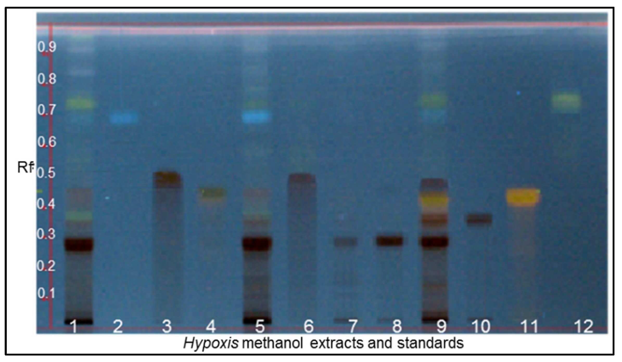
Figure 1. HPTLC plate with chemical profiles of the methanol extracts of wild Hypoxis species. Track 1: H. hemerocallidea, 2: orcinal glycoside, 3: curculigoside C, 4: hemerocalloside, 5: H. galpinii, 6: galpinoside, 7: colchicoside, 8: hypoxoside, 9: H. colchicifolia, 10: dehydroxyhypoxoside, 11: bis-dehydroxyhypoxoside, and 12: geraniol glycoside. Developing solvent: CHCl3-MeOH-H2O (70-30-4 v/v/v) and visualisation reagent: MeOH-H2SO4 (9-1 v/v).
Diversity 17 00729 g001
Figure 2. Identified standard compounds, their Rf values, and structures used for the HPTLC fingerprinting of methanol extracts of South African Hypoxis.
Diversity 17 00729 g002aDiversity 17 00729 g002b
Figure 3. HPTLC plate of wild Hypoxis chloroform extract and standards: track 1: H. hemerocallidea 2: β-sitosterol, 3: oleic acid, 4: H. galpinii, 5: 2-hydroxyethyl linoleate, and 6: H. colchicifolia. Developing solvent: CHCl3. Visualisation reagent: H2SO4-MeOH (1:9 v/v). Image captured at 366 nm.
Diversity 17 00729 g003
Figure 4. Structures of identified standard compounds and their Rf values used for the HPTLC fingerprinting of chloroform extracts of South African Hypoxis.
Diversity 17 00729 g004
Figure 5. Typical fingerprints of methanol extracts of Hypoxis species obtained by RP-UPLC-QTOF-MS. A: orcinal glycoside, B: curculigoside C, C: hemerocalloside, D: galpinoside, E: colchicoside, F: hypoxoside, G: dehydroxyhypoxoside, H: bis-dehydroxyhypoxoside, and I: geraniol glycoside.
Diversity 17 00729 g005
Figure 6. Annotated chromatogram obtained through GC-MS analysis of a chloroform extract of H. hemerocallidea. See Table 3 for the compounds represented by letters A–AA.
Diversity 17 00729 g006
Figure 7. An overview of the variations in polar secondary metabolites in medicinally important South African Hypoxis species. Red arrows: high percentages of hypoxoside, bis-dehydroxyhypoxoside, and geraniol glycoside. Black arrows: low percentages of hypoxoside, bis-dehydroxyhypoxoside, and geraniol glycoside.
Diversity 17 00729 g007
Figure 8. An overview of the variations in non-polar secondary metabolites in medicinally important South African Hypoxis species. Red arrows: high percentages of β-sitosterol, campesterol, and oleic acid and black arrows: low percentages of β-sitosterol, campesterol, and oleic acid.
Diversity 17 00729 g008
Figure 9. Scores scatter plot derived from PCA analysis of RP-UPLC-QTOF-MS data of wild South African Hypoxis species coloured according to species type and grouped based on similar phytoconstituents.
Diversity 17 00729 g009
Figure 10. Scores plot derived from an OPLS-DA model constructed from RP-UPLC-QTOF-MS data of six Hypoxis species.
Diversity 17 00729 g010
Figure 11. PLS-DA dendrogram visualising the three clusters of South African Hypoxis species.
Diversity 17 00729 g011
Figure 12. Loadings plot derived from the OPLS-DA model indicating chemical marker compounds that distinguish three chemotypes within Hypoxis species.
Diversity 17 00729 g012
Table 1. The location and altitude of each site where Hypoxis corms were collected.
Hypoxis SpeciesProvinceTownLocalityVoucher Specimen No.No. of SpecimenAltitude (mASL)
H. argenteaNorthern CapeKimberleyL3HA 007-HA 00931555
H. colchicifoliaEastern CapePort EdwardL22HC 070-HC 0723415
H. colchicifoliaKwaZulu-NatalMidmarL24HC 076-HC 07831036
H. galpiniiMpumalangaWakkerstroomL8HG 024-HG 02631791
H. galpiniiKwaZulu-NatalGroenvleiL9HG 027-HG 02931914
H. galpiniiKwaZulu-NatalWagendriftL17HG 054-HG 05741497
H. galpiniiKwaZulu-NatalUkahlambaL18HG 058-HG 06031662
H. galpiniiKwaZulu-NatalImpendleL26HG 082-HG 08431599
H. galpiniiKwaZulu-NatalUkahlambaL29HG 091-HG 09331714
H. hemerocallideaNorthern CapeBoshofL2HH 004-HH 00631555
H. hemerocallideaKwaZulu-NatalMount FrereL14HH 042-HH 04431100
H. hemerocallideaKwaZulu-NatalKokstadL20HH 064-HH 06631340
H. hemerocallideaEastern CapePort EdwardL23HH 073-HH 0753415
H. hemerocallideaGautengWalter Sisulu GardenL31HH 097-HH09931550
H. hemerocallideaGautengTshwane MarketL32HH 100-HH 10231280
H. hemerocallideaGauteng Marabastad MarketL33HH 103-HH 10531280
H. multicepsMpumalangaBreytenL7HM 021-HM 02331784
H. obtusaMpumalangaBreytenL6HO 018-HO 02031785
H. obtusaKwaZulu-NatalChelmsfordL11HO 033-HO 03531284
H. obtusaGautengRandfonteinL13HO 039-HO 04131709
H. obtusaKwaZulu-NatalMount FrereL15HO 045-HO 05061102
H. obtusaKwaZulu-NatalKokstadL19HO 061-HO 06331451
H. obtusaKwaZulu-NatalUkahlambaL28HO 088-HO 09031637
H. rigidula var. polissisimaKwaZulu-NatalDundeeL12HRP 036-HRP 03731284
H. rigidula var. polissisimaKwaZulu-NatalWagendriftL16HRP 051-HRP 05331345
H. rigidula var. rigidulaKwazulu-NatalBoshofL1HRP 001-HRP00331556
H. rigidula var. rigidulaNorthern CapeBoshofL4HRR 010-HRR 01231562
H. rigidula var. rigidulaMpumalangaBreytenL5HRR 013-HRR 01751780
H. rigidula var. rigidulaKwaZulu-NatalGroenvleiL10HRR030-RHH 03231915
H. rigidula var. rigidulaEastern CapePort EdwardL21HRR 67-HRR 0693418
H. rigidula var. rigidulaKwaZulu-NatalImpendleL25HRR 79-HRR 08131598
H. rigidula var. rigidulaKwaZulu-NatalUkahlambaL27HRR 085-HRR 08731455
H. rigidula var. rigidulaFree StateSterkfonteinL30HRR 94-HRR 09631694
Table 2. Data obtained by RP-UPLC-QTOF-MS, reflecting the chemical fingerprints of methanol extracts of Hypoxis corms.
PeaksRetention Time (min)Mass to Charge Ratio
[M − 1] (m/z)
UV (λmax) (nm)Hypoxis Species DetectedMass Fragmentation Pattern [M − H]Fragment IdentificationCompound
A1.64299279H. colchicifolia299, 163, 137[M − H], [ M − C7H5O3], [M − C6H11O5]orcinal glycoside
B2.48517210H. colchicifolia517, 481, 330, 197, 162[M − Cl2], [M − H], [M − C8H9O3], [M − C13H17O7], [M − C16H15O7]curculigoside C [32]
C3.50519255H. hemerocallidea and H. obtusa519, 461, 315, 299, 221, 205
163
[M − H], [M − C2H3O2], [M − C8H13O6], [M − C8H13O7], [M − C17H15O5] −, [M − C17H15O6], [M − C17H15O6 − C2H3O − 2H]hemerocalloside
D3.88663278Predominantly H. colchicifolia663, 627, 465,
315, 217
[M − H], [M − C2H2O2 − 5H], [M − C9H13O6 − H2O], [M − C9H13O6 − C8H15O2], [M − C22H23O10].colchicoside
E4.27623256, (299)H. galpinii and
H. rigidula var. rigidula
623, 461, 325, 299, 163, 160 [M − H], (M − C9H7O3], [M − C17H15O5], [C15H17O8], [M − C23H25O10], [M − C17H15O8 − C9H7O3]galpinoside
F4.58605257All 605, 443, 281[M − H], [M − C6H11O5], [M − 2C6H11O5],hypoxoside [33]
G4.89589258Most 589, 427, 256, 163[M − H], [M − C6H11O5] −, [M 2C6H11O5], [M − C23H23O8dehydroxyhypoxoside [31]
H5.20609259Most 609, 411, 248, 163[M − Cl], [M − C6H11O5], [M − 2C6H11O5], [M − C23H23O7]bis-dehydroxyhypoxoside [31]
I7.60447200All 483, 447, 295,
163, 155
[M − Cl2], [M − 1], [M − C10H19O], [M − C10H19O C11H19O9–C5H9O4], [M − C5H9O4]geraniol glycoside [34]
Table 4. An overview of metabolite variations in eight Hypoxis species.
Hypoxis SpeciesSecondary Metabolites Identified in the MeOH Extract of Hypoxis Samples
1234567
H. hemerocallideaXXX
H. obtusaXX
H. rigidula var. rigidulaXXX
H. rigidula var. polissisimaXX
H. galpiniiXXX
H. colchicifoliaX
H. multicepsXXXX
H. argenteaXX
√: presence and X: absence of secondary metabolite. 1: hemerocalloside, 2: colchicoside, 3: galpinoside, 4: hypoxoside, 5: dehydroxyhypoxoside, 6: bis-dehydroxyhypoxoside, and 7: geraniol glycoside.
Table 5. Classification of South African wild Hypoxis into three chemotypes from OPLS-DA analysis. Interchangeably used species (H. hemerocallidea and H. obtusa) were placed in the same chemotype.
Hypoxis SpeciesProvinceTownLocality IdVoucher Specimen NoChemotype
H. hemerocallideaGautengWestcliffL23HH 073
HH 074
HH 075
B
B
B
GautengWalter SisuluL31HH 097
HH 098
HH 099
B
B
B
GautengTshwane marketL32HH 100
HH 101
HH 102
B
B
B
GautengMarabastad MarketL33HH 103
HH 104
HH 105
B
B
B
Eastern CapeMount FrereL14HH 042
HH 043
HH 044
B
B
B
KwaZulu-NatalKokstadL20HH 064
HH 065
HH 066
B
B
B
Northern CapeBoshofL2HH 004
HH 005
HH 006
B
B
B
H. obtusaGautengRandfonteinL13HO 039
HO 040
HO 041
B
B
B
KwaZulu-NatalChelmsfordL11HO 033
HO 034
HO 035
B
B
B
Eastern CapeMount FrereL15HO 045
HO 046
HO 047
HO 048
HO 049
HO 050
B
B
B
B
B
B
KwaZulu-NatalKokstadL19HO 061
HO 062
HO 063
B
B
B
KwaZulu-NatalUkahlambaL28HO 088
HO 089
HO 090
B
B
B
MpumalangaBreytenL6HO 018
HO 019
HO 020
B
B
B
H. rigidula var. rigidulaEastern CapePort EdwardL21HRR 067
HRR 068
HRR 069
A
A
A
Free StateSterkfonteinL30HRR 094
HRR 095
HRR 096
A
A
A
KwaZulu-NatalGroenvleiL10HRR 030
HRR 031
HRR 032
A
A
A
KwaZulu-NatalImpendleL25HRR 079
HRR 080
HRR 081
B
B
B
KwaZulu-NatalUkahlambaL27HRR 85
HRR 86
HRR 87
B
B
B
MpumalangaBreytenL5HRR 013
HRR 014
HRR 015
HRR016
HRR 017
A
A
A
A
A
Northern CapeBoshofL4HRR 010
HRR011
HRR 012
A
A
A
H. rigidula var. polissisimaKwaZulu-NatalBoshofL1HRP 001
HRP 002
HRP 003
A
A
A
KwaZulu-NatalDundeeL12HRP 036
HRP 037
HRP 038
A
A
A
KwaZulu-NatalWagendriftL16HRP 051
HRP 052
HRP 053
B
A
B
H. galpiniiKwaZulu-NatalGroenvleiL9HG 027
HG 028
HG 029
A
A
A
KwaZulu-NatalWagendriftL17HG 054
HG 055
HG 056
HG 057
A
A
A
A
KwaZulu-NatalUkahlambaL18HG 058
HG 059
HG 060
A
A
A
KwaZulu-NatalImpendleL26HG 082
HG 083
HG 084
A
A
A
KwaZulu-NatalUkahlambaL29HG 091
HG 092
HG 093
A
A
A
MpumalangaWakkerstroomL8HG 024
HG 025
HG 026
A
A
A
H. colchicifoliaEastern CapePort EdwardL22HC 070
HC 071
HC 072
C
C
C
Eastern CapePort ElizabethL24HC 076
HC 077
HC 078
C
C
C
Disclaimer/Publisher’s Note: The statements, opinions and data contained in all publications are solely those of the individual author(s) and contributor(s) and not of MDPI and/or the editor(s). MDPI and/or the editor(s) disclaim responsibility for any injury to people or property resulting from any ideas, methods, instructions or products referred to in the content.