Article

Morphological and Molecular Identification of Porpita porpita (Hydrozoa: Porpitidae) Larval and Colonial Phases

by
Jeimy Denisse Santiago-Valentín
1,
Eric Bautista-Guerrero
2,
Alma Paola Rodríguez-Troncoso
2,
María del Carmen Franco-Gordo
1,
Mauricio Alejandro Razo-López
1 and
Enrique Godínez-Domínguez
1,*
1
Centro Universitario de la Costa Sur, Departamento de Estudios para el Desarrollo Sustentable de Zonas Costeras, Universidad de Guadalajara, Gómez Farías 82, San Patricio-Melaque, Jalisco 48980, Mexico
2
Centro Universitario de la Costa, Laboratorio de Ecología Marina, Universidad de Guadalajara, Av. Universidad No. 203. Puerto Vallarta, Jalisco 48280, Mexico
*
Author to whom correspondence should be addressed.
Diversity 2024, 16(7), 425; https://doi.org/10.3390/d16070425
Submission received: 19 April 2024 / Revised: 29 May 2024 / Accepted: 23 June 2024 / Published: 19 July 2024

Abstract

:
Porpita porpita is a colonial polymorphic hydrozoan distributed in temperate and tropical zones. This species, like most hydrozoans, possesses a metagenetic life cycle with alternating life forms: medusa stage, polypoid colony, and planula larva. However, a characterization of its early stages of development is still lacking. For this study, an integrative description of the larval stages and the hydroid colony was performed using molecular and histologic tools. The results show that P. porpita develops through three larval stages: preplanula, planula, and premetamorphic planula. The preplanula is distinguished by an absence of polarity, the planula by differentiation of the oral–aboral poles, and the premetamorphic stage by cellular differentiation. Furthermore, two morphologies of young hydroids with different developmental patterns of gonozooids and dactylozooids were observed; notably, it was not possible to observe the gastrozooid in either. Taxonomic identification was confirmed using mitochondrial (COI) and ribosomal (18S and 28S) markers. Our analysis indicates that the COI gene exhibits higher intraspecific variability compared to the 18s and 28s rDNA ribosomal genes. The presented results support the future identification of P. porpita based on morphological characteristics, regardless of the stage of development. Specifically, they shed light on the diversity of mesozooplankton in reef communities.

1. Introduction

The hydroid Porpita porpita (Linnaeus, 1758), commonly known as the blue button, belongs to the class Hydrozoa and, along with Velella velella (Linnaeus, 1758), constitutes the family Porpitidae [1,2]. Porpitidae family members are characterized by the formation of floating colonies, composed of many individuals (Zooides) who lose their individualized identity and specialize in different functions, such as dactylozooids, who protect the colony with stinging cells (Cnidocytes); gastrozooids that are responsible for feeding; and gonozooids, the individuals with the capacity to reproduce [1]. The division of labor in colonial integration allows the colony to function efficiently in the water column as a highly integrated individual [3]. Among Hydrozoa, only members of the Porpitidae family and Siphonophorae order form polymorphic pelagic colonies [1].
Porpita porpita adult colonies are free-floating and are propelled by sea currents and wind [4]. This has promoted the wide distribution of the species, which has been dispersed along tropical and temperate regions in the Atlantic Ocean, Mediterranean Sea, Indian Ocean, and Pacific Ocean [5,6,7,8,9,10]. The primary function of P. porpita in the ecosystem is to serve as a food source for small invertebrates, such as nudibranchs and snails [11,12]. It is also the preferred diet of neonatal loggerhead turtles [13]. In addition, it has been tested and found to contain biologically active substances with potential pharmacological uses [14]. However, this species has been scarcely studied, with most of the available data focusing on its distribution [6,7,9], and resolving taxonomic and phylogenetic issues [15,16,17,18], while basic physiological information regarding the species’ life cycle remains unknown.
The life cycles of many hydroids remain unknown as they are rare or difficult to cultivate. P. porpita possesses a metagenetic life cycle with an alternating life forms, including a polypoid colony, stage medusa, and planula larva. However, much of this life cycle is still unclear. An adult colony of Porpitidae family members produces large numbers of medusae, tiny zooxanthellate, and epipelagic medusae, which may sexually reproduce and supposedly generate planula larva [2,19,20]; however, there is no record of this occurring thus far.
Different techniques have been implemented to identify the species and their different life stages. Particularly, DNA barcoding is emerging as a tool to unravel hydrozoan life cycles through the matching of sequences obtained from medusae, hydroids, and larva [21,22,23,24]. This research describes the larva planula and colony hydroid in the different stages of development of P. porpita through the morphological, histological, and molecular identification and characterization of organisms collected in an insular and one coastal coral community from the Northeastern Tropical Pacific.

2. Materials and Methods

2.1. Study Area and Collected Samples

Zooplankton were sampled every month from February 2022 to June 2023 in a coral community located in the Central Mexican Pacific (CMP), Bahía Cuastecomates (19°14′ N, 104°45′ W), and during May 2017 at Isla Isabel (21°52′ N, 105°54′ W; Figure 1). The samples were collected using two plankton nets with a 30 cm diameter and mesh size of 150 µm. The net was towed manually by scuba divers for 20 min at 1–6 m deep. The filtered water was transferred to plastic containers. Subsequently, the samples were filtered using a 50 µm mesh net. The organisms were observed in vivo under a dissecting microscope (Axiovision version 4.2 Zeiss®, Oberkochen, Germany). All collected larva were individually photographed and described based on color, size, and external morphology. The adult specimens were manually collected from the surface of the water column using plastic containers. They were then identified and photodocumented based on their morphological characteristics, as outlined in the studies of Calder [25] and Schuchert [2]. Both adults and larva were separated according to their developmental stage, taking into account their external characteristics. In the case of the larval stages, different organisms were used for molecular and histological analyses due to their size (~200–600 µm). In contrast, the young hydroid colony stage was segmented in order to use the same organism for both types of analyses. Samples were preserved in 96% ethanol for molecular analysis and in 10% formaldehyde for histological analysis.

2.2. Histological Analysis

The histological processing of larva was carried out following the methodology of Santiago-Valentin et al. [26]. The organisms were dehydrated using an eight-stage ethanol series, cleared in two-stage xylene, and embedded in paraplast using a Leica® EG1160 tissue embedding machine, Wetzlar, Germany. Each stage lasted 1 h. Tissues were cut into 6 μm sections with a Leica® RM2125RT semiautomatic rotary microtome, Wetzlar, Germany, stained using Masson’s trichrome stain [27], and mounted with synthetic resin onto glass slides. All samples were examined and photo-documented using a Carl Zeiss® AxioScope optical microscope, Oberkochen, Germany. The structures of the organisms were evaluated according to the criteria of Bouillon et al. [1].

2.3. Molecular Identification

Genomic DNA from adults and larva were extracted using a PROMEGA Wizard®Genomic DNA Purification Kit. Partial sequences of the mitochondrial gene cytochrome c oxidase subunit 1 (COI) and two ribosomal genes (18s rDNA and 28s rDNA) were amplified with a PCR technique. The analysis was performed using the degenerate primers LCOI490 (5′-GGTCAACAAATCATAAAGAYATYGG-3′) and HCOI21908 (5′-TAAACTTCAGGGTGACCAAARAAYCA-3′) [28] for amplification of the COI fragment (~600 bp), while the primers 18sF (5′-CCTGCCAGTAGTCATATGCTT-3′) and 18sR (3′-CCTTGTTACGACTTTTACTTCCTC-5′) [29] were used for amplification of the 18s rDNA (~1800 bp), and the primers 28NL3F (5′-TCTAGTAGCTGGTTCCCTCCG-3′) and 28NL3R (5′-TTTACCGGACATTCAACCCT-3′) were used for amplification of the 28s rDNA (~1200 pb) [30]. The thermocycling protocol PCR conditions for all genes were as follows: 1 cycle at 94 °C for 5 min of initial denaturing; 35 cycles at 94 °C for 1 min; 48 °C (COI), 50 °C (18s), or 56 °C (28S) for 1 min; annealing; 72 °C for 1 min; and a final extending of 1 cycle at 72 °C for 5 min. PCR products were visualized by TAE (Tris acetate/EDTA) 2% agarose gel electrophoresis, and each reaction was categorized as either positive or negative. The PCR products were purified using the Wizard SV Gel and PCR clean-up System (Promega®, Madison, WI, USA). The purified PCR products were sequenced in both the forward and reverse directions by Macrogen Inc.® (Seoul, Republic of Korea). Forward and reverse sequences were manually edited to obtain a consensus sequence for each gene and organism using the Geneious Prime 2022.2.2 software. Sequences were submitted to NCBI (National Center for Biotechnology Information) with the following accession numbers: COI: PP663694-PP663707; 18s: PP663703-PP663707; and 28s: PP663712-PP663716.
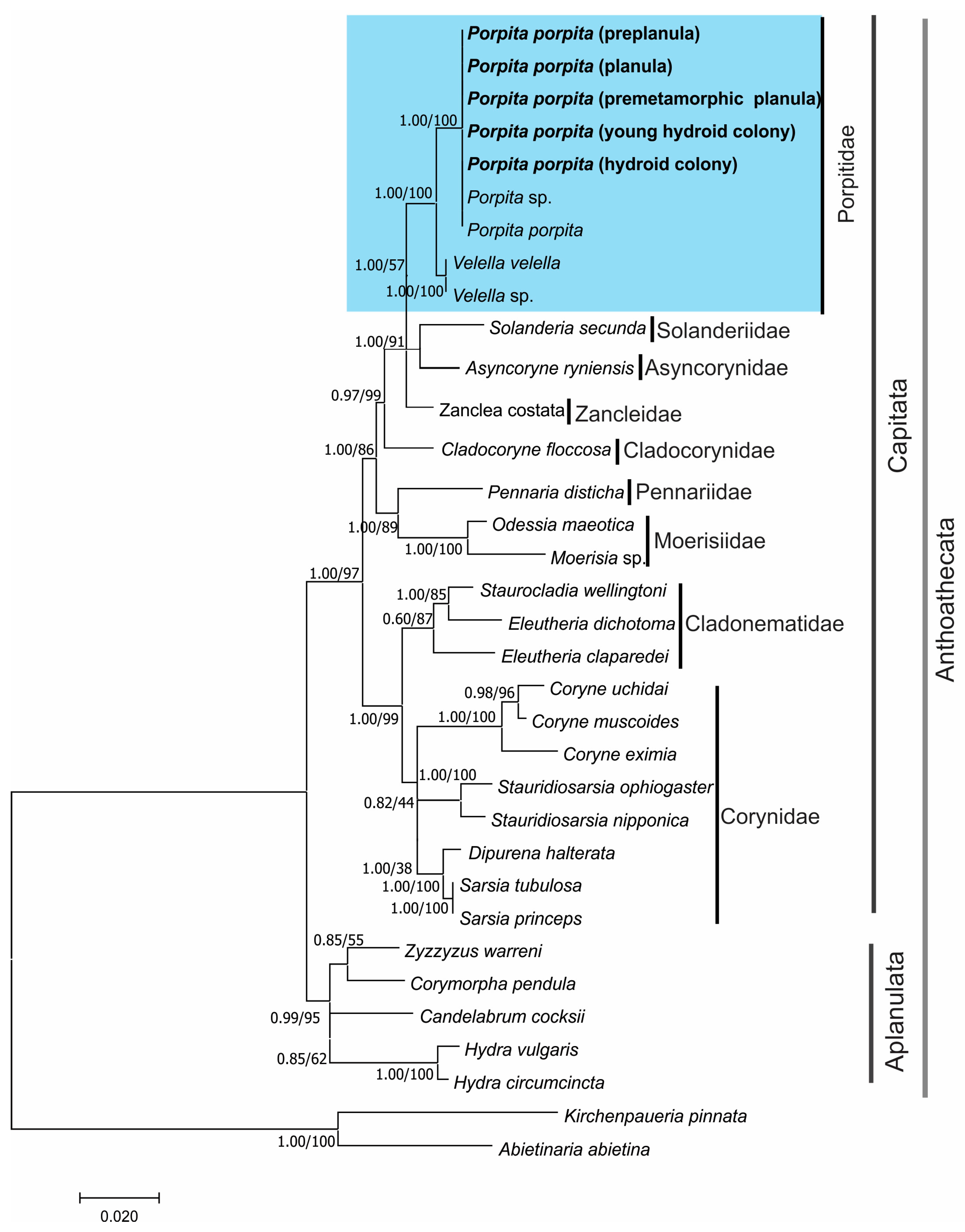
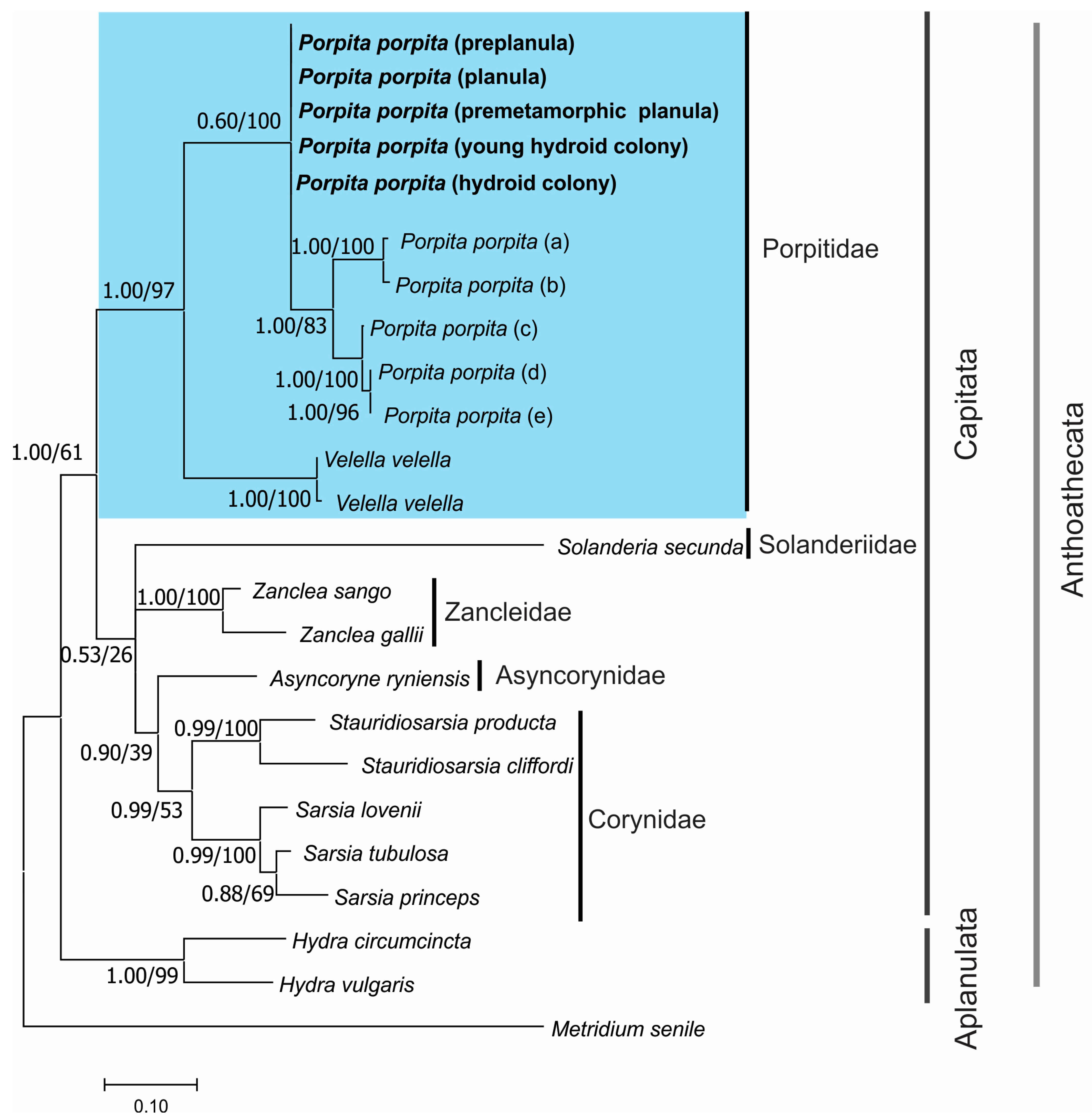
The sequences of the 18S rDNA and 28S rDNA genes were concatenated for analysis. The COI gene sequences were analyzed separately due to the lack of data on this gene for the species included in the ribosomal genes analysis. However, both analyses were used to determine the nucleotide relationship among samples (adults and larva). Sequences from NCBI of species belonging to the order Anthoathecata were downloaded and aligned with the sequences of different stage development of P. porpita used MEGA v.11 [31]. The best-fitting evolutionary substitution model for each selected region of the two data sets was determined using JModelTest 2.0 software; for the analysis of ribosomal genes, the model General Time Reversible + G + I and for COI gen Tamura-Nei+ G+ I model were using. Maximum likelihood (ML) analyses were performed using MEGA v.11 with 1000 bootstrap replicates [31]. Bayesian inference (BI) analysis was conducted using Mr. Bayes 3.2.1 [32] simulations of over 300,000 generations, with sampling every 10,000 generations. The appropriate burn-in value was determined by examining the standard deviation of split frequencies (<0.01). The posterior probabilities of trait values at ancestral nodes of phylogenies were derived using reversible jump Markov chain Monte Carlo (MCMC) methods. A majority rule consensus tree was constructed from all sampled generations after the burn-in period. The ribosomal (18s + 28s rDNA) gene tree was rooted using the sequence of Kirchenpaueria pinnata and Abietinaria abietina, while the COI tree was rooted with Metridium senile (= S1). The BI and ML trees are shown in posterior probability and bootstrap proportions, respectively, at each node. Finally, genetic distances with a Kimura two-parameter model (K2P) among sequences of larva and adults and sequences of P. porpita collected from other geographic areas and organisms in the Porpitidae family were obtained using MEGA v.11 software.

3. Results

3.1. Morphological and Histological Description

3.1.1. Hydroid Colony

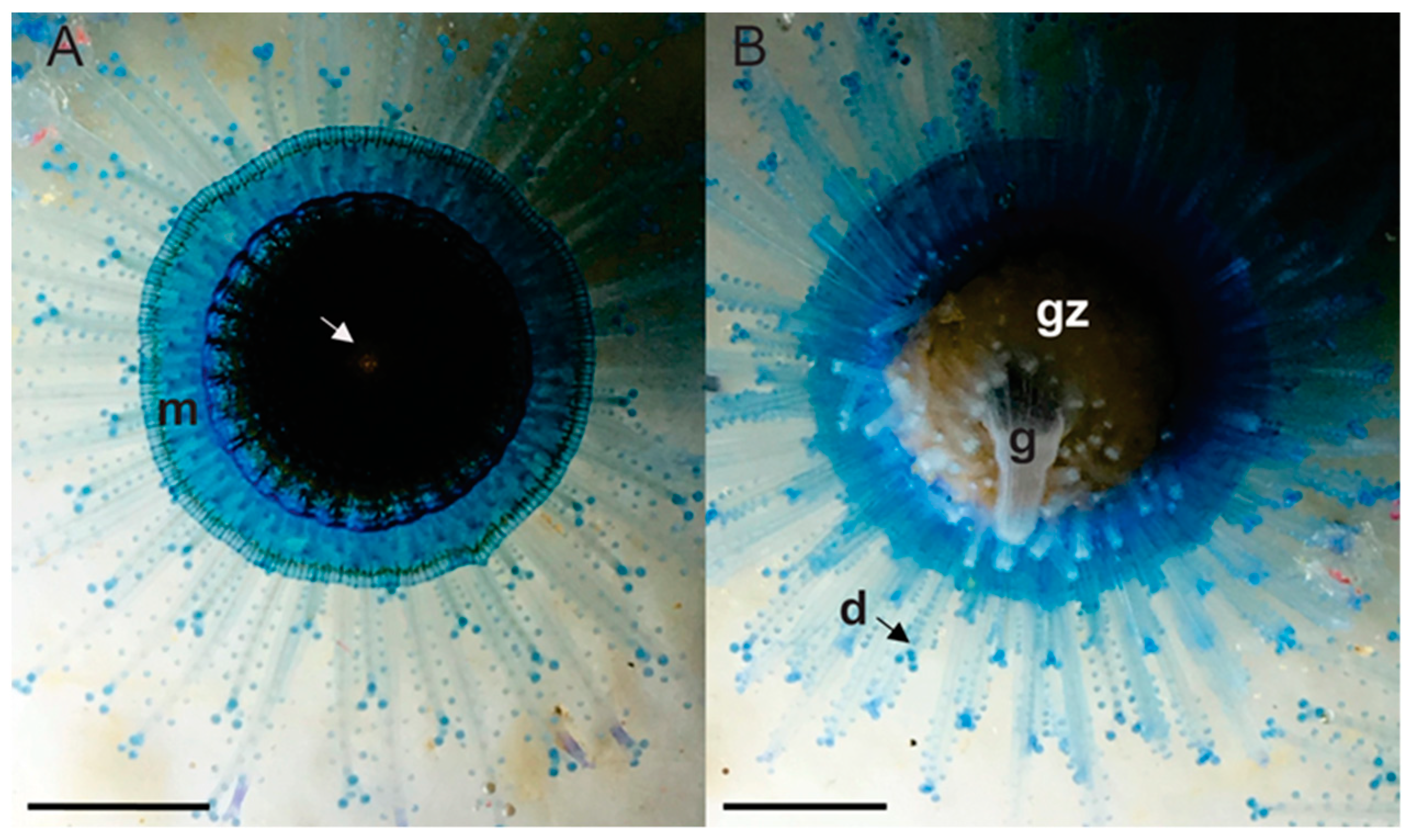
The hydroid colony (Figure 2) is pleustonic and polymorphic in the dorsal section, is slightly concave, and presents a disc-shaped clear blue mantle (6 mm wide), which overlies a chitinous float (29 mm diameter) of a dark brown color, and a central pore (pneumatophore) of a reddish-brown color (Figure 2A). The ventral undersurface has a single large central gastrozooid of a clear yellow color with a terminal mouth, a medial ring of gonozooids of a whitish–yellow color vermiform (Figure 2B), and a peripheral band of dactylozooids capitate with different longitudes of a bright blue color. Each dactylozooid has knobs of nematocysts arranged in longitudinal rows distally (Figure 2).

3.1.2. Young Hydroid Colony Stage

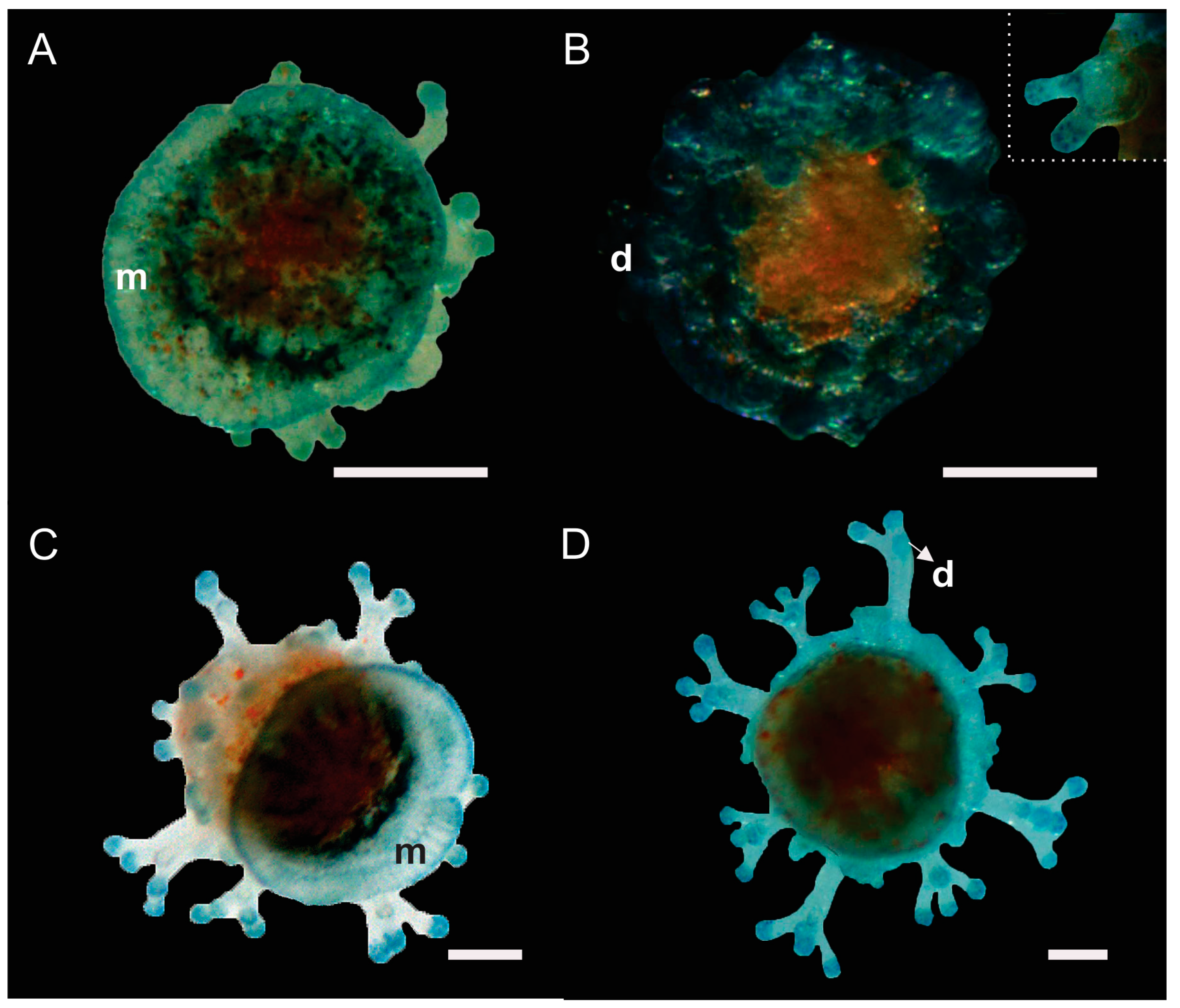
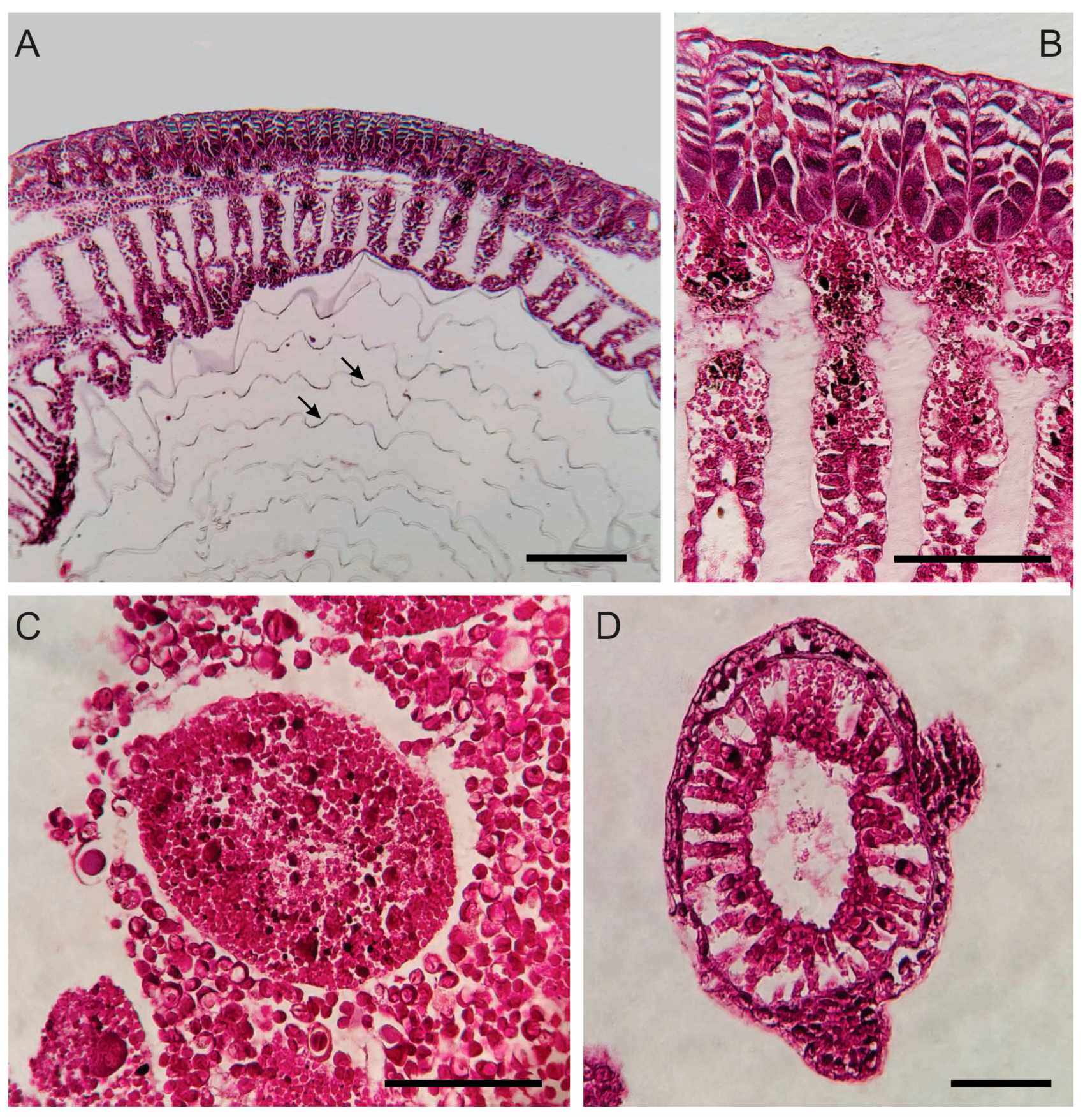
The young hydroid colony presented two (Figure 3) different stages of development. Both stage had a blue disk-shaped mantle and a developed chitinous float (Figure 3 and Figure 4A). The pneumatophore cavity is divided into chambers by vertical septa and branched cells that connect the two gastrodermal layers (Figure 4A,B), and the cavity of the float communicates with the exterior through an apical pore (Figure 2A). The first stage is the least developed, measures 5 mm, and has a light blue color with orange and dark brown spots on the dorsal region (Figure 3A). Gastrozooids and gonozooids were not observed in the ventral part (Figure 3B), but cell differentiation was observed (Figure 4C). Dactylozooids were present, with a rounded shape and only two branches (Figure 3B and Figure 4D). The second stage, measuring 6.5 mm, also did not develop gastrozooids or gonozooids (Figure 3C). In this stage, the ventral part was observed to be extended (Figure 3D), unlike the first stage, where the tissue was contracted (Figure 3B). The dactylozooids were more developed, showing greater length and branching (Figure 3D).

3.1.3. Planula Larva

The preplanula stage presents an ellipsoid form (210 µm in length and 175 µm in width). No distinction was observed between the oral and aboral poles. The endoderm appears reddish-brown and is separated from the translucent ectoderm by the mesoglea. The ectoderm is characterized by a poorly ciliated epithelium that is 35 µm wide (Figure 5A).
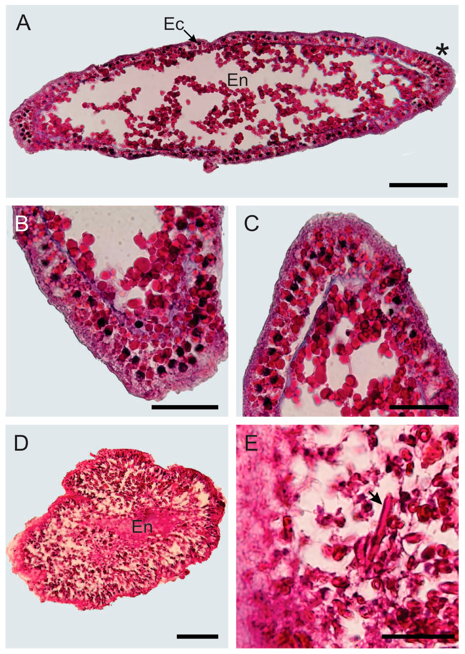
The motile planula stage larva is elongated and presents a fusiform shape (600 µm in length and 245 µm in width; Figure 5B). The ectoderm is pseudostratified (Figure 6) and 50 µm in width, while the endoderm contains yolk granules are separated by a thin and acellular mesogleal boundary (Figure 6A,C). The oral and aboral poles become distinguishable in the external morphology, with the oral pole being narrower (Figure 5B); however, they do not present differences in the conformation of their tissue (Figure 6B,C). Furthermore, the planula swims with a rounded aboral end forward, and the free-swimming planula gradually elongates.
The premetamorphic planula has a circular shape (650 µm in length and 500 µm in width). A circular furrow (125 µm wide) appears with bumps on the ectoderm of the oral pole during this phase (Figure 5C). At the cellular level, the oral pole, unlike the aboral pole, presents many well-developed cnidocytes and the cellular differentiation of the ectoderm is visible (Figure 6D,E).

3.2. Molecular Identification

The phylogenetic reconstruction using the maximum likelihood (ML) and Bayesian methods (BI) with the 18s and 28s rDNA (Figure 7) and COI mDNA (Figure 8) genes was congruent with moderate to high resolution for the Anthoathecata order and Capitata suborder.
The molecular phylogeny of the 18s and 28s rDNA was constructed using 35 sequences (2600 pb each). The phylogenetic analyses based BI and ML on this locus, placing the all-stage development of P. porpita of this study and P. porpita collected in the Western Pacific in the same subclade, which joined the subclade formed by organisms of V. vellela in a well-supported monophyletic clade corresponding to the family Porpitidae (see Figure 7).
The phylogenetic reconstructions resulting from the BI and ML analyses of the COI mDNA were constructed with 25 sequences (620 pb). The resulting topology (Figure 8) and ML analysis placed the planulae and hydroid colonies of P. porpita in a well-supported monophyletic clade with P. porpita from other geographic regions; however, three subclades were formed: the first subclade groups with the organisms sampled in the Mexican Pacific; the second subclade contains organisms from the Mediterranean Sea and North Atlantic; and the third subclade groups organisms from the Indian Ocean. The BI analysis also formed the same clades, but the posterior probability values (0.60) were not significant. The molecular analysis ML and BI with the COI data grouped P. porpita of all regions and V. velella within the Porpitidae family (Figure 8).
The genetic distance (K2P) calculated based on the mitochondrial DNA barcoding region (COI) and ribosomal DNA (18s and 28s rDNA) within congeneric species of the Porpitidae family revealed that sequences from the different larval and colony stages of P. porpita were identical (0.000 ± 0.000; see Table 1 and Table 2). The genetic distance calculated from the 18s and 28s rDNA showed no differences between the sequences of organisms from the CMP and the sequences of exemplars from the Western Pacific (0.000 ± 0.000; Table 1); meanwhile, the genetic distance between exemplars of P. porpita from V. velella was 0.008 ± 0.002 (Table 1). However, genetic distances using COI sequences indicated that the sampled P. porpita in the CMP region present genetic variation with respect to specimens collected in other regions (Table 2). The CMP organisms are genetically closer to P. porpita from the Indian Ocean (0.067 ± 0.004; 0.074 ± 0.004) and distant to organisms from the North Atlantic Ocean and Mediterranean Sea (0.087 ± 0.016; Table 2). Intergeneric comparison of the COI sequences revealed a genetic distance of 0.173 ± 0.013 between P. porpita and V. velella (Table 2).

4. Discussion

This study provides the first evidence of the presence of the larva of P. porpita and its ontogenic changes prior to the medusa stage. The evidence of planula larva in some orders, such Anthoathecata, is a diagnostic characteristic that distinguishes organisms in the suborder Capitata from those in suborder Aplanulata, which lack a larval stage [33]. Porpita porpita and V. velella are the only species of the Anthoathecata order with a distinct morphology, as they are holoplanktonic organisms. In fact, this has raised doubts regarding the membership of the family Porpitidae in the suborder Capitata, or even in the order Anthoathecata [33,34,35]. Consequently, the descriptions and observations of the larval stage in P. porpita are of significant importance to support their membership in the group Capitata.
Porpita porpita was recorded only during the cold temperature season from March to May. The presence of other pleustonic hydrozoans in the region [36,37,38,39] coincides with low-temperature periods. The Central Mexican Pacific is considered a transitional oceanographic zone, with high fluctuations in environmental conditions throughout the year [40]. Particularly during the end of the dry season, the water column presents low-temperature and high-nutrient concentrations due to the influence of coastal upwellings [41]; both of which are favorable conditions for the species.
The hydrozoan group exhibits high plasticity in their life cycle, with common changes including an absence of the medusa phase, loss of the polyp phase, and a range of diversity in polyp, colony, and medusa morphologies [42,43,44,45]. The life cycle of P. porpita includes planula larva, polypoid colony, and medusa stages, but it has been studied little in terms of morphology and physiology. This study describes the morphology of the larva during different developmental stages, including the preplanula, mobile larva, and premetamorphic larva. This is the first description of its kind. The absence of polarity is the primary characteristic defining a larva as preplanula. The cells of planula, polyp, and medusa are organized in the direction of a principal axis of polarity, termed oral–aboral. We observed differences between the oral and aboral poles in the planula stage. Due to its morphology and swimming habit, the planula swims forward with a rounded aboral end. Histological analysis of mobile planulae revealed an external pseudostratified ectoderm and an inner vacuolated endoderm separated by a thin, acellular mesogleal boundary. A distinctive feature of the ectoderm is its varying thickness at the anterior and posterior poles. These characteristics are consistent with the morphology of other cnidarian planulae [46,47,48,49]. Cellular differentiation was observed in the premetamorphic stage, as evidenced by more cells in the gastrodermis and the presence of cnidosites in the oral pole. There is cellular differentiation between poles; for example, the planula larva has an aboral specialization of the nervous system [50,51].
A general reorganization of the body and modification of the stem cell system characterize the transformation of the cnidaria planula into an adult. Metamorphosis involves the cessation of swimming, loss of cilia, and attachment to the substrate using glandular secretions and nematocytes [52,53,54,55]. In holoplanktonic species of the Siphonophores order, the planulae develop into a polymorphic colony without a primary polyp stage [56]. Although the changes during metamorphosis have not been characterized in P. porpita, the data obtained in this study allow us to establish evidence that this species—like the siphonophores—does not have a primary polyp stage. One piece of evidence is the formation of the pneumatophore in the aboral region initially. Planktonic larva are generally believed to have little or no control over their fate. In order to adapt to dynamic environments, the larva first generates the pneumatophore, which is of larval ectodermal origin, through which carbon monoxide can be released [57]. This allows the larva to regulate its float volume during vertical migration through controlling the amount of gas [58]. Thus, larva may have some control over their buoyancy early in their development, allowing them to maintain themselves within a preferred depth and temperature range. This optimization facilitates the development of the rest of the colony. The morphology of the pneumatophore of P. porpita was found in this study to be consistent with that of other holoplanktonic hydrozoans. The cavity is divided into chambers and, in the pneumatosaccus, it is covered by chitin. The function of the cells present in this region is still unknown. The cavity of the float communicates with the exterior through an apical pore. We did not observe developed gastrozooids, (i.e., feeding and digestive polyps) in young colonies. However, we did observe developing dactylozooids, suggesting that the colony prioritizes protection over ingesting food.
The COI barcoding and two other gene markers (18s and 28s) were amplified and employed to investigate the taxonomic status of the samples and the phylogenetic relationship of P. porpita within the order Anthotecata [59,60,61]. Our results indicated that the COI gene exhibits greater intraspecific variability compared to the 18s and 28s rDNA ribosomal genes. Specifically, we observed higher values of polymorphic sites among the sequences obtained in the CMP and other regions of the world. In particular, the 18s and 28s rDNA ribosomal genes presented only two polymorphic sites among the sequences obtained from the CMP and organisms distributed in the Western Pacific. These differences can be attributed to the type of molecular marker, as ribosomal markers (18s and 28s rDNA) are more conserved and diverge slower than mitochondrial markers, as reported by Hellberg et al. [62]; in contrast, in some taxa, the COI gene may exhibit significant nucleotide variations, including intraindividual heterogeneity [63,64]. These results are consistent with those reported in V. velella, which exhibited intraindividual heterogeneity among organisms from the Atlantic, Pacific, and Indian regions [65]. Therefore, further studies focused on the genetic variations in different populations of P. porpita is necessary in order to determine whether the observed inter-species changes are due to the selected molecular markers or whether P. porpita is undergoing a speciation process, which would result in multiple species being placed within the genus.
The taxonomy of the Porpita genus has changed considerably since it was first described, and 20 nominal species have been assigned at various times to this genus [66]. However, only three species have been recognized based on their morphological characteristics: Porpita porpita (Linnaeus, 1758) from the Indian Ocean, P. umbella (Müller, 1776b) from the Atlantic Ocean, and P. pacifica Lesson 1826 from the Pacific Ocean. However, since the works of Moser [15] and Totton [67], these three species have been considered conspecific. The COI gene analysis in this study indicated the presence of multiple species within the Porpita genus. However, confirmation is not yet possible due to the limited information from other genes (e.g., 18S and 28S) and the small number of specimens from other regions subjected to molecular analysis.
Detailed morphological information on zooplankton larva can avoid or correct identification problems commonly encountered in marine invertebrates with a metagenetic life cycle [1,43,68,69,70]. This research supports the future identification of P. porpita based on its morphological characteristics, regardless of the stage of development. Clarifying the morphology of the various stages of the life cycle will be useful for classification and may also aid our understanding of the evolutionary loss and convergence of these characteristics (both within and outside the group).

Supplementary Materials

The following supporting information can be downloaded at https://www.mdpi.com/article/10.3390/d16070425/s1, Table S1. Samples with GenBank (https://www.ncbi.nlm.nih.gov/genbank/ accessed on 3 February 2024) accession numbers. Blue box: samples from this study.

Author Contributions

Conceptualization, J.D.S.-V. and E.G.-D.; methodology, J.D.S.-V., M.A.R.-L. and E.B.-G.; resources, all authors; writing—original draft preparation, J.D.S.-V.; writing—all authors; funding acquisition, E.G.-D., M.d.C.F.-G. and A.P.R.-T. All authors have read and agreed to the published version of the manuscript.

Funding

The present study was funded by the Programa de Apoyo a la Mejora en las Condiciones de Producción de los Miembros del SNI y SNCA–PROSNI and the Programa de Fortalecimiento de Institutos, Centro y Laboratorios de Investigacion 2022 of the Universidad de Guadalajara, Mexico.

Institutional Review Board Statement

Not applicable.

Data Availability Statement

Data are contained within the article and Supplementary Materials.

Acknowledgments

JDSV was supported by a postdoctoral fellowship from the National Council of Humanities, Science, and Technology (CONAHCYT; ID: 619025). The authors would like to express their gratitude to the Laboratorio de Posgrado. Lab—209 “Equipamiento de laboratorio, para el estudio de sistemática molecular y poblacional en Ecología Marina of centro Universitario de la Costa Sur-UDG” and Laboratorio de Ecologia Marina the Centro Universitario de la costa-UDG, for the use of facilities for molecular and histological processing, and the anonymous reviewers for their valuable contributions that helped to improve the manuscript.

Conflicts of Interest

The authors declare no conflicts of interest. The funders had no role in the design of the study; in the collection, analyses, or interpretation of data; in the writing of the manuscript; or in the decision to publish the results.

References

  1. Bouillon, G.C.; Gili, F.P.; Boero, J.M. An Introduction to Hydrozoa. In Mémoires du Muséum National d’Histoire Naturelle; Muséum National d’Histoire Naturelle: Paris, France, 2006; Volume 194, pp. 1–591. ISBN 2-85653-580-1. [Google Scholar]
  2. Schuchert, P. The European anthecate hydroids and their medusa (Hydrozoa, Cnidaria): Capitata Part 2. Rev. Suisse Zool. 2010, 117, 337–555. [Google Scholar] [CrossRef]
  3. Dunn, C.W.; Wagner, G.P. The evolution of colony-level development in the Siphonophora (Cnidaria: Hydrozoa). Dev. Genes Evol. 2006, 216, 743–754. [Google Scholar] [CrossRef]
  4. Gürlek, M.; Uyan, A.; Doğdu, S.A.; Karan, S.; Gökçen, A.; Turan, C. Occurrence of the Blue Button Porpita porpita (Linnaeus, 1758) in the Iskenderun Bay, Northeastern Mediterranean Coast of Turkey. Acta Adriat. 2020, 61, 185–190. [Google Scholar] [CrossRef]
  5. Kirkendale, L.; Calder, D. Hydroids (Cnidaria: Hydrozoa) from Guam and the Commonwealth of the Northern Marianas Islands (CNMI). Micronesica 2003, 3536, 159–188. [Google Scholar]
  6. Gul, S.; Gravili, C. On the occurrence of Porpita porpita (Cnidaria: Hydrozoa) at Pakistan coast (North Arabian Sea). Mar. Biodivers. Rec. 2014, 7, 24. [Google Scholar] [CrossRef]
  7. Msn, C.; Sharifuzzaman, S.; Chowdhury, S.; Nabi, M.; Hossain, M. First Record of Porpita porpita (Cnidaria: Hydrozoa) from the coral reef ecosystem, Bangladesh. Ocean Sci. 2016, 51, 293–297. [Google Scholar] [CrossRef]
  8. Lillo, A.; Tiralongo, O.; Tondo, E. New records of Porpita porpita (Linnaeus, 1758) (Cnidaria: Hydrozoa) in the mediterranean sea. NESciences 2019, 4, 293–298. [Google Scholar] [CrossRef]
  9. Madkour, F.; Zaghloul, W.; Mohammad, S. First record of Porpita porpita (Linnaeus, 1758) (Cnidaria: Hydrozoa, Porpitidae) from the Red Sea of Egypt. J. Aquat. Sci. Mar. Biol. 2019, 2, 24–27. [Google Scholar] [CrossRef]
  10. Sivalingam, G. Notes on the occurrence of Porpita porpita (Blue button) from Pulicat Lagoon. J. Res. Biol. 2019, 4, 1487–1490. [Google Scholar]
  11. Bieri, R. Feeding preferences and rates of the snail, Ianthina prolongata, the barnacle, Lepas anserifera, the nudibranchs, Glaucus atlanticus and Fiona pinnata, and the food web in the marine neuston. Publ. Seto Mar. Biol. Lab. 1996, 14, 161–170. [Google Scholar] [CrossRef]
  12. Thompson, T.E.; Bennett, I. Observations on Australian Glaucidae (Mollusca: Opisthobranchia). Zool. J. Linn. Soc. 1970, 49, 187–197. [Google Scholar] [CrossRef]
  13. Sahu, B.K.; Baliarsingh, S.K.; Samanta, A.; Srichandan, S.; Singh, S. Mass beach stranding of blue button jellies (Porpita porpita, Linnaeus, 1758) along Odisha coast during summer season. Indian. J. Geo-Mar. Sci. 2020, 49, 1093–1096. [Google Scholar]
  14. Umamageswari, P.; Dineshkumar, R.; Jayasingam, P.; Sampathkumar, P. Antimicrobial activities of jellyfish, Porpita porpita from Southeast Coast of India. Pharm. Biotechnol. Microbiol. 2016, 2016, 1–4. [Google Scholar]
  15. Moser, F. Die Siphonophoren der Deutschen Südpolar-Expedition, 1901–1903. In Deutsche Südpolar-Expedition 1901–1903; Walter de Gruyter: Berlin, Germany, 1925; Volume 17, pp. 1–541. [Google Scholar]
  16. Cartwright, P.; Nawrocki, A. Character Evolution in Hydrozoa (phylum Cnidaria). Integr. Comp. Biol. 2010, 50, 456–472. [Google Scholar] [CrossRef]
  17. Ortman, B.D.; Bucklin, A.; Pages, F.; Youngbluth, M. DNA barcoding the Medusozoa using mtCOI. Deep-Sea Res. II Top. Stud. Oceanogr. 2010, 57, 2148–2156. [Google Scholar] [CrossRef]
  18. Khalturin, K.; Shinzato, C.; Khalturina, M.; Hamada, M.; Fujie, M.; Koyanagi, R.; Kanda, M.; Goto, H.; Anton-Erxleben, F.; Toyokawa, M.; et al. Medusozoan genomes inform the evolution of the jellyfish body plan. Nat. Ecol. Evol. 2019, 3, 811–822. [Google Scholar] [CrossRef]
  19. Brinckmann, A. Observations on the biology and development of Staurocladia portmanni sp. n. (Anthomedusae, Eleutheridae). Can. J. Zool. 1964, 42, 693–705. [Google Scholar] [CrossRef]
  20. Helm, R.R. The mysterious ecosystem at the ocean’s surface. PLoS Biol. 2021, 19, e3001046. [Google Scholar] [CrossRef]
  21. Pyataeva, S.V.; Hopcroft, R.R.; Lindsay, D.J.; Collins, A.G. DNA barcodes unite two problematic taxa: The meiobenthic Boreohydra simplex is a life-cycle stage of Plotocnide borealis (Hydrozoa: Aplanulata). Zootaxa 2016, 4150, 85–92. [Google Scholar] [CrossRef]
  22. Grossmann, M.M.; Lindsay, D.J.; Collins, A.G. The end of an enigmatic taxon: Eudoxia macra is the eudoxid stage of Lensia cossack (Siphonophora, Cnidaria). Syst. Biodivers. 2013, 11, 381–387. [Google Scholar] [CrossRef]
  23. Grossmann, M.M.; Collins, A.G.; Lindsay, D.J. Description of the eudoxid stages of Lensia havock and Lensia leloupi (Cnidaria: Siphonophora: Calycophorae), with a review of all known Lensia eudoxid bracts. Syst. Biodivers. 2014, 12, 163–180. [Google Scholar] [CrossRef]
  24. Schuchert, P. The polyps of Oceania armata identified by DNA barcoding (Cnidaria, Hydrozoa). Zootaxa 2016, 4175, 539–555. [Google Scholar] [CrossRef]
  25. Calder, D.R. Some anthoathecate hydroids and limnopolyps (Cnidaria, Hydrozoa) from the Hawaiian archipelago. Zootaxa. 2010, 2590, 1–91. [Google Scholar] [CrossRef]
  26. Santiago-Valentín, J.D.; Rodríguez-Troncoso, A.P.; Bautista-Guerrero, E.; López-Pérez, A.; Cupul-Magaña, A.L. Successful sexual reproduction of the scleractinian coral Porites panamensis: Evidence of planktonic larvae and recruitment. Invertebr. Biol. 2019, 138, 29–39. [Google Scholar] [CrossRef]
  27. Lynch, M.; Raphael, S.; Mellor, L.; Spare, P.; Inwood, M. Métodos de Laboratorio, 2nd ed.; Interamericana: Puebla, Mexico, 1972; 1522p. [Google Scholar]
  28. Folmer, O.; Black, M.; Hoeh, W.; Lutz, R.; Vrijenhoek, R. DNA primers for amplification of mitochondrial cytochrome c oxidase subunit I from diverse metazoan invertebrates. Mol. Mar. Biol. Biotechnol. 1994, 3, 294–299. [Google Scholar]
  29. Redmond, N.E.; Morrow, C.C.; Thacker, R.W.; Diaz, M.C.; Boury-Esnault, N.; Cárdenas, P.; Hajdu, E.; Lobo-Hajdu, G.; Picton, B.E.; Pomponi, S.A.; et al. Phylogeny and systematics of Demospongiae in light of new small-subunit ribosomal DNA (18S) sequences. Integr. Comp. Biol. 2013, 53, 388–415. [Google Scholar] [CrossRef]
  30. Medina, M.; Collins, A.G.; Silberman, J.D.; Sogin, M.L. Evaluating hypotheses of basal animal phylogeny using complete sequences of large and small subunit rRNA. Proc. Natl. Acad. Sci. USA 2001, 98, 9707–9712. [Google Scholar] [CrossRef]
  31. Kumar, S.; Stecher, G.; Li, M.; Knyaz, C.; Tamura, K. MEGA X: Molecular Evolutionary Genetics Analysis across Computing Platforms. Mol. Biol. Evol. 2018, 35, 1547–1549. [Google Scholar] [CrossRef]
  32. Ronquist, F.; Teslenko, M.; van der Mark, P.; Ayres, D.L.; Darling, A.; Höhna, S.; Larget, B.; Liu, L.; Suchard, M.A.; Huelsenbeck, J.P. MrBayes 3.2: Efficient Bayesian Phylogenetic Inference and Model Choice Across a Large Model Space. Syst. Biol. 2012, 61, 539–542. [Google Scholar] [CrossRef]
  33. Petersen, K.W. Evolution and taxonomy in capitate hydroids and medusae (Cnidaria: Hydrozoa). Zool. J. Linn. Soc. 1990, 100, 101–231. [Google Scholar] [CrossRef]
  34. Bouillon, J. Essai de classification des Hydropolypes-Hydroméduses (Hydrozoa-Cnidaria). Indo-Malay. Zool. 1985, 1, 29–243. [Google Scholar]
  35. Daly, M.; Brugler, M.R.; Cartwright, P.; Collins, A.G.; Dawson, M.N.; Fautin, D.G.; France, S.C.; Mcfadden, C.S.; Opresko, D.M.; Rodríguez, E.; et al. The phylum Cnidaria: A review of phylogenetic patterns and diversity 300 years after Linnaeus. Zootaxa 2007, 1668, 127–182. [Google Scholar] [CrossRef]
  36. Gómez, A.S. Contribución al estudio faunístico de celenterados y ctenóforos del plancton estuarino del noroeste de México. An. Inst. Biol. Univ. Nac. Autón. Mex. Bot. 1991, 62, 1–10. [Google Scholar]
  37. Gamero-Mora, E.; Ceballos-Corona, G.; Gasca, R.; Morales-Blake, A. Análisis de la comunidad del zooplancton gelatinoso (Hydrozoa, Ctenophora, Thaliacea) en el Pacífico central mexicano, abril-mayo 2011. Rev. Biol. Mar. Oceanogr. 2015, 50, 111–124. [Google Scholar] [CrossRef]
  38. Suárez-Morales, E.; Segura-Puertas, L.; Gasca, R. Medusan (Cnidaria) assemblages off the Caribbean coast of Mexico. J. Coast. Res. 1999, 15, 140–147. [Google Scholar]
  39. Fernández-Álamo, M.A.; Färber-Lorda, J. Zooplankton and the Oceanography of the Eastern Tropical Pacific: A Review. Prog. Oceanogr. 2006, 61, 318–359. [Google Scholar] [CrossRef]
  40. CONABIO, 2022. Biodiversidad Mexicana. Available online: https://www.biodiversidad.gob.mx/region/ecorregiones-marinas (accessed on 13 April 2024).
  41. Ambriz-Arreola, I.; Gómez-Gutiérrez, J.; del Carmen Franco-Gordo, M.; Lavaniegos, B.E.; Godínez-Domínguez, E. Influence of coastal upwelling downwelling variability on tropical euphausiid abundance and community structure in the inshore Mexican central Pacific. Mar. Ecol. Prog. Ser. 2012, 451, 119–136. [Google Scholar] [CrossRef]
  42. Dunn, C.W.; Pugh, P.R.; Haddock, S.H. Molecular phylogenetics of the Siphonophora (Cnidaria), with implications for the evolution of functional specialization. Syst. Biol. 2005, 54, 916–935. [Google Scholar] [CrossRef]
  43. Collins, A.G.; Schuchert, P.; Marques, A.C.; Jankowski, T.; Medina, M.; Schierwater, B. Medusozoan phylogeny and character evolution clarified by new large and small subunit rDNA data and an assessment of the utility of phylogenetic mixture models. Syst. Biol. 2006, 55, 97–115. [Google Scholar] [CrossRef]
  44. Leclère, L.; Schuchert, P.; Manuel, M. Phylogeny of the Plumularioidea (Hydrozoa, Leptothecata): Evolution of Colonial Organisation and Life Cycle. Zool. Scr. 2007, 36, 371–394. [Google Scholar] [CrossRef]
  45. Leclère, L.; Copley, R.R.; Momose, T.; Houliston, E. Hydrozoan insights in animal development and evolution. Curr. Opin. Genet. Dev. 2016, 39, 157–167. [Google Scholar] [CrossRef]
  46. Vandermeulen, J.H. Studies on reef corals. II. Fine structure of planktonic planula larva of Pocillopora damicornis, with emphasis on the aboral epidermis. Mar. Biol. 1974, 27, 239–249. [Google Scholar] [CrossRef]
  47. Hirose, M.; Kinzie, R.A.; Hidaka, M. Early development of zooxanthella-containing eggs of the corals Pocillopora verrucosa and P. eydouxi with special reference to the distribution of zooxanthellae. Biol. Bull. 2000, 199, 68–75. [Google Scholar] [CrossRef]
  48. Hirose, M.; Hidaka, M. Early development of zooxanthella-containing eggs of the corals Porites cylindrica and Montipora digitata: The endodermal localization of zooxanthellae. Zool. Sci. 2006, 23, 873–881. [Google Scholar] [CrossRef]
  49. Santiago-Valentín, J.D.; Rodríguez-Troncoso, A.P.; Bautista-Guerrero, E.; López-Pérez, A.; Cupul-Magaña, A.L. Internal ultrastructure of the planktonic larva of the coral Porites panamensis (Anthozoa: Scleractinia). Rev. Biol. Trop. 2022, 70, 222–234. [Google Scholar] [CrossRef]
  50. Freeman, G. The role of polarity in the development of the hydrozoan planula larva. Wilehm. Roux. Arch. Dev. Biol. 1981, 190, 168–184. [Google Scholar] [CrossRef] [PubMed]
  51. Piraino, S.; Zega, G.; Di Benedetto, C.; Leone, A.; Dell’Anna, A.; Pennati, R.; Carnevali, D.C.; Schmid, V.; Reichert, H. Complex neural architecture in the diploblastic larva of Clava multicornis (Hydrozoa, Cnidaria). J. Comp. Neurol. 2011, 519, 1931–1951. [Google Scholar] [CrossRef] [PubMed]
  52. Vandermeulen, J.H. Studies on reef corals. III. Fine structural changes of calicoblast cells in Pocillopora damicornis during settling and calcification. Mar. Biol. 1975, 31, 69–77. [Google Scholar] [CrossRef]
  53. Martin, V.; Chia, F.S.; Koss, R. A fine structural study of metamorphosis of the hydrozoan Mitrocomella polydiademata. J. Morphol. 1983, 176, 261–287. [Google Scholar] [CrossRef]
  54. Martin, V.J.; Archer, W.E. Stages of larval development and stem cell population changes during metamorphosis of a hydrozoan planula. Biol. Bull. 1997, 192, 41–52. [Google Scholar] [CrossRef]
  55. Strömberg, S.M.; Östman, C.; Larsson, A.I. The cnidome and ultrastructural morphology of late planulae in Lophelia pertusa (Linnaeus, 1758)—With implications for settling competency. Acta Zool. 2019, 100, 431–450. [Google Scholar] [CrossRef]
  56. Sherlock, R.E.; Robison, B.H. Effects of temperature on the development and survival of Nanomia bijuga (Hydrozoa, Siphonophora). Invertebr. Biol. 2000, 119, 379–385. [Google Scholar] [CrossRef]
  57. Pickwell, G.V. Physiological Dynamics of Siphonophores from Deep Scattering Layers: Size of Gas-Filled Floats and Rate of Gas Production; Report 1369; Navy Electronics Laboratory (NEL): San Diego, CA, USA, 1966; 50p. [Google Scholar]
  58. Mackie, G.O.; Pugh, P.R.; Purcell, J.E. Siphonophore biology. Adv. Mar. Biol. 1987, 24, 97–262. [Google Scholar] [CrossRef]
  59. Schuchert, P. DNA barcoding of some Pandeidae species (Cnidaria, Hydrozoa, Anthoathecata). Rev. Suisse Zool. 2020, 125, 101–127. [Google Scholar] [CrossRef]
  60. Moura, C.J.; Harris, D.J.; Cunha, M.R.; Rogers, A.D. DNA barcoding reveals cryptic diversity in marine hydroids (Cnidaria, Hydrozoa) from coastal and deep-sea environments. Zool. Scr. 2008, 37, 93–108. [Google Scholar] [CrossRef]
  61. Cartwright, P.; Evans, N.M.; Dunn, C.W.; Marques, A.C.; Miglietta, M.P.; Schuchert, P.; Collins, A.G. Phylogenetics of Hydroidolina (Hydrozoa: Cnidaria). J. Mar. Biol. Assoc. UK 2008, 88, 1663–1672. [Google Scholar] [CrossRef]
  62. Hellberg, M.E.; Burton, R.S.; Neigel, J.E.; Palumbi, S.R. Genetic assessment of connectivity among marine populations. Bull. Mar. Sci. 2002, 70, 273–290. [Google Scholar]
  63. Schroth, W.; Jarms, G.; Streit, B.; Schierwater, B. Speciation and phylogeography in the cosmopolitan marine moon jelly, Aurelia sp. BMC Evol. Biol. 2002, 2, 1. [Google Scholar] [CrossRef]
  64. Kim, D.H.; Abraham, A.; Cho, J.H. A hybrid genetic algorithm and bacterial foraging approach for global optimization. Inf. Sci. 2007, 177, 3918–3937. [Google Scholar] [CrossRef]
  65. Morello, B.; Bo, M.; Betti, F.; Bavestrello, G.; Abbiati, M.; Costantini, F. Spatial and temporal genetic structure of Velella velella (Hydrozoa, Porpitidae) and its predator Janthina pallida (Gastropoda, Epitoniidae). Hydrobiologia 2023, 850, 1751–1762. [Google Scholar] [CrossRef]
  66. Calder, D.R. Shallow-Water Hydroids of Bermuda: The Athecatae; Life sciences contributions; Royal Ontario Museum: Toronto, ON, Canada, 1988; Volume 148, pp. 1–107. [Google Scholar] [CrossRef]
  67. Totton, A.K. Siphonophora of the Indian Ocean together with systematic and biological notes on related specimens from other oceans. Discov. Rep. 1954, 27, 1–162. [Google Scholar]
  68. Schuchert, P. Revision of the European athecate hydroids and their medusae (Hydrozoa, Cnidaria): Families of Oceanidae and Pachycordylidae. Rev. Suisse. Zool. 2004, 111, 315–369. [Google Scholar] [CrossRef]
  69. Miranda, L.S.; Collins, A.G.; Marques, A.C. Molecules clarify a Cnidarian life cycle: The “Hydrozoan” Microhydrula limopsicola is an early life stage of the Staurozoan Haliclystus antarcticus. PLoS ONE 2010, 5, e10182. [Google Scholar] [CrossRef] [PubMed]
  70. Morandini, A.C.; Schiariti, A.; Stampar, S.N.; Maronna, M.M.; Straehler-Pohl, I.; Marques, A.C. Succession of generations is still the general paradigm for scyphozoan life cycles. Bull. Mar. Sci. 2016, 92, 343–351. [Google Scholar] [CrossRef]
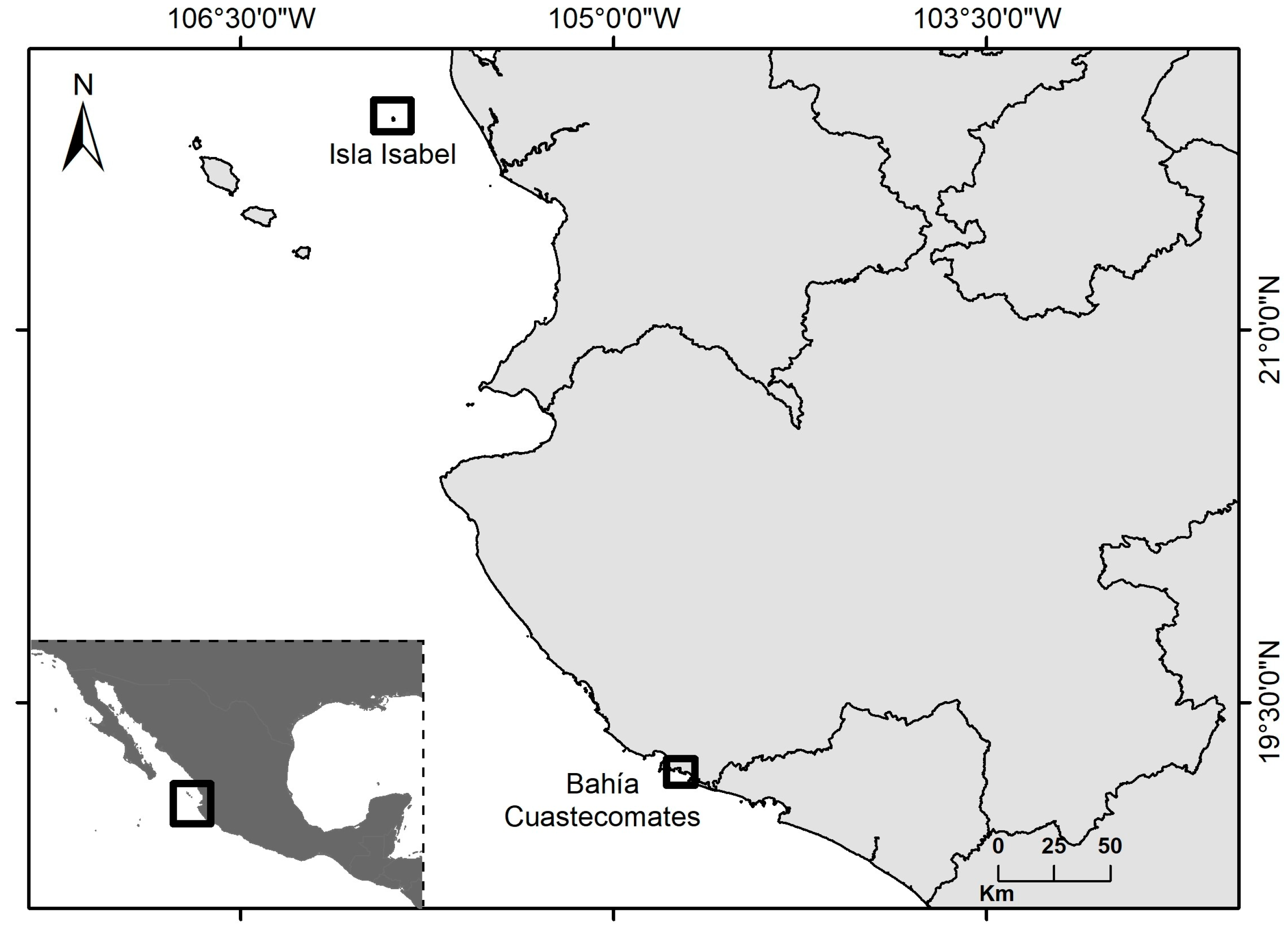
Figure 1. Sampling sites of larva and adult Porpita porpita in the Mexican Central Pacific.
Diversity 16 00425 g001
Figure 2. Hydrozoan colony of Porpita porpita. (A) Dorsal view: m, mantle. The arrow points to the pneumatophore. (B) Ventral view: gz, gonozooids; (g) gastrozooid; (d) dactylozooids. Scale = 3 mm.
Diversity 16 00425 g002
Figure 3. Young hydroid colony stage of Porpita porpita (m) mantle; (d) dactylozooids. (A) Ventral view of Stage 1. (B) Dorsal view of Stage 1, box: dactylozooid zoom. Scale = 2.5 mm. (C) Ventral view of Stage 2. (D) Dorsal view of Stage 2. Scale = 2 mm.
Diversity 16 00425 g003
Figure 4. Young hydroid colony of Porpita porpita. (A) Cross-section of the mantle and chitinous float (arrows). Scale = 100 µm; (B) Branched cells that connect the two gastrodermal layers. Scale = 50 µm; (C) Transverse section of gonozooid in morphogenesis. Note the cellular differentiation. Scale = 100 µm; (D) dactylozooids. Scale = 100 µm.
Diversity 16 00425 g004
Figure 5. Larval stage of Porpita porpita. (A) Preplanula. (B) Planula. (C) Premetamorphic planula. En: endoderm; Ec: ectoderm; * aboral pole. Scale = 100 µm.
Diversity 16 00425 g005
Figure 6. Larval stage of Porpita porpita. (A) Cross-section of planula. Scale = 100 µm. (B) Aboral pole of planula. Scale = 50 µm. (C) Oral pole of planula. Scale = 50 µm. (D) Cross-section for premetamorphic planula. Scale = 50 µm. (E): cnidocytes; En: endoderm; Ec: ectoderm; * aboral pole. Scale = 20 µm.
Diversity 16 00425 g006
Figure 7. Systematic position of Porpita porpita using concatenated sequences of ribosomal 18s rDNA and 28s rDNA genes. The topology was inferred using the Bayesian inference and maximum likelihood methods. The values of each node correspond to the posterior probability and bootstrap values of the major clades that were reconstructed with the BI and ML strict consensus tree, respectively (BI/ML). The blue boxes represent the Porpitidae family. GenBank accession numbers are provided in Table S1.
Diversity 16 00425 g007
Figure 8. Systematic position of Porpita porpita using sequences of the COI gene. The values of each node correspond to the posterior probability and bootstrap values of major clades that were reconstructed with BI and ML strict consensus trees, respectively (BI/ML). The blue boxes represent the Porpitidae family. GenBank accession numbers are provided in Table S1.
Diversity 16 00425 g008
Table 1. Pairwise genetic distance matrix for selected members of the Porpitidae family members, using ribosomal 18s and 28S rRNA concatenated genes (2600 pb). The matrix displays values of genetic distances (lower left diagonal) and their corresponding standard error estimates (upper right diagonal).
28s and 18s123456789
1Porpita porpita (preplanula)-0.0000.0000.0000.0000.0000.0000.0020.002
2Porpita porpita (planula)0.000-0.0000.0000.0000.0000.0000.0020.002
3Porpita porpita (premetamorphic planula)0.0000.000-0.0000.0000.0000.0000.0020.002
4Porpita porpita (young hydroid colony)0.0000.0000.000-0.0000.0000.0000.0020.002
5Porpita porpita (hydroid colony)0.0000.0000.0000.000-0.0000.0000.0020.002
6Porpita porpita0.0000.0000.0000.0000.000-0.0000.0020.002
7Porpita sp.0.0000.0000.0000.0000.0000.000-0.0020.002
8Vellella velella0.0080.0080.0080.0080.0080.0080.008-0.000
9Velella sp.0.0080.0080.0080.0080.0080.0080.0080.000-
Table 2. Pairwise genetic distance matrix for selected members of the Porpitidae family members, using a mitochondrial (COI) gene (620 pb). The matrix displays values of genetic distances (lower left diagonal) and their corresponding standard error estimates (upper right diagonal).
COI12345678910111213
1Porpita porpita (preplanula)-00.0000.0000.0000.0140.0140.0130.0120.0130.0200.0200.021
2Porpita porpita (planula)0.000-0.0000.0000.0000.0140.0140.0130.0120.0130.0200.0200.021
3Porpita porpita (premetamorphic planula)0.0000.000-0.0000.0000.0140.0140.0130.0120.0130.0200.0200.021
4Porpita porpita (young hydroid colony)0.0000.0000.000-0.0000.0140.0140.0130.0120.0130.0200.0200.021
5Porpita porpita (hydroid colony)0.0000.0000.0000.000-0.0140.0140.0130.0120.0130.0200.0200.021
6Porpita porpita (a) Mediterranean Sea0.0870.0870.0870.0870.087-0.0050.0130.0130.0130.0230.0220.024
7Porpita porpita (b) North Atlantic Ocean0.0870.0870.0870.0870.0870.012-0.0130.0130.0130.0230.0220.025
8Porpita porpita (c) Indian Ocean0.0740.0740.0740.0740.0740.0800.083-0.0040.0000.0230.0230.025
9Porpita porpita (d) Indian Ocean0.0670.0670.0670.0670.0670.0780.0810.010-0.0040.0100.0220.025
10Porpita porpita (e) Indian Ocean0.0740.0740.0740.0740.0740.0800.0830.0000.010- 0.0230.025
11Velella velella0.1710.1710.1710.1710.1710.2070.2010.2090.2070.209-0.0030.023
12Velella velella0.1710.1710.1710.1710.1710.2040.2010.2120.2040.2120.006 0.024
Disclaimer/Publisher’s Note: The statements, opinions and data contained in all publications are solely those of the individual author(s) and contributor(s) and not of MDPI and/or the editor(s). MDPI and/or the editor(s) disclaim responsibility for any injury to people or property resulting from any ideas, methods, instructions or products referred to in the content.