Article

Two New Lyophyllum Species from Yunnan, China

1
Faculty of Environmental Sciences and Engineering, Kunming University of Science and Technology, Kunming 650500, China
2
Biotechnology and Germplasm Resources Institute, Yunnan Academy of Agricultural Sciences, Kunming 650223, China
3
College of Agriculture and Biological Science, Dali University, Dali 671003, China
4
College of Biotechnology and Bioengineering, West Yunnan University, Lincang 677000, China
*
Author to whom correspondence should be addressed.
Diversity 2024, 16(4), 210; https://doi.org/10.3390/d16040210
Submission received: 28 February 2024 / Revised: 28 March 2024 / Accepted: 28 March 2024 / Published: 29 March 2024

Abstract

:
Two novel species of Lyophyllum are proposed based on morphological and phylogenetic investigations. Lyophyllum bulborhizum sp. nov. has abundant black scales on the surface, is a dry, dark grey at the center, grey at the margin, has a clear bulbous stipe base, and has a narrowly cylindrical or narrowly clavate pleurocystidia and cheilocystidia. Lyophyllum nigrum sp. nov. is tufted and has a dark grey pileus and subclavate, a greyish green-to-olive stipe, and a narrowly cylindrical or narrowly clavate pleurocystidia and cheilocystidia. In addition, L. rhombisporum is described with morphology and phylogeny and compared with the type species. Molecular analyses employing internal transcribed spacer (ITS) sequences strongly support the two new species to be unique in the genus Lyophyllum. Full descriptions, colour photos, illustrations, and phylogenetic analyses results of the two new taxa and the known taxon are presented in this study.

1. Introduction

Lyophyllum P. Karst belongs to the Lyophyllaceae family, within the Agaricales order [1]. The defining characteristics of this genus include basidiomata with colour that remains constant or darkens upon cutting, occurring in a scattered, gregarious, or solitary fashion. The pileus surface is smooth, while the stipe is solid, cylindrical, or tapers upwards. Lyophyllum species typically exhibit saprophytic and symbiotic habits, and their basidiospores contain colourless membranes and exhibit variable shapes such as globose, ellipsoid, or broadly fusiform and siderophile basidia in acetoferric carmine [2,3,4,5,6,7].
The initial identification of a Lyophyllum species in China was conducted by Dai in 1979 [6], who recognized L. cinerascens (Bull.) Konrad & Maubl., which is now referred to as L. decastes var. fumosum (Pers.) Gminder [8]. Subsequently, an additional 22 Lyophyllum species have been documented in China [6,9]. These identifications primarily relied on phenotypic resemblances to European Lyophyllum species, lacking comprehensive descriptions and molecular data.
Despite the number of studies performed within the genus, species identification, also by molecular data, remains challenging. Larsson and Sundberg [10] provided ITS and LSU data, which provided more evidence for identifying species. Later, several Lyophyllum species were published based on ITS and LSU data [11,12,13,14].
Commercially cultivated species of Lyophyllum include L. shimeji (Kawam.) Hongo, L. decastes and L. fumosum (Pers.) P.D. Orton. Lyophyllum shimeji naturally forms ectomycorrhizae and has been cultivated in synthetic media. This achievement is credited to the mycelium’s capacity for rapid growth in various media, such as potato dextrose agar (PDA) [15,16,17,18]. The classification of this genus and the cultivation techniques, particularly for L. shimeji, have been thoroughly investigated in countries such as Switzerland, Sweden, Japan, and Italy [19].
During a survey of Lyophyllum in China, two novel species, L. bulborhizum and L. nigrum, belonging to sec. Lyophyllum, were collected and are described in this paper based on morphological features and molecular data. In addition, L. rhombisporum is described with morphological characteristics and phylogenetic analyses and compared with the type species.

2. Materials and Methods

2.1. Morphological Studies

Eight specimens of Lyophyllum were gathered from Yunnan, China and immediately preserved in aluminum foil and collection boxes for subsequent examination. Macro-morphological characteristics were documented directly from the fresh collections, with colour specifications determined in accordance with the guidelines outlined by Kornerup and Wanscher [20], and morphological descriptions followed Vellinga [21]. Acetoferric carmine was also used to check the siderophilous granulations in the basidia [7]. Following collection, specimens were subjected to drying at 50 °C using a food drier and then securely stored in sealed plastic bags. These preserved specimens were deposited at the Biotechnology and Germplasm Resources Institute, Yunnan Academy of Agricultural Sciences in Kunming, and the Herbarium of Cryptogams at the Kunming Institute of Botany, Chinese Academy of Sciences. Micro-morphological characteristics were examined using dried material under a light microscope. All tissues underwent revitalization in 5–10% KOH and were mounted in Congo red. Twenty basidia and over 50 basidiospores were measured and photographed utilizing a Nikon Eclipse 80i microscope (Tokyo, Japan) at magnifications up to ×1000. The notation [x/y/z] indicates that measurements were conducted on ‘x’ basidiospores from ‘y’ basidiomata and ‘z’ collections. Basidiospore dimensions are expressed as ‘(a) b–n–c (d)’. Here, ‘a’ and ‘d’ represent the lower and upper limits of all measurements, respectively, ‘b–c’ denotes the range encompassing 95% of the measured basidiospores, and ‘n’ signifies the average dimension. Additionally, ‘Q’ represents the length/width ratio of basidiospores, while ‘Qm’ denotes the average Q calculated from all basidiospores.

2.2. DNA Extraction, PCR Amplification and Sequencing

Molecular analyses were conducted at the Yunnan Academy of Agricultural Sciences in China. Genomic DNA was extracted from dry specimens using the Ezup Column Fungi genomic DNA extraction kit (Sangon Biotech, Shanghai, China), according to the manufacturer’s protocol. The primer pair ITS1/ITS4 [22] was used to amplify the ITS region. PCR was executed on a C1000 thermal cycler (Bio-Rad, Kunming, China) with the following cycling program for ITS: initial denaturation at 95 °C for 5 min, 35 cycles of denaturation at 95 °C for 30 s, annealing at 55 °C for 30 s, extension at 72 °C for 90 s, and a final extension at 72 °C for 10 min [22].
The PCR products were subjected to electrophoresis on 1% agarose gels stained with ethidium bromide and visualized under UV light. Subsequently, the PCR amplicons were sent to Sangon Biotech (Shanghai, China) for Sanger sequencing in both directions, employing the PCR primers.

2.3. Sequence Alignment and Phylogenetic Analyses

The Lyophyllum sequences produced in this study were submitted to the NCBI database. The Basic Local Alignment Search Tool (BLAST) was employed on the GenBank database to verify whether the newly generated sequences represented amplified DNA from contaminants and to explore clusters with closely related sequences. DNA sequences were obtained and assembled using SeqMan. The sequence alignments were conducted using MAFFT version 7 (https://mafft.cbrc.jp/alignment/server/ accessed on 10 September 2023) [23], and individual gene analyses were performed using BioEdit version 7 [24]. The maximum likelihood (ML) analysis was executed using RAxML-HPC2 v. 8.2.12 [25] through the CIPRES v. 1.15 portal (https://www.phylo.org/portal2/login!input.action, (accessed on 10 September 2023)) [26]. The analysis utilized the GTR+G+I model, and 1000 rapid bootstrap (BS) replicates were performed for all genes.
To ensure consistency between tree topologies from individual genes, a reciprocal 70% bootstrap support method was employed. As no substantial incongruence in topology was observed among the maximum likelihood (ML) trees, the internal transcribed spacer (ITS) region was partitioned for subsequent phylogenetic analyses.
Bayesian inference (BI) employed the best substitution models for each partition, as determined by MrModeltest 2.2 [27]. The recommended models were ITS1: JC+I, 5.8S: GTR+G+I, and ITS2: K80+I+G. The Bayesian analysis was conducted using MrBayes ver. 3.2.7a through the CIPRES portal. To ensure convergence, four parallel runs were executed, each comprising one cold and three heated chains, over 10 million generations with sampling every 100 generations for single gene trees and 50 million generations with sampling every 1000. Parameter convergence (>200) was confirmed using Tracer v. 1.7 [28]. Phylogenetic clades were considered strongly supported if the bootstrap support value (BS) was ≥70% and/or the posterior probability (PP) was ≥0.95.

2.4. ITS Genetic Distances Calculation

After opening the alignment matrix, BioEdit software was used to check the number of different bases between the two specimens. The different numbers of bases were used to compare the length of the matrix bases to obtain ITS genetic distances.

3. Result

3.1. Phylogenetic Analyses

Based on the BLAST results of the length of the ITS region, two new species were found to share less than 98.5% similarity with the known species of Lyophyllum, respectively: 98.4% with L. sykosporum and 96.99% with L. cf. rhopalopodium.
Eight newly generated sequences and fifty-one sequences from GenBank were used as the ingroup. Three sequences of Calocybe gambosa, C. carnea, and C. persicolor, retrieved from GenBank, were used as the outgroup (Table 1).
ML and BI analyses generated nearly identical tree topologies with minimal variations in statistical support values. Thus, only the ML tree is displayed (Figure 1). Phylogenetic data and thorough morphological analysis (see below) showed that the two newly described taxa in this study are not conspecific with other known Lyophyllum species.

3.2. Taxonomy

Lyophyllum bulborhizum S.M. Tang & S.H. Li sp. nov.→Figure 2.
MycoBank: 852518.
Holotype: China, Yunnan Province, Chuxiong County, Shishan Town, Laoxiongqing Village, 101°52′31″ E, 25°11′26″ N, elev. 1902 m, in forest of Morella rubra, Song-Ming Tang, 6 September 2023 (L5083, holotype).
Etymology: bulborhizum refers to the unusually bulbous stipe base.
Pileus 3.0–5.0 cm diameter, fleshy, fragile, hemispherical, becoming convex with age, abundant black floccus on the surface, dry, dark grey (1F1) at the center, grey (1D1) at the margin, slightly depressed of center, involute of margin; pileus context thick, 0.3–0.5 cm wide, white (1A1). Lamellae moderately close together, triangular, subdecurrent to decurrent, ventricose to broadly ventricose, adnate to narrowly adnate, broad, white (1A1), dark grey when bruised, 3–4 tiers, 0.5–0.6 cm wide, edge even or entire. Stipe 1.0–3.1 × 1.0–1.9 cm, cylindrical to clavate, dark grey (1F1) points and lines on the surface, bulbous at the base, smooth, unchanging in colour when injured. Odor and taste not distinctive.
Basidiospores [93/2/2] 5.3–8.6 × 4.4–7.5 μm, (N = 93, Q = 1.1–1.3, Qm = 1.21 ± 0.14), av. 7.04 ± 0.73 × 5.85 ± 0.62 μm, very broadly fusiform to broadly fusiform, hyaline, smooth. Basidia 24–39 × 7–12 μm (N = 20), av. 29.9 ± 4.4 × 9.1 ± 1.64 μm, mostly 4–spored, rarely 2–spored, sterigmata long 1.9–3.9 μm, sometimes with basal clamp connections and clavate, siderophilous granulations. Subhymenium made up of moderately thin-walled hyphae, 40–50 μm thick, with 2–3 layers of ovoid, fusiform to narrowly cylindrical hyphae, 6–10 × 2–4 μm. Hymenophoral trama regular, 300–400 μm wide, consisting of thin and hyaline hyphae, some with clamp connections, narrowly cylindrical hyphae elements 2–5 μm wide. Cheilocystidia 17–23 (–39) × 3–4 μm, av. 21.3 ± 6.8 × 3.8 ± 0.5 μm, narrowly cylindrical or narrowly clavate, thin-walled. Pleurocystidia 14–25 × 3–5 μm, av. 19.3 ± 3.6 × 4.1 ± 0.6 μm, narrowly cylindrical or narrowly clavate, thin-walled. Pileipellis is an interwoven trichodermium to a subcutis composed of almost hyaline interwoven filamentous hyphae, terminal cells 70–115 × 6–9 μm, almost cylindrical to subcylindrical, occasional hyphal tips flexuous and sometimes inflate, some with clamp connections. Stipitipellis is a cutis of elongate hyphae, abundant caulocystidia present on the stipe, 53–153 × 6–8 µm, av. 103 ± 38 × 7.6 ± 1.0 µm, fusiform, irregular, with subacute apex, thin-walled. Clamp connections present at some septa.
Ecology and distribution: Mostly solitary, rarely scattered, growth in forests of Morella rubra. Summer to autumn (August to October). Known only from Yunnan Province, China.
Specimens examined: China, Yunnan Province, Chuxiong City, Shishan Town, elev. 1950 m, latitude 101°52′31, and longitude 25°11′26, in forest of Morella rubra, Song-Ming Tang, 6 September 2023, L5093; ibid, Shuai Wang, 6 September 2023, L5092.
Notes: Lyophyllum bulborhizum is distinguished from other Lyophyllum by grey dark points, lines on the stipe surface and bulbous at the stipe base, very broadly fusiform to broadly fusiform basidiospores (7.04 ± 0.73 × 5.85 ± 0.62 μm), narrowly cylindrical or narrowly clavate cheilocystidia and pleurocystidia.
Morphologically, L. bulborhizum is similar to L. subalpinarum in having light brown to greyish-brown pileus and irregular rhomboidal basidiospores. However, L. subalpinarum has a cylindrical stipe, and relatively narrower basidiospores (4.3–5.1 µm) [13].
In our phylogeny, L. bulborhizum is closely related to Lyophyllum sp. (TO 2011), specimen TO 2011 from Italy. The ITS genetic distance between Lyophyllum sp. TO2011 and L. bulborhizum (L5083 holotype) is 3.29% (19/577); thus, we considered them to be different species.
Lyophyllum nigrum S.M. Tang & S.H. Li sp. nov.→Figure 3.
MycoBank: 852519.
Holotype: China, Yunnan Province, Chuxiong County, Shishan Town, Laoxiongqing Village, 101°50′32″ E, 25°10′31″ N, elev. 1957 m, in a mixed forest of Michelia alba and Podocarpus macrophyllus, Song-Ming Tang, 6 September 2023, (L5091, holotype!).
Etymology: nigrum refers to lamellae becoming black when bruised.
Pileus 2.0–3.0 cm diameter, fleshy, fragile, hemispherical, becoming convex with age, smooth, dark grey (1F1), subumbonate of center, involute of margin; pileus context thick, 0.3–0.5 cm wide, white (1A1). Lamellae moderately close together, arcuate, subdecurrent to decurrent, even or entire of edge, white (1A1), grey dark when bruised, 1–2 tiers, 0.1–0.2 cm wide, edge even or entire. Stipe 5.0–7.1 × 1.0–1.9 cm, subclavate, 2–3 cm wide on the base, greyish green (1D3) to olive (1E3) on the surface, smooth, unchanging in colour when injured. Odor and taste not distinctive.
Basidiospores [89/2/2] 5.4–8.2 × 4.1–7.0 μm, (N = 89, Q = 1.2–1.5, Qm = 1.31 ± 0.13), av. 6.75 ± 0.66 × 5.20 ± 0.68 μm, very broadly fusiform to broadly fusiform, hyaline, smooth. Basidia 27–39 × 10–11 μm (N = 20), av. 35.1 ± 4.1 × 10.4 ± 0.25 μm, mostly 4–spored, rarely 2–spored, sterigmata long 1.4–2.1 μm, sometimes with basal clamp connections, clavate, siderophilous granulations. Subhymenium made up of moderately thin-walled hyphae, 50–60 μm thick, with 2–3 layers of ovoid, fusiform to narrowly cylindrical hyphae, 4–9 × 2–5 μm. Hymenophoral trama regular, 400–500 μm wide, consisting of thin and hyaline hyphae, some with clamp connections, narrowly cylindrical hyphae elements 2–4 μm wide. Cheilocystidia 15–34 × 2–5 μm, av. 23.4 ± 4.7 × 3.8 ± 1.1 μm, narrowly cylindrical or narrowly clavate, thin-walled. Pleurocystidia 15–31 × 2–6 μm, av. 23.4 ± 4.7 × 3.7 ± 1.1 μm, narrowly cylindrical or narrowly clavate, thin-walled. Pileipellis an interwoven trichodermium to a subcutis composed of almost hyaline interwoven filamentous hyphae, terminal cells 23–44 × 3–5 μm, almost cylindrical to subcylindrical, occasional hyphal tips flexuous, some with clamp connections. Stipitipellis a cutis of elongate hyphae, abundant caulocystidia present on the stipe, 28–82 × 3–5 µm, av. 43 ± 16 × 3.9 ± 0.8 µm, fusiform, irregular, with subacute apex, thin-walled. Clamp connections present at some septa.
Ecology and distribution: Tufted, growth in forests of Magnolia alba. Summer to Autumn (August to October). Known only from Yunnan Province, China.
Specimens examined: China, Yunnan Province, Chuxiong City, Shishan Town, elev. 2010 m, in forest of Morella rubra, Song-Ming Tang, 6 September 2023, L5186; ibid, Shuai Wang, 6 September 2023, L5187.
Notes: Lyophyllum nigrum is distinguished from other Lyophyllum species by its dark grey pileus surface; lamellae soon changing to grey dark when bruised, greyish green to olive and subclavate stipe; very broadly fusiform to broadly fusiform basidiospores 5.4–8.2 × 4.1–7.0 μm; narrowly cylindrical or narrowly clavate cheilocystidia and pleurocystidia.
According to phylogenetic analyses, L. nigrum clusters with L. sykosporum (IFO 30978). However, the original description of L. sykosporum from Japan has whitish pruinose on the stipe apex [33], and the ITS genetic distance between the holotype L. nigrum L5091 and L. sykosporum IFO 30978 is 2.08% (12/577); thus, L. nigrum is introduced as a distinct new species.
Lyophyllum rhombisporum S. H. Li & Y.C. Zhao→Figure 4.
MycoBank: 501463.
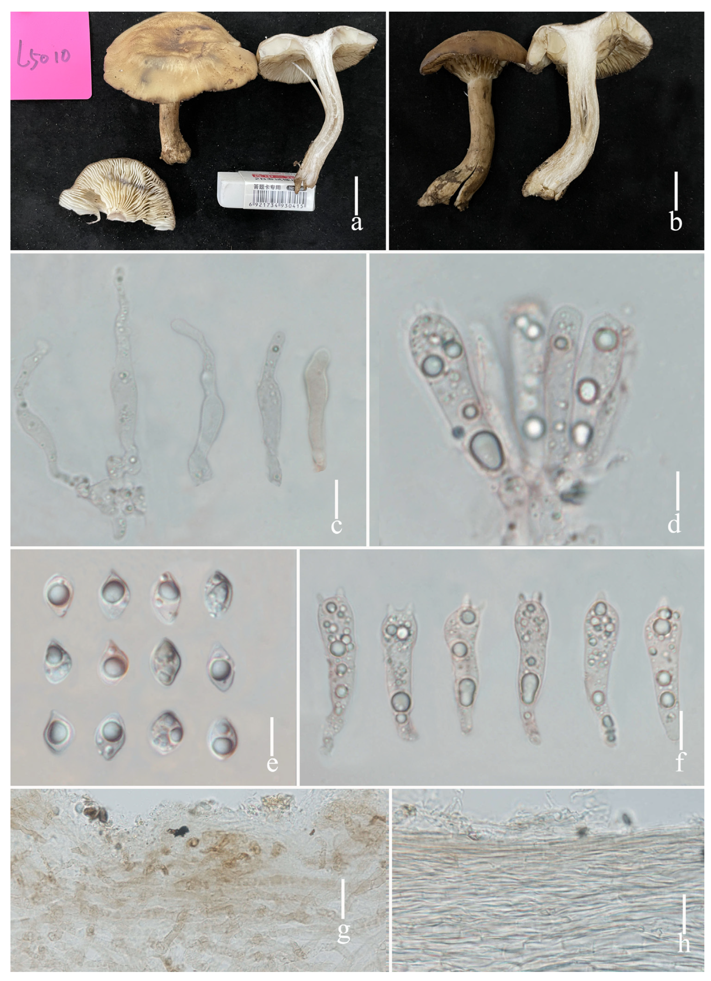
Pileus 5.0–7.0 cm diameter, fleshy, fragile, hemispherical, becoming to convex with age, smooth, greyish green (1C–D4), slightly depressed of center, deflexed to inflexed of margin; pileus context thick, 0.2–0.3 cm wide, white (1A1). Lamellae moderately close together, triangular, subdecurrent to decurrent, even or entire of edge, white (1A1), grey dark when bruised, 1–2 tiers, 0.7–0.9 cm wide, edge even or entire. Stipe 4.0–7.0 × 0.5–1.0 cm, cylindrical to clavate, greyish green (1C4) on the surface, smooth; stipe context white (1A1), fistulose, unchanging in colour when injured. Odor and taste not distinctive.
Basidiospores [111/2/2] 5.8–9.9 × 3.7–6.8 μm, (N = 111, Q = 1.3–2.0, Qm = 1.42 ± 0.16), av. 8.16 ± 0.99 × 5.69 ± 0.61 μm (L5010 and L5084); [50/1/1] 5.6–10.9 × 4.7–7.3 μm, (N = 50, Q = 1.2–1.8, Qm = 1.22 ± 0.11), av. 8.72 ± 0.57 × 5.87 ± 0.51 μm (holotype, L1763); very broadly fusiform to broadly fusiform, hyaline, smooth. Basidia 28–34 × 7–9 μm (N = 20), av. 28.1 ± 2.20 × 7.9 ± 0.98 μm, mostly 4–spored, rarely 2–spored, sterigmata long 1.3–4.3 μm, sometimes with basal clamp connections, clavate, siderophilous granulations. Subhymenium made up of moderately thin-walled hyphae, 15–21 μm thick, with 1–2 layers of ovoid, fusiform to narrowly cylindrical hyphae, 3–5 × 1–3 μm. Hymenophoral trama regular, 280–320 μm wide, consisting of thin and hyaline hyphae, some with clamp connections, narrowly cylindrical hyphae elements 4–7 μm wide. Cheilocystidia 28–40 × 5–8 μm, av. 37.2 ± 8.2 × 5.7 ± 0.7 μm, narrowly cylindrical or narrowly clavate, thin-walled. Pleurocystidia 20–46 × 4–6 μm, av. 31.9 ± 7.6 × 5.1 ± 0.7 μm, narrowly cylindrical or narrowly clavate, thin-walled. Pileipellis an interwoven trichodermium to a subcutis composed of almost hyaline interwoven filamentous hyphae, dark pigment inside, terminal cells 30–60 × 5–7 μm, almost cylindrical to subcylindrical, occasional hyphal tips flexuous and sometimes inflate, some with clamp connections. Stipitipellis a cutis of elongate hyphae, thin walled, caulocystidia absent. Clamp connections present at some septa.
Ecology and distribution: Mostly solitary, rarely scattered, growth in forests of Morella rubra. Summer to Autumn (August to October). Known only from Yunnan Province, China.
Specimens examined: China, Yunnan Province, Chuxiong City, from Chuxiong Mushroom Markets, Shu-Hong Li, 26 August 2023, L5010; ibid, Song-Ming Tang, 2 September 2023, L5084.
Notes: Lyophyllum rhombisporum was originally described from Yunnan, China, by Wang et al. [32]. In the original description, specimen L1763 had larger basidiospores (14.5–17 × 10–11.5 μm) and basidia (36.5–46 × 10–11.3 μm), but cheilocystidia and pleurocystidia were absent. It was surprising and puzzling due to the difficulty of finding such large basidiospores in the Lyophyllaceae. We rechecked the L. rhombisporum specimen (holotype, L1763) and compared it with our L. rhombisporum specimens L5010 and L5084; we found that the ITS sequences were 100% similar, thus, they belong to the same species.
According to phylogenetic analyses, L. rhombisporum is closely related to L. infumatum. However, L. infumatum, originally described from Italy, has relatively narrower basidia (25–35 × 8–9 μm) and a solid stipe context [32]. The ITS genetic distance between the holotype L. rhombisporum L1762 and L. infumatum IFO 30978 is 0.87% (5/577).

4. Discussion

We included 19 Lyophyllum species in the section Lyophyllum in the phylogenetic analyses of the present study, and 19 species formed a monophyletic clade (Figure 1). In China, only three Lyophyllum species in the section Lyophyllum were previously reported with molecular evidence: L. rhombisporum Shu H. Li & Y.C. Zhao from subtropical Yunnan Province, L. atrofuscum S.W. Wei, Q. Wang & Yu Li, and L. subalpinarum S.W. Wei, Q. Wang & Yu Li from Xizang Province [10]. Within the clade, in all the species of Lyophyllum, lamellae change to dark grey when bruised, basidiospores are variable, and in most of the species, basidiospores are very broadly fusiform to fusiform, and rarely globose to subglobose (L. moncalvoanum J.A. Cooper & P. Leonard).
Lyophyllum species are found globally with a notable concentration in Asia, where they have been extensively researched. China boasts a rich diversity of Lyophyllum species, making it a focal point for abundant species and comprehensive collections such as L. trigonosporum (Bres.) Kühner, L. pusillum Clémençon & A.H. Sm., L. pulvis-horrei E. Ludw. & Koeck, L. subalpinarum, L. semitale (Fr.) Kühner, L. rhombisporum, L. immundum (Berk.) Kühner, L. sykosporum Hongo & Clémençon, L. transforme (Lapl.) Singer, L. atrofuscum, L. infumatum (Bres.) Kühner, L. macrosporum Singer, L. loricatum (Fr.) Kühner, L. shimeji (Kawam.) Hongo, L. subdecastes, L. fumosum and L. decastes (Fr.) Singer [9,10,32,34].
We report L. rhombisporum based on two new collections (YAAS L5010 and YAAS L5084). Comparing our collections with the L. rhombisporum type specimen (holotype L1763) showed that basidiospores’ size varies significantly; thus, we assume that either the microscope calibrations were wrong, or the magnification was measured incorrectly in the original description.
In our study, we present morphological characteristics and phylogenetic data for two new Lyophyllum species and one known Lyophyllum species. Our findings, in line with previous studies, highlight the challenge of distinguishing Lyophyllum species based solely on morphology due to their similarities. Consequently, employing molecular methods becomes essential for accurately delineating species boundaries in Lyophyllum.
Collecting a diverse range of Lyophyllum species from various geographic locations, acquiring additional sequencing data, and thoroughly documenting their morphology are essential to enhance our understanding of their taxonomy and phylogeny.

Author Contributions

S.L. wrote the manuscript and phylogenetic analyses; S.T. and J.H. prepared the samples; D.Z. designed the article structure and correct manuscript. All authors have read and agreed to the published version of the manuscript.

Funding

This research was funded by the National Natural Science Foundation of China (Project ID: 32060006, 31560009, 31160010), China Agriculture Research System (Project ID: CARS-20), and central guidance for local scientific and technological development funds (202307AB110001).

Institutional Review Board Statement

Not applicable.

Informed Consent Statement

Not applicable.

Data Availability Statement

Data are contained within the article.

Acknowledgments

We would like to thank the three anonymous reviewers who gave us comments that have improved our manuscript.

Conflicts of Interest

The authors declare no conflicts of interest.

References

  1. Karsten, P.A. Hymenomycetes Fennici enumerati. Acta Soc. Fauna Flora Fenn. 1881, 2, 4. [Google Scholar]
  2. Dähncke, R.M.; Contu, M.; Vizzini, A. Two new species of Lyophyllum s.l. (Basidiomycota, Agaricomycetes) from La Palma (Canary Islands, Spain). Mycotaxon 2011, 115, 65–71. [Google Scholar] [CrossRef]
  3. Cooper, J.A. New species and combinations of some New Zealand agarics belonging to Clitopilus, Lyophyllum, Gerhardtia, Clitocybe, Hydnangium, Mycena, Rhodocollybia and Gerronema. Mycosphere 2014, 5, 263–288. [Google Scholar] [CrossRef]
  4. Bellanger, J.M.; Moreau, P.A.; Corriol, G.; Bidaud, A.; Chalange, R.; Dudova, Z.; Richard, F. Plunging hands into the mushroom jar: A phylogenetic framework for Lyophyllaceae (Agaricales, Basidiomycota). Genetica 2015, 143, 169–194. [Google Scholar] [CrossRef]
  5. Lomberg, M.L.; Renker, C.; Buchalo, A.S.; Solomko, E.F.; Kirchhoff, B. Micromorphological and Molecular Biological Study of Culinary-Medicinal Mushroom Hypsizygus marmoreus (Peck) Bigel. (Agaricomycetideae). Int. J. Med. Mushrooms 2003, 5, 307–312. [Google Scholar] [CrossRef]
  6. Dai, F. A Summary of Chinese Fungi; Science Press: Beijing, China, 1979. [Google Scholar]
  7. Kühner, R. Utilisation du carmin acétique dans la classification des agarics leucosporés. Publ. Société Linnéenne Lyon 1938, 7, 204–211. [Google Scholar] [CrossRef]
  8. Gminder, A. Nomenclatural novelties. Index Fungorum 2016, 302, 1. [Google Scholar]
  9. Li, X.; Li, Y. Research progress on fungus of the genus Lyophyllum in China. J. Edible Fungi 2009, 16, 75–79. [Google Scholar]
  10. Larsson, E.; Sundberg, H. Lyophyllum shimeji, a species associated with lichen pine forest in northern Fennoscandia. Mycoscience 2011, 52, 289–295. [Google Scholar] [CrossRef]
  11. Li, S.H.; Tang, S.M.; He, J.; Zhou, D. Two new edible Lyophyllum species from Tibetan areas, China. Diversity 2023, 15, 1027. [Google Scholar] [CrossRef]
  12. Ma, Y.H.; Liu, P.; Zhao, Z.Y.; Chen, W.M.; Zhao, Y.C. Lyophyllum deqinense (Lyophyllaceae, Agaricales), a new species from southwestern China. Phytotaxa 2023, 598, 219–228. [Google Scholar] [CrossRef]
  13. Wei, S.W.; Lu, B.Y.; Wang, Y.; Dou, W.J.; Wang, Q.; Li, Y. Morphology and phylogeny of Lyophylloid mushrooms in china with description of four new species. J. Fungi 2023, 9, 77. [Google Scholar] [CrossRef] [PubMed]
  14. Sesli, E.; Vizzini, A.; Contu, M. Lyophyllum turcicum (Agaricomycetes: Lyophyllaceae), a new species from Turkey. Turk. J. Bot. 2015, 39, 512–519. [Google Scholar] [CrossRef]
  15. Boa, E. Los Hongos Silvestres Comestibles: Perspectiva Global de Su USO E Importancia Para la Población; FAO: Roma, Italy, 2005; No. 17. [Google Scholar]
  16. Pokhrel, C.P.; Sumikawa, S.; Iida, S.; Ohga, S. Growth and productivity of Lyophyllum decastes on compost enriched with various supplements. Micol. Apl. Int. 2006, 18, 21–28. [Google Scholar]
  17. Yamanaka, K. Commercial cultivation of Lyophyllum shimeji. Mushroom news. In Proceedings of the 6th International Conference on Mushroom Biology and Mushroom Products, Bonn, Germany, 29 September–3 October 2008. [Google Scholar]
  18. Ohta, A. Some cultural characteristics of mycelia of a mycorrhizal fungus, Lyophyllum shimeji. Mycoscience 1994, 35, 83–87. [Google Scholar] [CrossRef]
  19. Visnovsky, S.B.; Cummings, N.; Guerin-Laguette, A.; Wang, Y.; Yamada, A.; Kobayashi, H.; Kawai, M.; Pitman, A.R. Detection of the edible ectomycorrhizal fungus Lyophyllum shimeji colonising seedlings of cultivated conifer species in New Zealand. Mycorrhiza 2014, 24, 453–463. [Google Scholar] [CrossRef] [PubMed]
  20. Kornerup, A.; Wanscher, J.H. Methuen Handbook of Colour, 3rd ed.; Eyre Methuen: London, UK, 1978. [Google Scholar]
  21. Vellinga, E.C. Glossary. In Flora Agaricina Neerlandica; Bas, C., Kuijper, T.W., Noordeloos, M.E., Vellinga, E.C., Eds.; CRC Press: Boca Raton, FL, USA, 1998; Volume 1, pp. 54–64. [Google Scholar]
  22. White, T.J.; Bruns, T.D.; Lee, S.; Taylor, J.W. Amplification and direct sequencing of fungal ribosomal RNA genes for phylogenetics science direct. PCR Protoc. 1990, 18, 315–322. [Google Scholar]
  23. Katoh, K.; Standley, D.M. MAFFT multiple sequence alignment software version 7: Improvements in performance and usability. Mol. Biol. Evol. 2013, 30, 772–780. [Google Scholar] [CrossRef] [PubMed]
  24. Hall, T. BioEdit v7. 2007. Available online: http://www.mbio.ncsu.edu/BioEdit/BioEdit.html (accessed on 10 September 2023).
  25. Stamatakis, A. RAxML version 8: A tool for phylogenetic analysis and post-analysis of large phylogenies. Bioinformatics 2014, 30, 1312–1313. [Google Scholar] [CrossRef] [PubMed]
  26. Miller, M.A.; Pfeiffer, W.; Schwartz, T. Creating the CIPRES Science Gateway for inference of large phylogenetic trees. In Proceedings of the Gateway Computing Environments Workshop (GCE), New Orleans, LA, USA, 14 November 2010. [Google Scholar]
  27. Nylander, J.A.A. MrModeltest, version 2.2.; Program distributed by the author; Uppsala University, Department of Systematic Zoology: Uppsala, Sweden, 2004. [Google Scholar]
  28. Rambaut, A.; Drummond, A.J.; Xie, D.; Baele, G.; Suchard, M.A. Posterior summarization in Bayesian phylogenetics using Tracer 1.7. Syst. Biol. 2018, 67, 901–904. [Google Scholar] [CrossRef]
  29. Hofstetter, V.; Clémençon, H.; Vilgalys, R.; Moncalvo, J.M. Phylogenetic analyses of the Lyophylleae (Agaricales, Basidiomycota) based on nuclear and mitochondrial rDNA sequences. Mycol. Res. 2002, 106, 1043–1059. [Google Scholar] [CrossRef]
  30. Ma, Y.H.; Liu, P.; Zhao, Z.Y.; Chen, W.M.; Zhao, Y.C. Lyophyllum pallidofumosum sp. nov. (Lyophyllaceae, Agaricales), from southwestern China. Phytotaxa 2022, 576, 173–183. [Google Scholar] [CrossRef]
  31. Tudor, D. Fungal Pigment Formation in Wood Substrate. Ph.D. Thesis, University of Toronto, Toronto, ON, Canada, 2013. [Google Scholar]
  32. Wang, X.Q.; Zhou, D.Q.; Zhao, Y.C.; Zhang, X.L.; Li, L.; Li, S.H. Lyophyllum rhombisporum sp. nov. from China. Mycotaxon 2013, 123, 473–477. [Google Scholar] [CrossRef]
  33. Hongo, T.; Clémençon, H. A new species of Lyophyllum from Japan. Mycol. Helv. 1983, 1, 43–46. [Google Scholar]
  34. Miura, T.; Kubo, M.; Itoh, Y.; Iwamoto, N.; Kato, M.; Park, S.R.; Yuuichi, U.; Yukio, K.; Ikukatsu, S.; Suzuki, I. Antidiabetic activity of Lyophyllum decastes in genetically type 2 diabetic mice. Biol. Pharm. Bull. 2002, 25, 1234–1237. [Google Scholar] [CrossRef] [PubMed]
Figure 1. Phylogram generated from maximum likelihood (RAxML) analysis based on a partial ITS1-5.8S-ITS2 sequence alignment of Lyophyllum. Calocybe persicolor, C. carnea, and C. gambosa were used as the outgroup. ML bootstrap support values/Bayesian posterior probability values equal to or greater than 70%/0.95 are indicated on the nodes. Names of the species in red represent new species, and the holotype of each species is in bold.
Diversity 16 00210 g001
Figure 2. Lyophyllum bulborhizum (a,b) basidiomata. (a) L5083, holotype; (b) L5092; (c) pleurocystidia and basidia; (d) cheilocystidia; (e) basidiospores; (f) basidia; (g) pileipellis; (h) stipitipellis. Scale bars: (ad) = 1 cm, (cf) = 10 µm, (g,h) = 20 µm.
Diversity 16 00210 g002
Figure 3. Fresh basidiomata of Lyophyllum nigrum (a,b) basidiomata. ((a,b) L5091); (c) cheilocystidia and basidia; (d) pleurocystidia; (e) basidiospores; (f) basidia; (g) pileipellis; (h) stipitipellis. Scale bars: (a,b) = 1 cm, (cf) = 10 µm, (g,h) = 20 µm.
Diversity 16 00210 g003
Figure 4. Lyophyllum rhombisporum (a,b) basidiomata. (a) YAAS L5010; (b) YAAS L5084; (c) pleurocystidia; (d) basidia and cheilocystidia; (e) basidiospores; (f) basidia; (g) pileipellis; (h) stipitipellis. Scale bars: (a) = 20 μm, (be) = 5 μm.
Diversity 16 00210 g004
Table 1. Lyophyllum taxa included in this study are listed by taxon, specimen number country, and GenBank accession number for DNA sequences used in the phylogenetic analyses. Newly generated sequences are listed in bold and a “*” after the specimen number indicates that the specimen is the holotype of that species.
TaxonSpecimen NumberCountryITSReference
Calocybe gambosaHC 78/64SwitzerlandAF357027[29]
C. carneaCBS 552.50SwitzerlandAF357028[29]
C. persicolorHC 80/99SwitzerlandAF357026[29]
L. ambustumCBS 452.87SwitzerlandAF357057[29]
L. anthracophilumHC 79/132SwitzerlandAF357055[29]
L. atratumCBS 709.87SwitzerlandAF357053[29]
L. atrofuscumHMJAU 63461ChinaOP605493[13]
L. atrofuscumHMJAU 63456 *ChinaOP605494[13]
L. bulborhizumL5083 *ChinaPP406873This study
L. bulborhizumL5092ChinaPP406874This study
L. bulborhizumL5093ChinaPP406875This study
L. caerulescensHC 80-140SwitzerlandAF357052[29]
L. caerulescensV.15759USAJF908339[14]
L. crassifoliumV.5077ItalyJF908331[14]
L. decastesDd 08054ChinaFJ810160[14]
L. decastesLd 418ChinaHM119485[14]
L. deqinenseYAAS M6949 *ChinaOQ418117[12]
L. deqinenseYAAS M6948ChinaOQ418116[12]
L. deliberatumV.15032SloveniaJF908338[14]
L. favreiBSI94cp2SwitzerlandAF357035[29]
L. favreiV.6334ItalyJF908333[14]
L. fumosumSJ02/006SwedenHM572539[11]
L. fumosumLAS00/144SwedenHM572541[11]
L. fumosumV.16077ItalyJF908340[14]
L. fumosumLfumNlf24JapanJN983977[14]
L. fumosumL2010512371ChinaJX966310[14]
L. fumosumYAAS M6135ChinaON681708[30]
L. fumosumYAAS M6340ChinaON681709[30]
L. gangraenosumV.12332ItalyJF908335[31]
L. heimoguL3026 *ChinaKY434100[11]
L. heimoguL3033ChinaKY434101[11]
L. heimoguL3035ChinaKY434102[11]
L. infumatumV.10152ItalyJF908334[14]
L. leucophaeatumHae251.97SwitzerlandAF357032[29]
L. littoralisCA 20091130ItalyJX280410[14]
L. lixiviumHKAS 129929ChinaOR506463Unpublished
L. loricatumV.13175USAJF908336[14]
L. loricatumCA 20090202.03ItalyJX280406[14]
L. loricatum01.12.09ItalyJX280407[14]
L. moncalvoanumPDD 96328 *New ZealandNR_137615[10]
L. moncalvoanumPDD 102581New ZealandKJ461912[10]
L. nigrumL5091 *ChinaPP406876This study
L. nigrumL5186ChinaPP406877This study
L. nigrumL5187ChinaPP406878This study
L. ochraceumBSI94.cp1SwitzerlandAF357033[29]
L. ochraceumV.537ItalyJF908329[14]
L. rhombisporumL1762 *ChinaJX966307[32]
L. rhombisporumL2082ChinaJX966308[32]
L. rhombisporumL5010ChinaPP406879This study
L. rhombisporumL5084ChinaPP406880This study
L. semitaleHC 85/13SwitzerlandAF357049[29]
L. semitaleEL 187-09SwedenHM572552[24]
L. shimejiOlsen 813006SwedenHM572530[24]
L. shimejiNZ4Q 88New ZealandJN983985[14]
L. shimejiL 2010512377ChinaJX966311[14]
L. sykosporumIFO 30978JapanAF357050[29]
L. subalpinarumHMJAU 63447 *ChinaOP605490[13]
L. subalpinarumHMJAU 63453ChinaOP605491[13]
L. subdecastesHMJAU 63470ChinaOP605488[13]
L. subdecastesHMJAU 63467 *ChinaOP605489[13]
L. turcicumKATO-2971 *TurkeyKJ158159[14]
L. yiqunyangL4206ChinaKY434104[11]
L. yiqunyangL2989 *ChinaKY434103[11]
Disclaimer/Publisher’s Note: The statements, opinions and data contained in all publications are solely those of the individual author(s) and contributor(s) and not of MDPI and/or the editor(s). MDPI and/or the editor(s) disclaim responsibility for any injury to people or property resulting from any ideas, methods, instructions or products referred to in the content.