Article

Detection of Avian Haemosporidian Parasites in Wild Birds in Slovakia

by
Lenka Minichová
1,
Vladimír Slobodník
2,
Roman Slobodník
3,
Milan Olekšák
4,
Zuzana Hamšíková
5,
Ľudovít Škultéty
1 and
Eva Špitalská
1,*
1
Institute of Virology, Biomedical Research Center, Slovak Academy Sciences, Dúbravska cesta 9, 845 05 Bratislava, Slovakia
2
State Nature Conservancy of the Slovak Republic, Dlhá 3, 971 01 Prievidza, Slovakia
3
Raptor Protection of Slovakia, Trhová 54, 841 04 Bratislava, Slovakia
4
National Park Slovak Karst, Hámossyho 188, 049 51 Brzotín, Slovakia
5
Institute of Zoology, Slovak Academy Sciences, Dúbravska cesta 9, 845 06 Bratislava, Slovakia
*
Author to whom correspondence should be addressed.
Diversity 2024, 16(2), 121; https://doi.org/10.3390/d16020121
Submission received: 11 December 2023 / Revised: 9 February 2024 / Accepted: 11 February 2024 / Published: 13 February 2024
(This article belongs to the Section Microbial Diversity and Culture Collections)

Abstract

:
Haemosporidians are a group of vector-borne parasites belonging to the order Haemosporida. These parasites infect avian hosts and require blood-sucking insects (Diptera) for transmission. The occurrence and diversity of haemosporidian parasites are shaped primarily by the specificity of the parasite and the susceptibility of the host/vector. In this study, the presence and distribution of haemosporidians in blood samples from birds in urbanized and natural habitats were estimated using microscopic and molecular approaches. Birds in urbanized habitats were infected with four different species of Plasmodium, P. relictum, P. vaughani, P. matutinum, and P. circumflexum, and one species of Haemoproteus, H. parabelopolskyi, and Leucocytozoon sp. The species H. attenuatus, H. concavocentralis, H. minutus, H. pallidus, H. noctuae, and H. tartakovskyi were additionally identified in birds in natural habitats. Typically, juvenile birds are essential markers of parasite species transmitted in the study area. The juveniles in the urbanized habitats carried P. relictum, P. vaughani, P. circumflexum, H. parabelopolskyi, and Leucocytozoon species. The most abundant parasite was H. parabelopolskyi, which was found in both habitat types. The prevalence of Haemoproteus/Plasmodium species determined by nested PCR in birds in natural habitats (43.80%; 53/121) was significantly greater than that in birds in urbanized habitats (21.94%; 43/196) (p < 0.05). There was no significant difference in the infection rate of Leucocytozoon sp. between the habitat types (p > 0.05; 10/121 vs. 19/196).

1. Introduction

Avian haemosporidians belonging to the order Haemosporida (Alveolata, Apicomplexa) are classified into four families: Haemoproteidae (Haemoproteus), Plasmodiidae (Plasmodium), Leucocytozoidae (Leucocytozoon), and Garniidae. They are a group of vector-borne parasites that infect avian hosts and require blood-sucking insects (Diptera) as vectors to complete their life cycle. Species of the genus Haemoproteus are transmitted by biting midges (Ceratopogonidae) and hippoboscid flies (Hippoboscidae), Plasmodium species by mosquitoes (Culicidae), and Leucocytozoon species by simuliid flies (Simuliidae). The life cycle of these parasites is complicated because they change hosts and reproduction modes and develop into different morphological stages [1,2]. Parasites can persist in birds for many years or in the chronic stage throughout their lifetime, and they serve as a source of infection for vectors, which can also lead to the infection of offspring at the site during the breeding season/reproduction period [1,3].
Most past studies have been based on blood smear microscopy, where a set of morphological characteristics served as the basis for species identification [1,2]. With the use of molecular methods in parasitology, the number of studies has increased rapidly. The most frequently used protocol for the detection of haemosporidians is based on the cytochrome b (cyt b) gene, which was established by Hellgren et al. [4]. The routine application of PCR, followed by sequencing for the detection and identification of haemosporidian lineages, has led to a massive expansion in the identification of parasite genetic diversity. Bensch et al. [5] created the MalAvi database [6], which summarizes 5123 unique parasite lineages and knowledge about their host ranges and geographical distributions, facilitating understanding of the evolutionary and ecological factors that drive this complex multi-host, multi-parasite system.
Information on the occurrence of haemosporidian parasites in Slovakia is currently limited. In the past, most studies on the presence of haemosporidians in Slovakia were based on microscopic analysis of blood smears [3,7,8,9,10]. PCR and sequencing were used for the first time in Slovakia by Berthová et al. [11] and expanded by Šujanová et al. [12].
Here, we investigated the presence of haemosporidians in wild birds, both adults and juveniles, in Slovakia using microscopic and molecular approaches. To enrich the knowledge of the occurrence and distribution of haemosporidians in Slovakia, it is necessary to identify the species whose transmission occurs here; thus, we compared the occurrence of haemosporidians in urbanized and natural habitats with local bird populations and migrating bird populations, respectively.

2. Materials and Methods
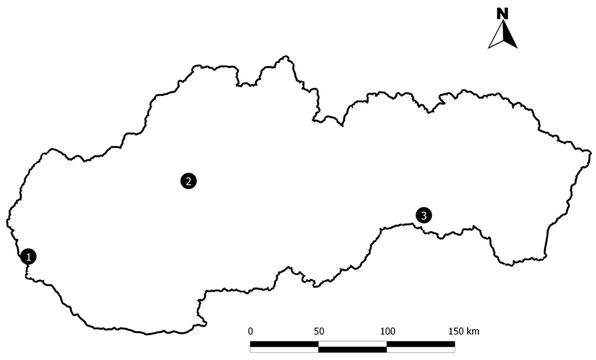
2.1. Study Sites

Birds were mist-netted in urbanized and natural habitat types (Figure 1) in Slovakia from April to August 2012 and from April to October 2013 in the morning (in urbanized habitats in April, June, July, August 2012 and from April to October 2013, and in natural habitats in April 2012). The birds were captured, ringed, blood-sampled, and released under permit No. 9368/2011-2.2 from the Ministry of Environment of the Slovak Republic. Bratislava and Prievidza represent urbanized habitats in city areas, with anthropogenic activities such as walking, cycling, hiking, and horse riding. Bratislava is located in the Small Carpathian Mountains in southwestern Slovakia at 202–334 m above sea level (asl) (259 m asl, 48°10′ N 17°03′ E). It is characterized by deciduous woodlands, oaks at lower altitudes, and beech at higher altitudes. The Prievidza district (289 m asl, 48°47′ N 18°34′ E), located in central Slovakia, is a forest–steppe rural area with Carpathian oak–hornbeam woods. Both study sites are characterized by local bird populations. Drienovec (181 m asl, 48°10′ N, 17°03′ E) represents a natural habitat with an ornithological observatory for research on migratory birds and is situated far from a city area and free from significant human activity. It is a forest–steppe natural area (181 m asl) in the Slovak Karst National Park in southeast Slovakia with sub-Mediterranean xerothermophilous oak woods and colline limestone grasslands [13,14].

2.2. Mist-Netting and Sampling Birds

After ringing the birds, the sex and age of each individual were determined using the field guides of Svensson [15] and Hromádko et al. [16,17,18]. A small amount of blood (approximately 50 µL) was taken by puncture of the vena ulnaris cutanea using a 1 mL X-tra-fine needle (HMD Healthcare, Warwick, UK), and blood smears were prepared, while the rest of the blood was stored in 96% ethanol. All birds were released immediately after sampling. All first-year individuals captured in the calendar year of hatching were considered juveniles. A total of 346 birds representing 42 species from 18 families were caught, including 114 juveniles [14]. Blood was collected from 317 individual birds.

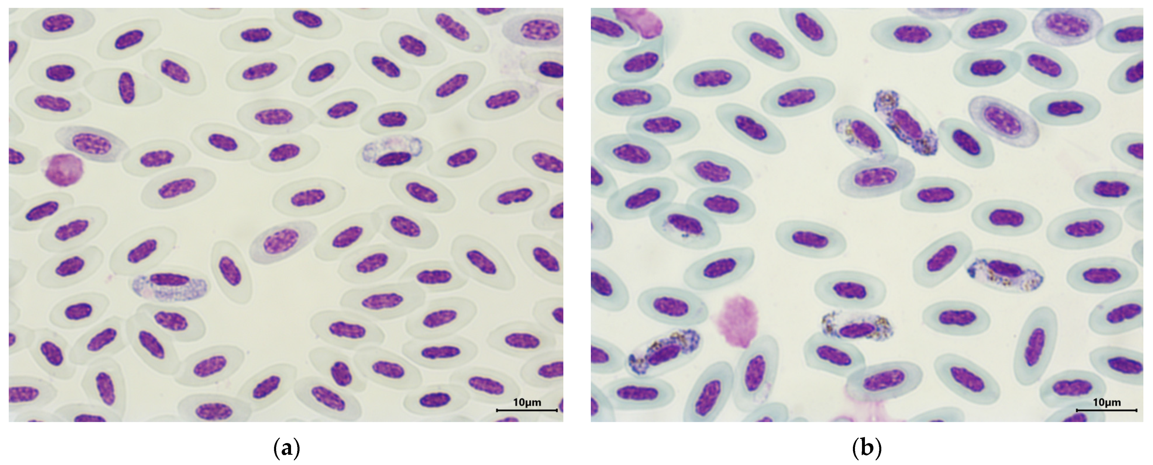
2.3. Microscopic Analysis of Haemosporidians

A drop of blood was used to prepare blood smears, and 2 thin smears were prepared per individual of a total of 302 birds. All smears were air-dried, fixed in methanol (96%), and stained with Giemsa stain, as described in Valkiūnas [1]. The smears were examined using a Leica DM4500B microscope (Wetzlar, Germany), and images were taken with a Leica DFC480 camera (Wetzlar, Germany). Approximately 100 fields were scanned under ×400 magnification, and at least 100 fields were subsequently examined under high magnification (×1000) (oil immersion). The intensity of parasitemia was determined according to the count of infected blood cells per 10,000 erythrocytes as follows: low (1–10 parasites), medium (11–100 parasites), or high (more than 100 parasites) according to Valkiūnas [1]. Images for the identification of parasites were edited using the program Leica Image Manager (Wetzlar, Germany). Determination of the haemosporidians was performed based on keys for morphological identification developed by Valkiūnas [1,2]. The smears were deposited at the Institute of Virology, Biomedical Research Center, SAS, Bratislava, Slovakia.

2.4. Molecular Analysis of Haemosporidians

DNA from the 317 blood samples was extracted using a NucleoSpin® Tissue commercial kit (Macherey–Nagel, Düren, Germany). Approximately 50 µL of the blood stored in ethanol was transferred to a sterile 1.5 mL Eppendorf tube, and the ethanol was allowed to evaporate. When the blood was completely dry, 200 µL of lysis buffer B3 and 25 µL of proteinase K were added. The mixture was gently vortexed and incubated at 70 °C for 1 h. The following procedure was performed according to the manufacturer’s instructions. The concentration and quality of the DNA were assessed using a NanoPhotometer Pearl (Implen, Munich, Germany). The DNA samples were stored at −20 °C until analysis. The presence of blood parasites was examined using a nested PCR assay targeting the cyt b gene mtDNA according to Bensch et al. [19] and Hellgren et al. [4]. Briefly, in the first step, the HaemNFI and HaemNR3 primers were used to amplify the 617-bp fragment. In the second step, nested PCR with the primers HaemF and HaemR2 amplified 480 bp fragments of Haemoproteus and Plasmodium, and the set of HaemFL and HaemR2L primers amplified a 478 bp fragment of Leucocytozoon. Sterile distilled water was used as a negative control, and the samples previously detected as PCR-positive and confirmed by sequencing [11] were used as positive controls. For the separation and visualization of the PCR products, 1% agarose gel stained with DNA Stain G (Serva, Heidelberg, Germany) was used. The PCR products of the expected size were considered positive and were sent for the purification and sequencing of both DNA strands at Macrogen (Amsterdam, Netherlands;). All the cyt b sequences were edited in MEGA6 version software [20] and identified by performing a nucleotide BLAST search in the GenBank NCBI database [21]. The obtained sequences were compared with those in the MalAvi database [5,6]. Poor-quality sequences were marked as unusable sequences (us), and sequences with double peaks, which denote co-infections, were marked as dp.
Samples that were microscopically positive for haemosporidians were examined for the intensity of haemosporidian infection using quantitative real-time PCR (qPCR), which targeted an 182 bp fragment of the cyt b gene [22]. All the reactions were carried out using GoTaq qPCR Master Mix (Promega, Madison, WI, USA) on a CFX96 real-time thermocycler (Bio-Rad, Hercules, CA, USA). The following cycling conditions were used: 95 °C for 2 min, 40 cycles of 95 °C for 30 s, and 64 °C for 35 s with a plate read, followed by a final melt curve analysis using the instrument’s default settings [12]. A synthetic double-stranded DNA product (Eurofins Genomics, Ebersberg, Germany) designed from a 220 bp fragment of the conserved rDNA region of Plasmodium relictum (accession number NC012426) was used as the positive control [22]. The synthetic DNA was diluted to a starting concentration of 106 copies/µL. The starting solution was serially diluted 10-fold to prepare a series of solutions from the 106 copies of genomic DNA (gDNA) per µL down to 1 copy/µL. All samples were run in duplicate [13]. Parasite intensity, which refers to the number of parasite DNA copies per 100 avian red blood cells, was determined for each sample based on the qPCR data and sample DNA concentration following the method of Friedl & Groscurth [23], with the assumption that the average genome size of a passerine bird is 2.8 pg [24]. An average of 1–1000 DNA copies per 100 avian red blood cells was marked as low parasite intensity, 1000–10,000 DNA copies per 100 avian red blood cells was marked as medium parasitemia, and more than 10,000 DNA copies per 100 avian red blood cells was marked as high parasitemia.

2.5. Statistical Analysis
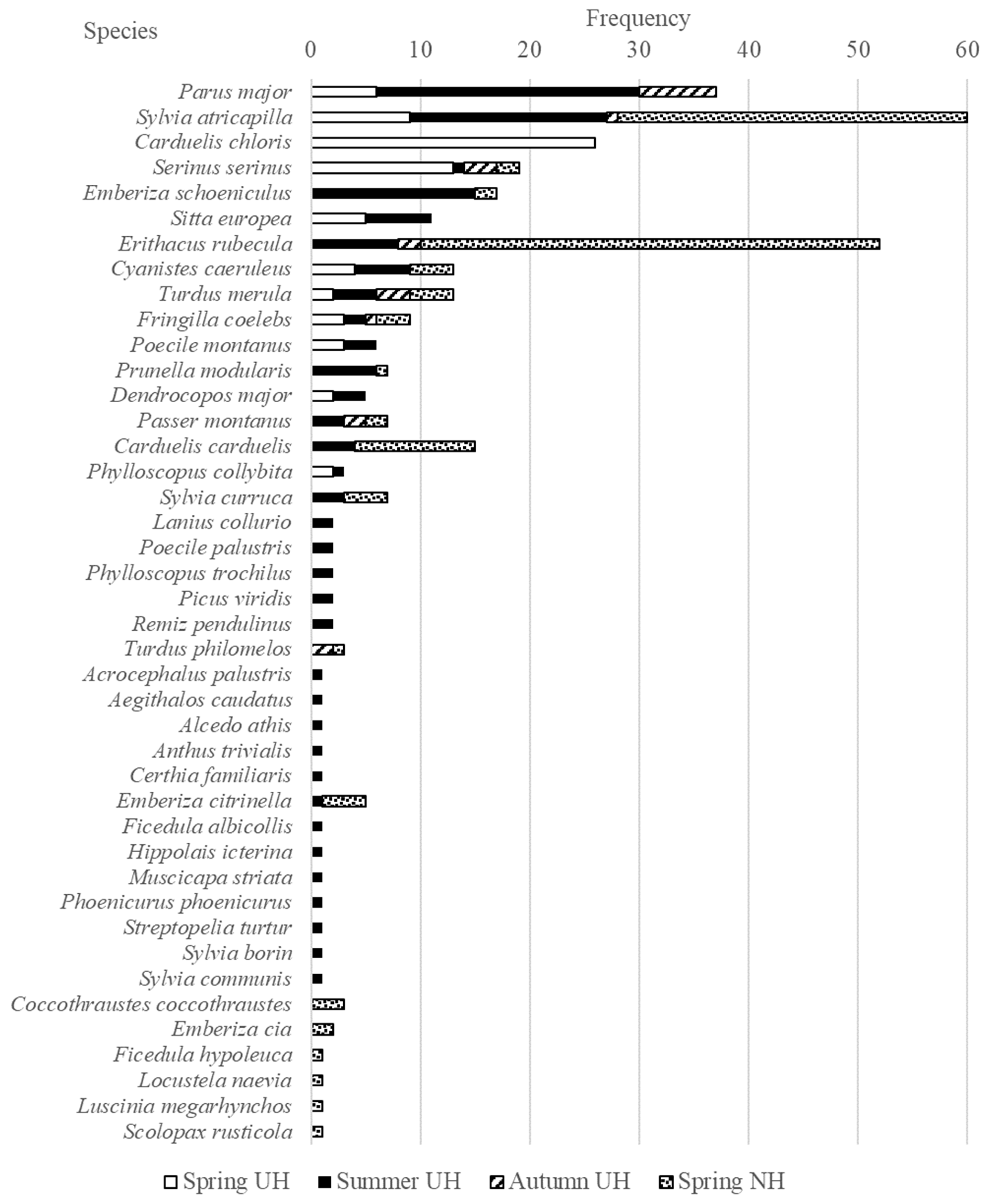
Statistical analysis was conducted to test the differences between the prevalence of birds trapped in urbanized and natural habitats with the χ2 test (p = 0.05) using Past version 2.17b software [25]. We calculated 95% confidence intervals (CIs) for each proportion individually [26].

3. Results

3.1. Haemosporidian Infections Based on Microscopic and Molecular Analyses

Overall, the most frequently captured bird species was the Eurasian blackcap, Sylvia atricapilla (60/346; 17.3%). However, of the birds captured in natural habitats, the most frequently captured species was the European robin Erithacus rubecula (42/121; 34.71%), and of the birds captured in urbanized habitats, the great tit, Parus major (37/225; 16.44%), was the most common (Figure 2).
The presence of haemosporidian parasites in the blood smears was detected in 52/302 individuals (17.22%). The most abundant species was H. parabelopolskyi in blood smears of 21 birds (Figure 3a), which is a host specific to the black cap S. atricapilla, and it was detected in birds from both habitat types. The second most abundant species was H. attenuatus (Figure 3b), which was detected in blood smears of 13 bird samples obtained from only 1 host species, the European robin E. rubecula, and only in birds from the natural habitats. No haemosporidians were identified in 18 bird samples. Plasmodium meronts were detected in the blood smears of five birds. Leucocytozoon was only detected in the blood smears of two individuals.
Parasite DNA was detected by nested PCR in 125/317 individuals (39.43%; CI 32.8–47.0%) of 18 bird species. Parasites of the genera Haemoproteus and Plasmodium were present in 96 individuals (30.28%; CI 25.2–35.3%), and parasites of the genus Leucocytozoon were present in 29 individuals (9.15%; CI 6.0–12.3%) (Table 1). Thirteen birds (4.10%; CI 1.9–6.3%) were co-infected (Table 1).
The infection rate of the genera Haemoproteus and Plasmodium in the birds caught in natural habitats was 43.80% (53/121; CI 34.8–53.1%). The overall infection rate of the birds from urbanized habitats was 21.94% (43/196; CI 16.4–28.4%). The prevalence of Haemoproteus/Plasmodium species in birds in natural habitats was significantly greater than that in birds in urbanized habitats (p < 0.05). Among the birds in natural habitats, 8.26% (10/121; CI: 4.0–14.7%) were infected with Leucocytozoon overall, while 8.00% (6/75; CI 3.0–16.6%) of the birds in urbanized habitats were infected with Leucocytozoon. There was no significant difference in infection rates for Leucocytozoon (p > 0.05). Among the birds in urbanized habitats, the co-infection rate was 3.57% (7/196; CI 1.5–7.2%). In natural habitats, 4.96% (6/121; CI 1.8–10.5%) of the birds had co-infections. There was no significant difference between the two habitat types (p > 0.05).
All 125 positive samples (PCR products of the expected size) were sequenced. After comparing the obtained 81 sequences with known sequences in the GenBank and MalAvi databases, we identified 11 haemosporidian species: H. attenuatus, H. concavocentralis, H. minutus, H. pallidus, H. parabelopolskyi, H. noctuae, H. tartakovskyi, P. circumflexum, P. relictum, P. vaughani, and P. matutinum. Sequences from 16 individuals were unusable, and 2 samples showed double peaks, indicating co-infections (Table S1).
Birds from the urbanized habitats were infected with five Haemoproteus spp./Plasmodium spp. (evaluated by nested PCR based on cyt b), and birds from the natural habitat were infected with eleven Haemoproteus spp./Plasmodium spp. (Table S1). The most abundant parasite species in the birds (evaluation only included species exceeding 10 individuals) from the urbanized habitats were the H. parabelopolskyi SYAT01, SYAT02, SYAT07, and SYAT10 lineages, with a prevalence of 39.29% in S. atricapilla; P. relictum SGS1 lineage (13.51%) in P. major; and P. circumflexum SYABOR02 lineage (13.33%) in E. schoeniculus. Among the birds from the natural habitat, infections with the H. parabelopolskyi SYAT01 and SYAT02 lineages were also the most prevalent (40.63%) in S. atricapilla, followed by the H. attenuatus ROBIN1/LULU1 lineage (23.8%) in E. rubecula, and the H. pallidus SYAT03 lineage (3.77%) in E. rubecula. The H. attenuatus ROBIN1/LULU1 lineage was present in a wide range of hosts (five species), though only in birds captured in the natural habitat (Table S1). Interestingly, we found this parasite in the blood of a seven-year-old female, which was confirmed by ringing data from 2006. No unique lineages were recorded.
Birds with long-distance migration were infected with the H. parabelopolskyi SYAT01, SYAT02, SYAT07, and SYAT10 lineages; H. attenuatus ROBIN1/LULU1 lineage; Haemoproteus sp. LWT1 lineage; P. relictum SGS1 lineage; and Leucocytozoon sp. PARUS4 and SCORUS01 lineages. In birds with short-distance migration and non-migratory birds, the presence of the H. attenuatus ROBIN1/LULU1 lineage; H. concavocentralis HAWF2 lineage; H. minutus TURDUS2 lineage; H. pallidus SYAT03 lineage; H. noctuae CIRCUM01 lineage; H. tartakovskyi HAWF1 lineage; Haemoproteus sp. CCF1, CCF2, CCF6, and EMCIR01 lineages; P. relictum SGS1 and COLL1 lineages; P. vaughani SYAT05 lineage; P. matutinum LINN1 lineage; P. circumflexum TURDUS1, BT7, and SYBOR02 lineages; and Leucocytozoon sp. PARUS4, PARUS18, PARUS19, PARUS28, PARUS50, STUR1/TURPEL01, PRUMOD01, BT2, BT5, SFC8, and SYCON06 lineages were identified.
The parasite intensity was verified via qPCR analysis with 52 microscopically positive samples. The intensity varied from 1 to 15,000 DNA copies per 100 avian red blood cells in both types of habitats. Low parasitemia (1–10 parasites per 10,000 erythrocytes) estimated from blood smears by microscopy corresponded on average to 1–1000 DNA copies per 100 avian red blood cells. Low parasitemia was recorded in the E. rubecula, S. atricapilla, T. merula, S. curruca, F. coelebs, and P. major bird species infected with the H. parabelopolskyi SYAT01, SYAT02, SYAT05, and SYAT10 lineages; H. attenuatus ROBIN1/LULU1 lineage; P. relictum COLL1 lineage; and Haemoproteus sp. CCF1 lineage. Medium parasitemia (11–100 parasites per 10,000 erythrocytes) estimated from blood smears by microscopy corresponded on average to 1000–10,000 DNA copies per 100 avian red blood cells. Medium parasitemia was identified in E. rubecula birds infected with the H. attenuatus ROBIN1/LULU1 lineage and in S. atricapilla birds infected with the H. parabelopolskyi SYAT01 and SYAT02 lineages. High parasitemia (more than 100 parasites per 10,000 erythrocytes) estimated from blood smears by microscopy corresponded to an average of more than 10,000 DNA copies per 100 avian red blood cells. High parasitemia was found in E. rubecula birds infected with the H. attenuatus ROBIN1/LULU1 lineage and in T. merula infected with the P. matutinum LINN1 lineage.
Co-infection with more than one haemosporidian genus was detected in 4.10% of the individuals (13/317; CI 1.9–6.3%). Co-infections with Haemoproteus and Leucocytozoon parasites were detected in five birds, and co-infections with Plasmodium and Leucocytozoon parasites were detected in eight individuals by molecular biology (Table 2). Co-infections with Haemoproteus and Plasmodium were not analyzed in detail in this study.

3.2. Haemosporidian Infections in Juvenile Birds Based on Microscopic and Molecular Analyses

Important markers of haemosporidians transmitted in Slovakia include juvenile birds captured in urbanized habitats. In our PCR-based study, 26 out of 114 juveniles were infected with Haemoproteus/Plasmodium (22.81%; CI 15.1–30.5%), 8 were infected with Leucocytozoon (7.02%; CI 2.3–11.7%), and 6 had co-infections (5.26%; CI 1.2–9.4%). Juvenile great tit P. major, Eurasian blue tit C. caeruleus, and Eurasian blackcap S. atricapilla caught in urbanized habitats carried the P. relictum SGS1 lineage, and the common blackbird T. merula carried the P. vaughani SYAT05 lineage. The song thrush T. philomelos was infected with the P. circumflexum SYBOR02 lineage, and the Eurasian blackcap S. atricapilla was infected with the H. parabelopolskyi SYAT02 lineage. The lack of gametocytes made species determination by microscopy impossible for six PCR-positive juveniles, the yellowhammer Emberiza citrinella, the common blackbird T. merula, and the common chaffinch Fringilla coelebs, which were infected with Haemoproteus spp. The European robin E. rubecula and the common blackbird T. merula were infected with Plasmodium spp., and Leucocytozoon spp. were found in the great tit P. major, the Eurasian blue tit C. caeruleus, the common blackbird T. merula, the Eurasian blackcap S. atricapilla, and the song thrush T. philomelos.
Mature gametocytes were confirmed by microscopy in the peripheral blood of 11 juvenile birds: H. parabelopolskyi in six Eurasian blackcap S. atricapilla, P. vaughani in one common blackbird T. merula, Haemoproteus sp. in one yellowhammer E. citrinella and two S. atricapilla, and Plasmodium sp. in one European robin, E. rubecula.
Parasite intensity in juveniles was positive according to nested PCR, and microscopically, it is shown in Table S2.

4. Discussion

In the present study, we aimed to extend the body of knowledge about the presence of haemosporidians in the blood samples of birds in urbanized and natural habitats and to identify which species are transmitted in Slovakia using microscopy and molecular detection. Quantitative PCR and microscopical analysis are only supplementary methods applied to a part of the samples. Here, we report eleven species of haemosporidian parasites detected in birds in Slovakia. We detected more different species among the birds in natural habitats, where most of the birds were migratory species, than among those in urbanized habitats with local populations (Table 1). In combination with suitable vectors, infected birds could serve as reservoirs of infection for other birds (because they share the same habitat for breeding, rest, and at night). Differences in prevalence may be due not only to vectors and hosts but also to abiotic environmental factors such as precipitation and mean annual temperature [27,28,29,30,31]. In our study, birds caught in natural habitats had significantly greater infection rates than birds from local populations in urbanized habitats. The highest prevalence of haemosporidian infections was recorded in the Eurasian blackcap S. atricapilla, the great tit P. major, the common chaffinch F. coelebs, the common blackbird T. merula, and the Eurasian blue tit C. caeruleus; these results are related to host abundance and are similar to the results of Šujanová et al. [12]. As previously reported, Haemoproteus species are the most frequently reported haemosporidian parasites in birds, and these results are not limited to Slovakia [32,33,34,35].
To determine which parasites are transmitted in Slovakia, it was necessary to carry out an analysis of juveniles. Unlike adult birds, which can become infected in any year or locality, young birds in their first year of life (before autumn migration) can be infected only by parasites actively transmitted by vectors in their breeding area [1,3,7,8].
In our study, juveniles from populations in the natural habitats were not mist-netted, so their infection status remains unknown. In the local populations in the urbanized habitats, blood analysis revealed the presence of H. parabelopolskyi, P. circumflexum, P. vaughani, P. relictum, and Leucocytozoon sp. in the caught juveniles.
Haemoproteus parasites are the most diverse group of avian haemosporidians [2,35]. Due to the limited knowledge of the life cycles and tissue merogony of the majority of Haemoproteus species, these parasites have usually been considered benign [36]. However, recent molecular studies indicate that they may cause severe and even lethal disease if infections occur in non-adapted avian hosts [37]. Our findings on H. parabelopolskyi and H. attenuatus are interesting, as H. parabelopolskyi was the most common species found only in the Eurasian blackcap S. atricapilla, and H. attenuatus was the second most prevalent Haemoproteus found in five host species. H. parabelopolskyi was the most common species found (24 individuals infected). This species is a strict host specialist for the Eurasian blackcap S. atricapilla [38]. However, H. parabelopolskyi can be occasionally found in birds of the family Acrocephalidae [2]. We recorded the occurrence of this parasite in both habitat types, likely because the Eurasian blackcap S. atricapilla is one of the most common bird species in Slovakia. Evidence of the presence of a suitable vector was confirmed by the infections of eight juveniles. Interestingly, we found a seven-year-old female Eurasian blackcap (S. atricapilla) infected with this parasite. The infection was confirmed by microscopy, PCR, and sequencing. Microscopy revealed a very low intensity of infection, and the ability to complete active flight indicated the chronic stage of infection. Although the time of the individual’s infection is unknown, this discovery supports the need for monitoring at the site when birds are migrating. In contrast, H. attenuatus (the second most prevalent Haemoproteus species, with 14 infected individuals from 5 host species) was detected in the natural habitat only, even though these species were common in the other habitats. The H. attenuatus ROBIN1 lineage is closely related to several lineages of H. balmorali that also parasitize birds of Muscicapidae. In our study, the obtained sequence fragments showed 100% identity with the sequences annotated in the database as both H. balmorali and H. attenuatus, and microscopic analysis confirmed the presence of H. attenuatus. For the first time, Hauptmanová et al. [9] noted the presence of this parasite in Slovakia in the blood of a European robin (E. rubecula) caught in the eastern part of Slovakia. Unfortunately, the authors did not specify the number of infected birds. Šujanová et al. [12] also identified ROBIN1 lineage in the C. caeruleus, Poecile palustris, Turdus iliacus, and Prunella modularis bird species. Thus, the host species reported for H. attenuatus has been extended. Hernández-Lara [39] described several species of the orders Passeriformes (Muscicapidae, Certhiidae, Acrocephalidae, Sylviidae, and Turdidae), and Coraciiformes (Alcedinidae) as vertebrate hosts of H. attenuatus, and the Diptera (Ceratopogonidae) species Culicoides festivipennis, C. obsoletus, and C. nubeculosus as vectors, which also occur in Slovakia and neighboring countries [40]. This could be a marker for the presence of a suitable vector in the east of the country, but further studies are needed to verify this premise.

5. Conclusions

This study contributes to the knowledge about the prevalence and morphological and molecular richness of haemosporidian parasites circulating in free-living birds in urbanized and natural habitats in Slovakia. The prevalence of Haemoproteus/Plasmodium species in birds in natural habitats was significantly greater than that in birds in urbanized habitats (p < 0.05), although the prevalence of Leucocytozoon was not greater in urban habitats. Among the 42 avian species, 11 haemosporidian species—H. attenuatus, H. concavocentralis, H. minutus, H. pallidus, H. parabelopolskyi, H. noctuae, H. tartakovskyi, P. circumflexum, P. relictum, P. vaughani, and P. matutinum, were identified. However, the H. parabelopolskyi, as well as three Plasmodium species, P. circumflexum, P. relictum, and P. vaughani, were also present in the juveniles, indicating the presence of suitable vectors and active local transmission of parasites in Slovakia.

Supplementary Materials

The following supporting information can be downloaded at: https://www.mdpi.com/article/10.3390/d16020121/s1, Table S1: List of the haemosporidian parasites detected by cyt b molecular analysis in the blood samples of the birds from urbanized and natural habitats (us—unusable sequences, dp—double peaks). Numbers in parentheses refer to the number of positive birds of a given lineage; Table S2. Parasite intensity in chosen juveniles was determined by qPCR and microscopy.

Author Contributions

Formal analysis, L.M. and E.Š.; Funding acquisition, E.Š.; Investigation, L.M., V.S., R.S., M.O. and Z.H.; Methodology, L.M. and E.Š.; Project administration, E.Š.; Supervision, Ľ.Š. and E.Š.; Writing—original draft, L.M.; Writing—review and editing, E.Š. All authors have read and agreed to the published version of the manuscript.

Funding

This research was funded by the Scientific Grant Agency of the Ministry of Education, Science, Research and Sport of the Slovak Republic and Slovak Academy of Science, grant number VEGA 2/0021/21.

Institutional Review Board Statement

The experiments presented in this paper comply with the current laws of the Slovak Republic. Birds were captured, ringed, blood sampled, and released under the permission of the Ministry of Environment of the Slovak Republic No. 9368/2011-2.2.

Data Availability Statement

The datasets used and/or analyzed during the current study are available from the first and corresponding author upon request.

Conflicts of Interest

The authors declare no conflicts of interest.

References

  1. Valkiūnas, G. Avian Malaria Parasites and Other Haemosporidia; CRC Press: Boca Raton, FL, USA, 2005. [Google Scholar]
  2. Valkiūnas, G.; Iezhova, T.A. Keys to the avian Haemoproteus parasites (Haemosporida, Haemoproteidae). Malar. J. 2022, 21, 269. [Google Scholar] [CrossRef]
  3. Kučera, J. Blood parasites of birds in Central Europe. 1. Survey of literature. The incidence in domestic birds and general remark to the incidence in wild birds. Folia Parasitol. 1981, 28, 13–22. [Google Scholar]
  4. Hellgren, O.; Waldenström, J.; Bensch, S. A new PCR assay for simultaneous studies of Leucocytozoon, Plasmodium, and Haemoproteus from avian blood. J. Parasitol. 2004, 90, 797–802. [Google Scholar] [CrossRef]
  5. Bensch, S.; Hellgren, O.; Pérez-Tris, J. MalAvi: A public database of malaria parasites and related haemosporidians in avian hosts based on mitochondrial cytochrome b lineages. Mol. Ecol. Resour. 2009, 9, 1353–1358. [Google Scholar] [CrossRef]
  6. MalAvi Database. Available online: http://130.235.244.92/Malavi/index.html (accessed on 28 November 2023).
  7. Kučera, J. Blood parasites of birds in Central Europe. 2. Leucocytozoon. Folia Parasitol. 1981, 28, 193–203. [Google Scholar]
  8. Kučera, J. Blood parasites of birds in Central Europe. 3. Plasmodium and Haemoproteus. Folia Parasitol. 1981, 28, 303–312. [Google Scholar]
  9. Hauptmanová, K.; Benedikt, V.; Literák, I. Blood Parasites in Passerine Birds in Slovakian East Carpathians. Acta Protozool. 2006, 45, 105–109. [Google Scholar]
  10. Haas, M.; Kisková, J. Absence of blood parasites in the Alpine Accentor Prunella collaris. Oecologia Mont. 2010, 19, 30–34. [Google Scholar]
  11. Berthová, L.; Országhová, Z.; Valkiūnas, G. The first report of nine species of haemosporidian parasites (Haemosporida: Haemoproteus, Plasmodium and Leucocytozoon) in wild birds from Slovakia. Biologia 2012, 67, 931–933. [Google Scholar] [CrossRef]
  12. Šujanová, A.; Špitalská, E.; Václav, R. Seasonal dynamics and diversity of haemosporidians in a natural woodland bird community in Slovakia. Diversity 2021, 13, 439. [Google Scholar] [CrossRef]
  13. Kazimírová, M.; Hamšíková, Z.; Kocianová, E.; Marini, G.; Mojšová, M.; Mahríková, L.; Berthová, L.; Slovák, M.; Rosá, R. Relative density of host-seeking ticks in different habitat types of south-western Slovakia. Exp. Appl. Acarol. 2016, 69, 205–224. [Google Scholar] [CrossRef]
  14. Berthová, L.; Slobodník, V.; Slobodník, R.; Olekšák, M.; Sekeyová, Z.; Svitálková, Z.; Kazimírová, M.; Špitalská, E. The natural infection of birds and ticks feeding on birds with Rickettsia spp. and Coxiella burnetii in Slovakia. Exp. Appl. Acarol. 2016, 68, 299–314. [Google Scholar] [CrossRef]
  15. Svensson, L. Identification Guide to European Passerines, 4th ed.; Lars Svensson: Stockholm, Sweden, 1992. [Google Scholar]
  16. Hromádko, M.; Horáček, J.; Chytil, J.; Pithart, K.; Škopek, J. Guide to the Determination of Our Passerines I; Publisher ZUV: Hradec Králové, Czech Republic, 1992. (In Czech) [Google Scholar]
  17. Hromádko, M.; Horáček, J.; Chytil, J.; Pithart, K.; Škopek, J. Guide to the Determination of Our Passerines II; Publisher ZUV: Hradec Králové, Czech Republic, 1993. (In Czech) [Google Scholar]
  18. Hromádko, M.; Horáček, J.; Chytil, J.; Pithart, K.; Škopek, J. Guide to the Determination of Our Passerines III; Publisher ZUV: Hradec Králové, Czech Republic, 1998. (In Czech) [Google Scholar]
  19. Bensch, S.; Stjernman, M.; Hasselquist, D.; Ostman, O.; Hansson, B.; Westerdahl, H.; Pinheiro, R.T. Host specificity in avian blood parasites: A study of plasmodium and haemoproteus mitochondrial DNA amplified from birds. Proc. Biol. Sci. 2000, 267, 1583–1589. [Google Scholar] [CrossRef]
  20. Kumar, S.; Stecher, G.; Li, M.; Knyaz, C.; Tamura, K. MEGA X: Molecular Evolutionary Genetics Analysis across Computing Platforms. Mol. Biol. Evol. 2018, 35, 1547–1549. [Google Scholar] [CrossRef]
  21. GenBank Database. Available online: http://blast.ncbi.nlm.nih.gov/ (accessed on 22 February 2023).
  22. Bell, J.A.; Weckstein, J.D.; Fecchio, A.; Tkach, V.V. A new real-time pcr protocol for detection of avian haemosporidians. Parasites Vectors 2015, 8, 383. [Google Scholar] [CrossRef]
  23. Friedl, T.W.P.; Groscurth, E. A real-time PCR protocol for simple and fast quantification of blood parasite infections in evolutionary and ecological studies and some data on intensities of blood parasite infections in a subtropical weaverbird. J. Ornithol. 2012, 153, 239–247. [Google Scholar] [CrossRef]
  24. Tiersch, T.R.; Wachtel, S.S. On the evolution of genome size of birds. J. Hered. 1991, 82, 363–368. [Google Scholar] [CrossRef]
  25. Hammer, Ø.; Harper, D.A.T.; Ryan, P.D. Past: Paleontological Statistics Software Package for Education and Data Analysis. Palaeo. Electronica 2001, 4, 4. Available online: https://palaeo-electronica.org/2001_1/past/issue1_01.htm (accessed on 15 September 2023).
  26. EasyCalculation. Available online: https://www.easycalculation.com/statistics (accessed on 22 February 2023).
  27. Basto, N.; Rodríguez, O.A.; Marinkelle, C.J.; Gutiérrez, R.; Matta, N. Haematozoa in birds from La Macarena National Natural Park (Colombia). Caldasia 2006, 28, 371–377. [Google Scholar]
  28. Doussang, D.; González-Acuña, D.; Torres-Fuentes, L.G.; Lougheed, S.C.; Clemente-Carvalho, R.B.; Greene, K.C.; Vianna, J.A. Spatial distribution, prevalence and diversity of haemosporidians in the rufous-collared sparrow, Zonotrichia capensis. Parasites Vectors 2019, 12, 2. [Google Scholar] [CrossRef]
  29. Ciloglu, A.; Ergen, A.G.; Inci, A.; Dik, B.; Duzlu, O.; Onder, Z.; Yetismis, G.; Bensch, S.; Valkiūnas, G.; Yildrim, A. Prevalence and genetic diversity of avian haemosporidian parasites at an intersection point of bird migration routes: Sultan Marshes National Park, Turkey. Acta Trop. 2020, 210, 105465. [Google Scholar] [CrossRef]
  30. Quillfeldt, P.; Arriero, E.; Martínez, J.; Masello, J.F.; Merino, S. Prevalence of blood parasites in seabirds—A review. Front. Zool. 2011, 8, 26. [Google Scholar] [CrossRef]
  31. Chakarov, N.; Kampen, H.; Wiegmann, A.; Werner, D.; Bensch, S. Blood parasites in vectors reveal a united blackfly community in the upper canopy. Parasites Vectors 2020, 13, 309. [Google Scholar] [CrossRef]
  32. Schumm, Y.R.; Bakaloudis, D.; Barboutis, C.; Cecere, J.G.; Eraud, C.; Fischer, D.; Hering, J.; Hillerich, K.; Lormée, H.; Mader, V.; et al. Prevalence and genetic diversity of avian haemosporidian parasites in wild bird species of the order Columbiformes. Parasitol. Res. 2021, 120, 1405–1420. [Google Scholar] [CrossRef]
  33. Carlson, J.; Martínez-Gómez, J.E.; Valkiūnas, G.; Loiseau, C.; Bell, D.A.; Sehgal, R.N. Diversity and phylogenetic relationships of hemosporidian parasites in birds of Socorro Island, México and their role in the re-introduction of the Socorro Dove (Zenaida graysoni). J. Parasitol. 2013, 99, 270–276. [Google Scholar] [CrossRef]
  34. Heym, E.C.; Kampen, H.; Krone, O.; Schäfer, M.; Werner, D. Molecular detection of vector-borne pathogens from mosquitoes collected in two zoological gardens in Germany. Parasitol. Res. 2019, 118, 2097–2105. [Google Scholar] [CrossRef]
  35. Valkiūnas, G.; Ilgūnas, M.; Bukauskaitė, D.; Chagas, C.R.F.; Bernotienė, R.; Himmel, T.; Harl, J.; Weissenböck, H.; Iezhova, T.A. Molecular characterization of six widespread avian haemoproteids, with description of three new Haemoproteus species. Acta Trop. 2019, 197, 105051. [Google Scholar] [CrossRef]
  36. Bennet, G.F.; Peirce, M.A.; Ashford, R.W. Avian haematozoa: Mortality and pathogenicity. J. Nat. Hist. 1993, 27, 993–1001. [Google Scholar] [CrossRef]
  37. Ortiz-Catedral, L.; Brunton, D.; Stidworthy, M.F.; Elsheikha, H.M.; Pennycott, T.; Schulze, C.; Braun, M.; Wink, M.; Gerlach, H.; Pendl, H.; et al. Haemoproteus minutus is highly virulent for Australasian and South American parrots. Parasit. Vectors 2019, 12, 40. [Google Scholar] [CrossRef]
  38. Pérez-Tris, J.; Hellgren, O.; Križanauskienė, A.; Waldenström, J.; Secondi, J.; Bonneaud, C.; Fjeldsȧ, J.; Hasselquist, D.; Bensch, S. Within-Host Speciation of Malaria Parasites. PLoS ONE 2007, 2, e235. [Google Scholar] [CrossRef]
  39. Hernández-Lara, C.; Duc, M.; Ilgūnas, M.; Valkiūnas, G. Massive Infection of Lungs with Exo-Erythrocytic Meronts in European Robin Erithacus rubecula during Natural Haemoproteus attenuatus Haemoproteosis. Animals 2021, 11, 3273. [Google Scholar] [CrossRef] [PubMed]
  40. de Jong, Y. Fauna Europaea; Checklist Dataset; Fauna Europaea Consortium: Copenhagen, Denmark, 2016; Available online: https://www.gbif.org/ (accessed on 12 January 2024). [CrossRef]
Figure 1. Map showing study areas: urbanized habitat (1—Bratislava, 2—Prievidza) and natural habitat (3—Drienovec) [14].
Diversity 16 00121 g001
Figure 2. Frequencies of the bird species captured in urbanized (UH) and natural (NH) habitats in Slovakia.
Diversity 16 00121 g002
Figure 3. Microscopical findings of gametocytes of Haemosporidium. (a) Gametocytes of H. parabelopolskyi (SAYT01 lineage) in the blood of the Eurasian blackcap Sylvia atricapilla; (b) Gametocytes of H. attenuatus (ROBIN1/LULU1 lineage) in the blood of the European robin Erithacus rubecula.
Diversity 16 00121 g003
Table 1. Haemoproteus/Plasmodium and Leucocytozoon infections in birds based on nested PCR. Birds with long-distance migration are marked with the asterisk symbol (*).
Bird SpeciesInfected with Haemoproteus/
Plasmodium/
Analyzed
Infected with Leucocytozoon/
Analyzed
Infected with Haemoproteus/
Plasmodium/Analyzed
Infected with Leucocytozoon/
Analyzed
Urbanized HabitatNatural Habitat
Parus major5/3712/37
Poecole montanus1/60/6
Cyanistes caeruleus1/93/91/41/4
Sylvia atricapilla * 15/281/2815/320/32
Sylvia communis * 1/10/1
Sylvia curruca * 1/40/4
Fringilla coelebs4/60/61/30/3
Emberiza citrinella1/10/12/40/4
Turdus merula8/91/94/40/4
Erithacus rubecula1/100/1019/425/42
Emberiza schoeniculus3/150/151/20/2
Turdus philomelos2/21/21/10/1
Prunella modularis1/61/6
Carduelis carduelis 3/110/11
Coccothraustes
coccothraustes
2/33/3
Scolopax rusticola * 1/11/1
Emberiza cia 1/20/2
Passer montanus 1/20/2
Uninfected
Total
0/660/660/60/6
43/19619/19653/12110/121
Table 2. List of birds co-infected with haemosporidian parasites based on nested PCR. Birds with long-distance migration are marked with the asterisk symbol (*).
Bird SpeciesHaemosporidian Species
Urbanized habitat
Turdus merulaP. vaughani SYAT05, L. sp.
Cyanistes caeruleusP. relictum SGS1, L. sp. PARUS19
Parus majorP. relictum SGS1, L. sp. PARUS4
Parus majorP. relictum SGS1, L. sp. PARUS18
Parus majorP. relictum SGS1, L. sp. STUR1/TURPEL01
Sylvia atricapilla * P. sp., L. sp. PARUS4
Turdus philomelosP. circumflexum BT7, L. sp.
Natural habitat
Erithacus rubeculaH. sp., L. sp. BT2
Scolopax rusticola * H. sp., L. sp.
Erithacus rubeculaH. attenuatus ROBIN1/LULU1, L. sp. SFC8
Coccothraustes coccothraustesH. tartakovskyi sp. HAWF1, L. sp.
Erithacus rubeculaP. circumflexum TURDUS1, L. sp. SYCON06
Coccothraustes coccothraustesH. concavocentralis HAWF2, L. sp.
Disclaimer/Publisher’s Note: The statements, opinions and data contained in all publications are solely those of the individual author(s) and contributor(s) and not of MDPI and/or the editor(s). MDPI and/or the editor(s) disclaim responsibility for any injury to people or property resulting from any ideas, methods, instructions or products referred to in the content.

