Article

Poseidonibacter ostreae sp. nov., Isolated from the Gut of Ostrea from the Seomjin River

Nakdonggang National Institute of Biological Resources (NNIBR), Sangju 37242, Republic of Korea
*
Author to whom correspondence should be addressed.
Diversity 2023, 15(8), 920; https://doi.org/10.3390/d15080920
Submission received: 11 July 2023 / Revised: 7 August 2023 / Accepted: 7 August 2023 / Published: 9 August 2023

Abstract

:
Three Gram-negative strains, SJOD-M-6T, SJOD-M-5, and SJOD-M-33, were isolated from Ostrea denselamellosa. These strains are oxidase- and catalase-positive coccoids that thrive aerobically. The three strains shared 100.0% 16S rRNA gene sequence similarity and showed average nucleotide identity (ANI) and digital DNA–DNA hybridization (dDDH) values of 99.7–99.8% and 93.8–96.8%, suggesting that they belonged to the same species. Phylogenetic analysis based on the 16S rRNA gene revealed that all three isolates belong to the genus Poseidonibacter. Their closest neighbors were Poseidonibacter parvus LPB0137T (98.8%), Poseidonibacter antarcticus SM1702T (98.7%), and Poseidonibacter lekithochrous LFT 1.7T (95.5%). However, the ANI and dDDH values between SJOD-M-6T (the representative strain of the novel species) and its closest phylogenetic relatives fell well below the established cut-off values of <95% (ANI) and <70% (dDDH) for species delineation. Furthermore, several phenotypic traits distinguish the novel strains from their closest relatives. Based on the combined genotypic and phenotypic data, strains SJOD-M-6T, SJOD-M-5, and SJOD-M-33 are classified as novel species within the genus Poseidonibacter. We propose the name Poseidonibacter ostreae sp. nov. for this novel species, with the type strain being SJOD-M-6T (=KCTC 72758T = NBRC 114334T = FBCC-B685).

1. Introduction

The genus Poseidonibacter, classified under the class Epsilonproteobacteria, was initially proposed by Diéguez et al. [1] with Poseidonibacter lekithochrous as the type species. As of June 2023, three species with validly published names have been identified, originating from various environmental niches. These species include P. lekithochrous LFT 1.7T, isolated from a molluscan hatchery in Norway [1,2]; P. antarcticus SM1702T, isolated from Antarctic intertidal sediment [3]; and P. parvus LPB0137T, isolated from a squid [4]. Members of the Poseidonibacter genus are characterized as Gram-stain-negative, oxidase- and catalase-positive, aerobic, and exhibit a rod or coccoid shape. The predominant fatty acids in these bacteria are summed feature 3 (C16:1 ω7c and/or C16:1 ω6c) and summed feature 8 (C18:1 ω7c and/or C18:1 ω6c). Common polar lipids include phosphatidylethanolamine and phosphatidylglycerol, while the major respiratory quinone is menaquinone MK-6.
The estuary of the Seomjin River, where Ostrea denselamellosa was collected, functions as a transition zone between river and marine environments, supporting a diverse mix of organisms adapted to freshwater, seawater, and brackish water. These ecosystems benefit from terrestrial nutrient abundance, promoting the thriving reproduction of phytoplankton and salt marsh organisms, thereby increasing productivity [5]. Consequently, microorganisms present in these environments are expected to exhibit various physiological activities [6]. During a study to screen symbiotic microorganisms from oyster samples, three strains designated SJOD-M-6T, SJOD-M-5, and SJOD-M-33 were isolated from the gut of Ostrea denselamellosa. Further polyphasic characterization of the new isolate confirms its classification as a novel species within the Poseidonibacter genus.

2. Material and Methods

2.1. Isolation and Ecology

A sample was collected from the gut of Ostrea denselamellosa in the Seomjin River, located in the Republic of Korea (35°0′26.3″ N, 127°47′11.7″ E) in March 2019. The Seomjin River is characterized by a natural mixture of seawater and freshwater, providing a diverse habitat for various species. In the river, O. denselamellosa can be found inhabiting rocks, gravel, sandy mud, or mud bottoms at depths ranging from 3 to 10 m [7,8].
The bacterial strains SJOD-M-6T, SJOD-M-5, and SJOD-M-33 were isolated from the collected sample using the standard dilution plating method on marine agar 2216 medium (MA; BD). The isolates were then incubated at temperatures of 20–25 °C for 3–7 days. Pure cultures of the isolates were maintained on MA at 25 °C for three days and preserved in glycerol suspensions (20% in distilled marine broth, w/v) at −80 °C. Strain SJOD-M-6T, representing the novel strain, was deposited at the Korean Collection for Type Culture (KCTC), the NITE Biological Resource Center (NBRC), and the Freshwater Bioresources Culture Collection (FBCC) under the accession numbers KCTC 72758T, NBRC 114334T, and FBCC-B685, respectively, for further systematic research. For physiological and chemotaxonomic comparison between strain SJOD-M-6T and the type strains of closely related species P. lekithochrous DSM 100870T, P. antarcticus KCTC 62796T, and P. parvus KACC 18888T, reference strains were obtained from the Deutsche Sammlung von Mikroorganismen und Zellkulturen (DSMZ), the Korean Collection for Type Culture (KCTC), and the Korean Agricultural Culture Collection (KACC).

2.2. 16S rRNA Phylogeny

For the 16S rRNA sequence analysis, genomic DNA of strains SJOD-M-6T, SJOD-M-5, and SJOD-M-33 was extracted using the DNeasy Blood and Tissue kit (Qiagen, Hulsterweg, The Netherlands), following the manufacturer’s instructions. The 16S rRNA gene sequencing was performed using two pairs of forward and reverse primers (27F, 5′-AGAGTTTGATCCTGGCTCAG-3′, and 1492R, 5′-GGTTACCTTGTTACGACTT-3′) [9]. PCR parameters included an initial denaturation at 94 °C for 5 min, followed by 25 cycles of denaturation at 94 °C for 30 s, annealing at 56 °C for 1 min, and extension at 72 °C for 1 min 30 s, with a final extension step at 72 °C for 5 min. Sequencing was performed in a core facility using a BigDyeTM terminator v.3.1 cycle sequencing kit and an ABI 3730 DNA analyzer (Applied Biosystems, Waltham, MA, USA). The amplified gene fragments were subjected to Sanger sequencing using four primers: 27F, 518F, 907R, and 1492R.
The obtained 16S rRNA gene sequences were compared to sequences of bacterial species with validly published names, available in the EzBioCloud database (www.ezbiocloud.net/eztaxon (accessed on 7 July 2023)) [10]. Multiple sequence alignments were performed using the EzEditor software version 6.1 [11]. Phylogenetic trees were constructed using the neighbor-joining (NJ) [12], maximum-likelihood (ML) [13], and maximum-parsimony (MP) [14] methods implemented in MEGA 7.0 software [15]. Bootstrap values were calculated based on 1000 replicates [16], and the evolutionary distances were determined using the Kimura two-parameter method [17].

2.3. Genome Features

For genome sequencing, microbial DNA was prepared using either the protocol published by the Joint Genome Institute (JGI) [18] or DNeasy Blood and Tissue kits (Qiagen). The whole-genome sequencing and analysis were performed following the proposed minimal standards [19]. The genome sequences of strains SJOD-M-6T, SJOD-M-5, and SJOD-M-33 were obtained using the NovaSeq 6000 system from Illumina at DNALink in Seoul, Republic of Korea. De novo assembly of the genome sequences was achieved using the Unicycler assembler [20]. The resulting contigs were annotated using the Rapid Annotation using the Subsystem Technology (RAST) server v.2.0 [21] and using the NCBI prokaryotic genome annotation pipeline [22]. To check for potential contamination in the genome assemblies, the Contamination Estimator by 16S (ContEst16S) tool [23] was used. The biosynthetic gene clusters (BGCs) of strain SJOD-M-6T were predicted and annotated using AntiSMASH v.7.0 [24]. Functional categorization of genes based on Clusters of Orthologous Group (COG) was performed by searching the KEGG (Kyoto Encyclopedia of Genes and Genomes) database [25]. Additionally, a genome map of strain SJOD-M-6T was generated using the EzBiCloud Server. The DNA G + C content of strain SJOD-M-6T was determined based on the whole genome sequence.
For comparative genomics, the genome sequences of related type strains of the genus Poseidonibacter were selected from the EzBioCloud genome database and NCBI genome database: Poseidonibacter parvus LPB0137T (GCA_001956695), Poseidonibacter antarcticus SM1702T (GCA_003667345), and Poseidonibacter lekithochrous DSM 100870T (GCA_013283835). To assess the genomic relatedness between strains SJOD-M-6T, SJOD-M-5, and SJOD-M-33 and the reference genomes of P. parvus LPB0137T, P. antarcticus SM1702T, and P. lekithochrous LMG 28652T, the ANI was calculated using the OrthoANI algorithm available on the EzBioCloud service [26]. Additionally, dDDH values were determined using the genome-to-genome distance calculator (GGDC) version 2.1 [27]. The Venn diagram and dendrogram were constructed using CLgenomicsTM software version 6.1 based on gene content (presence or absence) and Pan-genome Orthologous Groups (POGs), respectively. To infer a genome-based phylogenetic tree, the universal core gene set was extracted using the up-to-date bacterial core gene set (UBCG) pipeline [28] and subjected to FastTree [29] to reconstruct a phylogenetic tree based on amino acid alignment.

2.4. Morphological, Physiological, and Biochemical Characterization

The morphology of cells was observed after cultivation on MA plates at 25 °C for 5 days. Cellular morphology, cell size, and flagellation were observed using phase contrast microscopy (Axio Imager A2; Carl Zeiss, Jena, Germany) and transmission electron microscopy (TEM) (CM200; Philips, Amsterdam, Netherlands). Specimen for TEM analysis was prepared by mounting negatively stained cells with 2.0% (w/v) uranyl acetate on a carbon-coated copper grid. Gram reaction was determined using the Gram staining kit (bioMérieux, Marcy-l’Étoile, France) according to the manufacturer’s instructions. Oxidase and catalase activities were measured using an oxidase reagent (bioMérieux) and 3% (v/v) H2O2. The temperature range and optimum for growth were determined in MA at temperatures ranging from 4 to 45 °C (4, 10, 15, 20, 25, 30, 35, 37, 40, and 45 °C). To determine the pH range and optimum, the strains were maintained in an artificial seawater medium (ASW; basic formula, with NaCl, containing the following, L−1: 19.45 g NaCl, 5.9 g MgCl2·6H2O, 3.24 g MgSO4·7H2O, 1.8 g CaCl2·2H2O, 0.55 g KCl, 0.16 g NaHCO3, 0.08 g KBr, 0.034 g SrCl2·6H2O, 0.022 g H3BO3, 0.008 g Na2H2PO4, 0.004 g Na2SiO3, 0.0024 g NaF, 0.0016 g KNO3), described by Choo et al. [30], supplemented with 0.5% peptone and 0.1% yeast extract at different pH ranging from 5.0 to 10.0 (at intervals of 1.0 pH units). The buffers MES (pH 5.0–6.0), MOPS (pH 6.5–7.0), HEPES (pH 7.5–8.0), Tris (pH 8.5–9.0), and CHES (pH 9.5–10.0) were used at a final concentration of 0.05 M to maintain the pH. The requirement and tolerance to NaCl were determined by culturing the strains in NaCl-free ASW containing 0.5% peptone and 0.1% yeast extract, with different concentrations of NaCl (0–5% of NaCl at intervals of 0.5%; 5.0–15.0% of NaCl at intervals of 2.5%) [31]. Every day for up to 5 days, the turbidity increase in each culture was measured using a spectrophotometer (Optizen 2120UV; Mechasis, Ankara, Türkiye). Production of H2S was investigated using triple sugar iron agar (BD) supplemented with 2.0% NaCl. Hydrolysis of Tweens 20, 40, 60, and 80 (each 1.0%, v/v) was tested on MA supplemented with each component according to the method described in Smibert and Krieg [32]. Hydrolysis of casein (10% skimmed milk, w/v) and starch (1%, w/v) was determined based on the formation of clear zones around colonies after applying the suitable staining solutions [33]. The degradation of DNA was evaluated using DNase test agar (BD). According to the manufacturer’s manuals, other biochemical tests and carbon source utilization tests were performed using API 20NE, API ZYM (BioMérieux), and GN2 microplates (Biolog, Hayward, CA, USA).

2.5. Chemotaxonomic Characterization

Strains SJOD-M-6T, SJOD-M-5, SJOD-M-33, and related genus Poseidonibacter type strains were cultivated on MA at 25 °C to exponential phases, and cells are harvested for chemotaxonomic comparisons. Fatty acid methyl ester (FAME) analysis was performed using a Microbial Identification System (Microbial ID; MIDI) according to the manufacturer’s instructions. FAME extracts were analyzed by a GC (Agilent 6890) and identified by comparing fatty-acid profiles with the TSBA 6 database provided with the Sherlock Software version 6.1 [34]. Respiratory quinones were extracted from freeze-dried cells using chloroform/methanol (2:1, v/v) and analyzed using high-performance liquid chromatography (HPLC; Younglin, Anyang-City, Republic of Korea) equipped with a Spherisorb 5 μm ODS2 4.6 mm × 250 mm column (Waters, Milford, MA, USA). Respiratory quinones were eluted with a mixture of methanol/isopropyl ether (4:1, v/v) at a flow rate of 1 mL min−1 and temperature of 40 °C. Polar lipids were from lyophilized bacterial cells and examined using two-dimensional thin-layer chromatography (TLC) with the proper detection reagents [35] Polar lipids were extracted by the method described by Minnikin et al. [35] and identified by TLC on silica gel 60 F254 plates (10 × 10 cm; Merck, Rowe, NJ, USA). All polar lipids on the TLC plates were visualized by spraying phosphomolybdic acid ethanol solution (Sigma-Aldrich, St. Louis, MO, USA) and subsequent identification of specific lipids containing functional groups was performed by spraying zinzadze reagent (molybdenum blue spray reagent, 1.3%; Sigma-Aldrich) for phospholipids, 0.2% ninhydrin solution (Sigma-Aldrich) for aminolipids, and α-naphthol solution for glycolipids.

3. Results and Discussion

3.1. Isolation of Strains

The strains SJOD-M-6T, SJOD-M-5, and SJOD-M-33 were successfully isolated as single colonies from the gut of Ostrea denselamellosa using a standard dilution plating method. After an incubation period of 5 days, ivory-colored colonies were observed on the MA medium. The optimal temperature for growth was determined to be 25 °C, and subsequent cultivation of the strains was performed at this temperature on MA.

3.2. 16S rRNA Phylogeny

The nearly complete 16S rRNA gene sequences (1517 bp) of strains SJOD-M-6T, SJOD-M-5, and SJOD-M-33 were obtained and subjected to comparative analysis. A search in the EzBioCloud database revealed that these strains belonged to the genus Poseidonibacter. On the basis of 16S rRNA gene sequence similarity analysis, strains SJOD-M-6T, SJOD-M-5, and SJOD-M-33 shared 100.0% sequence similarity and the three strains were most closely related to P. parvus LPB0137T (98.8%), followed by to P. antarcticus SM1702T (98.7%) and P. lekithochrous LFT1.7T (95.5%). Phylogenetic trees were constructed using the NJ, ML, and MP methods to further assess the phylogenetic relationship of strains SJOD-M-6T, SJOD-M-5, and SJOD-M-33 with other related species. The trees showed that these strains, along with P. parvus LPB0137T, P. antarcticus SM1702T, and P. lekithochrous LFT1.7T, formed a distinct and well-supported clade, indicating their close relatedness within the genus Poseidonibacter. High bootstrap values supported the robustness of the clade formation (Figure 1).

3.3. Genome Features

The genome features of strains SJOD-M-6T, SJOD-M-5, and SJOD-M-33 were analyzed in this study. The DNA G + C content of strains SJOD-M-6T, SJOD-M-5, and SJOD-M-33 was 27.5%, 27.5%, and 27.6%, respectively. A summary of the genome properties and statistics of strains SJOD-M-6T, SJOD-M-5, and SJOD-M-33 and closely related taxa can be found in Table S1. The draft genome sequence of SJOD-M-6T, representing the novel strains, had a size of 2,858,436 base pairs (bp), consisting of 162 contigs with a contig N50 value of 55,001 bp. The genome contained a total of 2902 coding sequences, 3 rRNA genes, and 44 tRNA genes (Figure S1). Contamination was ruled out by comparing a fragment of the 16S rRNA gene with ContEst16S results. The genome sequence met the proposed minimum standards for bacterial taxonomy [19]. A RAST analysis (https://rast.nmpdr.org (accessed on 9 June 2023)) was performed to predict the functional gene content of strain SJOD-M-6T genome. The analysis revealed the presence of genes involved in various biological processes, including amino acids and derivatives (158 genes), protein metabolism (126 genes), and cofactors, vitamins, prosthetic groups, and pigments (124 genes) (Figure S2). Additionally, the genome was analyzed using AntiSMASH (https://antismash.secondarymetabolites.org (accessed on 9 June 2023)), which identified a redox-cofactor cluster located from nucleotide 1535 to 3612. This cluster showed 100% similarity to a known cluster responsible for the production of a vitamin C secondary metabolite. A total of 2864 protein-coding genes were identified in the genome of strain SJOD-M-6T, with 2585 genes (90.3%) assigned to COG categories. The distribution of genes among different COG categories revealed that a significant proportion of genes were associated with signal transduction mechanisms (T; 8.5%) and amino acid transport and metabolism (E; 8.0%), compared to other functional categories (Figure S3). These genome features provide insights into the genetic composition and potential metabolic capabilities of strain SJOD-M-6T, contributing to a better understanding of its physiological and ecological characteristics. In the pan-genome analysis, a total of 5019 POGs were obtained from the 14,087 CDSs of the 4 genomes. Analysis based on the POGs of strain SJOD-M-6T and the closely related strains identified 469 unique genes and 1636 core genes (Figure S4). The shared ness of POGs was the highest between strain SJOD-M-6T and Poseidonibacter parvus LPB0137T genomes (~78%). Due to their isolation from the gut of marine animals, these two strains (SJOD-M-6T and P. parvus LPB0137T) are hypothesized to exhibit a higher probability of sharing a more significant number of common genes compared to the type strains of other Poseidonibacter species. The unique POGs of strain SJOD-M-6T were distinctly different from the other three type strains because of the genes required for survival in a brackish water environment.
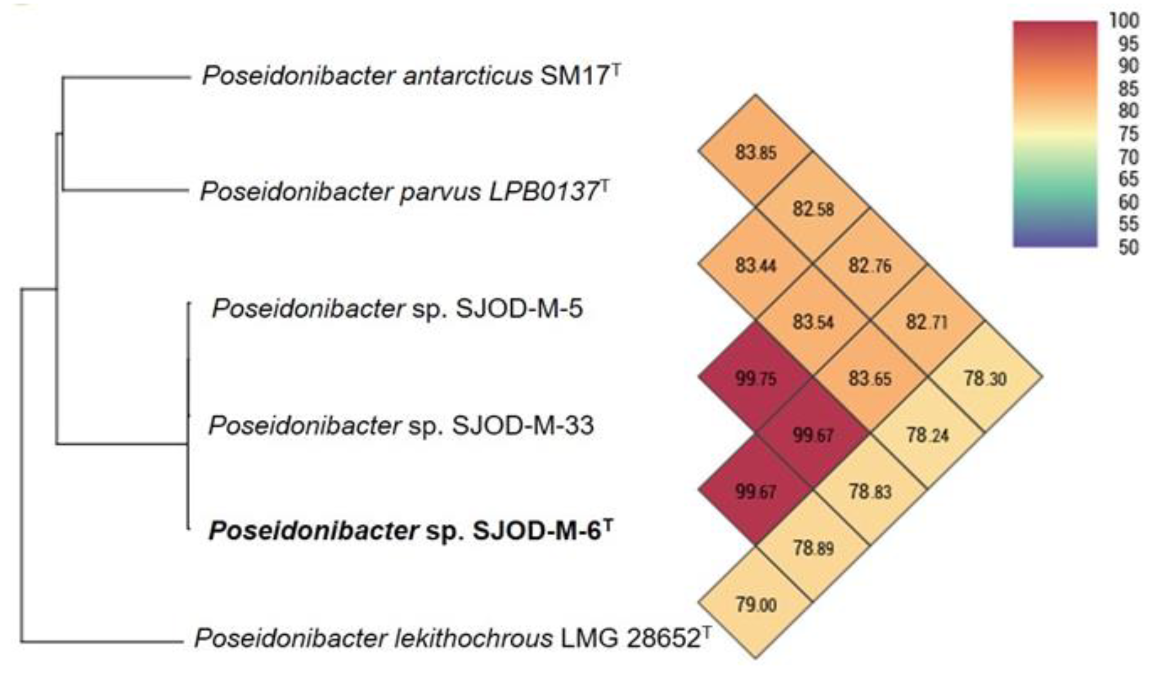
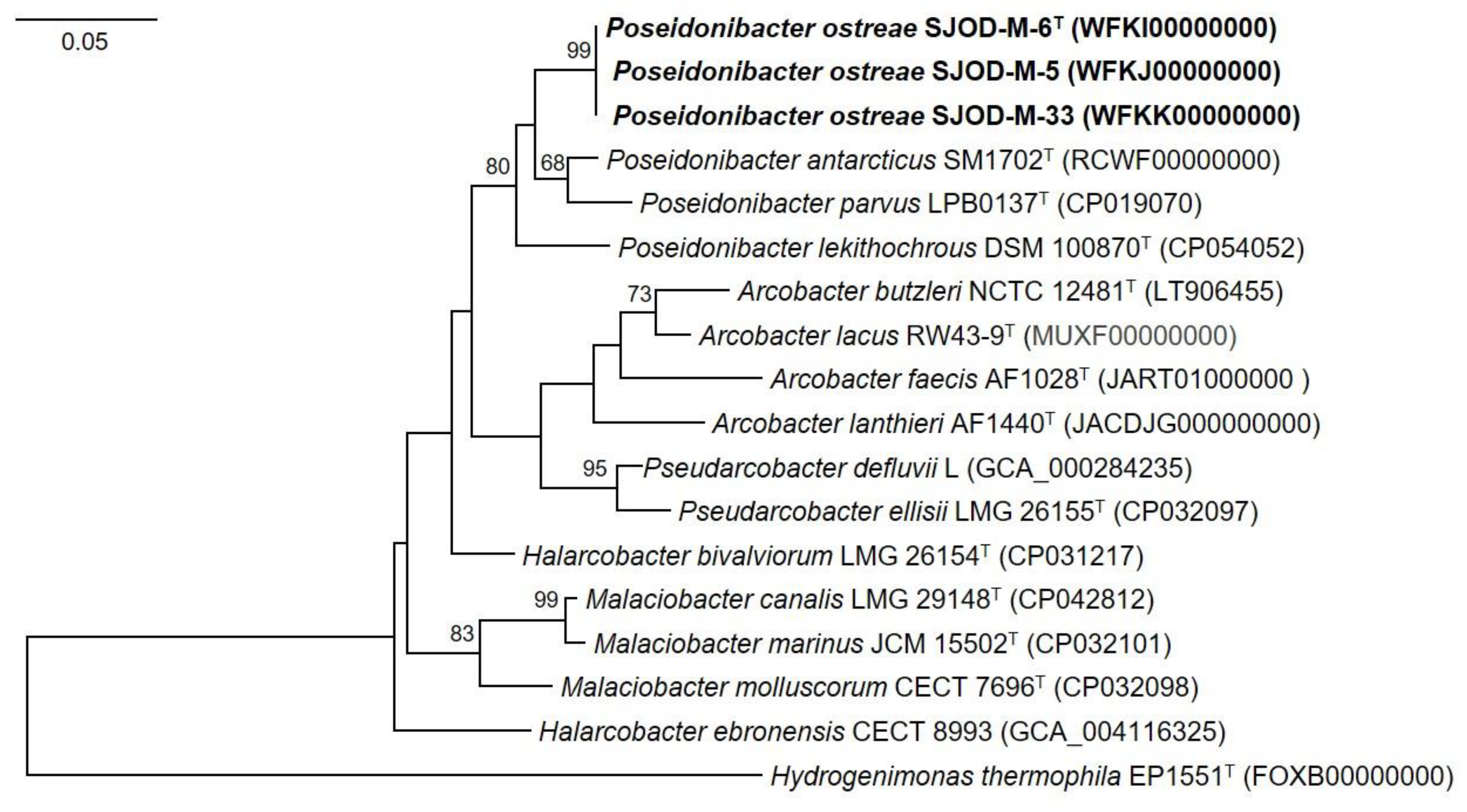
Further analysis using ANI values demonstrated that strains SJOD-M-6T, SJOD-M-5, and SJOD-M-33 share ANI values higher than 99.7%, indicating that they belong to the same species. However, the ANI values between these strains and other type species within the genus Poseidonibacter were 78.24% to 83.85%, that found to be lower than the 95% threshold (Figure 2). This supports the conclusion that these strains represent a novel species within the genus Poseidonibacter [36]. Digital DNA-DNA hybridization (dDDH) values were also calculated, and the results showed dDDH values of 36.9%, 20.3%, and 20.5% between SJOD-M-6T, and reference strains P. parvus LPB0137T, P. lekithochrous LFT 1.7T, and P. antarcticus SM1702T, respectively. These values are below the conventional threshold of 70% for species delineation [27,37]. Hence, these data further support the classification of these strains as distinct species within the genus Poseidonibacter. Furthermore, the phylogenomic tree showed that the genome of strains SJOD-M-6T, SJOD-M-5, and SJOD-M-33 formed a clade with the genome of Poseidonibacter species, confirming again that strain SJOD-M-6T represents a novel species of the genus Poseidonibacter (Figure 3). The overall genomic characteristics, particularly the ANI and dDDH values, suggest that strain SJOD-M-6T represents a novel species of the genus Poseidonibacter.

3.4. Morphological, Physiological, and Biochemical Characteristics

The cells of SJOD-M-6T, SJOD-M-5, and SJOD-M-33 were observed to be Gram-stain-negative and had a coccoid shape, with a size ranging from 0.5 to 1.0 µm (Figure S5). The colonies formed by the strain on MA medium appeared as regular, round, and ivory-colored after 3 days of incubation at 25 °C. Strain SJOD-M-6T exhibited a temperature range for growth of 10–30 °C, with an optimum growth temperature of 25 °C. It grew within a pH range of 5.5–8.0, with the optimal pH for growth being pH 7.0. The strain showed tolerance to NaCl concentrations in the range of 1.0–5.0% (w/v), with an optimum NaCl concentration for growth of 2.0%. The cells of SJOD-M-6T were positive catalase and oxidase activities. Among the tested strains, only SJOD-M-6T exhibited the ability to hydrolyze starch, while none of the strains showed H2S production or the ability to utilize tween 20, 40, 60, 80, and casein. Strain SJOD-M-6T could be differentiated from other members of the genus Poseidonibacter based on various characteristics, including the production of arginine dihydrolase, the activities of esterase (C4), and the assimilation of β-methyl-D-glucoside, quinic acid, glycyl-L-aspartic acid, and L-pyroglutamic acid. Additionally, strain SJOD-M-6T exhibited starch hydrolysis, which was not observed in its closest relatives. A summary of the phenotypic characteristics distinguishing strain SJOD-M-6T from its closest relatives can be found in Table 1.
These morphological, physiological, and biochemical characteristics provide important information for the identification and differentiation of strain SJOD-M-6T from related species within the genus Poseidonibacter.

3.5. Chemotaxonomic Characterization

The major cellular fatty acids of strains SJOD-M-6T, SJOD-M-5, and SJOD-M-33 were identified as summed feature 3 (C16:1 ω7c and/or C16:1 ω6c), followed by summed feature 8 (C18:1 ω7c and/or C18:1 ω6c) and C16:0. A comparison of the full fatty acid profiles between strains SJOD-M-6T, SJOD-M-5, and SJOD-M-33 and three reference strains can be found in Table 2.
The predominant respiratory quinone detected in strain SJOD-M-6T was indeed menaquinone-6 (MK-6). MK-6 is a common quinone found in the genus Poseidonibacter. Regarding the polar lipids, strain SJOD-M-6T was found to contain phosphatidylethanolamine, phosphatidylglycerol, an unidentified aminolipid, an unidentified aminophospholipid and two unidentified phospholipids (Figure S6). Among these, phosphatidylethanolamine and phosphatidylglycerol were identified as common polar lipids in the genus Poseidonibacter (Table 1). These chemotaxonomic characteristics, including the fatty acid composition, isoprenoid quinone profile, and polar lipid composition, align with the classification of most strains within the genus Poseidonibacter.

4. Conclusions

Based on the phylogenetic analysis, genome comparison, chemotaxonomic characteristics, and differential phenotypic and genetic features, it can be concluded that strain SJOD-M-6T, along with strains SJOD-M-5 and SJOD-M-33, belongs to the genus Poseidonibacter. However, the low genomic relatedness and distinct characteristics differentiate strain SJOD-M-6T from other type species within the genus Poseidonibacter. This designation highlights its isolation from the gut of Ostrea denselamellosa and emphasizes its distinct taxonomic position within the Poseidonibacter genus. Therefore, strain SJOD-M-6T represents a novel species, which is proposed to be named Poseidonibacter ostreae sp. nov.
Description of Poseidonibacter ostreae sp. nov.
Poseidonibacter ostreae (os.tre′a.e. L. gen. n. ostreae of/from an oyster).
Cells are gram-stain-negative, aerobic, catalase and oxidase positive, and coccoid-shaped (0.5–1.0 µm). The temperature range for its growth is 10–30 °C (optimum, 25 °C). The pH range for growth is 5.5–8.0 (optimum, 7.0). The range of NaCl concentration tolerated for growth is 1.0–5.0% (w/v), with an optimal NaCl concentration of 2.0% (w/v). Starch is hydrolyzed, but H2S, Tween 20, 40, 60, 80, casein, and DNA are not hydrolyzed. The API 20NE strip indicates positive for nitrate reduction and arginine dihydrolase activity, but indole production, glucose fermentation, urease, esculin hydrolysis, gelatinase, and β-galactosidase activity are negative. The API ZYM kit shows the presence of esterase (C4), esterase lipase (C8), and naphthol-AS-BI-phosphohydrolase activity, but alkaline phosphatase, lipase (C14), leucine arylamidase, valine arylamidase, cystine arylamidase, trypsin, α-chymotrypsin, acid phosphatase, α-galactosidase, β-galactosidase, β-glucuronidase, α-glucosidase, β-glucosidase, N-acetyl-β-glucosaminidase, α-mannosidase, and α-fucosidase activities are absent. The Biolog GN2 carbon source oxidation test indicates positive for D-arabitol, D-cellobiose, α-D-lactose, D-mannitol, D-mannose, D-melibiose, β-methyl-D-glucoside, L-rhamnose, D-sorbitol, malonic acid, propionic acid, quinic acid, succinic acid, L-aspartic acid, L-glutamic acid, glycyl-L-aspartic acid, L-pyroglutamic acid, and glycerol. Its major respiratory quinone is MK-6. The major fatty acids (>10%) are summed feature 3 (C16:1 ω7c and/or C16:1 ω6c) (56.7%), summed feature 8 (C18:1 ω7c and/or C18:1 ω6c) (17.5%), and C16:0 (11.7%). The polar lipids identified in the strain are phosphatidylethanolamine, phosphatidylglycerol, two unidentified phospholipids, an unidentified aminolipid, and an unidentified aminophospholipid.
The type strain of Poseidonibacter ostreae is SJOD-M-6T (=KCTC 72758T = NBRC 114334T = FBCC-B685), which was isolated from the gut of Ostrea denselamellosa. The G+C content of the type strain DNA is 27.5 mol%, and its genome size is 2.9 Mb. The 16S rRNA gene sequence is deposited in GenBank/EMBL/DDBJ under the accession number MN549522, and the draft genome sequence is available under the accession number WFKI00000000. Two closely related strains, SJOD-M-5 (accession number WFKJ00000000) and SJOD-M-33 (accession number WFKK00000000), were also isolated from the gut of Ostrea denselamellosa.

Supplementary Materials

The following supporting information can be downloaded at: https://www.mdpi.com/article/10.3390/d15080920/s1, Table S1. Genome statistics of strains SJOD-M-6T, SJOD-M-5, and SJOD-M-33 and their related strains of species of genus Poseidonibacter. Figure S1. Circular map of the Poseidonibacter sp. strain SJOD-M-6T genome. From outside to the center; the colored bands in ring 1 represent contigs; ring 2 represents the annotated genes on the forward strand (color determined by COG category); ring 3 shows the annotated genes on the reverse strand (color determined by COG category); ring 4 displays the RNA genes (rRNAs are displayed in red and tRNAs are displayed in purple); ring 5 shows the GC skew (higher-than-average values are displayed in green, while lower-than-average values are displayed in red) and ring 6 shows the GC ratio (higher-than-average values in blue and lower-than-average values in yellow). Figure S2. Subsystem category distribution of strain SJOD-M-6T based on the RAST annotation server (https://rast.nmpdr.org/) (accessed on 25 May 2023). Figure S3. COG functional classification of proteins in strain SJOD-M-6T genome. Figure S4. Venn diagram representing the core orthologous and unique genes for strains SJOD-M-6T and closely related type strainss. Figure S5. Phylogenomic tree based on concatenated multiple-alignment of 82 genes showing the relationship between SJOD-M-6T and members of the family Arcobacteraceae. The tree was generated using UBCG2 pipeline with the concatenation of 82 gene sequences. GenBank accession numbers are shown in parentheses. Percentage bootstrap values are 60 shown at the branch points. The tree was rooted with Hydrogenimonas thermophila EP 1551T (FOXB00000000). The Bar represents 0.05 substitutions per nucleotide position. Figure S6. Transmission electron micrograph of negatively stained cells of strain SJOD-M-6T. Bar, 200 nm. Figure S7. Two-dimensional thin-layer chromatogram showing polar lipids of SJOD-M-6T, which were stained with a phosphomolybdic acid solution for total lipids (A), spraying zinzadze reagent (molybdenum blue spray reagent, 1.3%) for phospholipids (B), and 0.2% ninhydrin solution for aminolipids (C), α-naphthol solution for glycolipids (D), respectively. Abbreviations: PE, phosphatidylethanolamine; PG, phosphatidylglycerol; PL1–2, unidentified phospholipids; AL, unidentified aminolipid.; APL, unidentified aminophospholipid.

Author Contributions

Conceptualization: K.B. and A.C.; methodology: K.B., S.J., E.J.C. and A.C.; formal analysis: K.B. and A.C.; resources: K.B. and A.C.; data curation: K.B. and A.C.; writing—original draft preparation: K.B. and A.C.; writing—review and editing: all authors; funding acquisition: S.H.R. All authors have read and agreed to the published version of the manuscript.

Funding

This work was supported by the Korea Environment Industry & Technology Institute (KEITI) through the project to make multi-ministerial national biological research resources more advanced program, funded by Korea Ministry of Environment (MOE) (2021003420001).

Institutional Review Board Statement

Not applicable.

Informed Consent Statement

Not applicable.

Data Availability Statement

Not applicable.

Conflicts of Interest

The authors declare no conflict of interest.

Abbreviations

ANI, average nucleotide identity; dDDH, digital DNA–DNA hybridization; MA, marine agar 2216; NJ, neighbor-joining; ML, maximum-likelihood; MP, maximum-parsimony; RAST, Rapid Annotation using the Subsystem Technology; BGCs, biosynthetic gene clusters; COGs, clusters of orthologous groups; GGDC, genome-to-genome distance calculator; POGs, pan-genome orthologous groups; UBCG, up-to-date bacterial core gene; TEM, transmission electron microscopy; ASW, artificial seawater medium; FAME, fatty acid methyl ester.

References

  1. Diéguez, A.L.; Balboa, S.; Magnesen, T.; Romalde, J.L. Arcobacter lekithochrous sp. nov., isolated from a molluscan hatchery. Int. J. Syst. Evol. Microbiol. 2017, 67, 1327–1332. [Google Scholar] [CrossRef]
  2. Diéguez, A.L.; Pérez-Cataluña, A.; Figueras, M.J.; Romalde, J.L. Arcobacter haliotis Tanaka et al. 2017 is a later heterotypic synonym of Arcobacter lekithochrous Diéguez et al. 2017. Int. J. Syst. Evol. Microbiol. 2018, 68, 2851–2854. [Google Scholar] [CrossRef] [PubMed]
  3. Guo, X.H.; Wang, N.; Yuan, X.X.; Zhang, X.Y.; Chen, X.L.; Zhang, Y.Z.; Song, X.Y. Poseidonibacter antarcticus sp. nov., isolated from Antarctic intertidal sediment. Int. J. Syst. Evol. Microbiol. 2019, 69, 2717–2722. [Google Scholar] [CrossRef] [PubMed]
  4. Kim, M.J.; Baek, M.-G.; Shin, S.-K.; Yi, H. Poseidonibacter parvus sp. nov., isolated from a squid. Int. J. Syst. Evol. Microbiol. 2021, 71, 004590. [Google Scholar] [CrossRef] [PubMed]
  5. Fisher, T.R.; Carlson, P.R.; Barber, R.T. Carbon and nitrogen primary productivity in three Noreth Carolina estuaries. Estuar. Coast. Shelf Sci. 1982, 15, 621–644. [Google Scholar] [CrossRef]
  6. Barlow, J.P. Physical and biological processes determining the distribution of zooplankton in a tidal estuary. Biol. Bull. 1955, 109, 211–225. [Google Scholar] [CrossRef]
  7. Min, D.K.; Lee, J.S.; Koh, D.B.; Je, J.G. Mollusks in Korea; Hangul Graphics: Busan, Republic of Korea, 2004; pp. 410–415. [Google Scholar]
  8. Noseworthy, R.G.; Lee, H.-J.; Choi, S.-D.; Choi, K.-S. Unique substrate preference of Ostrea denselamellosa Lischke, 1869 (Mollusca: Ostreidae) at Haechang Bay, on the south coast of Korea. Korean J. Parasitol. 2016, 32, 31–36. [Google Scholar] [CrossRef] [Green Version]
  9. Lane, D.J. 16S/23S rRNA sequencing. In Nucleic Acid Techniques in Bacterial Systematics; Stackebrandt, E., Goodfellow, M., Eds.; John Wiley and Sons: New York, NY, USA, 1991; pp. 115–175. [Google Scholar]
  10. Yoon, S.H.; Ha, S.M.; Kwon, S.; Lim, J.; Kim, Y.; Seo, H.; Chun, J. Introducing EzBioCloud: A taxonomically united database of 16S rRNA gene sequences and whole-genome assemblies. Int. J. Syst. Evol. Microbiol. 2017, 67, 1613–1617. [Google Scholar] [CrossRef]
  11. Jeon, Y.S.; Lee, K.; Park, S.C.; Kim, B.S.; Cho, Y.J.; Ha, S.M.; Chun, J. EzEditor: A versatile sequence alignment editor for both rRNA- and protein-coding genes. Int. J. Syst. Evol. Microbiol. 2014, 64, 689–691. [Google Scholar] [CrossRef] [PubMed] [Green Version]
  12. Saitou, N.; Nei, M. The neighbor-joining method: A new method for reconstructing phylogenetic trees. Mol. Biol. Evol. 1987, 4, 406–425. [Google Scholar]
  13. Felsenstein, J. Evolutionary trees from DNA sequences: A maximum likelihood approach. J. Mol. Evol. 1981, 17, 368–376. [Google Scholar] [CrossRef] [PubMed]
  14. Fitch, W.M. Toward defining the course of evolution: Minimum change for a specific tree topology. Syst. Zool. 1971, 20, 406–416. [Google Scholar] [CrossRef]
  15. Kumar, S.; Stecher, G.; Tamura, K. MEGA7: Molecular evolutionary genetics analysis version 7.0 for bigger datasets. Mol. Biol. Evol. 2016, 33, 1870–1874. [Google Scholar] [CrossRef] [PubMed] [Green Version]
  16. Felsenstein, J. Confidence limits on phylogenies: An approach using the bootstrap. Evolution 1985, 39, 783–791. [Google Scholar] [CrossRef] [PubMed]
  17. Kimura, M. A simple method for estimating evolutionary rates of base substitutions through comparative studies of nucleotide sequences. J. Mol. Evol. 1980, 16, 111–120. [Google Scholar] [CrossRef]
  18. William, S.; Feil, H.; Copeland, A. Bacterial genomic DNA Isolation Using CTAB. Joint Genome Institute, Walnut Creek, CA, USA, 2012. Joint Genome Institute. Bacterial Genomic DNA Isolation Using CTAB. 2012. Available online: https://jgi.doe.gov/wp-content/uploads/2014/02/JGI-Bacterial-DNA-isolation-CTAB-Protocol-2012.pdf (accessed on 7 July 2023).
  19. Chun, J.; Oren, A.; Ventosa, A.; Christensen, H.; Arahal, D.R.; da Costa, M.S.; Rooney, A.P.; Yi, H.; Xu, X.W.; De Meyer, S.; et al. Proposed minimal standards for the use of genome data for the taxonomy of prokaryotes. Int. J. Syst. Evol. Microbiol. 2018, 68, 461–466. [Google Scholar] [CrossRef]
  20. Wick, R.R.; Judd, L.M.; Gorrie, C.L.; Holt, K.E. Unicycler: Resolving bacterial genome assemblies from short and long sequencing reads. PLoS Comput. Biol. 2017, 13, e1005595. [Google Scholar] [CrossRef] [Green Version]
  21. Aziz, R.K.; Bartels, D.; Best, A.A.; DeJongh, M.; Disz, T.; Edwards, R.A.; Formsma, K.; Gerdes, S.; Glass, E.M.; Kubal, M.; et al. The RAST server: Rapid annotations using subsystems technology. BMC Genom. 2008, 9, 75. [Google Scholar] [CrossRef] [Green Version]
  22. Tatusova, T.; DiCuccio, M.; Badretdin, A.; Chetvernin, V.; Nawrocki, E.P.; Zaslavsky, L.; Lomsadze, A.; Pruitt, K.D.; Borodovsky, M.; Ostell, J. NCBI prokaryotic genome annotation pipeline. Nucleic Acids Res. 2016, 44, 6614–6624. [Google Scholar] [CrossRef]
  23. Lee, I.; Chalita, M.; Ha, S.M.; Na, S.I.; Yoon, S.H.; Chun, J. Contest16S: An algorithm that identifies contaminated prokaryotic genomes using 16S rRNA gene sequences. Int. J. Syst. Evol. Microbiol. 2017, 67, 2053–2057. [Google Scholar] [CrossRef]
  24. Blin, K.; Shaw, S.; Augustijn, H.E.; Reitz, Z.L.; Biermann, F.; Alanjary, M.; Fetter, A.; Terlouw, B.R.; Metcalf, W.W.; Helfrich, E.J.N.; et al. antiSMASH 7.0: New and improved predictions for detection, regulation, chemical structures and visualization. Nucleic Acids Res. 2023, 51, W46–W50. [Google Scholar] [CrossRef] [PubMed]
  25. Kanehisa, M.; Goto, S. KEGG: Kyoto encyclopedia of genes and genomes. Nucleic Acids Res. 2000, 28, 27–30. [Google Scholar] [CrossRef] [PubMed]
  26. Lee, I.; Kim, Y.O.; Park, S.-C.; Chun, J. OrthoANI: An improved algorithm and software for calculating average nucleotide identity. Int. J. Syst. Evol. Microbiol. 2016, 66, 1100–1103. [Google Scholar] [CrossRef] [PubMed]
  27. Auch, A.F.; von Jan, M.; Klenk, H.-P.; Göker, M. Digital DNA–DNA hybridization for microbial species delineation by means of genome-to-genome sequence comparison. Stand Genom. Sci. 2010, 2, 117–134. [Google Scholar] [CrossRef] [PubMed] [Green Version]
  28. Na, S.I.; Kim, Y.O.; Yoon, S.H.; Ha, S.M.; Baek, I.; Chun, J. UBCG: Up-to-date bacterial core gene set and pipeline for phylogenomic tree reconstruction. J. Microbiol. 2018, 56, 280–285. [Google Scholar] [CrossRef]
  29. Price, M.N.; Dehal, P.S.; Arkin, A.P. FastTree: Computing large minimum evolution trees with profiles instead of a distance matrix. Mol. Biol. Evol. 2009, 26, 1641–1650. [Google Scholar] [CrossRef]
  30. Choo, Y.-J.; Lee, K.; Song, J.; Cho, J.-C. Puniceicoccus vermicola gen. nov., sp. nov., a novel marine bacterium, and description of Puniceicoccaceae fam. nov., Puniceicoccales ord. nov., Opitutaceae fam. nov., Opitutales ord. nov. and Opitutae classis nov. in the phylum ‘Verrucomicrobia’. Int. J. Syst. Evol. Microbiol. 2007, 57, 532–537. [Google Scholar] [CrossRef]
  31. Kim, H.; Choo, Y.-J.; Cho, J.-C. Litoricolaceae fam. nov., to include Litoricola lipolytica gen. nov., sp. nov., a marine bacterium belonging to the order Oceanospirillales. Int. J. Syst. Evol. Microbiol. 2007, 57, 1793–1798. [Google Scholar] [CrossRef] [Green Version]
  32. Smibert, R.M.; Krieg, N.R. Phenotypic characterization. In Methods for General and Molecular Bacteriology; Gerhardt, P., Murray, R.G.E., Wood, W.A., Krieg, N.R., Eds.; American Society for Microbiology: Washington, DC, USA, 1994; pp. 607–654. [Google Scholar]
  33. Tindall, B.J.; Sikorski, J.; Smibert, R.A.; Krieg, N.R. Phenotypic Characterization and the Principles of Comparative Systematics. In Methods for General and Molecular Microbiology; Reddy, C.A., Ed.; ASM Press: Washington, DC, USA, 2007; pp. 330–393. [Google Scholar]
  34. Sasser, M. Identification of bacteria by gas chromatography of cellular fatty acids. Technol. Note 2001, 101. [Google Scholar]
  35. Minnikin, D.E.; O’Donnell, A.G.; Goodfellow, M.; Alderson, G.; Athalye, M.; Schaal, A.; Parlett, J. An integrated procedure for the extraction of bacterial isoprenoid quinones and polar lipids. J. Microbiol. Methods 1984, 2, 233–241. [Google Scholar] [CrossRef]
  36. Jain, C.; Rodriguez-R, L.M.; Phillippy, A.M.; Konstantinidis, K.T.; Aluru, S. High throughput ANI analysis of 90K prokaryotic genomes reveals clear species boundaries. Nat. Commun. 2018, 9, 5114. [Google Scholar] [CrossRef] [PubMed] [Green Version]
  37. Richter, M.; Rosselló-Móra, R. Shifting the genomic gold standard for the prokaryotic species definition. Proc. Natl. Acad. Sci. USA 2009, 106, 19126–19131. [Google Scholar] [CrossRef] [PubMed]
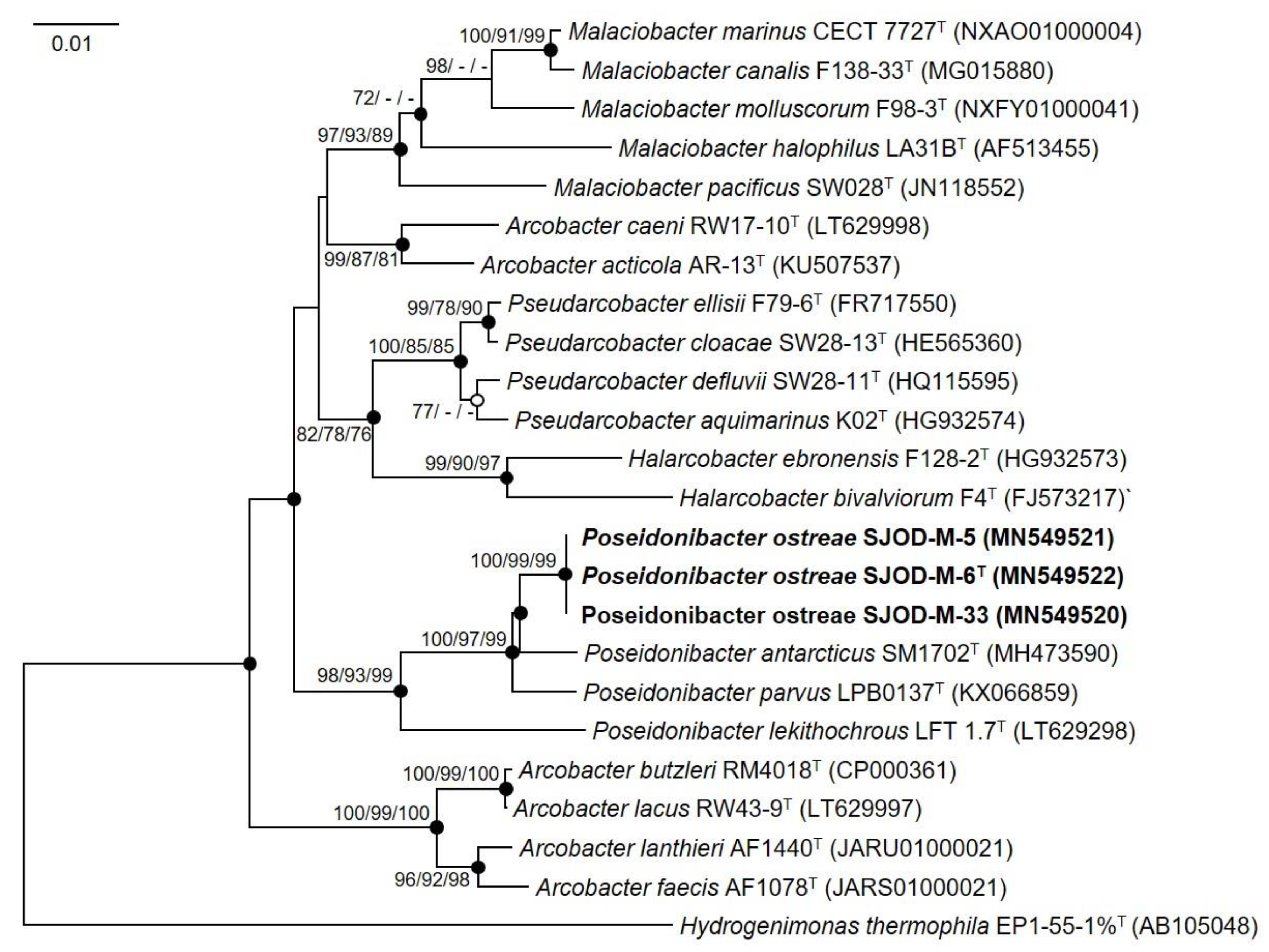
Figure 1. Neighbor-joining phylogenetic tree based on 16S rRNA gene sequences showing positions of strains SJOD-M-6T, SJOD-M-5, SJOD-M-33, and other closely related members of the family Arcobacteraceae. Bootstrap values (expressed as percentages of 1000 replications) over 70% are shown at nodes for neighbor−joining, maximum−likelihood and maximum−parsimony methods, respectively. Filled circles indicate that the corresponding nodes were recovered by all treeing methods. An open circle indicates that the corresponding node was recovered by the neighbor-joining and maximum−parsimony methods. Hydrogenimonas thermophile EP1-55-1%T (AB105048) was used as an out-group. Bar, 0.01 substitutions per nucleotide position.
Diversity 15 00920 g001
Figure 2. Heatmap of Orthologous Average Nucleotide Identity (OrthoANI) between strain SJOD-M-6T, SJOD-M-5, and SJOD-M-33, and closely related relatives. Values in the color scale indicate the similarity percentage among the genomes. The numerical values on the branches represent the calculated intergenomic genetic distance calculated by the Orthologous Average Nucleotide Identity Tool (OAT) software version 6.1.
Diversity 15 00920 g002
Figure 3. Phylogenomic tree based on concatenated multiple-alignment of 82 genes showing the relationship between SJOD-M-6T and members of the family Arcobacteraceae. The tree was generated using UBCG2 pipeline with the concatenation of 82 gene sequences. GenBank accession numbers are shown in parentheses. Percentage bootstrap values are 60 shown at the branch points. The tree was rooted with Hydrogenimonas thermophila EP 1551T (FOXB00000000). The Bar represents 0.05 substitutions per nucleotide position.
Diversity 15 00920 g003
Table 1. Characteristics that differentiate strains SJOD-M-6T, SJOD-M-5, and SJOD-M-33 from the type strains of species in the genus Poseidonibacter. Strains: 1, SJOD-M-6T; 2, SJOD-M-5; 3, SJOD-M-33; 4, P. parvus KACC18888T; 5, P. antarcticus KCTC 62796T; 6, P. lekithochrous DSM 100870T. Data are from this study or from, except where indicated. +, Positive; −, negative.
Characteristic123456
Isolation sourceOysterOysterOysterSquid Sediment Scallop §
Colony color on MAIvoryIvoryIvoryPinkish-brown Creamy yellow Brown §
Temperature range (Optimum) (°C)10–30 (25)10–30 (25)10–30 (25)10–25 (25) 0–30 (10–15) 15–25 (25) §
NaCl tolerance (%, w/v)1.0–5.01.0–5.01.0–5.02.0–4.5 0.5–5.0 0.0–8.0 §
API 20NE:
Arginine dihydrolase+++++
Indole production++
glucose fermentation +
Enzyme activity of:
Esterase (C4)+++ ++
Acid phosphatase+++
Alkaline phosphatase++
Leucine arylamidase+
Utilization of carbon sources (Biolog GN2):
D-sorbitol+++++
α-D-lactose, malonic acid, propionic acid, succinic acid, L-aspartic acid, L-glutamic acid, glycerol+++++
L-rhamnose+++++
D-arabitol++++
D-mannitol++++
D-cellobiose, D-mannose, D-melibiose++++
β-methyl-D-glucoside, quinic acid, glycyl-L-aspartic acid, L-pyroglutamic acid+++
L-arabinose, L-alanine++
Sucrose, D-trehalose, L-leucine, L-serine++
α-D-glucose, maltose, D-alanine++
Lactulose+
N-acetyl-D-glucosamine+
D-fructose, α-hydroxybutyric acid, D-saccharic acid, L-histidine, γ-amino butyric acid+
Hydrolysis of:
Starch+++
DNA++
DNA G + C content * (mol%)27.527.527.627.727.128.5
Genome size (Mb)2.862.963.212.872.923.57
* The G + C content was calculated based on the nucleotide content of the sequenced genomes. § Data from Diéguez et al. [1,2]. Data from Guo et al. [3]. Data from Kim et al. [4].
Table 2. Fatty acids profiles of strains SJOD-M-6T, SJOD-M-5, and SJOD-M-33 and the type strains of species of genus Poseidonibacter. Strains: 1, SJOD-M-6T; 2, SJOD-M-5; 3, SJOD-M-33; 4, P. parvus KACC18888T; 5, P. antarcticus KCTC 62796T; 6, P. lekithochrous DSM 100870T. All the strains were grown on MA at 25 °C and harvested in the late exponential phase. Values are percentages of the total fatty acids. Fatty acids that represented <1.0% in all strains are not shown. Tr, Trace (<1.0%); -, not detected.
Fatty Acid123456
C12:01.21.1Tr2.34.3Tr
C12:0 3-OHTrTr1.0-5.6-
C14:02.62.73.08.27.43.1
C16:011.711.911.98.511.912.0
C16:1 ω5c2.82.62.8TrTr1.4
Summed features *
2 (C14:0 3-OH and/or C16:1 iso-I)6.26.36.411.25.97.5
3 (C16:1 ω7c and/or C16:1 ω6c)56.757.256.856.845.154.8
8 (C18:1 ω7c and/or C18:1 ω6c)17.917.317.912.118.815.6
* Summed features represent groups of two fatty acids, which cannot be separated by the MIDI system.
Disclaimer/Publisher’s Note: The statements, opinions and data contained in all publications are solely those of the individual author(s) and contributor(s) and not of MDPI and/or the editor(s). MDPI and/or the editor(s) disclaim responsibility for any injury to people or property resulting from any ideas, methods, instructions or products referred to in the content.