Article

Molecular Phylogeny of the Genus Paracanthonchus (Nematoda: Chromadorida) with Description of P. yeongjongensis sp. nov. from Korea †

1
Biodiversity Research Department, National Institute of Biological Resources, Incheon 22689, Republic of Korea
2
Department of Environmental Science, Hanyang University, Seoul 04763, Republic of Korea
3
Department of Life Science, Hanyang University, Seoul 04763, Republic of Korea
4
Center for Creative Convergence Education, Hanyang University, Seoul 04763, Republic of Korea
5
Research Institute of Natural Science, Hanyang University, Seoul 04763, Republic of Korea
*
Author to whom correspondence should be addressed.
urn:lsid:zoobank.org:pub:0F527D3E-9780-4615-888D-96350C28CF86.
Diversity 2023, 15(5), 664; https://doi.org/10.3390/d15050664
Submission received: 24 March 2023 / Revised: 12 May 2023 / Accepted: 12 May 2023 / Published: 13 May 2023
(This article belongs to the Special Issue The Taxonomy, Evolution, and Phylogeography of Marine Invertebrates)

Abstract

:
During an investigation of the intertidal zone at Yeongjongdo Island, Incheon, Korea, a new species belonging to the genus Paracanthonchus was found and is reported. Paracanthonchus Mikoletzky, 1924 is the largest genus within the family Cyatholaimidae, and the species identification of this genus has been difficult mainly due to overlapping characteristics and a lack of genus/species-defining apomorphic characters. The new species is characterized by the buccal cavity, armed with one large dorsal tooth and two subventral teeth, the presence of lateral differentiation, seventy-six tubular precloacal supplements, and proximally paired gubernaculum. Alongside the description, we are updating the review of the genus by providing an up-to-date list of valid species, as well as a comprehensive tabular key to the genus with measurements of species-discerning characteristics. We also provide partial sequences of mtCOI, 18S and 28S rRNA to verify the new species belongs to the genus Paracanthonchus and to discuss the phylogeny of the family Cyatholaimidae as well as the genus Paracanthonchus. Our phylogeny agrees with previous findings that, while Cyatholaimidae forms a monophyletic clade, many genera within, including Paracanthonchus, are paraphyletic. For now, it appears that subfamilies are not well-represented by molecular means, and much more molecular data along with species defining morphological traits will have to be accumulated to meaningfully organize this family and the genus within.

1. Introduction

The nematode family Cyatholaimidae Filipjev, 1918 is a relatively diverse group of marine free-living nematodes [1] comprising 26 genera and 257 species [2]. It consists of free-living marine nematodes which inhabit marine sediments, and their distribution and dispersal has been the subject of many studies [3,4,5]. The family was first erected as a subfamily (Cyatholaimini) by Filipjev in 1918 [6], to be reorganized as a family by De Coninck and Schuurmans-Stekhoven (1933) [7]. According to the latest revision of the family by Cunha et al. (2022), there are four subfamilies which make up the family Cyatholaimidae: Cyatholaiminae, Paracanthonchinae, Pomponematinae and Xenocyatholaiminae. NeMys [8], however, lists a total of five subfamilies, including Nyctonema Bussau, 1993 with a sole species, Nyctonema portentosum Bussau, 1993. On the other hand, the most recent classification of the phylum Nematoda by Hodda (2022) only lists two subfamilies, Cyatholaiminae and Paracanthonchiinae, and replaced Xenocyatholaiminae by placing Xenocyatholaimus, a genus with lone species belonging to subfamily Xenocyatholiminae Gerlach & Riemann, 1973, in the subfamily Paracanthonchinae. Hodda (2022) does not include remarks on why he simplified the subfamilies, but it is clear he took gubernaculum structure as a key component in discerning the two subfamilies. Evidently, the validity of the subfamilies within Cyatholaimidae is not a fixed matter, but they are discerned by the structure of amphid, the structure of gubernaculum, the shape of the tail and the position of the vulva. The main problem, which means that this family does not have clear boundaries, is that these species, genera, and subfamilies’ dividing morphological features are based on a combination of multiple non-phylogenetic informative characters [4]. To make matters more complicated, recent phylogenetic analyses suggest that while the family itself maybe monophyletic, most genera belonging to Cyatholaimidae lack synapomorphic characters and are therefore non-monophyletic. Cunha et al. (2022)’s review of the family Cyatholaimidae provides a comprehensive revision that clarifies the delimitation of genera within the family, but the core problem of mixed topology remains unresolved.
Paracanthonchus is a large genus comprised of cosmopolitan species [3,4] which makes up a fifth of the whole family Cyatholaimidae [2,3,4]. Paracanthonchus was first established by Micoletzky (1924) [9], when he described the type species Paracanthonchus caecus. Since then, over 70 species have been described and many revisions of the genus have taken place [10,11,12,13,14]. The genus Paracanthonchus can be characterized by the following characteristics: (1) cuticle with transverse rows of fine dots and lateral differentiation with 2–4 rows of larger dots, (2) buccal cavity with an especially big dorsal tooth, (3) distally expended and dentate gubernaculum, and (4) tubular precloacal supplements [10,15,16]. However, due to a lack of apomorphic characters, diagnostic characters often overlap across other genera, making the species delimitation of this group a difficult task [4]. While Cunha et al., 2022, did briefly provide updated diagnoses of the genus with an updated number of valid species, it was still missing critical elements for species delimitation, such as a tabular key. Prior to Cunha et al., 2022, the most recent and complete revision of Paracanthonchus was by Miljutina and Miljutin, 2015. While an in-depth taxonomic history of the genus and tabular key was provided, considerable changes have taken place since, and phylogenetic aspects were not discussed. Much like its parent family, the number of valid species varies among different sources for the genus Paracanthonchus. NeMys currently lists 59 valid species [8], with relevant papers all citing different numbers (Hodda, 2022, 80 species [2]; Miljutina and Miljutin, 2015, 72 species (including 20 species inquirenda) [3]; Tchesunov, 2014, 67 species [15]; Tchesunov, 2015, 63 species [17]; Lee et al., 2016, 72 valid species [18]).
With various papers citing contradicting number of valid species, we have taken on the role of providing a comprehensive, updated review of the genus including an updated list of valid species and a tabular key for species delimitation. Along with the description and depiction of the new species, we also provide molecular sequences (mtCOI, 18S, 28S rRNA) of our new species, supplemented with a phylogenetic tree, to discuss the topology of the family Cyatholaimidae and the genus Paracanthonchus. Geographically, with approximately 70 species of marine nematodes described in Korea [18,19,20,21,22], this new species is the third species of Cyatholaimidae (with P. kamui Kito, 1981 and P. macrodon (Ditlevsen, 1918) previously reported from the east [18]) to be reported in Korea and the tenth species of Paracanthonchus to be reported in East Asia (P. brevicaudatus Gagarin and Nguyen Vu Thanh, 2016, Vietnam [23]; P. hawaiiensis Allgén, 1951, Japan [24]; P. heterocaudatus Huang and Xu, 2013, China [25]; P. kamui Kito, 1981, Japan [26]; P. macrodon (Ditlevsen, 1918) Micoletzky, 1924, Korea [18]; P. mamubiae Miljutina and Miljutin, 2015, North West Pacific [3]; P. multisupplementatus Gagarin, 2012, South China Sea [27]; P. perspicuus Kito, 1981, Japan [26]; P. securus Nguyen and Gagarin, 2018, South China Sea [28]).

2. Materials and Methods

2.1. Morphological Analysis

Free-living marine nematodes were collected from the intertidal zone on Yeongjongdo Island, Incheon, Republic of Korea, (37°26′13″ N, 126°23′03″ E) on 26 August 2022 (Figure 1). The sediments were collected using a spoon to scratch only the top layer, then fixed in 90% ethanol. Samples were brought back to the laboratory and stored in the freezer.
Nematodes were sorted in a Petri dish under a dissecting microscope (Olympus SZX-10, Olympus, Tokyo, Japan) then placed in a pool containing a solution of 10% glycerol and distilled water. The dish was left out at room temperature for dehydration to take place, which lasted 1 to 2 days. After dehydration, the specimens were mounted with very small beads on anhydrous glycerin using a standard wax-ring method [29].
For species identification, each specimen was observed under a 100X objective lens with immersion oil using an optical microscope (Nikon ECLIPSE 80i, Nikon, Tokyo, Japan) with Nomarski Differential Interference Contrast (DIC) illumination. All pencil drawings were prepared using a drawing tube equipped with an optical microscope. Line drawings were digitally prepared with a Wacom Cintiq 16 tablet (Wacom, Saitama, Japan) and Adobe Illustrator (Adobe, San Jose, CA, USA). All morphometric measurements were measured with Fiji software [30].
The classification of marine nematodes reflects Hodda (2022) [2], and species identification referred to pictorial keys of Platt and Warwick (1988) [16], as well as all the original descriptions.
For scanning electron microscopy, the identified specimens were transferred to a well of distilled water to be washed, and gradually transferred to ethanol series for dehydration (20%, 30%, 40% 50%, 60%, 70%, 80%, 90%, 95%, and 100%, each 1 h). Following dehydration, specimens were mounted on a stub with gold in an ion sputter coater after being critical-point dried (CPD300, Leica, Wetzlar, German). Observations and photographs were taken using a JSM-6390LV (JEOL, Japan) scanning electron microscope.

2.2. gDNA Extraction and Amplification

For genomic DNA extraction, specimens of interest were handpicked from samples separately fixed with pure ethanol. Two specimens were found and swiftly observed with a temporary slide to confirm species identification, then transferred to a pool of distilled water on a Petri dish to wash and remove excess ethanol. For DNA extraction, DNAeasy Blood & Tissue Kit (Qiagen, Hilden, Germany) was used following the manufacturer’s instructions. DNA concentration of the DNA templates were measured using NanoDrop 2000 (ThermoScientific, Wilmington, NC, USA), and the concentrations ranged from 2.5 to 4.4 ng/μL. For DNA amplification, AccuPower® PCR Master Mix (Bioneer, Daejeon, Republic of Korea) was used, achieving 20 μL of total reaction volume (10 μL premix, 1 μL for each primer, 6 μL DNA template and 2 μL ultra-pure water). Amplification protocol and respective primer set information are provided below (Table 1). Amplification success was checked by visualizing PCR product via 1% agarose gel electrophoresis. PCR products of both strands were sent to Bioneer (Daejeon, Republic of Korea) to be purified and sequenced. All sequences newly obtained in this study were uploaded to GenBank (Accession numbers listed in Table S1).

2.3. Phylogenetic Analysis

All sequences were visualized using FinchTV (v. 1.4.0, Geospiza, Inc.; Seattle, WA, USA; http://www.geospiza.com, accessed on 3 February 2023), and low-quality peaks were filtered by comparing both complementary strands. The two strands (forward and reverse) were aligned using ClustalW [36] implemented in MEGA (v. 11.0.13) [37]. Aligned sequences were compared against NCBI GenBank database using BLAST algorithm [38]. Pairwise distance between the two specimens of Paracanthonchus sp. nov. and other closely related species of mtCOI, 18S and 28S rRNA sequences were calculated with MEGA 11 using K2P model [39]. Phylogenetic trees were constructed using two nuclear genes (18S and 28S rRNA). Attempts were made to obtain longer 18S rRNA sequences, but with no success. Maximum likelihood (ML) and Bayesian Inference (BI) approaches were used when performing phylogenetic analyses. For ML analysis, IQ-TREE (multicore v. 2.0.3) [40] and the implemented ModelFinder [41] were used to determine the model of best-fit using Akaike information criterion (AIC). TIM3 + F + G4 and TIM3 + F + I + G4 models were determined to be the best-fit models for 18S and 28S datasets, respectively, and were selected for constructing ML tree with 1000 bootstrap replicates using IQ-Tree webservers using the ultrafast setting [42]. For BI analysis, jModelTest software (v. 2.1.7) [43] was used to find the best-fit model. BI tree was made utilizing MrBayes software (v. 3.2.6) [44] using the following model parameters for 18S rRNA dataset: Lset base = (0.2657, 0.2100, 0.2582, 0.2660) nst = 6 rmat = (1.0000, 1.7981, 1.0000, 1.0000, 3.2963, 1) rates = gamma shape = 0.4800 ncat = 4 pinvar = 0 with Enoplus sp. set as outgroup. For 28S rRNA dataset, the following model parameters were used: Lset base = (0.2115, 0.2252, 0.3175, 0.2459) nst = 6 rmat = (0.5077, 2.5356, 1.0000, 0.5077, 5.6541, 1) rates = gamma shape = 0.5890 ncat = 4 pinvar = 0.1870 with Enoploides sp. set as outgroup. Markov Chain Monte Carlo (MCMC) was run with ngen = 1,000,000, nchains = 4, samplefreq = 100, savebrlens = yes, printfreq = 1000, sump burnin = 250, and sumt burnin = 250. All trees were exported to FigTree (v. 1.4.4) [45], where visualizations and modifications were made.

3. Results

3.1. Systematics

Class: Chromadorea Inglis, 1983
Order: Chromadorida Chitwood, 1933
Family: Cyatholaimidae Filipjev, 1918 (De-Coninck and Schuurmans-Stekhoven, 1933)
Subfamily: Paracanthonchiinae De-Coninck, 1965
Genus: Paracanthonchus Micoletzky, 1923

3.2. Diagnosis (Followed Tchesunov, 2014)

Cyatholaimidae. Paracanthonchinae. Body cuticle composed of transverse rows of tiny fine dots, which laterally may be larger or irregularly arranged. Six outer labial and four cephalic seta jointed in one circle. Amphideal fovea multi-spiral. Cheilostom with twelve rugae. Buccal cavity with larger dorsal tooth and one or two pairs smaller subvetral teeth. Gubernaculum proximally paired, distally expanded and dentate. Tubular precloacal supplementary organs.
Type species: Paracanthonchus caecus (Bastian, 1865).

3.3. List of Valid Species

1. Paracanthonchus austrospectabilis Wieser, 1954 (Wieser, 1954: 19, Figure 100a–f; Chile, Islas Gueitecas, 3–5 m depth, small stones, sand and gravel; 5–7 m depth, from Mytilidae and echinids at stones with algae; from calcareous alga; Punta Arenas, algae, gravel and clay, mixed with mud; Talcahuano, rocks with small rock pools) [10].
2. Paracanthonchus barka Inglis, 1962 (Inglis, 1962: 271–272, Figure 66–67; France, Banyuls-sur-Mer, seaweeds with very little sediment on the rocks) [46].
3. Paracanthonchus batidus Gerlach, 1957 (Gerlach, 1957: 432, Figure 7e–i; Brazil, Säo Sebastiäo, fine sand) [47].
4. Paracanthonchus bipapillatus Kreis, 1928 (Kreis, 1928: 174–176, Taf. VI, Figure 24; Barents Sea, Kildin Island, few stones, a shell with algae) [48].
5. Paracanthonchus bothnicus Schiemer, Fritz, Jensen, Preben and Riemann, Franz, 1983 (Schiemer et al., 1983: 288, Figure 7; the Bothnian Bay, sand from 5 m depth, soft mud from 82 m) [49].
6. Paracanthonchus brevicaudatus Gagarin & Nguyen Vu Thanh, 2016 (Gagarin and Nguyen Vu Thanh, 2016: 9–14, Figure 1; Vietnam, artificial reservoirs) [23].
7. Paracanthonchus breviseta (Schuurmans Stekhoven, 1950) Hope & Murphy, 1972 (Schuurmans Stekhoven, 1950: 100–101, Figure 55a–c; (Paraseuratiella breviseta); Villefranche, 50m depth, grey mud. Hope and Murphy, 1972, transferred the species from the genus Paraseuratiella to Paracanthonchus) [11,12].
8. Paracanthonchus bulbicola Bussau, 1993 (Bussau, 1993: 160–165, abb. 50–52; Peru basin, a manganese lying on the seabed) [50].
9. Paracanthonchus caecus (Bastian, 1865) Micoletzky, 1924 (Bastian, 1865: 163, Plate VIII, fig, 213–214; (Cyatholaimus caecus); Falmouth, Marine surface-mud from estuary). Micoletzky, 1924, transferred the species from the genus Cyatholaimus to Paracanthonchus) [51].
10. Paracanthonchus canadensis Vincx, Sharma & Smol, 1982 (Vincx et al., 1982: 251, Figure 5A–G; Iona Island, Intertidal sand flat with medium-fine sand) [5].
11. Paracanthonchus cheynei Inglis, 1970 (Inglis, 1970: 6, Figure 6–11; Cheyne Beach, seaweed, and holdfasts with associated sand) [52].
12. Paracanthonchus cochlearis Gerlach, 1957 (Gerlach, 1957: 431–432, abb. 7a–d; Brazil. santos, fine sand) [47].
13. Paracanthonchus cristatus Wieser, 1954 (Wieser, 1954: 20, Figure 102a–c; Chile, Talcahuano, alga without detritus on rock) [10].
14. Paracanthonchus elongatus (de Man, 1907) Micoletzky, 1924 (de Man, 1907: 70–73, pl. IV, Figure 14; (Cyatholaimus elongates); Walcheren, tidal zone). Micoletzky, 1924, transferred the species from the genus Cyatholaimus to Paracanthonchus) [53].
15. Paracanthonchus filipjevi Micoletzky, 1924 (Filipjev, 1922: 113–114, Pl. 1, Figure 10a–b; (Cyatholaimus caecus); Black Sea, tidal zone) [54].
16. Paracanthonchus gerlachi Vincx, Sharma & Smol, 1982 (Vincx et al., 1982: 251–254, Figure 6A–G; (Paracanthonchus caecus sensu Gerlach 1965); Norway, Tromsø, tidal zone) [5].
17. Paracanthonchus gynodiporata Apolonio Silva De Oliveira, Decraemer, Moens, Dos Santos & Derycke, 2017 (Apolonio Silva De Oliveira et al., 2017: 1–17, Figure 5; Brazil, Cupe Beach, subtidal zone with brown seaweed) [55].
18. Paracanthonchus hartogi Inglis, 1970 (Inglis, 1970: 9–10, Figure 19–22; Australia, Shark Bay, 35 m depth, among mud and weed) [52].
19. Paracanthonchus hawaiiensis Allgén, 1951 (Allgén, 1951: 288–289, Figure 12; the coast of Honolulu, 10–40 m depth, tidal zone) [24].
20. Paracanthonchus heterocaudatus Huang & Xu, 2013 (Huang and Xu, 2013: 6–9, Figure 3; Yellow Sea, Yantai Coast, intertidal sandy sediment) [25].
21. Paracanthonchus heterodontus (Schulz, 1932) Vincx, Sharma & Smol, 1982 (Schulz 1932: 372–374, Figure 21a–d; (Cyatholaimus heterodontus) [56]; Bay of Kiel, tidal zone. Vincx et al., 1982: 254–256, Figure 7; Lake Grevelingen of the Netherlands, fine-medium sand) [5].
22. Paracanthonchus kamui Kito, 1981 (Kito, 1981: 273–275, Figure 12; Oshoro Bay of Japan, from Sargassum confusum) [26].
23. Paracanthonchus kartanum (Mawson, 1953) Wieser, 1959 (Mawson, 1953: 39–40, Figure 12–14; (Harveyjohnstonia karanum); Australia, Kangaroo Island, Pcnnington Bay, littoral rock. Wieser, 1959, transferred the species from the genus Harveyjohnstonia to Paracanthonchus) [13].
24. Paracanthonchus latens Gourbault, 1980 (Gourbault, 1980: 66–70, Figure 3; the South Atlantic Ocean, 2944–4180 m depth) [57].
25. Paracanthonchus lissus Gagarin & Klerman, 2008 (Gagarin and Klerman, 2008: 2–4, Figure 1; Mediterranean near Israel, 1–1.5 m depth, upper subtidal zone) [58].
26. Paracanthonchus longicaudatus Warwick, 1971 (Warwick, 1971: 99–100, Figure 2; England, the Northumberland coast, fine sand of 54m depth, silt of 80 m depth) [59].
27. Paracanthonchus longispiculum Pastor de Ward, 1985 (Pastor de Ward, 1985: 24–25, Figure 31; Argentina, Ria Deseado province, tidal and upper subtidal zones) [60].
28. Paracanthonchus macrospiralis Allgén, 1959 (Allgén, 1959: 228–229, abb. 4; Argentina, Fuegian Archipelago, Isla de los Estados, 36 m depth, gravel and shells) [61].
29. Paracanthonchus major (Kreis, 1928) Wieser, 1954 (Kreis, 1928: 176–177, Taf. II, III, VI, VII, Figure 25; (Paracyatholaimus major); Barents Sea, Kildin Island, few stones, a shell with algae. Wieser, 1954, transferred the species from the genus Paracyatholaimus to Paracanthonchus) [48].
30. Paracanthonchus mamubiae Miljutina & Miljutin, 2015 (Miljutina and Miljutin, 2015: North Pacific Ocean, Zenkevich Rise, ca. 5350 m depth) [3].
31. Paracanthonchus margaretae Inglis, 1970 (Inglis, 1970: 8–9, Figure 12–18; Australia, Cheyne Beach, weed and sand in 20 cm of silt) [52].
32. Paracanthonchus medius Galtsova, 1976 (Galtsova, 1976: 280–281, Figure 22; White Sea, slightly silted sand) [62].
33. Paracanthonchus micoletzkyi Schuurmans Stekhoven, 1943 (Schuurmans Stekhoven, 1943: 359–360, Abb. 28; Mediterranean, Alexandria) [63].
34. Paracanthonchus micropapillatus Wieser, 1954 (Wieser, 1954: 23, Figure 105a–f; Chile coast, littoral algae and sublittoral secondary substrate) [10].
35. Paracanthonchus miltommatus Leduc & Zhao, 2018 (Leduc and Zhao, 2018: New Zealand, Greta Point, low intertidal zone, from red seaweed on boulders) [1].
36. Paracanthonchus multisupplementatus Gagarin, 2012 (Gagarin, 2012: 60–68, Figure 1; Vietnam, Quang Ninh province, littoral zone, 2 m depth, sand) [27].
37. Paracanthonchus multitubifer Timm, 1961 (Timm, 1961: 56, Figure 42; Bay of Bengal, upper subtidal zone, bottom mud) [64].
38. Paracanthonchus mutatus Wieser, 1959 (Wieser, 1959: 40–41, Figure 39a–c; Seattle, patches of sand between the boulders) [14].
39. Paracanthonchus nannodontus (Schulz, 1932) Wieser, 1954 (Schulz, 1932: 370–372, Figure 20a–c; (Cyatholaimus nannodontus); Bay of Kiel, tidal zone, among seaweed. Wieser, 1954, transferred the species from the genus Cyatholaimus to Paracanthonchus) [56].
40. Paracanthonchus olgae Tchesunov, 2015 (Tchesunov, 2015: 356–361, Figure 10–13; Northern Mid-Atlantic Ridge, rainbow hydrothermal site, 2260–2350 m depth, washout from a druse of mussels Bathymodiolus azoricus) [17].
41. Paracanthonchus parahartogi Decraemer & Coomans, 1978 (Decraemer and Coomans, 1978: 531–535, Figure 7; Australia, Lizard Island, sand and algal mats from mangrove) [65].
42. Paracanthonchus perspicuus Kito, 1981 (Kito, 1981: 275–276, Figure 13; Oshoro Bay of Japan, from Sargassum confusum) [26].
43. Paracanthonchus platti Jayasree Vadhyar, 1980 (Jayasree Vadhyar, 1980: 376–378, Figure 1; Scotland, sandy beach) [66].
44. Paracanthonchus platypus Wieser & Hopper, 1967 (Wieser and Hopper, 1967: 267–268, Figure 31a–c; Florida, tidal zone) [67].
45. Paracanthonchus quinquepapillatus Wieser, 1959 (Wieser, 1959: 40, Figure 38a–b; Seattle, patches of sand between the boulders) [14].
46. Paracanthonchus ruens Wieser, 1954 (Wieser, 1954: 20, Figure 101a–d; Southern Chile, tidal belt, algae growing on boulders) [10].
47. Paracanthonchus sabulicolus Bouwman, 1981 (Bouwman, 1981: 52–53, Figure 20; Netherlands, the Ems estuary, low tidal zone) [68].
48. Paracanthonchus sandspitensis Nasira, Kamran & Shahina, 2007 (Nasira et al., 2007: 95–97, Figure 2; Pakistan, Sandspit beach, intertidal zone) [69].
49. Paracanthonchus securus Nguyen & Gagarin, 2018 (Nguyen and Gagarin, 2018: 90–94, Figure 3; Vietnam, Quang Ninh Province, artificial reservoirs, sand) [28].
50. Paracanthonchus sonadiae Timm, 1961 (Timm, 1961: 56–58, Figure 43; Bay of Bengal, upper subtidal zone, bottom mud) [64].
51. Paracanthonchus stateni Allgén, 1930 (Allgén, 1930: 27–28, Abb. 1; Fuegian Archipelago) [70].
52. Paracanthonchus stekhoveni Wieser, 1954 (Schuurmans Stekhoven, 1950: 101–103, Figure 56; (Praeacanthonchus micoletzkyi); France, Villefranche, coarse sand under Posidonia. Wieser, 1954, transferred the species from the genus Praeacanthonchus to Paracanthonchus) [11].
53. Paracanthonchus steueri (Micoletzky, 1922) Micoletzky, 1924 (Micoletzky, 1922: 86-88, Figure 4; (Cyatholaimus steueri); Egypt, coral reef, 1–2 m depth, sandy bottom with algae. Micoletzky, 1924, transferred the species from the genus Cyatholaimus to Paracanthonchus) [71].
54. Paracanthonchus sunesoni (Allgén, 1942) Wieser, 1954 (Allgén, 1942: 39–40, Abb. 9; (Cyatholaimus sunesoni); Mediterranean, Banyuls-sur-Mer, 0.5–1 m depth, under algae rock bottom. Wieser, 1954, transferred the species from the genus Cyatholaimus to Paracanthonchus) [72].
55. Paracanthonchus thaumasius (Schulz, 1932) Vincx, Sharma & Smol, 1982 (Schulz, 1932: 375–377, Figure 23a–c; (Cyatholaimus thaumasius); Bay of Kiel, tidal zone [56]. Vincx et al., 1982: 256–261, Figure 8–10; the Belgian and Netherlands coast, clean and fine-medium sand with some silt) [5].
56. P.tumepapillatus Timm 1957 (Timm, 1957: 133, Figure 1; Bay of Bengal, upper subtidal zone, bottom mud) [73].
57. Paracanthonchus tyrrhenicus (Brunetti, 1949) Gerlach, 1953 (Brunetti, 1949: 50–52, Figure 2B; (Paracyatholaimus tyrrhenicus) [74]; Mediterranean, tidal, or upper subtidal zone. Gerlach, 1953: 549, Abb. 15; Italy, Palermo, coastal waters) [75].
58. Paracanthonchus uniformis (Schuurmans Stekhoven, 1950) Wieser, 1954 (Schuurmans Stekhoven, 1950: 104, Figure 58A–E; (Praeacanthonchus uniformis); France, Villefranche, 5 m depth, sand. Wieser, 1954, transferred the species from the genus Praeacanthonchus to Paracanthonchus) [11].
59. Paracanthonchus wellsi Leduc & Zhao, 2023 (Leduc and Zhao, 2023: 92–97, Figure 52–55; Pāuatahanui Inlet, Wellington region, lower North Island, upper intertidal, gravelly sand) [76].
60. Paracanthonchus yeongjongensis Kim, Lee & Jeong, 2023 sp. nov. (Kim et al., 2023: Korea, Yeongjongdo Island, muddy-sand tidal zone) (This study).

3.4. Tabular Key to Valid Species (Table 2)

Based on the original list of Miljutina and Miljutin, 2015 [3], species newly reported since 2015 and re-evaluated species were added/removed, respectively. Species considered invalid (including species inquirenda) were omitted from the table, and characters considered detrimental for species delimitation within the genus were reviewed. Measurements were corrected (if erroneous) by reviewing original and related papers of all listed species.

3.5. Taxonomic Description

Paracanthonchus yeongjongensis sp. nov.
Zoobank registration:
urn:lsid:zoobank.org:act:A2502981-147C-4E5F-8276-92A8C88D7076
Type locality: Tidal zone on coast of Seonnyeobawi beach (37°26′13″ N, 126°23′03″ E), Inchoen, Republic of Korea, in fine muddy sediments.
Materials examined: Holotype 1♂ (NIBRIV0000903934) on one slide from Seonnyeobawi beach (37°26′13″ N, 126°23′03″ E) on 26 August 2022. Paratypes 2♂♂ and 2♀♀ (NIBRIV0000903935–NIBRIV0000903938) on each slide, all from Seonnyeobawi beach (37°26′13″ N, 126°23′03″ E) on 26 August 2022.
Etymology: The species name yeongjongensis is given as the species was discovered on Yeongjongdo Island, Republic of Korea.
Description:
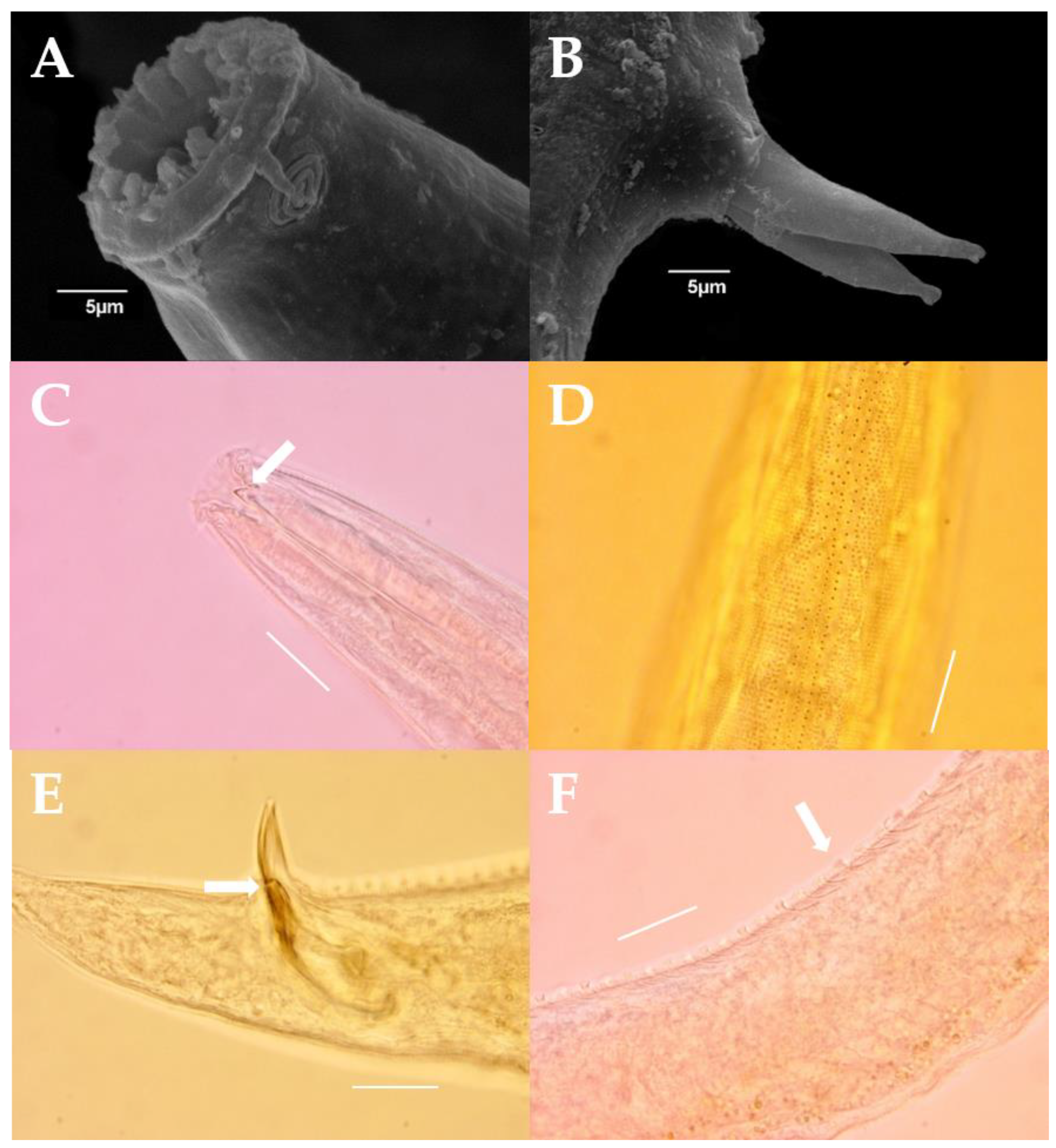
Male: Body roughly cylindrical, gradually growing from the anterior end to the nerve ring and then tapering from the anus to the tail (Figure 2A,B). Cuticle obviously ornamented with transverse rows of punctations at the whole body. Punctations aligned and laterally not differentiated until the posterior end of amphid. The lateral differentiation begins from under the amphid, with slightly larger punctations irregularly organized, three to four rows of punctations begin at nerve ring until tail region at the start point of cylindrical part (Figure 3D). Eight longitudinal rows of hypodermal pores present from anterior head end to the amphid body diameter depth until half of the tail (Figure 2C). Six inner labial setae short (only observed with SEM). Six outer labial setae and four cephalic setae present on the head and the same length, 4.5 μm long (head sensilla in the 6 + 10 pattern). Amphid located just below labial setae, multispiral with 2.5 turns, 6 μm wide (Figure 2C and Figure 3A). Buccal cavity conical shape with twelve distinct rugae (Figure 3A), armed with one large conspicuous dorsal tooth and two small subventral teeth (Figure 3C). Nerve ring situated slightly above the middle of the pharynx, 95 μm from anterior edge. Pharynx cylindrical, gradually expanding at two-thirds posterior region, 183 μm long. Excretory pore invisible. Ocelli absent. Reproduction system diorchic, opposed and outstretched. Spicule paired, curved, 58 μm long, slightly wider in the middle, shaped like a banana (Figure 3B,E). Gubernaculum paired, one on each side of the middle piece, 56 μm long (Figure 2D,G). The distal end of gubernaculum slightly extended from the upper-view, with a hook-like structure on the lateral distal part and some denticles on the inner half (Figure 2G). Seventy-six tubular precloacal supplemental organs, curved (Figure 2B and Figure 3F), with distal end strongly cuticularized and tubular part weakly cuticularized. Distance between each supplement gradually increases toward the anterior part. The most anterior and the most posterior supplement at distances 11 μm and 558 μm from the anus. Tail, 98 μm long, conico-cylindrical with a little swollen tip, the cylindrical part very short (Figure 2E).
Female: Similar to males in basic forms such as cuticle patterns, part of buccal cavity, and body shape; however, body length longer than males. Body length 1378–1401 μm. Didelphic, two opposed, reflexed ovaries (Figure 2A,F). Vulva located at 56–57% of body length, 776–800 μm from anterior end.

3.6. Molecular Analysis

The seven specimens of the new species used for molecular analysis were near identical in terms of mtCOI, 18S and 28S rRNA sequences. The K2P distance between the supposed intraspecies showed no difference from 18S and 28S regions (Tables S2 and S3), with mtCOI showing a difference of 0.39% (Table S4). All available sequences of congeneric species, as well as closely related genera such as Praecanthonchus, Acanthonchus and Cyatholaimus, were retrieved and compared against. In terms of mtCOI congeneric K2P distance, the new species differed from other congeneric species such as P. macrodon, P. caecus and P. gynodiporata by a range of 29.9–40% (Table S4). For 18S rRNA, the new species were compared to congeners such as P. macrodon, P. caecus and P. gynodiporata, as well as specimens only identified at genus level, and their distance differed by a range of 3.9–8.1% (Table S2). For 28S rRNA, new species were compared to congeners such as P. caecus, P. gynodiporata and P. miltommatus, which differed by a range of 17.7–36.8%, respectively (Table S3). When compared to other genera belonging to the family Cyatholaimidae, mtCOI, 18S and 28S differed by ranges of 32.8–42%, 4.9–14.8% and 14.6–22.4%, respectively.
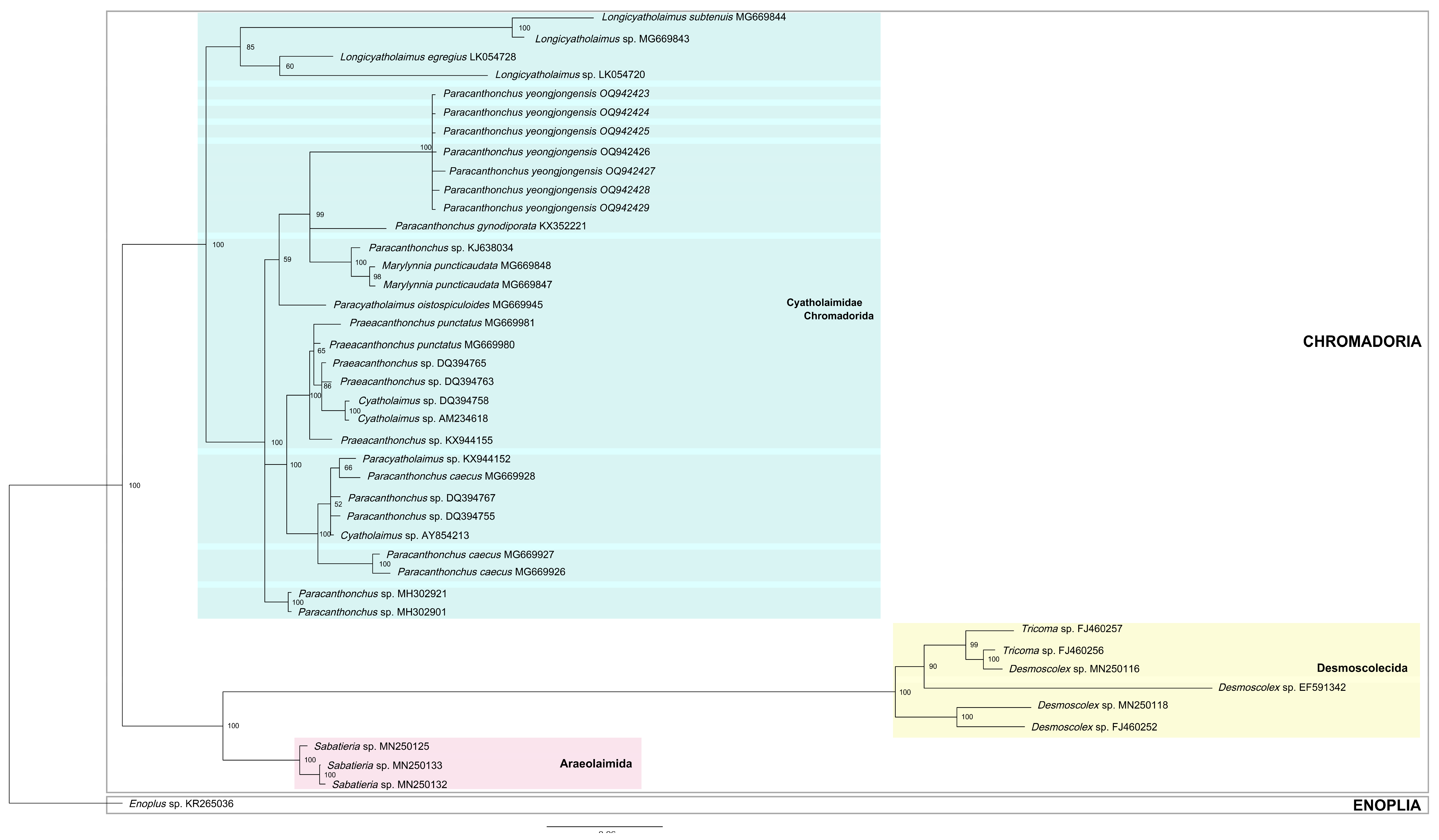
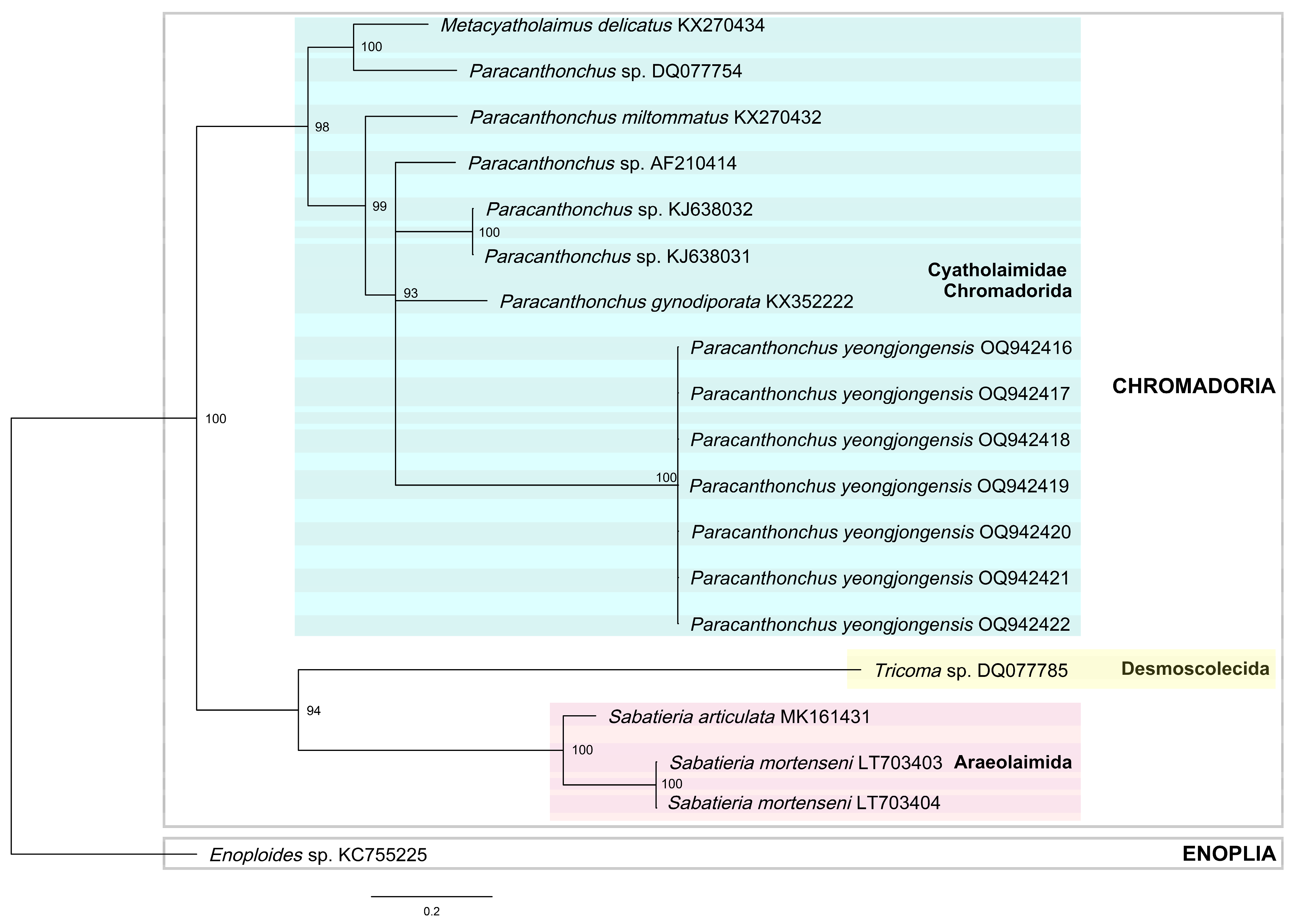
BI trees (Figure 4 and Figure 5) and ML trees (Figures S1 and S2) based on the 18S and 28S rRNA region, respectively, both produced trees with identical topology, albeit with different support values. All major nodes leading up to the family Cyatholaimidae had high posterior probability or UFboot support. As BI trees are identical in topology to ML trees with better support, BI trees of 18S and 28S rRNA sequences were used for discussion, with ML trees of 18S and 28S rRNA sequences being included as supplementary figures for reference. The sequence availability was significantly higher for 18S rRNA sequences compared to 28S rRNA sequences. The 18S rRNA phylogenetic tree included various genera belonging to the family Cyatholaimidae, such as other congeneric Paracanthonchus species, Longicyatholiamus, Marylynnia, Cyatholaimus and Praeacanthonchus, whereas the 28S rRNA tree only had other congeneric Paracanthoncus species and a single Metacyatholaimus sequence. All species belonging to the family Cyatholaimidae formed a monophyletic clade in both 18S and 28S trees with high support (pp = 100 and 98%, respectively) (Figure 4 and Figure 5). All seven sequences of the new species were included within this clade, and were seen grouped together with pp = 100% in both trees.

4. Discussion

4.1. List of Valid Species

Paracanthonchus is the most diverse and the largest genus within the family Cyatholaimidae [2]. The species identification of taxa belonging to the family Cyatholaimidae has been difficult, mainly due to their lack of apomorphic characters without clear interspecific/intergeneric boundaries. Many papers have discussed the morphological ambiguity of the genus Paracanthonchus [5,10], which stems from its type species, P. caecus. Paracanthonchus caecus has been mentioned in over a hundred papers, being reported in almost every marine habitat. Realizing this, Vincx et al., 1982, examined these highly variable species and determined that there are at least five different types of P. caecus and recognized four separate species from these types: P. heterodontus, P. gerlachi, P. thaumasius and P. canadensis. In addition to cases of species complex, many species have been transferred in and out of the genus [10,11,12,13,14]. To account for these frequent changes, many updated revisions of the genus exist. The most recent update to the genus was by Miljutina and Miljutin 2015, but the number of valid species has been inconsistent in most papers which followed [8,17,18]. Whether due to discrepancy or a lack of updated revisions, there is a clear need to re-evaluate the currently existing species to avoid any further convolution of an already complex genus. Accordingly, the species list was updated by referring to Miljutina and Miljutin 2015, NeMys, and original descriptions, as well as redescriptions. For the sake of clarity, any species already mentioned as being invalid in previous reviews were removed without remarks. Species which are ambiguous or require status updates are discussed below. After careful re-evaluation, from Miljutina and Miljutin, 2015,′s 52 valid species (excluding species inquirenda), we list a total of 60 species to be valid, including the new species.
Since Miljutina and Miljutin, 2015, seven species (including the new species) have been added to the genus: P. brevicaudatus, P. gynodiporata, P. miltommatus, P. olgae, P. secures, P. wellsi and P. yeongjongensis. Aside from newly described species, there are several species which must be discussed in terms of validity. Firstly, P. medius and P. sandspitensis were missing from Miljutina and Miljutin, 2015,′s revision, for no particular reason. The original descriptions have been checked, but with no apparent conflicts, we are considering the two species as valid. P. inglisi was described by Coles, 1965; however, it was considered synonymous to Praeacanthonchus by Platt and Warrick, 1988, as it bears unpaired gubernaculum. Despite rightfully being transferred to Praeacanthonchus, the species was still considered valid in the recent revision by Miljutina and Miljutin, 2015. P. steueri paracaecus (Micoletzky, 1922) was classified as a subspecies of P. steueri by the dark and pale differences in the color of the ocelli. While it may have been fine to erect subspecies based on minor morphological variation in 1922, it certainly is not acceptable now to erect subspecies without molecular evidence. While subspecies may differ in minor morphological variation, it should be noted that the specimens have been fixed in ethanol for over fourteen years. Color variation can be very subjective, and ethanol samples are highly prone to discoloration. Until molecular evidence becomes available, it is our decision to consider this species synonymous with its parent species, P. steueri. For P. tumepapillatus Timm, 1957, which was synonymized as Paracyatholaimus in 1966 by Murphy based on the structure of precloacal supplements (by comparison of the depiction), we agree with Miljutina and Miljutin, 2015, in reinstating the species as a valid species of Paracanthonchus. We agree with previous claims that the shape of the supplements is a characteristic more suitable for Paracanthonchus.
The list of valid species up until this point mainly relies on morphological accounts, with the exception of some species (P. caecus, P. gynodiporata, P. macrodon, P. mamubiae and P. miltommatus) which have substantiating molecular data. With much uncertainty and ambiguity in discerning species of this group morphologically, corroborating molecular data on existing and new species will help to organize and understand the species delineation of Cyatholaimidae.

4.2. Differential Morphological Diagnoses

The new species, Paracanthonchus yeongjongensis, is identified by genus characteristics: it has a buccal cavity armed with one large dorsal tooth and subventral teeth, precloacal tubular supplements and the gubernaculum paired distally expanded. Paracanthonchus yeongjongensis differs from other species by considering the number of supplements, the arrangement form and median pieces of the gubernaculum. Tubular supplement is one of the major characteristics of the genus; with the exception of two species (P. multisupplementatus and P. multitubifer), the rest of the congeners bear fewer than ten supplements. Evidently, P. yeongjongensis has a lot of supplements (76). It is evident from the tabular key (Table 3) that there are seven congeneric species similar to the new species in lacking a cusp on the distal end of the gubernaculum (P. longispiculum, P. major, P. micoletzkyi, P. mutantus, P. platypus, P. quinquepapillatus and P. ruens), and two species with a similar number of supplements (P. multisupplementatus and P. multitubifer). The new species is most closely related to P. multisupplementatus in terms of the number of supplements (76 vs. 57–62), lateral differentiation, the size of amphid and body length. However, it differs in the shape of the gubernaculum and the presence of cusp (none vs. numerous), the length interval between supplements (gradually increasing vs. equal) and its tail shape. It is also closely related to P. longispiculum in size and turns of amphid, and the shape of the distal part of the gubernaculum, but differs in body length (1186–1295 vs. 1780–1800) and number of supplements (76 vs. 8). Only P. elongatus has a median piece at the gubernaculum like the new species; however, it differs in most other morphological traits.

4.3. Phylogeny and Topology

While all available congeneric sequences were retrieved and used for the K2P analysis, some sequences of 18S (such as P. wellsi) had to be omitted from the analysis due to them also being short partial sequences targeting different region of 18S, with no overlap. Only sequences that correctly aligned with the seven sequences were used for further analysis. All three markers showed very low intraspecific K2P distances (0–0.3%), indicating the seven specimens analyzed here belong to the same species (Tables S2–S4). The usual interspecific difference for the mtCOI gene for nematodes is 5%, which is often used as a threshold to detect cryptic species [31]. Our results showed that mtCOI K2P distances between other congeners ranged from 29.9 to 40%, and from 32.8 to 42% between species of other genera of the family Cyatholaimidae (Table S4). Similarly, little difference in range was observed between the congeneric distance and intergeneric distance (different genera belonging to the family Cyatholaimidae) among 18S and 28S rRNA sequences (3.9–8.1% vs. 4.9–14.8%; 17.7–36.8% vs. 14.6–22.4%, respectively) (Tables S2 and S3). This is indicative of the fact that while the family Cyatholaimidae may be a monophyletic clade, its constituting genera are clearly not. When considering the availability of molecular data, it seems that 18S rRNA sequences are most readily available compared to mtCOI and 28S rRNA. However, there is no definite guideline as to which marker is most suitable for each group. Additionally, there have been instances, such as P. gynodiporata, where species show high morphological variation and dispersion, all whilst showing low genetic differentiation [55]. This goes to show that there is no one single data type that completely reflects inter/intra species delineation. It is thus imperative to make it common practice to obtain as much datatype (to supplement morphological data with molecular data of variety of markers) as possible to be able to resolve a complex genus such as Paracanthonchus.
While there are not large amounts of molecular data available for species belonging to Paracanthonchus, there were enough to build phylogenetic trees to gain some insight on their supposed position within the phylum Nematoda. Much like previous documentations [1,2,3], the family Cyatholaimidae did form a monophyletic group within the class Chromadorea in the BI tree based on 18S and 28S rRNA sequences with a high posterior probability of 100% (Figure 4 and Figure 5). However, its constituting genera formed mixed and low-support paraphyletic clades. This clearly questions the integrity of constituting genera within Cyatholaimidae and raises the need to re-evaluate highly ambiguous genera such as Paracanthonchus and Praeacanthonchus. In the latest classification of the phylum Nematoda by Hodda, 2022, the family Cyatholaimidae consisted of just two subfamilies: Cyatholaiminae and Paracanthonchinae. This was supported (although with low support) by a tree constructed by Leduc and Zhao, 2018, where the two subfamilies were seen forming two sister monophyletic clades. They did, however, mention the genus Praecanthonchus as being the sole exception. Their phylogenetic tree showed Praecanthonchus being included in the opposite subfamily, and based on this finding suggested that Praecanthonchus be placed in the subfamily Paracanthonchinae. Several years have passed since their publication, but species lists on NeMys as well as the classification of Hodda, 2022, still list Praecanthonchus as a constituent of the subfamily Cyatholaiminae. While Hodda, 2022, does acknowledge the suggestion of Leduc and Zhao, 2018, he argues that it is with a low level of support and with sparse taxon sampling. In the SSU phylogenetic tree built by Leduc and Zhao, 2018, it is evident that Cyatholaimus is also seen as an exception, like Praecanthonchus; however, they argued that Cyatholaimus itself is a very vague group which has been transferred in and out of different groups, such as Acanthonchus, Metacyatholaimus, Paracanthonchus and Longicyatholaimus. Based on this reason, they claimed that little credibility can be asserted to Cyatholaimus sequences. While this is true, to confirm the monophyletic nature of the two subfamilies we included more sequences of species belonging to the subfamily Cyatholaiminae, such as Cyatholaimus, Marylynnia and Longicyatholaimus, to our phylogenetic tree. However, many of the sequences retrieved from NCBI used in the phylogenetic tree were only identified to genus level, and erroneous species identification cannot be ruled out. Our BI 18S tree (Figure 4) showed that while Longicyatholaimus does form a monophyletic sister group (as the subfamily Cyatholaiminae) with the subfamily Paracanthonchinae, it is with pp of 85% and there is more than one exception: Cyatholaimus, Marylynnia and Praecanthonchus, all of which are known to belong to Cyatholaiminae, are included within the Paracanthonchinae clade. We could suggest, based on our molecular results, that the three genera be placed in Paracanthonchinae, but as with the case of Cyatholaimus, the validity of the sequences can be argued. We instead acknowledge that the existing subfamilies of Cyatholaimidae cannot be represented well with a molecular phylogenetic tree, at least currently, with limited molecular data. Most species of this group still lack associated molecular sequences, and even those with available sequences are mostly partial without additional marker regions. As more molecular data accumulate over time, a more complete phylogeny may unearth insights that can be used to resolve these ambiguous genera/families. Granted, morphology and molecular phylogenies may not always match perfectly [77,78], but it is clear that no one datatype can resolve the issue alone. Both morphological and molecular results must be used to substantiate one another. The morphological distinction of the two subfamilies by the type of gubernaculum (unpaired/paired), may not be represented well phylogenetically, but it is still, nonetheless, currently the most distinctive group-defining character. It is uncertain whether complex topology of the group can be resolved by morphological or molecular means, as both are full of ambiguity and lack data. In all, a lack of apomorphic morphological traits and molecular data makes it extremely difficult to resolve this group, currently, but as more datatypes begin to accumulate, hopefully the topology of this group may become clearer in the future.

Supplementary Materials

The following supporting information can be downloaded at: https://www.mdpi.com/article/10.3390/d15050664/s1, Figure S1: Rooted maximum likelihood phylogenetic tree of 18S rRNA gene with Enoplus sp. set as an outgroup (with UFboot support shown at each node); Figure S2: Rooted maximum likelihood phylogenetic tree of 28S rRNA gene with Enoploides sp. set as an outgroup (with UFboot support shown at each node); Table S1. GenBank accession number of mtCOI, 18S and 28S rRNA sequences obtained and used in this study; Table S2. Kimura 2-parameter distance between other congeneric species as well as closely related genera based on 18S rRNA alignment with 1000 bootstrap. Standard deviation marked in blue; Table S3. Kimura 2-parameter distance between other congeneric species as well as closely related genera based on 28S rRNA alignment with 1000 bootstrap. Standard deviation marked in blue; Table S4. Kimura 2-parameter distance between other species belonging to the genus Paracanthonchus and Praeacanthonchus based on mtCOI alignment with 1000 bootstrap. Standard deviation marked in blue.

Author Contributions

Conceptualization, H.K., R.J. and W.L.; methodology, H.K.; software, R.J.; validation, H.K., R.J. and W.L.; formal analysis, H.K. and R.J.; investigation, H.K.; resources, H.K. and R.J.; data curation, H.K., R.J. and W.L.; writing—original draft preparation, H.K. and R.J.; writing—review and editing, H.K., R.J. and W.L.; visualization, H.K. and R.J.; supervision, R.J.; project administration, H.K. and R.J.; funding acquisition, H.K., R.J. and W.L. All authors have read and agreed to the published version of the manuscript.

Funding

This research was funded by the National Institute of Biological Resources (NIBR), by the Ministry of Environment (MOE) of the Republic of Korea (NIBR202304103), and by grant (NRF-2021R1I1A1A01040377) from the National Research Foundation of Korea.

Institutional Review Board Statement

Not applicable.

Data Availability Statement

The voucher specimens examined within this study were deposited in the National Institute of Biological Resources (NIBR), Korea. Partial sequences of mtCOI, 18S, and 28S rRNA were deposited in GenBank. The GenBank accession numbers are listed in Table S1.

Conflicts of Interest

The authors declare no conflict of interest. The funders had no role in the design of the study; in the collection, analyses, or interpretation of data; in the writing of the manuscript, or in the decision to publish the results.

Abbreviations

a—body length divided by maximum body diameter; abd—anal body diameter, (µm); amp—transversal diameter of amphid, (µm); amp’—diameter of amphid divided by corresponding body diameter; amp cbd—corresponding body diameter at the level of amphid, (µm); b—body length divided by esophagus length; bcl—distance from anterior edge to base of buccal cavity; c—body length divided by tail length; c’—tail length divided by anal body diameter; cyln—length of cylindrical tail portion (µm); CSL—cephalic sensilla length, (µm); das—distance from anus to most anterior supplement; dps—distance from anus to most posterior supplement; EL—distance from anterior edge to excretory pore, (µm); hd—head diameter, (µm); L—total body length, (µm); LSL—outer labial sensilla length, (µm); mbd—maximum body diameter, (µm); NL—distance from anterior edge to nerve ring, (µm); na—number of turns in amphid; ns—number of supplements; ncbd—corresponding body diameter at the level of nerve ring, (µm); PL—pharynx length, (µm); pcbd—corresponding body diameter at base of pharynx, (µm); s’—spicule length as arc length divided by anal body diameter; spic—spicule length as arc, (µm); gub—gubernaculum length as arc, (µm); TL—tail length, (µm); V—vulva distance from anterior end divided by total body length; VL—distance from anterior end to vulva, (µm).

References

  1. Leduc, D.; Zhao, Z. Phylogenetic relationships within the Cyatholaimidae (Nematoda: Chromadorida), the taxonomic significance of cuticle pore and pore-like structures, and a description of two new species. Mar. Biodivers. 2018, 48, 217–230. [Google Scholar] [CrossRef]
  2. Hodda, M. Phylum Nematoda: A classification, catalogue and index of valid genera, with a census of valid species. Zootaxa 2022, 5114, 1–289. [Google Scholar] [CrossRef] [PubMed]
  3. Miljutina, M.; Miljutin, D. A revision of the genus Paracanthonchus (Cyatholaimidae, Nematoda) with a tabularkey to species and a description of P. mamubiae sp. n. from the deep North-Western Pacific. Deep.-Sea Res. Pt. II 2015, 111, 104–118. [Google Scholar]
  4. Cunha, B.P.; Fonseca, G.; Amaral, A.C.Z. Diversity and Distribution of Cyatholaimidae (Chromadorida: Nematoda): A Taxonomic and Systematic Review of the World Records. Front. Mar. Sci. 2022, 9, 836670. [Google Scholar] [CrossRef]
  5. Vincx, M.; Sharma, J.; Smol, N. On the Identity and Nomenclature of Paracanthonchus caecus (Bastian, 1865), with a Redefinition of the Genus Paracanthonchus Micoletzky (Nematoda, Cyatholaimidae). Zool. Scr. 1982, 11, 243–263. [Google Scholar] [CrossRef]
  6. Filipjev, I.N. Free-living marine nematodes of the Sevastopol area. Trans. Zool. Lab. Sevastopol. Biol. Stat. Rus. Acad. Sci. 1918, 2, 1–255. [Google Scholar]
  7. De Coninck, L.A.; Schuurmans-Stekhoven, J.H. The freeliving marine nemas of the Belgian Coast. II With general remarks on the structure and the system of nemas. Mém. Mus. R. Hist. Nat. Belg. 1933, 58, 3–163. [Google Scholar]
  8. Bezerra, T.; Eisendle, U.; Hodda, M.; Holovachov, O.; Leduc, D.; Mokievsky, V. Nemys: World Database of Nematodes. 2022. Available online: http://nemys.ugent.be (accessed on 1 February 2023).
  9. Micoletzky, H. Letzter Bericht über freilebende Nematoden aus Suez. Sber. Akad. Wiss. Wien (I) 1924, 133, 137–179. [Google Scholar]
  10. Wieser, W. Free-Living Marine Nematodes II. Chromadoroidea; Gleerup: Lund, Sweden, 1954; Volume 50, pp. 1–148. [Google Scholar]
  11. Stekhoven, J.H. The free-living marine nemas of the Mediterranean. I. The Bay of Villefranche. Mem. Inst. R. Sci. Nat. Belg. Ser. 1950, 2, 1–220. [Google Scholar]
  12. Hope, W.D.; Murphy, D.G. A taxonomic hierarchy and checklist of the genera and higher taxa of marine nematodes. Smithson. Contrib. Zool. 1972, 137, 1–101. [Google Scholar] [CrossRef]
  13. Mawson, P.M. Some marine freeliving nematodes from the Australian coast. Trans. R. Soc. S. Aust. 1953, 76, 34–40. [Google Scholar]
  14. Wieser, W. Free-Living Nematodes and Other Small Invertebrates of Puget Sound Beaches; University of Washington Press: Seattle, WA, USA, 1959; pp. 1–179. [Google Scholar]
  15. Tchesunov, A. Order Chromadorida Chitwood, 1933. In Handbook of Zoology; Schmidt-Rhaesa, A., Ed.; De Gruyter: Berlin, Germany, 2014; Volume 2, pp. 373–398. [Google Scholar]
  16. Platt, H.; Warwick, R. Free-Living Marine Nematodes; Part 2: British Chromadorids; Brill, E.J., Backhuys, W., Eds.; Linnean Society of London: London, UK; Estuarine and Brackish-water Sciences Association: Leiden, The Netherlands, 1988; p. 502. [Google Scholar]
  17. Tchesunov, A. Free-living nematode species (Nematoda) dwelling in hydrothermal sites of the North Mid-Atlantic Ridge. Helgoland Mar. Res. 2015, 69, 343–384. [Google Scholar] [CrossRef]
  18. Lee, H.; Jung, J.; Rho, H. Two unrecorded marine nematode species of Paracanthonchus (Nematoda: Cyatholaimidae) from the East Sea of Korea. J. Species Res. 2016, 5, 503–513. [Google Scholar] [CrossRef]
  19. Kim, H.; Tchesunov, A.; Lee, W. A new species of the genus Marylynnia (Nematoda: Chromadorida: Cyatholaimidae) from Gwangyang Bay, Korea. Proc. Biol. Soc. Wash. 2015, 128, 227–238. [Google Scholar] [CrossRef]
  20. Jeong, R.; Tchesunov, A.; Lee, W. A new species of the genus Thalassironus (Nematoda: Enoplida: Ironidae) from the coasts of South Korea. Zootaxa 2019, 4563, 516–530. [Google Scholar] [CrossRef]
  21. Tchesunov, A.; Jeong, R.; Lee, W. Two New Marine Free-Living Nematodes from Jeju Island Together with a Review of the Genus Gammanema Cobb 1920 (Nematoda, Chromadorida, Selachinematidae). Diversity 2020, 12, 19. [Google Scholar] [CrossRef]
  22. Hong, J.; Tchesunov, A.; Lee, W. Revision of Cervonema Wieser, 1954 and Laimella Cobb, 1920 (Nematoda: Comesomatidae) with descriptions of two species from East Sea, Korea. Zootaxa 2016, 4098, 333–357. [Google Scholar] [CrossRef]
  23. Gagarin, V.; Nguyen Vu Thanh. Paracanthonchus brevicaudatus sp. n. (Nematoda, Chromadorida, Cyatholaimidae) from artificial reservoirs in Vietnam. Int. J. Nematol. 2016, 26, 9–14. [Google Scholar]
  24. Allgén, C. Pacific Freeliving Marine Nematodes. (Papers from Dr. Th. Mortensen’s Pacific Expedition 1914–16. LXXVI). Vidensk. Meddel. Naturhist. Foren. Kjobenhavn 1951, 113, 263–411. [Google Scholar]
  25. Huang, Y.; Xu, K. Two new free-living nematode species (Nematoda: Cyatholaimidae) from intertidal sediments of the Yellow Sea, China. Cah. Biol. Mar. 2013, 54, 1–10. [Google Scholar]
  26. Kito, K. Studies on the free-living marine nematodes from Hokkaido, IV. J. Fac. Sci. Hokkaido Univ. Ser. VI Zool. 1981, 22, 250–278. [Google Scholar]
  27. Gagarin, V. Paracanthonchus multisupplementatus sp.n. and Cyatholaimus minor sp.n. (Nematoda) from the coast of Vietnam. Zootaxa 2012, 3392, 60–68. [Google Scholar] [CrossRef]
  28. Nguyen Dinh Tu; Gagarin, V. Two new species of nematodes of the order chromadorida (Nematoda: Chromadorea) from Vietnam. Russ. J. Mar. Biol. 2018, 44, 86–95. [Google Scholar]
  29. Hopper, D. Drawing and measuring nematodes. In Laboratory Methods for Work with Plant and Soil Nematodes; Ministry of Agriculture, Fisheries and Food: Her Majesty’s Stationery Office: London, UK, 1970; pp. 59–65. [Google Scholar]
  30. Schindelin, J.; Arganda-Carreras, I.; Frise, E.; Kaynig, V.; Longair, M.; Pietzsch, T.; Preibisch, S.; Rueden, C.; Saalfeld, S.; Schmid, B.; et al. Fiji: An Open-Source Platform for Biological-Image Analysis. Nat. Methods 2012, 9, 676–682. [Google Scholar] [CrossRef] [PubMed]
  31. Derycke, S.; Vanaverbeke, J.; Rigaux, A.; Backeljau, T.; Moens, T. Exploring the Use of Cytochrome Oxidase c Subunit 1 (COI) for DNA Barcoding of Free-Living Marine Nematodes. PLoS ONE 2010, 5, e13716. [Google Scholar] [CrossRef] [PubMed]
  32. Bowles, J.; Bowles, J.; Blair, D.; McManus, D.P. Genetic variants within the genus Echinococcus identified by mitochondrial DNA sequencing. Mol. Biochem. Parasitol. 1992, 54, 165–173. [Google Scholar] [CrossRef]
  33. Carta, L.K.; Li, S. Improved 18S small subunit rDNA primers for problematic nematode amplification. J. Nematol. 2018, 50, 533–542. [Google Scholar] [CrossRef] [PubMed]
  34. Carta, L.K.; Li, S. PCR amplification of a long rDNA segment with one primer pair in agriculturally important nematodes. J. Nematol. 2019, 51, e2019–e2026. [Google Scholar] [CrossRef]
  35. De Ley, P.; De Ley, I.M.; Morris, K.; Abebe, E.; Mundo-Ocampo, M.; Yoder, M.; Heras, J.; Waumann, D.; Rocha-Olivares, A.; Jay Burr, A.H.; et al. An integrated approach to fast and informative morphological vouchering of nematodes for applications in molecular barcoding. Phil. Trans. R. Soc. B 2005, 360, 1945–1958. [Google Scholar] [CrossRef]
  36. Thompson, J.D.; Higgins, D.G.; Gibson, T.J. CLUSTAL W: Improving the sensitivity of progressive multiple sequence alignment through sequence weighting, position-specific gap penalties and weight matrix choice. Nucleic Acids Res. 1994, 22, 4673–4680. [Google Scholar] [CrossRef]
  37. Tamura, K.; Stecher, G.; Kumar, S. MEGA11: Molecular evolutionary genetics analysis version 11. Mol. Boil. Evol. 2021, 38, 3022–3027. [Google Scholar] [CrossRef]
  38. Altschul, S.F.; Gish, W.; Miller, W.; Myers, E.W.; Lipman, D.J. Basic local alignment search tool. J. Mol. Boil. 1990, 215, 403–410. [Google Scholar] [CrossRef]
  39. Kimura, M. A simple method for estimating evolutionary rates of base substitutions through comparative studies of nucleotide sequences. J. Mol. Evol. 1980, 16, 111–120. [Google Scholar] [CrossRef]
  40. Minh, B.Q.; Schmidt, H.A.; Chernomor, O.; Schrempf, D.; Woodhams, M.D.; von Haeseler, A.; Lanfear, R. IQ-TREE 2: New models and efficient methods for phylogenetic inference in the genomic era. Mol Biol Evol. 2020, 37, 1530–1534. [Google Scholar] [CrossRef]
  41. Kalyaanamoorthy, S.; Minh, B.Q.; Wong, T.K.F.; von Haeseler, A.; Jermiin, L.S. ModelFinder: Fast model selection for accurate phylogenetic estimates. Nat. Methods 2017, 14, 587–589. [Google Scholar] [CrossRef]
  42. Minh, B.Q.; Nguyen, M.A.T.; von Haeseler, A. Ultrafast approximation for phylogenetic bootstrap. Mol. Biol. Evol. 2013, 30, 1188–1195. [Google Scholar] [CrossRef] [PubMed]
  43. Darriba, D.; Taboada, G.L.; Doallo, R.; Posada, D. jModelTest 2: More models, new heuristics and high-performance computing. Nat. Methods 2012, 9, 772. [Google Scholar] [CrossRef] [PubMed]
  44. Huelsenbeck, J.P.; Ronquist, F. Mrbayes: Bayesian inference of phylogenetic trees. Bioinformatics 2001, 17, 754–755. [Google Scholar] [CrossRef]
  45. Rambaut, A. FigTree Version 1.4.4. 2018. Available online: http://tree.bio.ed.ac.uk/software/figtree/ (accessed on 1 March 2023).
  46. Inglis, W. Marine nematodes from Banyuls-sur-mer: With a review of the genus Eurystomina. Bull. Br. Mus. Nat. Hist. 1962, 8, 209–287. [Google Scholar]
  47. Gerlach, S. Die Nematodenfauna des Sandstrandes an der Küste von Mittelbrasilien (BrasilianischeMeeres-NematodenIV). Mitt. Zool. Mus. Berl. 1957, 33, 411–459. [Google Scholar] [CrossRef]
  48. Kreis, H. Die freilebenden marinen Nematoden der Spitzbergen-Expedition von F. Roemer und F. Schaudinnim Jahre 1898. Mitt. Zool. Mus. Berl. 1928, 14, 131–197. [Google Scholar]
  49. Schiemer, F.; Jensen, P.; Riemann, F. Ecology and systematics of free-living nematodes from the Bothnian Bay, northern Baltic Sea. Ann. Zool. Fenn. 1983, 20, 277–291. [Google Scholar]
  50. Bussau, C. Taxonomische und ökologische Untersuchungen an Nematoden des Peru-Beckens. Ph.D. Thesis, University of Kiel, Kiel, Germany, 1993. [Google Scholar]
  51. Bastian, H.C. Monograph on the Anguillulidae, or free nematoids, marine, land, and fresh water; with descriptions of 100 new species. Trans. Linn. Soc. Lond. 1865, 25, 73–184. [Google Scholar] [CrossRef]
  52. Inglis, W.G. Cyatholaimidae (Nematoda) from the coast of Western Australia. Rec. S. Austr. Mus. 1970, 16, 1–13. [Google Scholar]
  53. De Man, J.G. Sur quelques espèces nouvelles ou peu connues de nématodes libres vivant sur les côtes de la Zélande. Tijdschr. Ned. Dierk. Vereen. 1907, 10, 227–244. [Google Scholar]
  54. Filipjev, I.N. Encore sur les Nématodes libres de la mer Noire. Tr. Stravrop. Skh. Inst. Zool. 1922, 1, 83–184. [Google Scholar]
  55. Oliveira, D.A.S.; de Decraemer, W.; Moens, T.; Dos Santos, G.A.P.; Derycke, S. Low genetic but high morphological variation over more than 1,000 km coastline refutes omnipresence of cryptic diversity in marine nematodes. BMC Evolution. Biol. 2017, 17, 71. [Google Scholar] [CrossRef]
  56. Schulz, E. Beitrage zur Kenntnis mariner Nematoden aus der Kieler Bucht. Zool. Jb. (Syst.) 1932, 62, 331–430. [Google Scholar]
  57. Gourbault, N. Nématodes abyssaux (Campagne Walda du navire océanographique Jean-Charcot). I. Espèces nouvelles de Cyatholaimidae. Cah. Biol. Mar. 1980, 21, 61–71. [Google Scholar]
  58. Gagarin, V.G.; Klerman, A.K. Two new species of free-living nematodes (Nematoda) from the Mediterranean Sea. Zool. Bespozvon. 2008, 5, 1–7. [Google Scholar]
  59. Warwick, R.M. The Cyatholaimidae (Nematoda, Chromadoroidea) off the coast of Northumberland. Cah. Biol. Mar. 1971, 12, 95–110. [Google Scholar]
  60. Pastor de Ward, C.T. Free-living nematodes of the Deseado River estuary (Chromadoroidea: Chromadoridae, Etholaimidae, Cyatholaimidae and Choniolaimidae) Santa Cruz, Argentina. 5. Cent. Nac. Patagon. Publ. Espec. 1985, 6, 1–83. [Google Scholar]
  61. Allgén, C. Über einige, meistens neue, freilebende marine Nematoden aus dem Feuerland-Archipel (Schwedische Südpolar-Expedition 1901–1903). Zool. Anz. 1959, 163, 222–243. [Google Scholar]
  62. Galtsova, V.V. Free-living marine nematodes as components of the meiofauna of the Chupa Bay, White Sea. Issled. Fauny Morej 1976, 15, 165–270. [Google Scholar]
  63. Stekhoven, J.H. Freilebende marine Nematoden des Mittelmeeres. IV. Freilebende marine Nematoden der Fischereigründe bei Alexandrien. Zool. Jb. (Syst.) 1943, 76, 323–378. [Google Scholar]
  64. Timm, R.W. The marine nematodes of the Bay of Bengal. Proc. Pak. Acad. Sci. 1961, 1, 25–88. [Google Scholar]
  65. Decraemer, W.; Coomans, A.C. Scientific report on the Belgian expedition to the Great Barrier Reefin 1967. Nematodes, XIII. A description of four new species and a redescription of four known species from in and around mangroves on LizardIsland. Aust. J. Mar. Freshw. Res. 1978, 29, 509–541. [Google Scholar] [CrossRef]
  66. Jayasree Vadhyar, K. A new species of Paracanthonchus Micoletzky, 1924 and a known species of Paracyatholaimus Micoletzky, 1922 (Nematoda, Cyatholaimidae, Paracanthonchinae) from polluted intertidal sand in Scotland. Cah. Bid. Mar. 1980, 21, 375–381. [Google Scholar]
  67. Wieser, W.; Hopper, B. Marine nematodes of the east coast of North America. I. Florida. Bull. Mus. Comp. Zool. Harv. 1967, 135, 239–344. [Google Scholar]
  68. Bouwman, L.A. A survey of nematodes from the Ems estuary. Part I: Systematics. Zool. Jb. (Syst.) 1981, 108, 335–385. [Google Scholar]
  69. Nasira, K.; Kamran, M.K.; Shanina, F. Two new species of free-living marine nematodes of the family Cytaholaimidae (Nematoda: Chromadorida) from the Arabian sea of Pakistan. Pak. J. Nematol. 2007, 25, 91–98. [Google Scholar]
  70. Allgén, C. Freilebende marine Nematoden von der Stateninsel (Feuerland-Archipel). II. Zool. Anz. 1930, 90, 27–38. [Google Scholar]
  71. Micoletzky, H. Neue freilebende Nematoden aus Suez. Sber. Akad. Wiss. Wien. 1922, 131, 77–103. [Google Scholar]
  72. Allgén, C. Die freilebenden Nematoden des Mittelmeers. Zool. Jb. (Syst.) 1942, 76, 1–102. [Google Scholar]
  73. Timm, R.W. New Marine Nematodes from St. Martin’s Island. Pak. J. Sci. 1957, 9, 133–138. [Google Scholar]
  74. Brunetti, B. Contributo alla conoscenza dei nematode del M. Tirreno. II. Alcune specie appartenenti alle famiglie: Enoplidae, Cyatholaimidae, Chromadoridae, Axonolaimidae. Monit. Zool. Ital. 1949, 57, 41–59. [Google Scholar]
  75. Gerlach, S.A. Die Nematodenbesiedlung des Sandstrandes und des Küstengrundwassers an der italienischen Küste. I. Systematischer Teil. Archo Zool. Ital. 1953, 37, 517–640. [Google Scholar]
  76. Leduc, D.; Zhao, Z.Q. The Marine Biota of Aotearoa New Zealand; Ngā toke o Parumoana: Common free-living Nematoda of Pāuatahanui Inlet, Te-Awarua-o-Porirua Harbour, Wellington; NIWA Biodiversity Memoir: Auckland, New Zealand, 2023; Volume 135, p. 212. [Google Scholar]
  77. Patterson, C.; Williams, D.M.; Humphries, C.J. Congruence between molecular and morphological phylogenies. Annu. Rev. Ecol. Syst. 1993, 24, 153–188. [Google Scholar] [CrossRef]
  78. Cohen, B.L. Match and mismatch of morphological and molecular phylogenies: Causes, implications, and new light on cladistics. Zool. J. Linn. Soc. 2018, 184, 516–527. [Google Scholar] [CrossRef]
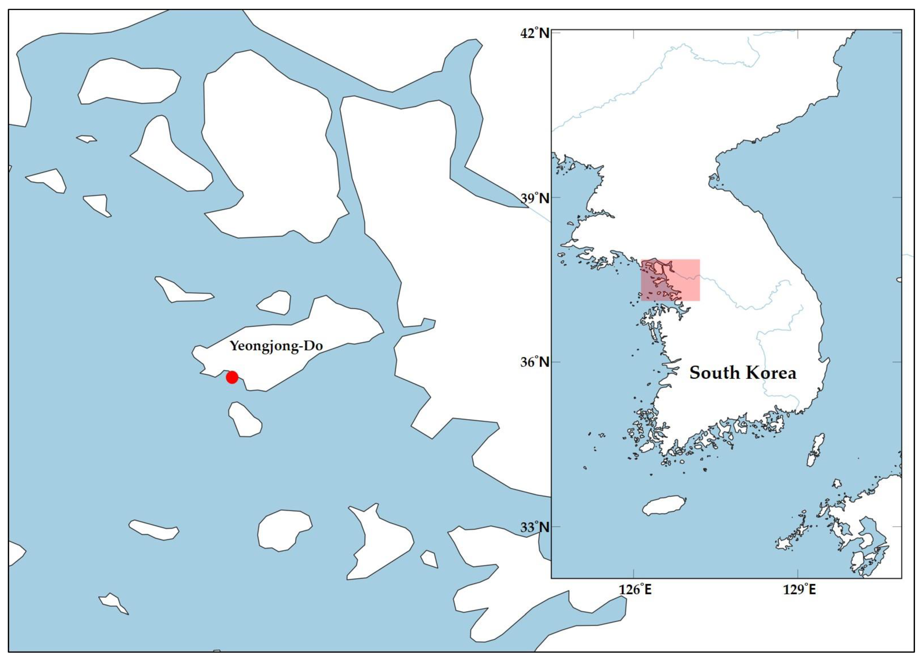
Figure 1. Map of the sample locality. The red box indicates region zoomed-in on the map; The red dot indicates the type locality of the new species. The map was made with QGIS v. 3.28.2 (downloaded from https://qgis.org/en/site/forusers/download.html, accessed on 3 February 2023).
Diversity 15 00664 g001
Figure 2. Paracanthonchus yeongjongensis sp. nov.: (A), female habitus (paratype 1); (B), male habitus (holotype); (C), female anterior part (paratype 2); (D), male cloacal region (holotype); (E), female tail region (paratype 2); (F), female reproductive system (paratype 1); (G), male spicule and gubernaculum, upper-side view. Scale bars: (AF) = 50 μm.
Diversity 15 00664 g002
Figure 3. Paracanthonchus yeongjongensis sp. nov.: SEM micrographs―(A) anterior part with amphid, (B) cloacal part with distal part of spicule; light micrographs―(C) head with dorsal tooth, (D) lateral differentiation of punctations at mid, (E) spicule and gubernaculum (arrow, cuticularized with distal part of gubernaculum), (F) tubular supplements. Scale bars: (A,B) = 5 μm; (CF) = 25 μm.
Diversity 15 00664 g003
Figure 4. Rooted Bayesian Inference phylogenetic tree of 18S rRNA gene with Enoplus sp. set as an outgroup (with posterior probability support shown at each node).
Diversity 15 00664 g004
Figure 5. Rooted Bayesian Inference phylogenetic tree of 28S rRNA gene with Enoploides sp. set as an outgroup (with posterior probability as percentage shown at each node).
Diversity 15 00664 g005
Table 1. Primer-related information used in this study.
MarkerPrimer (Direction)Sequence 5′-3′Amplification ConditionSequence Length (bp)Reference
mtCOIJB3 (f)TTTTTTGGGCATCCTGAGGTTTAT94 °C for 5 min, 5 cycles of 94 °C for 30 s, 54 °C for 30 s and 72 °C for 30 s, followed by 35 cycles of denaturation at 94 °C for 30 s, annealing at 50 °C for 30 s, and extension at 72 °C for 30 s, followed by final step of 72 °C for 10 min330Derycke et al., 2010 [31]; Bowles et al., 1992 [32]
JB5 (r)AGCACCTAAACTTAAAACATAATGAAAATG
18s18S-CL-F (f)TCAAAGATTAAGCCATGCAT95 °C for 3 min followed by 36 cycles of denaturation at 95 °C for 30 s, annealing at 50 °C for 45 s, and extension at 72 °C for 3 min, followed by final step at 72 °C for 7 min494Carta and Li, 2018; Carta and Li, 2019 [33,34]
530R ®GCGGCTGCTGGCACCACACTT
28SD2A (f)ACAAGTACCGTGAGGGAAAGTTG95 °C for 5 min followed by 37 cycles of denaturation at 95 °C for 30 s, annealing at 56 °C for 1 min, and extension at 72 °C for 1 min 30 s min, followed by final step at 72 °C for 5 min731–741 bpDe Ley et al., 2005 [35]
D®(r)TCGGAAGGAACCAGCTACTA
Table 2. Tabular key of valid Paracanthonchus species with morphological characters (amended from Miljutina and Miljutin, 2015).
SpeciesBody Length
(Male)
Body Length
(Female)
Lateral
Differentiation
of Cuticle
Number of
Subventral Teeth
Distance from
Anterior
to Amphid
Amphid WidthTurn Number
of Amphid
Spicule Length
as Arc
Gubernaculum LengthNumber of Cusps on
Distal End
of Gubernaculum
Number of SupplementsTail Shape
P. austrospectabilis1440–20901370–2280none/except on the tailn/a9134.56565numerous6conical
P. barka860 presencen/a57–84.5–5.5191824conical
P. batidus888 nonen/a65.52.53122n/a6conical
P. bipapillatus2046 nonenone10132.570.259.825conical
P. bothnicus1568–18001715–1784presence49106504545conical
P. brevicaudatus956–1230992–1413none31113–154.3–4.5302716–183conical
P. breviseta10921072nonen/a20145.544n/a14conical
P. bulbicola13401230–1960presence411156.253823numerous5conical
P. caecus976–1470presence2–4107–134.5–5.540–4835–448–95conical
P. canadensis960–11601089–1170none477639.1–40.635.5–40.6numerous5conical
P. cheynei1240–12801090none4149–104.2546–4839–42numerous6conical
P. cochlearis11231162presence210176363025conical–cylindrical
P. cristatus1050–13601180–1300presencen/a9–1382.75–34035n/a8–9conical
P. elongatus3025presencenone15154–586 (as chord)n/anumerous5conical
P. filipjevi11501300n/an/a794–5.5464123conical
P. gerlachi1194–13911045–1480none412107.538–393675conical
P. gynodiporata1001–11461075–1238presence411.3–12.97.4–10.7438.9–42.134.5–40.8numerous4conical
P. hartogi1240–1420 nonenone1512–133.2544–4939–4446conical
P. hawaiiensis1625 presencen/a1310340–4439–44numerous4conical
P. heterocaudatus1335–15701555–1750presence279–135–631–3225–2726conical–cylindrical
P. heterodontus1042–16681129–1842none413134.556–6746–54numerous5conical
P. kamui1658–17281761–1816presence46104.2553–6045–55numerous6conical
P. kartanum870–16001450none41593.5–4.523–3826–36numerous6conical
P. latens1733–2458none48106.584–8743–50numerous4conical
P. lissus1165–12521334nonen/a1010–124–4.539–4345–4944conical
P. longicaudatus1330–15101570–1790none4129–105.3–6.2543–5140–42numerous5conical–cylindrical
P. longispiculum1780–18001900n/a2116–102.5–370–7670none8conical
P. macrospiralis3000 presencen/a122036532n/an/aconical
P. major26082676nonen/an/a6.5–7.82.559.849.4none4conical
P. mamubiae1610–18311638–1971none43–77–144.1560–6546–51numerous3–5conical–cylindrical
P. margaretae1280–14801210presence41010–114.7547–4844–45numerous6conical
P. medius913–12541232nonen/an/an/an/a4844.4numerous4conical
P. micoletzkyi10201220presencen/a12124.54125none4conical
P. micropapillatus960–1350950–1400none/except on the tailn/a58.5–10.53.25–2.5342527conical
P. miltommatus1827–20511885–2062none/except on the tail47–1210–114.5–553–5748–555–86conical
P. multisupplementatus1027–13081181–1365presence2(4?)3.5–6.58–134–4.556–6136–43numerous57–62conical
P. multitubifer1100–12001100–1240nonen/a87.53.53628321–22conical–cylindrical
P. mutatus9301100none/except anterior1 or 2109–1053024none5conical
P. nannodontus16001430n/an/a1793.5–46057numerous3?conical
P. olgae1300–18301445–2065none46–9.96–11544.5–6442–69numerous5 (rarely 2–4)conical
P. parahartogi1350–14401410–1560presencen/a88–123.7549–5250–5366conical
P. perspicuus1269–1287 presencenone15114.253122–24numerous5conical
P. platti1500–19201820none212–1412–135.5–640–45359–115conical
P. platypus1180–1320nonen/a13–159–113.53635none4conical
P. quinquepapillatus1360presence2?13134.53828none5conical
P. ruens16101450–1530none/except on the tailn/a8937572none7conical
P. sabulicolus1700–18001500–1700none2912540–4537124conical
P. sandspitensis1200–14001000–1600none4108–105–632–4125–30numerous5conical
P. securus931–1112944–1205none21511–134.3–4.539–4324–267–103conical–cylindrical
P. sonadiae1150–12701110–1820n/an/a595.53225n/a6conical
P. stateni920–1720presencen/a595.5332225conical
P. stekhoveni1132 presencen/a12104.545 (as chord)36numerous5conical
P. steueri9401070presence2–4?1073.53225n/a6conical
P. sunesoni800–1390presencen/a1672.5–33226numerous5–7conical
P. thaumasius1511–19021418–2001none41410–155–6.543–6536–4995conical–cylindrical
P. tumepapillatus1200-1440 none2121152216n/a3conical–cylindrical
P. tyrrhenicus17001700–1800n/a4?1512–176424225conical
P. uniformis12721200n/anone?117–124.5–5.52118n/a4conical
P. wellsi1485–15121465–1595presence48–1310536–3728–31numerous3conical–cylindrical
P. yeongjongensis1186–12951378–1401presence255.5–82.551–5849–56None (little denticle)76conical–cylindrical
Table 3. Measurements of Paracanthonchus yeongjongensis sp. nov. (all measurements in μm, “-” indicates unavailable information, “n/a” indicates not applicable).
CharactersHolotypeParatype (m1)Paratype (m2)Paratype (f1) Paratype (f2)
L11861253129514011378
hd2019202518.5
LSL4.555.575
CSL 4.555.575
bcl2217141821
amp65.587
na2.52.52.52.5
amp cbd25212721.3
NL95807691104
ncbd4540415352
PL183194198232212
pcbd574752.57060
mbd6254658464.5
VLn/an/an/a800776
ovn/an/an/a768/987662/884
abd4140415044.5
spia585154n/an/a
gub564952n/an/a
ns767676
dps111613
das558611599
TL98106103121117
a19.1323.2019.9216.6821.36
b6.486.466.546.046.50
c12.1011.8212.5711.5811.78
c’2.392.652.512.422.63
V0.570.56
amp’0.240.260.300.33
s’1.411.281.32
Disclaimer/Publisher’s Note: The statements, opinions and data contained in all publications are solely those of the individual author(s) and contributor(s) and not of MDPI and/or the editor(s). MDPI and/or the editor(s) disclaim responsibility for any injury to people or property resulting from any ideas, methods, instructions or products referred to in the content.