Brief Report

First Worldwide Evidence of Bronchopulmonary Strongyle Nematodes and the First Report on Italy of Cryptosporidium sp. in Allochthonous Nutria (Myocastor coypus)

by
Silvia De Michelis
1,
Claudio De Liberato
2,
Cristina Amoruso
2,
Corrado Battisti
3,* and
Monica Carosi
1,*
1
Department of Sciences, Roma Tre University, Viale Guglielmo Marconi 446, 00146 Rome, Italy
2
Laboratorio di Parassitologia, Istituto Zooprofilattico Sperimentale del Lazio e della Toscana “M. Aleandri”, Via Appia Nuova 1411, 00178 Rome, Italy
3
“Torre Flavia” LTER (Long Term Ecological Research) Station, Città Metropolitana di Roma, Protected Areas Service, Via G. Ribotta 41, 00144 Rome, Italy
*
Authors to whom correspondence should be addressed.
Diversity 2023, 15(5), 611; https://doi.org/10.3390/d15050611
Submission received: 18 February 2023 / Revised: 18 April 2023 / Accepted: 25 April 2023 / Published: 29 April 2023

Abstract

:
Nutria (or coypu, Myocastor coypus), is a semi-aquatic rodent that is native to South America and has been introduced almost all over the world since the end of the 19th century. In Europe, this rodent is considered an invasive species. In this report, we analyzed nutria fecal samples in a small coastal wetland of Central Italy, using different techniques (fresh smear, direct immunofluorescence, Baermann technique, flotation, ethyl acetate sedimentation) to obtain an arrangement of eukaryote endoparasites (Protozoa and Helminths) and compare them with data available in the literature for both Italy and worldwide. We recorded five taxa, with a dominant occurrence (>70%) of nematodes of the genus Strongyloides. Moreover, we reported for the first time in nutria a bronchopulmonary strongyle nematode (Muellerius vel. Angiostrongylus) and, for the first time in Italy, protozoans of the genus Cryptosporidium. Since nutria co-occurs with humans and domestic animals in the study area, we highlighted possible sanitary and management implications.

1. Introduction

The nutria (or coypu), Myocastor coypus (Molina, 1782) (Rodentia, Myocastoridae), is a semi-aquatic rodent that is native to South America and has been introduced almost all over the world since the end of the 19th century, due to the high interest in its meat and fur. Following the release and escape of many individuals from farms and due to its great adaptability, this rodent has been establishing and persisting outside its native range and is currently considered among the 100 worst invasive species in the world [1,2].
Invasive allochthonous species represent one of the most serious threats to biodiversity, altering the structure and functioning of the ecosystems, interacting with native species both directly and indirectly (e.g., modifying the habitat, potentially making it more suitable for the parasites) or through the spread of pathogens [3,4]. In this regard, invasive species can either become reservoir hosts and vectors of new parasites and transmit them to native species or ease the spreading of local parasites [5,6]; thus, they can pose a threat to human health as well [4]. Recent data about invasive mammal species and their impact on human health ranked the nutria very high, after mice and rats only [7]. On the other hand, autochthonous parasites can represent an enemy for invasive species, since in some cases they are able to impede or limit their spreading, thus becoming an important resource for ecosystem communities [3,5]. Nutria’s pathogens (i.e., endo- and ectoparasites, viruses, and bacteria) have been extensively studied worldwide both in the wild and in captivity, by analyzing fecal, histological, cellular, blood, and other biological matrices and by macroscopic examinations of carcasses (e.g., [8,9,10,11,12,13,14,15,16,17,18,19,20,21]).
The most updated report of nutria distribution in Italy [22] accounts for the presence of nutria in all regions but one, Apulia, therefore supporting its widespread distribution in this country. Nevertheless, in Italy, investigations on nutria’s pathogens have been carried out in only 5 out of a total of 20 regions, and mainly in the North (Tuscany, Emilia-Romagna, Piedmont, Veneto, and Lombardy) (e.g., [20,21,23,24]).
We studied a nutria population in Central Italy (Lazio region), within the borders of a small Tyrrhenian coastal wetland (“Palude di Torre Flavia”) which hosts a rich assemblage of non-native animal species, both invertebrates and vertebrates [25,26,27,28]. Here, nutria was first detected in 2004, and the actual population originated from specimens probably coming from the Tiber River, about 27 km away [29]. The area is adjacent to a low-level urbanized mosaic landscape (with croplands and small towns) and is connected through a system of reclamation canals to other neighboring lowland irrigation systems: this may explain the presence of nutria in the “Palude di Torre Flavia” wetland. This population has been defined as a sink population, which is a population inhabiting “sink” habitats, with ecological conditions outside the species’ niche and therefore with a death rate exceeding the birth rate [30]. In fact, at this location, population dynamics appear mainly controlled by climatic factors, especially cold winters, causing periodic local extinction [31]. A variety of studies have been carried out on this population, and a fair amount of data with respect to diet [32,33], yearly dynamics [31,34], impact on bird nests [35], microplastics in feces [36], and sexual behavior [37] are available. The “Palude di Torre Flavia” has been a regional nature reserve since 1997 (Special Protection Area, according to the EU 147/2009/ EU “Birds” Directive; hereafter “Torre Flavia” SPA) because it hosts about 220 bird species, mainly water-related (waders, waterfowls, rails, and small reedbed-related passerines), 42 species of which are of high conservation concern (Annex 1 of “Birds” Directive) [25]. The main aim of the present study was to enlarge the nutria endoparasite (eukaryote endoparasites, i.e., protozoans and helminths) database, thus contributing to the main picture characterizing the persistence and ecological role of this invasive species, and possibly providing useful information for future management interventions. In order to do so, the specific goals of our investigation were to (i) investigate and include data about the richness and prevalence of gastrointestinal parasites (fecal protozoans and helminths) from the Lazio region, where no nutria population has ever undergone a parasitological investigation; (ii) evaluate the parasitism in this population by comparing our findings with those available in the literature, for populations both on the Italian territory and worldwide; and finally, (iii) evaluate this population role as a host of zoonotic diseases potentially risky for local wild and domestic species and possibly human health.

2. Materials and Methods

2.1. Study Area

“Torre Flavia” SPA (Lazio, Central Italy; 41°58′ N, 12°03′ E) is a small wetland (40 ha) located along the Tyrrhenian coast, relict of a formerly larger wet ecosystem, later drained and transformed by land reclamation [29,38]. At a local scale, it shows a seminatural heterogeneity with Phragmites australis reedbeds and ponds used for fish farming (mainly managing stocks of Anguilla anguilla and three species of mullets, Mugil cephalus, Chelon saliens, and Chelon ramada). In 2004, activities of fish stock management such as flooding, reedbed mowing, and burning, were completely abandoned [25,29]. Flooded meadows are adjacent to the reedbeds, with Carex hirta, Juncus acutus, and Cyperaceae corresponding to the Juncetalia maritimi habitat type, according to the “Habitat” Directive 92/43/EC [39,40]. Along the coastline, patches of the EU Habitat type “Embryonic shifting dunes” (code 2110) are present [39,41]. The climate is xeroteric thermo-Mediterranean/meso-Mediterranean [42]. The water flooding the coastal wetland is mainly of meteoric and sea storm origin [43] (see [44] for details on chemistry and water quality).

2.2. Methods

We gathered data about parasites in nutria only using a non-invasive approach (i.e., fecal sampling) which, therefore, did not require the capture or any direct contact with the animals. Thirty-one samples were collected between 24 May and 13 July 2021. On each sampling day, all the areas frequented by nutria—based both on field observations (direct and indirect observations of nutria’s presence) and on previous sampling designs at the same field site [32,34]—were searched. Only fresh feces were collected, easily recognizable by their translucency, due to the presence of mucus and superficial desquamation cells of the intestine, and by their soft consistency. A single nutria’s fecal sample may be composed of one or more droppings, usually found close together when produced by the same individual and characterized by similar appearance (3 criteria used: consistency, translucency, and color). In order to minimize the chance of erroneously combining droppings of different fecal samples together, we labeled as a single sample only droppings found close together and which followed those criteria. Droppings were collected from the ground, while those found in water were ignored. Immediately after collection, the droppings were weighed (by means of a digital portable mini scale, Accuweight 255), and each sample was placed in one test tube and labeled with an alphanumeric code. All samples were delivered to the Istituto Zooprofilattico Sperimentale del Lazio e della Toscana (IZSLT) on the same day of field collection, and laboratory analyses were carried out either on the same day or the following one, after overnight storing at 4 °C.
Parasitological examination of the fecal samples was carried out with the following techniques: (i) fresh smear [45], flotation [46], and ethyl acetate sedimentation [47] were all used for detection of helminth eggs and protozoan cysts: the floatation method exploits the floating tendency of eggs and oocysts when suspended in a solution with a high specific weight (1300 density sucrose solution), whereas the sedimentation uses solutions of lower specific gravity than parasitic organisms and allows them to concentrate in the sediment after centrifugation; (ii) direct immunofluorescence (MeriFluor C/G) was used to detect Giardia duodenalis Stiles, 1902, and Cryptosporidium Tyzzer, 1907 spp.: this technique is based on the use of specific monoclonal antibodies which, labeled with fluorescein, bind the cyst and oocyst wall antigens, thus making them visible under a fluorescence microscope; and finally, (iii) the Baermann technique [46] was used to detect nematode larvae: this method exploits the active migration of larvae towards the warm water in which the feces are immersed and their subsequent sedimentation by gravity. Depending on taxa, morphological identification of the parasites, performed according to Zajac and Conboy (2012) [48], reached either the species, genus, family, or higher taxonomic levels. A Spearman rank correlation test (α level = 0.05) was performed to evaluate the correlation between the weight of fecal samples and the number of endoparasite taxa detected [49]. We calculated prevalence (referring to fecal samples, not individuals) for each detected parasite taxon and used a χ2 test to compare taxa prevalence. We evaluated parasite richness for comparison with other nutria populations.
We also reviewed the literature about endoparasites detected in the nutria (both wild and captive ones) and summarized all information in two tables: (1) one based on parasite data deriving from a variety of biomatrices (i.e., feces, blood, organs, or other tissues), limited to nutria populations within Italy, therefore useful for describing nutria parasitological condition in our country so far; (2) one based on parasite data deriving only from the same biomatrix used in the present study (i.e., feces), including all studied populations worldwide (Italy included). Based on this, papers reporting Italian data and using a variety of biomatrices (including feces) were (a) cited in both tables and (b) reporting different parasite taxa in the two tables, since this depended on biomatrix used. It is important to stress that the second table was based on studies published from 1984 onwards, since older studies (from 1973 back) were not available on the web. In fact, they could not be included in the table since this featured additional details about each study beyond the mere presence/absence data and which could not be retrieved (i.e., number of fecal samples used; fecal sample collection substrate, whether G = ground or R = rectum; population living condition, whether W = wild or C = captive; and studied population geographic area). These studies have nevertheless been mentioned and used in the discussion for parasite taxa occurrence, for completeness of data. Similarly, studies reporting absence of parasites were not included in the tables; however, they are mentioned in the results and used to discuss the present study. In order to place our findings in the main picture known for nutria fecal parasites so far (therefore limiting our comparisons to fecal data only), we highlighted new findings for this species, and we compared the study population parasite richness with that reported in the literature for both wild and captive animals, also checking for differences in these two conditions (Mann–Whitney test, PAST statistical software [50]). Studies resulting in no parasites found but deriving from targeted analyses (e.g., looking for Giardia spp. only and not finding them) were not included as zero richness in the analyses. Although we performed a cautious fecal sample collection (see above), our samples could not be assigned to specific nutria individuals, and we could not exclude repeated sampling from the same individual; therefore, in this study, prevalence refers to fecal samples. For the same reason, prevalence data could not be compared with other studies, since prevalence was not always explicitly attributed to either individuals (extremely difficult for these animals in the wild without capture) or fecal samples. Results will be discussed in the wider context of endoparasites found in nutria so far. Finally, within the text and tables, we reported parasite nomenclature as found in the original studies cited, even if we acknowledge the possibility of synonymy, especially in old studies (e.g., [51]).

3. Results

We detected a total of five taxa of protozoans and nematodes in nutria’s feces, here presented from higher to lower prevalence. Eggs and larvae of nematodes of the genus Strongyloides Grassi, 1879 (fam. Strongyloididae), were detected in 22 out of the 31 examined fecal samples, showing a significantly higher prevalence (71%, χ2 = 10.903, p = 0.002) when compared to the other taxa reported. Four samples (12.9%) were positive for Eimeria Schneider, 1875, oocysts (fam. Eimeriidae); three (9.7%) for gastrointestinal strongyle (fam. Trichostrongylidae) larvae; one (3.2%) for larvae of bronchopulmonary strongyles (superfam. Metastrongyloidea) of the genera Muellerius Cameron, 1927 vel. Angiostrongylus Kamensky, 1905 (Figure 1); and one sample (3.2%) was positive for Cryptosporidium oocysts (fam. Cryptosporidiidae).
All samples were negative for G. duodenalis (fam. Hexamitidae). Although the amount of fecal matter analyzed would differ among samples (either one or more droppings per sample), the weight of fecal samples did not correlate with the number of endoparasite taxa detected (Spearman rank correlation test, n.s.).
Nutria protozoan and helminth endoparasites reviewed from the literature are summarized in Table 1 and Table 2. Nutria endoparasites in Table 1 include data belonging to studied populations within the Italian national territory (data available only in three regions: Tuscany—Central Italy; Piedmont and Lombardy—Northern Italy) deriving from a variety of biomatrices (i.e., feces, blood, organs, and other tissues), useful to have a picture of nutria parasites found in Italy so far. In a population of a northern region (Lombardy), targeted analyses reported Giardia spp. and Cryptosporidium spp. absence in a significant number of individuals (N = 153) [52].
Table 2 includes all available data on nutria endoparasites, covering worldwide populations studied (Europe (including Italy), Asia, North America, and South America), however focusing on parasites from fecal samples only, to make a comparison with the present study population. No data were available for Africa (an update on nutria’s current African distribution can be found in [54]). No evidence about nutria’s occurrence in Australia is available [55].
Table 2. Helminths and protozoans detected in nutria’s feces worldwide (including Italy) (+ = parasite presence). Next to the reference number, in parentheses, is the geographic area/country of studied nutria population. a [17] (several sites/China); b [56] (several sites/China); c [11] (Atlantic Loire/France); d [15] (several sites/Czech Republic); e [11] (Atlantic Loire/France); f [15] (several sites/Czech Republic); g [51] (Norfolk, Suffolk/UK); h [57] (Texas/USA); i [58] (Curitiba/Brazil); j [12] (Malargüe/Argentine); k [13] (Buenos Aires, Argentina); l [18] (several sites/Czech Republic and Slovakia); m [52] (Lombardy/Italy); n this study (Lazio/Italy). The number after each taxon indicates author and year of description. 1 Tyzzer, 1907; 2 Ježková, Limpouchová, Prediger, Holubová, Sak, Konečný, Květoňová, Hlásková, Rost, McEvoy, Rajský, Feng, and Kváč, 2021; 3 Fayer, Santin, and Macarisin, 2010; 4 Tyzzer, 1912; 5 Schneider, 1875; 6 Prasad, 1960; 7 Yakimoff, 1933; 8 Obitz and Wadowski, 1937; 9 Seidel, 1954; 10 Lewis and Ball, 1984; 11 Desportes, Le Charpentier, Galian, Bernard, Cochand-Priollet, Lavergne, Ravisse, and Modigliani, 1985; 12 Künstler, 1882; 13 Stiles, 1902; 14 Cameron, 1927, and Kamensky, 1905; 15 Looss, 1905; 16 Rossin, Timi, and Malizia, 2006; 17 Grassi, 1879; 18 Artigas and Pacheco, 1933; 19 Roederer, 1761; 20 Linnaeus, 1758.
No. Fecal Samples308 a308 b304 c200 d438 e20 f252 g30 h16 i9 j108 k150 l153 m31 n
Rectum (R)/Ground (G)GGRGRGRRGGRRGRG
Wild (W)/Captivity (C)CCCCWWWWWWWWWW
Taxon
Cryptosporidium sp. 1 + +
C. myocastoris 2 +
C. ubiquitum 3 +
C. parvum 4 +
Eimeriidae indet. + +
Eimeria sp. 5 +
E. nutriae 6 + ++
E. myopotami 7 + +
E. coypi 8 + ++ +
E. seideli 9 + + +
E. fluviatilis 10 +
Enterocytozoon bieneusi 11 +
Giardia sp. 12 + +
G. duodenalis 13+
Ascarididae indet. +
Muellerius vel. Angiostrongylus 14 +
Trichostrongylidae +
Strongyloidea indet. +
Trichostrongylus sp. 15 + +
T. duretteae 16 +
Strongyloides sp. 17 + + ++
S. myopotami 18 +
Trichuris sp. 19 + +
Fasciola hepatica 20 + + ++
Cestoda indet. +
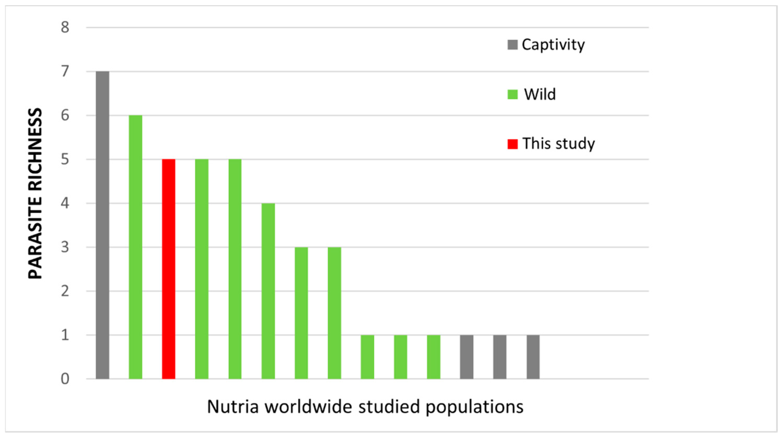
Limiting comparisons to studies which used fecal samples as the source biomatrix, the study population showed a parasite richness (five taxa) in the upper range of wild populations worldwide at different locations (from one to six taxa) and within the range reported in captive animals (from one to seven taxa) (Figure 2).
Mean parasite richness in reviewed wild and captive data did not differ ( x w ¯ = 3.4 taxa vs. x c ¯ = 2.5 taxa, Mann–Whitney test, n.s.), although the dataset might be biased by the large proportion of data deriving from the wild (71.4%). We are not aware of studies searching for eukaryote parasites by using multiple techniques (as we did in this study) and finding no parasites at all.

4. Discussion

In our study, the fecal endoparasite detected with the highest prevalence (71% of the samples) was Strongyloides sp., which is not new to the Italian populations (Table 1 and Table 2) [52]. Overall, four species of Strongyloides have been reported in nutria worldwide: S. myopotami Artigas and Pacheco, 1933; S. chapini Sandground, 1925; S. papillosus (Wedl, 1856); and S. ratti Sandground, 1925 (e.g., [8,13,15,52]). These Strongyloides species do not represent a sanitary risk for humans, causing only mild, short-term skin infections [59]. Gastrointestinal strongyles are among the more frequently detected parasites in ruminants [47,60,61], hence their finding in the study population could be due to the presence of a flock of sheep (the only ruminants occurring in the study area) sharing the same grazing pasture with nutrias. Worldwide, four congener gastrointestinal strongyles have been found in nutria, in addition to the genus Strongyloides, but of a different family: Trichostrongylus duretteae Rossin, Timi, and Malizia, 2006; T. colubriformis (Giles, 1892); T. sigmodontis Baylis, 1945; and T. retortaeformis (Zeder, 1800) (fam. Trichostrongylidae) (e.g., [8,13,52]). The genus Trichostrongylus Looss, 1905, has been already detected in one Northern Italian population (Lombardy region) with the T. duretteae species (Table 1 and Table 2) [52]. In humans, trichostrongylosis is reported in Asia and Africa in people living in close contact with domestic animals (sheep, goats, and donkeys); however, a few cases have also been observed in other parts of the world, including some northern regions in Italy (Lombardy, Piedmont, Veneto) [47,62].
Morphological identification of larvae found in feces did not allow discriminating between the two genera Muellerius and Angiostrongylus. Species of the genus Muellerius are commonly found worldwide in wild and domestic ruminants and are of substantial economic and veterinary importance. No nematode belonging to the genus Muellerius appears to be transmissible to humans [47,63]. In the present study, based on the frequent contact of nutrias with the local flock of sheep, we could tentatively hypothesize that the detected larvae were Muellerius capillaris (Mueller, 1889), a common parasite of sheep and goats [60]. As for the species of the genus Angiostrongylus, they are naturally present in various families of rodents and in tupaidids, mephithids, mustelids, procyonids, felids, and canids. Sometimes, however, they were also detected in various species of birds and other mammalian species, including humans. Currently, the zoonotic species known for the genus Angiostrongylus are A. cantonensis (Chen, 1935) and A. costaricensis Morera and Céspedes, 1971 [64]. Only the former species has been reported in Italy so far; however, it was a unique case (probably not indigenous) referring to a single person who had traveled to an endemic area of the tropics [65,66]. In the present study, we could tentatively hypothesize that Angiostrongylus larvae might belong to A. vasorum (Baillet, 1866), a species that is widespread in Europe, including Italy [66,67] where it has been found in domestic dogs and foxes (Vulpes vulpes), species both present in our study area [25]. This nematode would not represent a risk to people, as it is not a zoonotic parasite. To the best of our knowledge, this is the first report of bronchopulmonary nematodes of the superfamily Metastrongyloidea in nutria so far.
As for protozoa, two genera were detected in our study: Eimeria and Cryptosporidium (subclass Coccidia), both already reported in nutria worldwide (e.g., [13,15,18,51,52]). Eimeria, the causative agent of mammal (e.g., rodents, ruminants) and bird coccidiosis, was detected in both wild and captive nutria worldwide, accounting for a variety of species (reviewed in [51]). The species E. coypi Obitz and Wadowski, 1937, and E. seideli (Seidel, 1954) have already been described in nutria populations in Italy (Lombardy region, Table 1 and Table 2) [52]. On the contrary, Cryptosporidium represents the first report for nutria in Italy as a whole, although already targeted in other studies, for example in Lombardy [52]. However, while Eimeria species are incredibly host-specific and the rare human infections have not been proven pathogenic [68], the genus Cryptosporidium includes species of paramount relevance in human medicine, infecting millions of people worldwide (via contaminated food and water) and causing thousands of deaths, mainly in immunocompromised subjects [47]. Although poorly studied in wildlife, Cryptosporidium is a common parasite of vertebrates, with C. parvum Tyzzer, 1912, by far the more relevant species and most common cause of mammalian cryptosporidiosis [47], and C. ubiquitum Fayer, Santin, and Macarisin, 2010, an emerging zoonotic parasite [69]. Both species have been detected in nutria from the Czech Republic and Slovakia (Table 2) [18]. In these countries, in addition, a new Cryptosporidium species found in nutria (C. myocastoris Ježková, Limpouchová, Prediger, Holubová, Sak, Konečný, Květoňová, Hlásková, Rost, McEvoy, Rajský, Feng, and Kváč, 2021) was described as genetically and biologically distinct from already known species, since it was located in a part of the digestive tract never observed before in other species [18]. In mammals, in fact, most species of gastric and intestinal Cryptosporidium are characterized by a specific adaptation to different parts of the digestive tract, as a type of host specificity. We are unable at this time to exclude that Cryptosporidium found in the fecal samples of our study population could be C. myocastoris.
The protozoan G. duodenalis was not detected in the “Torre Flavia” SPA population. This is in line with no cases of Giardia spp. reported in other Italian study populations, although limited to the northern region Lombardy (e.g., [52]). Worldwide, the genus Giardia Künstler, 1882, has been detected several times in nutria (e.g., in the American continent) [13,57]; however, the zoonotic species G. duodenalis was reported in just one study on a captive population (Table 2) [17].
The nutria population of “Torre Flavia” SPA showed a parasite richness well within data reported in the literature, regardless of geographic area and animal living conditions (wild vs. captive). Nevertheless, we may hypothesize that at least part of the reason why richness was close to the upper range of wild populations could be due to the variety of other domestic and wild species which inhabit the protected area (e.g., grazing sheep, horses, donkeys, foxes, mice, rats, various bird species), many of which are potentially directly involved in the life cycle of some of the parasites found.
Comparison with the published literature was only possible at a general level due to the heterogeneity of (i) biomatrices used, (ii) study aims, (iii) techniques used for the sample collection and analyses, (iv) number of samples available, and (v) nutria living conditions (only wild subjects in Italy; both captive and wild animals worldwide). For example, studies interested in targeting particular parasite taxa by using species-specific techniques (e.g., antigen detection) could only report about the presence of those taxa, not providing information on any other parasite, and only provide limited results in the case of absence [10,19].
In the future, it would be advisable to take into consideration different types of biological samples (also considering dead animals, if available) and new analytical techniques to detect the possible presence of viruses and bacteria in the nutrias of Torre Flavia as well. Further and more detailed studies could also provide more information on the risk of zoonoses within our nature reserve, especially by making use of molecular analyses for the identification of parasites at the species level. The risk of zoonosis can be high in an area characterized by the co-presence of wild and domestic animals and, above all, with high human attendance and a considerable amount of people bringing along domestic dogs [29].

Author Contributions

Conceptualization, M.C.; methodology, S.D.M., C.D.L. and M.C.; investigation, S.D.M. and C.A.; data curation, S.D.M., C.D.L. and C.A.; formal analysis, S.D.M., C.B. and M.C.; writing—original draft preparation, S.D.M., C.D.L. and C.A.; writing—review and editing, S.D.M., C.D.L., C.B. and M.C.; supervision, C.B. and M.C. All authors have read and agreed to the published version of the manuscript.

Funding

This research received no external funding.

Institutional Review Board Statement

Not applicable.

Informed Consent Statement

Not applicable.

Data Availability Statement

The data presented in this study are available on request from the corresponding authors.

Acknowledgments

We are grateful to Martina D’Agostino and Giovanni Di Bella for their support in the fieldwork. Three anonymous reviewers provided useful comments and suggestions which greatly improved the first and the second draft of the manuscript.

Conflicts of Interest

The authors declare no conflict of interest.

References

  1. Cocchi, R.; Riga, F. Linee Guida per il Controllo della Nutria (Myocastor coypus); No. 5; Ministero dell’Ambiente e della Tutela del Territorio, Servizio Conservazione Natura: Roma, Italy, 2001. (In Italian)
  2. Adhikari, P.; Kim, B.-J.; Hong, S.-H.; Lee, D.-H. Climate change induced habitat expansion of nutria (Myocastor coypus) in South Korea. Sci. Rep. 2022, 12, 3300. [Google Scholar] [CrossRef] [PubMed]
  3. Frainer, A.; McKie, B.G.; Amundsen, P.-A.; Knudsen, R.; Lafferty, K.D. Parasitism and the Biodiversity-Functioning Relationship. Trends Ecol. Evol. 2018, 33, 260–268. [Google Scholar] [CrossRef]
  4. Landaeta-Aqueveque, C.; Moreno Salas, L.; Henríquez, A.; Silva-de la Fuente, M.C.; González-Acuña, D. Parasites of Native and Invasive Rodents in Chile: Ecological and Human Health Needs. Front. Vet. Sci. 2021, 8, 643742. [Google Scholar] [CrossRef] [PubMed]
  5. Chalkowski, K.; Lepczyk, C.A.; Zohdy, S. Parasite Ecology of Invasive Species: Conceptual Framework and New Hypotheses. Trends Parasitol. 2018, 34, 655–663. [Google Scholar] [CrossRef] [PubMed]
  6. Llaberia-Robledillo, M.; Balbuena, J.A.; Sarabeev, V.; Llopis-Belenguer, C. Changes in native and introduced host–parasite networks. Biol. Invasions 2022, 24, 543–555. [Google Scholar] [CrossRef]
  7. Capizzi, D.; Monaco, A.; Genovesi, P.; Scalera, R.; Carnevali, L. Impact of alien mammals on human health. In Invasive Species and Human Health; CAB International: Wallingford, UK, 2018; pp. 130–150. [Google Scholar] [CrossRef]
  8. Babero, B.B.; Lee, J.W. Studies on the helminths of nutria, Myocastor coypus (Molina), in Louisiana with checklist of other worm parasites from this host. J. Parasitol. 1961, 47, 378–390. [Google Scholar] [CrossRef]
  9. Newson, R.M.; Holmes, R.G. Some Ectoparasites of the Coypu (Myocastor coypus) in Eastern England. J. Anim. Ecol. 1968, 37, 471–481. [Google Scholar] [CrossRef]
  10. Howerth, E.W.; Reeves, A.J.; McElveen, M.R.; Austin, F.W. Survey for Selected Diseases in Nutria (Myocastor coypus) from Louisiana. J. Wildl. Dis. 1994, 30, 450–453. [Google Scholar] [CrossRef]
  11. Ménard, A.; Agoulon, A.; L’Hostis, M.; Rondelaud, D.; Collard, S.; Chauvin, A. Myocastor coypus as a reservoir host of Fasciola hepatica in France. Vet. Res. 2001, 32, 499–508. [Google Scholar] [CrossRef]
  12. Issia, L.; Pietrokovsky, S.; Sousa-Figueiredo, J.; Stothard, J.R.; Wisnivesky-Colli, C. Fasciola hepatica infections in livestock flock, guanacos and coypus in two wildlife reserves in Argentina. Vet. Parasitol. 2009, 165, 341–344. [Google Scholar] [CrossRef]
  13. Martino, P.; Radman, N.; Parrado, E.; Bautista, E.; Cisterna, C.; Silvestrini, M.; Corba, S. Note on the occurrence of parasites of the wild nutria (Myocastor coypus, Molina, 1782). Helminthologia 2012, 49, 164–168. [Google Scholar] [CrossRef]
  14. Martino, P.E.; Radman, N.E.; Gamboa, M.I.; Samartino, L.E.; Parrado, E.J. Ectoparasites from some Myocastor coypus (Molina, 1782) populations (Coypus or Nutria) in Argentina. Rev. Bras. Parasitol. Vet. 2018, 27, 254–257. [Google Scholar] [CrossRef] [PubMed]
  15. Nechybová, S.; Langrová, I.; Tůmová, E. Parasites of Myocastor coypus—A Comparison in Farm Animals and Their Feral Counterparts. Sci. Agric. Bohem. 2018, 49, 21–25. [Google Scholar] [CrossRef]
  16. Lim, S.R.; Lee, D.H.; Park, S.Y.; Lee, S.; Kim, H.Y.; Lee, M.S.; Lee, J.R.; Han, J.E.; Kim, H.K.; Kim, J.H. Wild nutria (Myocastor coypus) is a potential reservoir of carbapenem-resistant and zoonotic Aeromonas spp. in Korea. Microorganisms 2019, 7, 224. [Google Scholar] [CrossRef]
  17. Cui, Z.; Wang, D.; Wang, W.; Zhang, Y.; Jing, B.; Xu, C.; Chen, Y.; Qi, M.; Zhang, L. Occurrence and Multi-Locus Analysis of Giardia duodenalis in Coypus (Myocastor coypus) in China. Pathogens 2021, 10, 179. [Google Scholar] [CrossRef] [PubMed]
  18. Ježková, J.; Limpouchová, Z.; Prediger, J.; Holubová, N.; Sak, B.; Konečný, R.; Květoňová, D.; Hlàskovà, L.; Rost, M.; McEvoy, J.; et al. Cryptosporidium myocastoris n. sp. (Apicomplexa: Cryptosporidiidae), the Species Adapted to the Nutria (Myocastor coypus). Microorganisms 2021, 9, 813. [Google Scholar] [CrossRef]
  19. Nardoni, S.; Angelici, M.C.; Mugnaini, L.; Mancianti, F. Prevalence of Toxoplasma gondii infection in Myocastor coypus in a protected Italian wetland. Parasites Vectors 2011, 4, 240. [Google Scholar] [CrossRef]
  20. Serracca, L.; Battistini, R.; Rossini, I.; Mignone, W.; Peletto, S.; Boin, C.; Pistone, G.; Ercolini, R.; Ercolini, C. Molecular Investigation on the Presence of Hepatitis E Virus (HEV) in Wild Game in North-Western Italy. Food Environ. Virol. 2015, 7, 206–212. [Google Scholar] [CrossRef]
  21. Scaglione, F.E.; Pregel, P.; Masoero, L.; Caruso, C.; Starvaggi Cucuzza, L.; Dondo, A.; Zoppi, S.; Sereno, A.; Chiappino, L.; Zanet, S.; et al. Nutria (Myocastor coypus) health status in the natural park “La Mandria”. Anatomopathological and microbiological investigations. In Proceedings of the 72nd Convegno SISVet: Tutte le Novità in Ambito Veterinario, Torino, Italy, 20–22 June 2018; p. 239. [Google Scholar]
  22. Bertolino, S.; Cocchi, R. Piano di Gestione Nazionale della Nutria Myocastor coypus; Istituto Superiore per la Protezione e la Ricerca Ambientale: Rome, Italy, 2018. (In Italian) [Google Scholar]
  23. Bertelloni, F.; Cilia, G.; Turchi, B.; Pinzauti, P.; Cerri, D.; Fratini, F. Epidemiology of leptospirosis in North-Central Italy: Fifteen years of serological data (2002–2016). Comp. Immunol. Microbiol. Infect. Dis. 2019, 65, 14–22. [Google Scholar] [CrossRef]
  24. Arcangeli, G. La nutria selvatica quale potenziale “reservoir” di agenti trasmissibili all’uomo: Situazione in Italia e nel mondo. La gestione delle specie alloctone in Italia: Il caso della nutria e del gambero rosso della Louisiana. Quad. Padule Fucecchio 2002, 2, 31. (In Italian) [Google Scholar]
  25. Battisti, C.; Cento, M.; Fraticelli, F.; Hueting, S.; Muratore, S. Vertebrates in the “Palude di Torre Flavia” special protection area (Lazio, Central Italy): An updated checklist. Nat. Hist. Sci. 2021, 8, 3–28. [Google Scholar] [CrossRef]
  26. Amori, G.; Battisti, C. An invaded wet ecosystem in Central Italy: An arrangement and evidence for an alien food chain. Rend. Lincei 2008, 19, 161–171. [Google Scholar] [CrossRef]
  27. Ferri, V.; Battisti, C.; Soccini, C.; Santoro, R. A hotspot of xenodiversity: First evidence of an assemblage of non-native freshwater turtles in a suburban wetland in Central Italy. Lakes Reserv. Res. Manag. 2020, 25, 250–257. [Google Scholar] [CrossRef]
  28. Di Blasio, L.; Santoro, R.; Ferri, V.; Battisti, C.; Soccini, C.; Egidi, A.; Scalici, M. First successful reproduction of the Chinese striped-necked turtle Mauremys sinensis (Gray, 1834) in a European wetland. BioInvasions Rec. 2021, 10, 721–729. [Google Scholar] [CrossRef]
  29. Battisti, C. Biodiversità, Gestione, Conservazione di Un’area Umida del Litorale Tirrenico: La Palude di Torre Flavia; Provincia di Roma, Assessorato alle Politiche Agricole e dell’Ambiente, Gangemi Editore: Roma, Italy, 2006. (In Italian) [Google Scholar]
  30. Gundersen, G.; Johannesen, E.; Andreassen, H.; Ims, R. Source-sink dynamics: How sinks affect demography of sources. Ecol. Lett. 2001, 4, 14–21. [Google Scholar] [CrossRef]
  31. Battisti, C.; Marini, F.; Vignoli, L. A five-year cycle of coypu abundance in a remnant wetland: A case of sink population collapse? Hystrix 2015, 26, 37–40. [Google Scholar]
  32. Marini, F.; Gabrielli, E.; Montaudo, L.; Vecchi, M.; Santoro, R.; Battisti, C.; Carpaneto, G.M. Diet of coypu (Myocastor coypus) in a Mediterranean coastal wetland: A possible impact on threatened rushbeds? Vie Milieu 2013, 63, 97–103. [Google Scholar]
  33. De Michelis, S.; Ceschin, S.; Carosi, M.; Battisti, C. Coypu (Myocastor coypus Molina, 1782) feeding on algae: First evidence for Europe. Vie Milieu 2022, 72, 31–34. [Google Scholar]
  34. Marini, F.; Ceccobelli, S.; Battisti, C. Coypu (Myocastor coypus) in a Mediterranean remnant wetland: A pilot study of a yearly cycle with management implications. Wetl. Ecol. Manag. 2011, 19, 159–164. [Google Scholar] [CrossRef]
  35. Angelici, C.; Marini, F.; Battisti, C.; Bertolino, S.; Capizzi, D.; Monaco, A. Cumulative impact of rats and coypu on nesting waterbirds: First evidences from a small Mediterranean wetland (Central Italy). Vie Milieu 2012, 62, 137–141. [Google Scholar]
  36. Gallitelli, L.; Battisti, C.; Pietrelli, L.; Scalici, M. Anthropogenic particles in coypu (Myocastor coypus; Mammalia, Rodentia)’ faeces: First evidence and considerations about their use as track for detecting microplastic pollution. Environ. Sci. Pollut. Res. 2022, 29, 55293–55301. [Google Scholar] [CrossRef]
  37. Grillo, G.; Sartori, G.; Battisti, C.; Ferri, V.; Luiselli, L.; Amori, G.; Carpaneto, G.M. Attempted copulatory behaviour between two phylogenetically unrelated alien species (Coypu, Myocastor coypus, and Pond slider, Trachemys scripta): First evidence. Zool. Ecol. 2020, 30, 165–168. [Google Scholar] [CrossRef]
  38. Redolfi De Zan, L.; Battisti, C.; Carpaneto, G.M. Inter-annual and intra-seasonal patterns of abundance in a set of common waterbirds: Along term study in a Mediterranean wetland. Vie Milieu 2011, 61, 101–106. [Google Scholar]
  39. Guidi, A. Introduzione alla flora e alle comunità vegetali. In Biodiversità, Gestione E Conservazione di Un’area Umida del Litorale Tirrenico: La Palude di Torre Flavia; Battisti, C., Ed.; Provincia di Roma, Assessorato alle Politiche Agricole e dell’Ambiente, Gangemi Editore: Roma, Italy, 2006; pp. 169–187. (In Italian) [Google Scholar]
  40. Fanelli, G.; Bianco, P.M. Memoria Illustrativa della Carta della Vegetazione della Provincia di Roma; Provincia di Roma, Dip. VI-Governo del Territorio, Serv. 3., Sistema Informativo Geografico: Roma, Italy, 2007. (In Italian) [Google Scholar]
  41. Ioni, S.; Battisti, C.; Fanelli, G. Mapping vegetation dynamics on embryonic sand dunes: A fine-grained atlas for periodic plant monitoring in a Mediterranean protected area. Quad. Mus. Civ. St. Nat. Ferrara 2020, 8, 37–42. [Google Scholar]
  42. Blasi, C.; Michetti, L. Biodiversità e Clima. In Stato della Biodiversità in Italia; Contributo alla Strategia Nazionale per la Biodiversità; Blasi, C., Boitani, L., La Posta, S., Manes, F., Marchetti, M., Eds.; Ministero dell’Ambiente e della Tutela del territorio, F.lli Palombi Editori: Roma, Italy, 2005. (In Italian) [Google Scholar]
  43. Battisti, C.; Luiselli, L.; Teofili, C. Quantifying threats in a Mediterranean wetland: Are there any changes in their evaluation during a training course? Biodivers. Conserv. 2009, 18, 3053–3060. [Google Scholar] [CrossRef]
  44. Sabia, G.; Petta, L.; Moretti, F.; Ceccarelli, R. Combined statistical techniques for the water quality analysis of a natural wetland and evaluation of the potential implementation of a FWS for the area restoration: The Torre Flavia case study, Italy. Ecol. Indic. 2018, 84, 244–253. [Google Scholar] [CrossRef]
  45. Sloss, M.W.; Kemp, R.L. Parassiti in Medicina Veterinaria: Metodi di Identificazione ed Indagine Microscopica; Ed. Ermes: Milan, Italy, 1982. (In Italian) [Google Scholar]
  46. Ambrosi, M. Parassitologia Zootecnica; Edagricole: Bologna, Italy, 1995. (In Italian) [Google Scholar]
  47. De Carneri, I. Parassitologia Generale e Umana, 11th ed.; Casa Editrice Ambrosiana: Milan, Italy, 1992. (In Italian) [Google Scholar]
  48. Zajac, A.M.; Conboy, G. Veterinary Clinical Parasitology, 8th ed.; Wiley-Blackwell Publishing: Oxford, UK, 2012. [Google Scholar]
  49. Dytham, C. Choosing and Using Statistics: A Biologist’s Guide; John Wiley & Sons: Oxford, UK, 2011. [Google Scholar]
  50. Hammer, Ø.; Harper, D.A.; Ryan, P.D. PAST: Paleontological statistics software package for education and data analysis. Palaeontol. Electr. 2001, 4, 9. [Google Scholar]
  51. Lewis, D.C.; Ball, S.J. Eimeria fluviatilis n.sp and other species of Eimeria in wild coypus in England. Syst. Parasitol. 1984, 6, 191–198. [Google Scholar] [CrossRef]
  52. Zanzani, S.A.; Di Cerbo, A.; Gazzonis, A.L.; Epis, S.; Invernizzi, A.; Tagliabue, S.; Manfredi, M.T. Parasitic and Bacterial Infections of Myocastor coypus in a Metropolitan Area of Northwestern Italy. J. Wildl. Dis. 2016, 52, 126–130. [Google Scholar] [CrossRef]
  53. Bollo, E.; Pregel, P.; Gennero, S.; Pizzoni, E.; Rosati, S.; Nebbia, P.; Biolatti, B. Health status of a population of nutria (Myocastor coypus) living in a protected area in Italy. Res. Vet. Sci. 2003, 75, 21–25. [Google Scholar] [CrossRef]
  54. Pedruzzi, L.; Schertler, A.; Giuntini, S.; Leggiero, I.; Mori, E. An update on the distribution of the coypu, Myocastor coypus, in Asia and Africa through published literature, citizen-science and online platforms. Mamm. Biol. 2022, 102, 109–118. [Google Scholar] [CrossRef]
  55. Murillo Peixoto-Couto, R.; Correa-Branco, A.; Cabrera-Miguel, M. First record of Myocastor coypus (Molina 1782) (Mammalia, Rodentia) for the state of Mato Grosso do Sul, Brazil, and its distribution in southern South America. Act. Biol. 2022, 44, 1–6. [Google Scholar] [CrossRef]
  56. Yu, F.; Cao, Y.; Wang, H.; Liu, Q.; Zhao, A.; Qi, M.; Zhang, L. Host-adaptation of the rare Enterocytozoon bieneusi genotype CHN4 in Myocastor coypus (Rodentia: Echimyidae) in China. Parasites Vectors 2020, 13, 578. [Google Scholar] [CrossRef] [PubMed]
  57. Dunlap, B.G.; Thies, M.L. Giardia in beaver (Castor canadensis) and nutria (Myocastor coypus) from east Texas. J. Parasitol. 2002, 88, 1254–1258. [Google Scholar] [CrossRef]
  58. El-Kouba, M.M.A.N.; Marques, S.M.T.; Pilati, C.; Hamann, W. Presence of Fasciola hepatica in Feral Nutria (Myocastor coypus) Living in a Public Park in Brazil. J. Zoo Wildl. Med. 2009, 40, 103–106. [Google Scholar] [CrossRef]
  59. Centers for Disease Control and Prevention. Strongyloides—Biology. 2019. Available online: https://www.cdc.gov/parasites/strongyloides/biology.html (accessed on 1 December 2022).
  60. Cringoli, G.; Rinaldi, L.; Veneziano, V. Mappe con omogenea distribuzione dei punti—Point distribution maps. In Mappe Parassitologiche; University of Naples Federico II: Naples, Italy, 2001; Volume 4. (In Italian) [Google Scholar]
  61. Hoberg, E.P.; Kocan, A.A.; Rickard, L.G. Gastrointestinal Strongyles in Wild Ruminants. In Parasitic Diseases of Wild Mammals, 2nd ed.; Samuel, W.M., Pybus, M.J., Kocan, A.A., Eds.; Iowa State University Press: Ames, IA, USA, 2001. [Google Scholar]
  62. Buonfrate, D.; Angheben, A.; Gobbi, F.; Mistretta, M.; Degani, M.; Bisoffi, Z. Four clusters of Trichostrongylus infection diagnosed in a single center, in Italy. Infection 2017, 45, 233–236. [Google Scholar] [CrossRef]
  63. Zafari, S.; Mohtasebi, S.; Sazmand, A.; Bahari, A.; Sargison, N.D.; Verocai, G.G. The Prevalence and Control of Lungworms of Pastoral Ruminants in Iran. Pathogens 2022, 11, 1392. [Google Scholar] [CrossRef]
  64. Spratt, D.M. Species of Angiostrongylus (Nematoda: Metastrongyloidea) in wildlife: A review. Int. J. Parasitol. Parasites Wildl. 2015, 4, 178–189. [Google Scholar] [CrossRef]
  65. Federspiel, F.; Skovmand, S.; Skarphedinsson, S. Eosinophilic meningitis due to Angiostrongylus cantonensis in Europe. Int. J. Infect. Dis. 2020, 93, 28–39. [Google Scholar] [CrossRef]
  66. Morgan, E.R.; Modry, D.; Paredes-Esquivel, C.; Foronda, P.; Traversa, D. Angiostrongylosis in Animals and Humans in Europe. Pathogens 2021, 10, 1236. [Google Scholar] [CrossRef]
  67. Morgan, E.; Shaw, S. Angiostrongylus vasorum infection in dogs: Continuing spread and developments in diagnosis and treatment. J. Small Anim. Pract. 2010, 51, 616–621. [Google Scholar] [CrossRef] [PubMed]
  68. Kiskaddon, R.M.; Renshaw, R.J.F. Human coccidiosis. J. Am. Med. Assoc. 1945, 128, 731–732. [Google Scholar] [CrossRef]
  69. Li, N.; Xiao, L.; Alderisio, K.; Elwin, K.; Cebelinski, E.; Chalmers, R.; Santin, M.; Fayer, R.; Kvac, M.; Ryan, U.; et al. Subtyping Cryptosporidium ubiquitum, a Zoonotic Pathogen Emerging in Humans. Emerg. Infect. Dis. 2014, 20, 217. [Google Scholar] [CrossRef] [PubMed]
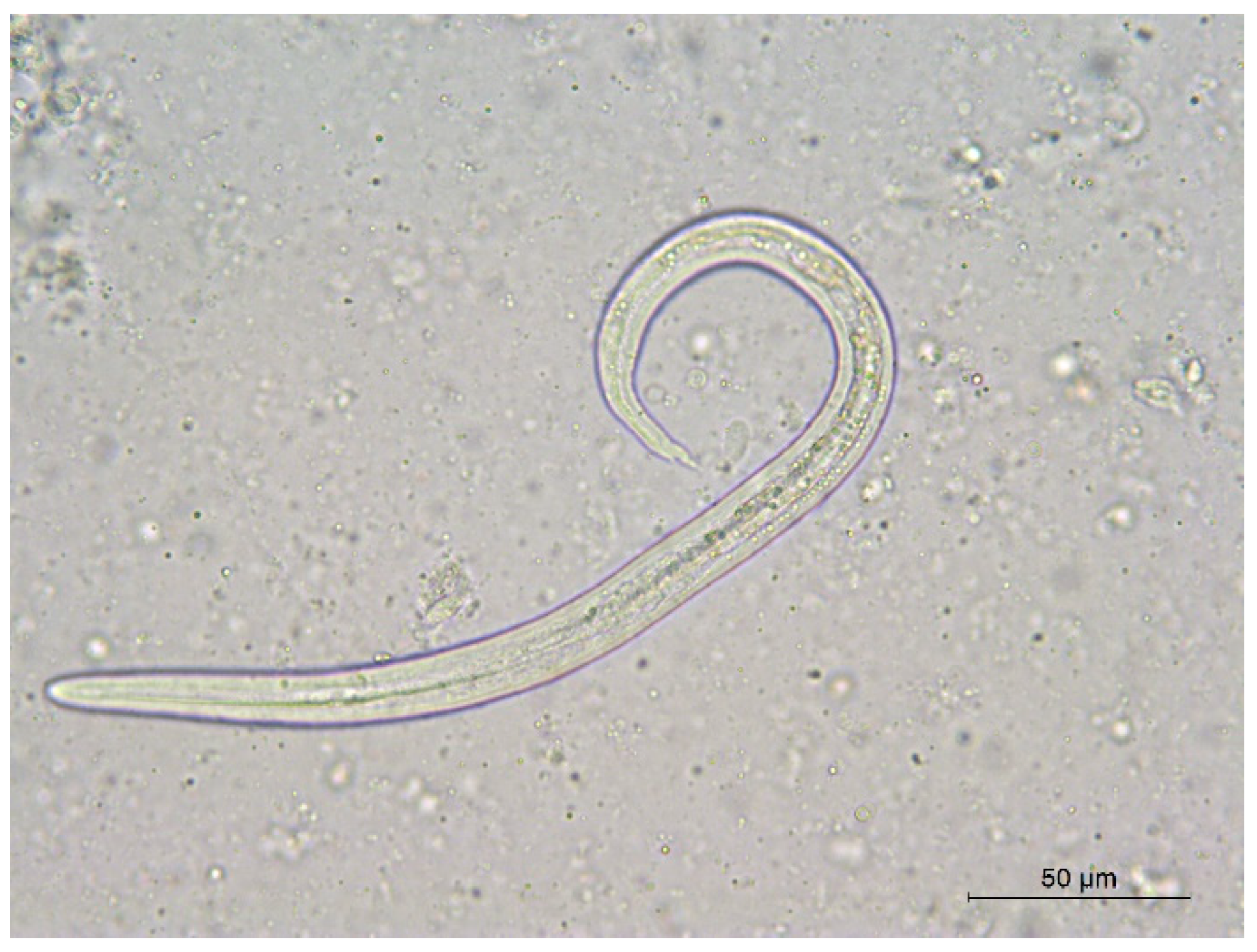
Figure 1. Nematode Muellerius vel. Angiostrongylus L1 larval stage.
Diversity 15 00611 g001
Figure 2. Eukaryote fecal parasite richness in nutria worldwide study populations (wild, captive, this study). Populations are those found in Table 2.
Diversity 15 00611 g002
Table 1. Helminths and protozoans reported for wild nutrias in Italy (+ = parasite presence). a [53]; b [19]; c [52]; d [21]; e this study. * = F: feces, B: blood, O: organs, T: other tissues. The number after each taxon indicates author and year of description. 1 Tyzzer, 1907; 2 Schneider, 1875; 3 Obitz and Wadowski, 1937; 4 Seidel, 1954; 5 Nicolle and Manceaux, 1908; 6 Cameron, 1927, and Kamensky, 1905; 7 Grassi, 1879; 8 Artigas and Pacheco, 1933; 9 Looss, 1905; 10 Rossin, Timi, and Malizia, 2006.
Regions (Italy)Piedmont aTuscany bLombardy cPiedmont dLazio e
Biomatrices *F/B/O/TB/O/TF/B/O/TB/TF
Taxon
Coccidia indet.+
Cryptosporidium sp. 1 +
Eimeria sp. 2 +
Eimeria coypi 3 +
Eimeria seideli 4 +
Toxoplasma gondii 5++++
Muellerius vel. Angiostrongylus 6 +
Trichostrongylidae +
Strongyloides sp. 7 + +
Strongyloides myopotami 8 +
Trichostrongylus sp. 9 +
Trichostrongylus duretteae 10 +
Disclaimer/Publisher’s Note: The statements, opinions and data contained in all publications are solely those of the individual author(s) and contributor(s) and not of MDPI and/or the editor(s). MDPI and/or the editor(s) disclaim responsibility for any injury to people or property resulting from any ideas, methods, instructions or products referred to in the content.