Interesting Images

Evidence of Coral Diseases, Phase Shift, and Stressors in the Atolls of Lakshadweep Islands, Arabian Sea—With Geographical Notes on Their Occurrence within the Indian EEZ and Contiguous International Waters

by
Rocktim Ramen Das
1,2,*,
Chemmencheri Ramakrishnan Sreeraj
1,3,
Gopi Mohan
1,
Nina Tabitha Simon
1,4,5,
Purvaja Ramachandran
1,
Ramesh Ramachandran
1,
Pandian Krishnan
1,6 and
Deepak Samuel Vijay Kumar
1,*
1
National Centre for Sustainable Coastal Management (NCSCM), Ministry of Environment, Forests and Climate Change (MoEF&CC), Chennai 600-025, India
2
Molecular Invertebrates, Systematics and Ecology (MISE) Laboratory, Graduate School of Engineering and Science, University of the Ryukyus, Nishihara 903-0213, Okinawa, Japan
3
Western Ghat Regional Centre, Zoological Survey of India (ZSI), Kozhikode 673-006, India
4
Marine Biology Regional Centre (MBRC), Zoological Survey of India (ZSI), Chennai 600-028, India
5
Department of Zoology, The University of Madras, Chennai 600-005, India
6
Bay of Bengal Program Inter-Governmental Organisation (BOBP-IGO), Chennai 600-018, India
*
Authors to whom correspondence should be addressed.
Diversity 2023, 15(3), 382; https://doi.org/10.3390/d15030382
Submission received: 8 December 2022 / Revised: 17 February 2023 / Accepted: 21 February 2023 / Published: 7 March 2023
(This article belongs to the Special Issue Indo-Pacific Coral Diseases: Diversity, Impacts and Solutions)

Abstract

:
Photographic evidence of some important coral diseases (black band disease, black disease/Terpios hoshinota, white syndrome, pink line syndrome, pink spots, invertebrate galls, skeletal growth anomalies, tissue loss), coral competing sponges, and coral–algal phase shifts (competitive overgrowth of the seaweed Caulerpa spp. over corals and competitive scleractinian interactions such as with Halimeda spp.) have been collected during field observations in a few atolls within the Lakshadweep archipelago, Arabian Sea. Further, earlier reports of similar diseases and other stressors within the Indian Exclusive Economic Zone (EEZ) and its contiguous international waters, including the reefs of the Maldives and Sri Lanka, are highlighted and their distributional ranges are shown.

Coral reefs in the Lakshadweep atolls, located in the Arabian Sea, were severely bleached following the 1998 global mass bleaching event [1,2] and due to the rise in sea surface temperature following climate change. Several similar events have followed in subsequent years, greatly affecting these reefs [2,3,4,5,6]. However, information on coral diseases, which have historically been proven to be positively correlated with post-bleaching events on Indian reefs [7] and elsewhere, viz., the Persian Gulf or the Caribbean [8,9], seems to have been documented only a few times in this region (e.g., [10,11]). Apart from hindering disease resistance, such large-scale events can also give rise to other stressors and lead to shifts in community composition [1]. They also provide space for opportunistic species to overcome scleractinian dominance. Herein, we provide photographic evidence of coral diseases in the Lakshadweep atolls, especially at Bangaram and Thinnakara (BT), Cheriyam (uninhabited) (CHE) and Kalpeni (KAL) (Kalpeni Atoll), and Suheli (uninhabited) (SUH) (Figure 1). These areas were surveyed in 2016 as a part of a national project to assess the ecologically sensitive areas (ESAs) and critically vulnerable coastal areas (CVCAs) within the Indian exclusive economic zone (EEZ). Further, we have also performed comparisons with earlier reports published on the occurrence of these diseases, phase shifts, and stressors in the other reefs within the Indian EEZ and its contiguous international waters, which include the Maldives and Sri Lanka (Figure 1), to highlight the distributional ranges of those reef health impacts.
The Indian EEZ harbors four major coral reef ecosystems located in the Gulf of Mannar Biosphere Reserve (GoMBR) and Marine National Park (GoMMNP), the Andaman and Nicobar Islands (ANI), the Gulf of Kachchh (GoK), and the Lakshadweep atolls [12,13]. It is to be noted that only limited information is available on coral diseases within the Indian EEZ [13] and that the same is the case for the broader Indian Ocean region (IOR) [14].
Approximately 20~30 min timed snorkel surveys or until the area was covered were conducted in the outer lagoon areas of these atolls to record the habitat characteristics, as described by Raymundo et al., with some modifications [15] (Figure 1). The underwater pictures were taken (with a Canon Power-shot G15 placed in an underwater housing) at each survey location (BT = 34; KAL = 34; CHE = 26; SUH = 51); the survey area ranged from 10 × 10 to 50 × 50 m depending on the reef topography. Visual morphological observations such as ”disease-like”, ”infected”, or ”unhealthy conditions” were later evaluated from the photographs, and disease prevalence was calculated from the photographs by counting disease colonies/total number of colonies × 100 [16] (Supplementary Table S1–S3). As no diving was involved, deeper reefs were not considered. Overall, a total of 2239 colonies belonging to 20 different genera were evaluated (Supplementary Table S4). Water quality parameters (sea surface temperature, pH, and DO) were additionally acquired through a multi-parameter water quality sonde (YSI optic probe no. 15K100034) and triplicate observations were made at each of the surveyed atolls at one or two selected locations (Figure 1A–C). The environmental parameters are provided in Table 1. Major diseases, stressors, and phase shifts observed in this study are summarized in Table 2.
The photo-documentation provides evidence regarding the occurrence of coral diseases, stressors, coral competing sponges, and coral–algal phase shifts, even in reefs where human presence is minimal. Nevertheless, recent reports have suggested that live coral cover has increased in the atolls since 2006 [27]. Coral disease-specific surveys to quantify the damage caused by pathogens, reef-associated invertebrates, and sedimentation in the Lakshadweep atolls have been scarce. Along with studies on ecologically sensitive areas under a project, we have availed ourselves of the opportunity to explore and record coral diseases and other stressed conditions with photographic evidence. Our observations were compared with other records gleaned from previous literature related to the region, which would help to describe the possible human impacts on the reefs observed in this survey and those reported in other studies.
Growth Anomalies (GAs): During our observation, nodular GAs were observed in tabular Acropora spp. (0.9%; 4, Tot. Col = 447) (at BT) (Figure 2H), while the overall prevalence of GAs in the common host Porites was (0.33%; 2, Tot. Col = 603)) (Figure 4F). Four colonies of Platygyra spp., including P. pini (Total Col. = 37), were also noted with GAs and BT) (Figure 5D). The classification of GAs based on morphology is available in the literature [28]. GAs within the Indian EEZ have been reported in T. mesenterina, Pavona sp., A. humilis, and Porites spp. [29,30,31], with no current reports from Maldivian atolls and a lack of specific details from the reefs of Sri Lanka.
Pink Line Syndrome (PLS): Our observation of PLS in A. ocellata at BT (Figure 2D) is the first report from the Indian EEZ. We have also observed PLS in P. solida and mushroom coral at SUH. Not much comprehensive information is available on PLS from the Maldives or Sri Lanka.
Trematodiasis/Pink Spots (PS): Our observation of PS was mostly limited to P. solida at the BT and SUH atolls (Figure 2E and Figure 5B), as seen elsewhere in the Indo-Pacific [32]. PLS/PS combined gave a prevalence on the common host Porites ranging from 5.5% (16, Total Col. = 294 at BT) up to 23.4% (37, Total Col. = 158 at SUH) (Figure 2E and Figure 5B).
Black Band Disease (BBD): Within the atolls, BBD has greatly influenced tissue loss in massive corals, which showed coral mortality after the 1998 global bleaching event [7,11] as well as in GoMMNP [33]. Surveys in the Maldives have shown BBD to have a broader host specificity [34], with limited information from Sri Lankan reefs. We observed BBD in the genera Hydnophora (Figure 2B) and Goniopora (at BT). BBD was also observed in P. varians (at KAL) (Figure 4C). However, no epizootic occurrence of BBD was observed (RRD pers. obs.) as noted elsewhere in the Indo-Pacific [16]. The prevalence of BBD in the Maldives is much lower; reports suggest that although it is widespread in the atolls, it has not exceeded 0.5% [34].
Invertebrate Galls (IGs): There has been no report on gall formation in scleractinians within the Indian Reefs, nor from the Maldives or Sri Lanka. We observed IGs in Pocillopora in the atolls of BT and Montipora, along with Pocillopora at SUH (Figure 5A).
White Syndrome (WS): WS was observed in several instances during our surveys. I. palifera seemed to be one of the major hosts for WS, with an overall prevalence of 4.4% (9, Total Col. = 205), and in some cases associated with H. opuntia. WS was additionally noted in G. edwarsi, (Figure 2A), Acropora, and Porites. Our observation of focal to multifocal tissue loss among branching Acropora spp. (Figure 3G,H) can also be categorized under the general category of WS [18]. Acroporids are major hosts for WS in the Maldives, and it has also been observed in the genera Echinopora, Psammocora, Pavona, and Pocillopora [14,35].
Terpios hoshinota/Black Disease (BD): The genera Terpios, first mentioned in the Maldives in 2012 [36]; confirmation of its presence and outbreak were only available in 2015 [37]. Within the EEZ, initial reports were from Palk Bay [38,39], and followed by GoMMNP [40] and Lakshadweep atolls [21]. With a relatively short larval dispersal strategy [41], it is unlikely that BD has spread over such distances, but climate change-driven habitat variability might have allowed BD to overtake several scleractinian dominant habitats. [21,39,40]. Our observation of BD on Porites cylindrica was previously overlooked (Figure 2F), while the highest prevalence of BD/T. hoshinota was observed in Acropora spp. (3.6%, 16, Tot. Col = 447 at BT).
Coral Competing Sponges (CCS): CCS-like Cliona sp. complex, Clathria (Microciona) aceratoobtusa, Cliona kempi, had severely overgrown common scleractinian colonies in the Indian EEZ [42,43]. In the atolls, however, the presence of CCS is not recent; its spread has been observed since the early 1980s [44]. In several instances, we observed CCS overgrowing Acropora spp., A. muricata, and Porites (at CHE and KAL) (Figure 3D, Figure 4D and Figure 5F), and further recommend assessing the spicules to understand their taxonomy.
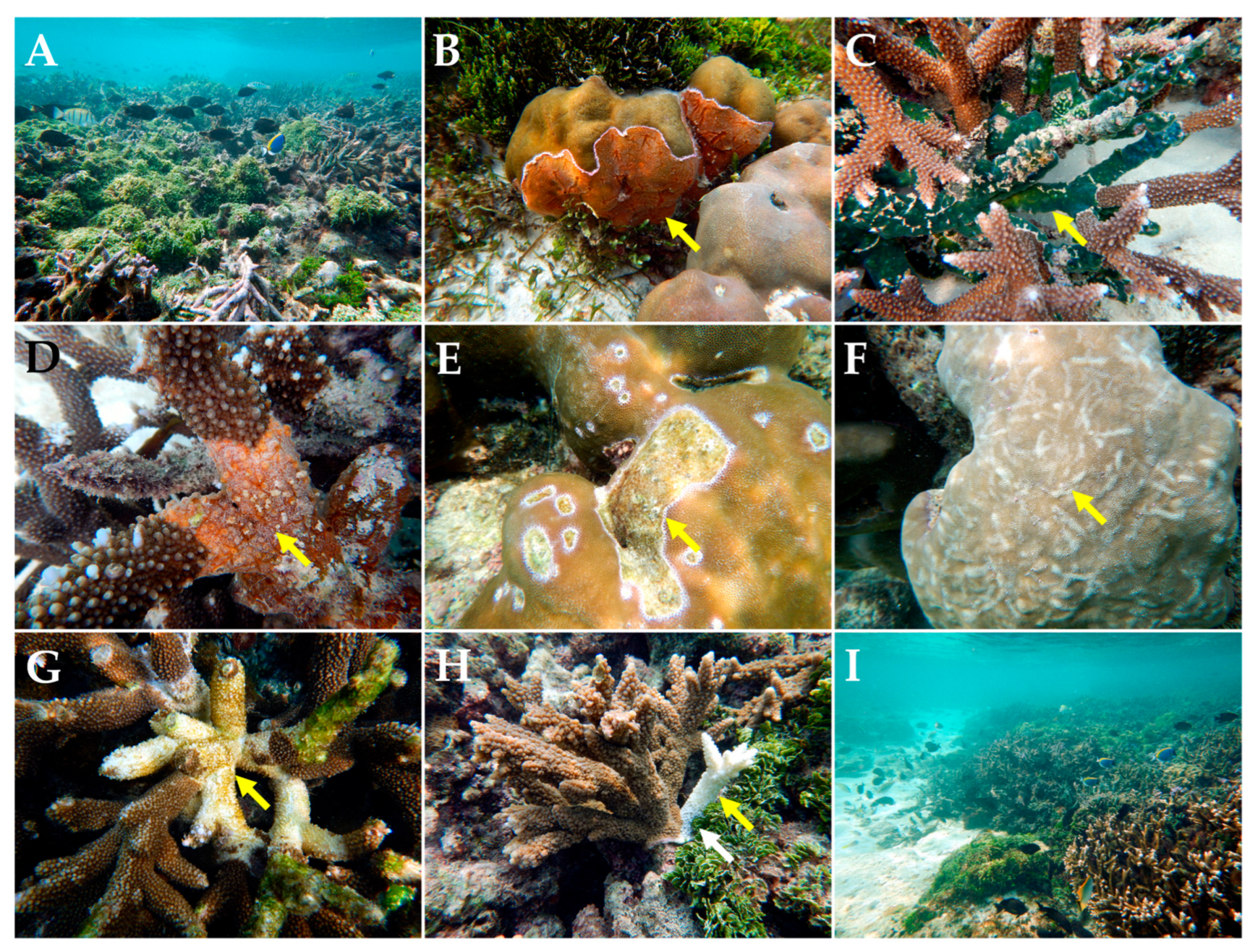
Figure 3. Cheriyam: (A) C. racemosa overgrowth in a coral reef habitat; (B) Overgrowth of unknown coral competing sponge on P. solida; (C) Overgrowth of colonial ascidian species on A. muricata; (D) Coral competing for sponge over Acropora sp.; (E) Tissue loss in P. solida; (F) Predation/fish bites in Porites sp.; (G) Acropora white syndrome depicting multi-focal tissue loss; (H) Acropora depicting tissue loss (yellow arrow) in areas in contact with H. opuntia (white arrow); (I) Macroalgae overgrowing reefs [Photographs by R.R. Das, C.R. Sreeraj, and G. Mohan].
Diversity 15 00382 g003
Figure 4. Kalpeni: (A) White syndrome in I. palifera (yellow arrow) along with overgrowth and coral contact with H. opuntia (red arrow); (B) C. racemosa in Porites spp.; (C) BBD in P. varians (yellow arrow depicting progressing cyanobacterial band); (D) C. racemosa overgrowing Acropora dominated reefs; (E) coral competing sponge overgrowing Porites sp.; (F) GAs in Porites sp.; (G) coral mortality due to C. racemosa overgrowth (H) Multifocal acute tissue loss/White Syndrome in A. muricata (Yellow arrow) followed by algal overgrowth (red arrow); (I) Algal overgrowth of white syndrome affected I. palifera (yellow arrow), along with the presence of H. opuntia (Red arrow) [Photographs by R.R. Das, C.R. Sreeraj, and G. Mohan].
Diversity 15 00382 g004
Figure 5. Suheli: (A) Invertebrate galls in Pocillopora damicornis; (B) PLS/PS-like condition in Porites sp.; (C) Predation and PS in Porites sp.; (D) GAs in Platygyra pini; (E) PLS in Herpolitha sp.; (F) Sponge overgrowth on Acropora sp.; (G) GAs in Porites cylindrica (yellow arrow); (H) CCA overgrowth Pocillopora grandis (red arrow) in association with H. opuntia (yellow arrow); (I) Algal overgrowth on Acropora beds [Photographs by R.R. Das, C.R. Sreeraj, and G. Mohan].
Diversity 15 00382 g005
Crown of Thorns Starfish (COTS): No COTS were observed during the present study, but previous records of COTS outbreaks can be visualized in Figure 1 based on [45,46,47,48,49,50,51,52].
Coral–Algal Phase Shift: The invasion of algal species within the India EEZ has been well documented [53,54]. The extensive spread of C. racemosa is observed in the northernmost, southern, and central regions of Kalpeni Atoll overgrowing Acropora reefs has resulted in severe mortality (Figure 1C,D and Figure 3A,I) [21]. Within the Maldivian archipelago, the devastating effect of C. racemosa on reefs of A. muricata has been noted since 2011 [22]. In the reefs of Sri Lanka, the rapid growth of algae has been observed, comprising Turbinaria sp., Caulerpa sp., and Sargassum sp., [12,55]. As a matter of fact, phase-shift has been occurring at an alarming rate at GoMMNP, where almost 50% of the benthic component is algae [54].
In general, the surveys revealed that the shallow reefs within the atolls of Lakshadweep are generally abundant with common Indo-Pacific genera, viz. Acropora, Porites, Isopora, and Pocillopora. Although site-specific variation might be expected, overall, the presence of 20 genera was noted. Specific details of disease prevalence can be found in the Supplementary Tables (Tables S1–S4). High coral mortality was noted in BT (Acropora = 21.0%; 94, Total Col. 447; Porites = 5.1%; 15, Total Col. 294; Isopora = 8.8%; 15, Total Col. = 171), KAL (Acropora = 28.5%; 85, Total Col. 298), and SUH (Acropora = 13.1%; 29, Total Col. 222). Although it is hard to confirm the cause of this mortality, it can be conjectured that the reefs were not in a very healthy state.
Further, information on coral diseases within the Indian Ocean region is limited when compared to other areas [13,14,34,35]. In this paper, we have provided some interesting photographic evidence, apart from disease prevalence supplements, which highlights the variety of diseases and poor health conditions within the Lakshadweep atolls. Additionally, their presence within other reefs in India and contiguous international waters has been visualized through the application of GIS. Hughes et al. [6] did mention this region (the northernmost Maldivian atolls) as being severely affected by the 2015/16 mass bleaching event; however, such direct mass bleaching observation was not performed, and it would be ignorant to make such a correlation at this stage. Even within Sri Lankan waters, which are considered to possess diverse tropical coral reef ecosystems, there is surprisingly little information. Thus, our observation calls for continuous monitoring in these regions to understand the outbreaks of coral disease epizootics, along with regular monitoring of bleaching events. Further, when performing coral disease assessments, there needs to be a clear understanding of different concepts, such as prevalence and incidence, and consistency should be maintained in disease terminology to avoid confusion while collecting data [17,56]. As a concluding note, through our observations in the atolls, we were able to highlight several diseases and symptoms, but we were not able to detect a very high prevalence of BBD as mentioned by Ranith et al., [11]. Similarly, with the diplomatic cooperation of Mauritius, India’s plans for development in the remote island of Agalela (Western Indian Ocean) calls for extensive and mandatory EIA studies (Figure 1).

Supplementary Materials

The following supporting information can be downloaded at: https://www.mdpi.com/article/10.3390/d15030382/s1, Table S1: Coral disease prevalence in Bangaram and Thinnakara Atoll; Table S2: Coral disease prevalence in Kalpeni Atoll; Table S3: Coral disease prevalence in Suheli Atoll; Table S4: Overall coral disease prevalence. (Acronyms, NFB = Non-Focal Bleaching; BBD = Black Band Disease; GA = Growth Anomalies; Pre = Predation; WS = White Syndrome; BD = Black Disease/T. hoshinota; IG = Invertebrate Galls; TL = Tissue Loss; PLS/PS = Pink Line Syndrome/Pink Spots; CHS = Compromised Health Signs).

Author Contributions

Conceptualization, R.R.D.; methodology, C.R.S., G.M. and R.R.D.; software, R.R.D.; validation, R.R.D.; formal analysis, R.R.D.; investigation, R.R.D., C.R.S., G.M.; resources, P.R., R.R. and P.K.; writing—original draft presentation, R.R.D.; writing—reviewing and editing, R.R.D.,C.R.S., N.T.S., P.R., P.K. and D.S.V.K.; visualization, R.R.D.; supervision, D.S.V.K.; project administration, P.R., R.R., D.S.V.K. and P.K.; funding acquisition, P.R., R.R., P.K. and D.S.V.K. All authors have read and agreed to the published version of the manuscript.

Funding

This research was funded by the Ministry of Environment, Forest & Climate Change (MoEF&CC), Govt. of India, and the World Bank under the India Coastal Zone Management (ICZM) grant number [F.No. 22-29/2008-WBICZM-IA-III; 19 June 2014]. The APC was funded by the National Centre for Sustainable Coastal Management (NCSCM), MoEF&CC, Govt. of India.

Institutional Review Board Statement

This article was formally approved by the internal publication review board of NCSCM, India.

Informed Consent Statement

Not Applicable.

Data Availability Statement

Data are presented in the paper.

Acknowledgments

The authors acknowledge James Davis Reimer (MISE, University of the Ryukyus, Okinawa, Japan) and R. Soundararajan (Bay of Bengal Program Inter-Governmental Organization, BOBP-IGO, Chennai, India) for comments and suggestions. The authors are further thankful to the three anonymous referees and the Indo-Pacific Coral Disease special issue guest editor Simone Montano (University of Milano-Bicocca, Italy/MaRHE center, Maldives) for critically enhancing an earlier version of this manuscript.

Conflicts of Interest

The authors declare no conflict of interest. The funders had no role in the design of the study; in the collection, analyses, or interpretation of data; in the writing of the manuscript; or in the decision to publish the results.

References

  1. Arthur, R. Coral bleaching and mortality in three Indian reef regions during El Nino southern oscillation event. Curr. Sci. 2000, 79, 1723–1729. [Google Scholar]
  2. Arthur, R.; Done, T.J.; Marsh, H.; Harriott, V. Local processes strongly influence post-bleaching benthic recovery in the Lakshadweep Islands. Coral Reefs 2006, 25, 427–440. [Google Scholar] [CrossRef]
  3. Harithsa, S.; Raghukumar, C.; Dalal, S.G. Stress response of two coral species in the Kavaratti atoll of the Lakshadweep Archipelago, India. Coral Reefs 2005, 24, 463–474. [Google Scholar] [CrossRef] [Green Version]
  4. Arthur, R.; Karkarey, R.; Lobo, A.S.; Alcoverro, T.; Kelkar, N. Coral Reef Resilience: Recovery and Resistance across Lakshadweep Archipelago; Ocean and coasts program; Nature Conservation Foundation: Mysuru, India, 2010; p. 24. [Google Scholar]
  5. Vinoth, R.; Gopi, M.; Kumar, T.T.A.; Thangaradjou, T.; Balasubramanian, T. Coral reef bleaching at Agatti Island of Lakshadweep Atolls, India. J. Ocean Univ. China 2012, 11, 105–110. [Google Scholar] [CrossRef]
  6. Hughes, T.P.; Anderson, K.; Conolly, S.R.; Heron, S.F.; Kerry, J.T.; Louch, J.M.; Baird, A.H.; Baum, J.K.; Berumen, M.L.; Bridge, T.C.; et al. Spatial and temporal patterns of mass bleaching of corals in the Anthropocene. Science 2018, 359, 80–83. [Google Scholar] [CrossRef] [Green Version]
  7. Ravindran, J.; Raghukumar, C.; Raghukumar, S. Disease and stress-induced mortality of corals in Indian reefs and observations on bleaching of corals in the Andamans. Curr. Sci. 1999, 76, 233–237. [Google Scholar]
  8. Hazraty-Kari, S.; Tavakoli-Kolour, P.; Das, R.R.; Farhadi, M.; Barkhordari-Ahmadi, A.; Yahyavi, M.; Rezai, H. Baseline assessment of coral diseases in an environmentally extreme environment of the northern Persian Gulf. Mar. Poll. Bull. 2021, 171, 112707. [Google Scholar] [CrossRef]
  9. Croquer, A.; Weil, E. Changes in Caribbean coral disease prevalence after the 2005 bleaching event. Dis. Aquat. Org. 2009, 87, 33–43. [Google Scholar] [CrossRef]
  10. Ravindran, J.; Raghukumar, C. Pink line syndrome (PLS) in the scleractinian coral Porites lutea. Coral Reefs 2002, 21, 252. [Google Scholar] [CrossRef]
  11. Ranith, R.P.; Senthilnathan, L.; Machendiranathan, M.; Thangaradjou, T.; Sasamal, S.; Choudhury, S.B. Sources and threats of chronic tissue loss on coral reefs in the Lakshadweep Islands, Indian Ocean. Mar. Ecol. 2017, 38, e12436. [Google Scholar] [CrossRef]
  12. Rajasuriya, A.; Zahir, H.; Muley, E.V.; Subramanian, B.R.; Venkataraman, K.; Wafar, M.V.M.; Munjurul Hannan Khan, S.M.; Whittingham, E. Status of coral reefs in South Asia: Bangladesh, India, Maldives, and Sri Lanka. In Status of Coral Reefs of the World, Proceedings of the Ninth International Coral Reef Symposium, Bali, Indonesia, 23–27 October 2000; Moosa, M.K., Soemodihardjo, S., Soegiarto, A., Romimohtarto, K., Nontji, A., Soekarno, S., Eds.; Global Coral Reef Monitoring Network and Reef and Rainforest Research Centre: Townsville, Australia; Volume 2, pp. 23–27.
  13. Sharma, D.; Ravindran, C. Diseases and pathogens of marine invertebrate corals in Indian reefs. J. Invertebr. Pathol. 2020, 173, 107373. [Google Scholar] [CrossRef]
  14. Montano, S.; Strona, G.; Seveso, D.; Galli, P. First report of coral diseases in the Republic of Maldives. Dis. Aquat. Org. 2012, 101, 159–165. [Google Scholar] [CrossRef] [Green Version]
  15. Raymundo, L.J.; Couch, C.S.; Harvell, C.D. Coral Disease Handbook: Guidelines for Assessment, Monitoring, and Management; Coral reef targeted research and capacity building for management program; University of Queensland: St. Lucia, QLD, Australia, 2008. [Google Scholar]
  16. Das, R.R.; Wada, H.; Masucci, G.D.; Singh, T.; Tavakoli-Kolour, P.; Wada, N.; Tang, S.L.; Yamashiro, H.; Reimer, J.D. Four-year field survey of black band disease and skeletal growth anomalies in encrusting Montipora spp. corals around Sesoko Island, Okinawa. Diversity 2022, 14, 32. [Google Scholar] [CrossRef]
  17. Sutherland, K.P.; Porter, J.W.; Torres, C. Disease and immunity in Caribbean and Indo-Pacific zooxanthellate corals. Mar. Ecol. Prog. Ser. 2004, 266, 273–302. [Google Scholar] [CrossRef] [Green Version]
  18. Bourne, D.G.; Ainsworth, T.D.; Pollock, F.J.; Willis, B.L. Towards a better understanding of white syndromes and their causes on Indo-Pacific coral reefs. Coral Reefs 2015, 34, 233–242. [Google Scholar] [CrossRef]
  19. Bythell, J.; Pantos, O.; Richardson, R. White Plague, White Band and other “White” Diseases. In Coral Health and Diseases; Rosenberg, E., Loya, Y., Eds.; Springer: Berlin/Heidelberg, Germany, 2004. [Google Scholar]
  20. Adhavan, D.; Chandran, R.; Tikadar, S.; Sivakumar, K. Trematode infestation in coral colonies at Poshitra reef, Gulf of Kachchh marine national park, Gujarat, India. J. Threat. Taxa 2017, 9, 10345–10346. [Google Scholar] [CrossRef] [Green Version]
  21. Das, R.R.; Sreeraj, C.R.; Mohan, G.; Abhilash, K.R.; Samuel, V.K.D.; Ramachandran, P.; Ramachandran, R. Incursion of killer sponge Terpios hoshinota Rützler & Muzik, 1993 on the coral reefs of the Lakshadweep archipelago, Arabian Sea. J. Threat. Taxa 2020, 12, 17009–17013. [Google Scholar]
  22. Montano, S.; Seveso, D.; Strona, G.; Arrigoni, R.; Galli, P. Acropora muricata mortality associated with extensive growth of Caulerpa racemosa in Magoodhoo Island, Republic of Maldives. Coral Reefs 2012, 31, 793. [Google Scholar] [CrossRef] [Green Version]
  23. Manikandan, B.; Ravindran, J. Differential response of coral communities to Caulerpa spp. bloom in the reefs of Indian Ocean. Environ. Sci. Poll. Res. 2017, 24, 3912–3922. [Google Scholar] [CrossRef]
  24. Thinesh, T.; Jose, P.A.; Ramasamy, P.; Meenatchi, R.; Selvan, K.M.; Selvin, J. Differential coral response to algae contact: Porites tissue loss, praise for Halimeda interaction at southeast coast of India. Environ. Sci. Poll. Res. 2019, 26, 17845–17852. [Google Scholar] [CrossRef]
  25. Nugues, M.M.; Smith, G.W.; Hooidonk, R.J.V.; Seabra, M.I.; Bak, R.P.M. Algal contact as a trigger for coral disease. Ecol. Lett. 2004, 7, 919–923. [Google Scholar] [CrossRef]
  26. Thaha, P.P.; Rathod, J.L. Report of coral diseases in the reef flats of Chetlat Island, Lakshadweep. J. Mar. Biol. Ass. India 2019, 61, 51–53. [Google Scholar] [CrossRef]
  27. Gopi, M.; Jeevamani, J.J.J.; Goutham, S.; Simon, N.T.; Samuel, V.D.; Abhilash, K.R.; Robin, R.S.; Hariharan, G.; Muruganandam, R.; Krishnan, P.; et al. Status of health and conservation classification of tropical coral reefs in Lakshadweep archipelago. Wetl. Ecol. Manag. 2021, 29, 653–668. [Google Scholar] [CrossRef]
  28. Ricci, F.; Leggat, W.; Page, C.E.; Ainsworth, T.D. Coral growth anomalies, neoplasms, and tumors in the Anthropocene. Trends Microbiol. 2022, 30, 1160–1173. [Google Scholar] [CrossRef] [PubMed]
  29. Hussain, A.; De, K.; Thomas, L.; Nagesh, R.; Mote, S.; Ingole, B. Prevalence of skeletal tissue growth anomalies in a scleractinian coral: Turbinaria mesenterina of Malvan marine sanctuary, eastern Arabian Sea. Dis. Aquat. Org. 2016, 121, 79–83. [Google Scholar] [CrossRef] [Green Version]
  30. Thinesh, T.; Mathews, G.; Patterson Edward, J.K. Coral disease prevalence in Mandapam group of islands, Gulf of Mannar, Southeastern India. Ind. J. Geo. Mar. Sci. 2009, 38, 444–450. [Google Scholar]
  31. Ramesh, C.H.; Mohanraju, R.; Murthy, K.N.; Karthick, P. Preliminary survey of diseases in the coral reefs of Burmanallah coast, Andaman’s. Ind. J. Geo. Mar. Sci. 2014, 43, 1972–1976. [Google Scholar]
  32. Kubomura, T.; Wee, H.B.; Reimer, J.D. Investigating incidence and possible cause of pink and purple pigmentation response in hard coral genus Porites around Okinawajima Island, Japan. Reg. Stud. Mar. Sci. 2021, 41, 101569. [Google Scholar] [CrossRef]
  33. Thinesh, T.; Mathews, G.; Diraviya-Raj, K.; Patterson-Edward, J.K. Variation in black and white band disease progression in corals of the Gulf of Mannar and Palk Bay, Southeastern India. Dis. Aquat. Org. 2014, 110, 227–234. [Google Scholar] [CrossRef] [Green Version]
  34. Montano, S.; Strona, G.; Seveso, D.; Galli, P. Prevalence, host range, and spatial distribution of black band disease in the Maldivian Archipelago. Dis. Aquat. Org. 2013, 105, 65–74. [Google Scholar] [CrossRef] [Green Version]
  35. Montano, S.; Strona, G.; Seveso, D.; Maggioni, D.; Galli, P. Widespread occurrence of coral diseases in the central Maldives. Mar. Freshw. Res. 2015, 67, 1253–1262. [Google Scholar] [CrossRef] [Green Version]
  36. Tkachenko, K.S. The northernmost coral frontier of the Maldives: The coral reefs of Ihavandippolu atoll under long-term environmental change. Mar. Environ. Res. 2012, 82, 40–42. [Google Scholar] [CrossRef]
  37. Montano, S.; Chou, W.-H.; Chen, C.A.; Galli, P.; Reimer, J.D. First record of the coral killing sponge Terpios hoshinota in the Maldives and the Indian Ocean. Bull. Mar. Sci. 2015, 91, 97–98. [Google Scholar] [CrossRef] [Green Version]
  38. Thinesh, T.; Mathews, G.; Raj, K.D.; Edward, J.K.P. Outbreaks of Acropora white syndrome and Terpios sponge overgrowth combined with coral mortality in Palk Bay, southeast coast of India. Dis. Aquat. Org. 2017, 126, 63–70. [Google Scholar] [CrossRef]
  39. Thinesh, T.; Arul Jose, P.; Hassan, S.; Muthamizh Selvan, K.; Selvin, J. Intrusion of coral-killing sponge (Terpios hoshinota) on the reefs of Palk Bay. Curr. Sci. 2015, 109, 1030–1032. [Google Scholar]
  40. Diraviya-Raj, K.; Selva Bharath, M.; Mathews, G.; Aeby, G.S.; Patterson Edward, J.K. Coral-killing sponge Terpios hoshinota invades the corals of Gulf of Mannar, Southeast India. Curr. Sci. 2018, 114, 117–119. [Google Scholar] [CrossRef]
  41. Wang, J.-T.; Hirose, E.; Hsu, C.-M.; Chen, Y.-Y.; Meng, P.-J.; Chen, C.A. A coral killing sponge, Terpios hoshinota, releases larvae harboring cyanobacterial symbionts: An implication of dispersal. Zool. Stud. 2012, 51, 314–320. [Google Scholar]
  42. Ashok, A.M.; Schonberg, C.H.L.; Diraviya-Raj, K.; Bhoopathi, M.; Selva Bharath, M.; Patterson Edward, J.K. A sponge of the Cliona viridis complex invades and excavates corals of the Gulf of Mannar, south-eastern India. Mar. Freshw. Res. 2018, 69, 874–882. [Google Scholar] [CrossRef]
  43. Ashok, A.M.; Calcinai, B.; Edward, J.K.P. The coral-killing sponge Clathria (Microciona) aceratoobtusa (Porifera: Demosponigiae) invades various coral communities of Gulf of Mannar Marine National Park, southeast India. Eur. Zool. J. 2020, 87, 1–11. [Google Scholar] [CrossRef] [Green Version]
  44. Thomas, P.A. Boring sponges destructive to economically important molluscan beds and coral reefs in Indian seas. Ind. J. Fish. 1979, 26, 163–200. [Google Scholar]
  45. Venkataraman, K.; Rajan, P.T. Coral reefs of Mahatma Gandhi marine national park and crown of thorn starfish phenomenon. In Island Ecosystem and Sustainable Development; Gangwar, B., Chandra, K., Eds.; Andaman Science Association and Department of Science and Technology: Port Blair, India, 1998; pp. 124–132. [Google Scholar]
  46. Jeyabaskaran, R. Disturbances to coral reef communities of Andaman & Nicobar Islands. In National Symposium of Conservation and Valuation of Marine Biodiversity; The Director, ZSI, Ed.; Zoological Survey of India: Kolkata, India, 2007; pp. 117–124. [Google Scholar]
  47. Adam, M.S. Status Report and Survey Results; COT busters program, Marine research section; Ministry of Fisheries and Agriculture: Male’, Republic of Maldives, 1989; 12p. [Google Scholar]
  48. Saponari, L.; Montano, S.; Seveso, D.; Galli, P. The occurrence of an Acanthaster planci outbreak in Ari Atoll, Maldives. Mar. Biodivers. 2015, 45, 599–600. [Google Scholar] [CrossRef]
  49. Pernetta, J.; Wells, S. (Eds.) Marine Protected Area Needs in the South Asian Sea’s Region: Maldives; A Marine Conservation and Development Report; IUCN: Gland, Switzerland, 1993; Volume 3. [Google Scholar]
  50. Jaleel, A. The status of the coral reefs and the management approaches: The case of the Maldives. Ocean Coast. Manag. 2013, 82, 104–118. [Google Scholar] [CrossRef]
  51. Rajasuriya, A.; White, A.T. Coral reefs of Sri-lanka: Review of their extent, condition, and management status. Coast. Manag. 1995, 23, 77–90. [Google Scholar] [CrossRef]
  52. De Bruin, G.H.P. The crown-of-thorns starfish Acanthaster planci (Linne’) in Ceylon. Bull. Fish. Res. Stn. 1972, 23, 37–41. [Google Scholar]
  53. Kamalakannan, B.; Jeevamani, J.J.J.; Nagendran, N.A.; Pandiaraja, D.; Krishnan Kutty, N.; Chandrasekaran, S. Turbinaria sp. as victims to Kappaphycus alvarezii in reefs of Gulf of Mannar, India. Coral Reefs 2010, 29, 1077. [Google Scholar] [CrossRef] [Green Version]
  54. Krishnan, P.; Abhilash, K.R.; Sreeraj, C.R.; Samuel, V.D.; Purvaja, R.; Anand, A.; Mahapatra, M.; Sankar, R.; Raghuraman, R.; Ramesh, R. Balancing livelihood enhancement and ecosystem conservation in seaweed farmed areas: A case study from Gulf of Mannar Biosphere Reserve, India. Ocean Coast. Manag. 2021, 207, 105590. [Google Scholar] [CrossRef]
  55. Arulananthan, A.; Herath, V.; Kuganathan, S.; Upasantha, A.; Harishchandra, A. The status of the coral reefs of the Jaffna peninsula (Northern Sri Lanka), with 36 coral species to Sri Lanka confirmed by DNA bar-coding. Oceans 2021, 2, 509–529. [Google Scholar] [CrossRef]
  56. Rogers, C.S. Words matter: Recommendations for clarifying coral disease nomenclature and terminology. Dis. Aquat. Org. 2010, 167–175. [Google Scholar] [CrossRef]
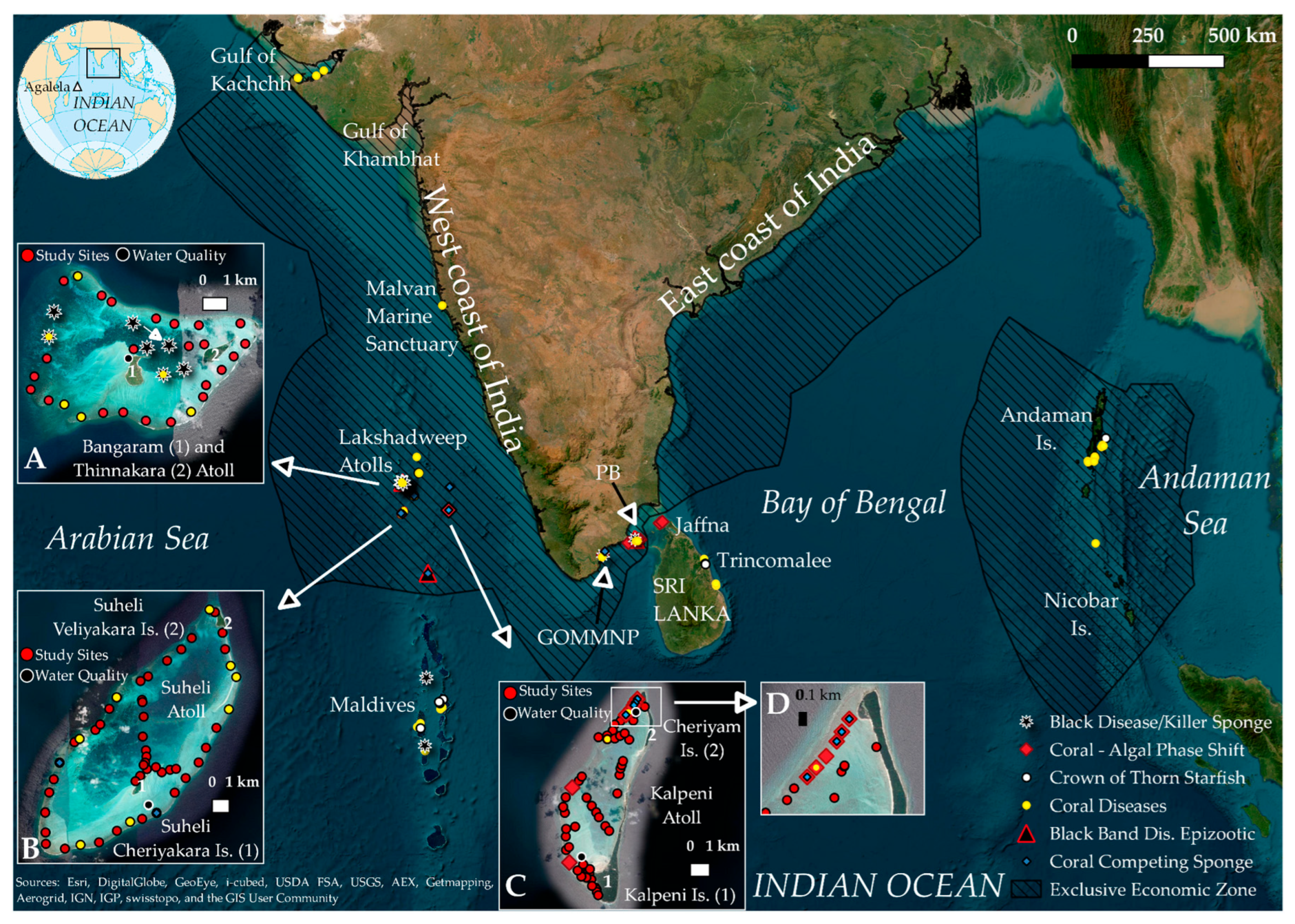
Figure 1. The Indian Exclusive Economic Zone (EEZ) indicates the current study locations and studies pertaining to different aspects of coral diseases, coral competing sponges (CCS), and coral–algal phase shift within the EEZ and contiguous international waters. (A) Sites at Bangaram and Thinnakara Atoll; adapted and updated from [21], white arrow, a previously unreported BD site. (B) Sites at Suheli Atoll. (C) Sites at Kalpeni Atoll. (D) Zoomed on the location of reefs near Cheriyam Is. (Kalpeni Atoll) [GOMMNP: Gulf of Mannar Marine National Park; PB: Palk Bay; black disease = T. hoshinota)]. Globe top-left: https://www.worldatlas.com/aatlas/infopage/oceans/indian.gif (Accessed on 6th December 2022).
Diversity 15 00382 g001
Figure 2. Bangaram and Thinnakara (BT Atoll): (A) White syndrome in G. edwarsi (yellow arrow); (B) Black band disease in Hydnophora sp. (yellow arrow); (C) Tissue loss in G. edwarsi (red arrow); (D) PLS in A. ocellata (yellow arrow); (E) Trematodiasis/PS in P. solida (yellow arrow); (F) BD/T. hoshinota and CHS on P. cylindrica (yellow arrow) [21]; (G) BD/T. hoshinota overgrowing D. lizardensis (yellow arrow) [21]; (H) Gas in Acropora sp. (yellow arrow); (I) CHS (yellow arrow) in Fungiidae [Photos by R.R.Das, C.R.Sreeraj, G.Mohan].
Diversity 15 00382 g002
Table 1. Environmental parameters recorded at each surveyed site (November 2016).
Location (Islands)Sea Surface Temperature (SST;°C)pHDissolved Oxygen (mg/L)Figures
Bangaram and Thinnakara (BT)29.97 (±1.29)7.63 (±0.04)6.82 (±1.2)Figure 1A
Suheli (SUH)29.5 (±1.34)7.6 (0)7.72 (±0.5)Figure 1B
Cheriyam (CHE)29.6 (±0.28)7.6 (±0.05)5.9 (±0.03)Figure 1C
Kalpeni (KAL)29.1 (±1.34)7.5 (0)6.63 (±2.3)Figure 1C
Table 2. Disease and coral health conditions observed in Lakshadweep Atolls and its associated significance.
LocationGPS CoordinatesGenus/Sp.Disease/Stressors/TransitionsFiguresDate of SurveyAdditional Ref./ReadingComments/Remarks
Bangaram and Thinnakara (BT)10.94999 N,
72.29684 E
Goniastrea edwarsiWhite syndromeFigure 2A12 November 2016[17,18,19]Following Bourne et al., [18] and Bythell et al., [19] this observation is placed under the general category of white syndrome
Hydnophora sp.Black band disease (BBD)Figure 2B First report from the Arabian sea
Goniastrea edwarsiTissue lossFigure 2C
Astreopora ocellataPink line syndrome (PLS)Figure 2D [10]First report from Indian EEZ
Porites solidaTrematodiasis/pink spots (PS)Figure 2E [20]Requires lab verification for confirmation
Isopora palifera; Cyphastrea spp.; Porites luteaBlack disease (BD)/T. hoshinota [21]
Porites cylindricaBlack disease (BD)/T. hoshinotaFigure 2F [21] BD presence overlooked at this site by Das et al., [21] (Figure 1A—White arrow)
Dipsastraea lizardensisBlack disease (BD)/T. hoshinotaFigure 2G [21]
Acropora spp.Skeletal growth anomalies (GAs)Figure 2H
Fungia sp.; Herpolitha sp. Compromised health signs (CHS)Figure 2I
Pocillopora sp.Invertebrate galls (IGs) No reports from the Indian EEZ, Maldives, or Sri Lanka.
Goniopora sp.Black band disease (BBD)
Cheriyam (CHE)10.14353 N, 73.65811 EAcropora dominated reefMassive overgrowth of Caulerpa racemosaFigure 3A7 November 2016[22,23]Similar opportunistic invasion reported in Maldives [22] and mainland India [23].
Porites sp. Coral competing sponge (CCS) overgrowthFigure 3B
Acropora muricataOvergrowth of colonial asicidianFigure 3C
Acropora muricataCoral competing sponge (CCS) overgrowthFigure 3D
Porites solidaTissue lossFigure 3E
Porites sp.Predation/fish bitesFigure 3F [11]
Acropora sp.Multifocal tissue loss/white syndrome followed by growth of turf algae.Figure 3G [17,18,19] Further studies related to white syndrome, involving multiple techniques remains necessary [18].
Acropora sp.Association with H. opuntia leading to tissue loss (white syndrome?)Figure 3H [17,18,19,24,25]Necessary to study interaction and pathogen reservoir potential of H. opuntia [25].
Reef habitatC. racemosa overgrowing Acropora spp. dominated reef habitatFigure 3I [22]
Kalpeni (KAL)10.08134 N, 73.63442 EIsopora paliferaWhite syndrome in association with Halimeda opuntiaFigure 4A7 November 2016[17,18,19] Reported as white band disease by Thaha and Rathod [26], needs lab verification/confirmation.
Porites spp.Caulerpa racemosa overgrowthFigure 4B [23]
Pavona variansBlack band disease (BBD)Figure 4C [26]
Acropora spp.C. racemosa overgrowthFigure 4D [22]
Porites sp.Coral competing sponge (CCS)Figure 4E
Porites sp.Skeletal growth anomalies (GAs)Figure 4F
Reef habitatMacroalgae causing coral mortalityFigure 4G
Acropora spp.Multifocal tissue loss, followed by algal overgrowthFigure 4H
Isopora paliferaWhite syndrome following algal overgrowth. Presence of H. opuntia noted.Figure 4I [17,18,19]
Suheli (SUH)10.07667 N, 72.29111 EPocillopora damicornisInvertebrate galls (IGs)Figure 5A9–10 November 2016 Not reported from the Indian EEZ, Maldives, or Sri Lanka.
Porites solidaPink line syndrome (PLS)/pink spot (PS)-like conditionFigure 5B [10,20]
Porites sp.Fish bites and pink spots (PS)Figure 5C [11]
Platygyra piniSkeletal growth anomalies (GAs)Figure 5D
Herpolitha sp.Pink line syndrome (PLS)Figure 5E
Acropora sp.Coral competing sponge (CCS) overgrowthFigure 5F
Porites cylindricaSkeletal growth anomalies (GAs)Figure 5G First report from the Indian EEZ
Pocillopora grandisCCA-related partial mortality in association with H. opuntiaFigure 5H
Acropora spp.Tips of Acropora showing algal overgrowth. A possible scenario following tissue loss.Figure 5I
Disclaimer/Publisher’s Note: The statements, opinions and data contained in all publications are solely those of the individual author(s) and contributor(s) and not of MDPI and/or the editor(s). MDPI and/or the editor(s) disclaim responsibility for any injury to people or property resulting from any ideas, methods, instructions or products referred to in the content.