Article

Expanding the Fossil Record of Soldier Fly Larvae—An Important Component of the Cretaceous Amber Forest

1
Faculty of Biology, Biocenter, Ludwig-Maximilians-Universität München (LMU Munich), Großhaderner Str. 2, 82152 Munich, Germany
2
GeoBio-Center at LMU, Richard-Wagner Str. 10, 80333 Munich, Germany
3
Zoological Institute and Museum, Cytology and Evolutionary Biology, University of Greifswald, Soldmann Str. 23, 17489 Greifswald, Germany
4
Medical Biology and Electron Microscopy Center, University Medical Center Rostock, Strempel Str. 14, 18057 Rostock, Germany
*
Author to whom correspondence should be addressed.
These authors contributed equally to this work.
Diversity 2023, 15(2), 247; https://doi.org/10.3390/d15020247
Submission received: 4 January 2023 / Revised: 4 February 2023 / Accepted: 6 February 2023 / Published: 9 February 2023
(This article belongs to the Special Issue Paleoecology of Insects)

Abstract

:
Larvae of soldier flies and their closest relatives (Diptera: Stratiomyomorpha) are important decomposers of organic material, including wood, that take part in carbon cycling. They also play a certain role in the modern-day animal and human food industry, representing economic value. Larvae of Stratiomyomorpha are considered to be rather rare in the fossil record. Indeed, only very few larvae have been reported so far. Here, we demonstrate that larvae of Stratiomyomorpha are in fact abundant in the Cretaceous, at least in Myanmar amber (about 100 million years old), based on more than 100 amber pieces containing larvae. The specimens could be differentiated into three morphotypes, two already described in a previous work, and a new one. For one morphotype, three larval stages could be distinguished by analysing the dimensions of the head capsules. A quantitative analysis of body shapes indicates a lower morphological diversity of the fossil sample in comparison to the extant fauna, but suggests that they might have had a different ecology in the past. It appears that the data set is not yet saturated, i.e., that more fossil larvae of this group, including different morphologies, are expected to be found.

1. Introduction

Insecta is an ingroup of Euarthropoda with an extremely high species richness. Most of this richness is concentrated in a single ingroup of Insecta, namely Holometabola [1], the group which includes, among others, bees, beetles, and butterflies. This group is characterized by highly specialized early post-embryonic stages, larvae (see [2] for challenges of the term), and the often very differently appearing adults.
One of the more species-rich ingroups of Holometabola, with about 159,000 formally described species, is Diptera, the group of flies [3]. Flies can be found in great quantities in almost every habitat; they only struggle to prevail in greater numbers in the marine realm, and the Arctic and Antarctic areas [4]. This distribution shows their strong evolutionary adaptability and success. Larvae of Diptera contribute to a large extent to most modern terrestrial and freshwater ecosystems, for example, by purifying water or decomposing dead organic matter [5]. They also function as an important food source for aquatic and terrestrial predators. In short, with the diversity of their ecological roles the existence of flies including their larvae is indispensable in nearly any continental ecosystem.
Stratiomyomorpha is one of the ingroups of Diptera, more precisely of Brachycera, the group of true flies. The group Stratiomyomorpha includes three major lineages in the extant fauna: Xylomyidae, wood soldier flies; Stratiomyidae, true soldier flies; and Panthophthalmidae, giant timber flies [6,7]. Although being one of the smaller ingroups of Diptera, Stratiomyomorpha still has quite a high species diversity and includes more than 3000 formally described species [4].
Larvae of Stratiomyidae live in either aquatic or terrestrial habitats, decomposing organic matter. In some cases, it has been reported that they also feed on living plants, such as grass roots [8]. Larvae of Xylomyidae are commonly found in forests under the bark of trees [9]. They feed on wood as implied by the name (“xylo” = wood). Larvae of Panthophthalmidae can be found living on or in dead trees, feasting on accumulated organic matter or the wood fermentation products in dead timber [10].
Larvae of Stratiomyomorpha have been found as fossils reaching back to the early Cretaceous of the late Mesozoic era, dated to an age of about 125 million years [11,12]. Such fossil larvae of Stratiomyomorpha were considered rather rare until Baranov et al. [13] reported new fossils from various deposits, significantly expanding the record by six new morphotypes from no less than five different deposits: Kachin amber (Myanmar, Cretaceous, 100 million years old), Baltic amber (Eocene, 40–35 million years old), Messel (Germany, Eocene, 48 million years old), Dominican amber (Miocene, 20–15 million years old) and Činžat (Slovenia, Miocene, 17–16 million years old). Still, each of these morphotypes was represented by few individuals (slightly more than 20 specimens in total). Hence, fossil stratiomyomorphan larvae seemed no longer to be particularly rare, but still could not be considered abundant.
So far, 17 formally described species of stratiomyomorphans have been recorded in amber deposits of Myanmar: five species of Stratiomyidae, seven of Xylomyidae, and five of the exclusively fossil group Zhangsolvidae [14,15,16,17]. The fossil larvae could not be identified as conspecific with any of the species known as adults [12,13].
This study expands the record of stratiomyomorphan larvae found in Cretaceous Kachin amber, Myanmar, of about 100 million years old. The new finds demonstrate that such larvae were in fact common and rather abundant in the Mesozoic era, at least in Myanmar amber. Furthermore, parts of the ontogeny could be reconstructed, and a quantitative morphological comparison to extant specimens is provided.

2. Materials and Methods

2.1. Material

All specimens examined in this study are part of the Palaeo–Evo–Devo Research Group Collection of Arthropods, Ludwig Maximilian University of Munich, Germany. The specimens were legally purchased on the trading platform ebay.com from two traders (burmite-miner and macro-cretaceous). All fossil specimens reported here are preserved in Kachin amber from Myanmar, which was dated to an age of about 100 million years [18,19,20]. In total, 118 amber pieces with a total of 143 specimens were examined; of these, 131 individuals in 102 amber pieces were identified as larvae of Stratiomyomorpha (details in Table S1 and File S2). Additionally, 71 extant specimens retrieved from the literature were included in the analysis. For details of all specimens, see electronic Supplement (Table S1 and File S2).

2.2. Documentation Methods

All fossil specimens were documented on a Keyence VHX 6000 digital microscope. Amber pieces were fixed on microscope slides with plasticine and then covered with a film of Glycerin and a cover slip on top. This evens out the often not perfectly plane surface of the amber. Images were recorded with different magnifications (100×–500×), depending on the optical properties (especially transparency) of the amber piece. Different backgrounds (white or black) and different light conditions (unpolarized ring light, cross-polarized coaxial light) were tested. All images were recorded as compound images [21]. Single frames were processed and stitched automatically by the built-in software of the Keyence VHX 6000. For further visual illustration, including manual digital colour-coding, the resulting compound images were edited with Photoshop CS2.

2.3. Measurements

In total, four dimensions were measured: (1) the total length of body, (2) the total width of body, (3) the length of head capsule, (4) the width of head capsule. The total length of the larvae was measured from the most anterior tip of the labrum to the most posterior tip of the trunk end. Only the lengths of well-preserved larvae that were relatively straight were measured [13]. The widths of the bodies were measured on the widest point and only on the best-preserved and most robust larvae. The length of the head capsule was measured from the tip of the labrum to the posterior edge of the head capsule [13]. The width of the head capsule was measured at the widest point of the head capsule.

2.4. Shape Analysis

For shape analysis, black-and-white outlines of the specimens (from either ventral or dorsal view) were made with Inkscape, which is freeware for editing vector graphics. The best-preserved halves of each specimen were outlined, copied, mirrored and merged to obtain a symmetrical shape of the larvae. In cases in which the larvae were slightly bent, shapes were straightened by outlining each segment of the convex side of the body and rearranging while maintaining the original features of the larvae [22]. Most shapes were re-used from Baranov et al. [13], others were based on new material reported here and from the literature (details in Table S1 and File S2). These shapes were converted into mathematical functions with the freeware Shape and further processed using a principal component analysis. Resulting values were plotted for comparison. Plots were edited in Adobe Illustrator CS2.

3. Results

3.1. The General Structure of Larvae of Stratiomyomorpha Reported Here

Three different morphotypes (1, 2, 7) could be differentiated among 131 specimens that were documented for this study. The first two morphotypes (morphotype 1, 2) were already recognised and described by Baranov et al. [13]. A new morphotype (morphotype 7, referring to the counting of [13]) has some similarities to morphotype 2, but also differs in certain aspects. In not so well-preserved specimens, it can be difficult to distinguish between morphotypes 2 and 7 (see details below).
All three morphotypes share certain general features (cf. [13]): Small larva, dorsoventrally flattened with a spindle-shaped body, and covered in pellets, presumably of calcium carbonate (Figure 1F). Body with twelve visible units, head capsule, and eleven trunk units; three segments of the thorax (prothorax, mesothorax, metathorax) and eight units of the abdomen (seven abdomen segments and trunk end = compound structure of several undifferentiated segments; Figure 1A).
Head capsule sclerotized, differing between morphotypes. Mandibular–maxillary complex located at anterior part of head capsule, with “brush” formed by setae at the apical part. Posterior parts retracted into thorax (metacephalic rod, paired tentorial arms). In some cases (only of morphotype 1), anterior paired prothoracic spiracles visible laterally on the prothorax (Figure 1B). Paired posterior spiracles in trunk end (Figure 1D). Different shape of setae between the three morphotypes (Figure 1A,C,E,G).
The identity of the specimens was determined based on the synapomorphies recognized by Yeates [6] and the key provided by Teskey [23].

3.2. Morphotype 1

3.2.1. Material Examined

We examined 110 specimens: PED 0490, PED 0497, PED 0511, PED 0548, PED 0550, PED 0551, PED 0567-1, PED 0567-2, PED 0567-3, PED 0599-1, PED 0599-2, PED 0601, PED 0611-1, PED 0611-2, PED 0611-3, PED 0614, PED 0617, PED 0620, PED 0622, PED 0628, PED 0653, PED 0678, PED 0681, PED 0685, PED 0691, PED 0701, PED 0712, PED 0714, PED 0721, PED 0729, PED 0738, PED 0743, PED 0749, PED 0752, PED 0760, PED 0761-1, PED 0761-2, PED 0761-3, PED 0762, PED 0764, PED 0766, PED 0770, PED 0771, PED 0773-1, PED 0773-2, PED 0773-3, PED 0777, PED 0799, PED 0811-1, PED 0811-2, PED 0811-3, PED 0817, PED 0819-1, PED 0819-2, PED 0819-3, PED 0819-4, PED 0819-5, PED 0819-6, PED 0819-7, PED 0819-8, PED 0819-9, PED 0846, PED 0850, PED 0861, PED 0873-1, PED 0873-2, PED 0921, PED 0923, PED 0927, PED 0934, PED 0962, PED 0966, PED 0976-1, PED 0976-2, PED 0978, PED 0987, PED 0990, PED 1001, PED 1007-1, PED 1007-2, PED 1007-3, PED 1010, PED 1014, PED 1018, PED 1025, PED 1044, PED 1129, PED 1137, PED 1140, PED 1167, PED 1174, PED 1185, PED 1192, PED 1200-1, PED 1200-2, PED 1200-3, PED 1200-4, PED 1247, PED 1274, PED 1276, PED 1279, PED 1282, PED 1349, PED 1353, PED 1405, PED 1535, PED 1548-1, PED 1548-2, PED 1560, PED 1567.

3.2.2. General Description of Morphotype 1

General structure of larvae of Stratiomyomorpha as outlined above. Body longer and softer appearing (in comparison to morphotypes 2 and 7) despite also being covered in pellets or scales. Specimens often stretched, crushed or ripped apart. Body mostly yellowish-brown and slightly transparent; in few cases very dark, pigmented. Head capsule strongly sclerotized, therefore well-preserved in most cases. Head capsule longer than wide with labrum, mandibular–maxillary complex and labium visible on the anterior end of head capsule (see description of individual specimens of morphotype 1). Posterior part of head capsule with metacephalic rod and paired tentorial arms usually retracted into thorax, only visible through rather transparent thorax cuticle. Thoracic spiracles on lateral sides of prothorax (Figure 1B). Segments of thorax with multiple rows of short spine-like setae dorsally and laterally (Figure 1E). Abdomen segments 1–7 with longer ventral spine-like setae used for locomotion (Figure 1D). Length and number differ between the segments (Figure 2A–C). Trunk end with spine-like setae facing posteriorly. Posterior paired spiracles located on trunk end (Figure 1F). Dimension range: Body length 1.3 mm–12.57 mm (n = 27), body width 0.5 mm–3.33 mm (n = 71).

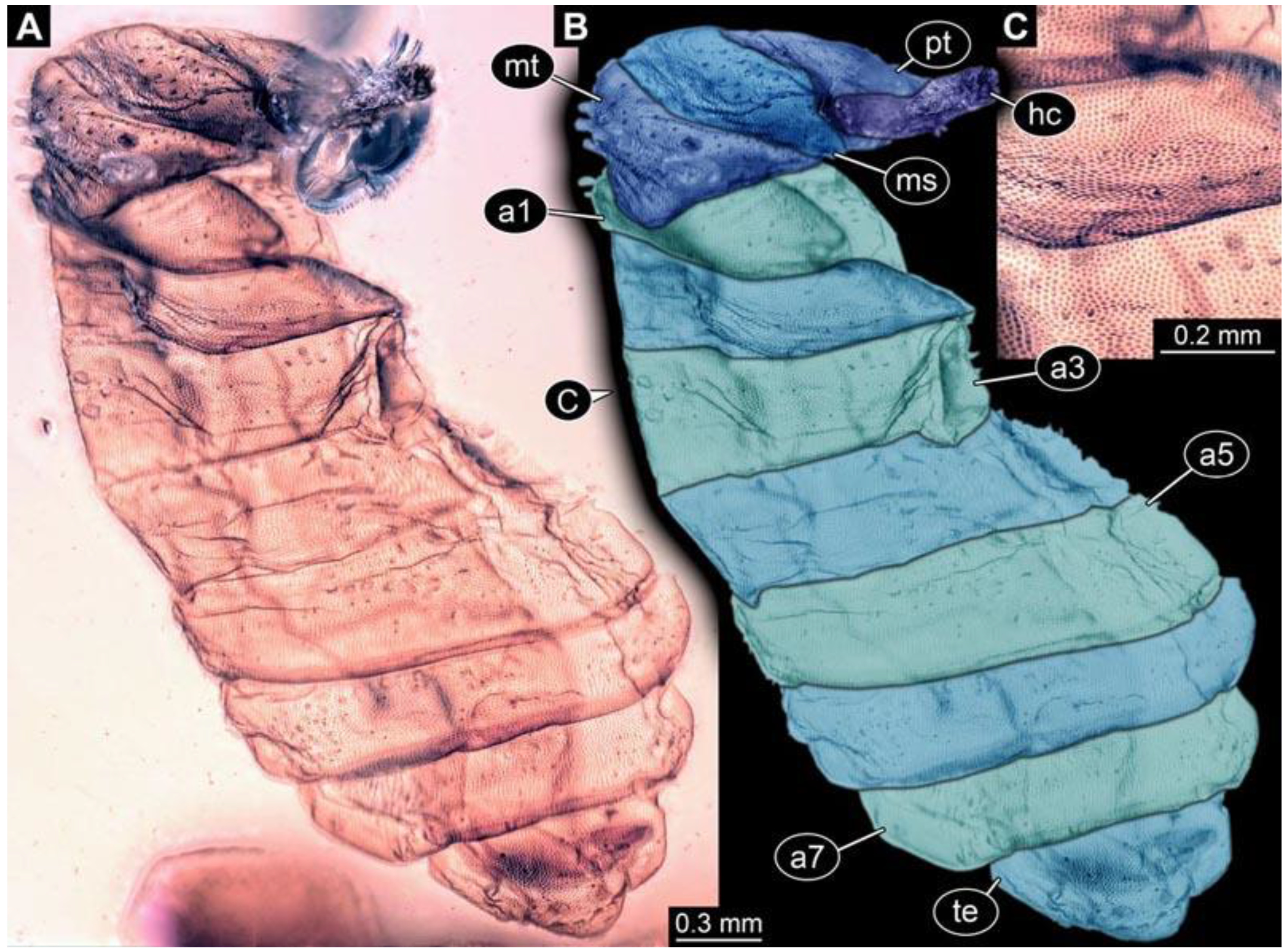
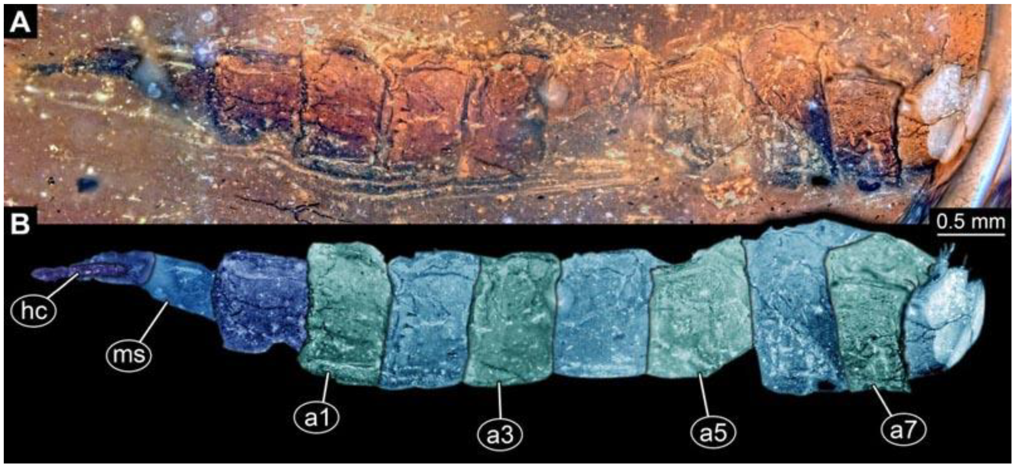
3.2.3. Short Descriptions of Individual Specimens of Morphotype 1

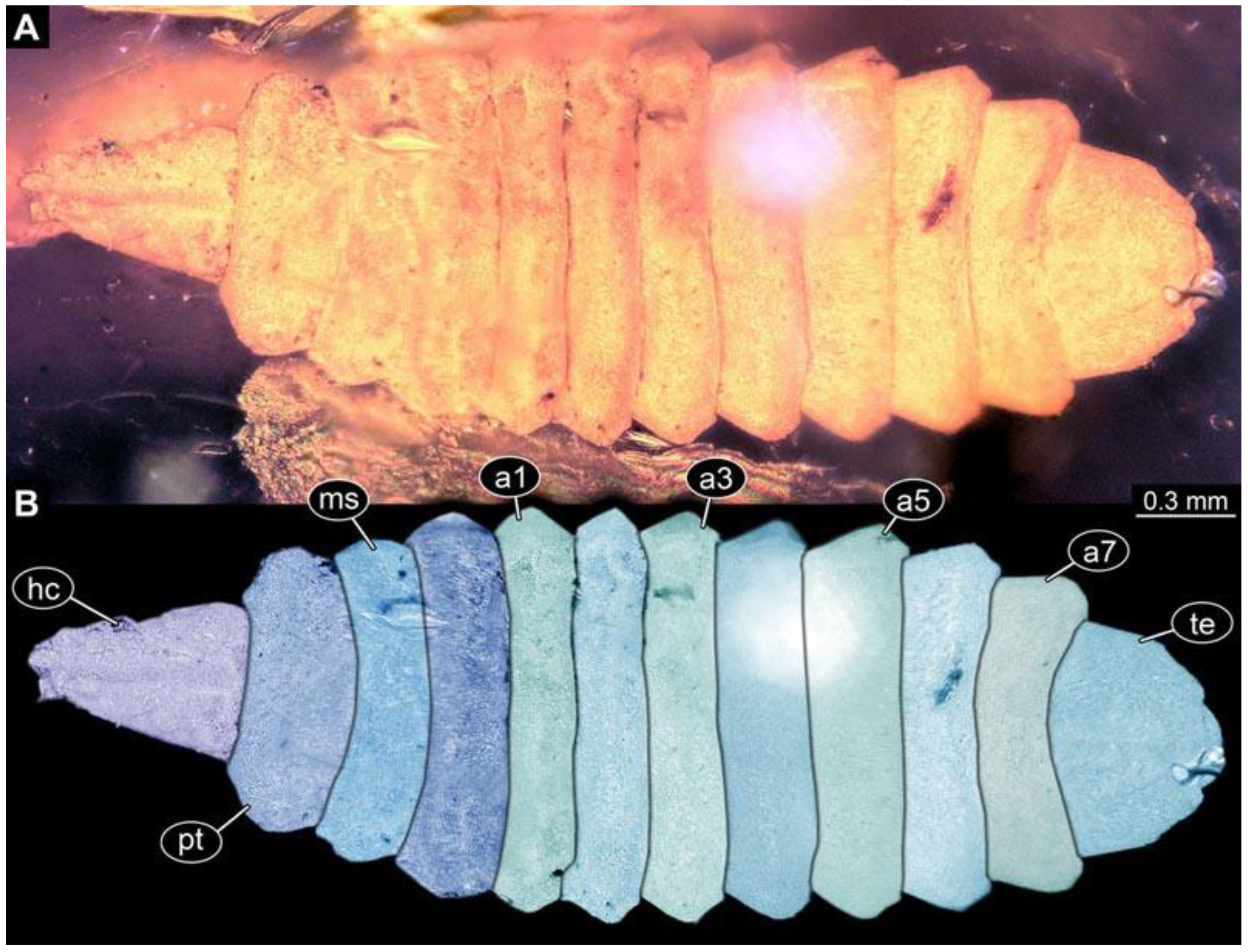
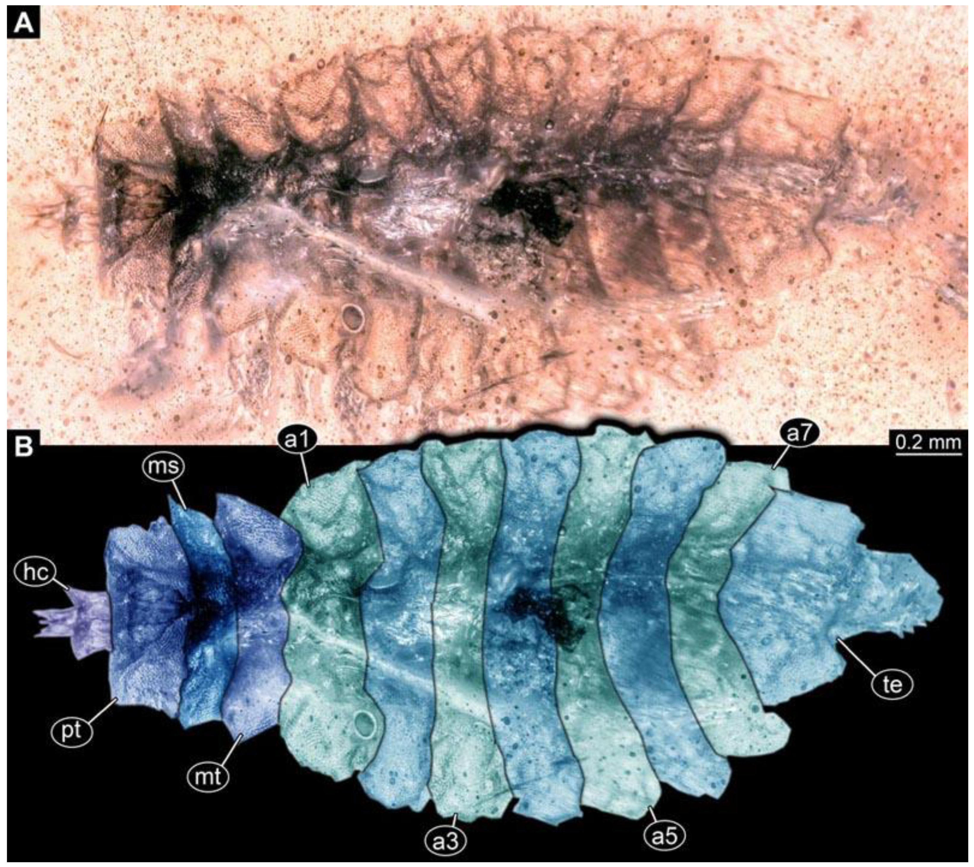
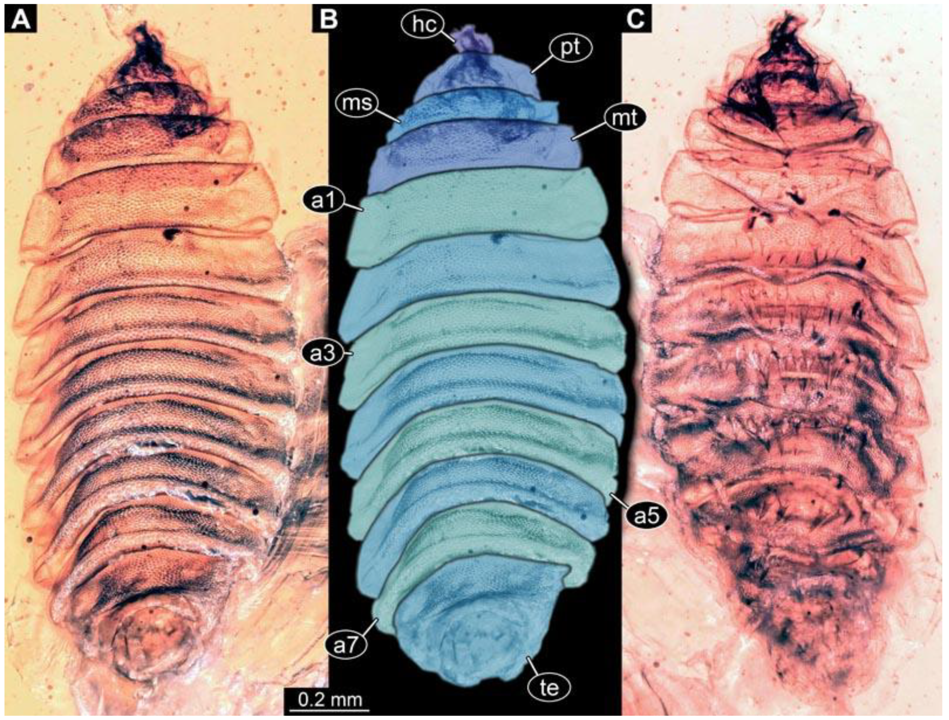
Each specimen of morphotype 1 is briefly described, including the condition of the amber piece and preservation quality of the larvae. Visible details of every specimen and dimensions of the body and head capsule are provided. Although every specimen should have all the mentioned details available, if the detail was not visible, for example due to being covered by dirt particles or being only accessible from one side, it is not mentioned.
(1) PED 0617: Specimen strongly bent on anterior part (Figure 2). Surface structure apparent. Head capsule well preserved; due to position, length could not be reliably measured. This also applies to total body length. Short spine-like setae on thorax segments well developed, longer ventral spine-like setae on abdomen segments well developed. Paired spiracles at trunk end well apparent. Dimensions: body width 1.58 mm, head capsule width 0.13 mm.
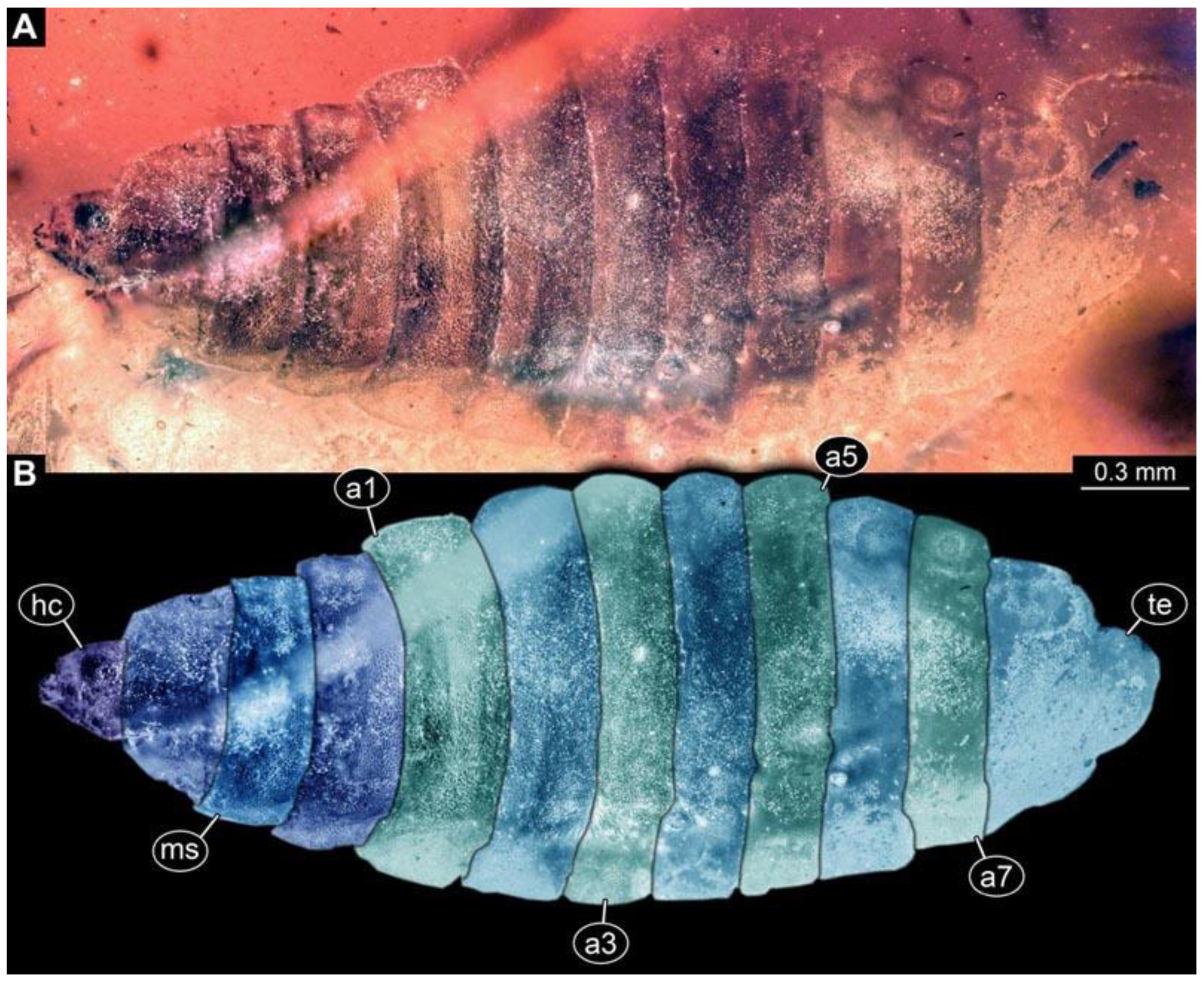
(2) PED 0685: Specimen appears slightly stretched (Figure 3), partly concealed by blurry amber and particles. Long ventral spine-like setae on abdomen segments well developed. Surface structure apparent. Dimensions: body length 8.27 mm, body width 1.05 mm, head capsule length 0.79 mm, head capsule width 0.11 mm.
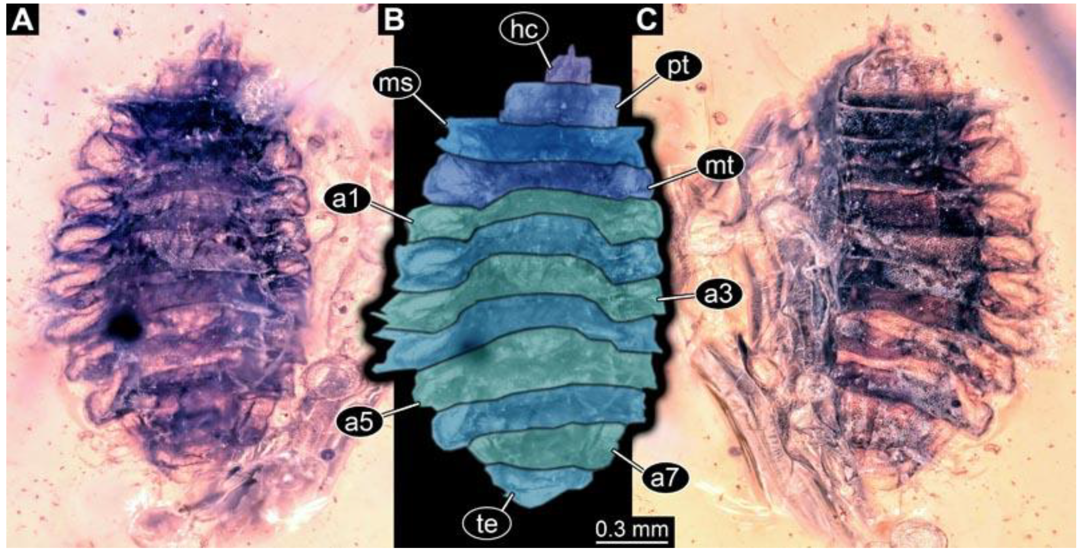
(3) PED 0701: Specimen torn apart ventrally in the more posterior region, also slightly bent (Figure 4). Surface structure apparent. Head capsule and thorax well preserved. Short spine-like setae on thorax segments and longer ventral spine-like setae on abdomen segments well developed (Figure 4C). Dimensions: body length 5.08 mm, body width 0.57 mm, head capsule length 0.67 mm, head capsule width 0.13 mm.
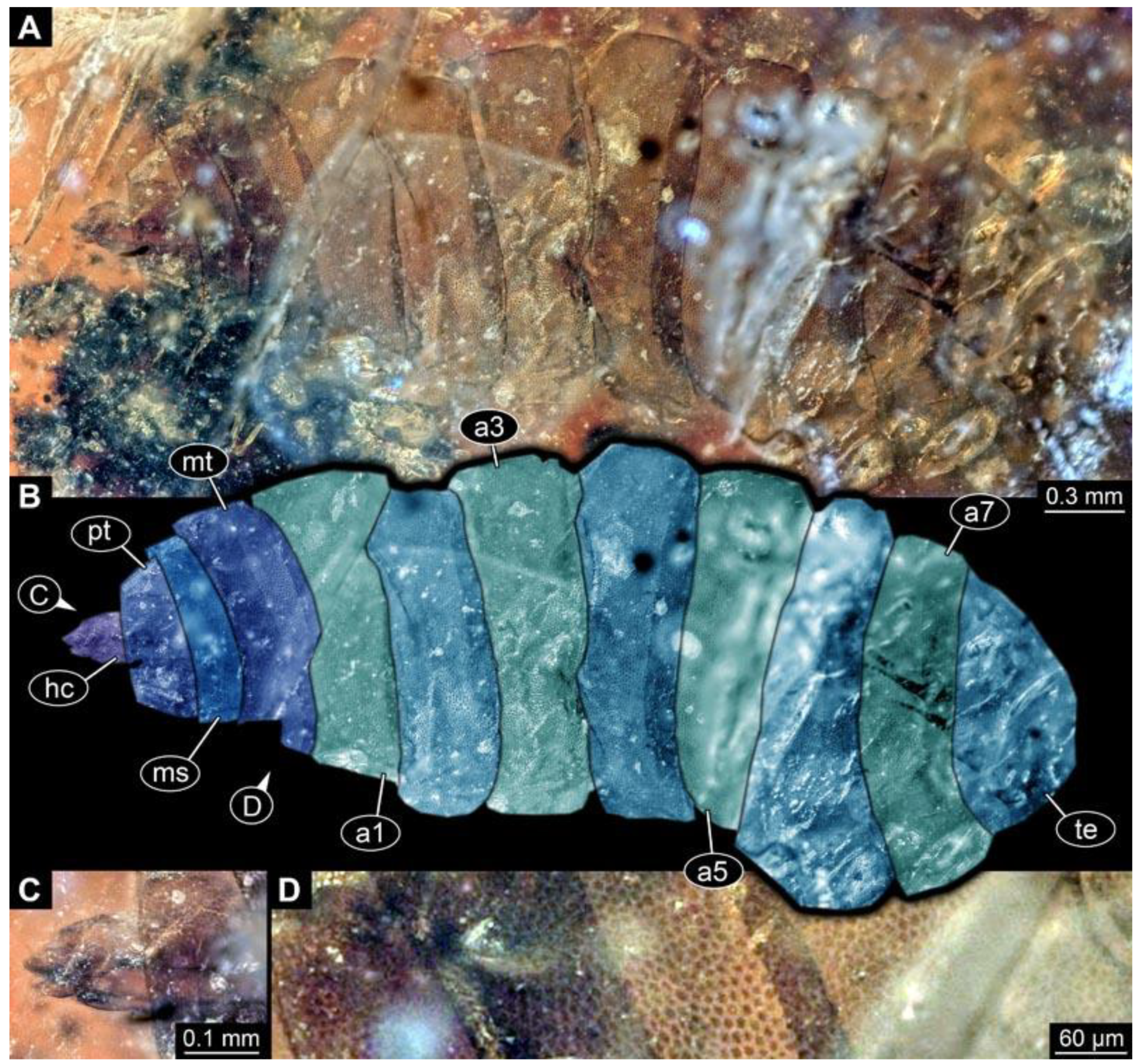
(4) PED 1200-4: Specimen relatively straight, slightly damaged on lateral side of abdomen segment 5 (Figure 5). Surface structure apparent (Figure 5D). Head capsule well preserved (Figure 5C). Two rows of short, spine-like setae per segment of abdomen on ventral and dorsal sides (Figure 5D). Paired spiracle visible on trunk end. Dimensions: body length 1.3 mm, body width 0.77 mm, head capsule length 0.28 mm, head capsule width 0.13 mm.
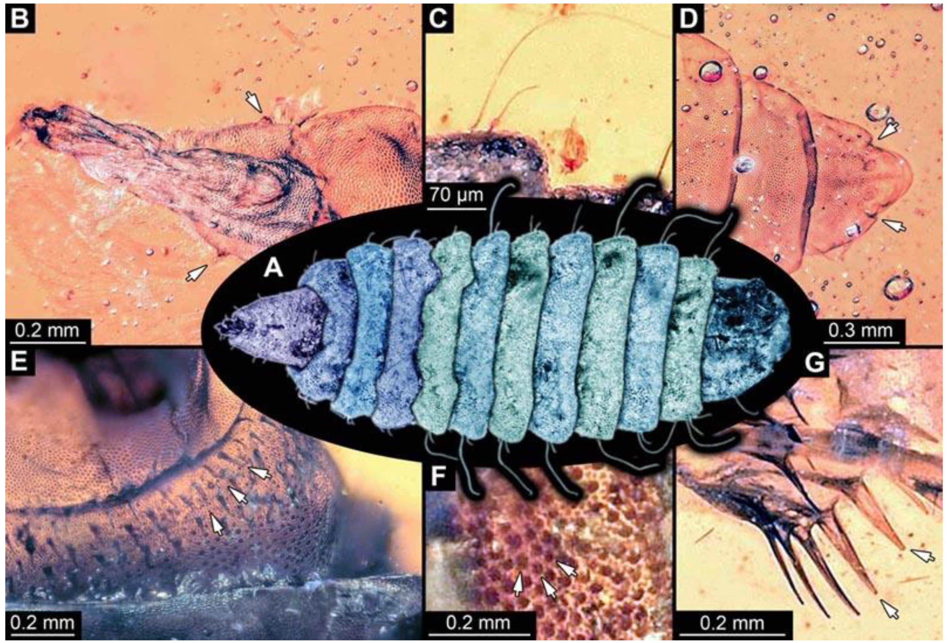
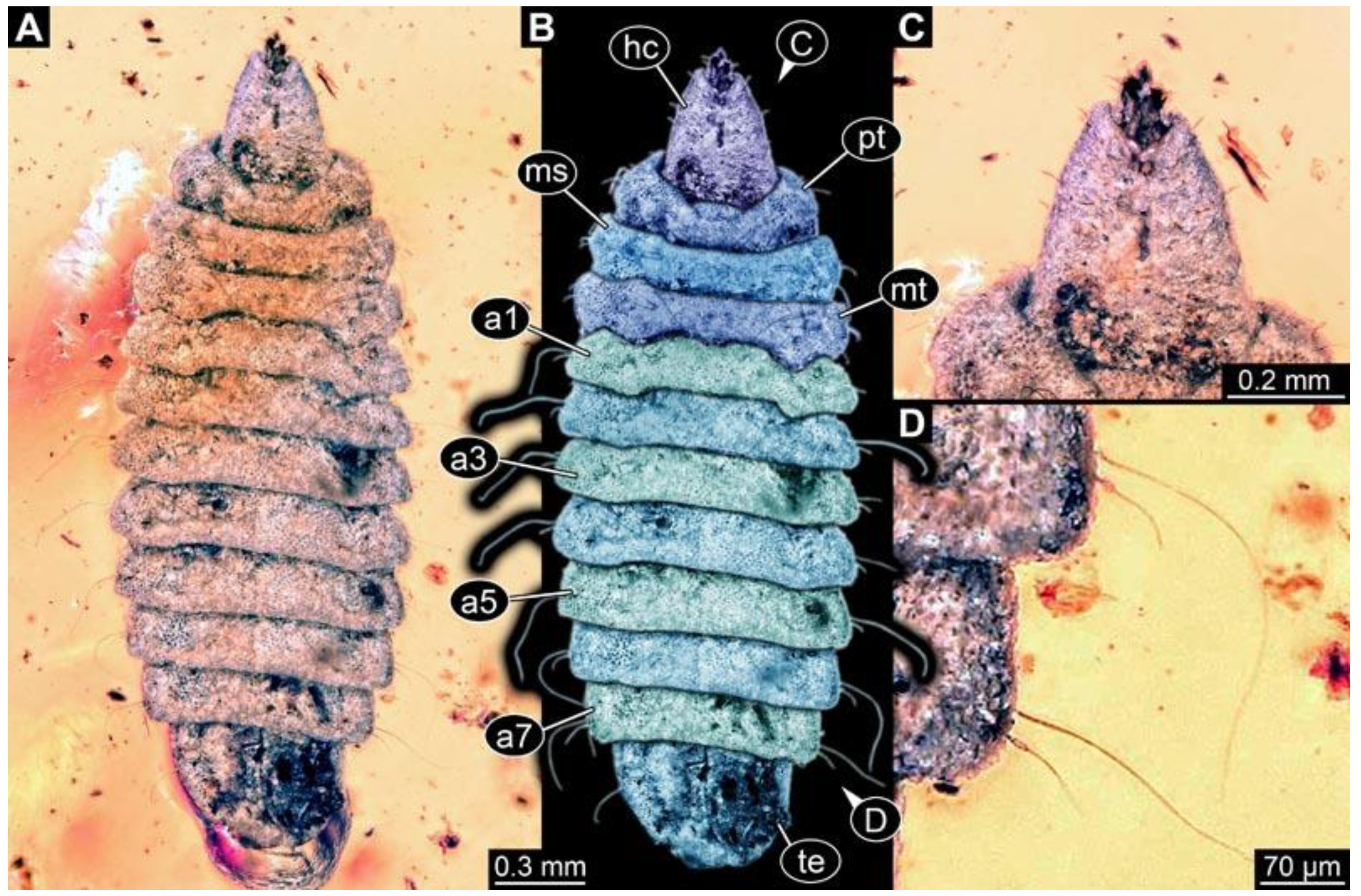
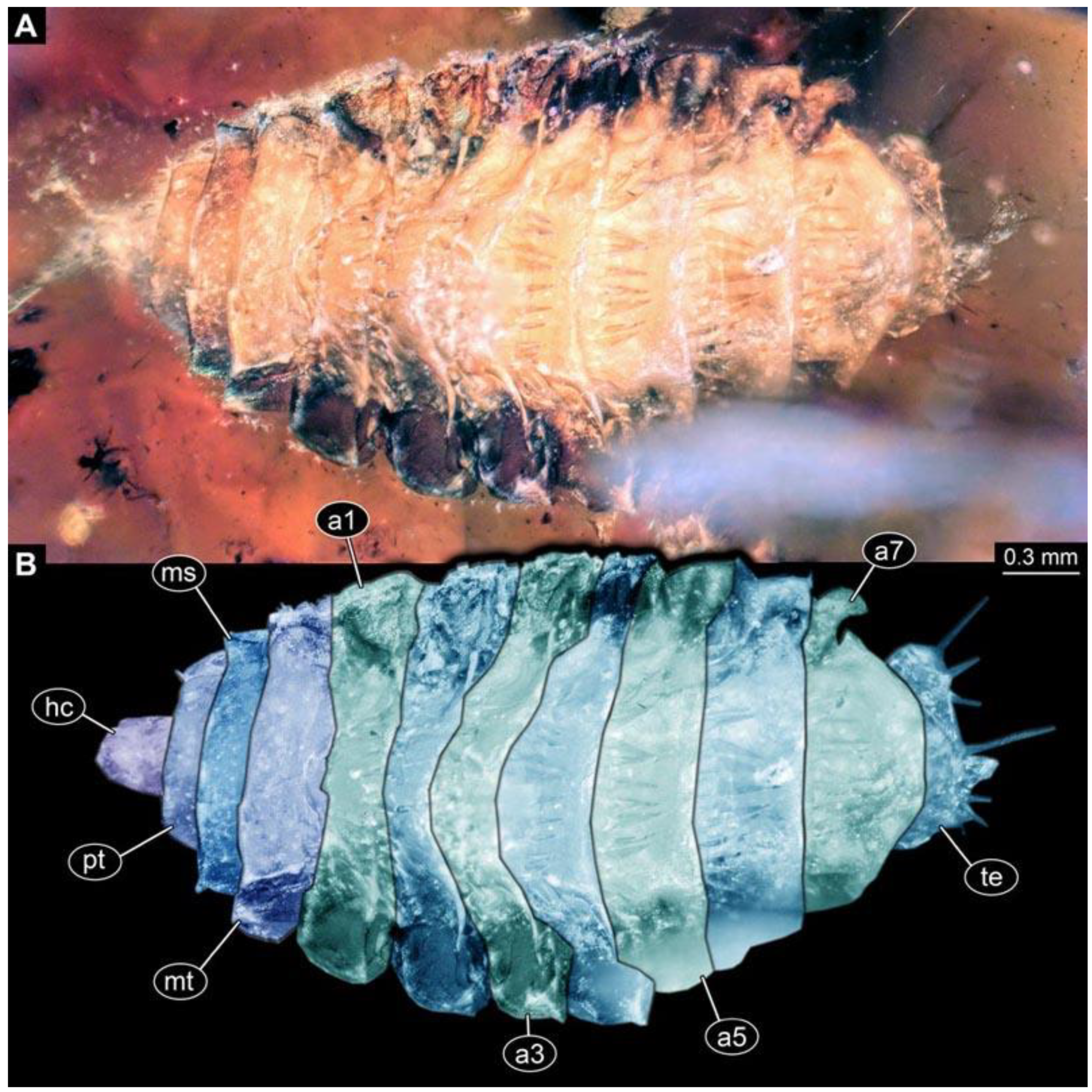
(5) PED 1276: Specimen visible in lateral view, most detail-rich specimen of morphotype 1 (Figure 6). Surface structure apparent. Head capsule and trunk well preserved (Figure 6F). One anterior spiracle visible on prothorax. Short spine-like setae on thorax segments and longer ventral spine-like setae on abdomen segments well developed (Figure 6C–E). Ventral spines on abdomen segment 1 appear shorter; two rows instead of one. Spine-like setae visible on the trunk end. Dimensions: body length 5.58 mm, body width 1.29 mm, head capsule length 0.68 mm, head capsule width 0.25 mm.
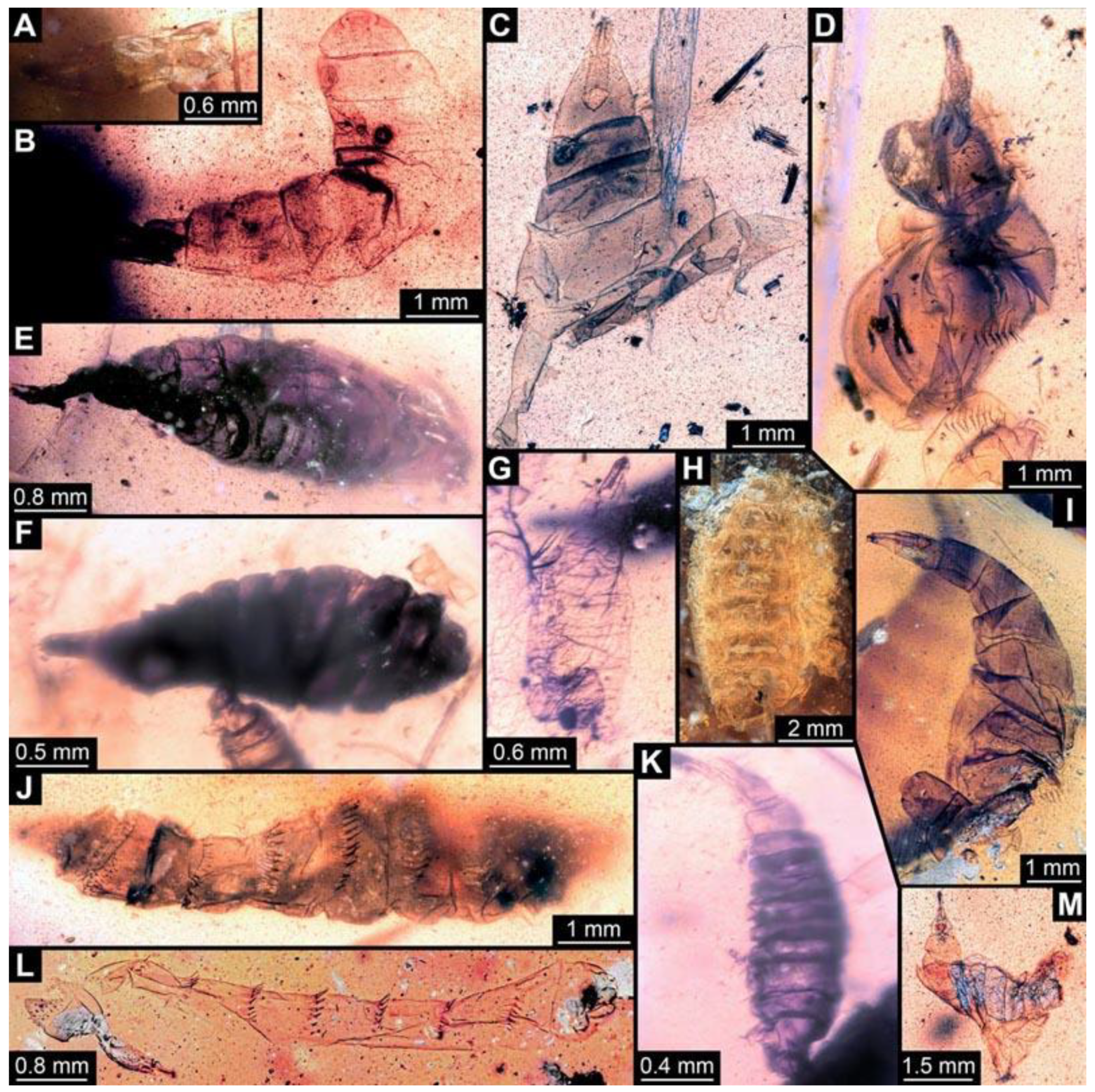
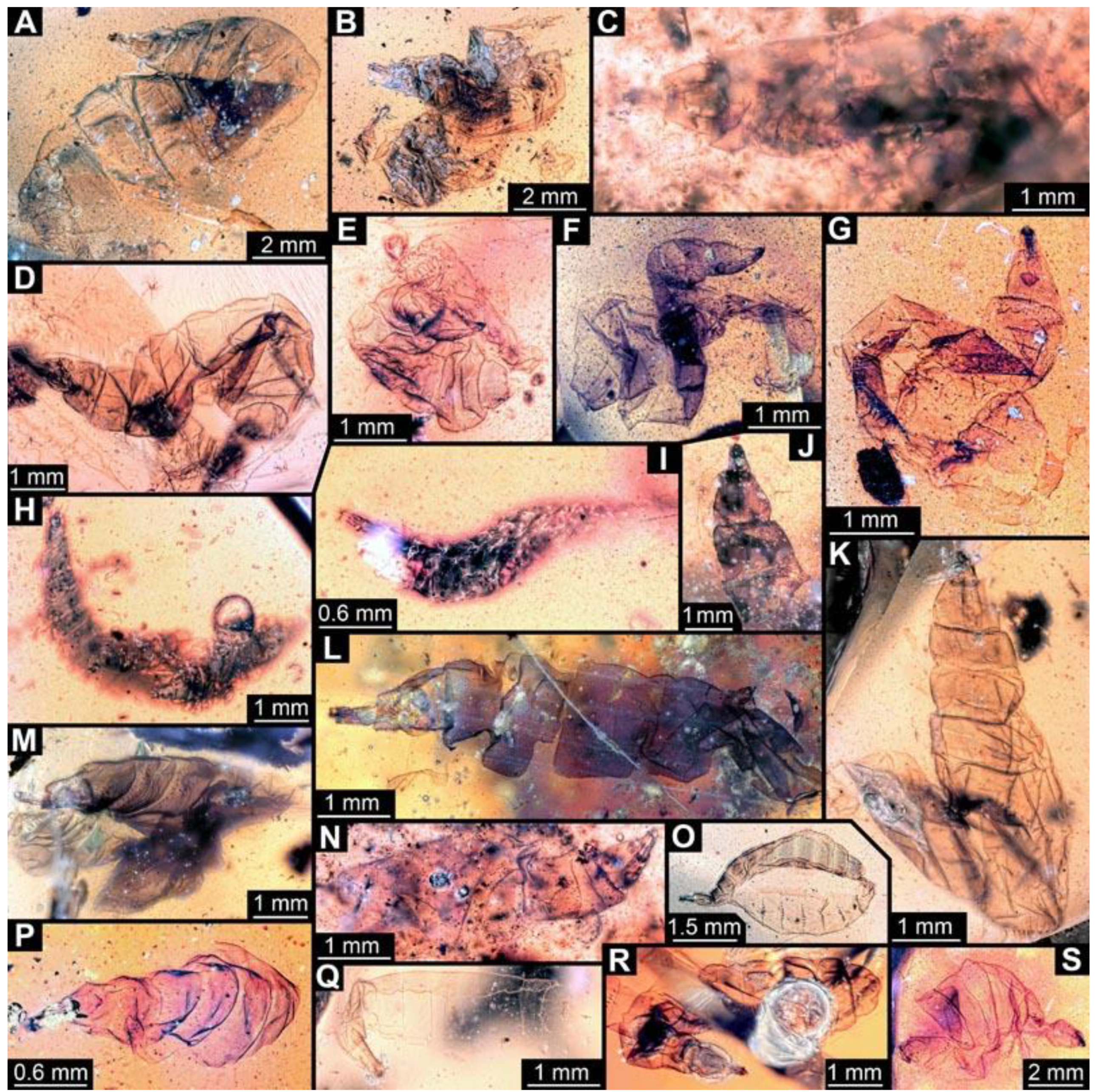
(6) PED 0511: Specimen oblique in the amber (Figure 7A), numerous particles conceal details. Surface structure apparent. Head capsule and thorax dark pigmented. Ventral spine-like setae on abdomen segments well developed. Specimen bent in the middle of the trunk. No measurements possible due to position of the larva.
(7) PED 0628: Specimen bent in the anterior region, torn apart at the posterior end, slightly concealed by irregularities and particles in the amber (Figure 7B). Surface structure apparent. Head capsule well preserved. Long ventral spine-like setae on abdomen segments well developed. Dimensions: body width 1.23 mm, head capsule length 0.84 mm, head capsule width 0.14 mm.
(8) PED 1349: Specimen oblique in the amber piece, only accessible in antero-dorsal view (Figure 7E). Surface structure apparent. Head capsule well-preserved. No measurements could be recorded due to the orientation of the specimen.
(9) PED 0490: Specimen accessible only in dorsal view (Figure 7F). Numerous particles conceal details, surface structure apparent. Head capsule well preserved. Short spine-like setae on thorax segments well developed. Specimen is crumpled at the posterior part; total body length could not be measured. Dimensions: body width 1.57 mm, head capsule length 1.4 mm, head capsule width 0.42 mm.
(10) PED 0548: Specimen accessible only in ventral view (Figure 7G), slightly bent, surrounded by cracks/air inclusions that conceal details. Body and head capsule well preserved. Dimensions: body length 1.3 mm, body width 0.5 mm, head capsule length 0.27 mm, head capsule width 0.11 mm.
(11) PED 0714: Specimen torn apart in the trunk region, only head capsule, thorax and abdomen segments 1 and 2 visible and well-preserved. (Figure 7H). Surface structure apparent. One anterior spiracle apparent in prothorax. Short spine-like setae on thorax segments and longer ventral spine-like setae on abdomen segments well developed. Total body length could not be measured. Dimensions: body width 1.39 mm, head capsule length 0.98 mm, head capsule width 0.15 mm.
(12) PED 0567-1: Specimen ripped apart at the posterior end (Figure 7I), therefore total body length could not be reliably measured. Surface structure apparent. Head capsule well preserved. Dimensions: body width 1.2 mm, head capsule length 0.57 mm, head capsule width 0.15 mm.
(13) PED 0567-3: Specimen cut off at the edge of the amber, only prothorax, mesothorax, metathorax and abdomen segment 1 apparent (Figure 7J). Head capsule well preserved. Short spine-like setae on thorax segments well developed. Dimensions: body width 1.7 mm, head capsule length 1.08 mm, head capsule width 0.32 mm.
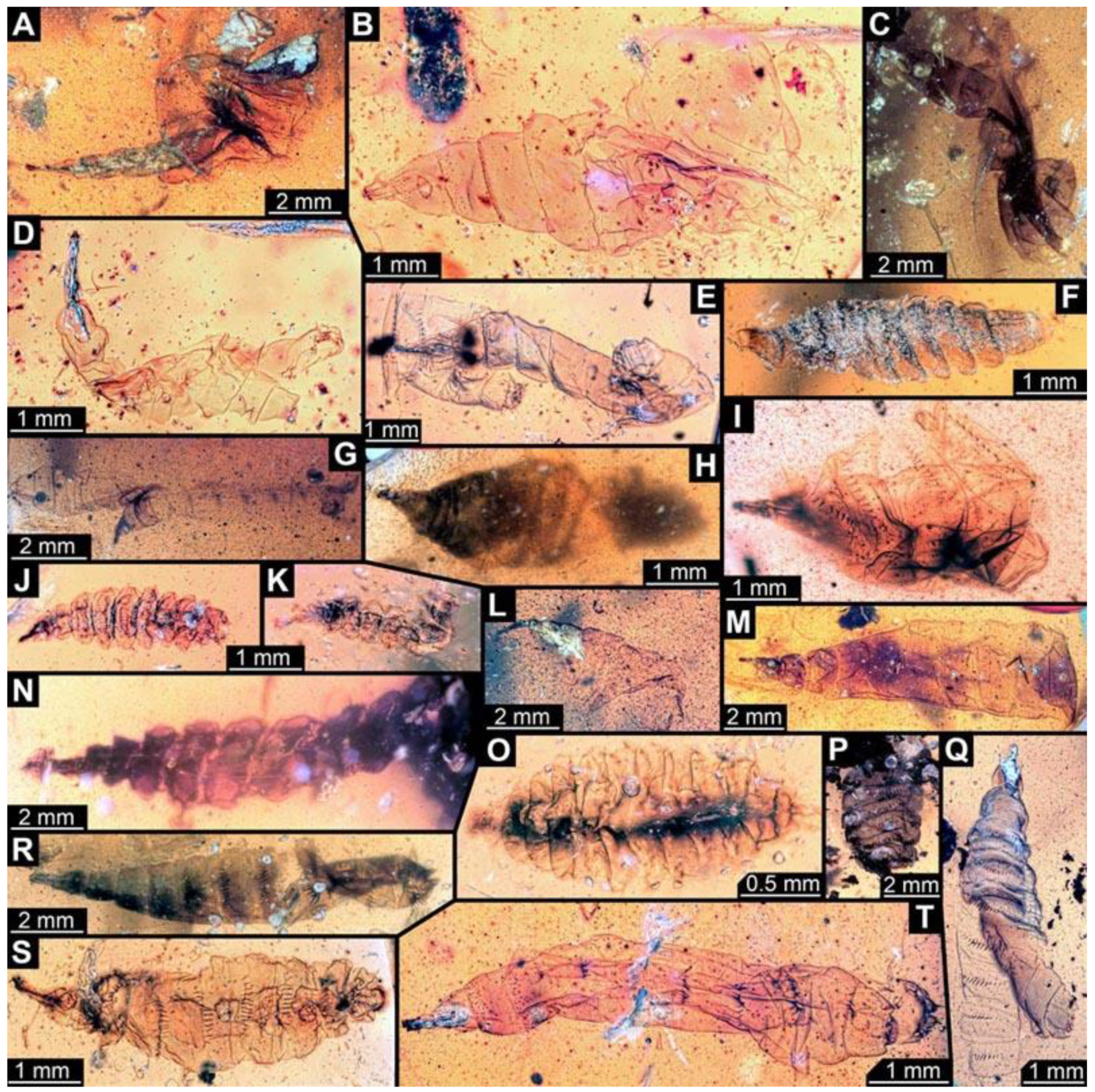
(14) PED 0497: Specimen accessible in dorsal and ventral view, better visible in dorsal view (Figure 8B). Numerous dirt particles in the amber, one large dark object conceals the head capsule, which is only visible with transmitted light and enhanced brightness (Figure 8A). Short spine-like setae on thorax segments well developed. Paired posterior spiracles in trunk end. Trunk is bent by about 90° in the middle, so the total body length could not reliably be measured. Dimensions: body width 1.67 mm, head capsule length 1.8 mm, head capsule width 0.5 mm.
(15) PED 0599-1: Specimen torn off at abdomen segment 2. Head, thorax and abdomen segment 1 well apparent (Figure 8C). Surface structure apparent. Head capsule well preserved. Short spine-like setae on thorax segments well developed. Total body width was measured on the widest point of the last intact segment. Dimensions: body width 2.33 mm, head capsule length 1.4 mm, head capsule width 0.24 mm.
(16) PED 0599-2: Specimen very crumpled (Figure 8D), therefore no measurements of the body possible. Surface structure apparent. Head capsule well preserved. Long ventral spine-like setae on abdomen segments well developed. Dimensions: head capsule length 1.14 mm, head capsule width 0.46 mm.
(17) PED 0550: Specimen oblique in the amber (Figure 8E). Head capsule is slightly stretched. Posterior part of body concealed by particles in the amber; therefore body length could not be reliably measured. Dimensions: body width 1.76 mm, head capsule length 1.51 mm, head capsule width 0.29 mm.
(18) PED 0611-1: Specimen very darkly pigmented (Figure 8F), dirt particles and blurry amber conceal most details. Head capsule well preserved. Dimensions: body length 3 mm, body width 1 mm, head capsule length 0.42 mm, head capsule width 0.2 mm.
(19) PED 0611-3: Specimen is slightly bent, parts concealed by blurry amber (Figure 8G). Head capsule and trunk well preserved. Long ventral spine-like setae on abdomen segments well developed. Dimensions: body length 2.18 mm, body width 0.56 mm, head capsule length 0.67 mm, head capsule width 0.09 mm.
(20) PED 0620: Specimen seemingly dorso-ventrally compressed (Figure 8H), many cracks and air inclusions in the amber piece concealing details. Trunk end slightly folded inwards but still apparent. Head capsule bent by 90° to the trunk, but still visible and well-preserved. Dimensions: body length 5.78 mm, body width 2.63 mm, head capsule length 1.2 mm, head capsule width 0.13 mm.
(21) PED 0601: Specimen slightly bent, the posterior part is torn apart (Figure 8I). Surface structure apparent. Head capsule well preserved. Ventral spine-like setae visible on torn posterior part. Total length of the body could not be reliably measured. Dimensions: body width 2.18 mm, head capsule length 1.2 mm, head capsule width 0.36 mm.
(22) PED 0551: Specimen accessible in dorsal and ventral view, better visible in ventral view (Figure 8J), surrounding amber blurry in some areas. Head capsule well preserved, but partly concealed by irregularities in the amber. Short spine-like setae on thorax segments well developed, longer ventral spine-like setae on abdomen segments well developed. Dimensions: body length 8 mm, body width 1.78 mm, head capsule length 1.34 mm, head capsule width 0.34 mm.
(23) PED 0611-2: Specimen concealed by dirt particles and blurry amber, posterior part crumpled (Figure 8K). Head capsule and trunk well preserved. Total body length could not be reliably measured. Dimensions: body width 0.74 mm, head capsule length 0.33 mm, head capsule width 0.1 mm.
(24) PED 0721: Specimen appears stretched (Figure 8L). Surface structure apparent. Head capsule, prothorax and parts of mesothorax torn off from the rest of the trunk. Head capsule well preserved. Short spine-like setae on thorax segments and longer ventral spine-like setae on abdomen segments well developed. Dimensions: body width 0.58 mm, head capsule length 0.86 mm, head capsule width 0.14 mm.
(25) PED 0691: Specimen crumpled and damaged (Figure 8M). Surface structure visible. Head capsule well preserved. Paired anterior spiracles visible on prothorax. Short spine-like setae on thorax segments well developed. No measurements of the body were recorded. Dimensions: head capsule length 0.92 mm, head capsule width 0.17 mm.
(26) PED 0622: Specimen strongly bent between abdomen segments 2 and 3 (Figure 9A). Surface structure apparent. Head capsule well preserved. Ventral spine-like setae on abdomen segments well developed. The total body length could not be reliably measured. Dimensions: body width 3.33 mm, head capsule length 1.37 mm, head capsule width 0.27 mm.
(27) PED 0653: Specimen strongly crumpled (Figure 9B), air inclusions and particles in the amber conceal most details, no measurements of the body were recorded. Head capsule well preserved. Short spine-like setae on thorax segments and longer ventral spine-like setae on abdomen segments well developed. Dimensions: head capsule length 1.31 mm, head capsule width 0.33 mm.
(28) PED 0761-1: Mostly concealed by dirt particles, only head capsule, thorax and abdomen segment 1 visible (Figure 9C). Surface structure apparent. Head capsule well preserved. Paired anterior spiracles visible on prothorax. Total body length could not be measured. Dimensions: body width 1.21 mm, head capsule length 0.98 mm, head capsule width 0.26 mm.
(29) PED 0729: Specimen very crumpled (Figure 9D). Surface structure apparent. Head capsule well preserved. Short spine-like setae on thorax segments well developed. Measurements of the body could not be recorded. Dimensions: head capsule length 1.36 mm, head capsule width 0.17 mm.
(30) PED 0743: Specimen very crumpled (Figure 9E). Surface structure apparent. Head capsule well preserved. Short spine-like setae on thorax segments well developed. Measurements of the body could not be recorded. Dimensions: head capsule length 0.75 mm, head capsule width 0.14 mm.
(31) PED 0749: Specimen very crumpled (Figure 9F). Surface structure apparent. Head capsule well preserved. Short spine-like setae on thorax segments well developed. Some of the longer ventral spine-like setae on abdomen segments well developed. No measurements of the body could be recorded. Dimensions: head capsule length 1.01 mm, head capsule width 0.33 mm.
(32) PED 0752: Specimen very crumpled and torn apart (Figure 9G). Surface structure apparent. Head capsule well preserved. Short spine-like setae on thorax segments well developed. Some of the longer ventral spine-like setae on abdomen segments visible. No measurements of the body could be recorded. Dimensions: head capsule length 1.43 mm, head capsule width 0.5 mm.
(33) PED 0771: Only head capsule and thorax visible, rest of trunk torn off (Figure 9J). Surface structure apparent. Head capsule well preserved. Short spine-like setae on thorax segments well developed. The total body length could not be measured. Dimensions: body width 1.53 mm, head capsule length 1.45 mm, head capsule width 0.48 mm.
(34) PED 0770: Specimen strongly crumpled and torn apart (Figure 9K). Surface structure apparent. Head capsule well preserved. Short spine-like setae on thorax segments well developed. Total body length could not be measured. Dimensions: body width 1.53 mm, head capsule length 1.09 mm, head capsule width 0.36 mm.
(35) PED 0761-2: Specimen strongly damaged, one half of the trunk torn off (Figure 9L). Surface structure apparent. Head capsule well preserved. Short spine-like setae on thorax segments well developed. Total body length could not be measured. Dimensions: body width 1.61 mm, head capsule length 1.44 mm, head capsule width 0.26 mm.
(36) PED 0762: Specimen oblique in the amber piece, strongly crumpled (Figure 9M). Surface structure apparent. Head capsule well preserved. Short spine-like setae on thorax segments well developed. No measurements of the body could be recorded. Dimensions: head capsule length 1.41 mm, head capsule width 0.23 mm.
(37) PED 0761-3: Mostly concealed by dirt particles, only head capsule, thorax and some abdomen segments visible (Figure 9N). Surface structure apparent. Head capsule well preserved. One anterior spiracle apparent on prothorax. Short spine-like setae on thorax segments well developed. Dimensions: body width 0.87 mm, head capsule length 0.88 mm, head capsule width 0.25 mm.
(38) PED 0846: Specimen separated into ventral and dorsal halves (Figure 9O). Surface structure apparent. Head capsule and prothorax well preserved. Short spine-like setae on thorax segments and longer ventral spine-like setae on abdomen segments well developed. Paired spiracles visible on trunk end. No measurements of the body could be recorded. Dimensions: head capsule length 0.77 mm, head capsule width 0.12 mm.
(39) PED 0766: Specimen torn in the posterior region (Figure 9P). Surface structure apparent. Head capsule well preserved. Short spine-like setae on thorax segments well developed. No measurements of the body could be recorded. Dimensions: head capsule length 0.68 mm, head capsule width 0.17 mm.
(40) PED 0811-3: Head capsule and thorax of specimen bent, rest of body straight and relatively transparent (Figure 9Q). Surface structure apparent. Head capsule well preserved. Short spine-like setae on thorax segments well developed. Total body length could not be reliably measured. Dimensions: body width 1.08 mm, head capsule length 0.79 mm, head capsule width 0.26 mm.
(41) PED 0817: Specimen bent in posterior region, separated into ventral and dorsal halves (Figure 9R). Surface structure apparent. Head capsule well preserved. Short spine-like setae on thorax segments and longer ventral spine-like setae on abdomen segments well developed. Paired spiracles visible on trunk end. Total body length could not be reliably measured. Dimensions: body width 1.42 mm, head capsule length 0.89 mm, head capsule width 0.22 mm.
(42) PED 0923: Specimen crumpled and torn apart (Figure 9S). Head capsule appears inflated. No measurements could be recorded. Short spine-like setae on thorax segments and longer ventral spine-like setae on abdomen segments well developed. Surface structure apparent.
(43) PED 0850: Specimen crumpled and torn apart, dirt particles conceal details (Figure 10A). Head capsule slightly stretched. Long ventral spine-like setae on abdomen segments well developed. No measurements of the body could be recorded. Dimensions: head capsule length 1.29 mm, head capsule width 0.19 mm.
(44) PED 0873-1: Specimen crumpled and torn apart from abdomen segment 2 to trunk end (Figure 10B). Surface structure apparent. Head capsule slightly damaged. Paired anterior spiracles visible on prothorax. Short spine-like setae on thorax segments and longer ventral spine-like setae visible on remains of abdomen segments. Total body length could not be measured. Dimensions: body width 1.42 mm, head capsule length 0.95 mm, head capsule width 0.25 mm.
(45) PED 1018: Specimen very crumpled, air inclusions cover some parts (Figure 10C). Surface structure apparent on thorax segments. Head capsule well preserved, with bright pigmentation. Paired anterior spiracles are visible on prothorax. Short spine-like setae on thorax segments and longer ventral spine-like setae on abdomen segments well developed. Total body length could not be reliably measured. Dimensions: body width 1.33 mm, head capsule length 1.44 mm, head capsule width 0.31 mm.
(46) PED 0873-2: Specimen crumpled and torn apart from mesothorax to trunk end (Figure 10D). Surface structure apparent. Head capsule stretched. Short spine-like setae on thorax segments and longer ventral spine-like setae on abdomen segments well developed. No measurements of body could be recorded. Dimensions: head capsule length 1.71 mm, head capsule width 0.27 mm.
(47) PED 0962: Specimen separated into ventral and dorsal halves (Figure 10E). Surface structure apparent. Head capsule well preserved. Paired anterior spiracles visible on prothorax. Short spine-like setae on thorax segments and longer ventral spine-like setae on abdomen segments well developed. No measurements of the body could be recorded. Dimensions: head capsule length 0.69 mm, head capsule width 0.14 mm.
(48) PED 1137: Specimen only accessible in dorsal view, concealed by numerous bubbles (Figure 10F). Surface structure apparent. Head capsule missing, but width could be approximated by opening of prothorax. Short spine-like setae on thorax segments well developed. Dimensions: body width 1.18 mm, head capsule width 0.21 mm.
(49) PED 1560: Specimen torn apart in ventral and dorsal halves from thorax to trunk end (Figure 10G). Numerous dirt particles and air inclusions conceal details. Surface structure apparent. Head capsule well preserved. Short spine-like setae on thorax segments and longer ventral spine-like setae on abdomen segments well developed. Total body length could not be measured. Dimensions: body width 1.33 mm, head capsule length 0.83 mm, head capsule width 0.12 mm.
(50) PED 0987: Specimen twisted in the posterior trunk region, concealed by numerous dirt particles (Figure 10H). Head capsule not completely visible because of state of the amber and darker pigmentation on the larva. Short spine-like setae on thorax segments well developed. Total body length and length of head capsule could not be reliably measured. Dimensions: body width 2.87 mm, head capsule width 0.28 mm.
(51) PED 1167: Specimen crumpled, numerous dirt particles in the amber conceal details (Figure 10I). Surface structure apparent. Head capsule well preserved but concealed by blurry amber. Short spine-like setae on thorax segments and longer ventral spine-like setae on abdomen segments well developed. Total body length could not be measured. Dimensions: body width 1.35 mm, head capsule length 1.42 mm, head capsule width 0.26 mm.
(52) PED 0921: Specimen visible in ventral view, overall well preserved (Figure 10J). Long ventral spine-like setae on abdomen segments well developed. Surface structure apparent. Dimensions: body length 2.95 mm, body width 0.88 mm, head capsule length 0.46 mm, head capsule width 0.08 mm.
(53) PED 0934: Specimen crumpled and torn apart in posterior region (Figure 10K). Surface structure apparent. Head capsule not fully visible, concealed by dirt particles and bubbles. Total body length could not be measured. Dimensions: body width 0.61 mm, head capsule width 0.06 mm.
(54) PED 1001: Specimen torn apart in the posterior region, numerous particles conceal more details (Figure 10L). Surface structure apparent on thorax segments. Head capsule slightly bent but overally well preserved. Thorax segments with bright pigmentation. Long ventral spine-like setae on abdomen segments well developed. Total body length could not be measured. Dimensions: body width 1.2 mm, head capsule length 1.12 mm, head capsule width 0.19 mm.
(55) PED 1140: Specimen relatively straight, visible in dorsal view, the largest of all specimens (Figure 10M). Surface structure apparent. Head capsule well preserved. Short spine-like setae on thorax segments and longer ventral spine-like setae on abdomen segments well developed. Dimensions: body length 12.57 mm, body width 3.07 mm, head capsule length 1.24 mm, head capsule width 0.24 mm.
(56) PED 0990: Specimen poorly recognizable due to numerous particles and bubbles in the amber piece (Figure 10N). Head capsule mostly covered by bubbles, length of head capsule could not be measured. Posterior part of trunk completely covered by large particles. Total body length could not be measured. Dimensions: body width 2.57 mm, head capsule width 0.45 mm.
(57) PED 1279: Specimen crumpled, numerous particles conceal parts of the head capsule and most of the thorax (Figure 10P). Surface structure apparent. Head capsule strongly bent, not fully visible, length could not be measured. Long ventral spine-like setae on abdomen segments well developed. Dimensions: body width 3.05 mm, head capsule width 0.33 mm.
(58) PED 1174: Specimen separated into ventral and dorsal halves at posterior part of abdomen (Figure 10Q). Head capsule missing. Metathorax and abdomen segments 1–3 with bright pigmentation. Surface structure apparent. Short spine-like setae on thorax segments and longer ventral spine-like setae on abdomen segments well developed. Dimensions: body length 6.01 mm, body width 1.25 mm.
(59) PED 1014: Specimen damaged and twisted in posterior part of abdomen, numerous particles and bubbles conceal more details (Figure 10R). Surface structure apparent. Head capsule well preserved. Short spine-like setae on thorax segments and longer ventral spine-like setae on abdomen segments well developed. Total body length could not be reliably measured. Dimensions: body width 2.05 mm, head capsule length 1.55 mm, head capsule width 0.36 mm.
(60) PED 1025: Specimen only accessible in ventral view, numerous particles conceal some details (Figure 10S). Surface structure apparent. Head capsule partially concealed by dirt particles. Short spine-like setae on thorax segments and longer ventral spine-like setae on abdomen segments well developed. Paired spiracles visible on trunk end. Dimensions: body length 5.15 mm, body width 1.57 mm, head capsule length 0.87 mm, head capsule width 0.18 mm.
(61) PED 1044: Specimen crumpled, trunk end split (Figure 10T). Paired anterior spiracles visible on prothorax. Surface structure apparent. Head capsule well preserved. Short spine-like setae on thorax segments and longer ventral spine-like setae on abdomen segments well developed. Paired spiracles visible on trunk end. Dimensions: body length 6.32 mm, body width 1.21 mm, head capsule length 0.81 mm, head capsule width 0.18 mm.
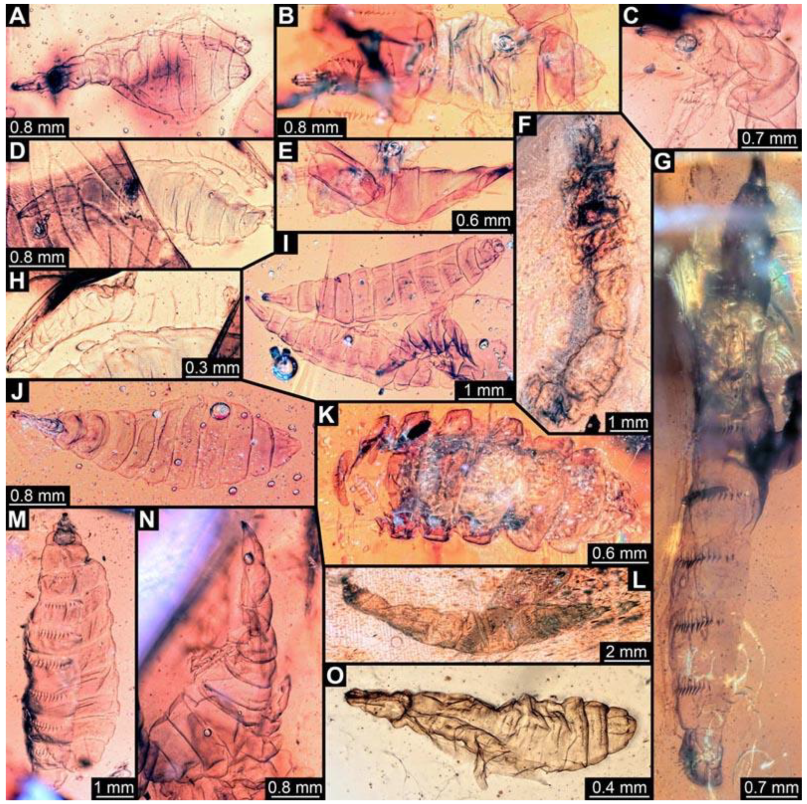
(62) PED 0819-1: Specimen slightly inflated, separated into ventral and dorsal halves (Figure 11A). Surface structure apparent. Head capsule well preserved. Paired anterior spiracles visible on prothorax. Paired posterior spiracles visible on the trunk end. Ventral spine-like setae on abdomen segments well developed. Body width could not be reliably measured. Dimensions: body length 3.36 mm, head capsule length 0.69 mm, head capsule width 0.19 mm.
(63) PED 0819-4: Specimen partly concealed by other specimens in the amber piece (Figure 11B). Surface structure apparent. Head capsule well preserved. Paired spiracles on trunk end apparent. Dimensions: body length 3.88 mm, body width 1.13 mm, head capsule length 0.61 mm, head capsule width 0.17 mm.
(64) PED 1200-1: Specimen stretched and bent (Figure 11C). Surface structure apparent. Head capsule well preserved, but partially covered by particles and bubbles. Short spine-like setae on thorax segments well developed. Total body length could not be measured. Dimensions: body width 0.95 mm, head capsule length 0.64 mm, head capsule width 0.13 mm.
(65) PED 0819-2: Specimen slightly bent (Figure 11D). Surface structure apparent. Head capsule well preserved. Short spine-like setae on thorax segments well developed. Dimensions: body length 3.51 mm, body width 0.99 mm, head capsule length 0.44 mm, head capsule width 0.19 mm.
(66) PED 0819-5: Specimen partly concealed by other specimens in the amber piece, also posterior part torn off (Figure 11E). Surface structure apparent. Head capsule well preserved. Long ventral spine-like setae on abdomen segments well developed. Total body length could not be measured. Dimensions: body width 0.71 mm, head capsule length 0.72 mm, head capsule width 0.24 mm.
(67) PED 1274: Specimen crumpled and partly concealed by dirt in the amber (Figure 11F). Most details not accessible, therefore not measurable.
(68) PED 1247: Specimen stretched, bubbles conceal parts of thorax (Figure 11G). Surface structure apparent. Head capsule well preserved, but concealed by blurry amber, length of head capsule could not be measured. Short spine-like setae on thorax segments and longer ventral spine-like setae on abdomen segments well developed. Spine-like setae are visible on trunk end. Dimensions: body length 8.3 mm, body width 0.99 mm, head capsule width 0.28 mm.
(69) PED 0819-3: Specimen slightly bent, slightly stretched, oblique in the amber piece (Figure 11H). Head capsule well preserved. Ventral spine-like setae on abdomen segments well developed, appear rather short. Total body length could not be reliably measured. Dimensions: head capsule length 0.36 mm, head capsule width 0.23 mm.
(70) PED 0819-6: Specimen crumpled and torn apart in trunk region, overlapping with two specimens (0819-7 and 0819-8; Figure 11I). Surface structure apparent. Head capsule well preserved, but concealed by other specimen. Total body length could not be measured. Dimensions: body width 0.75 mm, head capsule length 0.56 mm, head capsule width 0.25 mm.
(71) PED 0819-7: Specimen overlapping with other specimens in the amber piece (Figure 11I). Surface structure apparent. Head capsule well preserved. One anterior spiracle apparent on prothorax. Short spine-like setae on thorax segments and longer ventral spine-like setae on abdomen segments well developed. Paired spiracles visible in the trunk end. Dimensions: body length 4.6 mm, body width 1 mm, head capsule length 0.55 mm, head capsule width 0.2 mm.
(72) PED 0819-8: Specimen overlapping with other specimens in the amber piece (Figure 11I). Surface structure apparent. Head capsule well preserved. Paired anterior spiracles are visible on prothorax. Short spine-like setae on thorax segments and longer ventral spine-like setae on abdomen segments well developed. Dimensions: body length 3.75 mm, body width 0.88 mm, head capsule length 0.55 mm, head capsule width 0.17 mm.
(73) PED 0819-9: Specimen accessible in dorsal and ventral view, better visible in dorsal view, some parts concealed by bubbles (Figure 11J). Surface structure apparent. Head capsule well preserved. Paired anterior spiracles are visible on prothorax. Paired spiracles visible on trunk end. Dimensions: body length 4.1 mm, body width 1.29 mm, head capsule length 0.69 mm, head capsule width 0.15 mm.
(74) PED 1282: Specimen slightly twisted and bent (Figure 11L). Numerous particles conceal most details. Surface structure visible in some areas of the abdomen. Head capsule well preserved. Long ventral spine-like setae on abdomen segments well developed. Total body length could not be measured. Dimensions: body width 1.02 mm, head capsule length 0.74 mm, head capsule width 0.2 mm.
(75) PED 1185: Specimen separated into ventral and dorsal halves from thorax to trunk end (Figure 11M). Surface structure apparent. Head capsule well preserved. Short spine-like setae on thorax segments and longer ventral spine-like setae on abdomen segments well developed. Dimensions: body length 7.38 mm, body width 1.55 mm, head capsule length 0.83 mm, head capsule width 0.19 mm.
(76) PED 1200-2: Specimen slightly bent (Figure 11N). Surface structure apparent. Head capsule well preserved. Short spine-like setae on thorax segments well developed. Some small ventral spine-like setae are visible on abdomen segment 6. Dimensions: body length 5.03 mm, body width 1.42 mm, head capsule length 0.72 mm, head capsule width 0.13 mm.
(77) PED 1567: Specimen damaged on ventral side, slightly twisted (Figure 11O). Surface structure barely visible. Head capsule well preserved. Two rows of short spine-like setae visible on dorsal side of abdomen segments, one row of short spine-like setae visible on ventral parts of abdomen segments. Paired spiracles visible on trunk end. Dimensions: body length 1.96 mm, body width 0.5 mm, head capsule length 0.29 mm, head capsule width 0.13 mm.
(78) PED 0567-2: Specimen cut off at the edge of the amber, only prothorax and head capsule apparent (Figure 12A). Dimensions: head capsule length 1.04 mm, head capsule width 0.5 mm.
(79) PED 0681: Specimen torn apart and partly scattered in the amber piece (Figure 12B). Surface structure apparent. Head capsule well preserved, still attached to prothorax and mesothorax, this anterior region separated from the rest of the trunk. One anterior spiracle apparent on prothorax. Short spine-like setae on thorax segments well developed. No measurements of the body could be recorded. Dimensions: head capsule length 1.12 mm, head capsule width 0.41 mm.
(80) PED 0773-1: Multiple crumpled specimens overlapping in this amber piece, only head capsule of specimen apparent (Figure 12C). Dimensions: head capsule length 0.42 mm, head capsule width 0.1 mm.
(81) PED 0773-2: Multiple crumpled specimens overlapping in this amber piece, only head capsule and prothorax of specimen apparent (Figure 12D). Surface structure apparent on prothorax. Dimensions: head capsule length 0.69 mm, head capsule width 0.15 mm.
(82) PED 0773-3: Multiple crumpled specimens overlapping in this amber piece, only head capsule and thorax of specimen apparent (Figure 12E). Surface structure apparent on thorax. Dimensions: head capsule length 0.6 mm, head capsule width 0.14 mm.
(83) PED 0811-1: Specimen very crumpled and torn apart (Figure 12F). Only the head capsule and the thorax of the specimen well preserved. Surface structure apparent on thorax. Short spine-like setae on thorax segments well developed. Dimensions: head capsule length 1.62 mm, head capsule width 0.52 mm.
(84) PED 0764: Specimen very crumpled and torn apart (Figure 12G). Surface structure apparent. Head capsule well preserved. Short spine-like setae on thorax segments well developed. No measurements of the body could be recorded. Dimensions: head capsule length 0.87 mm, head capsule width 0.19 mm.
(85) PED 0811-2: Specimen very crumpled and torn apart (Figure 12H). Only the head capsule and the prothorax of the specimen well preserved. Surface structure apparent on prothorax. Short spine-like setae on the prothorax well developed. No measurements on body could be recorded. Dimensions: head capsule length 1.46 mm, head capsule width 0.26 mm.
(86) PED 0978: Specimen stretched, crumpled, bent at thorax (Figure 12I). Surface structure apparent. Head capsule stretched and bent, length of the head capsule could not be reliably measured. Short spine-like setae on thorax segments and longer ventral spine-like setae on abdomen segments well developed. No measurements of the body could be recorded. Dimensions: head capsule width 0.21 mm.
(87) PED 1200-3: Specimen mostly concealed by other specimens, only head capsule and thorax well visible (Figure 12J). Surface structure apparent. Head capsule well preserved. Short spine-like setae on thorax segments well developed. No measurements of the body could be recorded. Dimensions: head capsule length 0.82 mm, head capsule width 0.25 mm.
(88) PED 1353: Specimen separated into ventral and dorsal halves from thorax to trunk end (Figure 12K). Numerous particles and bubbles conceal details. Surface structure apparent. Head capsule well preserved, but not completely visible due to dark pigmented thorax segments, length of the head capsule could not be measured. Short spine-like setae on thorax segments and longer ventral spine-like setae on abdomen segments well developed. Dimensions: body width 1.96 mm, head capsule width 0.18 mm.
(89) PED 1535: Specimen only accessible in lateral view, torn apart posteriorly, particles conceal some details (Figure 12L). Surface structure apparent. Head capsule well preserved, but partly concealed by blurry amber. Long ventral spine-like setae on abdomen segments well developed. Dimensions: body length 3.37 mm, body width 0.46 mm, head capsule length 0.4 mm, head capsule width 0.14 mm.
(90) PED 0614: Specimen oblique within the amber piece, bent in multiple directions (Figure 12M), numerous cracks and air inclusions conceal most details. Surface structure barely visible on some parts of the larva. No measurements could be recorded.
(91) PED 1548-1: Specimen torn apart posteriorly, numerous particles and bubbles conceal details (Figure 12N). Surface structure apparent. Head capsule well preserved, partially with bright pigmentation. Long ventral spine-like setae on abdomen segments well developed. Total body length could not be measured. Dimensions: body width 0.61 mm, head capsule length 0.71 mm, head capsule width 0.11 mm.
(92) PED 1548-2: Specimen torn apart posteriorly, numerous particles and bubbles conceal details (Figure 12O). Surface structure barely visible. Head capsule well preserved, partially with bright pigmentation. Paired spiracles barely visible on trunk end. Total body length could not be measured. Dimensions: body width 0.49 mm, head capsule length 0.33 mm, head capsule width 0.13 mm.
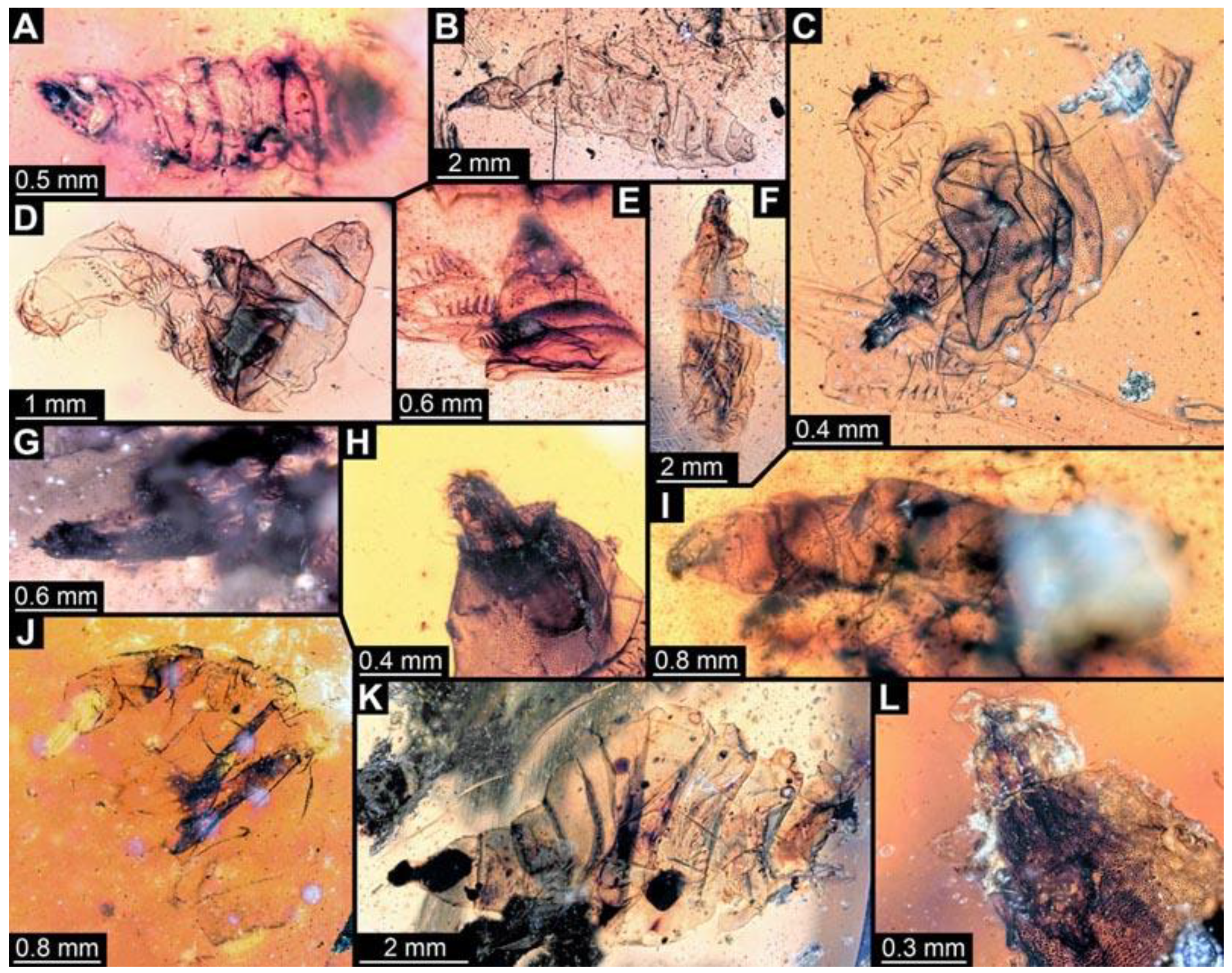
(93) PED 0799: Specimen very crumpled and torn apart (Figure 12P). Numerous particles conceal most details. Surface structure apparent. Head capsule barely visible and crumpled. Short spine-like setae on thorax segments and longer ventral spine-like setae on abdomen segments well developed. No measurements could be recorded.
(94) PED 0678: Specimen strongly crumpled and damaged (Figure 12Q), large cracks and bubbles conceal most parts of the larva. Trunk end and abdomen segments 6 and 7 are the only visible, well-preserved parts. Some ventral spine-like setae are visible through the larva. Surface structure visible on the well-preserved segments. No measurements could be recorded.
(95) PED 0738: Specimen barely visible, very crumpled, numerous particles in the amber conceal details of the body (Figure 12R). Head capsule slightly inflated, with bright pigmentation. No measurements could be recorded.
(96) PED 0760: Specimen torn apart and crumpled (Figure 12S). Surface structure visible on remnants of the larva. Head capsule mostly covered by cracks and air inclusions. No measurements could be recorded.
(97) PED 0712: Specimen mostly concealed by large cracks and bubbles (Figure 12T). Mostly thorax visible. Surface structure apparent. Short spine-like setae on thorax segments well developed. No measurements could be recorded.
(98) PED 0927: Specimen crumpled, stretched and bent in multiple directions (Figure 12U). Concealed by numerous dirt particles and blurry amber. Head capsule appears stretched. No measurements could be recorded.
(99) PED 0777: Specimen twisted, bent, damaged (Figure 12V). Numerous particles conceal details. Head capsule stretched. No measurements could be recorded.
(100) PED 0861: Specimen damaged (Figure 13B). Surface structure apparent. Only head capsule, prothorax and mesothorax are well preserved. Short spine-like setae on thorax segments and longer ventral spine-like setae on abdomen segments well developed. No measurements could be recorded.
(101) PED 1007-3: Specimen crumpled and torn apart (Figure 13C). Surface structure apparent. Head capsule partially covered by bubbles, but well preserved. Long ventral spine-like setae on abdomen segments well developed. Spine-like setae are visible on trunk end. No measurements could be recorded.
(102) PED 0966: Specimen torn apart and crumpled (Figure 13D). Surface structure apparent. Head capsule oblique, could not be measured. Long ventral spine-like setae on abdomen segments well developed. Spine-like setae visible on trunk end. Paired spiracles visible on trunk end. No measurements could be recorded.
(103) PED 0976-1: Specimen crumpled and torn apart from abdomen segment 2 to trunk end; oblique in the amber piece (Figure 13E). Numerous particles conceal most parts of specimen. Surface structure apparent. Ventral spine-like setae visible on abdomen. No measurements could be recorded.
(104) PED 1010: Specimen very crumpled (Figure 13F). Surface structure apparent. Head capsule missing. Short spine-like setae on thorax segments and longer ventral spine-like setae on abdomen segments well developed.
(105) PED 0976-2: Specimen crumpled and torn apart, most parts concealed by particles (Figure 13G). No measurements could be recorded.
(106) PED 1007-1: Specimen torn apart (Figure 13H). Surface structure apparent. Head capsule and thorax visible and well-preserved. Short spine-like setae on thorax segments well developed. No measurements could be recorded.
(107) PED 1129: Specimen twisted and mostly concealed by blurry amber (Figure 13I). Surface structure barely visible. Head capsule missing. Paired spiracles barely visible on trunk end. No measurements could be recorded.
(108) PED 1192: Specimen crumpled, bent and torn apart (Figure 13J). Details concealed by numerous particles and dark colour of the amber. No measurements could be recorded.
(109) PED 1405: Specimen crumpled, torn apart at trunk end, accessible in ventral view (Figure 13K). Large particles conceal major parts of the specimen. Surface structure apparent. Head capsule and part of prothorax very dark. Long ventral spine-like setae on abdomen segments well developed. No measurements could be recorded.
(110) PED 1007-2: Only head capsule and prothorax of specimen visible, partially covered by bubbles (Figure 13L). Surface structure apparent on prothorax. Paired anterior spiracles visible in prothorax. No measurements could be recorded.

3.3. Morphotype 2

3.3.1. Material Examined

We examined 2 specimens: PED 0789, PED 0821.

3.3.2. General Description of Morphotype 2

General structure of larvae of Stratiomyomorpha as outlined above. Body longer than wide and more robust (in comparison to morphotype 1), covered in pellets or scales. Specimens mostly well preserved and straight. Body mostly with bright pigmentation. Head capsule rather triangular, strongly sclerotized. Head capsule longer than wide, prominent, with labrum, mandibular–maxillary complex and labium visible on the anterior end of head capsule. Posterior part of head capsule with metacephalic rod and paired tentorial arms usually retracted into thorax, not visible due to bright pigmentation and surface structure on thorax. Segments of trunk with multiple long hair-like setae, one pair on each lateral side, multiple on dorsal and ventral sides (Figure 1A). Trunk end with hair-like setae. Posterior paired spiracles not visible on trunk end. Dimension range: body length 2.65 mm–3.6 mm (n = 2), body width 1.01 mm–1.23 mm (n = 2).

3.3.3. Short Descriptions of Individual Specimens of Morphotype 2

Each specimen of morphotype 2 is briefly described, including the condition of the amber piece and preservation quality of the larvae. Visible details of every specimen and dimensions of the body and head capsule are provided. Although every specimen should have all the mentioned details available, if the detail was not visible, for example due to being covered by particles or only accessible from one side, it is not mentioned.
(111) PED 0789: Specimen accessible in dorsal view (Figure 14A,B). Surface structure apparent, bright pigmentation on the entire body. Head capsule well-preserved (Figure 14C). Short hair-like setae on head capsule (Figure 14D). Long hair-like setae well developed on entire trunk. Paired hair-like setae laterally on each trunk segment, one longer than the other one for each pair. Dimensions: body length 2.65 mm, body width 1.01 mm, head capsule length 0.57 mm, head capsule width 0.35 mm.
(112) PED 0821: Specimen accessible in dorsal and ventral view, better visible in dorsal view (Figure 15). Surface structure apparent, bright pigmentation on the whole body. Head capsule well-preserved. No setae visible on the whole body. Segments of thorax and abdomen slightly triangular on lateral sides. Dimensions: body length 3.6 mm, body width 1.23 mm, head capsule length 0.65 mm, head capsule width 0.46 mm.

3.4. Morphotype 7

3.4.1. Material Examined

We examined 6 specimens: PED 0723, PED 1107, PED 1109, PED 1218, PED 1295, PED 1346.

3.4.2. General Description of Morphotype 7

General structure of larvae of Stratiomyomorpha as outlined above. Body longer than wide and more robust (in comparison to morphotype 1), covered in pellets or scales. Specimens mostly well preserved and relatively straight. Body mostly yellowish-brown. Head capsule more strongly sclerotized, therefore mostly well-preserved. Head capsule longer than wide with labrum, mandibular–maxillary complex and labium visible on the anterior end of head capsule. Posterior part of head capsule with metacephalic rod and paired tentorial arms usually retracted into thorax, not visible due to dominant surface structure on thorax. Dorsal body side without setae; ventral body side with spine-like setae for locomotion. Posterior paired spiracles on trunk end. Dimension range: Body length 1.64–6.95 mm (n = 5), body width 0.71–2.44 mm (n = 5).

3.4.3. Short Descriptions of Individual Specimens of Morphotype 7

Each specimen of morphotype 7 is briefly described, including the condition of the amber piece and preservation quality of the larvae. Visible details of every specimen and dimensions of the body and head capsule are provided. Although every specimen should have all the mentioned details available, if the detail was not visible, for example due to being covered by dirt particles or only accessible from one side, it is not mentioned.
(113) PED 1107: Specimen accessible only in ventral view (Figure 16). Numerous particles conceal details, surface structure apparent. Head capsule well preserved. Ventral spine-like setae well developed. Dimensions: body length 6.95 mm, body width 2.44 mm, head capsule length 0.38 mm, head capsule width 0.23 mm.
(114) PED 1218: Specimen accessible in dorsal and ventral view, better visible in dorsal view (Figure 17). Numerous particles conceal details, surface structure apparent. Head capsule well preserved. Ventral spine-like setae well developed. Dimensions: body length 2.31 mm, body width 1.04 mm, head capsule length 0.38 mm, head capsule width 0.16 mm.
(115) PED 1295: Specimen accessible in dorsal and ventral view (Figure 18). Surface structure apparent. Head capsule well preserved. Ventral spine-like setae well developed (Figure 18C). Paired spiracles visible on trunk end. Dimensions: body length 1.64 mm, body width 0.71 mm, head capsule length 0.18 mm, head capsule width 0.06 mm.
(116) PED 1346: Specimen accessible in dorsal and ventral view (Figure 19). Bubbles conceal details on some areas of specimen. Surface structure apparent. Head capsule well preserved. Ventral spine-like setae well developed (Figure 19A). Dimensions: body length 1.92 mm, body width 1.13 mm, head capsule length 0.27 mm, head capsule width 0.19 mm.
(117) PED 0723: Specimen oblique in the amber piece (Figure 7C). Head capsule missing. Dark pigmentation on trunk. Surface structure apparent. Some ventral spine-like setae visible. No measurements could be recorded due to orientation of the specimen.
(118) PED 1109: Specimen accessible in dorsal and ventral view, better visible in dorsal view (Figure 10O). Bubbles conceal parts of specimen. Surface structure apparent. Head capsule largely concealed by blurry amber. Ventral spine-like setae well developed. Dimensions: body length 2.06 mm, body width 0.94 mm, head capsule length 0.09 mm, head capsule width 0.14 mm.

3.5. Specimens of Morphotype 2 or 7

Some specimens could not be identified as a specific morphotype, they are most likely morphotype 2 or 7. The specimens do not have all details preserved or accessible that would allow a clear identification of the specimens.

3.5.1. Material Examined

13 specimens: PED 0480 (2?), PED 0513 (?), PED 0593 (7?), PED 0730 (2?), PED 0806 (7?), PED 0822, PED 0881 (?), PED 0744-1 (7?), PED 0744-2 (7?), PED 0744-3 (7?), PED 1188 (?), PED 1254 (7?), PED 1262 (7?).

3.5.2. Short Descriptions of Individual Specimens of Morphotype 2 or 7

Each specimen of morphotype 2 or 7 is briefly described, including the condition of the amber piece and preservation quality of the larvae. Visible details of every specimen and dimensions of the body and head capsule are provided. Although every specimen should have all the mentioned details available, if the detail was not visible, for example due to being covered by dirt particles or only accessible from one side, it is not mentioned.
(119) PED 0480: Specimen accessible in dorsal and ventral view, better visible in dorsal view (Figure 20). Numerous particles conceal details, surface structure apparent. Head capsule well preserved. Ventral spine-like setae barely visible. Dimensions: body length 4.33 mm, body width 1.72 mm, head capsule length 0.87 mm, head capsule width 0.32 mm.
(120) PED 0513: Specimen accessible in dorsal and ventral view, better visible in ventral view (Figure 21). Bubbles and particles conceal most parts of the dorsal side. Surface structure apparent. Head capsule concealed by bubbles. Ventral spine-like setae well developed. Multiple spine-like setae on trunk end. Dimensions: body length 3.2 mm, body width 1.7 mm, head capsule length 0.5 mm, head capsule width 0.23 mm.
(121) PED 0593: Specimen accessible in dorsal view (Figure 22). Numerous particles and bubbles conceal details, surface structure apparent (Figure 22D). Head capsule well preserved (Figure 22C). Dimensions: body length 3.48 mm, body width 1.23 mm, head capsule length 0.79 mm, head capsule width 0.39 mm.
(122) PED 0730: Specimen accessible in dorsal view (Figure 23). Numerous particles and bubbles conceal details, surface structure apparent. Head capsule well preserved. Dimensions: body length 3.05 mm, body width 1.16 mm, head capsule length 0.24 mm, head capsule width 0.28 mm.
(123) PED 0806: Specimen accessible in dorsal and ventral view, better visible in ventral view (Figure 24). Ventral part mostly with bright pigmentation. Surface structure apparent. Head capsule well preserved. Ventral spine-like setae well developed. Dimensions: body length 5.32 mm, body width 2.05 mm, head capsule length 0.43 mm, head capsule width 0.21 mm.
(124) PED 1254: Specimen accessible in ventral view (Figure 25). Surface structure well apparent (Figure 25F). Head capsule well preserved with mouthparts (Figure 25C,D). Ventral spine-like setae well developed (Figure 25E). Dimensions: body length 8.05 mm, body width 3.07 mm, head capsule width 0.33 mm.
(125) PED 1262: Specimen accessible only in dorsal view (Figure 26). Numerous particles, cracks and bubbles conceal details, surface structure apparent. Head capsule well preserved, slightly bent. Dimensions: body length 5.06 mm, body width 1.85 mm, head capsule length 0.55 mm, head capsule width 0.12 mm.
(126) PED 1188: Specimen partly crumpled at the rim, otherwise outstretched (Figure 7D). Posterior region partly concealed by particles. Head capsule recognisable. Dimensions: body length 2.3 mm, body width 0.96 mm, head capsule width 0.14 mm.
(127) PED 0744-1: Specimen oblique, largely concealed by blurry amber (Figure 9H). Numerous particles and bubbles conceal most details. Head capsule well preserved. Partially concealed behind PED 0744-2. No measurements could be recorded.
(128) PED 0744-2: Specimen oblique, largely concealed by blurry amber (Figure 9H). Numerous dirt particles and bubbles conceal details. Head capsule well preserved. No measurements could be recorded.
(129) PED 0744-3: Specimen oblique, largely concealed by blurry amber (Figure 9I). Numerous dirt particles and bubbles conceal details. Surface structure apparent. Head capsule well preserved. No measurements could be recorded.
(130) PED 0881: Specimen accessible only in ventral view (Figure 11K). Numerous particles and bubbles conceal details, surface structure apparent. Head capsule and thorax torn off. Ventral spine-like setae well developed. No measurements could be recorded.
(131) PED 0822: Specimen partly oblique, posteriorly concealed by blurry amber (Figure 13A). Most details not accessible. No measurements possible.

3.6. Larval Stages

Since the number of specimens identified as morphotype 1 is much larger than for the other two morphotypes, the larval development could be best examined for this morphotype. According to Dyar’s rule (or Brooke’s law) the growth of representatives of Euarthropoda occurs (in most cases) at a similar rate in each developmental stage; for holometabolans this mostly accounts for the larvae [24]. The head capsule of the larvae is generally considered to be the most reliable part of the specimens to be measured and plotted, as it is more sclerotized and therefore more robust and hence less deformed ([25] for Sylvicola fenestralis). The length and width of the head capsules were plotted, and instars were recognised based on visible gaps in the plot [24,25,26]. In this case, three consecutive instars were recognised (Figure 27 and Figure 28).

3.7. Shape Analysis

In total, 93 shapes were used in the shape analysis, of 33 fossil and 60 modern-day larvae (details in Table S1 and File S2). We used ten harmonics. The analysis resulted in four effective principal components, together explaining 90.43% of the overall shape variation in the data set (details in Files S3–S5).
PC 1 explains 63.41% of the overall variation. It is dominated by the shape of the head and trunk end. Lower values indicate a more slender head and trunk end, higher values a wider head and trunk end.
PC 2 explains 13.55% of the overall variation. It is dominated by the relative width of the trunk. Lower values indicate a wider mid-trunk region in comparison to width of head and trunk end, higher values indicate a similar width along the entire body.
PC 3 explains 8.50% of the overall variation. It is dominated by the shape of the trunk end. Lower values indicate a more pointed trunk end, higher values indicate a more rounded trunk end.
PC 4 explains 5.00% of the overall variation. It is dominated by the shape of the head. Lower values indicate a broader head, higher values indicate a narrower head.

4. Discussion

4.1. Identity of Specimens

Three different morphotypes (1, 2, 7) could be recognized in the studied material, based on qualitative characters. Specimens of all three morphotypes are soft in appearance (are therefore often deformed), lack locomotory appendages on the thorax (legs), and the head is rather indistinct and partly retracted into the thorax, immediately indicating that these larvae are immature flies (Diptera; [27]). Specimens of all three morphotypes (also the ones which could not be identified as morphotypes 2 or 7 with certainty) share a distinct mandibular–maxillary complex on the anterior part of the head capsule and prominent pellets or scales (presumably calcium carbonate) on the entire trunk arranged in a honeycomb-like pattern. These characters are well-known autapomorphies of the group Stratiomyomorpha [6,13]. Therefore, all studied specimens are interpreted as larval representatives of Stratiomyomorpha, which includes soldier flies and their closer relatives. Two of the three morphotypes recognised here correspond to the morphotypes 1 and 2 recognized in a previous work [13], while morphotype 7 represents a new morphotype.
As discussed in detail in Baranov et al. [13], morphotypes 1 and 2 could not be confidently interpreted as representatives of any narrower ingroup of Stratiomyomorpha. Morphotype 1 has oval pellets on all the extensions of the thoracic integument, differing from extant representatives of Xylomyidae, which have smooth areas on the pro- and mesothoracic tergites [23]. It also lacks, together with the other morphotypes here described, a row of spines surrounding the anus, a diagnostic feature of larvae of Xylomyidae. It differs from extant larvae of Stratiomyidae by having a strongly elongated head capsule, with tentorial arms and a metacephalic rod extending posteriorly across the prothorax, and by the presence of rows of long spines on the tergites. In addition, morphotype 1 differs from the fossil larval forms described by Liu et al. [12] by the presence of such spines and the absence of long and thick setae along the body.
Morphotype 2 has the general appearance and diagnostic features of larvae of Stratiomyidae and has been considered as such by Baranov et al. [13]. As discussed by Grimaldi [28], Cretaceous representatives of Stratiomyidae seem to be early radiations of the group (“stem lineage”, not part of any modern ingroup commonly interpreted as “subfamily”), thus any further interpretation is not possible. For this reason we do not refer to any key of extant groups and/or species (e.g., [29]).
Morphotype 7 resembles morphotype 2 in many aspects, yet the head capsule is significantly smaller, and it bears rows of spines on the ventral region of the trunk. Similarly to morphotype 1, it has a peculiar morphology not found in any major extant stratiomyomorphan lineage, likely also representing an early branch within Stratiomyidae (as indicated by the similarities to morphotype 2).
Zhangsolvidae is an extinct family with species described based on adults found in Myanmar amber [14,28]. The possibility that at least some of the larvae described in the present study are representatives of this group cannot be excluded. Yet, to further substantiate this possibility we would need a pupa preserved together with larval exuviae, and pupal exuviae preserved together with an ecclosing adult (or a pharate adult). Hence, the larvae can in general be interpreted as early representatives of Stratiomyomorpha, but further interpretations are still challenging.
In general, differences between the three morphotypes are mostly restricted to the shape of the head capsule and trunk width. Additionally, the bright pigmentation that was recognised in specimens of morphotype 2 [13] seems absent in morphotype 1.
Another point differentiating morphotypes 2 and 7 from morphotype 1 is the rigidity of the trunk region. Morphotype 1 specimens appear to be overall softer and therefore more vulnerable to external influences. This aspect is demonstrated by the large percentage of damaged specimens within the material. On the contrary, representatives of morphotypes 2 and 7 were mostly well-preserved and relatively straight. Therefore, we can assume that these larvae have a thicker, stronger, more sclerotized cuticle than larvae of morphotype 1. Differentiating between morphotypes 2 and 7 with qualitative characters proved difficult (for example due to limited accessibility of the ventral side), and some specimens remained unclear in identity (see also further below).

4.2. Size Differences and Ontogeny: Morphotype 1

Studied specimens showed a considerable size difference. This was already noted for the smaller sample size reported in Baranov et al. [13]. The rather large sample size available for morphotype 1 facilitates further exploration of this aspect.
Three distinct size clusters could be recognized based on head size (Figure 27 and Figure 28), following the concept of a steady size gain with each moult [24] (cf. ‘Brook’s law’, e.g., [25,30]; see also discussion in [26]). The size range within each cluster is quite considerable. It is therefore well possible that more larval stages (instars) are in fact represented within the material, but these could simply not be differentiated. Size variation within a larval stage leading to overlapping size ranges for each stage, but also the softness of the specimens leading to uncertainties in the measurements of body size, are possible causes for possible additional stages being obscured. Hence, there are at least three different larval stages present, based on head size, and possibly more. Modern stratiomyomorphans have up to nine larval stages [31], hence the observed number and the possibility of an even higher number are well within this range. Besides larvae growing proportionally bigger within each stage, some qualitative differences in the size clusters representing changes during ontogeny could be observed: the smallest larvae of stage 1 appear more transparent, as the pellets or scales on the surface seem not fully developed; the short spine-like setae on the thoracic segments appear not yet developed; the ventral spine-like setae appear shorter than in larger specimens but are higher in number. Hence, it appears that the locomotory ventral spine-like setae decrease in number, but increase in relative size. Comparable changes seem well-known in extant dipteran larvae and therefore these likely represent true ontogenetic changes.

4.3. Lifestyle and Ecological Role

In the group of Stratiomyomorpha, extant larvae are generally differentiated, roughly, into three functional groups: terrestrial, aquatic, and wood-boring. Yet, these categories are partly functionally overlapping (e.g., wood-boring larvae also occur mostly in the terrestrial realm, although some aquatic forms live on sunken wood). Not surprisingly, also the morphospaces of the larvae adapted to these three general roles overlap (Figure 29). Similarly to the morphospaces in Baranov et al. ([13]: Figure 14A,B), the terrestrial larvae occupy the largest area, fully embracing the area of the larvae living in wood, but also strongly overlapping with the area occupied by aquatic larvae.
Slightly different from the earlier study, the areas of aquatic and wood-associated larvae overlap strongly. This result fits with the idea of an evolutionary connection between the two (see discussion in [13]). A stronger difference from the earlier study is that in the new analysis many or, in fact, most fossil larvae plot outside the area occupied by modern-day larvae. In the earlier study, the fossil larvae plotted well within the area of the modern forms. The enlarged data set used here likely polarised the analysis differently, providing a different result. This new pattern makes it more difficult to use this comparison to infer the life habits of the fossils. Yet, it indicates that certain larval morphologies have been lost, possibly meaning that also specific ecological functions have been lost (see discussions in [32,33]).
We must accept that morphotypes 2 and 7 especially differ quite strongly from their modern relatives, possibly representing early offshoots of the lineage toward modern representatives of Stratiomyidae. Hence, we have no good basis for estimating their ecology. For morphotype 1, the situation is slightly better. First, these largely plot closer to the modern larvae. More importantly, we know some details of the mouthparts of these larvae ([13]: Figure 3A–C) indicating a wood-associated lifestyle. Since amber, in which the larvae are preserved, is formed from tree resin, it is likely that these larvae had a lifestyle related to trees. With an assumed wood-associated lifestyle (contributing to the decay of wood), the fossil stratiomyomorphan larvae possessed very important ecological roles back in the Cretaceous, as decomposers of dead organic matter, possibly playing a major role in carbon cycling [13,34].

4.4. Carbon Cycling in the Cretaceous

Peris and Rust [35] pointed out that many beetles in the Cretaceous had a wood-associated lifestyle, but that wood-consuming (xylophagous) beetle larvae in amber had remained rare [36]. In the meantime, several beetle larvae either boring into hardwood [22] or decaying wood [37], or a least potentially associated with wood [38], have been reported. Wood-associated beetle larvae together with termites [39] and stratiomymorphan larvae seem to have performed wood decomposition back in the Cretaceous. Although generally considered rather rare, all these forms seem to be much more common than previously anticipated.
In fact, with more than 100 specimens of stratiomyomorphan larvae of morphotype 1, this seems so far the most abundant morphotype of a holometabolan larva in Myanmar amber when compared with numbers of larvae in other groups. While there are finds of numerous beetle larvae even in a single amber piece (e.g., [40]), or numerous specimens of lacewing larvae, in some cases with several dozen specimens (e.g., [41,42,43,44,45]), none of these morphotypes is known from even close to 100 specimens. This emphasizes not only the assumed ecological importance of these larvae, but also that dipteran larvae in terrestrial habitats are in fact not rare in the fossil record, rather they have so far not been given enough attention [5,46].
It remains puzzling that the two types of stratiomyomorphan larvae first reported from Kachin amber (morphotypes A and B in Figure 27; [12]) have remained single finds. The high number of specimens of stratiomyomorphan larvae here “only” allowed to differentiate one additional morphotype (morphotype 7) that is still very similar to an already known one (morphotype 2). This might give the impression that concerning the morphological diversity of stratiomyomorphan larvae, we might move towards a saturation. Yet, the absence of further specimens of morphotype A and B reported by Liu et al. [12] as well as the significantly different new occupation of the morphospace (Figure 30) indicates that the point of saturation might be still further away, and more discoveries are to be expected.
Another aspect makes it likely that stratiomyomorphan larvae had a larger role in carbon recycling in the past, especially related to necrophagous habits. Nowadays, the most important carrion decomposer flies cited in forensic studies are in the groups Calliphoridae, Sarcophagidae, Muscidae, Piophilidae, Phoridae and Stratiomyidae [47,48]. The first four of these did not arise before the Palaeocene (66–56 million years old) [49,50]. Phoridae only had a major radiation by the end of the Cretaceous [51]. Hence, it is likely that stratiomyomorphan larvae fulfilled this specific role, while the other groups had not yet diversified.

4.5. Food Web

The high abundance of the stratiomyomorphan larvae in Myanmar amber also implies that they represented an important food source for predators living in the Cretaceous. In modern times, it is well known that holometabolan larvae represent an important food source for multiple larger animals, depending on the habitat of the larvae (see discussion in [52]).
Terrestrial larvae, for example, are more likely to be eaten by birds or frogs, while aquatic larvae are eaten by fishes, tadpoles or turtles [53]. Wood-associated larvae seem to be more protected, but there are also predators specializing in these larvae. In the modern fauna, woodpeckers represent a well-known example [54], but these had not yet evolved in the Cretaceous. However, a single fossil species from the Cretaceous, Parapengornis eurycaudatus, an early representative of birds, has been suggested to have had a woodpecker-like lifestyle [55]. As pointed out, not all of the stratiomyomorphan larvae were wood-associated, and among modern birds such non-wood-associated larvae are also an attractive food. Most reports of modern birds feeding on stratiomyomorphan larvae are agricultural studies [56,57] as they seem to be a common food for poultry.
Lopes et al. [58] reported that larvae of Stratiomyidae are seasonally consumed by tropical birds. The slender antbird (Rhopornis ardesiacus) and scalloped antbird (Myrmeciza ruficauda) especially seem to prey upon the larvae during the summer months. Some other birds (Conophaga melops, Drymophila squamata and Xiphocolaptes albicollis) also feed on stratiomyomorphan larvae, but in much lower quantities. These larvae seem to, at least seasonally, present an important part in the diet of some birds.
Alvarezsauroidean dinosaurians have been hypothesized to have specialized on eating termites (see [59] and references therein) and possessing specialization for digging, which would also allow them to tear rotting wood (as demonstrated by modern anteaters; [60]). As early termites lived inside rotting wood, it seems quite likely that alvarezsauroideans would also have been predators of wood-associated larvae, including stratiomyomorphans. While alvarezsauroideans are known from the Cretaceous, so far no more direct indications have been found that such animals ever lived in the forests of Myanmar. Hence, they remain possible predators, yet a clear interaction can so far not be substantiated.
Of course, it also needs to be considered that other representatives of Insecta were possible predators of the fossil stratiomyomorphan larvae. Checker beetles, Cleridae, are well known to have larvae living inside wood, preying on xylophagous larvae. Such larvae are well known in younger ambers [61]. Additionally, in Myanmar amber, larvae of Cleridae have been reported [62], yet these larvae appear to parasitise bees and not to prey on wood-associated larvae.

5. Conclusions

The new fossils expand our knowledge on stratiomyomorphan larvae in various ways:
-
The unprecedented number of stratiomyomorphan larvae changes our perception of diversity of Stratiomyomorpha in the Cretaceous. In short: they are not rare, but common.
-
It remains challenging to systematically interpret the different morphotypes of the larvae (cf. discussion in [13]).
-
Differences in quantitative morphology between fossils and extant larvae indicate that they could have had different ecological roles in the past.
-
Ecomorphological reconstructions need more than mere taxonomic identification in order to understand the ecological functions of the larvae in the past and their evolution.

Supplementary Materials

The following supporting information can be downloaded at: https://www.mdpi.com/article/10.3390/d15020247/s1, Table S1: Dataset of specimens analysed in the present study; File S2: Additional references for table in File S1 not referred to in main text; File S3: Results of the principal component analysis; File S4: Graphical representation of the factor loadings of the principal component analysis; File S5: Files resulting from the shape analysis, including chain codes, aligned shapes, and principal component analysis. High-resolution figures are available at https://doi.org/10.5281/zenodo.7566733.

Author Contributions

Conceptualization, J.T.H.; methodology, A.P.A., D.G., G.T.H., C.H., J.G., M.K.H. and J.T.H.; investigation, A.P.A., D.G., G.T.H., C.H., J.G., M.K.H. and J.T.H.; resources, C.H. and J.T.H.; writing—original draft preparation, A.P.A., D.G., J.G. and J.T.H.; writing—review and editing, A.P.A., D.G., G.T.H., C.H., J.G., M.K.H. and J.T.H.; visualization, D.G., G.T.H. and J.T.H.; funding acquisition, A.P.A., J.G. and J.T.H. All authors have read and agreed to the published version of the manuscript.

Funding

This research was funded by the German Research Foundation (DFG Ha 6300/6-1) and by the Volkswagen Foundation with a Lichtenberg professorship to J.T.H.; A.P.A. is supported by the Graduate School Life Science Munich (LSM) and by the German Academic Exchange Service (DAAD) with a PhD scholarship. J.G. is supported by the Landesgraduiertenförderung Mecklenburg-Vorpommern with a PhD scholarship.

Institutional Review Board Statement

Not applicable.

Data Availability Statement

All data from this study are available in this paper, the Supplementary Materials, and the associated papers.

Acknowledgments

We are grateful to Viktor Baranov, Sevilla, for advice and discussion. Our thanks go to Steffen Harzsch, Greifswald, and J. Matthias Starck, München, for long-term support. We thank all people dedicating their time for providing low-cost, open-access or open-source software.

Conflicts of Interest

The authors declare no conflict of interest.

References

  1. Grimaldi, D.A.; Engel, M.S. Evolution of the Insects; Cambridge University Press: Cambridge, UK; New York, NY, USA, 2005; ISBN 978-0-521-82149-0. [Google Scholar]
  2. Haug, J.T. Why the term “larva” is ambiguous, or what makes a larva? Acta Zool. 2020, 101, 167–188. [Google Scholar] [CrossRef]
  3. Borkent, A.; Brown, B.V.; Adler, P.H.; Amorim, D.D.S.; Barber, K.; Bickel, D.; Boucher, S.; Brooks, S.E.; Burger, J.; Burington, Z.L.; et al. Remarkable fly (Diptera) diversity in a patch of Costa Rican cloud forest: Why inventory is a vital science. Zootaxa 2018, 4402, 53. [Google Scholar] [CrossRef] [PubMed]
  4. Pape, T.; Blagoderov, V.; Mostovski, M.B. Order Diptera Linnaeus, 1758. Zootaxa 2011, 3148, 222–229. [Google Scholar] [CrossRef]
  5. Baranov, V.; Hoffeins, C.; Hoffeins, H.-W.; Haug, J. Reaching across the ocean of time: A midge morphotype from the Cretaceous of Gondwana found in the Eocene Baltic Amber. Palaeontol. Electron. 2019, 22, 38A. [Google Scholar] [CrossRef]
  6. Yeates, D.K. Relationships of extant lower Brachycera (Diptera): A quantitative synthesis of morphological characters. Zool. Scr. 2002, 31, 105–121. [Google Scholar] [CrossRef]
  7. Shin, S.; Bayless, K.M.; Winterton, S.L.; Dikow, T.; Lessard, B.D.; Yeates, D.K.; Wiegmann, B.M.; Trautwein, M.D. Taxon sampling to address an ancient rapid radiation: A supermatrix phylogeny of early brachyceran flies (Diptera). Syst. Entomol. 2018, 43, 277–289. [Google Scholar] [CrossRef]
  8. Woodley, N. A World Catalog of the Stratiomyidae. MYIA 2001, 11, 1–475. [Google Scholar]
  9. Stuke, J.H. Die Stratiomyidae und Xylomyidae (Diptera) Niedersachsens und Bremens. Braunschw. Nat. Schr. 2003, 6, 831–856. [Google Scholar]
  10. Carvajal-Chitty, H.; Cabrera, G.A.C. New record of the giant timber flies (Diptera: Pantophathalmidae) from Sartenejas Valley, Miranda, Venezuela. Open Biol. J. 2019, 7, 21–24. [Google Scholar] [CrossRef]
  11. Whalley, P.E.S.; Jarzembowski, E.A. Fossil insects from the Lithographic Limestone of Montsech (Late Jurassic-Early Cretaceous), Lérida Province, Spain. Bull. Br. Mus. Nat. Hist. Geol. 1985, 38, 381–412. [Google Scholar] [CrossRef]
  12. Liu, Y.; Hakim, M.; Huang, D. First stratiomyomorphan larvae in the mid-Cretaceous amber from Myanmar (Diptera: Brachycera). Cretac. Res. 2020, 106, 104265. [Google Scholar] [CrossRef]
  13. Baranov, V.; Wang, Y.; Gašparič, R.; Wedmann, S.; Haug, J.T. Eco-morphological diversity of larvae of soldier flies and their closest relatives in deep time. PeerJ 2020, 8, e10356. [Google Scholar] [CrossRef]
  14. Ross, A.J. Burmese (Myanmar) Amber Taxa, On-Line Supplement v.2018.2. Available online: https://www.researchgate.net/publication/327403117 (accessed on 15 January 2023).
  15. Ross, A.J. Burmese (Myanmar) Amber Taxa, On-Line Supplement v.2019.1. Available online: https://www.researchgate.net/publication/333641677 (accessed on 15 January 2023).
  16. Ross, A.J. Burmese (Myanmar) Amber Taxa, On-Line Supplement v.2019.2. Available online: https://www.researchgate.net/publication/336375924 (accessed on 15 January 2023).
  17. Ross, A.J. Burmese (Myanmar) Amber Taxa, On-Line Supplement v.2022.1. Available online: https://www.researchgate.net/publication/364658627 (accessed on 15 January 2023).
  18. Cruickshank, R.D.; Ko, K. Geology of an amber locality in the Hukawng Valley, Northern Myanmar. J. Asian Earth Sci. 2003, 21, 441–455. [Google Scholar] [CrossRef]
  19. Shi, G.; Grimaldi, D.A.; Harlow, G.E.; Wang, J.; Wang, J.; Yang, M.; Lei, W.; Li, Q.; Li, X. Age constraint on Burmese amber based on U–Pb dating of zircons. Cretac. Res. 2012, 37, 155–163. [Google Scholar] [CrossRef]
  20. Yu, T.; Thomson, U.; Mu, L.; Ross, A.; Kennedy, J.; Broly, P.; Xia, F.; Zhang, H.; Wang, B.; Dilcher, D. An ammonite trapped in Burmese amber. Proc. Natl. Acad. Sci. USA 2019, 116, 11345–11350. [Google Scholar] [CrossRef]
  21. Haug, C.; Mayer, G.; Kutschera, V.; Waloszek, D.; Maas, A.; Haug, J.T. Imaging and documenting gammarideans. Int. J. Zool. 2011, 2011, 380829. [Google Scholar] [CrossRef]
  22. Haug, C.; Haug, G.T.; Zippel, A.; van der Wal, S.; Haug, J.T. The earliest record of fossil solid-wood-borer larvae—Immature beetles in 99 million-year-old Myanmar amber. Palaeoentomology 2021, 004, 390–404. [Google Scholar] [CrossRef]
  23. Teskey, H.J. Key to families—Larvae. In Manual of Neartic Diptera. Monograph; Research Branch Agriculture Canada: Quebec, QC, Canada, 1981; Volume 27, pp. 125–147. [Google Scholar]
  24. Dyar, H.G. The number of molts of lepidopterous larvae. Psyche J. Entomol. 1890, 5, 420–422. [Google Scholar] [CrossRef]
  25. Coombs, R.M.; Cleworth, M.A.; Davies, D.H. Determination of instar for the window gnat Sylvicola fenestralis (Diptera: Anisopodidae). Water Res. 1997, 31, 186–193. [Google Scholar] [CrossRef]
  26. Baranov, V.; Schädel, M.; Haug, J.T. Fly palaeo-evo-devo: Immature stages of bibionomorphan dipterans in Baltic and Bitterfeld amber. PeerJ 2019, 7, e7843. [Google Scholar] [CrossRef]
  27. Hill, S.B.; Stehr, F.W.; Enns, W.R. Key to orders of immature insects and selected arthropods. In Immature Insects; Kendall/Hunt Publishing Company: Dubuque, IA, USA, 1987; pp. 19–44. [Google Scholar]
  28. Grimaldi, D.A. Diverse orthorrhaphan flies (Insecta: Diptera: Brachycera) in amber from the Cretaceous of Myanmar: Brachycera in Cretaceous amber, part VII. Bull. Amer. Mus. Nat. Hist. 2016, 2016, 1–131. [Google Scholar] [CrossRef]
  29. Rozkošný, R. Family Stratiomyidae. In Contributions to a Manual of Palaearctic Diptera (with Special Reference to Flies of Economic Importance); Nematocera and Lower Brachycera; Science Herald: Budapest, Hungary, 1997; Volume 2, pp. 388–411. [Google Scholar]
  30. Fowler, G.H., XII. Biscayan plankton. Part XII.-The ostracode. Trans. Linn. Soc. Lond. 2nd Ser. Zool. 1909, 10, 219–336. [Google Scholar] [CrossRef]
  31. Bull, R.M. The larval stages of the pasture and yellow soldier flies, Inopus rubriceps (Macq.) and I. flavus (James) (Diptera, Stratiomyidae). Bull. Entomol. Res. 1976, 65, 567–572. [Google Scholar] [CrossRef]
  32. Haug, G.T.; Baranov, V.; Wizen, G.; Pazinato, P.G.; Müller, P.; Haug, C.; Haug, J.T. The morphological diversity of long-necked lacewing larvae (Neuroptera: Myrmeleontiformia). Bull. Geosci. 2021, 96, 431–457. [Google Scholar] [CrossRef]
  33. Haug, J.T.; Baranov, V.; Müller, P.; Haug, C. New extreme morphologies as exemplified by 100 million-year-old lacewing larvae. Sci. Rep. 2021, 11, 20432. [Google Scholar] [CrossRef]
  34. Mángano, M.G.; Buatois, L.A.; Claps, G.L. Grazing trails formed by soldier fly larvae (Diptera: Stratiomyidae) and their paleoenvironmental and paleoecological implications for the fossil record. Ichnos 1996, 4, 163–167. [Google Scholar] [CrossRef]
  35. Peris, D.; Rust, J. Cretaceous beetles (Insecta: Coleoptera) in amber: The palaeoecology of this most diverse group of insects. Zool. J. Linn. Soc. 2020, 189, 1085–1104. [Google Scholar] [CrossRef]
  36. Kirejtshuk, A.G.; Azar, D. Current knowledge of Coleoptera (Insecta) from the Lower Cretaceous Lebanese amber and taxonomical notes for some Mesozoic groups. Terr. Arthropod Rev. 2013, 6, 103–134. [Google Scholar] [CrossRef]
  37. Zippel, A.; Haug, C.; Müller, P.; Haug, J.T. The first fossil false click beetle larva preserved in amber. PalZ 2022, 1–7. [Google Scholar] [CrossRef]
  38. Zippel, A.; Haug, C.; Hoffeins, C.; Hoffeins, H.-W.; Haug, J.T. Expanding the record of larvae of false flower beetles with prominent terminal ends. Riv. Ital. Paleontol. E Stratigr. 2022, 128, 81–104. [Google Scholar] [CrossRef]
  39. Krishna, K. Biology of Termites; Academic Press Elsevier: Amsterdam, The Netherlands, 2012. [Google Scholar]
  40. Batelka, J.; Prokop, J.; Pohl, H.; Bai, M.; Zhang, W.; Beutel, R.G. Highly specialized Cretaceous beetle parasitoids (Ripiphoridae) identified with optimized visualization of microstructures: Cretaceous primary larvae of Ripiphoridae. Syst. Entomol. 2019, 44, 396–407. [Google Scholar] [CrossRef]
  41. Wang, B.; Xia, F.; Engel, M.S.; Perrichot, V.; Shi, G.; Zhang, H.; Chen, J.; Jarzembowski, E.A.; Wappler, T.; Rust, J. Debris-carrying camouflage among diverse lineages of Cretaceous insects. Sci. Adv. 2016, 2, e1501918. [Google Scholar] [CrossRef] [Green Version]
  42. Haug, G.T.; Haug, C.; Pazinato, P.; Braig, F.; Perrichot, V.; Gröhn, C.; Müller, P.; Haug, J.T. The decline of silky lacewings and morphological diversity of long-nosed antlion larvae through time. Palaeontol. Electron. 2020, 23, a39. [Google Scholar] [CrossRef]
  43. Pérez-de la Fuente, R.; Engel, M.S.; Delclòs, X.; Peñalver, E. Straight-jawed lacewing larvae (Neuroptera) from Lower Cretaceous Spanish amber, with an account on the known amber diversity of neuropterid immatures. Cretac. Res. 2020, 106, 104200. [Google Scholar] [CrossRef]
  44. Haug, J.T.; Linhart, S.; Haug, G.T.; Gröhn, C.; Hoffeins, C.; Hoffeins, H.-W.; Müller, P.; Weiterschan, T.; Wunderlich, J.; Haug, C. The diversity of aphidlion-like larvae over the last 130 million years. Insects 2022, 13, 336. [Google Scholar] [CrossRef]
  45. Haug, C.; Posada Zuluaga, V.; Zippel, A.; Braig, F.; Müller, P.; Gröhn, C.; Weiterschan, T.; Wunderlich, J.; Haug, G.T.; Haug, J.T. The morphological diversity of antlion larvae and their closest relatives over 100 million years. Insects 2022, 13, 587. [Google Scholar] [CrossRef]
  46. Baranov, V.; Engel, M.; Hammel, J.; Hörnig, M.; van de Kamp, T.; Zuber, M.; Haug, J. Synchrotron-radiation computed tomography uncovers ecosystem functions of fly larvae in an Eocene forest. Palaeontol. Electron. 2021, 24, a07. [Google Scholar] [CrossRef]
  47. Amendt, J.; Krettek, R.; Zehner, R. Forensic entomology. Naturwissenschaften 2004, 91, 51–65. [Google Scholar] [CrossRef]
  48. de Carvalho, C.J.B.; Mello-Patiu, C.A. de Key to the adults of the most common forensic species of Diptera in South America. Rev. Bras. Entomol. 2008, 52, 390–406. [Google Scholar] [CrossRef]
  49. Wiegmann, B.M.; Trautwein, M.D.; Winkler, I.S.; Barr, N.B.; Kim, J.W.; Lambkin, C.; Yeates, D.K. Episodic radiations in the fly tree of life. Proc. Natl. Acad. Sci. USA 2011, 108, 5690–5695. [Google Scholar] [CrossRef]
  50. Han, H.-Y.; Ro, K.-E. Molecular phylogeny of the superfamily Tephritoidea (Insecta: Diptera) reanalysed based on expanded taxon sampling and sequence data. J. Zool. Syst. Evol. Res. 2016, 54, 276–288. [Google Scholar] [CrossRef]
  51. Grimaldi, D.A. Basal Cyclorrhapha in amber from the Cretaceous and Tertiary (Insecta: Diptera), and their relationships: Brachycera in Cretaceous amber, Part IX. Bull. Am. Mus. Nat. Hist. 2018, 423, 1–97. [Google Scholar] [CrossRef]
  52. Gauweiler, J.; Haug, C.; Müller, P.; Haug, J.T. Lepidopteran caterpillars in the Cretaceous: Were they a good food source for early birds? Palaeodiversity 2022, 15, 45–59. [Google Scholar] [CrossRef]
  53. Marten, G.G. Turtles. J. Am. Mosq. Control Assoc. 2007, 23, 221–224. [Google Scholar] [CrossRef]
  54. Beckwith, R.C.; Bull, E.L. Scat analysis of the arthropod component of Pileated Woodpecker diet. Murrelet 1985, 66, 90. [Google Scholar] [CrossRef]
  55. Hu, H.; O’Connor, J.K.; Zhou, Z. A new species of Pengornithidae (Aves: Enantiornithes) from the Lower Cretaceous of China suggests a specialized scansorial habitat previously unknown in early birds. PLoS ONE 2015, 10, e0126791. [Google Scholar] [CrossRef]
  56. Schiavone, A.; Dabbou, S.; De Marco, M.; Cullere, M.; Biasato, I.; Biasibetti, E.; Capucchio, M.T.; Bergagna, S.; Dezzutto, D.; Meneguz, M.; et al. Black soldier fly larva fat inclusion in finisher broiler chicken diet as an alternative fat source. Animal 2018, 12, 2032–2039. [Google Scholar] [CrossRef]
  57. Bellezza Oddon, S.; Biasato, I.; Imarisio, A.; Pipan, M.; Dekleva, D.; Colombino, E.; Capucchio, M.T.; Meneguz, M.; Bergagna, S.; Barbero, R.; et al. Black soldier fly and yellow mealworm live larvae for broiler chickens: Effects on bird performance and health status. J. Anim. Physiol. Anim. Nutr. 2021, 105, 10–18. [Google Scholar] [CrossRef]
  58. Lopes, L.; Fernandes, A.; Marini, M. Diet of some Atlantic Forest birds. Rev. Bras. Ornitol.-Braz. J. Ornithol. 2013, 13, 95–103. [Google Scholar]
  59. Qin, Z.; Zhao, Q.; Choiniere, J.N.; Clark, J.M.; Benton, M.J.; Xu, X. Growth and miniaturization among alvarezsauroid dinosaurs. Curr. Biol. 2021, 31, 3687–3693.e5. [Google Scholar] [CrossRef]
  60. Shaw, J.H.; Machado-Neto, J.; Carter, T.S. Behavior of free-living giant anteaters (Myrmecophaga Tridactyla). Biotropica 1987, 19, 255–259. [Google Scholar] [CrossRef]
  61. Weitschat, W. Jäger, Gejagte, Parasiten und Blinde Passagiere: Momentaufnahmen aus dem Bernsteinwald. Denisia 2009, 26, 243–256. [Google Scholar]
  62. Poinar, G., Jr.; Brown, A. Dating the co-evolution between bees and beetle triungulins (Coleoptera: Cleridae) to the mid-Cretaceous. Biosis Biol. Syst. 2020, 1, 2–7. [Google Scholar] [CrossRef]
Figure 1. Overview of general structure and details of larvae of Stratiomyomorpha. (A). Dorsal view of specimen PED 0789. (B). Prothorax of specimen PED 0819-9; arrows indicate spiracles. (C). Hair-like setae of PED 0789. (D). Trunk end of specimen PED 0819-9; arrows indicate spiracles. (E). Spine-like setae on the mesothorax of specimen PED 1007-1 (arrows). (F). Surface structure (pellets or scales, arrows) of specimen PED 1254. (G). Ventral spine-like setae (arrows) of specimen PED 0701.
Diversity 15 00247 g001
Figure 2. Fossil larva of morphotype 1, specimen PED 0617. (A). Full view of specimen. (B). Colour-marked. (C). Close-up of surface structure. Abbreviations: a1–a7 = abdomen units 1–7; hc = head capsule; ms = mesothorax; mt = metathorax; pt = prothorax; te = trunk end.
Diversity 15 00247 g002
Figure 3. Fossil larva of morphotype 1, specimen PED 0685. (A). Full view of specimen. (B). Colour-marked. Abbreviations: a1–a7 abdomen units 1–7; hc = head capsule; ms = mesothorax.
Diversity 15 00247 g003
Figure 4. Fossil larva of morphotype 1, specimen PED 0701. (A). Lateral view. (B). Colour-marked. (C). Close-up of ventral spine-like setae. Abbreviations: a1–a7 = abdomen units 1–7; hc = head capsule; ms = mesothorax.
Diversity 15 00247 g004
Figure 5. Fossil larva of morphotype 1, specimen PED 1200-4. (A). Full view of specimen. (B). Colour-marked. (C). Close-up of head capsule. (D). Close-up of surface structure and spine-like setae. Abbreviations: a1–a7 = abdomen units 1–7; hc = head capsule; ms = mesothorax; mt = metathorax; pt = prothorax; te = trunk end.
Diversity 15 00247 g005
Figure 6. Fossil larva of morphotype 1, specimen PED 1276. (A). Lateral view. (B). Colour-marked. (CE). Close-up of spine-like setae. (F). Close-up of head capsule. Abbreviations: a1–a7 = abdomen units 1–7; hc = head capsule; ms = mesothorax; mt = metathorax; pt = prothorax; te = trunk end.
Diversity 15 00247 g006
Figure 7. Several fossil larvae. (A). PED 0511. (B). PED 0628. (C). PED 0723. (D). PED 1188. (E). PED 1349. (F). PED 0490. (G). PED 0548. (H). PED 0714. (I). PED 0567-1. (J). PED 0567-3.
Diversity 15 00247 g007
Figure 8. Several fossil larvae. (A,B). PED 0497. (A). Close-up of head capsule. (B). Overview. (C). PED 0599-1. (D). PED 0599-2. (E). PED 0550. (F). PED 0611-1. (G). PED 0611-3. (H). PED 0620. (I). PED 0601. (J). PED 0551. (K). PED 0611-2. (L). PED 0721. (M). PED 0691.
Diversity 15 00247 g008
Figure 9. Several fossil larvae. (A). PED 0622. (B). PED 0653. (C). PED 0761-1. (D). PED 0729. (E). PED 0743. (F). PED 0749. (G). PED 0752. (H). PED 0744-1 and PED 0744-2. (I). PED 0744-3. (J). PED 0771. (K). PED 0770. (L). PED 0761-2. (M). PED 0762. (N). PED 0761-3. (O). PED 0846. (P). PED 0766. (Q). PED 0811-3. (R). PED 0817. (S). PED 0923.
Diversity 15 00247 g009
Figure 10. Several fossil larvae. (A). PED 0850. (B). PED 0873-1. (C). PED 1018. (D). PED 0873-2. (E). PED 0962. (F). PED 1137. (G). PED 1560. (H). PED 0987. (I). PED 1167. (J). PED 0921. (K). PED 0934. (L). PED 1001. (M). PED 1140. (N). PED 0990. (O). PED 1109. (P). PED 1279. (Q). PED 1174. (R). PED 1014. (S). PED 1025. (T). PED 1044.
Diversity 15 00247 g010
Figure 11. Several fossil larvae. (A). PED 0819-1. (B). PED 0819-4. (C). PED 1200-1. (D). PED 0819-2. (E). PED 0819-5. (F). PED 1274. (G). PED 1247. (H). PED 0819-3. (I). PED 0819-6, PED 0819-7, PED 0819-8. (J). PED 0819-9. (K). PED 0881. (L). PED 1282. (M). PED 1185. (N). PED 1200-2. (O). PED 1567.
Diversity 15 00247 g011
Figure 12. Several fossil larvae. (A). PED 0567-2. (B). PED 0681. (C). PED 0773-1. (D). PED 0773-2. (E). PED 0773-3. (F). PED 0811-1. (G). PED 0764. (H). PED 0811-2. (I). PED 0978. (J). PED 1200-3. (K). PED 1353. (L). PED 1535. (M). PED 0614. (N). PED 1548-1. (O). PED 1548-2. (P). PED 0799. (Q). PED 0678. (R). PED 0738. (S). PED 0760. (T). PED 0712. (U). PED 0927. (V). PED 0777.
Diversity 15 00247 g012
Figure 13. Several fossil larvae. (A). PED 0822. (B). PED 0861. (C). PED 1007-3. (D). PED 0966. (E). PED 0976-1. (F). PED 1010. (G). PED 0976-2. (H). PED 1007-1. (I). PED 1129. (J). PED 1192. (K). PED 1405. (L). PED 1007-2.
Diversity 15 00247 g013
Figure 14. Fossil larva of morphotype 2, specimen PED 0789. (A). Dorsal view. (B). Colour-marked. (C). Close-up of head capsule. (D). Close-up of lateral hair-like setae. Abbreviations: a1–a7 = abdomen units 1–7; hc = head capsule; ms = mesothorax; mt = metathorax; pt = prothorax; te = trunk end.
Diversity 15 00247 g014
Figure 15. Fossil larva of morphotype 2, specimen PED 0821. (A). Full view of specimen. (B). Colour-marked. Abbreviations: a1–a7 = abdomen units 1–7; hc = head capsule; ms = mesothorax; pt = prothorax; te = trunk end.
Diversity 15 00247 g015
Figure 16. Fossil larva of morphotype 7, specimen PED 1107. (A). Ventral view. (B). Colour-marked. Abbreviations: a1–a7 = abdomen units 1-7; hc = head capsule; ms = mesothorax; mt = metathorax; pt = prothorax; te = trunk end.
Diversity 15 00247 g016
Figure 17. Fossil larva of morphotype 7, specimen PED 1218. (A). Dorsal view. (B). Colour-marked. Abbreviations: a1–a7 = abdomen units 1–7; hc = head capsule; ms = mesothorax; mt = metathorax; pt = prothorax; te = trunk end.
Diversity 15 00247 g017
Figure 18. Fossil larva of morphotype 7, specimen PED 1295. (A). Dorsal view. (B). Colour-marked. (C). Ventral view. Abbreviations: a1–a7 = abdomen units 1–7; hc = head capsule; ms = mesothorax; mt = metathorax; pt = prothorax; te = trunk end.
Diversity 15 00247 g018
Figure 19. Fossil larva of morphotype 7, specimen PED 1346. (A). Ventral view. (B). Colour-marked. (C). Dorsal view. Abbreviations: a1–a7 = abdomen units 1–7; hc = head capsule; ms = mesothorax; mt = metathorax; pt = prothorax; te = trunk end.
Diversity 15 00247 g019
Figure 20. Fossil larva or morphotype 2 or 7, specimen PED 0480. (A). Dorsal view. (B). Colour-marked. Abbreviations: a2–a6 = abdomen units 1–7; hc = head capsule; ms = mesothorax; mt = metathorax; te = trunk end.
Diversity 15 00247 g020
Figure 21. Fossil larva of morphotype 2 or 7, specimen PED 0513. (A). Ventral view. (B). Colour-marked. Abbreviations: a1–a7 = abdomen units 1–7; hc = head capsule; ms = mesothorax; mt = metathorax; pt = prothorax; te = trunk end.
Diversity 15 00247 g021
Figure 22. Fossil larva of morphotype 2 or 7, specimen PED 0593. (A). Full view of specimen. (B). Colour-marked. (C). Close-up of head capsule. (D). Close-up of surface structure. Abbreviations: a1–a7 = abdomen units 1–7; hc = head capsule; ms = mesothorax; mt = metathorax; pt = prothorax; te = trunk end.
Diversity 15 00247 g022
Figure 23. Fossil larva of morphotype 2 or 7, specimen PED 0730. (A). Dorsal view. (B). Colour-marked. Abbreviations: a1–a7 = abdomen units 1–7; hc = head capsule; ms = mesothorax; te = trunk end.
Diversity 15 00247 g023
Figure 24. Fossil larva of morphotype 2 or 7, specimen PED 0806. (A). Ventral view. (B). Colour-marked. Abbreviations: a1–a7 = abdomen units 1–7; hc = head capsule; ms = mesothorax; te = trunk end.
Diversity 15 00247 g024
Figure 25. Fossil larva of morphotype 2 or 7, specimen PED 1254. (A). Ventral view. (B). Colour-marked. (C). Close-up of head capsule. (D). Close-up of head capsule, colour-marked. (E). Close-up of ventral spine-like setae. (F). Close-up of surface structure. Abbreviations: a1–a7 = abdomen units 1–7; hc = head capsule; lr = labrum; ms = mesothorax; mt = metathorax; mx = mandibular–maxillary complex; pt = prothorax; te = trunk end.
Diversity 15 00247 g025
Figure 26. Fossil larva of morphotype 2 or 7, specimen PED 1262. (A). Dorsal view. (B). Colour-marked. Abbreviations: a1–a7 = abdomen units 1–7; hc = head capsule; ms = mesothorax; mt = metathorax; pt = prothorax; te = trunk end.
Diversity 15 00247 g026
Figure 27. Restorations of all known types of fossil stratiomyomorphan larvae. Morphotypes 1, 2 and 7 based on specimens in this paper. Morphotypes A and B based on [12].
Diversity 15 00247 g027
Figure 28. Plotting head width over head length of morphotype 1 larvae. Three more or less distinct clusters can be recognised.
Diversity 15 00247 g028
Figure 29. Morphospace comparing body shapes of modern and fossil stratiomyomorphan larvae. Modern larvae are marked based on their ecologies.
Diversity 15 00247 g029
Figure 30. Same morphospace as in Figure 29, highlighting the new occupation of morphospace of the fossils.
Diversity 15 00247 g030
Disclaimer/Publisher’s Note: The statements, opinions and data contained in all publications are solely those of the individual author(s) and contributor(s) and not of MDPI and/or the editor(s). MDPI and/or the editor(s) disclaim responsibility for any injury to people or property resulting from any ideas, methods, instructions or products referred to in the content.

