Article

The Curious Case of Fritillaria sonnikovae (Liliaceae) in South Siberia: New Insights into Its Origin and Phylogeny

by
Marina Protopopova
1,*,
Denis Sandanov
2,3,
Vasiliy Pavlichenko
1,
Inessa Selyutina
4 and
Nikolay Stepanov
5
1
Siberian Institute of Plant Physiology and Biochemistry, Siberian Branch of the Russian Academy of Sciences, Lermontov St. 132, Irkutsk 664033, Russia
2
Institute of General and Experimental Biology, Siberian Branch of the Russian Academy of Sciences, Sakhyanovoi St. 6, Ulan-Ude 670047, Russia
3
Tunkinsky National Park, Lenina St. 130, Kyren 671010, Russia
4
Central Siberian Botanical Garden, Siberian Branch of the Russian Academy of Sciences, Zolotodolinskaya St. 101, Novosibirsk 630090, Russia
5
Department of Aquatic and Terrestrial Ecosystems, Siberian Federal University, Svobodnyi 79, Krasnoyarsk 660041, Russia
*
Author to whom correspondence should be addressed.
Diversity 2023, 15(2), 193; https://doi.org/10.3390/d15020193
Submission received: 31 December 2022 / Revised: 26 January 2023 / Accepted: 27 January 2023 / Published: 31 January 2023
(This article belongs to the Topic Plant Systematics and Taxonomy)

Abstract

:
Fritillaria Tourn. ex L. is a genus of Liliaceae including a little more than 150 species occurring in the temperate Holarctic. Fritillaria sonnikovae Shaulo & Erst is the most recently described Siberian species in the genus. In the affinity section of the F. sonnikovae diagnosis, only F. dagana Turcz. and F. roylei Hook. are mentioned. Our study is an original attempt to shed light on the F. sonnikovae origin and its evolutionary relationships with other Fritillaria using nuclear (ITS) and plastid (matK + rps16 + trnH-psbA) DNA markers. Our results showed that F. sonnikovae together with F. dagana and F. maximowiczii Freyn belongs to the North Asian lineage of the Liliorhiza subgenus and produced no evidence supporting relationship between F. sonnikovae and F. roylei. Monophyly of Fritillaria sonnikovae was not reliably confirmed in our study since its close affinity with F. maximowiczii was demonstrated by phylogenetic analysis and morphology. Fritillaria dagana was shown to be a sister to the F. maximowiczii + F. sonnikovae group. Most authors of the present study suggest considering F. sonnikovae as a synonym for F. maximowiczii. In this view, F. sonnikovae may be considered a narrow endemic and one of the light-perianth morphs of F. maximowiczii, which has emerged in the Western Sayan and remained there as a tertiary relict.

1. Introduction

Fritillaria Tourn. ex L. is the genus in the Liliaceae family that includes a little more than 150 species [1,2] of perennial herbaceous flowering plants occurring in most temperate regions of the Holarctic, mainly in the western part of North America, the Mediterranean region, Asia Minor, and Central, Northeast, and East Asia [1,3], with the defined center of diversity being located in southwestern and Himalayan Asia [4].
According to the latest revision, Fritillaria is divided into eight subgenera [5]. The division was overall supported by the subsequent molecular phylogenetic analyses, with the only exception being the monophyletic status of the Fritillaria subgenus and other small exceptions, including the rather doubtful description of Fritillaria maximowiczii Freyn into the Liliorhiza (Kellogg) Benth. & Hook. f. subgenus based on the ncDNA dataset [4,6,7]. The species whose ranges include Siberia are distributed between two subgenera. Two species, Fritillaria dagana Turcz. and F. maximowiczii, inhabiting the eastern territories of Siberia and North Asia (Figure 1), group together, forming a clade that is nested in Liliorhiza [7,8] which is predominantly represented by the New World taxa.
Fritillaria camschatcensis (L.) Ker Gawl., which is also included in Liliorhiza, apart from North America, can also be found in Northeast Asia. However, its range does not reach Siberia. Chinese Fritillaria anhuiensis S.C. Chen & S.F. Yin, which was previously included in Liliorhiza, turned out to be nested in one of the subgenus Fritillaria clades based on molecular phylogenetic reconstructions [7]. The native distribution of all other Liliorhiza species is limited to the western part of North America. Thus, F. dagana and F. maximowiczii appear to be the only taxa in Liliorhiza with no entrance to the North American continent.
Four species (F. meleagris L., F. meleagroides Patrin ex Schult. & Schult.f., F. ruthenica Wikstr. and F. verticillata Willd.), for which the easternmost part of the range lies in Siberia, belong to two different clades within the subgenus Fritillaria, embracing Old World taxa. The Yenisey riverbed can be considered a conditional boundary between the mentioned western (F. meleagris, F. meleagroides, F. ruthenica, and F. verticillata) and eastern (F. dagana and F. maximowiczii) groups of species occurring in Siberia.
The seventh known Siberian species belonging to the aforementioned eastern group is Fritillaria sonnikovae Shaulo & Erst described not long ago [10]. This species may be considered a narrow endemic in the Western Sayan Mountains. Before F. sonnikovae was described in 2010, the rare findings belonging to this species were defined as F. dagana (Amai mountain, near the present Sayanogorsk city [former Oznachennoye settlement], the Republic of Khakassia, Russia, collected by V.V. Reverdatto, 01 June 1927; the vouchers deposited in TK herbarium) or F. maximowiczii (NS0000198!, NS0000199!). Nevertheless, in the affinity section of the F. sonnikovae diagnosis, only F. dagana of the two aforementioned species is listed, from which F. sonnikovae differs by having linear-lanceolate or linear but not oblong ovate-lanceolate leaves, closed but not broadly bell-shaped perianth, which is light greenish-yellow on the outside and bright yellow with purple specks on the inside but not orange-purple with a light small checkerboard pattern. Moreover, these two species differ in their altitude preferences. In particular, the known F. sonnikovae habitats are found at altitudes ranging from around 300 to 800 m [10], while the closest F. dagana localities are in the Western Sayan, where it is found at altitudes higher than (600) 800 m and mostly in the subalpine belt at 2000 m and higher [11,12,13]. Regarding the flower color, the authors of the species considered F. sonnikovae to be the most similar to F. roylei Hook. [10], which may also be considered a synonym of Fritillaria cirrhosa D. Don or its subspecies F. cirrhosa subsp. roylei (Hook.) Ali. Nevertheless, until present, F. sonnikovae has never been involved in molecular phylogenetic studies, and our paper is an original attempt to shed light on the possible origin of F. sonnikovae and its evolutionary relationships with other Fritillaria species using molecular genetic analysis.

2. Materials and Methods

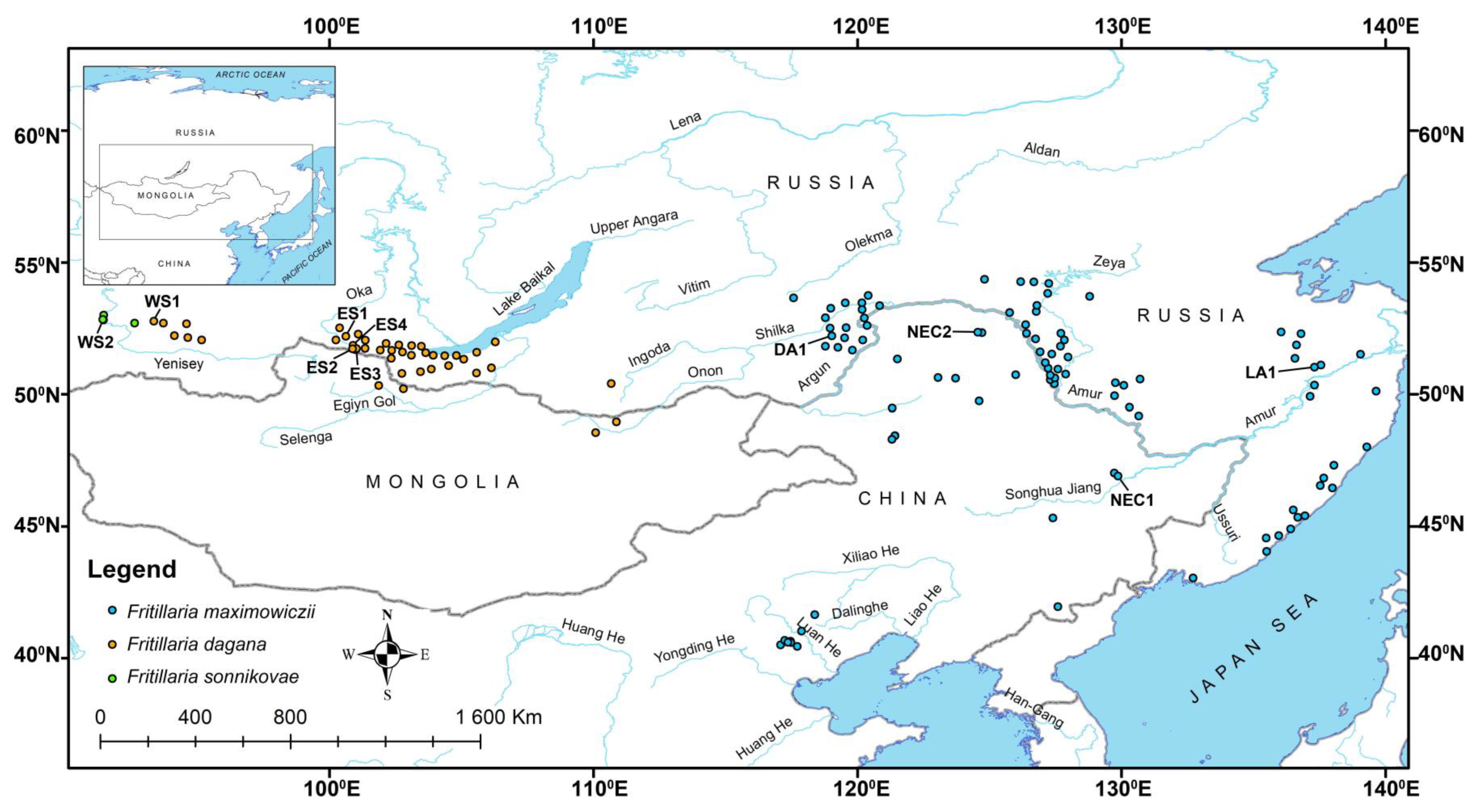
2.1. Distribution Data and Plant Material Collection

Species distribution data were extracted from the database of vascular plants for Asian Russia [14] and original studies [15,16]. Localities in China were georeferenced from the Chinese Virtual Herbarium (https://www.cvh.ac.cn/, accessed on 10 November 2022). Fritillaria samples for DNA sequencing were collected from 7 localities distributed between some presumptive isolated fragments of the South Siberia range: the Eastern Sayan (ES1–ES4), the Western Sayan (WS1, WS2) Mountains, and Argun Dahuria (DA1) (Figure 1, Table 1).
The herbarium-preserved specimens (ES and WS localities) and living plants (DA1) were used for the study. In the following DNA analysis, one (ES1, ES2, ES4), two (ES3), three (WS1), or four individuals (DA1 and WS2) from each population/herbarium sheet were used. For F. sonnikovae isotypes, specimens (WS2) stored in the NS herbarium were used. The analysis pattern depended on the number of available specimens, the level of DNA degradation in preserved samples, and related PCR sufficiency. The samples collected from living plants during fieldwork were put in individual filter paper bags (23 g·m−2), dried, and stored in silica gel until DNA isolation.

2.2. DNA Isolation, PCR

Total DNA was isolated from silica-dried leaf tissue following the cetyltrimethylammonium bromide (CTAB) method [17], with some authors’ modifications [18]. For phylogenetic reconstruction, the ITS1-ITS2 region (ITS) of nuclear DNA (ncDNA) and a fragment of the matK gene, the rps16 intron region, and the trnH-psbA intergenic spacers of plastid DNA (ptDNA) were used as molecular markers. A complete ITS region was amplified using the forward ITS1-P2 [19] and the reverse ITS4 [20] primers, complementary to the rDNA 18S and 26S flanking regions. PCR was performed in 25 µL of a reaction mixture containing 1X Q5 Reaction Buffer and 0.5 units of Q5 High-Fidelity DNA Polymerase (New England BioLabs, Ipswich, MA, USA) with final concentrations of 200 µM of each dNTP and 500 nM of each primer. Amplification conditions for the ITS region were 98 °C for 30 s, 30 cycles at 98 °C for 10 s, 52 °C for 20 s, and 72 °C for 15 s each, with a final elongation for 2 min at 72 °C. All used ptDNA regions were amplified using GoTaq Flexi DNA Polymerase (Promega, Madison, WI, USA). The fragment of matK was amplified using the 390F and 1326R primer pair [21,22], the rps16 intron was amplified using 6-forward and H-reverse primers [23], and the trnH-psbA spacer was amplified using a trnH2 [24] and psbAF primer pair [25]. The reaction mixture contained 1X Green GoTaq Flexi Buffer, 0.5 units of GoTaq polymerase, 2.5 mM of MgCl2, 250 µM of each dNTP, and 250 nM of each primer in the final volume of 20 µL. The amplification conditions for all ptDNA regions and all primer pairs were 95 °C for 2 min, 37 cycles of 95 °C for 20 s, 52 °C (matK), 53 °C (trnH-psbA), and 57 °C (rps16) for 30 s, and 72 °C for 1 min (trnH-psbA) or 1 min 10 s (matK and rps16) each, followed by final elongation for 5 min at 72 °C. Amplicons were either directly purified from the PCR mixture (ITS) using the GeneJET PCR Purification Kit (Thermo Fisher Scientific, Vilnius, Lithuania) or first visualized in a 1% agarose gel stained with ethidium bromide after electrophoresis and then gel-purified (ptDNA) using a GeneJET Gel Extraction Kit (Thermo Fisher Scientific, Vilnius, Lithuania). In order to reduce PCR-mediated recombination between ITS clones and to improve PCR accuracy, a proofreading polymerase was used together with lower initial template concentrations (not more than 5 ng per reaction) and PCR cycle numbers (not more than 30 cycles) as recommended by Lahr and Katz [26].

2.3. Cloning and Sequencing

Purified amplicons were either directly sequenced (ptDNA) or first cloned in Escherichia coli cells in the case of ITS analysis. For molecular cloning, amplicons were ligated into the plasmid vector pMiniT 2.0 (New England Biolabs, USA) according to the manufacturer’s recommendations using the insert-to-vector molar ratio of 3:1 in 5 µL of reaction mixture containing 12.5 ng of vector. Further, 50 µL of One Shot TOP10 E. coli chemically competent cells (Invitrogen, Waltham, MA, USA) was heat shock transformed at 42 °C for 35 s using 2.5 µL of the obtained ligation mixture. After transformation, the cell culture was incubated in a SOC liquid medium at 37 °C for 1.5 h with horizontal stirring at 250 rpm and plated onto LB (containing 15 g·L−1 of agar and 100 mg·L−1 of ampicillin) Petri dishes. Because the pMiniT 2.0 vector carries a toxic minigene in the cloning site, all grown colonies were considered to contain the insert. Eight colonies from each plate were picked with a sterile pipette tip and inoculated into 5 mL of liquid LB medium containing 100 mg·L−1 of ampicillin. In the case of low transformation efficiency, all detected colonies were used for further analysis. Cells were grown overnight at 37 °C with stirring at 250 rpm. Plasmids were isolated from overnight cultures using a GeneJet Plasmid Miniprep Kit (Thermo Fisher Scientific, Vilnius, Lithuania). Isolated plasmids and original amplicons were Sanger-sequenced using a BigDye Terminator Cycle Sequencing Kit v. 3.1 (Applied Biosystems, Austin, TX, USA) in a 3500 Genetic Analyzer (Applied Biosystems and Hitachi, Tokyo, Japan). All amplicons and plasmids were sequenced in both forward and reverse directions using the same region-specific primers that were used for PCR. For rps16 intron sequencing, in addition to PCR primers, the G-reverse primer [23] was also used to read the middle part of the intron.

2.4. Sequence Alignment and Phylogenetic Analysis

Raw sequencing data were processed using SnapGene Viewer software v. 2.6.2 (GSL Biotech, San Diego, CA, USA) and deposited in GenBank of the National Center for Biotechnology Information (NCBI, https://www.ncbi.nlm.nih.gov, accessed on 13 January 2023). Sequences available in GenBank (http://www.ncbi.nlm.nih.gov/genbank, accessed on 10 December 2022) belonging to the other Fritillaria species were used as a reference. The data on the taxa and sequences used in the analysis are summarized in Table 2. For analysis, we also used sequences for F. maximowiczii from GenBank in addition to those obtained in our study.
Multiple alignments of nucleotide sequences using the MUSCLE application with a gap opening penalty of 500 and an extension penalty of 4.0 were performed in MEGA v. 7.0.16 [39], followed by manual editing. ITS alignment was trimmed to obtain the matrix from the beginning of ITS1 to the end of ITS2 regions by removing 18S and 26S gene flanking partitions. For the ptDNA, the left and right ends of alignments were trimmed to correspond to the minimal length of the reference sequences obtained from GenBank. ITS, matK, and joint ptDNA sequences were analyzed both separately and together. The joint ptDNA dataset was based on the combined alignment of the sequences of matK, rps16, and trnH-psbA fragments obtained for each specimen used in the analysis. The joint ptDNA and ITS + ptDNA analyses were prepared on the limited sample size because, for this purpose, the GenBank records containing only complete ptDNA genome information were used to cover all markers sequenced from each specimen (Table 2). The ribotypes (R) and plastotypes (P) used in the joint analysis were indicated in bold in the figures.
Every insertion/deletion in each alignment was considered a single evolutionary event. They were binary encoded (with ‘1′ indicating presence and ‘0′ indicating absence of a gap) and included as separate binary data at the end of the alignment matrix. In particular, for the ITS-only phylogenetic analysis, 9 indels (positions no. 60–64, 99–100, 101–117, 229, 420, 421, 431, 432, 556) in the final alignment numbering 637 positions were coded as binary data; for the network based on multiple ITS variants revealed by molecular cloning three closely related Fritillaria species with no outgroup taxa, only 1 indel (position no. 396) in the final alignment numbering 610 positions was coded. For matK analysis, 2 indels (positions no. 137–154, 331–339) from a total length of 834 alignment positions were coded. For ptDNA analysis 35 indels (positions no. 137–154, 331–339, 871, 979, 986–990, 1077, 1163, 1196, 1212–1217, 1243, 1244–1252, 1253, 1267, 1279–1280, 1281–1282, 1283, 1591, 1592–1600, 1658, 1704–1706, 1707, 1708, 1858–1862, 1920–1923, 1924, 1925–1930, 1931–1961, 1962–1963, 1964–1969, 1970–2038, 2039–2042, 2043–2049, 2050–2052, 2053–2055, 2094–2098) in the final alignment numbering 2135 positions were coded as binary data. For ITS + ptDNA analysis 42 indels (positions no. 60–64, 99–100, 101–117, 419, 420, 430, 554, 772–789, 966–974, 1506, 1614, 1621–1625, 1712, 1798, 1831, 1847–1852, 1878, 1879–1887, 1888, 1902, 1914–1915, 1916–1917, 1918, 2226, 2227–2235, 2293, 2339–2341, 2342, 2343, 2493–2497, 2555–2558, 2559, 2560–2565, 2566–2596, 2597–2598, 2599–2604, 2605–2673, 2674–2677, 2678–2684, 2685–2687, 2688–2690, 2729–2733) in the final alignment numbering 2770 positions were coded.
Phylogenetic reconstructions were obtained independently by the Bayesian inference method (BI) based on the matrices combining the nucleotide alignments and binary (gaps) datasets in MrBayes v. 3.2.5 [40] and the maximum likelihood method (ML) based on multiple nucleotide sequence alignments in MEGA independently. The best-fit model of nucleotide substitutions based on the lowest Bayesian information criterion (BIC) calculated using the ‘find best DNA/protein models’ tool in MEGA (neighbor-joining tree to use and ML as a statistical method were applied as the settings) was selected and then used to perform the analysis. Nucleotide frequencies calculated using the aforementioned tool were also included to optimize the models implemented in MrBayes in the case of BI.
A BI analysis of nucleotide datasets was performed using the models implemented in MrBayes with optimized parameters to better correspond with the models used in the ML analysis (see below). The analyses were performed by specifying separately the model and parameters for each partition of the DNA datasets using the ‘applyto’ option. In particular, we optimized the HKY-like model [41] implemented in MrBayes (lset nst = 2 setting) to get Tamura three-parameter-like model (T92) [42] with gamma-distribution of substitution rate variation among sites (+G) for ITS and rps16 or no rate variation for matK and trnH-psbA datasets. The base frequencies (A, C, G, and T) were fixed as 0.18, 0.32, 0.32, and 0.18 for ITS; 0.35, 0.15, 0.15, and 0.35 for matK and rps16; and 0.34, 0.16, 0.16, and 0.34 for trnH-psbA. Binary data (indels) were analyzed using the F81-like model [43] implemented in MrBayes with equal stationary state frequencies to match the JC69 model [44]. Analysis of the joint ptDNA and ITS + ptDNA dataset was performed using the above-mentioned models and parameters individually set for each DNA region using the ‘applyto’ option. For each dataset, two simultaneous and completely independent Markov chain Monte Carlo (MCMC) analyses were run with four parallel chains up to 10,000,000 generations, with sampling every 100 generations and diagnostic calculations every 1000 generations. The first 25% of the samples from the cold chain were discarded (relburnin = yes and burninfrac = 0.25). The standard deviation of split frequencies below 0.01 was regarded as a sufficient convergence level, and reaching this value was considered a criterion for reaching the chain stationary state. The fluctuations of the cold chain likelihood in the stable range were also considered for the estimate of reaching stationarity. The sampled trees from both analyses were pooled, and 50% majority-rule consensus trees were constructed from 150,002 trees for ITS, 150,001 trees for matK, 3047 trees for joint ptDNA, and 232 trees for the ITS + ptDNA datasets to estimate clade posterior probability values (PP). The final phylogenetic trees were edited in FigTree v. 1.4.3 [45].
For the ML analysis, the T92 model for the matK and trnH-psbA datasets and the T92 + G model for the ITS, rps16, ptDNA, and ITS + ptDNA joint datasets were used, omitting the binary matrix. In all analyses, the initial tree for the heuristic search was inferred using the neighbor-joining method based on a pairwise distance matrix estimated using the maximum composite likelihood (MCL) method. All positions, including the indels, were used in the analysis. A bootstrap test for phylogeny including 1000 replicates was used.
The final phylogenetic trees are presented in the figures as BI phylograms, with the additional indication of bootstrap values (BS) for the clades on the corresponding ML trees. The Cardiocrinum species were used as an outgroup, based on the previous phylogenetic reconstructions [3,4,6,7,36]. The data matrices and trees obtained in the study are available in TreeBASE via the following link: http://purl.org/phylo/treebase/phylows/study/TB2:S30013 (accessed on 30 December 2022).
A phylogenetic analysis based on the alignment of the multiple ITS region variants revealed by molecular cloning and sequencing was carried out in two variants: (a) a phylogram based on only the main ITS variants and (b) a network constructed using all the identified ITS variants. The first analysis was prepared as mentioned above. The network was built using the TCS [46] method implemented in PopART software v. 1.7 [47].

3. Results

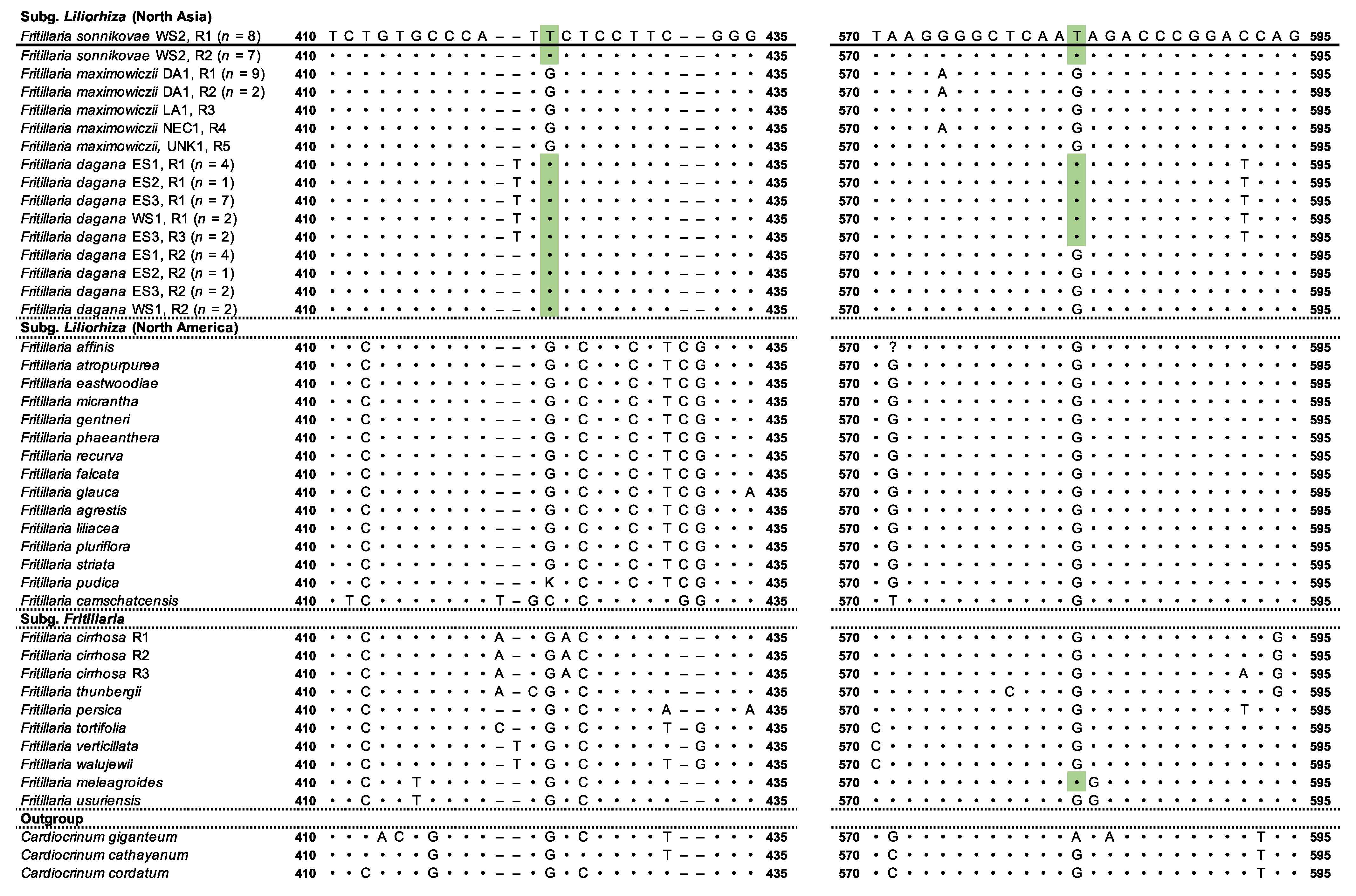
3.1. Phylogenetic Analysis Based on Nuclear DNA

The evolutionary history of the nuclear genome was inferred based on the analysis of the ribosomal DNA polymorphism (ITS region). At the start, we faced the problem of having double peaks for a number of positions in the ITS sequenograms obtained from the direct sequencing of PCR products, which made it impossible to unambiguously reconstruct each individual ITS nucleotide sequence. Moreover, many sequences belonging to different Fritillaria species, which were imported from the GenBank, also contained positions with degenerate nucleotides. We suppose that these degenerate signals may have appeared due to the high level of intragenomic polymorphism known for this region [18,48]. To be able to split the double peaks, molecular cloning of the ITS region was performed. Phylogenetic analysis (Figure 2a) showed that all species used in the analysis were distributed between the two main groups of the Fritillaria: the clade defined by node A, corresponding to the Liliorhiza subgenus, and the clade defined by node B, corresponding to the Fritillaria subgenus designated in the previous studies [4,6,7,8]. In Liliorhiza, the North Asian group presented by F. dagana + (F. sonnikovae + F. maximowiczii) (node C) looked to be a sister group to the group combining the North American species (node D). The ribotypes of F. sonnikovae showed close affinity with the ribotypes of F. maximowiczii (node G; PP, 1.00; BS, 82). The clade encompassing exclusively F. maximowiczii ribotypes (R1–R5) was supported only by PP (0.97) and not by BS, whereas the clade encompassing F. sonnikovae ribotypes (R1, R2) was supported only by BS (91) and not by PP.
Despite the close relationship between the two species, the analysis also showed that two positions in the multiple alignment of the ITS2 region seemed to be synapomorphies shared by F. sonnikovae and F. dagana/F. meleagroides but not F. maximowiczii (Figure 3).
The ribotypes of F. dagana (Figure 2a) were presented by two well-supported groups of paralogues (nodes H and J) laying claim to be sister to the F. maximowiczii + F. sonnikovae group (node G). The obtained results showed that both types of ITS paralogues were present in the F. dagana from all localities studied. As can be seen on the tree, the estimated genetic distances suggest that the F. dagana R2 ribotype is, in fact, the closest one to the common ancestor of the joint F. dagana + (F. maximowiczii + F. sonnikovae) group (node C) among all other ribotypes.
Some low-copy ITS variants belonging to F. dagana, F. maximowiczii, and F. sonnikovae formed an additional structure within the network (Figure 2b) and might be transitional variants between the main homologous variants described here (Figure 2a). The ribotypes of F. maximowiczii and F. sonnikovae showed high affinity between them, forming a haplogroup within the network that corresponded to node G on the tree. The ribotypes of F. dagana formed two haplogroups corresponding to the clades H and J.
The Fritillaria subgenus was represented by the two subgroups defined by nodes E and F. The species representing the westernmost group of Siberia taxa (F. verticillata and F. meleagroides) were distributed between these two clades (Figure 2a).

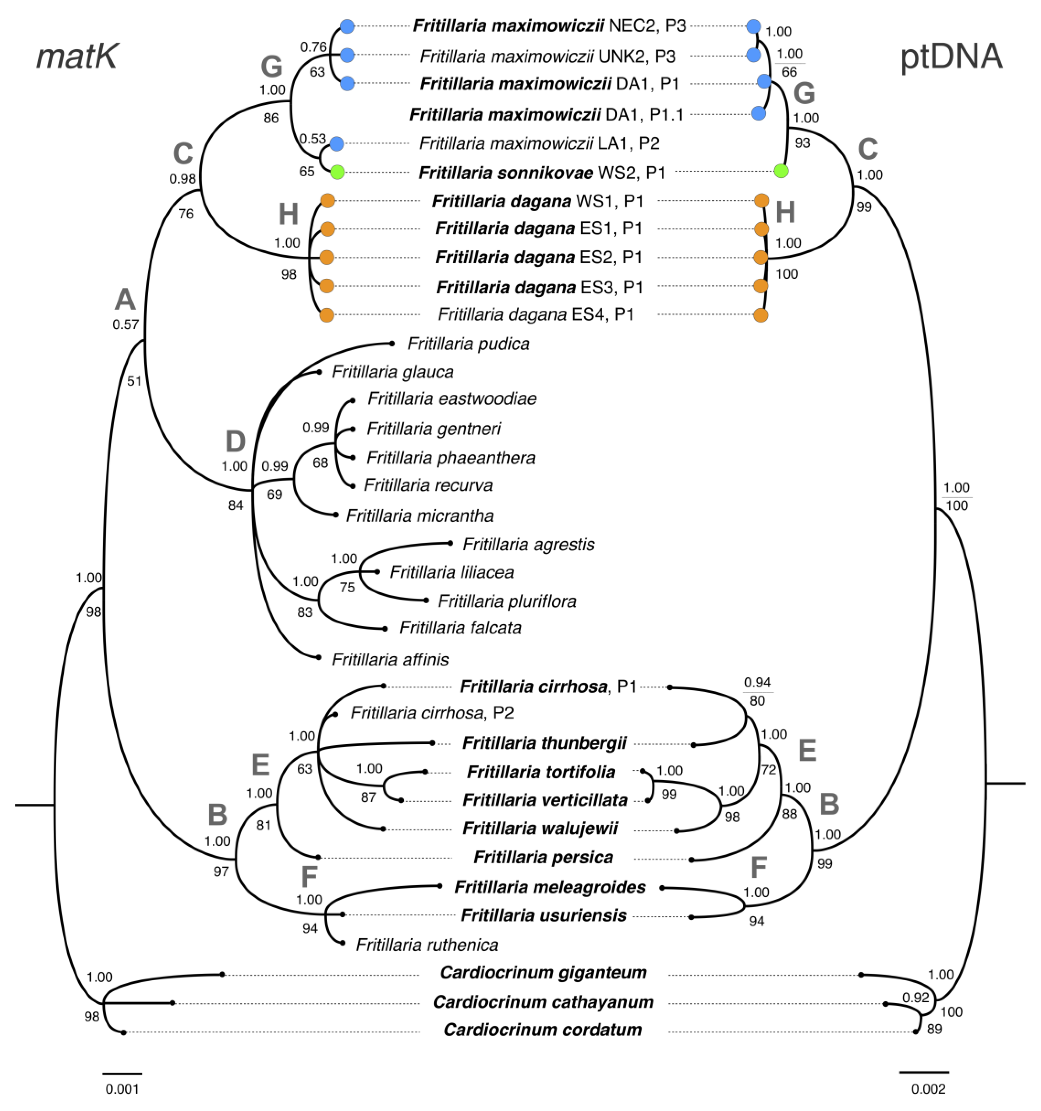
3.2. Phylogenetic Analysis Based on Plastid DNA

The reconstruction tracing the maternal lineage is presented in two variants (see Section 2.4). The first tree was based on the matK-only dataset (Figure 4, to the left), and the second one was based on the joint ptDNA (matK + rps16 + trnH-psbA) dataset (to the right). Although both phylogenies showed an overall similarity to the ITS tree, some points should still be emphasized. In particular, similarly to the ITS tree, the groups defined by nodes B–H were still presented in the ptDNA trees (Figure 4).
Similar to the ITS tree, the Fritillaria subgenus (clade B) was represented by the two subgroups on the ptDNA tree. The species representing the westernmost group of Siberian taxa and belonging to the Fritillaria subgenus were distributed between clade E (F. verticillata) and clade F (F. meleagroides and F. ruthenica).
Group A representing the Liliorhiza subgenus did not get significant support on the matK tree obtained by both BI and ML. In the Liliorhiza group, Fritillaria dagana from the different localities shared the same plastotype and formed a sister group (node H) to the well-supported clade of F. maximowiczii + F. sonnikovae (node G). The P1 plastotype of F. sonnikovae was identical to the P2 plastotype of F. maximowiczii, although the sequences clustered together with a moderate level of support, possibly due to their high resemblance to the other F. maximowiczii sequences, from which they differed by only a single substitution. The sequences of trnH-psbA were identical between the two species. The plastotypes of rps16 belonging to F. sonnikovae and F. maximowiczii were extremely similar differing only by the length of the poly(A) and poly(T) regions (Figure 5).
It is important to note here that length variability within the rps16 region was also observed in the plastotypes of different F. maximowiczii specimens. Thus, since the polymorphism of ptDNA markers is expressed only in the number of A/Ts in the homopolymeric regions of the rps16 intron, the identified differences between F. maximowiczii and F. sonnikovae are better correlated to the interpopulation level than to the interspecies level.

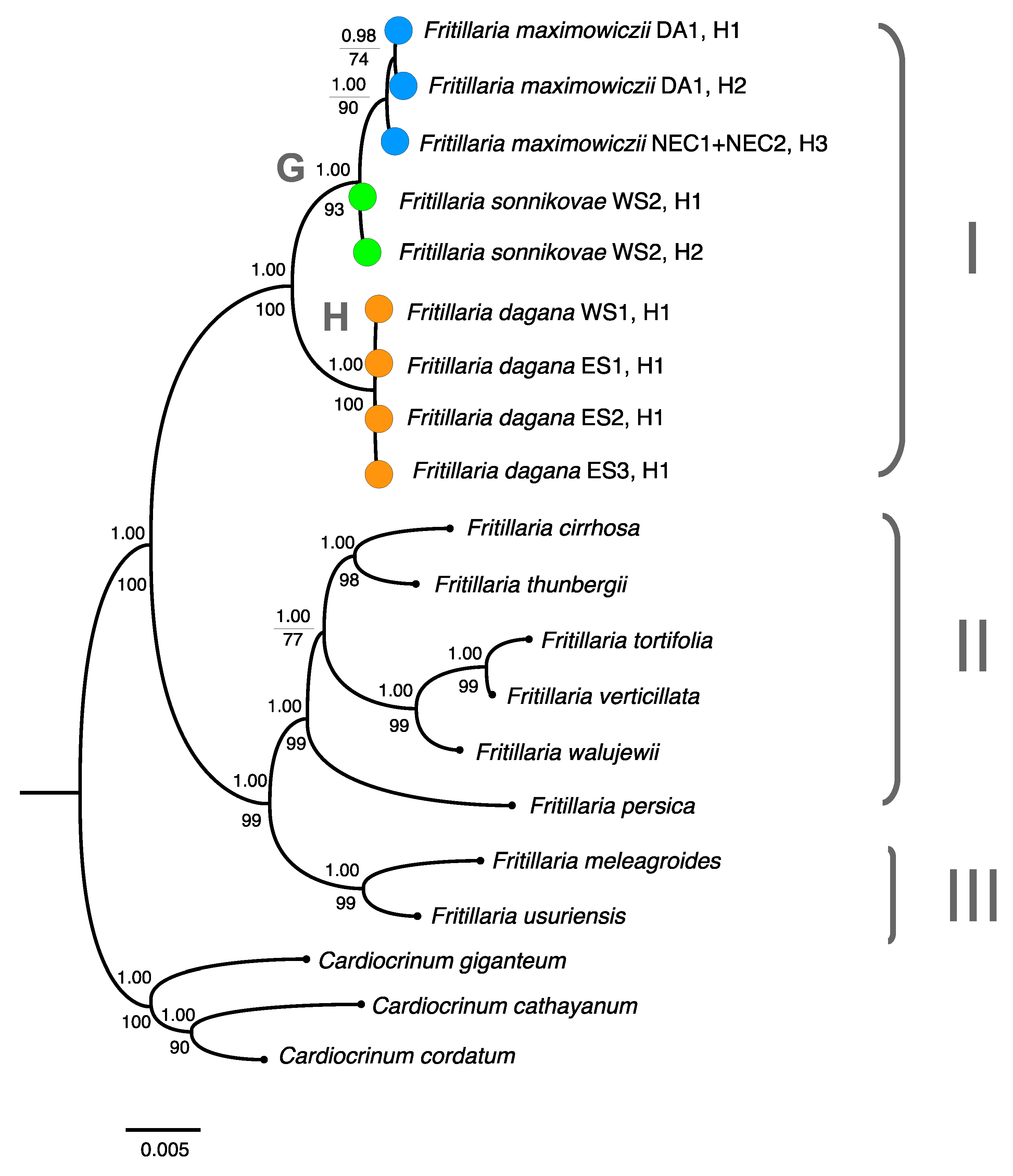
3.3. Combined Phylogenetic Analysis

The topology of the phylogenetic tree built based on the joint ITS + ptDNA dataset was well defined and similar to that of both the ITS and ptDNA trees (Figure 6).
The species occurring in Siberia segregated into three main lineages: lineage I, represented by F. dagana + (F. sonnikovae + F. maximowiczii), lineage II, including F. verticillata, and lineage III, including F. meleagroides. The lineage I is a well-supported group where F. dagana (node H) is sister to the group combining the two other species (node G).

4. Discussion

4.1. Phylogenetic Relationships between F. sonnikovae and Other Fritillaria Species

Our results correlate well with the previous studies that suggest three phylogenetic lineages of Fritillaria in Siberia [4,6,7,8]. The group, which includes F. dagana, F. sonnikovae, and F. maximowiczii, inhabiting the North Asian territories eastwards from the right bank of the Yenisey River represents lineage I (Figure 6), which nests in the Liliorhiza clade together with the New World taxa. At the same time, the matK tree on which Liliorhiza encompassed the F. dagana + (F. sonnikovae + F. maximowiczii) group and New World taxa got low statistical support. Some previous reconstructions have also rather doubtfully placed F. maximowiczii in Liliorhiza [4,7]. The Siberian species with westernmost ranges are distributed between the two groups of the non-monophyletic Fritillaria subgenus [7] encompassing the Old World taxa: F. verticillata (Figure 6) represents the lineage II and F. meleagris [4,6,7], F. meleagroides (Figure 6), and F. ruthenica (Figure 4) represents the lineage III. The distribution of these three species among Fritillaria subgroups on the phylogenetic trees is well correlated with some previous studies [4,6,7,8].
We found a close affinity between F. sonnikovae and F. maximowiczii based on both ncDNA and ptDNA. Namely, the plastotypes belonging to F. sonnikovae and F. maximowiczii were just slightly different or even identical for some populations. Neither the analysis based on ncDNA, nor the analysis based on ptDNA could reliably confirm the haplotype monophyly of F. sonnikovae. These findings suggest that the identified difference between F. maximowiczii and F. sonnikovae plastotypes corresponds to the interpopulation level. For ITS, only two positions in the ITS2 region brought F. sonnikovae closer to other Siberian species, with the other synapomorphies being shared with F. maximowiczii. However, we have found out quite surprisingly that in the affinity section of the F. sonnikovae diagnosis, only F. dagana and F. roylei Hook. but not F. maximowiczii were mentioned by the authors [10]. Moreover, in the subsequent studies, no possible similarity between F. sonnikovae and F. maximowiczii was mentioned either. Our study did not produce any evidence of a close relationship between F. sonnikovae and F. roylei (F. cirrhosa subsp. roylei) or F. cirrhosa as well. According to our results and the previous studies [4,6,7], F. cirrhosa and F. cirrhosa subsp. roylei are in fact nested in the distant clade of lineage II belonging to the subgenus Fritillaria and have no close relationship with lineage I encompassing F. dagana, F. sonnikovae, and F. maximowiczii.

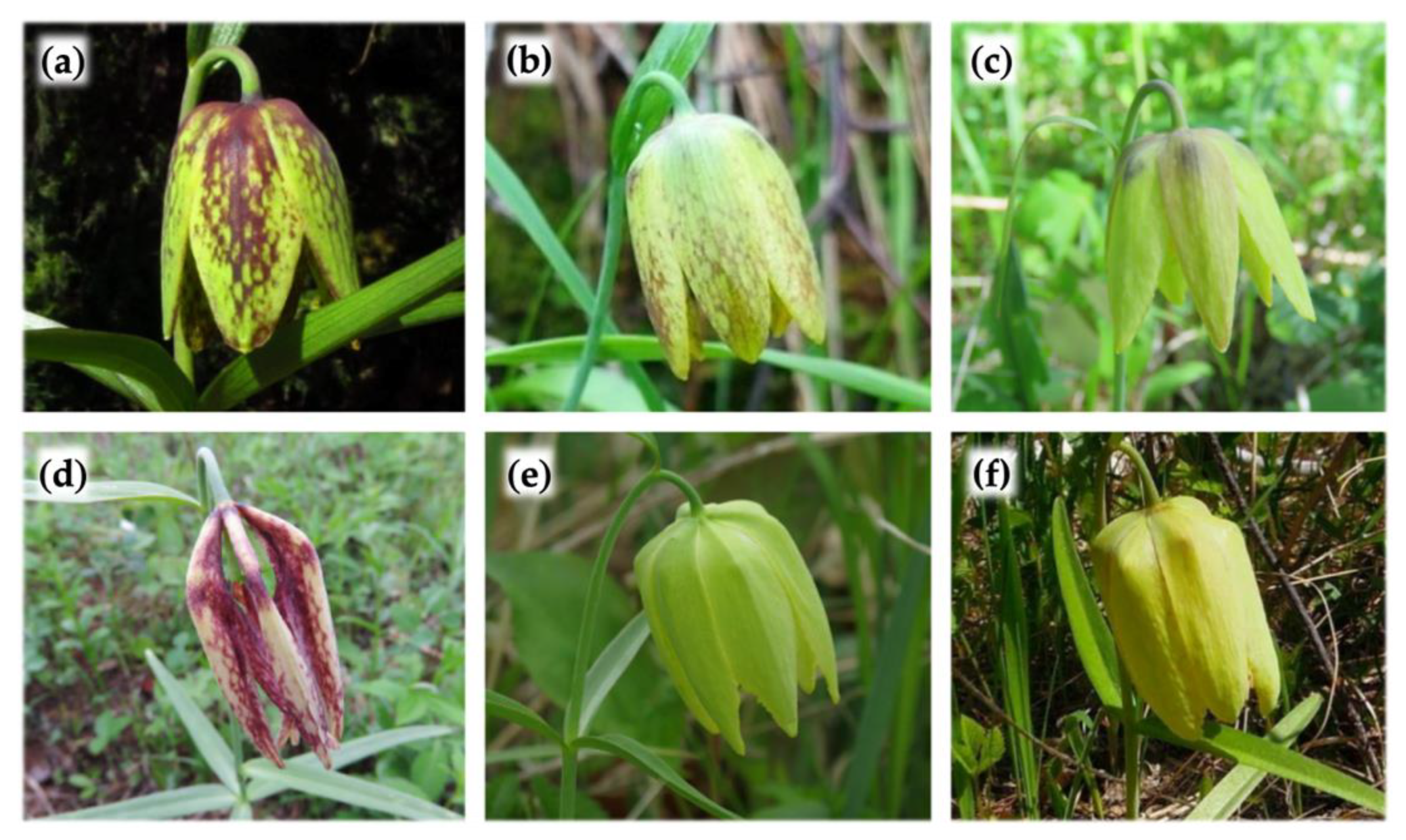
4.2. Comparative Morphological Features of Three Closely Related Species of Siberian Fritillaria

The affinity of F. sonnikovae and F. maximowiczii on the molecular phylogenetic tree inspired us to look at their morphological features and those of the most closely related F. dagana (Table 3).
The analysis of relevant morphological features revealed that being a sister species, F. dagana can be clearly distinguished from F. sonnikovae and F. maximowiczii by the oblong-ovate-lanceolate leaves always assembled in a single whorl located around the middle of the stem, the tepals, which are oblong-obovate, the triangular nectaries, and the capsule with narrow wings [49,50]. It is also worth noting that the phenology of the species is quite different. F. sonnikovae and F. maximowiczii, on average, bloom earlier (May—the first half of June) [53,54] than F. dagana (June—the first half of July) [55].
On the other hand, F. sonnikovae and F. maximowiczii have rather similar morphology. In particular, both species have linear or lanceolate-linear leaves assembled in one, or sometimes two, whorls located above the middle of the stem, oblong-elliptical perianth segments with slightly finely sinuate fine-squiggly erose margins, oval nectaries, and capsules with wide wings (Table 3).
Fritillaria sonnikovae individuals sometimes seem to be higher (up to 52 cm), have bigger leaves (70–120 × 3–13 mm) and longer tepals, and a few more leaves than it was described for W. maximowiczii inhabiting the Russian Far East [51]. At the same time, F. maximowiczii in China can reach up to 55 cm in height with a leaf length of 45–100 mm and a leaf width of 3–13 mm [52]. Hence, our analysis did not reveal any specific morphological differences between F. sonnikovae and F. maximowiczii. The plants of both species (except minimum and maximum values) can have very similar morphological features. Some morphology exceptions can be explained by habitat conditions or age variation. The same situation has been observed for Fritillaria montana Hoppe ex W.D.J. Koch and its close relatives. The detailed biometric analysis showed that some described species of Fritillaria have no taxonomic value and can be included in F. montana [56].
One of the main qualitative differences between F. sonnikovae and F. maximowiczii is perianth color. However, in this case, the fact of the presence of light and slightly pigmented F. maximowiczii forms should be considered (Figure 7).
Colorless perianth forms are also known for other species, e.g., in F. meleagris, cream-white or sulphurous yellow-colored flower morphs with no tessellation are sometimes observed in up to 7% of individuals within the population [60]. The colorless morphs may possibly arise as a result of the inability to synthesize anthocyanin, caused by the expression of a recessive allele in a homozygous state. The study performed by Mucciarelli et al. [61] has also shown that notwithstanding having differently colored flowers, the two intraspecies taxa of F. tubaeformis Gren. & Godr. are genetically very similar and share a considerable part of their gene pool. Additionally, the light-yellow morph of F. dagana (ES3 locality) included in our phylogenetic analysis (Figure 7c) showed no differences in the nucleotide sequences compared to the common morphs (Figure 7a, ES1, ES2, ES4, WS1 localities).
In this context, based on the presence of colorless forms among Fritillaria, most authors of the present study suggest that F. sonnikovae may be considered one of the light-perianth morphs of F. maximowiczii. At the same time, one of the authors (N.S.) has an alternative opinion regarding the morphological features of the two species. According to this point of view, the morphological differences between the two taxa seem to be stable and include, in addition to the different perianth colors, the larger maximum sizes of plants and individual organs in F. sonnikovae compared to F. maximowiczii, differing flower proportions, and some flower quality characteristics, such as tepals with a wide, obtuse apex in F. sonnikovae and a pointed apex in F. maximowiczii. On the other hand, these arguments have not been statistically proven, and comparative biometric analysis of morphological features has not been carried out. The discussed observations have not been published either; hence, they may be considered just a personal opinion. Most of the authors of the present study still consider the published data (Table 3) in this regard and adhere to the fact that the comparisons made revealed more similarities than differences between F. sonnikovae and F. maximowiczii. Most of the authors also believe that the quantitative differences (e.g., maximum sizes) may be associated with the habitat conditions (see above), while some qualitative features of F. sonnikovae, e.g., no splitting of the flower color, with the homozygous recessive state of the corresponding genes as a result of the possible bottleneck event in the F. sonnikovae history considered to a local endemic with a narrow range (see Section 4.3). As regards the tepals form, we also found some intrapopulation variations of this feature in closely related F. dagana. In particular, the light-perianth F. dagana specimens from the ES3 locality (Figure 7c) mentioned above and included in the phylogenetic analysis have tepals with pointed apex but not obtuse as usual form (Figure 7a) of species has.
In any case, the authors, who described F. sonnikovae, were supposed to discuss the differences, which would make it possible to distinguish the new taxon from F. maximowiczii, in the diagnosis presented by them [10]. Remarkably, due to the morphological similarity between F. sonnikovae and F. maximowiczii, some specimens, which are now attributed to F. sonnikovae, were originally identified as F. maximowiczii (the findings dating back to 1980 and 2006, vouchers NS0000198! and NS0000199!, respectively), and later re-identified as F. sonnikovae when this species was described in 2010. These specimens have the morphological characters of F. maximowiczii, but the perianth is light yellow on the exterior, with translucent checkerboard or linear patterns. However, as it was mentioned above, the obvious similarities between two closely related species were not listed in the F. sonnikovae diagnosis [10]. The absence of F. maximowiczii in the affinity section of F. sonnikovae diagnosis does not allow making judgements as to the clarity of the latter species identification. Thus, most authors of the present study believe that the authors of F. sonnikovae did not provide sufficient evidence to claim it to be a separate species, and before convincing arguments are presented, F. sonnikovae should be considered a synonym of F. maximowiczii.

4.3. The Possible Origin of F. sonnikovae and Diversification of Siberian Taxa

Molecular clock estimations based on ptDNA revealed that the New World Fritillaria clade (Liliorhiza), which also includes the North Asian F. dagana, F. sonnikovae, and F. maximowiczii, separated from the other lineages in the Early Miocene approximately 21 (16–25) Mya (mean, [95% highest posterior density, HPD]) on the Qinghai–Tibet Plateau (QTP) [7]. The results of the combined ITS + ptDNA analysis presented by the authors in the above-mentioned study were generally consistent with the data obtained using the ptDNA only and estimated the time when the stem leading to this group was formed to be between 13 and 23 Mya (95% HPD). The diversification of Liliorhiza lineages and the crown formation are considered to have happened around 8–14 Mya (the range of means found using different combinations of DNA regions) out of the QTP [7]. When climate cooling started in about the Middle Miocene [62], plants might have adapted to cold habitats and expanded their ranges outside the QTP to the new temperate areas that became accessible in North Asia. Around that time (5–15 Mya) [7], the colonization of North America could happen via the Bering Land Bridge, which connected North Asia and western North America. The F. dagana + F. maximowiczii group crown formation might have, probably, started around 6 Mya (mean) according to the cpDNA data [7]. The haplotypes belonging to the groups G (F. sonnikovae) and H (F. dagana) are both present in South Siberia, whereas the easternmost part of North Asia is populated by the species carrying exclusively the haplotypes from the group G (F. maximowiczii) (Figure 6). Moreover, the estimated genetic distances on the phylogenetic trees revealed that F. dagana ribotypes, in fact, appear to be the most closely located to the common ancestor of lineage I (F. dagana + [F. maximowiczii + F. sonnikovae] group) compared to all others (Figure 2), and F. maximowiczii looks to carry the youngest haplotypes among the other species of the lineage (Figure 2 and Figure 6). These findings taken together allow to hypothesize that diversification within the F. dagana + (F. maximowiczii + F. sonnikovae) group could happen in South Siberia with the unidirectional dispersal to the Far East. Further diversification might have happened via allopatric speciation, which was, probably, promoted by the expansion of mountainous and alpine habitats as a result of the surface uplift during the mountain building in South Siberia and the formation of the Yenisey valley, which were remarkably intense in the late Neogene–early Quaternary [63]. Under these conditions, F. dagana could arise from populations preferentially inhabiting the mountain-forest or subalpine zones in the South Siberian mountains. Traditionally, mountainous areas are considered to be the centers of plant diversity and species diversification. Large-scale patterning of species diversity in Asian Russia also revealed high species richness in the mountains of South Siberia [64]. The common ancestor of F. sonnikovae + F. maximowiczii could inhabit the forest belt at lower altitudes in Siberia. At that time, broad-leaved deciduous forests were predominant in South Siberia [65]. The analysis of the bottom sediments of Lake Baikal provided evidence for the dominance of the nemoral dark coniferous–broad-leaved complex on this territory till the middle of the Late Pliocene. As a result of the progressive worsening of climatic conditions, the broad-leaved forests demonstrating increasingly more boreal features were replaced by the coniferous forests, which included nemoral elements by the end of the Pliocene–Eopleistocene [65,66,67]. Considering the current F. maximowiczii habitats, which include the East Asian mesophylic larch, birch, and broad-leaved deciduous forests, it can be assumed that the Pleistocene environmental conditions in South Siberia were not suitable for this species, which led to the regression of its range. In this view, F. sonnikovae, a narrow endemic in the Western Sayan Mountains, can be considered a tertiary relict that emerged in the remnant of the Paleo-range of its common ancestor with F. maximowiczii. The mountains of southern Siberia playing the role of refugia were of great importance for the survival of mesophylic broad-leaved forest remnants at that time [65,66,67]. The possible nemoral relict nature of F. sonnikovae was also pointed out by the authors, who described this species [10]. The specific combination of features in F. sonnikovae, for example, the distinguishing perianth color, may, in our opinion, be a result of a possible bottleneck effect. Genetic drift associated with the bottleneck event could lead to the fixation of haplotypes by chance, which may explain the fact that F. sonnikovae retained only one of the ancestral morphs. At the same time, F. maximowiczii did not pass through the bottleneck and retained a wider range of morphs. However, as mentioned above, the genetic data do not provide any clear evidence that F. sonnikovae and F. maximowiczii have diverged enough to be currently considered separate species. From this point of view, F. sonnikovae may be considered a ‘stray’ population of F. maximowiczii. Based on our results, we may speculate that genetic introgression from other Fritillaria species could contribute to the formation of the somewhat blurred morphological traits of F. sonnikovae. In particular, the two positions in ITS2 in the multiple alignment seem to be the synapomorphies shared by F. sonnikovae and the closely related F. dagana, with no clear hybridization patterns observed (Figure 3). Since ITS is a marker of biparental inheritance while ptDNA marks only the maternal lineage, the absence of common plastotypes may indicate that possible contacts between the populations of these two species could probably occur through pollen transfer. Notwithstanding the fact that differences in phenophases between F. sonnikovae and F. dagana [53,54,55] objectively reduce the possibility of cross-pollination between these species, some late-flowering individuals of F. sonnikovae still have a chance to be pollinated by the early-flowering plants of F. dagana.
The migration pathways and current distribution of Liliorhiza in Asia are well separated from other Fritillaria species [6]. Thus, four other species occurring in Siberia (F. meleagris, F. meleagroides, F. ruthenica, and F. verticillata) have origins and histories that differ from those of the North Asian branch of Liliorhiza. These species belong to two different clades within the Fritillaria subgenus, encompassing Old World taxa. The ancestral migration routes from the QTP to Siberia for these Fritillaria lineages could connect the Irano-Turanian region to the Mediterranean Basin and Europe [7].

5. Conclusions

Our data have clearly shown that F. sonnikovae belongs to the North Asian lineage together with F. dagana and F. maximowiczii in Liliorhiza. Our study did not produce any evidence for the close relationship between F. sonnikovae and F. roylei (F. cirrhosa subsp. roylei) listed in the F. sonnikovae diagnosis. We suggest that diversification within the F. dagana + (F. sonnikovae + F. maximowiczii) group started in South Siberia during the late Neogene with the unidirectional dispersal to the Far East. Within the aforementioned group, F. sonnikovae showed a close affinity with F. maximowiczii based on both molecular phylogenetic analysis and morphology. F. dagana is sister to the F. maximowiczii + F. sonnikovae group and could not be directly considered as the closest relative of F. sonnikovae. However, the possible incidents of genetic introgression from F. dagana into the gene pool of F. sonnikovae cannot be excluded. Neither the analysis based on ncDNA nor the analysis based on ptDNA could reliably confirm the haplotype monophyly of F. sonnikovae and its segregation from F. maximowiczii. Most authors of the present study suggest that the known morphological and identified genetic differences between F. maximowiczii and F. sonnikovae correspond to the interpopulation level but not the interspecies level. The absence of F. maximowiczii in the affinity section of F. sonnikovae diagnosis does not allow making judgements as to the clarity of the latter species identification. Thus, most authors of the present study believe that the authors of F. sonnikovae did not provide sufficient evidence to claim it to be a separate species, and before convincing arguments are presented, F. sonnikovae should be considered a synonym of F. maximowiczii. In this regard, F. sonnikovae may be considered a narrow endemic and one of the light-perianth morphs of F. maximowiczii that arose in the Western Sayan Mountains and remained there as a tertiary relict.

Author Contributions

Conceptualization, M.P.; methodology, M.P., D.S. and V.P.; validation, M.P. and D.S.; formal analysis, M.P.; investigation, M.P., D.S., V.P., I.S. and N.S.; resources, M.P. and V.P.; data curation, M.P. and D.S.; writing—original draft preparation, M.P.; writing—review and editing, M.P., D.S., V.P., I.S. and N.S.; visualization, M.P., D.S. and V.P.; supervision, M.P.; project administration, M.P. and D.S.; funding acquisition, M.P. and V.P. All authors have read and agreed to the published version of the manuscript.

Funding

The research was funded by Russian state assignments, the project numbers 122041100049-0, 121030900138-8, and 2-22-128-1.

Institutional Review Board Statement

Not applicable.

Data Availability Statement

Original sequence data are available at GenBank (http://www.ncbi.nlm.nih.gov/genbank, accessed on 13 January 2023) by their accession numbers presented in Table 2. Alignments and trees are also available at TreeBASE (http://purl.org/phylo/treebase/phylows/study/TB2:S30013, accessed on 30 December 2022).

Acknowledgments

The research was done using the equipment of the Core Facilities Center ‘Bioanalitika’ and within the frameworks of the state assignment of Siberian Institute of Plant Physiology and Biochemistry, Siberian Branch of the Russian Academy of Sciences (No 122041100049-0, Marina Protopopova, Vasiliy Pavlichenko); Russian state projects No 121030900138-8 and 2-22-128-1 (Denis Sandanov); and state assignments of other institutes listed in the author affiliations (Inessa Selyutina, Nikolay Stepanov). Authors express gratitude to KRSU, NS, TK, and UUH herbaria for allowing us to deposit the vouchers and collect samples for DNA analysis, Natalya Shvetsova for the lab assistance, Zhiheng Wang (Peking University) for providing distribution data for F. maximowiczii in China, Aleksandr Ebel for helping us with vouchers searching in TK herbarium, Daba Chimitov and Lubov Bel’mas for the Fritillaria photos providing.

Conflicts of Interest

The authors declare no conflict of interest.

References

  1. Hassler, M. World Plants. Synonymic Checklist and Distribution of the World Flora. Version 14.2. Last Update 15 October 2022. Available online: https://www.worldplants.de (accessed on 10 December 2022).
  2. The Royal Botanic Gardens, Kew. The World Checklist of Vascular Plants (WCVP). In Catalogue of Life Checklist (4.0); Available online: https://doi.org/10.48580/dfpk-4nz (accessed on 10 December 2022).
  3. Bi, Y.; Zhang, M.; Xue, J.; Dong, R.; Du, Y.; Zhang, X. Chloroplast Genomic Resources for Phylogeny and DNA Barcoding: A Case Study on Fritillaria. Sci. Rep. 2018, 8, 1184. [Google Scholar] [CrossRef] [PubMed] [Green Version]
  4. Rønsted, N.; Law, S.; Thornton, H.; Fay, M.F.; Chase, M.W. Molecular Phylogenetic Evidence for the Monophyly of Fritillaria and Lilium (Liliaceae; Liliales) and the Infrageneric Classification of Fritillaria. Mol. Phylogenetics Evol. 2005, 35, 509–527. [Google Scholar] [CrossRef] [PubMed]
  5. Rix, E.M. Fritillaria: A Revised Classification Together with an Updated List of Species; Fritillaria Group of the Alpine Garden Society Press: Pershore, UK, 2001. [Google Scholar]
  6. Day, P.D.; Berger, M.; Hill, L.; Fay, M.F.; Leitch, A.R.; Leitch, I.J.; Kelly, L.J. Evolutionary Relationships in the Medicinally Important Genus Fritillaria, L. (Liliaceae). Mol. Phylogenetics Evol. 2014, 80, 11–19. [Google Scholar] [CrossRef] [PubMed]
  7. Huang, J.; Yang, L.-Q.; Yu, Y.; Liu, Y.-M.; Xie, D.-F.; Li, J.; He, X.-J.; Zhou, S.-D. Molecular Phylogenetics and Historical Biogeography of the Tribe Lilieae (Liliaceae): Bi-Directional Dispersal between Biodiversity Hotspots in Eurasia. Ann. Bot. 2018, 122, 1245–1262. [Google Scholar] [CrossRef] [PubMed]
  8. Ryan, S.P. Molecular Phylogeny and Character Evolution of Fritillaria Subgenus Liliorhiza (Liliaceae). Master’s Thesis, San Diego State University, San Diego, CA, USA, 2014. [Google Scholar]
  9. Hill, L. Fritillaria Icones. Available online: http://www.fritillariaicones.com (accessed on 10 December 2022).
  10. Shaulo, D.N.; Erst, A.S. A New Species of Fritillaria L. (Liliaceae) from West Sayan. Turczaninowia 2010, 13, 46–49. [Google Scholar]
  11. Shaulo, D.N. Flora of the Western Sayan. Turczaninowia 2006, 9, 5–336. (In Russian) [Google Scholar]
  12. Cherepnin, L.M. Flora Yuzhnoy Chasti Krasnoyarskogo Kraya [Flora of the Southern Part of Krasnoyarskiy Kray]; Krasnoyarsk State Pedagogical Institute: Krasnoyarsk, Russia, 1959; Volume 2, p. 211. (In Russian) [Google Scholar]
  13. Krasnoborov, I.M. Flora Alpina Montium Sajanesium Occidentalium; Nauka: Novosibirsk, Russia, 1976. (In Russian) [Google Scholar]
  14. Sandanov, D.V. Developing the Database of Vascular Plants Distribution for Asian Russia. Information Technology in Biodiversity Research. In Proceedings of the III National Conference with International Participation, Dedicated to the 100th Anniversary of the Birth of Russian Academician Pavel Gorchakovskii, Ekaterinburg, Russia, 5–10 October 2020; University for the Humanities: Ekaterinburg, Russia, 2020; pp. 470–472. (In Russian). [Google Scholar]
  15. Sandanov, D.V. Finding of Fritillaria dagana (Lilliaceae) in Mongolia. Rastit. Mir Aziat. Ross. 2013, 1, 44–46. (In Russian) [Google Scholar]
  16. Baasanmunkh, S.; Oyuntsetseg, B.; Tsegmed, Z.; Oyundelger, K.; Urgamal, M.; Gantuya, B.; Javzandolgor, C.; Nyambayar, N.; Kosachev, P.; Choi, H.J. Distribution of Vascular Plants in Mongolia—I Part. Mong. J. Biol. Sci. 2022, 20, 3–28. [Google Scholar] [CrossRef]
  17. Doyle, J.J.; Doyle, J.L. A Rapid DNA Isolation Procedure for Small Quantities of Fresh Leaf Tissue. Phytochem. Bull. 1987, 19, 11–15. [Google Scholar]
  18. Protopopova, M.V.; Pavlichenko, V.V. Eranthis Salisb. (Ranunculaceae) in South Siberia: Insights into Phylogeography and Taxonomy. Diversity 2022, 14, 779. [Google Scholar] [CrossRef]
  19. Utelli, A.; Roy, B.; Baltisberger, M. Molecular and Morphological Analyses of European Aconitum Species (Ranunculaceae). Plant Syst. Evol. 2000, 224, 195–212. [Google Scholar] [CrossRef]
  20. White, T.J.; Bruns, T.; Lee, S.; Taylor, J. Amplification and Direct Sequencing of Fungal Ribosomal RNA Genes for Phylogenetics. In PCR Protocols: A Guide to Methods and Applications; Innis, M.A., Gelfand, D.H., Sninsky, J.J., White, T.J., Eds.; Academic Press: San Diego, CA, USA, 1990; pp. 315–322. [Google Scholar]
  21. Cuenoud, P.; Savolainen, V.; Chatrou, L.W.; Powell, M.; Grayer, R.J.; Chase, M.W. Molecular Phylogenetics of Caryophyllales Based on Nuclear 18S RDNA and Plastid RbcL, AtpB, and MatK DNA Sequences. Am. J. Bot. 2002, 89, 132–144. [Google Scholar] [CrossRef] [PubMed]
  22. Burgess, K.S.; Fazekas, A.J.; Kesanakurti, P.R.; Graham, S.W.; Husband, B.C.; Newmaster, S.G.; Percy, D.M.; Hajibabaei, M.; Barrett, S.C.H. Discriminating Plant Species in a Local Temperate Flora Using the RbcL+MatK DNA Barcode. Methods Ecol. Evol. 2011, 2, 333–340. [Google Scholar] [CrossRef]
  23. Lee, C.-S.; Downie, S.R. Phylogenetic Relationships within Cicuta (Apiaceae Tribe Oenantheae) Inferred from Nuclear RDNA ITS and CpDNA Sequence Data. Can. J. Bot. 2006, 84, 453–468. [Google Scholar] [CrossRef]
  24. Tate, J.A.; Simpson, B.B. Paraphyly of Tarasa (Malvaceae) and Diverse Origins of the Polyploid Species. Syst. Bot. 2003, 28, 723–737. [Google Scholar]
  25. Sang, T.; Crawford, D.J.; Stuessy, T.F. Chloroplast DNA Phylogeny, Reticulate Evolution, and Biogeography of Paeonia (Paeoniaceae). Am. J. Bot. 1997, 84, 1120–1136. [Google Scholar] [CrossRef] [Green Version]
  26. Lahr, D.J.G.; Katz, L.A. Reducing the Impact of PCR-Mediated Recombination in Molecular Evolution and Environmental Studies Using a New-Generation High-Fidelity DNA Polymerase. Biotechniques 2009, 47, 857–866. [Google Scholar] [CrossRef]
  27. Dubouzet, J.G.; Shinoda, K. ITS DNA Phylogeny and Infrageneric Classification of Genus Lilium; Vegetable and Ornamental Crops Laboratory, Hokkaido National Agricultural Experiment Station: Sapporo, Japan, 1998. [Google Scholar]
  28. Lu, R.-S.; Li, P.; Qiu, Y.-X. The Complete Chloroplast Genomes of Three Cardiocrinum (Liliaceae) Species: Comparative Genomic and Phylogenetic Analyses. Front. Plant Sci. 2017, 7, 2054. [Google Scholar] [CrossRef] [Green Version]
  29. Yang, L.Q.; He, X.J.; Zhou, S.D. Molecular Phylogeny and Biogeography of the Tribe Lilieae (Liliaceae S. Str.) Focusing on the Chinese Species; College of Life Science, Sichuan University: Sichuan, China, 2015. [Google Scholar]
  30. Gao, Y.-D.; Hohenegger, M.; Harris, A.; Zhou, S.-D.; He, X.-J.; Wan, J. A New Species in the Genus Nomocharis Franchet (Liliaceae): Evidence That Brings the Genus Nomocharis into Lilium. Plant Syst. Evol. 2012, 298, 69–85. [Google Scholar] [CrossRef]
  31. Li, Q.; Li, Y.; Song, J.; Xu, H.; Xu, J.; Zhu, Y.; Li, X.; Gao, H.; Dong, L.; Qian, J.; et al. High-Accuracy de Novo Assembly and SNP Detection of Chloroplast Genomes Using a SMRT Circular Consensus Sequencing Strategy. New Phytol. 2014, 204, 1041–1049. [Google Scholar] [CrossRef]
  32. Zhang, J. Identify Original Species of Four Fritillaria Species and a Primary Discussion on Cultivation Situation; College of Pharmacy and Chemistry, Dali University: Dali, China, 2020. [Google Scholar]
  33. Chen, Q.; Wu, X.; Zhang, D. Comparison of the Abilities of Universal, Super, and Specific DNA Barcodes to Discriminate among the Original Species of Fritillariae cirrhosae Bulbus and Its Adulterants. PLoS ONE 2020, 15, e0229181. [Google Scholar] [CrossRef]
  34. Gochar, M.; Goyal, P.; Yusuf, M.; Gupta, S. GenBank Direct Submission. Available online: https://www.ncbi.nlm.nih.gov/nuccore/MT539156.1 (accessed on 10 December 2022).
  35. Chen, Q.; Hu, H.; Zhang, D. DNA Barcoding and Phylogenomic Analysis of the Genus Fritillaria in China Based on Complete Chloroplast Genomes. Front. Plant Sci. 2022, 13, 764255. [Google Scholar] [CrossRef] [PubMed]
  36. Huang, J.; Yu, Y.; Liu, Y.-M.; Xie, D.-F.; He, X.-J.; Zhou, S.-D. Comparative Chloroplast Genomics of Fritillaria (Liliaceae), Inferences for Phylogenetic Relationships between Fritillaria and Lilium and Plastome Evolution. Plants 2020, 9, 133. [Google Scholar] [CrossRef] [PubMed] [Green Version]
  37. Luo, L.; Li, H.; Wei, X.; Wei, J.; Liu, H.; Li, X.; Fan, C.; Zhang, B.; Qi, Y. Study on the Phylogenetic Relationship of Fritillaria L; From Xinjiang; Key Laboratory of Bioactive Substances and Resources Utilization of Chinese Herbal Medicine: Beijing, China, 2018. [Google Scholar]
  38. Li, Y.; Zhang, Z.; Lv, G. The Complete Chloroplast Genome of Fritillaria Yuminensis, a Rare and Endangered Species Endemic to China. Mitochondrial DNA Part B 2017, 2, 913–914. [Google Scholar] [CrossRef] [Green Version]
  39. Kumar, S.; Stecher, G.; Tamura, K. MEGA7: Molecular Evolutionary Genetics Analysis Version 7.0 for Bigger Datasets. Mol. Biol. Evol. 2016, 33, 1870–1874. [Google Scholar] [CrossRef] [PubMed]
  40. Ronquist, F.; Teslenko, M.; van der Mark, P.; Ayres, D.L.; Darling, A.; Höhna, S.; Larget, B.; Liu, L.; Suchard, M.A.; Huelsenbeck, J.P. MrBayes 3.2: Efficient Bayesian Phylogenetic Inference and Model Choice across a Large Model Space. Syst. Biol. 2012, 61, 539–542. [Google Scholar] [CrossRef] [PubMed] [Green Version]
  41. Hasegawa, M.; Kishino, H.; Yano, T. Dating of the Human-Ape Splitting by a Molecular Clock of Mitochondrial DNA. J. Mol. Evol. 1985, 22, 160–174. [Google Scholar] [CrossRef]
  42. Tamura, K. Estimation of the Number of Nucleotide Substitutions When There Are Strong Transition-Transversion and G+C-Content Biases. Mol. Biol. Evol. 1992, 9, 678–687. [Google Scholar] [CrossRef] [PubMed] [Green Version]
  43. Felsenstein, J. Evolutionary Trees from DNA Sequences: A Maximum Likelihood Approach. J. Mol. Evol. 1981, 17, 368–376. [Google Scholar] [CrossRef]
  44. Jukes, T.H.; Cantor, C.R. Evolution of Protein Molecules; Academic Press: New York, NY, USA, 1969; Volume 3, pp. 21–132. [Google Scholar]
  45. Rambaut, A. FigTree: Tree Figure Drawing Tool, Version 1.4.3. Available online: http://tree.bio.ed.ac.uk/software/figtree/ (accessed on 15 July 2022).
  46. Templeton, A.R.; Crandall, K.A.; Sing, C.F. A Cladistic Analysis of Phenotypic Associations with Haplotypes Inferred from Restriction Endonuclease Mapping and DNA Sequence Data. III. Cladogram Estimation. Genetics 1992, 132, 619–633. [Google Scholar] [CrossRef]
  47. Leigh, J.W.; Bryant, D. PopART: Full-Feature Software for Haplotype Network Construction. Methods Ecol. Evol. 2015, 6, 1110–1116. [Google Scholar] [CrossRef]
  48. Rogers, S.O.; Bendich, A.J. Ribosomal RNA Genes in Plants: Variability in Copy Number and in the Intergenic Spacer. Plant Mol. Biol. 1987, 9, 509–520. [Google Scholar] [CrossRef]
  49. Losina-Losinskaja, A.S. Fritillaria L. In Flora of the USSR, vol. 4; Komarov, V.L., Ed.; Academy of Sciences of the USSR: Leningrad, USSR, 1935; pp. 302–320. (In Russian) [Google Scholar]
  50. Vlasova, S.N. Fritillaria L. In Flora Sibiriae. Araceae-Orchidaceae; Malyschev, L.I., Peschkova, G.A., Eds.; Nauka: Novosibirsk, USSR, 1987; pp. 99–101. (In Russian) [Google Scholar]
  51. Barkalov, V.Y. Fritillaria L. In Plantae Vasculares Orientis Extremi Sovietici, Vol. 2; Charkevicz, S.S., Ed.; Nauka: Leningrad, USSR, 1987; pp. 370–373. (In Russian) [Google Scholar]
  52. Xinqi, C.; Mordak, H. Fritillaria maximowiczii Freyn. In Flora of China, vol. 24 (Flagellariaceae–Marantaceae); Wu, Z.-Y., Raven, P.H., Deyuan, H., Eds.; Science Press and Missouri Botanical Garden Press: Beijing, China; St. Louis, MO, USA, 2000; p. 133. [Google Scholar]
  53. Leonova, T.V.; Barsukova, I.N.; Ankipovich, E.S. Some Aspects of the Population Biology Study of Fritillaria sonnikovae Schaulo et A. Erst (Liliaceae) on the Territory of the Western Sayan. Vestn. KRASGAU Her. Krasn. State Agric. Univ. 2016, 2, 3–7. (In Russian) [Google Scholar]
  54. Gileva, M.V. Fritillaria maximowiczii Freyn. In Red Data Book of Zabaikalsky Krai. Plants; OOO Dom Mira: Novosibirsk, Russia, 2017; pp. 48–49. (In Russian) [Google Scholar]
  55. Sandanov, D.V.; Gileva, M.V. Fritillaria Dagana Turcz. In Red Data Book of Zabaikalsky Krai. Plants; OOO Dom Mira: Novosibirsk, Russia, 2017; pp. 46–47. (In Russian) [Google Scholar]
  56. Bartolucci, F.; Caparelli, K.F.; Peruzzi, L. Biometric Study of Fritillaria montana Hoppe ex W.D.J. Koch s.l. (Liliaceae) Shows a Single Polymorphic Species, with no Infraspecific Taxa. Plant Biosyst. Int. J. Deal. All Asp. Plant Biol. 2009, 143, 516–527. [Google Scholar] [CrossRef]
  57. Chimitov, D. Fritillaria Dagana Turcz. Available online: https://www.gbif.org/occurrence/3873414270 (accessed on 30 December 2022).
  58. Arkhipov, V. Fritillaria Maximowiczii Freyn. Available online: https://www.gbif.org/occurrence/3889051817 (accessed on 30 December 2022).
  59. Bel’mas, L. Fritillaria sonnikovae Shaulo & Erst. Available online: https://www.plantarium.ru/page/image/id/598372.html (accessed on 30 December 2022).
  60. Day, P.D. Studies in the Genus Fritillaria L. (Liliaceae). Ph.D. Thesis, Queen Mary, University of London, London, UK, 2018. [Google Scholar]
  61. Mucciarelli, M.; Ferrazzini, D.; Belletti, P. Genetic Variability and Population Divergence in the Rare Fritillaria Tubiformis Subsp. Moggridgei Rix (Liliaceae) as Revealed by RAPD Analysis. PLoS ONE 2014, 9, e101967. [Google Scholar] [CrossRef] [PubMed] [Green Version]
  62. Milne, R.I. Northern Hemisphere Plant Disjunctions: A Window on Tertiary Land Bridges and Climate Change? Ann. Bot. 2006, 98, 465–472. [Google Scholar] [CrossRef]
  63. Alter, S.P. K Istorii Formirovaniya Doliny Eniseya [to the History of the Yenisey Valley Formation]. Dokl. Inst. Geogr. Sib. I Dal’nego Vost. Rep. Inst. Geogr. Sib. Far East 1965, 8, 38–44. (In Russian) [Google Scholar]
  64. Sandanov, D.V. Modern Approaches to Modeling Plant Diversity and Spatial Distribution of Plant Species: Implication Prospects in Russia. Vestn. Tomsk. Gos. Univ. Biol. 2019, 46, 82–114. (In Russian) [Google Scholar] [CrossRef]
  65. Polozhii, A.V.; Krapivkina, E.D. Relikty Tretichnyh Shirokolistvennyh Lesov vo Flore Sibiri [Relics of Tertiary Deciduous Forests in the Flora of Siberia]; Tomsk University Press: Tomsk, USSR, 1985. (In Russian) [Google Scholar]
  66. Belov, A.V.; Bezrukova, E.V.; Sokolova, L.P.; Abzayeva, A.A.; Letunova, P.P.; Fisher, E.E.; Orlova, L.A. Vegetation of the Baikal Region as an Indicator of Global and Regional Changes in Natural Conditions of North Asia in the Late Cainozoic. Geogr. Nat. Resour. 2006, 6, 5–18. (In Russian) [Google Scholar]
  67. Krestov, P.V.; Barkalov, V.Y.; Omelko, A.M.; Yakubov, V.V.; Nakamura, Y.; Sato, K. Relic Vegetation Complexes in the Modern Refugia of Northeast Asia. Komar. Chtenia V. L. Komar. Meml. Lect. 2009, 56, 5–63. (In Russian) [Google Scholar]
Figure 1. The map of the three Fritillaria species’ distribution and sample collection sites for phylogenetic analysis. The abbreviated locality names and numbers are the same as those listed in the figures and tables further in the paper. The localities ES1–ES2 (the Eastern Sayan Mountains), WS1 and WS2 (the Western Sayan Mountains), and DA1 (Argun Dahuria) correspond to sampling sites for DNA analysis carried out in the present study; the localities LA1 (the lower Amur river) and NEC1 and NEC2 (NE China) correspond to the sequences obtained from GenBank and are indicated according to the previously published voucher information [4,6,7,9]. The map was drawn using QGIS 3.10, datum WGS84.
Diversity 15 00193 g001
Figure 2. Diversity and phylogenetic relationships between ITS ribotype variants (R) in Fritillaria from different localities. Color patterns and abbreviated names correspond to those in Figure 1 (UNK1 indicates unknown locality). Capital letters indicate the groups discussed in the text. (a) BI phylogram of main ITS ribotypes based on the 50% majority-rule consensus tree. Posterior probabilities ≥0.90 are indicated above the branches, bootstrap values of the respective clades on the ML tree ≥70, below the branches. The number of ribotype copies found by molecular cloning is indicated in brackets next to the branch names. ITS variants used in the subsequent joint ITS + ptDNA analysis are indicated in bold. Scale bar represents the number of expected changes (substitutions and/or indels) per site corresponding to the unit of branch length; (b) TCS network based on multiple ITS variants identified by molecular cloning. Different ribotypes are presented as colored circles connected by lines, where hatch marks correspond to the number of evolutionary events (substitutions/indels). Circle size corresponds to the number of identical ribotypes sampled (see the circular scale). Unidentified dark dots show network vertices.
Diversity 15 00193 g002
Figure 3. Two parts of the multiple alignment of the ITS2 region. The number of haplotype copies found by molecular cloning is indicated in brackets. The dots indicate the bases, the same as in the reference sequence (F. sonnikovae, R1), the characters indicate the different bases, the dashes indicate the gaps, and the numbers indicate positions in the multiple alignment. The possible synapomorphies shared by F. sonnikovae and F. dagana/F. meleagroides are shown in light green.
Diversity 15 00193 g003
Figure 4. BI phylogram of ptDNA based on the 50% majority-rule consensus tree. Color patterns and names correspond to those in Figure 1 (UNK2 indicates unknown locality). Posterior probabilities are indicated above branches, bootstrap values for the corresponding clades on the ML tree (if relevant) below the branches. Capital letters at the nodes correspond to the groups discussed in the text and those in Figure 2. The plastotypes used in the subsequent joint ITS + ptDNA analysis are indicated in bold. Scale bar indicates the number of expected changes (substitutions or/and indels) per site corresponding to the unit of branch length.
Diversity 15 00193 g004
Figure 5. The fragment of the rps16 multiple alignment for three closely related Fritillaria species. The dots indicate bases same as in the reference sequence (F. sonnikovae), characters indicate the differences, the dashes indicate the gaps, and the numbers indicate positions in the alignment. The polymorphic poly(A) and one poly(T) regions mentioned in the text are shown in light green.
Diversity 15 00193 g005
Figure 6. BI phylogram constructed using the joint ITS + ptDNA dataset based on the 50% majority-rule consensus tree. Color patterns and names correspond to those in Figure 1, capital letters at the nodes, to those in Figure 2 and Figure 4. Posterior probabilities are indicated above the branches, bootstrap values for the corresponding clades on the ML tree (if relevant) are below the branches. Scale bar indicates the number of expected changes (substitutions or/and indels) per site corresponding to the unit of branch length.
Diversity 15 00193 g006
Figure 7. Different color morphs of the studied Fritillaria species. (a) F. dagana, the form with the perianth brown-purple on the exterior (photo by N.V. Stepanov, locality WS1); (b) F. dagana, the form with the light-colored perianth on the exterior (photo by D. Chimitov) [57]; (c) F. dagana, light-yellow-colored morph (photo by I.Yu. Selyutina, locality ES3); (d) F. maximowiczii, the common form with the perianth purple on the exterior (photo by D.V. Sandanov, locality DA1, late flowering stage); (e) F. maximowiczii, the form with the light perianth on the exterior (photo by V. Arkhipov) [58], (f) F. sonnikovae, the light-yellow form from locus classicus (photo by L. Bel’mas) [59].
Diversity 15 00193 g007
Table 1. The list of Fritillaria sampling for DNA analysis.
Locality Abbr.Herbarium Voucher InformationCoordinates,
Altitude 1
ES1Fritillaria dagana Turcz. Russia, the Republic of Buryatia, Tunkinskiy Raion, the Eastern Sayan Mts, near Mondy settlement., 11 July 1992, Shvetsova (UUH004118).Unknown
ES2Fritillaria dagana Turcz. Russia, the Republic of Buryatia, Tunkinskiy Raion, the Eastern Sayan Mts, near Mondy settlement., 7 July 2003, Yu.A. Rupyshev (UUH004121).Unknown
ES3Fritillaria dagana Turcz. Russia, the Republic of Buryatia, Tunkinskiy Raion, the Eastern Sayan Mts, near Mondy settlement., 23 June 2021, D. Sandanov (UUH019871).51.70000° N, 100.96719° E,
1612 m alt.
ES4Fritillaria dagana Turcz. Russia, the Republic of Buryatia, Tunkinskiy Raion, the Eastern Sayan Mts, near Mondy settlement., 20 July 1991, Yu. Rupyshev (UUH004123).Unknown
WS1Fritillaria dagana Turcz. Russia, Krassnoyarskiy Kray, Ermakovskiy Raion, the Western Sayan Mts., Ergaki Nature Park., Tushkanchik Mt., 14 June 2019, N. Stepanov (KRSU T52012).52.766371° N, 93.350444° E,
1080 m alt.
WS2Fritillaria sonnikovae Shaulo & Erst Russia, Krassnoyarskiy Kray, Schushennskiy Raion, the Western Sayan Mts., near Maina reservoir, 20 May 2010, A.S. Erst, Yu.N. Danylov, A.E. Sonnikova; isotypes (NS0000192).52.52° N,
91.26° E,
765 m alt.
DA1Fritillaria maximowiczii Freyn Russia, Zabaykalsiy Kray, Gazimuro-Zavodskoy Raion, Argun Dahuria, near Kurleya settlement., 11 June 2022, D. Sandanov (UUH019872).52.21448° N, 119.02705° E,
635 m alt.
1 The geographic coordinates and altitude data of our original sampling sites (ES3, WS1, DA1) were referenced by GPS positioning, datum WGS84.
Table 2. The taxa and DNA sequences used for the phylogenetic reconstructions.
Species Name Used in This StudyLocality 1Haplotype GenBank Accession Numbers
RibotypePlastotypeRef.
ITS matKrps16trnH-psbA
Cardiocrinum giganteum (Wall.) MakinoAF092515.1KX528334.1KX528334.1KX528334.1[27,28]
Cardiocrinum cathayanum (E.H.Wilson) StearnKP712016.1KX575836.1KX575836.1KX575836.1[28,29]
Cardiocrinum cordatum (Thunb.) MakinoKP712019.1KX575837.1KX575837.1KX575837.1[7,28]
Fritillaria affinis (Schult. & Schult.f.) SealyAY616710.1LM993042.1[4,6]
Fritillaria agrestis GreeneMW025089.1LM993043.1[6,8]
Fritillaria atropurpurea Nutt.MW025090.1[8]
Fritillaria camschatcensis (L.) Ker Gawl.MW025094.1[8]
Fritillaria cirrhosa D.DonR1HM045469.1P1KF769143.1KF769143.1KF769143.1[30,31]
Fritillaria cirrhosa D.DonR2KP711997.1P2MT806755[29,32]
Fritillaria cirrhosa D.DonR3MH588409.1[33]
Fritillaria cirrhosa subsp. roylei (Hook.) AliMT539156.1[34]
Fritillaria dagana Turcz.ES1H1R1OQ244464P1OQ267631OQ267639OQ267647this study
Fritillaria dagana Turcz.ES1R2OQ244465this study
Fritillaria dagana Turcz.ES2H1R1OQ244466P1OQ267632OQ267640OQ267648this study
Fritillaria dagana Turcz.ES2R2OQ244467this study
Fritillaria dagana Turcz.ES3H1R1OQ244468P1OQ267633OQ267641OQ267649this study
Fritillaria dagana Turcz.ES3R2OQ244469this study
Fritillaria dagana Turcz.ES3R3OQ244470this study
Fritillaria dagana Turcz.ES4P1OQ267634OQ267642OQ267650this study
Fritillaria dagana Turcz.WS1H1R1OQ244471P1OQ267635OQ267643OQ267651this study
Fritillaria dagana Turcz.WS1R2OQ244472this study
Fritillaria eastwoodiae R.M.Macfarl.MW025096.1LM993068.1[6,8]
Fritillaria falcata (Jeps.) D.E.BeetleAY616720.1AY624436.1[4]
Fritillaria gentneri GilkeyAY616721.1AY624437.1[4]
Fritillaria glauca GreeneAY616723.1AY624439.1[4]
Fritillaria liliacea Lindl.MW025102.1LM993087.1[6,8]
Fritillaria maximowiczii FreynDA1H1R1OQ244473P1OQ267636OQ267644OQ267652this study
Fritillaria maximowiczii FreynDA1H2P1.1OQ267637OQ267645OQ267653this study
Fritillaria maximowiczii FreynDA1R2OQ244474this study
Fritillaria maximowiczii FreynLA1R3AY616729.1P2AY624444.1[4]
Fritillaria maximowiczii FreynNEC1H3R4MG525328.1[7]
Fritillaria maximowiczii FreynNEC2H3P1MN810992.1MN810992.1MN810992.1[35]
Fritillaria maximowiczii FreynUNK1R5HM045471.1[30] 2
Fritillaria maximowiczii FreynUNK2P1MK258138.1MK258138.1MK258138.1[36]
Fritillaria meleagroides Patrin ex Schult.f.MG946144.1MF947710.1MF947710.1MF947710.1[3,37]
Fritillaria micrantha A.HellerAY616732.1LM993091.1[4,6]
Fritillaria persica L.AY616736.1MF947709.1MF947709.1MF947709.1[3,4]
Fritillaria phaeanthera PurdyAY616737.1AY624452.1[4]
Fritillaria pluriflora Torr. ex Benth.MW025109.1LM993104.1[6,8]
Fritillaria pudica (Pursh) Spreng.MW025110.1LM993107.1[6,8]
Fritillaria recurva Benth.MW025113.1AY624455.1[4,8]
Fritillaria ruthenica Wikstr.LM993110.1[6]
Fritillaria sonnikovae Shaulo & ErstWS2H1R1OQ244475P1OQ267638OQ267646OQ267654this study
Fritillaria sonnikovae Shaulo & ErstWS2H2R2OQ244476this study
Fritillaria striata Eastw.AY616743.1[4]
Fritillaria thunbergii Miq.MH588428.1MH244914.1MH244914.1MH244914.1[33]
Fritillaria tortifolia X.Z.Duan & X.J.ZhengMG946151.1MN810987.1MN810987.1MN810987.1[35,37]
Fritillaria usuriensis Maxim.MH588434.1MH593369.1MH593369.1MH593369.1[33]
Fritillaria verticillata Willd.KP712007.1MG211823MG211823MG211823[29,38]
Fritillaria walujewii RegelKP712008.1MN810990.1MN810990.1MN810990.1[29,35]
1 The locality abbreviations if presented are the same as in Figure 1 and Table 1. UNK1 and UNK2 indicate unknown localities. 2 The sequence is absent in the original study referred in the related record of GenBank.
Table 3. Comparative morphological features of three closely related Fritillaria species.
FeatureF. dagana
[49,50]
F. maximowiczii
[51]
F. sonnikovae
[10]
Plant height, cm20–35Up to 4526–52
Stem colorationSpottedNon-spottedNon-spotted
Leaves number
(bracts excluded)
2–52–63–7
Whorls number,
possition on the stem
1, in the middle1–2 1 (few), above the middle1–2, above the middle
Bracts number11–2 21–2
Leaves shapeOblong-(ovate)-lanceolateLinear-lanceolate, linear 2Linear-lanceolate, linear
Leaves length, mm8050–9070–120
Leaves width, mm154–93–13
Outer perianth colorBrown-purple 3Purple 3Light greenish yellow with translucent checkerboard or linear patterns
Inner perianth colorYellowish with a checkerboard patterns, mottledYellowish with a checkerboard patterns, mottledBright yellow with purple speckles
Tepals length, mmUp to 4032–4540–65
Tepals width, mm1310–1511–16
Tepals shapeOblong-obovateOblong-elliptical, margin eroseOblong-elliptical, margin erose
Nectaries shapetriangularovaloval
Capsule length, mm1515–2510
Capsule shapeWith oblong narrow wingsWith wide wings
blunted at the end
With wide wings
blunted at the end
1 One-two whorls instead of few according to Xinqi and Mordak [52], our observations and herbarium/databases revisions. 2 According to Xinqi and Mordak [52]. 3 According to our observations and herbarium/databases revisions plants with the greenish yellow color of the outer part of the perianth may also occur but are very rare.
Disclaimer/Publisher’s Note: The statements, opinions and data contained in all publications are solely those of the individual author(s) and contributor(s) and not of MDPI and/or the editor(s). MDPI and/or the editor(s) disclaim responsibility for any injury to people or property resulting from any ideas, methods, instructions or products referred to in the content.

