Abstract
In the area of the Neretva delta in the eastern Adriatic, where the European eel, Anguilla anguilla (Linnaeus, 1758) has been traditionally fished for centuries, a decline in its population has been observed, as in most of Europe. Despite several studies, systematic monitoring was not performed, and the causes of population decline are attributed to anthropogenic stressors, mainly overfishing and interventions that disrupt the migration. With the stock at a low level, there is a need for a detailed assessment of biological data and the determination of the “zero state” of the eel population in the areas where monitoring was not previously performed, such as the Neretva delta. This data would serve as a basis for the development of an appropriate monitoring and eel management plan. One of the under-researched aspects is still the eel’s morphology, which is closely related to all basic life functions. The aim of this work was to analyze in detail the morphological parameters of yellow and silver eels from the mouth of the Neretva River in different seasons and the relationships between the measured morphometric parameters and physiological indicators and to compare them with previously published results for different life stages across Europe. The samples were collected during spring, summer and autumn of 2021, and winter of 2022. Yellow eels were present in the catch throughout the sampling period, while silver eels were caught in the autumn and winter. Yellow and silver eels were significantly different regarding 22 morphometric measures that were analyzed. Isometric growth was recorded for yellow eels in the spring and autumn of 2021, and positive allometric growth was recorded for yellow eels in the summer and silver eels in the autumn of 2021 and winter of 2022. PCA showed that the main factor that separates the eels grouped by life stage in different seasons is the intestine length (IL), whereas the rest of the factors (weight—W; intestine weight—IW; liver weight—LW; and total length—TL) affect the groupings almost equally. Seasonal averages of the condition factor (CF) for yellow and silver eels did not differ statistically. Three indicators were used to describe intestine morphology: relative gut weight (RGW), relative gut length (RGL), and Zihler’s index (ZHI); and the only statistically significant difference between yellow and silver eels was recorded for the RGW. The hepatosomatic index (HSI) was significantly different between silver eels in winter and yellow eels in spring. In addition to supplementing the already known facts, this paper provides new information on the functional morphology of the European eel. Monitoring of these characteristics is crucial for management of the European eel fisheries as they are directly related to functional performance and affect the ability to maintain sustainable populations in anthropogenically altered environments.
1. Introduction
The European eel (Anguilla anguilla L.) is a catadromous species that inhabits the Eastern Atlantic, from the Scandinavian peninsula to Morocco, including the Mediterranean, Adriatic, and Black Seas with the connected freshwater bodies [1,2]. Small-scale fisheries for eel throughout its distribution area have been developed for millennia [3,4] and rapidly expanded in the mid-1900s [5]. Due to very low genetic diversity, the entire population is considered a single stock [6] which makes the monitoring and establishment of mitigation measures for the scattered substocks even more challenging [7]. The development of fishing tools and increased fishing pressure, environmental changes, disease outbreaks, pollution, construction of obstacles for migration, illegal fishing, and poor control resulted in a drastic decline in population numbers [8,9]. This caused the eel to be listed on the IUCN red list as a critically endangered species in 2008 [10] and in Appendix II of the Convention on the Conservation of Migratory Species of Wild Animals (CMS) in 2014 [11]. In order to support the recovery of the eel population, some countries accepted recommendations of the Council Regulation for Establishing measures for the recovery of the European eel stock [12] by reducing commercial fishing activity, limiting recreational fishing, combating predators, temporary shutting down hydroelectric turbines, and introducing measures related to aquaculture. Since this directive did not succeed in improving the status of the eel population, in the year 2023, closures of commercial fishing were extended from three to six months, and a complete ban of recreational eel fishing was introduced in the EU [13].
In the eastern Adriatic, the river Neretva and its delta have had a long tradition of eel fishing. Despite several studies, systematic monitoring of eel population in this area was not performed, and the causes of the population decline are attributed to anthropogenic stressors, mainly overfishing and interventions that disrupt the migration. One of the aspects that remains generally understudied is the eel’s morphology [14]. Functional morphology is closely related to how species perform basic life functions, such as feeding and locomotion, and consequently, it provides key insights into their survival and fitness [15,16,17,18]. Morphology can allow us to determine the potential effects of environmental alterations on a species’ performance [19,20].
As the life cycle of the European eel consists of five stages, it is necessary to collect as much data as possible about its morphology, in order to understand the physiological processes in each stage and to distinguish between yellow and silver eels more easily. The eel larvae are transported from the Sargasso Sea by the Gulf stream to the European shelf. After metamorphosis to glass eels, they enter estuarine environments where they metamorphose into elvers. Elvers normally colonize rivers and lakes where they feed and metamorphose into yellow sedentary eels [21]. After the age of 6 to 12 for males and 9 to 20 for females, they metamorphose into silver migratory eels which migrate to the Sargasso Sea for spawning [22]. Each metamorphosis is characterized by a series of internal and external morphological, physiological and behavioral changes driven by the neuroendocrine system [23]. The last metamorphosis, also known as silvering, is often recognized by the contrasting coloration of the back and belly. Some bigger specimens, however, do not change color so drastically and remain similarly colored as the yellow eel [24]. Other changes that occur during the silvering include an enlargement of the eyes, changes in the lateral line, a reduction of the digestive tract, an accumulation of fat in the muscles and various other changes [23,24,25]. After silvering, eels congregate in estuarine areas to prepare for spawning migration [1]. During that period, fishing pressure is highest as less fishing effort is needed to capture silver eels due to their strong migratory behavior and abundance [26]. Therefore, it is crucial to correctly differentiate between yellow and silver eels and to monitor their seasonal presence for good stock management [26].
The European eel has very low genetic diversity but high phenotypic plasticity [27], which is often seen in morphological adaptations that enable the transition between different habitats [14]. Morphological variations observed in eels are usually associated with trade-offs between different performance traits and can affect feeding, condition, health, reproduction and susceptibility to natural predators and fishing [14]. Major morphological changes also occur during ontogenesis. Periods of linear growth are interrupted by sudden morphological changes [28], which are accompanied by physiological, anatomical and behavioral changes, and can also cause changes in habitat preferences [14,19,29,30,31]. Morphometric analyses are used to determine phenotypic characteristics that may be variable due to genetic variability in populations and environmental factors. Most studies on the morphometrics of the European eel focused only on a few parameters that are often used to distinguish between yellow and silver eels, such as eye height and width and pectoral fin length [23,25,32,33,34,35]. Substantial research has also been carried out on head morphology [36,37,38,39]. İlhan et al. [40] analyzed morphometric characteristics of the European eel in the eastern Mediterranean in more detail but did not compare them for different life stages.
Weight–length relationships and the condition factor have been reported by Gorgo and Jorge [41] in the Aveiro lagoon in Portugal, Koutrakis and Tsikliras [42] in the Rihios estuary in Greece, by Yalçın-Özdilek et al. [43] in the river Asi in Turkey, by Kara et al. [44] in Izmir Bay in Turkey, by İlhan et al. [40] in western Turkey, by Veiga et al. [45] in Arade estuary in Portugal, by Verreycken et al. [46] in the Flanders region in Belgium, by Moreno-Valcárcel [47] in the Guadalquivir estuary in Spain, by Durif et al. [48] and Durif et al. [23] in western France and by Simon [49] in lakes on the river Havel in Germany. In Croatia, weight–length relationships were reported by Dulčić and Glamuzina [50], Popović et al. [51], Glamuzina and Dobroslavić [52] and Piria et al. [53]. Kužir et al. [54] reported intestine characteristics in eels from the Zrmanja river in Croatia. Gonadal development has been described for lagoons in the North Adriatic by Gentile et al. [55]. However, for adequate stock management, it is necessary to investigate all of the mentioned parameters together, especially because fish, compared to other vertebrates, have greater morphological variability within the same species [14,56,57].
Although numerous parameters have been analyzed in the literature, few studies have provided detailed analyses of all parameters simultaneously. In addition, a detailed analysis of morphological characteristics between yellow and silver eels was not found in the available literature. To obtain as comprehensive data as possible, the goal of this research was to analyze in detail all relevant morphological parameters and physiological indicators for yellow and silver eels in different seasons, compare them with available data from the literature and determine possible differences which could be used as indicators in further monitoring.
2. Materials and Methods
Samples were collected at the mouth of the river Neretva (Figure 1) in the south Adriatic during regular commercial fishing operations in the spring, summer, autumn of 2021 and winter of 2022.
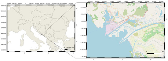
Figure 1.
Mouth of river Neretva—sampling location on the eastern side of the Adriatic Sea.
All the fish were weighed. For the determination of the life stage, the total length (TL) of each specimen was measured to the nearest mm, and eye height and eye width were measured to the nearest 0.1 mm. The coloration of the skin and differentiation of the lateral line were recorded according to Acou et al. [24]. The life stage was determined by qualitative (skin coloration and lateral line differentiation) and quantitative criteria (ocular index) according to Acou et al. [24]. Ocular index (OI) was computed using the formula [25] OI = {[(AR + BR)/4)] × [(AL + BL)/4] × π/TL} × 100, where OI is the ocular index, A and B are the horizontal and vertical eye diameters in mm, TL is the total body length and R and L indicate left and right eyes. A value of 6.5 for was considered as the threshold separating the life stages of yellow and silver eels as determined by Pankhurst [25]. The eels were considered to be silver if they had at least two of the criteria present. After determination of the life stage, all further analyses were carried out separately for yellow and silver eels.
Morphometric parameters were measured using an ichthyometer and a digital caliper. Measurements that were taken are shown in Figure 2.

Figure 2.
Representation of measured characteristics; TL—total length; SL—standard length; CL—head length; CH—head height; CW—head width; EH—eye height; EW—eye width; IO—interocular length; ML—mouth length; LS—snout length; HS—snout height; MH—mouth height; MW—mouth width; H—maximum body height; LP—length of pectoral fin; LDA—length from the beginning of the dorsal to the beginning of the anal fin; PA—preanal length; PD—predorsal length; LD—dorsal fin length; HD—maximum dorsal fin height; LA—anal fin length; HA—maximum anal fin height.
Weight–length relationships were determined with the equation [58] W = a × TLb, where W is body mass, TL is total length, a is coefficient of body shape, and b is coefficient of growth. Fulton’s condition factor was computed using the formula [59] CF = W/TL3 × 100, where CF is Fulton’s condition factor, W is body mass, and TL is total length.
After dissection, gut length was measured and the empty intestines, liver and gonads were weighed. In order to investigate morphological properties of the digestive system, relative gut length (RGL) and weight (RGW) and Zihler’s index (ZHI) were used. RGL was calculated using the formula [60] RGL = IL/TL × 100, where RGL is relative gut length, IL is intestine length, and TL is total length. RGW was calculated using the formula [61] RGW = IW/W × 100, where RGW is relative gut length, GW is empty intestine weight, and W is body mass. ZHI was calculated using the formula [62] ZHI = GL/(10 × ∛ W), where ZHI is Zihler’s index IL is intestine length, and W is body mass.
To access the energy reserves stored in liver, hepatosomatic index (HSI) was used. HSI was calculated using the formula [63] HSI = LW/W × 100, where HSI is hepatosomatic index, LW is liver weight, and W is body weight.
Data were analyzed using R (2023) [64]. Normality of the data and normality of residuals in used models were checked by examining histograms and by using Shapiro–Wilk test. Fligner–Killeen Test was used to check for homogeneity of variances. Durbin–Watson test was used to check for autocorrelation of residuals for regressions.
To describe the relationship between the measured morphometric parameters and TL, a linear regression was made for each parameter in relation to TL for both life stages, determined by the equation Y = aTL + b, where Y is a morphometric parameter, a is the slope, TL is total length, and b is the intercept.
To enable multivariate comparison of morphometric parameters of silver and yellow eels, due to size variation, data were transformed to common logarithms [65]. Another advantage of the transformation is that linearity and normality are usually more closely approximated by logarithmic rather than original values [66]. No outliers were found in the data by inspection of dot plots. A linear regression was made for all measured morphometric parameters in relation to TL and all regressions were statistically significant (p < 0.001). Analysis of covariance (ANCOVA) was used to test for differences in allometric relationships among yellow and silver eels for the analyzed morphometric parameters [67]. When the test was significant, meaning that heterogeneity between silver and yellow eels existed, regressions were made for both life stages separately, otherwise, common regression was used instead [68]. The measurements of morphometric parameters were adjusted to those expected for the overall mean total length with a modification of the allometric formula [69] ACi = logOCi − [ β × (logTLi − logMTL)], where ACi is the adjusted logarithmic parameter measurement of the itch specimen, OCi is the unadjusted parameter measurement of the itch specimen, β is the regression coefficient of that parameter against total length after the logarithmic transformation of both variables, TLi is the total length of the ith specimen, and MTL is the overall mean total length. To analyze the external morphometric data, analysis of similarities (ANOSIM) based on Bray–Curtis dissimilarity with 999 permutations was used. Level of similarity among groups was visualized using Multidimensional scaling (MDS). Additionally, similarity percentage (SIMPER) was calculated based on pairwise comparisons to find the contribution of each parameter to the overall dissimilarity [70].
ANCOVA test was used to investigate the effect of TL on W, IL, IW and LW. Principal components analysis (PCA) was performed using the log-transformed data with length effect removed for W, IL, IW, LW and TL.
Weight–length relationships were analyzed using ANOVA to determine if the b coefficient of the weight–length relationships differed from the isometric growth (b = 3). If the difference was statistically significant, allometric growth (positive or negative) was determined. ANCOVA was used to test for difference in determined b coefficients.
ANOVA test was used to analyze parameters that were normally distributed (CF, RGW, RGL, ZHI and HSI). If ANOVA was statistically significant, Tukey’s post hoc test was performed to determine which groups were different. If data were not normally distributed (TL, IO, W), Kruskal–Wallis test and Dunn’s post hoc test were used instead.
3. Results
A total of 77 specimens of eel were captured. Individual body weight of all captured eels ranged from 47 g to 1130 g. The TL ranged from 29.1 to 86.6 cm. The left eye height ranged from 2.6 to 11 mm and the right eye height ranged from 2.5 to 11.2 mm. The left eye width ranged from 3.0 to 11.9 mm and the right eye width ranged from 2.9 to 12.0 mm.
Of all the eels that were caught, 28 (36.4%) had an OI above the threshold of 6.5. Two silver eels with an OI greater than 6.5 and contrasting coloring in January did not have differentiated lateral lines, while the rest had both criteria present (Figure 3).

Figure 3.
Values of the OI of yellow (empty circles) and silver eels (black circles) with the threshold line set at 6.5 [21].
The median weight value of the yellow eels was the highest in the autumn and the lowest in the summer. The median weight value of the silver eels was the highest in the winter and the lowest in the autumn (Table 1). The differences in weight grouped by life stage in different seasons were determined to be statistically significant using the Kruskal–Wallis test (chi-squared = 12.52, p = 0.01). Dunn’s post hoc test showed that silver eels from the winter were significantly different from yellow eels in the summer (Supplementary Table S3).

Table 1.
Seasonal weight values of silver and yellow eels from the mouth of river Neretva (n—number of specimens; SD—standard deviation).
Yellow eels had the highest median value of total length and weight in spring and the lowest in summer. Silver eels had the highest median value of total length and weight in winter and the lowest in the autumn (Table 1). Differences in the TL between eels grouped by life stage in different seasons were determined by Kruskal–Wallis test to be statistically significant (chi-squared = 22.46, p = 0.00001). Dunn’s post hoc test showed that the TL of silver eels in the winter differed statistically from the TL of yellow eels in spring, summer and autumn (Supplementary Table S1). Yellow eels had the highest median value of OI in the spring and the lowest in the summer. Silver eels had the highest median value of OI in the autumn and smallest in the winter (Table 2). The differences in the OI between yellow and silver eels in different seasons were determined by the Kruskal–Wallis test to be statistically significant (53.84, p < 0.0001). Dunn’s post hoc test showed that the OI of silver eels from all seasons differed from the OI of yellow eels from all seasons (Supplementary Table S2).

Table 2.
Seasonal TL and OI values of silver and yellow eels from the mouth of river Neretva (n—number of specimens; SD—standard deviation).
The length of yellow eels ranged from 29.1 cm to 60.3 cm, while most of them (28.6%) belonged to a size class of 36–40 cm. The length of the silver eels ranged from 36.2 to 86.6 cm and most of the specimens (30.0%) were in the size class of 36–40 cm (Figure 4).
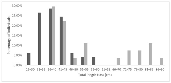
Figure 4.
Size class distribution of yellow (black columns) and silver eels (grey columns).
The values of measured morphometric parameters for yellow and silver eels are given in Table 3. Linear regressions of all the morphometric parameters with the TL had values of R2 between 0.56 and 0.999. The length–length relationships were found to be significantly linear in all cases (p < 0.001). A positive increase with an increase in the TL was determined for all morphometric parameters. The highest values of regression slopes were observed for the SL (a = 0.99 and a = 0.96 for yellow and silver eels), LD (a = 0.65 and 0.69 for yellow and silver eels) and LA (a = 54 and a = 55 for yellow and silver eels).

Table 3.
Minimum, maximum, median and standard deviation of the morphometric parameters (cm) and a—slope; b—intercept; and R2—coefficient of determination of linear regression of morphometric parameters with TL for yellow and silver eels.
ANOSIM showed significant differences in analyzed log-transformed parameters between yellow and silver eels (R = 0.6487, p = 0.001). The MDS plot showed that a small overlap in the analyzed parameters exists, but generally, the two groups were separated (stress value = 0.16). The within-group distance was smaller among the yellow than among the silver eels (Figure 5).
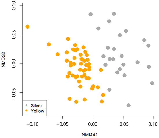
Figure 5.
MDS plot of similarity of analyzed morphometric parameters for yellow and silver eels.
Parameters that contributed the most to the dissimilarity between the groups are EW (13%), EH (10.9%), IO (8.2%), HD (7.1%), LP (6.3%), HS (6%) and HA (5.3%) (Table 4).

Table 4.
Results of SIMPER analysis of the external morphometric parameters that contributed to the dissimilarity between groups with more than 5%. Average—average contribution of this parameter to the average dissimilarity of the two groups; SD—standard deviation of the contribution of this parameter; ratio—ratio of average to SD; ava, avb—average value of every parameter in each of the two groups, cumulative contribution of this and all previous parameters in the table.
Values of IL, IW and LW are given in Table 5. Linear regressions (p < 0.001) showed an increase in all the measured parameters with the TL. Regressions of the IL with TL had R2 values between 0.45 and 0.63 depending on the life stage and season, meaning that the TL accounted for 45–63% of explained variation. Regressions of the IW with TL showed that the TL accounted for 39–78% of the explained variation. Regressions of the LW with TL showed that the TL accounted for 27–80% of the explained variation, depending on the life stage and season. ANCOVA showed that the differences in the regression coefficients between life stages in different seasons were statistically significant for all three parameters (p < 0.05).

Table 5.
Minimum, maximum, median and standard deviation of intestine length (IL), intestine weight (IW) and liver weight (LW). a—slope; b—intercept; and R2—coefficient of determination of linear regression of measured parameters with TL for yellow and silver eels.
Overall PCA analysis showed that PC1 and PC2 axes explained more than 90% of the variability of the data. In addition, silver eels from the winter are grouped separately from the rest. The main factor that separates the groups is the IL, whereas the rest of the factors (weight, intestine weight, liver weight and total length) affect the groupings almost equally (Figure 6).
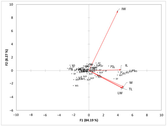
Figure 6.
PCA biplot based on standardized log transformed parameters: body weight (W), total length (TL), intestine length (IL), intestine weight (IW) and liver weight (LW). SY—yellow eels from the spring; SUY—yellow eels from the summer; AY—yellow eels from the autumn; WY—yellow eels from the winter; AS—silver eels from the autumn; WS—silver eels from the winter.
Positive allometric growth was determined for all the eels caught throughout the sampling period and described with the overall equation W = 0.0008 × TL3.20 (R2 = 0.97, p < 0.05) (Figure 7). Isometric growth was determined for yellow eels for the entire sampling period and described with the equation W = 0.0012 × TL3.01 (R2 = 0.96, p < 0.05), and positive allometric growth for silver eels was described by the equation W = 0.0004 × TL3.37 (R2 = 0.95 p < 0.05).
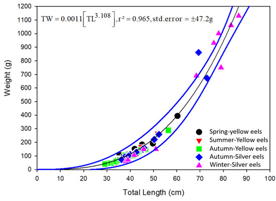
Figure 7.
Length–weight relationship of the European eel grouped by season and life stage (black line—regression line, blue lines—confidence interval).
The coefficient b values for yellow eels ranged from 2.98 in autumn to 3.29 in summer and for silver eels from 3.36 in autumn to 3.6 in winter. Isometric growth was determined for yellow eels in spring and autumn. Positive allometric growth was determined for yellow eels in the summer and silver eels in autumn and winter (Table 6). The differences in the b coefficient grouped by life stage in different seasons were statistically significant (ANCOVA, p < 0.05).

Table 6.
Weight–length relationship parameters of silver and yellow eels from the mouth of river Neretva (n—number of specimens; a and b—coefficients of weight–length relationships; R2—coefficient of determination) and one-way ANOVA to test for deviation from the isometric growth.
The Mean value of the CF for yellow eels in every season and silver eels in the autumn was the same, and it equaled 0.17 ± 0.02. The Mean value of the CF for silver eels in the winter was 0.16 ± 0.04. Seasonal differences in the CF were determined not to be statistically significant using ANOVA (Supplementary Table S4).
From the three indices used to describe intestine morphology, the only statistically significant difference between yellow and silver eels was determined for RGW, while no statistically significant differences were found for RGL and ZHI (Table 7).

Table 7.
Intestine morphology indices for silver and yellow eel from the mouth of river Neretva (SD—standard deviation; RGL—relative gut length; RGW—relative body weight; ZHI—Zihler’s index) and one-way ANOVA output (f and p-values).
Yellow eels had the highest mean HSI in the spring and the lowest in the summer. Silver eels had the biggest HSI in the autumn (Table 8). Differences between the eels grouped by life stage in different seasons were found to be statistically significant using ANOVA (Supplementary Table S5). Tukey’s post hoc test showed that the HSI of silver eels in the winter differed statistically from the HSI of yellow eels in spring (Supplementary Table S6).

Table 8.
Seasonal values of HSI of yellow and silver eels from the mouth of river Neretva (HSI—hepatosomatic index; SD—standard deviation).
4. Discussion
The transition from yellow to silver eels, also called silvering, encompasses a series of adaptations that enables their spawning migration and successful reproduction [22,23]. It is a gradual process that normally lasts from spring to autumn and the changes happen chronologically [71,72]. An increase in the eye diameter is one of the first changes, usually correlated with an increase in the gonad weight, and contrast coloring occurs among the last changes [33,72]. For that reason, the combination of three different criteria was used in this study for the determination of the life stage. Two silver eels were found in the river Neretva delta in February 2022 with OI values of 10.71 and 9.22 and contrast coloring but an undifferentiated lateral line. Acou et al. [24] also reported two specimens with the same description among 214 analyzed specimens in the river Frémur in France. This indicates variability in the chronological order of changes during the silvering process for a small portion of the subpopulations. Although no yellow eel had an OI greater than the threshold, three specimens with an OI over 5.0 and one with an OI over 6.0 were recorded in the spring of 2021. This could indicate that they were caught on the onset of the silvering process. Acou et al. [24] also found that yellow eels without any silvering criteria were the most abundant and the eels in transition constituted only a small percentage in the rivers Frémur, Oir and Loire in France in the autumn.
The presence of yellow eels in the catch in this study is in accordance with Glamuzina et al. [73] who reported the presence of yellow eels throughout the year in Baćinska lakes and the Parila lagoon at the mouth of the river Neretva. The presence of silver eels in the autumn and winter is consistent with the literature, as it is the period when the migration towards the Sargasso Sea starts [1]. Morović [74] recorded that the catch in the lower flow of the river Neretva in the autumn exclusively consisted of silver eels in the years 1943, 1946 and 1947. Glamuzina et al. [73] reported the presence of only a small percentage (9.8–16.4%) of silver eels in the autumn/winter catch during 2016–2019, while in this research, silver eels constituted 48.1% and 93.8% in autumn and winter. These differences might be related to the sampling location and sampling methods, as well as environmental factors, but there is currently no way of knowing the progression of changes in the appearance of the two different life stages due to gaps in the available data.
Most of the yellow eels that were caught during this research belonged to the length class of 36–40 cm, which is similar to the results of Glamuzina and Dobroslavić [52] and Glamuzina et al. [73], but the authors do not distinguish between different life stages. Metamorphosis largely depends on body size [75,76]. A difference in metamorphosis timing and growth is also present among the sexes. Female eels grow larger, and the timing of metamorphosis is more flexible as compared to males that metamorphosize at smaller sizes [77]. Some eels never metamorphosize; instead, they remain in the yellow eel stage until death [78]. An overlap found in this research in the size classes of yellow and silver eels can be explained as a consequence of different sizes that the sexes have to reach for metamorphosis. De Leo and Gatto [79] reported that female silver eels sampled from Valli di Comacchio lagoons in the eastern Adriatic were longer than males, which averaged around 40 cm in length. Body size strongly influences swimming performance. Generally, continuous swimming speed increases with body size, but maneuverability decreases [14]. On the contrary, acceleration, which is important for predator avoidance, does not change with body size [80]. It is also known that eels are burrowing into the substrate to shelter from predators and fishing tools [81,82]. Not enough is known about the influence of body size on digging behavior; although, larger individuals theoretically have more force and can penetrate harder and denser substrates, but a larger body produces more drag during digging [83]. For that reason, the substrate preference of different sedentary life stages of the European eel changes with size, and larger individuals generally prefer fine gravel substrates [81].
This research, to our knowledge, presents the first comprehensive external morphometric analysis of the European eel. Yellow and silver eels differed significantly with regard to the 22 morphometric measures that were analyzed. The EH and EW, which accounted for the biggest differences between yellow and silver eels, have already been well described in the literature [23,25,32,33,34,35,48]. Durif et al. [48] also reported that LP is a discriminant factor for the determination of the two life stages, while Rad et al. [34] did not reach the same conclusion. Other morphometric parameters that were responsible for the differences greater than 5% between the two life stages in this research were two related to the head area, IO and HS, and two related to fin heights: HD and HA. Variations in eel head shape have been well studied because they strongly influence feeding, burrowing and competitive behavior [39,82,84,85]. Bite force increases with increasing width of the head which shifts food preferences towards more nutritious, larger pray, thereby increasing the trophic level of eels with wider heads [86]. European eels were reported to have a dimorphic head shape with broad- and narrowheaded individuals [36,37]. Such a phenomenon was not observed in this research, nor was it observed by Verhelst et al. [38]. Instead, CW showed a slow linear increase following the increase in the TL for both silver and yellow eels.
Research in the river Neretva by Dulčić and Glamuzina [50] showed positive allometric growth for the used individuals in the study, while Glamuzina and Dobroslavić [52] reported isometric growth. Piria et al. [53] sampled the eel from five rivers in the eastern Adriatic basin and reported negative allometric growth with the b coefficient ranging from 2.60 to 2.82. Seasonal data and classification of life stages, which in this research were shown to affect growth, are missing in the mentioned research. The eel is an opportunistic species with regard to feeding [87], and feeding increases with increasing temperature [88]. Therefore, the positive allometric growth recorded in the summer might be the result of increased food availability and increased temperature. The positive allometric growth observed for silver eels can be explained as a consequence of increased feeding prior to metamorphosis which is necessary to build up fat reserves which are used as an energy source for migration [89]. Other research conducted in Europe reported b coefficient values ranging from 2.60 to 3.47 [34,40,43,44,45,46,47].
Seasonal values of the CF in this research showed little to no variation for both life stages. This finding is in accordance with Simon [49] who reported that the mean value of the CF was 0.16–0.18 in six lakes on the river Havel in Germany. Similar values were reported by Milošević et al. [90] who found the CF to be 0.166 ± 0.02 for females and 0.173 ± 0.011 for males in Lake Skadar, Montenegro.
The morphology of the intestine of fish is determined by phylogeny but also to a certain extent by the influence of external factors during ontogenesis [61]. In general, intestine length is a factor of nutrition and digestibility of the predominant food components [91,92]. Some studies [93,94] showed that when suffering food shortages, some fish may reduce the size and function of the digestive tract. Kužir et al. [54] found that the intestine of reared eels is almost twice as long as that of a wild-caught eel. This suggests that feeding intensity and dominant food items have a strong effect on the intestine length for yellow eels. For the wild-caught eels from the river Zrmanja (east Adriatic) with an average length of 51.9 ± 7.7 cm, Kužir et al. [54] determined the RGL to be 29.9, which is similar to the results of this paper. It is generally accepted that silver eels stop feeding after metamorphosis, their intestinal digestion function is lost, the walls reduce in width, the intestine shortens, and its remaining functions are water resorption and osmoregulation [22]. Shortening of the intestine of silver eels was not observed in this research. Instead, silver eels had RGL values higher than yellow eels, and the difference was close to statistical significance (p = 0.09). This could mean that feeding in silver eels does not completely stop immediately after metamorphosis, but it strongly reduces as reported by Beulleus et al. [95]. The intestine shortens later when the feeding stops completely [96]. A statistically significant reduction in the RGW was recorded for silver eels compared to yellow eels in this research. This change is a consequence of the drastic reduction in the wall thickness, number of villi, number of mucus cells, and structural changes in epithelial cells [97]. These results could indicate that the intestine weight reduces before length in response to reduced or complete cessation of feeding. The RGW values in this research were similar to those reported by Lionetto et al. [98] who determined the RGL to be 1.81 ± 0.13 for yellow and 0.46 ± 0.03 for silver eels.
Mean seasonal HSI values in this research are similar to those reported by Svedäng and Wickström [99] from southeast Sweden. Mean values of the HSI in this research were significantly different between silver eels in the winter and yellow eels in the spring. Higher values of the HSI in spring may be due to increased feeding with rising temperature and increased prey availability [88]. Similar to the assumptions of Righton and Metcalfe [22], the results of this study indicated that silver eels stop or drastically decrease feeding after metamorphosis which could explain the low HSI values of silver eels in winter.
A substantial amount of the analyzed morphological parameters were not statistically significant. The insignificance of these parameters could be due to the sample size. For this reason, statistical tests were performed based on “small sample size” transformations (for example, tests between means) whenever this was possible and available. At the same time, analyses based on sub-groups were performed (by life stage and by season) to reduce the error caused by the small sample size and at the same time to stratify the data and increase the accuracy of the estimated parameters (for example, averages and standard deviations) and PCA. Furthermore, the outlier analysis made it possible to remove individuals which reduce the effectiveness of the relationships.
The PCA showed that the factor most responsible for grouping eels based on season and life stage was the IL due to the silver eels from the winter. The value of this parameter was higher for the silver eels but probably decreases in the more advanced silver stage of the eels.
This extensive research with regard to a number of morphological parameters represents the “zero state” for the investigated locality and at the same time provides an example of the basis for the development of an appropriate eel monitoring and management plan in such areas.
5. Conclusions
This research, to our knowledge, presents the first comprehensive external morphometric analysis of the European eel. The results of this work suggest that the intestine of the silver eels decreases in weight before it decreases in length. Therefore, the RGW could be more useful for differentiating yellow and silver eels earlier in the silvering process. The results showed that IO, HD and HS could be used in addition to the EH, EW and LP that are commonly used to distinguish yellow and silver eels. This would make the two life stages more morphologically different than previously thought. However, future research is needed to confirm this statement. Monitoring the characteristics investigated in this paper is crucial for the management of the European eel fisheries as they are directly related to functional performance and affect the ability to maintain sustainable populations in anthropogenically altered environments.
Supplementary Materials
The following supporting information can be downloaded at: https://www.mdpi.com/article/10.3390/d15121223/s1, Table S1: Comparison of TL for yellow and silver eels in different seasons using Dunn’s multiple comparison post hoc test; Table S2: Comparison of OI for yellow and silver eels in different seasons using Dunn’s multiple comparison post hoc test; Table S3: Comparison of W for yellow and silver eels in different seasons using Dunn’s multiple comparison post hoc test; Table S4: ANOVA output for CF of yellow and silver eels in different seasons; Table S5: ANOVA output for HSI of yellow and silver eels in different seasons; Table S6: Comparison of HSI for yellow and silver eels in different seasons using Tukey’s range post hoc test.
Author Contributions
Conceptualization and supervision, A.G. and J.J.-D.; methodology, A.G., A.C. and O.B.; software, O.B.; validation, T.R. and J.J.-D.; formal analysis, O.B., T.R., N.K. and A.C.; investigation, O.B., T.R. and A.G.; writing—original draft preparation, O.B.; Writing—review and editing, J.J.-D., A.C. and A.G. All authors have read and agreed to the published version of the manuscript.
Funding
This study was funded by the Ministry of Agriculture of the Republic of Croatia within the framework of Measure I.3. “Partnership between scientists and fishermen for the period 2017–2020” as a grant for the project “Fishermen—Scientist Network of the City of Ploče”; activity: “Case study: Status of eel population in Neretva Delta” (grant number 324-01/20-01/1244).
Institutional Review Board Statement
The animal study protocol was approved by the Ministry of Agriculture of the Republic of Croatia (protocol number 525-13/1269-2-2 from 5 July 2020 and protocol number 525-13/0797-21-2 from 6 May 2021).
Data Availability Statement
Data is contained within the Supplementary Material.
Acknowledgments
The results presented in the paper are outputs from the research project “Fishermen and Scientists Network of the City of Ploče” within the framework of Measure I.3. “Partnership between scientists and fishermen for the period 2017–2020”. For the sample collection, authors express a special gratitude to the fisherman Ante Kapović.
Conflicts of Interest
The research was conducted in the absence of any commercial or financial relationships that could be construed as a potential conflict of interest. Jurica Jug-Dujaković worked at a few Croatian Universities, and he is an expert, still the guest lecturer in Croatia, and this manuscript has nothing to do with his work in the USA.
References
- Deelder, C.L. Synopsis of biological data on the eel, Anguilla anguilla (Linnaeus, 1758). FAO Fish. Synop. 1984, 80, 73. [Google Scholar]
- Rochard, E.; Elie, P. La Macrofaune Aquatique de L’Estuaire de la Gironde. Contribution au Livre Blanc de L’Agence de L’Eau Adour Garonne. In État des Connaissances sur L’Estuaire de la Gironde; Mauvais, J.L., Guillaud, J.F., Eds.; Agence de L’Eau Adour-Garonne, Éditions Bergeret: Bordeaux, France, 1994; Volume 56, p. 115. [Google Scholar]
- Kettle, A.J.; Heinrich, D.; Barrett, J.H.; Benecke, N.; Locker, A. Past distributions of the European freshwater eel from archaeological and palaeontological evidence. Quat. Sci. Rev. 2008, 27, 1309–1334. [Google Scholar] [CrossRef]
- Gabriel, O.; Wendt, T. Fishing methods. In The Eel, 3rd ed.; Tesch, F.W., Thorpe, J.E., Eds.; Blackwell Science Ltd.: Hoboken, NJ, USA, 2003; pp. 243–293. [Google Scholar] [CrossRef]
- Dekker, W. The history of commercial fisheries for European eel commenced only a century ago. Fish Manag. Ecol. 2019, 26, 6–19. [Google Scholar] [CrossRef]
- Palm, S.; Dannewitz, J.; Prestegaard, T.; Wickström, H. Panmixia in European eel revisited: No genetic difference between maturing adults from southern and northern Europe. Heredity 2009, 103, 82–89. [Google Scholar] [CrossRef]
- Dekker, W. The fractal geometry of the European eel stock. ICES J. Mar. Sci. 2000, 57, 109–121. [Google Scholar] [CrossRef]
- International Council for the Exploration of the Sea. European Eel (Anguilla anguilla) throughout Its Natural Range; JNCC: Peterborough, UK, 2019. [Google Scholar] [CrossRef]
- Froehlicher, H.; Kaifu, K.; Rambonilaza, T.; Daverat, F. Eel translocation from a conservation perspective: A coupled systematic and narrative review. Glob. Ecol. Conserv. 2023, 46, e02635. [Google Scholar] [CrossRef]
- The IUCN Red List of Threatened Species. Available online: https://www.iucnredlist.org/ (accessed on 19 November 2023).
- Convention on the Conservation of Migratory Species of Wild Animals (CMS). Available online: https://www.cms.int/sites/default/files/basic_page_documents/appendices_cop13_e_0.pdf (accessed on 19 November 2023).
- Council Regulation (EC) No 1100/2007 of 18 September 2007 Establishing Measures for the Recovery of the Stock of European Eel. Available online: https://eur-lex.europa.eu/legal (accessed on 19 November 2023).
- Council Regulation (EU) 2023/194 of 30 January 2023 Fixing for 2023 the Fishing Opportunities for Certain Fish Stocks, Applicable in Union Waters and, for Union Fishing Vessels, in Certain Non-Union Waters, as Well as Fixing for 2023 and 2024 Such Fishing Opportunities for Certain Deep-Sea Fish Stocks. Available online: https://eur-lex.europa.eu/legal-content/EN/TXT/?uri=CELEX:32023R0194 (accessed on 19 November 2023).
- De Meyer, J.; Verhelst, P.; Adriaens, D. Saving the European eel: How morphological research can help in effective conservation management. Integr. Comp. Biol. 2020, 60, 467–475. [Google Scholar] [CrossRef]
- Arnold, S.J. Morphology, performance and fitness. Am. Zool. 1983, 23, 347–361. [Google Scholar] [CrossRef]
- Arnold, S.J. Performance surfaces and adaptive landscapes. Integr. Comp. Biol. 2003, 43, 367–375. [Google Scholar] [CrossRef]
- Irschick, D.J. Measuring performance in nature: Implications for studies of fitness within populations. Integr. Comp. Biol. 2003, 43, 396–407. [Google Scholar] [CrossRef]
- Schoenfuss, H.L.; Blob, R.W. The importance of functional morphology for fishery conservation and management: Applications to Hawaiian amphidromous fishes. Bish. Mus. Bull. Cult. Environ. Stud. 2007, 3, 125–141. [Google Scholar]
- Holland, L.E. Effects of barge traffic on distribution and survival of ichthyoplankton and small fishes in the Upper Mississippi River. Trans. Am. Fish. Soc. 1986, 115, 162–165. [Google Scholar] [CrossRef]
- Wolter, C.; Arlinghaus, R. Navigation impacts on freshwater fish assemblages: The ecological relevance of swimming performance. Rev. Fish Biol. Fish. 2003, 13, 63–89. [Google Scholar] [CrossRef]
- Cresci, A. A comprehensive hypothesis on the migration of European glass eels (Anguilla anguilla). Biol. Rev. 2020, 95, 1273–1286. [Google Scholar] [CrossRef] [PubMed]
- Righton, D.A.; Metcalfe, J.D. Fish migrations|Eel Migrations. Encycl. Fish Physiol. 2011, 3, 1937–1944. [Google Scholar] [CrossRef]
- Durif, C.; Guibert, A.; Elie, P. Morphological discrimination of the silvering stages of the European eel. In American Fisheries Society Symposium; American Fisheries Society: New York, NY, USA, 2009; Volume 58, pp. 103–111. [Google Scholar]
- Acou, A.; Boury, P.; Laffaille, P.; Crivelli, A.J.; Feunteun, E. Towards a standardized characterization of the potentially migrating silver European eel (Anguilla anguilla L.). Arch. Hydrobiol. 2005, 164, 237–255. [Google Scholar] [CrossRef]
- Pankhurst, N.W. Relation of visual changes to the onset of sexual maturation in the European eel Anguilla anguilla (L.). J. Fish Biol. 1982, 21, 127–140. [Google Scholar] [CrossRef]
- Bevacqua, D.; Melià, P.; Schiavina, M.; Crivelli, A.J.; De Leo, G.A.; Gatto, M. A demographic model for the conservation and management of the European eel: An application to a Mediterranean coastal lagoon. ICES J. Mar. Sci. 2019, 76, 2164–2178. [Google Scholar] [CrossRef]
- Enbody, E.D.; Pettersson, M.E.; Sprehn, C.G.; Palm, S.; Wickström, H.; Andersson, L. Ecological adaptation in European eels is based on phenotypic plasticity. Proc. Natl. Acad. Sci. USA 2021, 118, e2022620118. [Google Scholar] [CrossRef]
- Sagnes, P.; Gaudin, P.; Statzner, B. Shifts in morphometrics and their relation to hydrodynamic potential and habitat use during grayling ontogenesis. J. Fish Biol. 1997, 50, 846–858. [Google Scholar] [CrossRef]
- Kawamura, G.; Tsuda, R.; Kumai, H.; Ohashi, S. The visual cell morphology of Pagrus major and its adaptive changes with shifts from pelagic to benthic habitats. Bull. Jpn. Soc. Sci. Fish. 1984, 50, 1975–1980. [Google Scholar] [CrossRef]
- Kawamura, G.; Washiyama, N. Ontogenic changes in behaviour and sense organ morphogenesis in largemouth bass and Tilapia nilotica. Trans. Am. Fish. Soc. 1989, 118, 203–213. [Google Scholar] [CrossRef]
- Norton, S.F.; Luczkovitch, J.J.; Motta, P.J. The role of ecomorphological studies in the comparative biology of fishes. Environ. Biol. Fishes 1995, 44, 287–304. [Google Scholar] [CrossRef]
- Frost, W.E. The Age and Growth of Eels (Anguilla anguilla) from the Windermere Catchment Area. J. Anim. Ecol. 1945, 14, 106–124. [Google Scholar] [CrossRef]
- Han, Y.S.; Liao, I.C.; Huang, Y.S.; He, J.T.; Chang, C.W.; Tzeng, W. Synchronous changes of morphology and gonadal development of silvering Japanese eel Anguilla japonica. Aquaculture 2003, 219, 783–796. [Google Scholar] [CrossRef]
- Rad, F.; Barış, M.; Bozaoğlu, S.A.; Temel, G.O.; Üstündağ, M. Preliminary investigation on morphometric and biometric characteristics of female silver and yellow, Anguilla anguilla, from eastern Mediterranean (Göksu delta/Turkey). J. Fish Sci. 2013, 7, 253–265. [Google Scholar] [CrossRef]
- Lythgoe, J.N. The Ecology of Vision; Clarendon Press: Oxford, UK, 1979; p. 244. ISBN 0198545290/9780198545293. [Google Scholar]
- Törlitz, H. Anatomische und entwicklungsgeschichtliche Beitr€age zur Artfrage unseres Flussaales. Z Fisch 1992, 21, 1–48. [Google Scholar]
- Proman, J.M.; Reynolds, J.D. Differences in head shape of the European eel, Anguilla anguilla (L.). Fish. Manag. Ecol. 2000, 7, 349–354. [Google Scholar] [CrossRef]
- Ide, C.; De Schepper, N.; Christiaens, J.; Van Liefferinge, C.; Herrel, A.; Goemans, G.; Meire, P.; Belpaire, C.; Geeraerts, C.; Adriaens, D. Bimodality in head shape in European eel. J. Zool. 2011, 285, 230–238. [Google Scholar] [CrossRef]
- Verhelst, P.; De Meyer, J.; Reubens, J.; Coeck, J.; Goethals, P.; Moens, T.; Mouton, A. Unimodal head-width distribution of the European eel (Anguilla anguilla L.) from the Zeeschelde does not support disruptive selection. PeerJ 2018, 6, e5773. [Google Scholar] [CrossRef]
- İlhan, A.; İlhan, D.; Hammed, R.O. Comparisons of morphometric characteristics and length-weight relationship of european eel (Anguilla anguilla L., 1758) in Turkish inland waters. Egypt. J. Zool. 2020, 74, 13–21. [Google Scholar] [CrossRef]
- Gordo, L.S.; Jorge, M.I. Age and growth of the European eel, Anguilla anguilla (Linnaeus, 1758) in the Aveiro Lagoon, Portugal. Sci. Mar. 1991, 55, 389–395. [Google Scholar]
- Koutrakis, E.T.; Tsikliras, A.C. Length–weight relationships of fishes from three northern Aegean estuarine systems (Greece). J. Appl. Ichthyol. 2003, 19, 258–260. [Google Scholar] [CrossRef]
- Yalçın-Özdilek, Ş.; Gümüş, A.; Dekker, W. Growth of European eel in a Turkish river at the south-eastern limit of its distribution. Electron. J. Ichthyol. 2006, 2, 55–64. [Google Scholar]
- Kara, A.; Sağlam, C.; Acarli, D.; Cengız, Ö. Length-weight relationships for 48 fish species of the Gediz estuary, in Izmir Bay (Central Aegean Sea, Turkey). J. Mar. Biol. Assoc. UK 2018, 98, 879–884. [Google Scholar] [CrossRef]
- Veiga, P.; Machado, D.; Almeida, C.; Bentes, L.; Monteiro, P.; Oliveira, F.; Ruano, M.; Erzini, K.; Gonçalves, J.M.S. Weight–length relationships for 54 species of the Arade estuary, southern Portugal. J. Appl. Ichthyol. 2009, 25, 493–496. [Google Scholar] [CrossRef]
- Verreycken, H.; Van Thuyne, G.; Belpaire, C. Length–weight relationships of 40 freshwater fish species from two decades of monitoring in Flanders (Belgium). J. Appl. Ichthyol. 2011, 27, 1416–1421. [Google Scholar] [CrossRef]
- Moreno-Valcárcel, R.; Oliva-Paterna, F.J.; Arribas, C.; Fernández-Delgado, C. Length–weight relationships for 13 fish species collected in the Doñana marshlands (Guadalquivir estuary, SW Spain). J. Appl. Ichthyol. 2012, 28, 663–664. [Google Scholar] [CrossRef]
- Durif, C.; Dufour, S.; Elie, P. The silvering process of Anguilla anguilla: A new classification from the yellow resident to the silver migrating stage. J. Fish Biol. 2005, 66, 1025–1043. [Google Scholar] [CrossRef]
- Simon, J. Age, growth, and condition of European eel (Anguilla anguilla) from six lakes in the River Havel system (Germany). ICES J. Mar. Sci. 2007, 64, 1414–1422. [Google Scholar] [CrossRef]
- Dulčić, J.; Glamuzina, B. Length–weight relationships for selected fish species from three eastern Adriatic estuarine systems (Croatia). J. Appl. Ichthyol. 2006, 22, 254–256. [Google Scholar] [CrossRef]
- Popović, J.; Fašaić, K.; Homen, Z. Komparativno ispitivanje odnosa dužina/masa u jegulja (Anguilla anguilla L. 1758) iz dva različita ekosistema. Eel (Anguilla anguilla L. 1758) comparative research of length-mass relation from two different ecosystems. Ichthyologia 1984, 16, 29–41. [Google Scholar]
- Glamuzina, L.; Dobroslavić, T. Summer fish migrations in the River Neretva (South-Eastern Adriatic coast, Croatia) as a consequence of salinization. Naše More 2020, 67, 103–116. [Google Scholar] [CrossRef]
- Piria, M.; Šprem, N.; Tomljanović, T.; Slišković, M.; Jelić Mrčelić, G.; Treer, T. Length weight relationships of the European eel Anguilla anguilla (linnaeus, 1758) from six karst catchments of the Adriatic basin, Croatia. Croat. J. Fish. Ribar. 2014, 72, 32–35. [Google Scholar] [CrossRef]
- Kužir, S.; Gjurčević, E.; Nejedli, S.; Baždarić, B.; Kozarić, Z. Morphological and histochemical study of intestine in wild and reared European eel (Anguilla anguilla L.). Fish Physiol. Biochem. 2012, 38, 625–633. [Google Scholar] [CrossRef]
- Gentile, L.; Casalini, A.; Emmanuele, P.; Brusa, R.; Zaccaroni, A.; Mordenti, O. Gonadal Development in European Eel Populations of North Adriatic Lagoons at Different Silvering Stages. Appl. Sci. 2022, 12, 2820. [Google Scholar] [CrossRef]
- Allendorf, F.; Ryman, N.; Utter, F. Genetics and fishery management: Past, present, and future. In Population Genetics and Fishery Management; Ryman, N., Utter, F., Eds.; Washington Sea Grant Publications; University of Washington Press: Seattle, WA, USA; London, UK, Reprinted by The Blackburn Press: Caldwell, NJ, USA; 1987; pp. 1–19. [Google Scholar]
- Wiberg, U.H. Sex determination in the European eel (Anguilla anguilla, L.) A hypothesis based on cytogenetic results, correlated with the findings of skewed sex ratios in eel culture ponds. Cytogenet. Genome Res. 1983, 36, 589–598. [Google Scholar] [CrossRef]
- Le Cren, E.D. The length-weight relationship and seasonal cycle in gonad weight and condition in the perch (Perca fluviatilis). J Anim. Ecol. 1951, 20, 201–219. [Google Scholar] [CrossRef]
- Ricker, W.E. Computation and interpretation of biological statistics of fish populations. Fish. Res. Board Can. Bull. 1975, 191, 1–382. [Google Scholar]
- Karachle, P.K.; Stergiou, K.I. Intestine morphometrics of fishes: A compilation and analysis of bibliographic data. Acta Ichthyol. Piscat. 2010, 40, 45–54. [Google Scholar] [CrossRef]
- Karachle, P.K.; Stergiou, K.I. Gut length for several marine fish: Relationships with body length and trophic implications. Mar. Biodivers. Rec. 2010, 3, e106. [Google Scholar] [CrossRef]
- Zihler, F. Gross morphology and configuration of digestive tracts of Cichlidae (Teleostei, Perciformes): Phylogenetic and functional, significance. Neth. J. Zool. 1981, 32, 544–571. [Google Scholar] [CrossRef]
- Wootton, R.J.; Evans, G.W.; Mills, L. Annual cycle in female three-spined sticklebacks (Gasterosteus aculeatus L.) from an upland and lowland population. J. Fish Biol. 1978, 12, 331–343. [Google Scholar] [CrossRef]
- R Core Team. R: A Language and Environment for Statistical Computing; R Foundation for Statistical Computing: Vienna, Austria, 2023; Available online: https://www.R-project.org/ (accessed on 15 November 2023).
- Thorpe, R.S. Biometric analysis of geographic variation and racial affinities. Biol. Rev. Cambr. Phil. Soc. 1976, 51, 407–452. [Google Scholar] [CrossRef]
- Hair, J.F.; Anderson, R.E., Jr.; Tatham, R.L.; Black, W.C. Multivariate Data Analysis, 5th ed.; Prentice-Hall: Upper Saddle River, NJ, USA, 1998. [Google Scholar]
- Sokal, P.R.; Rohlf, F.J. Biometry: The Principles and Practice of Statistics in Biological Research, 3rd ed.; W.H. Freeman: New York, NY, USA, 1995. [Google Scholar]
- Reist, J.D. An empirical evaluation of coefficients used in residual and allometric adjustment of size covariation. Can. J. Zool. 1986, 64, 1363–1368. [Google Scholar] [CrossRef]
- Claytor, R.R.; MacCrimmon, H.R. Partitioning size from morphometric data: A comparison of five statistical procedures used in fisheries stock identification research. Can. Tech. Rep. Fish. Aq. Serv. 1987, 1531, 1–23. [Google Scholar]
- Deli, T.; Bahles, H.; Said, K.; Chatti, N. Patterns of genetic and morphometric diversity in the marbled crab (Pachygrapsus marmoratus, Brachyura, Grapsidae) populations across the Tunisian coast. Acta Oceanol. Sin. 2015, 34, 49–58. [Google Scholar] [CrossRef]
- Fontaine, Y.A. L’argenture de l’anguille: Métamorphose, anticipation, adaptation. Bull. Fr. Pêche Piscic. 1994, 335, 171–185. [Google Scholar] [CrossRef][Green Version]
- Durif, C.; Elie, P.; Dufour, S.; Marchelidon, J.; Vidal, B. Analysis of morphological and physiological parameters during the silvering process of the European eel (Anguilla anguilla) in the Lake of Grand-Lieu, France). Cybium 2000, 24, 63–74. [Google Scholar]
- Glamuzina, L.; Pećarević, M.; Dobroslavić, T.; Tomšić, S.; Glamuzina, B. The study of European eel, Anguilla anguilla in the River Neretva estuary (Eastern Adriatic Sea, Croatia) using traditional fishery gear. Acta Adriat. 2022, 63, 35–44. [Google Scholar] [CrossRef]
- Morović, D. Nekoja opažanja o duljini i težini jegulje iz Neretve. Croat. J. Fish. Ribar. 1955, 10, 28–30. [Google Scholar]
- Vøllestad, L.A.; Jonsson, B. Life-history characteristics of the European eel Anguilla anguilla in the Imsa River, Norway. Trans. Am. Fish. Soc. 1986, 115, 864–871. [Google Scholar] [CrossRef]
- Vøllestad, L.A. Geographic variation in age and length at metamorphosis of maturing European eel: Environmental effects and phenotypic plasticity. J. Anim. Ecol. 1992, 61, 41–48. [Google Scholar] [CrossRef]
- Davey, A.J.; Jellyman, D.J. Sex determination in freshwater eels and management options for manipulation of sex. Rev. Fish Biol. Fish. 2005, 15, 37–52. [Google Scholar] [CrossRef]
- Rossi, R.; Colombo, G. Sex ratio, age and growth of silver eels in two brackish lagoons in the northern Adriatic (Valli di Comacchio and Valle Nuova). Arch. Oceanogr. Limnol. 1976, 18, 227–310. [Google Scholar]
- Leo, G.D.; Gatto, M. A size and age-structured model of the European eel (Anguilla anguilla L.). Can. J. Fish. Aquat. Sci. 1995, 52, 1351–1367. [Google Scholar] [CrossRef]
- Domenici, P. The scaling of locomotor performance in predator–prey encounters: From fish to killer whales. Comp. Biochem. Physiol. A Mol. Integr. Physiol. 2001, 131, 169–182. [Google Scholar] [CrossRef]
- Steendam, C. The Burrowing Eel: Effects of Life Stage and Head Shape. Master’s Thesis, University of Ghent, Ghent, Belgium, 2019; pp. 1–79. [Google Scholar]
- Herrel, A.; Choi, H.F.; Dumont, E.; De Schepper, N.; Vanhooydonck, B.; Aerts, P.; Adriaens, D. Burrowing and subsurface locomotion in anguilliform fish: Behavioral specializations and mechanical constraints. J. Exp. Biol. 2011, 214, 1379–1385. [Google Scholar] [CrossRef]
- Vogel, S. Life in Moving Fluids: The Physical Biology of Flow; Princeton University Press: Princeton, NJ, USA, 1994. [Google Scholar]
- Cooper, W.E.; Vitt, L.J. Female mate choice of large male broad-headed skinks. Anim. Behav. 1993, 45, 683–693. [Google Scholar] [CrossRef][Green Version]
- Vanhooydonck, B.; Boistel, R.; Fernandez, V.; Herrel, A. Push and bite: Trade-offs between burrowing and biting in a burrowing skink (Acontias percivali). Biol. J. Linn. Soc. 2011, 102, 91–99. [Google Scholar] [CrossRef]
- De Meyer, J.; Belpaire, C.; Boeckx, P.; Bervoets, L.; Covaci, A.; Malarvannan, G.; De Kegel, B.; Adriaens, D. Head shape disparity impacts pollutant accumulation in European eel. Environ. Pollut. 2018, 240, 378–386. [Google Scholar] [CrossRef] [PubMed]
- Bouchereau, J.L.; Marques, C.; Pereira, P.; Guelorget, O.; Lourié, S.M.; Vergne, Y. Feeding behaviour of Anguilla anguilla and trophic resources in the Ingril Lagoon (Mediterranean, France). Cah. Biol. Mar. 2009, 50, 319–332. [Google Scholar]
- Barak, N.E.; Mason, C.F. Population density, growth and diet of eels, Anguilla anguilla L., in two rivers in eastern England. Aquac. Res. 1992, 23, 59–70. [Google Scholar] [CrossRef]
- Larsson, P.; Hamrin, S.; Okla, L. Fat content as a factor inducing migratory behavior in the eel (Anguilla anguilla L.) to the Sargasso Sea. Naturwissenschaften 1990, 77, 488–490. [Google Scholar] [CrossRef]
- Milošević, D.; Mrdak, D.; Causevic, D. Lenght-weight relationship and condition factor of silver stage of European eel, Anguilla anguilla (Linnaeus, 1758) from Lake Skadar (Montenegro). Stud. Mar. 2022, 35, 5–11. [Google Scholar] [CrossRef]
- Ward-Campbell, B.M.S.; Beamish, F.W.H.; Kongchaiya, C. Morphological characteristics in relation to diet in five coexisting Thai fish species. J. Fish Biol. 2005, 67, 1266–1279. [Google Scholar] [CrossRef]
- Al-Hussaini, A.H. The feeding habits and the morphology of the alimentary tract of some teleosts living in the neighbourhood of the Marine Biological Station, Ghardaqa, Red Sea. Publ. Mar. Biol. Stat. Ghardap. 1947, 5, 1–61. [Google Scholar]
- Day, R.D.; Tibbetts, I.R.; Secor, S.M. Physiological responses to short-term fasting among herbivorous, omnivorous, and carnivorous fishes. J. Comp. Physiol. B. 2014, 184, 497–512. [Google Scholar] [CrossRef]
- German, D.P.; Neuberger, D.T.; Callahan, M.N.; Lizardo, N.R.; Evans, D.H. Feast to famine: The effects of food quality and quantity on the gut structure and function of a detritivorous catfish (Teleostei: Loricariidae). Comp. Biochem. Physiol. Part A Mol. Integr. Physiol. 2010, 155, 281–293. [Google Scholar] [CrossRef]
- Beulleus, K.; Eding, E.H.; Gilson, P.; Ollevier, F.; Komen, J.; Richter, C.J.J. Gonadal differentiation, intersexuality and sex ratios of European eel (Anguilla Anguilla L.) maintained in captivity. Aquaculture 1997, 153, 135–150. [Google Scholar] [CrossRef]
- Marchelidon, J.; Schmitz, M.; Houdebine, L.M.; Vida, B.; Belle, N.L.; Dufour, S. Development of a radioimmunoassay for European eel growth hormone and application to the study of silvering and experimental fasting. Gen. Comp. Endocrinol. 1996, 102, 360–369. [Google Scholar] [CrossRef] [PubMed]
- Pankhurst, N.W.; Sorensen, P.W. Degeneration of the alimentary tract in sexually maturing European Anguilla anguilla (L.) and American eels Anguilla rostrata (LeSueur). Can. J. Zool. 1984, 62, 1143–1149. [Google Scholar] [CrossRef]
- Lionetto, M.G.; Maffia, M.; Vignes, F.; Storelli, C.; Schettino, T. Differences in Intestinal Electrophysiological Parameters and Nutrient Transport Rates between Eels (Anguilla anguilla) at Yellow and Silver Stages. J. Exp. Zool. 1996, 275, 399–405. [Google Scholar] [CrossRef]
- Svedäng, H.; Wickström, H. Low fat contents in female silver eels: Indications of insufficient energetic stores for migration and gonadal development. J. Fish Biol. 1997, 50, 475–486. [Google Scholar] [CrossRef]
Disclaimer/Publisher’s Note: The statements, opinions and data contained in all publications are solely those of the individual author(s) and contributor(s) and not of MDPI and/or the editor(s). MDPI and/or the editor(s) disclaim responsibility for any injury to people or property resulting from any ideas, methods, instructions or products referred to in the content. |
© 2023 by the authors. Licensee MDPI, Basel, Switzerland. This article is an open access article distributed under the terms and conditions of the Creative Commons Attribution (CC BY) license (https://creativecommons.org/licenses/by/4.0/).