
Naturally Occurring Simple Oxygenated Benzophenones: Structural Diversity, Distribution, and Biological Properties


Abstract
1. Introduction
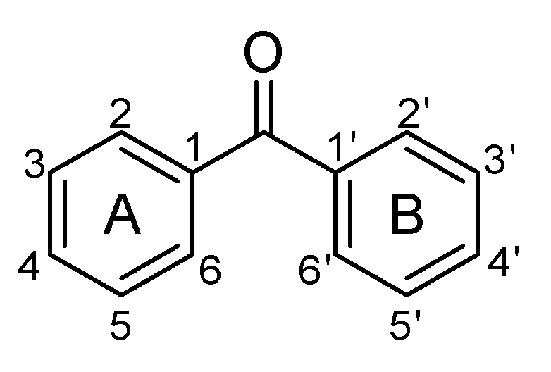
2. Structural Diversity of Simply Oxygenated Benzophenones
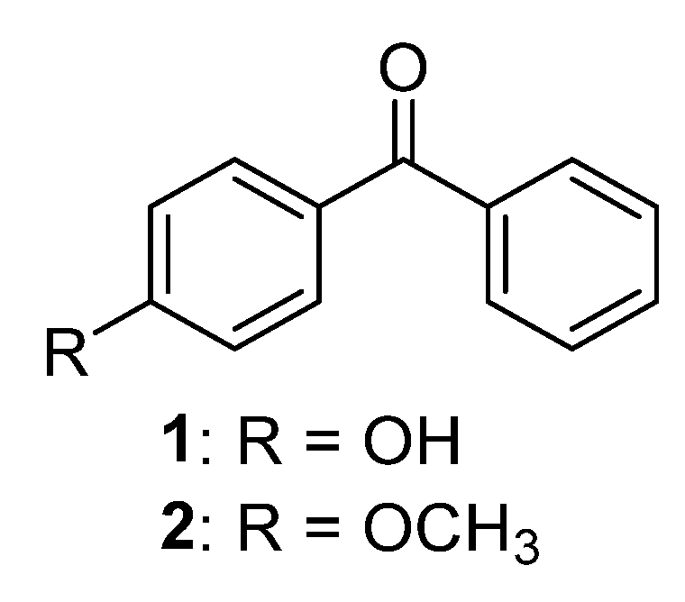
2.1. Monooxygenated Benzophenones
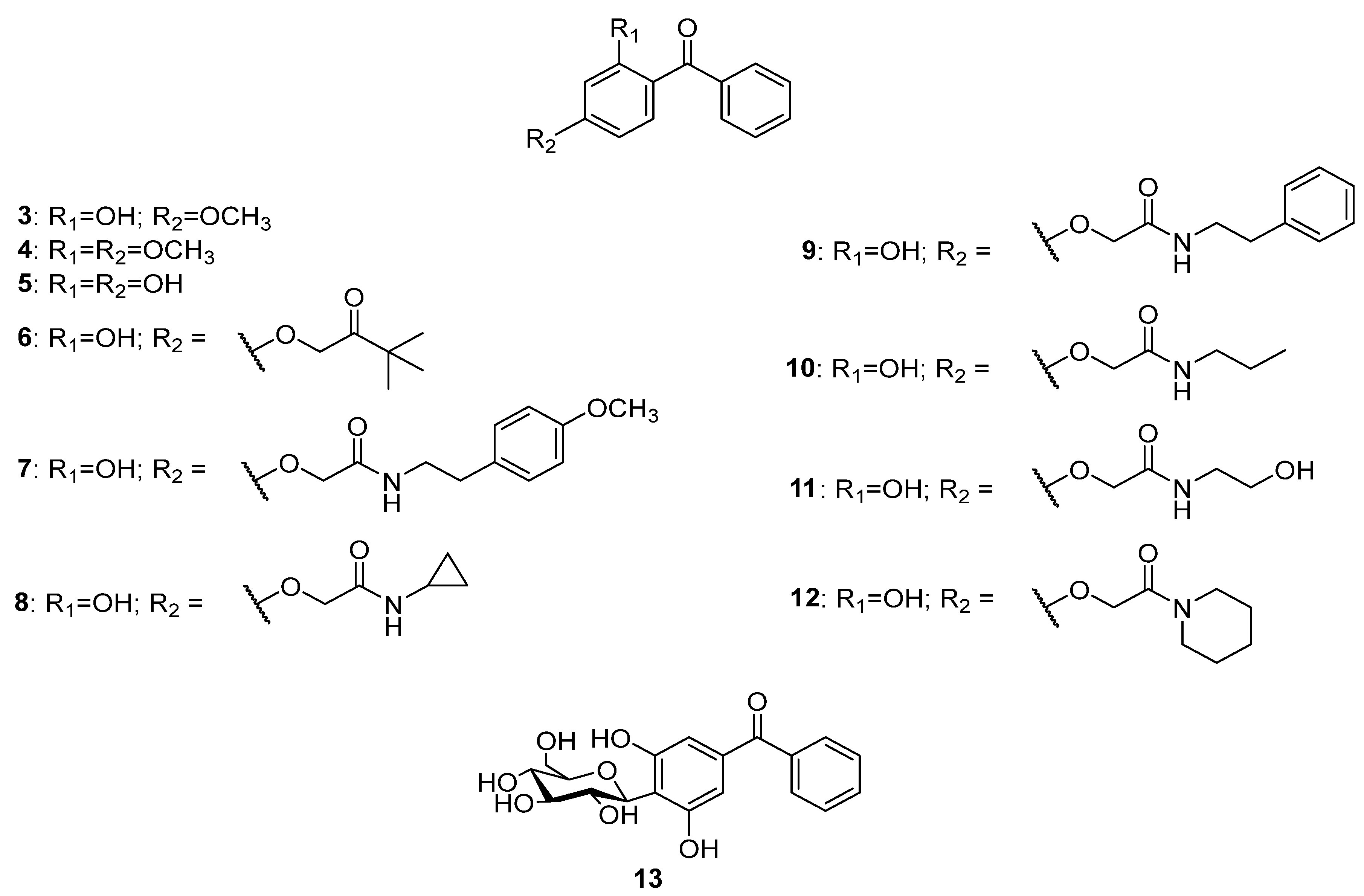
2.2. Dioxygenated Bezophenones
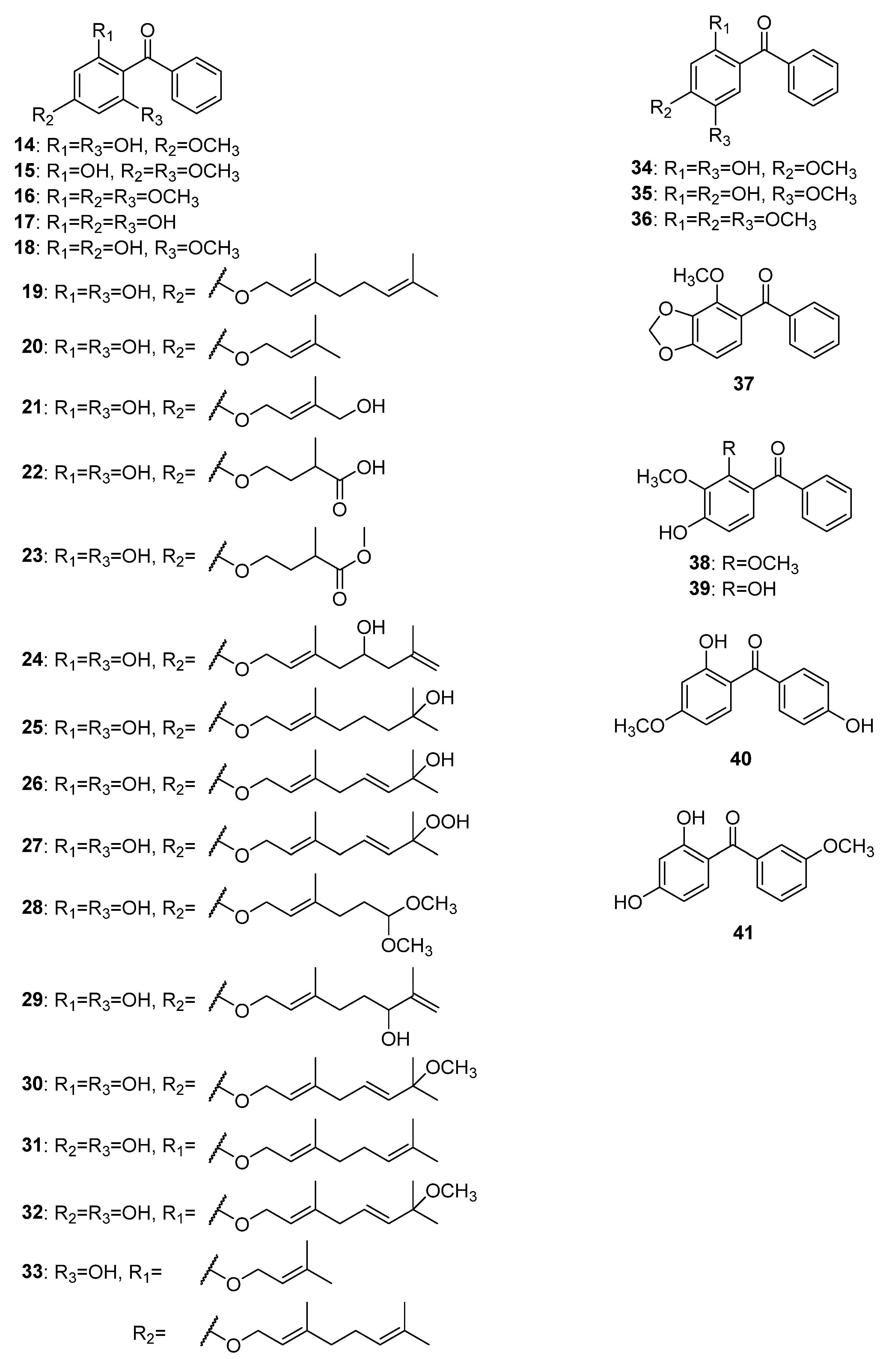
2.3. Trioxygenated Bezophenones
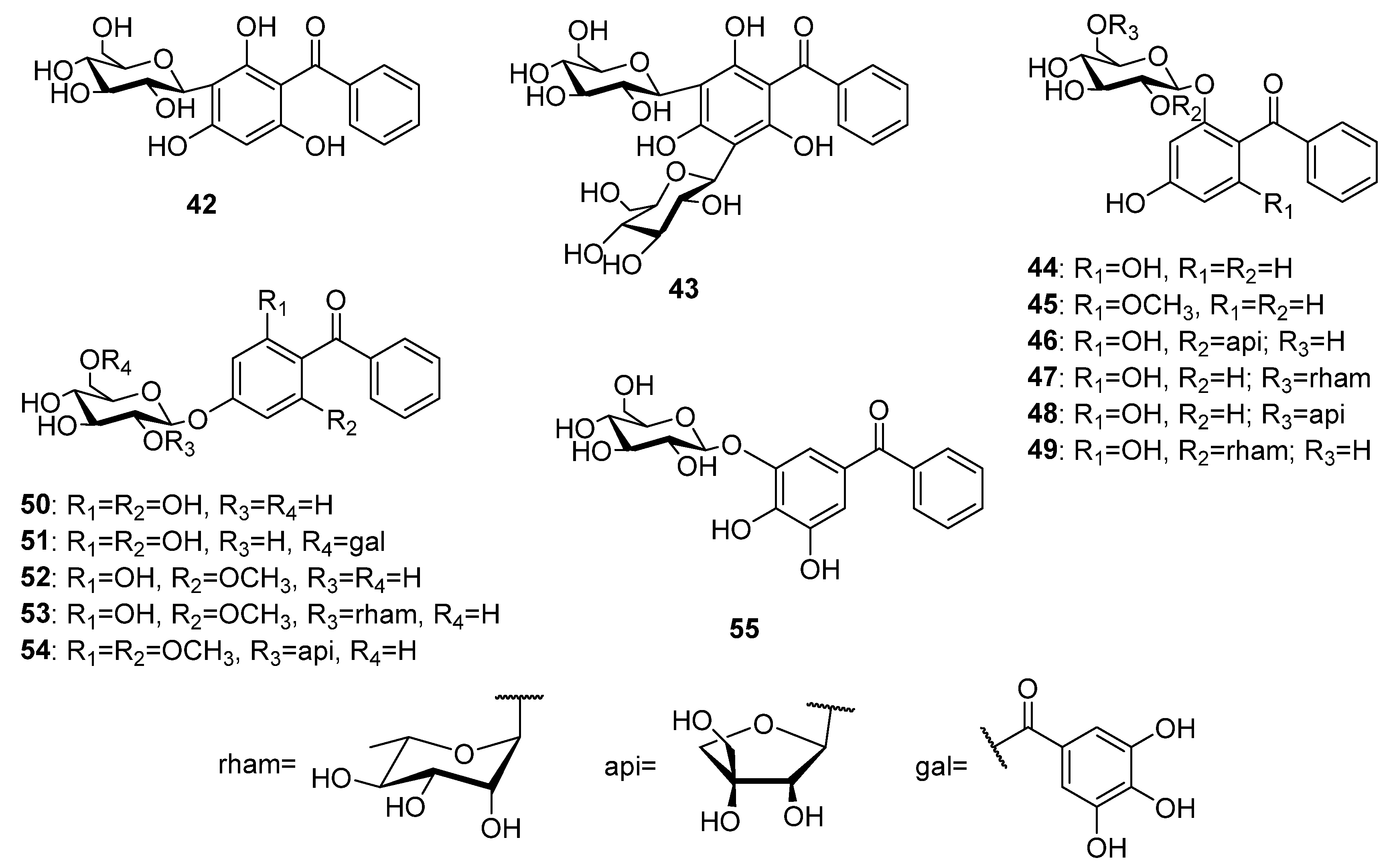
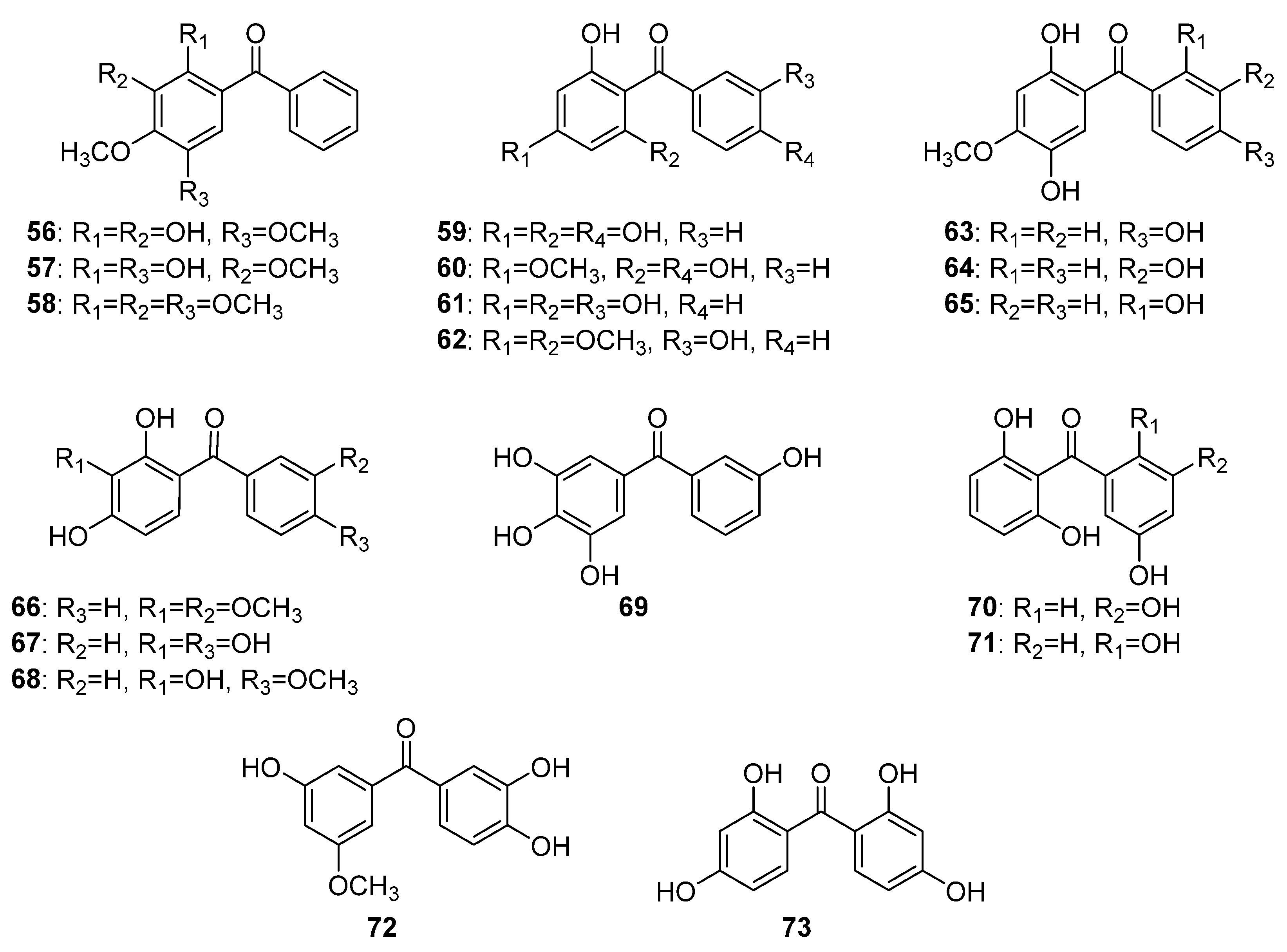
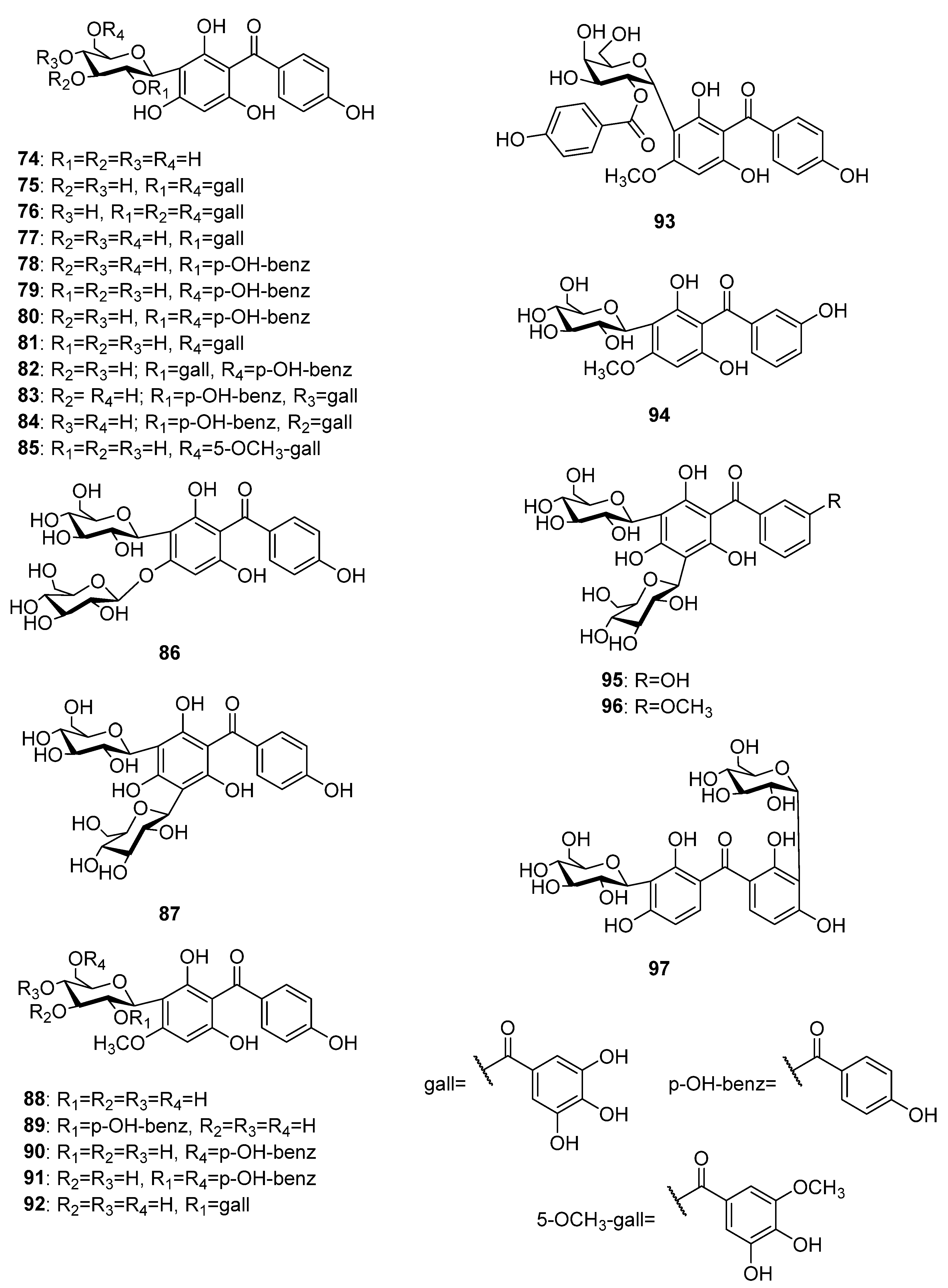
2.4. Tetraoxygenated Bezophenones
2.5. Pentaoxygenated Benzophenones
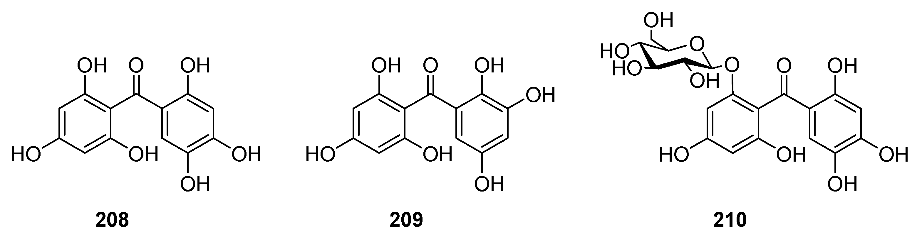
2.6. Hexaxygenated Benzophenones
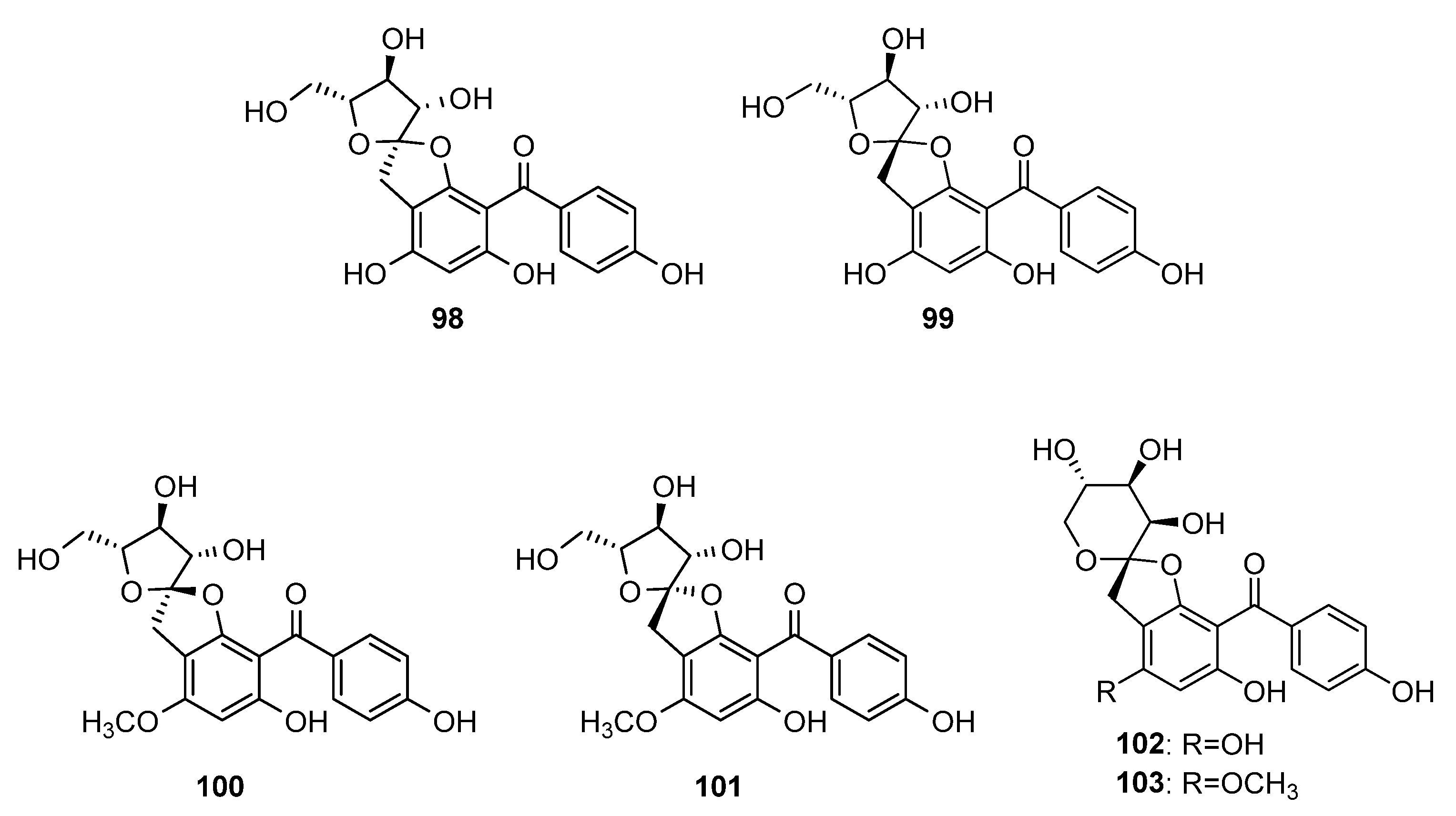
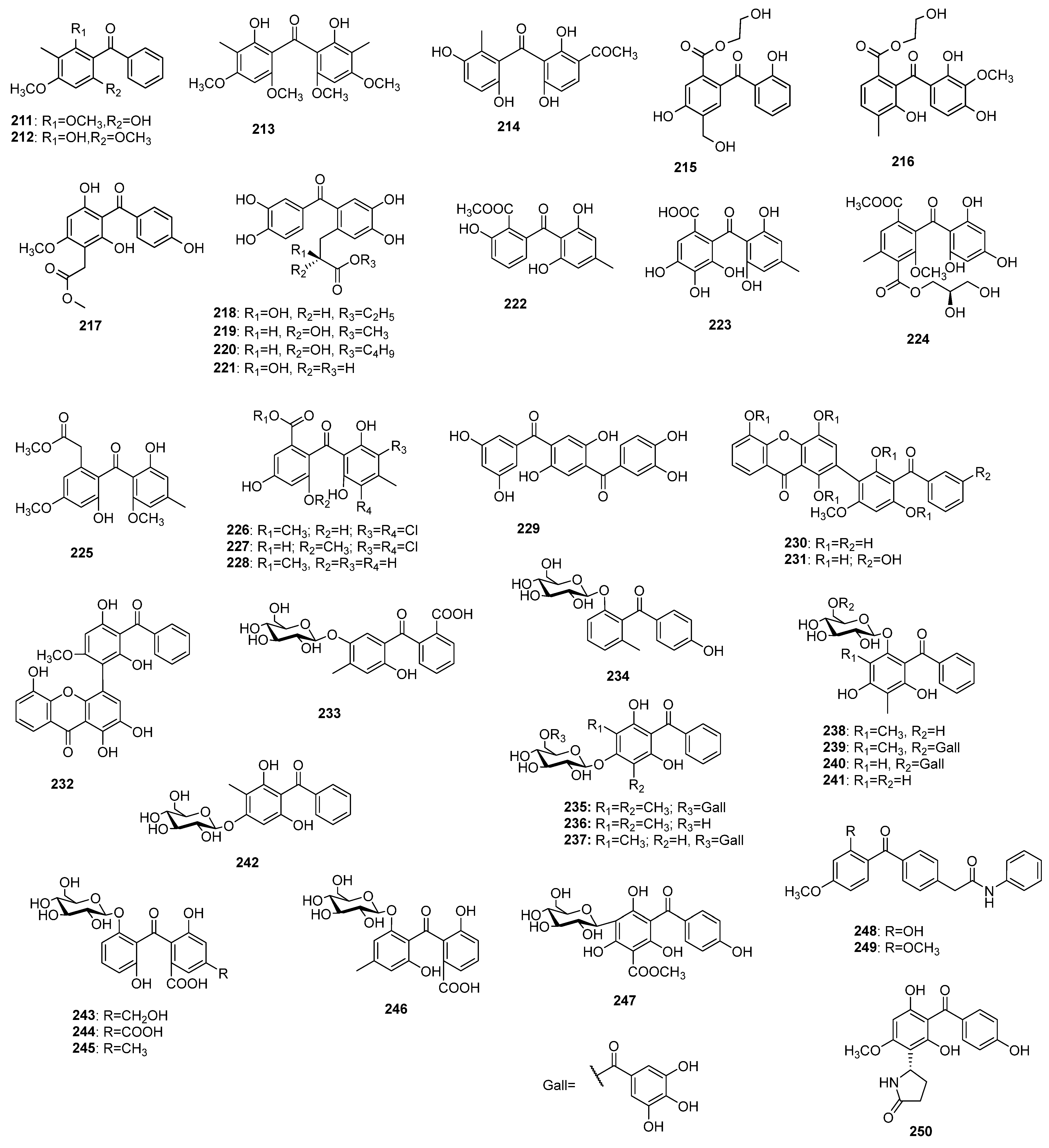
2.7. Uncategorized and Miscellaneous Benzophenones
3. Distribution of Naturally Occurring Simply Oxygenated Benzophenones
4. Biological Properties of Naturally Occurring Simply Oxygenated Benzophenones
4.1. Antioxidant Activities
4.1.1. DPPH Radical Scavenging Activity
4.1.2. ABTS Radical Scavenging Activity
4.1.3. FRAP (Ferric Reducing Antioxidant Power) Assay
4.1.4. Inhibition of Lipid Peroxidation
4.2. Antiallergic Activity
4.3. Immunosuppressive Activity
4.4. Anti-Inflammatory Activity
4.5. Estrogenic Activity
4.6. Cytotoxic and Antitumor Activities
4.6.1. Cytotoxicity on Human Cancer Cell Lines
4.6.2. Cytotoxicity on Animal Cell Lines and Models
4.7. Cytoprotective Effects
4.8. Antimicrobial Activity
4.8.1. Antibacterial Activity
4.8.2. Antimycotic Activity
4.8.3. Antiviral Activity
4.8.4. Antiparasitic Activity
4.8.5. Antimalarial Activity
4.9. Metabolic Syndrome
4.9.1. Antidiabetic Activity
- α-Glucosidase inhibitory activity
- 2.
- α-Amylase inhibitory activity
- 3.
- The efficiency of glucose uptake
4.9.2. Antihyperlipidemic Activity
4.9.3. Antihypertensive Activity
4.10. Inhibitory Activities on Platelet Aggregation
4.11. MAO-A Inhibition Activity
4.12. Antiarthritic Activity
4.13. Anticholinesterase Activity
4.14. Anti-Atherosclerotic Activity
4.15. Laxative Effect
4.16. Negative Effects of Naturally Occurring Simply Oxygenated Benzophenones on Human and Animal Health and the Environment
5. Biogenetic Significance of Simply Oxygenated Benzophenones
6. Conclusions
Author Contributions
Funding
Institutional Review Board Statement
Informed Consent Statement
Data Availability Statement
Conflicts of Interest
References
- Bukvički, D.; Novaković, M.; Ab Ghani, N.; Marin, P.D.; Asakawa, Y. Secondary Metabolites from Endemic Species Iris Adriatica Trinajstić Ex Mitić (Iridaceae). Nat. Prod. Res. 2018, 32, 1849–1852. [Google Scholar] [CrossRef] [PubMed]
- Nandy, S.; Mukherjee, A.; Pandey, D.K.; Ray, P.; Dey, A. Indian Sarsaparilla (Hemidesmus Indicus): Recent Progress in Research on Ethnobotany, Phytochemistry and Pharmacology. J. Ethnopharmacol. 2020, 254, 112609. [Google Scholar] [CrossRef] [PubMed]
- Cuesta-Rubio, O.; Piccinelli, A.L.; Rastrelli, L. Chemistry and Biological Activity of Polyisoprenylated Benzophenone Derivatives. In Studies in Natural Products Chemistry; Atta-ur-Rahman, Ed.; Elsevier: Amsterdam, The Netherlands, 2005; Volume 32, pp. 671–720. ISBN 1572-5995. [Google Scholar]
- Acuna, M.U.; Jancovski, N.; Kennelly, J.E. Polyisoprenylated Benzophenones from Clusiaceae: Potential Drugs and Lead Compounds. Curr. Top. Med. Chem. 2009, 9, 1560–1580. [Google Scholar] [CrossRef] [PubMed]
- Wu, S.-B.; Long, C.; Kennelly, E.J. Structural Diversity and Bioactivities of Natural Benzophenones. Nat. Prod. Rep. 2014, 31, 1158–1174. [Google Scholar] [CrossRef]
- Singh, P.I.; Bharate, S.B. Phloroglucinol Compounds of Natural Origin. Nat. Prod. Rep. 2006, 23, 558–591. [Google Scholar] [CrossRef]
- Sultanbawa, M.U.S. Xanthonoids of Tropical Plants. Tetrahedron 1980, 36, 1465–1506. [Google Scholar] [CrossRef]
- Kitanov, G.M.; Nedialkov, P.T. Benzophenone O-Glucoside, a Biogenic Precursor of 1,3,7-Trioxygenated Xanthones in Hypericum annulatum. Phytochemistry 2001, 57, 1237–1243. [Google Scholar] [CrossRef]
- Mandal, S.; Das, P.C.; Joshi, P.C. Naturally Occurring Xanthones from Terrestrial Flora. J. Indian Chem. Soc. 1992, 69, 611–636. [Google Scholar]
- Heurung, A.R.; Raju, S.I.; Warshaw, E.M. Benzophenones. Dermatitis 2014, 25, 3–10. [Google Scholar] [CrossRef]
- Pallares, E.S.; Hector, M.G. Study of Yoloxochitl. Arch. Inst. Cardiol. Mex. 1947, 17, 833–849. [Google Scholar]
- Sharma, M.L.; Nigam, M.C.; Hande, K.L. Essential Oil of Costus speciosus. Perfum. Essent. Oil Rec. 1963, 54, 579–580. [Google Scholar]
- Liu, L.-N.; Zhang, X.-W.; Hou, F.-J.; Duan, X.-H.; Zhao, J.-C.; Chen, Y.-L. Benzophenones from Endohydric Moss Polytrichastrum Formosum and Their Cytotoxic Activities. Chin. Tradit. Herb. Drugs 2022, 53, 667–670. [Google Scholar]
- Duan, X.-H.; He, P.; Qin, M.; Li, L.; Pei, L.; Zhao, J.-C. A New Benzophenone from Endohydric Moss Pogonatum Inflexum. Chin. Tradit. Herb. Drugs 2019, 50, 1291–1293. [Google Scholar] [CrossRef]
- Duan, X.-H.; Zhao, J.-C.; Li, L.; Pei, L.; He, P.; Wang, R. Two New Benzophenones from Endohydric Moss Polytrichum Commune. Nat. Prod. Res. 2019, 33, 2750–2754. [Google Scholar] [CrossRef] [PubMed]
- Duan, X.-H.; Zhang, X.-W.; Qin, M.; He, P.; Pei, L.; Zhao, J.-C.; Chen, Y.-L. Two New Benzophenones from the Endohydric Moss Polytrichastrum formosum. Nat. Prod. Commun. 2021, 16, 1934578X211002623. [Google Scholar] [CrossRef]
- Gamiotea-Turro, D.; Cuesta-Rubio, O.; Prieto-González, S.; De Simone, F.; Passi, S.; Rastrelli, L. Antioxidative Constituents from the Leaves of Hypericum styphelioides. J. Nat. Prod. 2004, 67, 869–871. [Google Scholar] [CrossRef]
- Rusby, H.H. New Species of Trees of Medical Interest from Bolivia. Bull. Torrey Bot. Club 1922, 49, 259–264. [Google Scholar] [CrossRef]
- Jobst, J. Ueber Coto-Rinden Und Deren Krystallisirbare Bestandtheile. Berichte Der Dtsch. Chem. Ges. 1876, 9, 1633–1634. [Google Scholar] [CrossRef]
- Jobst, J.; Hesse, O. Ueber Die Cotorinden Und Ihre Charakteristischen Bestandtheile. Justus Liebigs Ann. Der Chem. 1879, 199, 17–96. [Google Scholar] [CrossRef]
- Ciamician, G. Ueber Die Constitution Des Cotoïns. Berichte Der Dtsch. Chem. Ges. 1894, 27, 409–426. [Google Scholar] [CrossRef]
- Ciamician, G.; Silber, P. Ueber Das Hydrocotoïn, Einen Bestandtheil Der Cotorinde. Berichte Der Dtsch. Chem. Ges. 1891, 24, 299–301. [Google Scholar] [CrossRef]
- Ciamician, G.; Silber, P. Synthese Des Benzophloroglucintrimethyläther. (Methylhydrocotoïn Oder Benzoylhydrocoton). Berichte Der Dtsch. Chem. Ges. 1894, 27, 1497–1501. [Google Scholar] [CrossRef]
- Ciamician, G.; Silber, P. Ueber Die Constitution Einiger in Der Paracotorinde Enthaltenen Bestandtheile. Berichte Der Dtsch. Chem. Ges. 1892, 25, 1119–1138. [Google Scholar] [CrossRef]
- Randriaminahy, M.; Proksch, P.; Witte, L.; Wray, V. Lipophilic Phenolic Constituents from Helichrysum Species Endemic to Madagascar. Z. Für Naturforschung C 1992, 47, 10–16. [Google Scholar] [CrossRef]
- Bohlmann, F.; Suwita, A. Neue Phloroglucin-Derivate Aus Leontonyx-Arten Sowie Weitere Verbindungen Aus Vertretern Der Tribus Inuleae. Phytochemistry 1978, 17, 1929–1934. [Google Scholar] [CrossRef]
- Jakupovic, J.; Zdero, C.; Grenz, M.; Tsichritzis, F.; Lehmann, L.; Hashemi-Nejad, S.M.; Bohlmann, F. Twenty-One Acylphloroglucinol Derivatives and Further Constituents from South African Helichrysum Species. Phytochemistry 1989, 28, 1119–1131. [Google Scholar] [CrossRef]
- Don, M.-J.; Huang, Y.-J.; Huang, R.-L.; Lin, Y.-L. New Phenolic Principles from Hypericum sampsonii. Chem. Pharm. Bull. 2004, 52, 866–869. [Google Scholar] [CrossRef]
- Zhu, H.; Chen, C.; Tan, D.; Li, D.; Guo, Y.; Wei, G.; Zhang, J.; Wang, J.; Luo, Z.; Xue, Y.; et al. Sampbenzophenones A–G, Prenylated Benzoylphloroglucinol Derivatives from Hypericum sampsonii. RSC Adv. 2016, 6, 86710–86716. [Google Scholar] [CrossRef]
- Huang, C.-Y.; Chang, T.-C.; Wu, Y.-J.; Chen, Y.; Chen, J.-J. Benzophenone and Benzoylphloroglucinol Derivatives from Hypericum Sampsonii with Anti-Inflammatory Mechanism of Otogirinin A. Molecules 2020, 25, 4463. [Google Scholar] [CrossRef]
- Wu, F.-S.; Hung, C.-J.; Lin, C.-L.; Huang, H.-Y.; Kuo, Y.-H.; Chang, T.-H.; Chen, C.-L.; Sung, P.-J.; Cheng, M.-J.; Kuo, C.-W.; et al. A New Benzophenone and Bioactive Constituents of Hypericum nokoense. Chem. Nat. Compd. 2021, 57, 645–649. [Google Scholar] [CrossRef]
- Nedialkov, P.T.; Zheleva-Dimitrova, D.; Momekov, G.; Karlov, K.; Girreser, U.; Kitanov, G.M. Elegaphenone and 7-Epi-Clusianone, the Major Cytotoxic Constituents of Hypericum elegans. Nat. Prod. Res. 2011, 25, 1743–1750. [Google Scholar] [CrossRef]
- Wu, F.-S.; Wang, I.-C.; Liaw, C.-C.; Huang, H.-Y.; Chang, T.-H.; Chen, C.-L.; Sung, P.-J.; Cheng, M.-J.; Kuo, C.-W.; Chen, J.-J. New Benzophenone and Bioactive Constituents from Hypericum Nagasawae. Chem. Nat. Compd. 2022, 58, 833–838. [Google Scholar] [CrossRef]
- Ollis, W.D. Proceedings of the 6th Annual Symposium of the Plant Phenolics Group of North America, 1966; Mabry, T.J., Alston, R.E., Runeckles, V.C., Eds.; Recent Advances in Phytochemistry; Appleton–Century–Crofts: New York, NY, USA, 1968; Volume 1. [Google Scholar]
- Wang, W.; Weng, X.; Cheng, D. Antioxidant Activities of Natural Phenolic Components from Dalbergia odorifera T. Chen. Food Chem. 2000, 71, 45–49. [Google Scholar] [CrossRef]
- Dibwe, D.F.; Awale, S.; Kadota, S.; Morita, H.; Tezuka, Y. Heptaoxygenated Xanthones as Anti-Austerity Agents from Securidaca longepedunculata. Bioorganic Med. Chem. 2013, 21, 7663–7668. [Google Scholar] [CrossRef] [PubMed]
- Kang, W.-Y.; Wang, Z.-M.; Li, Z.-Q.; Xu, X.-J. Three New Compounds from Securidaca inappendiculata. Helv. Chim. Acta 2005, 88, 2771–2776. [Google Scholar] [CrossRef]
- Casu, L.; Solinas, M.N.; Saba, A.R.; Cottiglia, F.; Caboni, P.; Floris, C.; Laconi, S.; Pompei, R.; Leonti, M. Benzophenones from the Roots of the Popoluca Amerindian Medicinal Plant Securidaca Diversifolia (L.) S.F. Blake. Phytochem. Lett. 2010, 3, 226–229. [Google Scholar] [CrossRef]
- Triana, J.; López, M.; Pérez, F.J.; Platas, J.G.; Estévez, F.; León, J.F.; Hernández, J.C.; Brouard, I.; Bermejo, J. Chemical Constituents of Tolpis Species. Fitoterapia 2009, 80, 437–441. [Google Scholar] [CrossRef]
- Wu, X.-D.; Cheng, J.-T.; He, J.; Zhang, X.-J.; Dong, L.-B.; Gong, X.; Song, L.-D.; Zheng, Y.-T.; Peng, L.-Y.; Zhao, Q.-S. Benzophenone Glycosides and Epicatechin Derivatives from Malania Oleifera. Fitoterapia 2012, 83, 1068–1071. [Google Scholar] [CrossRef]
- Wu, Z.-J.; Ouyang, M.-A.; Yang, C.-R. Oligosaccharide Esters and Phenol Compounds from Polygala Arillata. Acta Bot. Yunnan. 2000, 22, 482–494. [Google Scholar] [CrossRef]
- Huang, Y.-L.; Chen, C.-C.; Chen, Y.-J.; Huang, R.-L.; Shieh, B.-J. Three Xanthones and a Benzophenone from Garcinia mangostana. J. Nat. Prod. 2001, 64, 903–906. [Google Scholar] [CrossRef]
- Li, J.; Jiang, Y.; Tu, P.-F. Xanthone O-Glycosides and Benzophenone O-Glycosides from the Roots of Polygala tricornis. J. Nat. Prod. 2005, 68, 1802–1804. [Google Scholar] [CrossRef]
- Dai, Y.; He, X.-J.; Zhou, G.-X.; Kurihara, H.; Ye, W.-C.; Yao, X.-S. Acylphloroglucinol Glycosides from the Fruits of Pyracantha fortuneana. J. Asian Nat. Prod. Res. 2008, 10, 111–117. [Google Scholar] [CrossRef]
- Shi, Y.-N.; Shi, Y.-M.; Yang, L.; Li, X.-C.; Zhao, J.-H.; Qu, Y.; Zhu, H.-T.; Wang, D.; Cheng, R.-R.; Yang, C.-R.; et al. Lignans and Aromatic Glycosides from Piper wallichii and Their Antithrombotic Activities. J. Ethnopharmacol. 2015, 162, 87–96. [Google Scholar] [CrossRef] [PubMed]
- Fu, H.Z.; Yang, J.Z.; Li, C.J.; Zhang, D.M. A New Benzophenone Glycoside from the Leaves of Psidium Guajava L. Chin. Chem. Lett. 2011, 22, 178–180. [Google Scholar] [CrossRef]
- Matsuzaki, K.; Ishii, R.; Kobiyama, K.; Kitanaka, S. New Benzophenone and Quercetin Galloyl Glycosides from Psidium guajava L. J. Nat. Med. 2010, 64, 252–256. [Google Scholar] [CrossRef] [PubMed]
- Shi, T.-X.; Wang, S.; Zeng, K.-W.; Tu, P.-F.; Jiang, Y. Inhibitory Constituents from the Aerial Parts of Polygala Tenuifolia on LPS-Induced NO Production in BV2 Microglia Cells. Bioorganic Med. Chem. Lett. 2013, 23, 5904–5908. [Google Scholar] [CrossRef]
- Jiang, Y.; Tu, P. Four New Phenones from the Cortexes of Polygala tenuifolia. Chem. Pharm. Bull. 2005, 53, 1164–1166. [Google Scholar] [CrossRef][Green Version]
- Zhou, L.-Y.; Wang, J.-M.; Huang, Y.-J.; Yu, X.-H.; Lu, B.; Hua, Y. Two New Glycosides Isolated from Polygala sibirica L. Var. megalopha Fr. Phytochem. Lett. 2016, 16, 174–177. [Google Scholar] [CrossRef]
- An, H.; Thanh, L.N.; Khanh, L.Q.; Ryu, S.H.; Lee, S.; Yeon, S.W.; Lee, H.H.; Turk, A.; Lee, K.Y.; Hwang, B.Y.; et al. Characterization of Antioxidant and α-Glucosidase Inhibitory Compounds of Cratoxylum formosum ssp. pruniflorum and Optimization of Extraction Condition. Antioxidants 2023, 12, 511. [Google Scholar] [CrossRef]
- Gottlieb, O.R.; Fineberg, M.; Salignac De Souza Guimaraes, I.; Taveira Magalhaes, M.; Ollis, W.D.; Eyton, W.B. The chemistry of the Brazilian leguminosae. VII. The Constituents of Machaerium scleroxylon. An. Acad. Bras. Cienc. 1964, 36, 33–34. [Google Scholar]
- Eyton, W.B.; Ollis, W.D.; Fineberg, M.; Gottlieb, O.R.; Salignac de Souza Guimarães, I.; Taveira Magalhães, M. The Neoflavanoid Group of Natural Products—II: The Examination of Machaerium Scleroxylon and Some Biogenetic Proposal Regarding the Neoflavanoids. Tetrahedron 1965, 21, 2697–2705. [Google Scholar] [CrossRef]
- Ochora, D.O.; Kakudidi, E.; Namukobe, J.; Heydenreich, M.; Coghi, P.; Yang, L.J.; Mwakio, E.W.; Andagalu, B.; Roth, A.; Akala, H.M.; et al. A New Benzophenone, and the Antiplasmodial Activities of the Constituents of Securidaca longipedunculata Fresen (Polygalaceae). Nat. Prod. Res. 2022, 36, 2758–2766. [Google Scholar] [CrossRef] [PubMed]
- Spada, A.; Cameroni, R.; Bernabei, M.T. The Pigments of Morus Alba. Gazz. Chim. Ital. 1956, 86, 46–55. [Google Scholar]
- De Barros Corrêa, D.; Gottlieb, O.R. Duckein, an Alkaloid from Aniba Duckei. Phytochemistry 1975, 14, 271–272. [Google Scholar] [CrossRef]
- Atkinson, J.E.; Gupta, P.; Lewis, J.R. Some Phenolic Constituents of Gentiana Lutea. Tetrahedron 1969, 25, 1507–1511. [Google Scholar] [CrossRef]
- Locksley, H.D.; Murray, I.G. Extractives from Guttiferae. Part XIX. The Isolation and Structure of Two Benzophenones, Six Xanthones and Two Biflavonoids from the Heartwood of Allanblackia floribunda Oliver. J. Chem. Soc. C 1971, 1332–1340. [Google Scholar] [CrossRef]
- Donnelly, D.M.X.; O’Reilly, J.; Whalley, W.B. Neoflavanoids of Dalbergia melanoxylon. Phytochemistry 1975, 14, 2287–2290. [Google Scholar] [CrossRef]
- Lin, S.; Liu, R.-H.; Ma, G.-Q.; Mei, D.-Y.; Shao, F.; Chen, L.-Y. Two New Compounds from the Heartwood of Dalbergia Melanoxylon. Nat. Prod. Res. 2020, 34, 2794–2801. [Google Scholar] [CrossRef]
- Pathak, V.; Shirota, O.; Sekita, S.; Hirayama, Y.; Hakamata, Y.; Hayashi, T.; Yanagawa, T.; Satake, M. Antiandrogenic Phenolic Constituents from Dalbergia cochinchinensis. Phytochemistry 1997, 46, 1219–1223. [Google Scholar] [CrossRef]
- Wu, D.-L.; Liao, Z.-D.; Chen, F.-F.; Zhang, W.; Ren, Y.-S.; Wang, C.-C.; Chen, X.-X.; Peng, D.-Y.; Kong, L.-Y. Benzophenones from Anemarrhena Asphodeloides Bge. Exhibit Anticancer Activity in HepG2 Cells via the NF-ΚB Signaling Pathway. Molecules 2019, 24, 2246. [Google Scholar] [CrossRef]
- Smitha, C.; Udayan, P. GC-MS and HR-LCMS Fingerprinting of Various Parts of Oroxylum Indicum (L.) Vent. A Comparative Phytochemical Study Based on Plant Part Substitution Approach. J. Pharmacogn. Phytochem. 2020, 9, 1817–1824. [Google Scholar]
- Jiang, H.Z.; Quan, X.F.; Tian, W.X.; Hu, J.M.; Wang, P.C.; Huang, S.Z.; Cheng, Z.Q.; Liang, W.J.; Zhou, J.; Ma, X.F.; et al. Fatty Acid Synthase Inhibitors of Phenolic Constituents Isolated from Garcinia Mangostana. Bioorganic Med. Chem. Lett. 2010, 20, 6045–6047. [Google Scholar] [CrossRef] [PubMed]
- Jantan, I.; Saputri, F.C. Benzophenones and Xanthones from Garcinia Cantleyana Var. Cantleyana and Their Inhibitory Activities on Human Low-Density Lipoprotein Oxidation and Platelet Aggregation. Phytochemistry 2012, 80, 58–63. [Google Scholar] [CrossRef] [PubMed]
- Wabo, H.K.; Kowa, T.K.; Lonfouo, A.H.N.; Tchinda, A.T.; Tane, P.; Kikuchi, H.; Frédérich, M.; Oshima, Y. Phenolic Compounds and Terpenoids from Hypericum lanceolatum. Rec. Nat. Prod. 2012, 6, 94–100. [Google Scholar]
- Mian, J.V.Y.; Lian, E.G.C.; Aspollah, S.M.; Hin, T.-Y.Y.; Yen, K.H.; Yok, C.M.K. Benzophenone Constituents from the Roots of Garcinia eugenifolia. Res. J. Chem. Environ. 2012, 16, 36–39. [Google Scholar]
- Azlini, I.; Erlena, N.A.A.R.; Muhammad, N.O.; Wan, A.N.W.A. Antihypertensive Assay-Guided Fractionation of Syzygium Polyanthum Leaves and Phenolics Profile Analysis Using LCQTOF/ MS. Pharmacogn. J. 2020, 12, 1670–1692. [Google Scholar] [CrossRef]
- Murakami, T.; Tanaka, N.; Wada, H.; Saiki, Y.; Chen, C.-M. Chemical and Chemotaxonomical Studies on Filices. LXIII. Yakugaku Zasshi 1986, 106, 378–382. [Google Scholar] [CrossRef]
- Tanaka, T.; Sueyasu, T.; Nonaka, G.; Nishioka, I. Tannins and Related Compounds. XXI. Isolation and Characterization of Galloyl and p-Hydroxybenzoyl Esters of Benzophenone and Xanthone C-Glucosides from Mangifera indica L. Chem. Pharm. Bull. 1984, 32, 2676–2686. [Google Scholar] [CrossRef]
- Barreto, J.C.; Trevisan, M.T.S.; Hull, W.E.; Erben, G.; de Brito, E.S.; Pfundstein, B.; Würtele, G.; Spiegelhalder, B.; Owen, R.W. Characterization and Quantitation of Polyphenolic Compounds in Bark, Kernel, Leaves, and Peel of Mango (Mangifera indica L.). J. Agric. Food Chem. 2008, 56, 5599–5610. [Google Scholar] [CrossRef]
- Zhang, Y.; Qian, Q.; Ge, D.; Li, Y.; Wang, X.; Chen, Q.; Gao, X.; Wang, T. Identification of Benzophenone C-Glucosides from Mango Tree Leaves and Their Inhibitory Effect on Triglyceride Accumulation in 3T3-L1 Adipocytes. J. Agric. Food Chem. 2011, 59, 11526–11533. [Google Scholar] [CrossRef]
- Zhang, Y.; Han, L.; Ge, D.; Liu, X.; Liu, E.; Wu, C.; Gao, X.; Wang, T. Isolation, Structural Elucidation, MS Profiling, and Evaluation of Triglyceride Accumulation Inhibitory Effects of Benzophenone C-Glucosides from Leaves of Mangifera indica L. J. Agric. Food Chem. 2013, 61, 1884–1895. [Google Scholar] [CrossRef] [PubMed]
- Beelders, T.; Brand, D.J.; de Beer, D.; Malherbe, C.J.; Mazibuko, S.E.; Muller, C.J.F.; Joubert, E. Benzophenone C- and O-Glucosides from Cyclopia Genistoides (Honeybush) Inhibit Mammalian α-Glucosidase. J. Nat. Prod. 2014, 77, 2694–2699. [Google Scholar] [CrossRef] [PubMed]
- Hara, H.; Ise, Y.; Morimoto, N.; Shimazawa, M.; Ichihashi, K.; Ohyama, M.; Iinuma, M. Laxative Effect of Agarwood Leaves and Its Mechanism. Biosci. Biotechnol. Biochem. 2008, 72, 335–345. [Google Scholar] [CrossRef]
- Pan, J.; Yi, X.; Zhang, S.; Cheng, J.; Wang, Y.; Liu, C.; He, X. Bioactive Phenolics from Mango Leaves (Mangifera indica L.). Ind. Crops Prod. 2018, 111, 400–406. [Google Scholar] [CrossRef]
- Xu, M.; Zhang, M.; Wang, D.; Yang, C.-R.; Zhang, Y.-J. Phenolic Compounds from the Whole Plants of Gentiana rhodantha (Gentianaceae). Chem. Biodivers. 2011, 8, 1891–1900. [Google Scholar] [CrossRef]
- Li, C.-J.; Zhang, D.-M.; Yu, S.-S. Benzophenone C-Glucosides from Polygala glomerata Lour. J. Asian Nat. Prod. Res. 2008, 10, 293–297. [Google Scholar] [CrossRef]
- Panidthananon, W.; Chaowasku, T.; Sritularak, B.; Likhitwitayawuid, K. A New Benzophenone C-Glucoside and Other Constituents of Pseuduvaria Fragrans and Their α-Glucosidase Inhibitory Activity. Molecules 2018, 23, 1600. [Google Scholar] [CrossRef]
- Qi, J.; Lu, J.-J.; Liu, J.-H.; Yu, B.-Y. Flavonoid and a Rare Benzophenone Glycoside from the Leaves of Aquilaria sinensis. Chem. Pharm. Bull. 2009, 57, 134–137. [Google Scholar] [CrossRef]
- Pan, J.; Yi, X.; Wang, Y.; Chen, G.; He, X. Benzophenones from Mango Leaves Exhibit α-Glucosidase and NO Inhibitory Activities. J. Agric. Food Chem. 2016, 64, 7475–7480. [Google Scholar] [CrossRef]
- Gu, C.; Yang, M.; Zhou, Z.; Khan, A.; Cao, J.; Cheng, G. Purification and Characterization of Four Benzophenone Derivatives from Mangifera Indica L. Leaves and Their Antioxidant, Immunosuppressive and α-Glucosidase Inhibitory Activities. J. Funct. Foods 2019, 52, 709–714. [Google Scholar] [CrossRef]
- Ito, H.; Nishitani, E.; Konoshima, T.; Takasaki, M.; Kozuka, M.; Yoshida, T. Flavonoid and Benzophenone Glycosides from Coleogyne ramosissima. Phytochemistry 2000, 54, 695–700. [Google Scholar] [CrossRef] [PubMed]
- Lee, S.-S.; Tseng, C.-C.; Chen, C.-K. Three New Benzophenone Glucosides from the Leaves of Planchonella Obovata. Helv. Chim. Acta 2010, 93, 522–529. [Google Scholar] [CrossRef]
- Sun, J.; Wang, S.; Xia, F.; Wang, K.-Y.; Chen, J.-M.; Tu, P.-F. Five New Benzophenone Glycosides from the Leaves of Aquilaria sinensis (Lour.) Gilg. Chin. Chem. Lett. 2014, 25, 1573–1576. [Google Scholar] [CrossRef]
- Yuan, H.; Zhao, J.; Wang, M.; Khan, S.I.; Zhai, C.; Xu, Q.; Huang, J.; Peng, C.; Xiong, G.; Wang, W.; et al. Benzophenone Glycosides from the Flower Buds of Aquilaria sinensis. Fitoterapia 2017, 121, 170–174. [Google Scholar] [CrossRef] [PubMed]
- Sun, H.; Zhang, Y.-F.; Huo, H.-X.; Guan, P.-W.; Wang, C.-C.; Yao, H.-N.; Zhao, Y.-F.; Tu, P.-F.; Li, J. Benzophenone Glycosides from the Pericarps of Aquilaria Yunnanensis S. C. Huang. Nat. Prod. Res. 2020, 34, 2030–2036. [Google Scholar] [CrossRef]
- Feng, J.; Yang, X.-W.; Wang, R.-F. Bio-Assay Guided Isolation and Identification of α-Glucosidase Inhibitors from the Leaves of Aquilaria Sinensis. Phytochemistry 2011, 72, 242–247. [Google Scholar] [CrossRef]
- Rancon, S.; Chaboud, A.; Darbour, N.; Comte, G.; Bayet, C.; Simon, P.-N.; Raynaud, J.; Di Pietro, A.; Cabalion, P.; Barron, D. Natural and Synthetic Benzophenones: Interaction with the Cytosolic Binding Domain of P-Glycoprotein. Phytochemistry 2001, 57, 553–557. [Google Scholar] [CrossRef]
- Ferrari, J.; Terreaux, C.; Sahpaz, S.; Msonthi, J.D.; Wolfender, J.-L.; Hostettmann, K. Benzophenone Glycosides from Gnidia involucrata. Phytochemistry 2000, 54, 883–889. [Google Scholar] [CrossRef]
- Zhang, S.-Y.; Zhang, Q.-H.; Zhao, W.; Zhang, X.; Zhang, Q.; Bi, Y.-F.; Zhang, Y.-B. Isolation, Characterization and Cytotoxic Activity of Benzophenone Glucopyranosides from Mahkota Dewa (Phaleria macrocarpa (Scheff.) Boerl). Bioorganic Med. Chem. Lett. 2012, 22, 6862–6866. [Google Scholar] [CrossRef]
- Osman, A.G.; Ali, Z.; Fantoukh, O.; Raman, V.; Kamdem, R.S.T.; Khan, I. Glycosides of Ursane-Type Triterpenoid, Benzophenone, and Iridoid from Vangueria Agrestis (Fadogia Agrestis) and Their Anti-Infective Activities. Nat. Prod. Res. 2020, 34, 683–691. [Google Scholar] [CrossRef]
- Zhang, Y.-B.; Xu, X.-J.; Liu, H.-M. Chemical Constituents from Mahkota Dewa. J. Asian Nat. Prod. Res. 2006, 8, 119–123. [Google Scholar] [CrossRef]
- Kaya, D.; Yalçın, F.N.; Bedir, E.; Çalış, İ.; Steinhauser, L.; Albert, K.; Ersöz, T. New Benzophenone Glucosides from the Aerial Parts of Gentiana Verna L. Subsp. Pontica (Soltok.) Hayek. Phytochem. Lett. 2011, 4, 459–461. [Google Scholar] [CrossRef]
- Mohamed, G.A.; Ibrahim, S.R.M. Garcixanthone E and Garcimangophenone C: New Metabolites from Garcinia Mangostana and Their Cytotoxic and Alpha Amylase Inhibitory Potential. Life 2022, 12, 1875. [Google Scholar] [CrossRef] [PubMed]
- Otsuka, H.; Kijima, K. An Iridoid Gentiobioside, a Benzophenone Glucoside and Acylated Flavone C-Glycosides from Tripterospermum japonicum (SIEB. et ZUCC.) MAXIM. Chem. Pharm. Bull. 2001, 49, 699–702. [Google Scholar] [CrossRef] [PubMed][Green Version]
- Duan, Y.; Dai, Y.; Wang, G.; Chen, H.; Gao, H.; Chen, J.; Yao, X.; Zhang, X. Xanthone and Benzophenone Glycosides from the Stems of Cratoxylum formosum ssp. pruniflorum. Chem. Pharm. Bull. 2011, 59, 231–234. [Google Scholar] [CrossRef] [PubMed]
- Jo, Y.H.; Kim, S.B.; Ahn, J.H.; Liu, Q.; Hwang, B.Y.; Lee, M.K. Inhibitory Activity of Benzophenones from Anemarrhena asphodeloides on Pancreatic Lipase. Nat. Prod. Commun. 2013, 8, 1934578X1300800419. [Google Scholar] [CrossRef]
- Wagner, R. Ueber Die Farbstoffe Des Gelbholzes (Morus Tinctoria). J. Für Prakt. Chem. 1850, 51, 82–106. [Google Scholar] [CrossRef]
- Hlasiwetz, H.; Pfaundler, L. Ueber Das Morin Und Die Moringerbsäure. Justus Liebigs Ann. Der Chem. 1863, 127, 351–361. [Google Scholar] [CrossRef]
- Ciamician, G.; Silber, P. Ueber Die Constitution Des Maclurins Und Phloretins. Berichte Der Dtsch. Chem. Ges. 1895, 28, 1393–1398. [Google Scholar] [CrossRef][Green Version]
- Minami, H.; Kinoshita, M.; Fukuyama, Y.; Kodama, M.; Yoshizawa, T.; Sugiura, M.; Nakagawa, K.; Tago, H. Antioxidant Xanthones from Garcinia subelliptica. Phytochemistry 1994, 36, 501–506. [Google Scholar] [CrossRef]
- Chiang, Y.-M.; Kuo, Y.-H.; Oota, S.; Fukuyama, Y. Xanthones and Benzophenones from the Stems of Garcinia multiflora. J. Nat. Prod. 2003, 66, 1070–1073. [Google Scholar] [CrossRef]
- Nguyen, L.H.D.; Venkatraman, G.; Sim, K.Y.; Harrison, L.J. Xanthones and Benzophenones from Garcinia Griffithii and Garcinia Mangostana. Phytochemistry 2005, 66, 1718–1723. [Google Scholar] [CrossRef]
- Muriithi, E.; Bojase-Moleta, G.; Majinda, R.R.T. Benzophenone Derivatives from Garcinia Livingstonei and Their Antioxidant Activities. Phytochem. Lett. 2016, 18, 29–34. [Google Scholar] [CrossRef]
- Choodej, S.; Koopklang, K.; Raksat, A.; Chuaypen, N.; Pudhom, K. Bioactive Xanthones, Benzophenones and Biphenyls from Mangosteen Root with Potential Anti-Migration against Hepatocellular carcinoma Cells. Sci. Rep. 2022, 12, 8605. [Google Scholar] [CrossRef]
- Minami, H.; Hamaguchi, K.; Kubo, M.; Fukuyama, Y. A Benzophenone and a Xanthone from Garcinia subelliptica. Phytochemistry 1998, 49, 1783–1785. [Google Scholar] [CrossRef]
- Fouotsa, H.; Lannang, A.M.; Dzoyem, J.P.; Tatsimo, S.J.N.; Neumann, B.; Mbazoa, C.D.; Razakarivony, A.A.; Nkengfack, A.E.; Eloff, J.N.; Sewald, N. Antibacterial and Antioxidant Xanthones and Benzophenone from Garcinia smeathmannii. Planta Medica 2015, 81, 594–599. [Google Scholar] [CrossRef]
- Rao, A.V.R.; Sarma, M.R.; Venkataraman, K.; Yemul, S.S. A Benzophenone and Xanthone with Unusual Hydroxylation Patterns from the Heartwood of Garcinia pedunculata. Phytochemistry 1974, 13, 1241–1244. [Google Scholar] [CrossRef]
- Nargis, J.; Wong, K.-C.; Khairuddin, M.; Chantrapromma, S.; Fun, H.-K. (2,4-Dihydroxy-6-Methoxyphenyl)(3,5-Dihydroxyphenyl)Methanone Monohydrate. Acta Crystallogr. Sect. E 2011, 67, o2717–o2718. [Google Scholar] [CrossRef]
- Pailee, P.; Kuhakarn, C.; Sangsuwan, C.; Hongthong, S.; Piyachaturawat, P.; Suksen, K.; Jariyawat, S.; Akkarawongsapat, R.; Limthongkul, J.; Napaswad, C.; et al. Anti-HIV and Cytotoxic Biphenyls, Benzophenones and Xanthones from Stems, Leaves and Twigs of Garcinia speciosa. Phytochemistry 2018, 147, 68–79. [Google Scholar] [CrossRef]
- Nguyen Viet, D.; Le Ba, V.; Nguyen Duy, T.; Pham Thi, V.A.; Tran Thi, H.; Le Canh, V.C.; Bach Long, G.; Kim, Y.H.; Tuan Anh, H.L. Bioactive Compounds from the Aerial Parts of Hypericum sampsonii. Nat. Prod. Res. 2021, 35, 646–648. [Google Scholar] [CrossRef]
- Wang, M.; Ma, G.; Shao, F.; Liu, R.; Chen, L.; Liu, Y.; Yang, L.; Meng, X. Neoflavonoids from the Heartwood of Dalbergia melanoxylon. Nat. Prod. Res. 2022, 36, 735–741. [Google Scholar] [CrossRef] [PubMed]
- Yoshimura, M.; Ninomiya, K.; Tagashira, Y.; Maejima, K.; Yoshida, T.; Amakura, Y. Polyphenolic Constituents of the Pericarp of Mangosteen (Garcinia mangostana L.). J. Agric. Food Chem. 2015, 63, 7670–7674. [Google Scholar] [CrossRef] [PubMed]
- Li, J.-C.; Nohara, T. Benzophenone C-Glucosides from Polygala telephioides. Chem. Pharm. Bull. 2000, 48, 1354–1355. [Google Scholar] [CrossRef] [PubMed][Green Version]
- Ma, T.-J.; Shi, X.-C.; Jia, C.-X. Telephenone D, A New Benzophenone C-Glycoside from Polygala telephioides. Chin. J. Nat. Med. 2010, 8, 9–11. [Google Scholar] [CrossRef]
- Rouis, Z.; Abid, N.; Aouni, M.; Faiella, L.; Dal Piaz, F.; De Tommasi, N.; Braca, A. Benzophenone Glycosides from Hypericum Humifusum ssp. austral. J. Nat. Prod. 2013, 76, 979–982. [Google Scholar] [CrossRef] [PubMed]
- Cheng, Z.-Q.; Yang, D.; Ma, Q.-Y.; Yi, X.-H.; Zhou, J.; Zhao, Y.-X. A New Benzophenone from Dobinea delavayi. Chem. Nat. Compd. 2013, 49, 46–48. [Google Scholar] [CrossRef]
- Wu, X.-R.; Lang, L.-J.; Shen, Y.; Dong, X.; Xiao, C.-J.; Jiang, B. Four New Phenolic Glycosides from Dobinea delavayi. Nat. Prod. Res. 2023, 37, 1146–1153. [Google Scholar] [CrossRef]
- Nedialkov, P.T.; Kitanov, G.M. Two Benzophenone O-Arabinosides and a Chromone from Hypericum annulatum. Phytochemistry 2002, 59, 867–871. [Google Scholar] [CrossRef]
- Demirkiran, O.; Ahmed Mesaik, M.; Beynek, H.; Abbaskhan, A.; Iqbal Choudhary, M. Cellular Reactive Oxygen Species Inhibitory Constituents of Hypericum thasium Griseb. Phytochemistry 2009, 70, 244–249. [Google Scholar] [CrossRef]
- Demirkiran, O. Three New Benzophenone Glycosides with MAO-A Inhibitory Activity from Hypericum thasium Griseb. Phytochem. Lett. 2012, 5, 700–704. [Google Scholar] [CrossRef]
- Nedialkov, P.T.; Zheleva-Dimitrova, D.; Girreser, U.; Kitanov, G.M. Benzophenone O-Glycosides from Hypericum elegans. Nat. Prod. Res. 2009, 23, 1176–1180. [Google Scholar] [CrossRef] [PubMed]
- Tanaka, N.; Kubota, T.; Kashiwada, Y.; Takaishi, Y.; Kobayashi, J. Petiolins F—I, Benzophenone Rhamnosides from Hypericum pseudopetiolatum var. kiusianum. Chem. Pharm. Bull. 2009, 57, 1171–1173. [Google Scholar] [CrossRef] [PubMed][Green Version]
- Xia, J.; Hu, B.; Qian, M.; Zhang, J.; Wu, L. Benzophenone Rhamnosides and Chromones from Hypericum Seniawinii Maxim. Molecules 2022, 27, 7056. [Google Scholar] [CrossRef] [PubMed]
- Yang, L.; Wang, Z.-M.; Wang, Y.; Li, R.-S.; Wang, F.; Wang, K. Phenolic Constituents with Neuroprotective Activities from Hypericum wightianum. Phytochemistry 2019, 165, 112049. [Google Scholar] [CrossRef] [PubMed]
- Petrunak, E.; Kester, A.C.; Liu, Y.; Bowen-Forbes, C.S.; Nair, M.G.; Henry, G.E. New Benzophenone O-Glucoside from Hypericum ellipticum. Nat. Prod. Commun. 2009, 4, 1934578X0900400412. [Google Scholar] [CrossRef]
- Alhakamy, N.A.; Mohamed, G.A.; Fahmy, U.A.; Eid, B.G.; Ahmed, O.A.; Al-Rabia, M.W.; Khedr, A.I.; Nasrullah, M.Z.; Ibrahim, S.R. New Alpha-Amylase Inhibitory Metabolites from Pericarps of Garcinia mangostana. Life 2022, 12, 384. [Google Scholar] [CrossRef]
- Mohamed, G.A.; Ibrahim, S.R.M. New Benzophenones and a Dihydroflavanonol from Garcinia Mangostana Pericarps and Their Antioxidant and Cytotoxic Activities. Phytochem. Lett. 2020, 39, 43–48. [Google Scholar] [CrossRef]
- Powell, V.; Sutherland, M. Substituted Benzophenones from Leptospermum luehmannii (F. M. Bailey). Aust. J. Chem. 1963, 16, 282–284. [Google Scholar] [CrossRef]
- de Corréa, D.B.; Guerra, L.F.B.; Gottlieb, O.R.; Maia, J.G.S. C-Methyl Phenolics from Qualea Species. Phytochemistry 1981, 20, 305–307. [Google Scholar] [CrossRef]
- Gong, S.; Liu, C.; Liu, S.; Du, Y.; Kang, W.; Dong, X. Studies on constituents of the Chinese traditional drug baishouwu (Cynanchum auriculatum Royle ex Wight). Yao Xue Xue Bao 1988, 23, 276–280. [Google Scholar]
- Deng, Y.; Chin, Y.-W.; Chai, H.; Keller, W.J.; Kinghorn, A.D. Anthraquinones with Quinone Reductase-Inducing Activity and Benzophenones from Morinda citrifolia (Noni) Roots. J. Nat. Prod. 2007, 70, 2049–2052. [Google Scholar] [CrossRef] [PubMed]
- Liao, Z.-D.; Xu, F.-Q.; Wu, D.-L.; Zhang, W.; Huang, Q. A new benzophenone isolated from fibrous roots of Anemarrhena asphodeloides. Zhongguo Zhong Yao Za Zhi 2019, 44, 1392–1396. [Google Scholar] [CrossRef] [PubMed]
- Xiong, Y.; Deng, K.Z.; Gao, W.Y.; Guo, Y.Q.; Zhang, T.J. A Novel Alkenoic Acid Ester and a New Benzophenone from Ranunculus ternatus. Chin. Chem. Lett. 2007, 18, 1364–1366. [Google Scholar] [CrossRef]
- Deng, K.-Z.; Xiong, Y.; Zhou, B.; Guan, Y.-M.; Luo, Y.-M. Chemical Constituents from the Roots of Ranunculus Ternatus and Their Inhibitory Effects on Mycobacterium tuberculosis. Molecules 2013, 18, 11859–11865. [Google Scholar] [CrossRef]
- Wu, B.-L.; Zou, H.-L.; Qin, F.-M.; Li, H.-Y.; Zhou, G.-X. New Ent-Kaurane-Type Diterpene Glycosides and Benzophenone from Ranunculus muricatus Linn. Molecules 2015, 20, 22445–22453. [Google Scholar] [CrossRef]
- Kawahara, N.; Sekita, S.; Satake, M.; Udagawa, S.I.; Kawai, K.I. Structures of a New Dihydroxanthone Derivative, Nidulalin A, and a New Benzophenone Derivative, Nidulalin B, from Emericella nidulans. Chem. Pharm. Bull. 1994, 42, 1720–1723. [Google Scholar] [CrossRef]
- Liu, H.-X.; Tan, H.-B.; Liu, Y.; Chen, Y.-C.; Li, S.-N.; Sun, Z.-H.; Li, H.-H.; Qiu, S.-X.; Zhang, W.-M. Three New Highly-Oxygenated Metabolites from the Endophytic Fungus Cytospora rhizophorae A761. Fitoterapia 2017, 117, 1–5. [Google Scholar] [CrossRef]
- Form, I.C.; Bonus, M.; Gohlke, H.; Lin, W.; Daletos, G.; Proksch, P. Xanthone, Benzophenone and Bianthrone Derivatives from the Hypersaline Lake-Derived Fungus Aspergillus wentii. Bioorganic Med. Chem. 2019, 27, 115005. [Google Scholar] [CrossRef]
- Liu, B.; Chen, N.; Chen, Y.; Shen, J.; Xu, Y.; Ji, Y. A New Benzophenone with Biological Activities Purified from Aspergillus fumigatus SWZ01. Nat. Prod. Res. 2021, 35, 5710–5719. [Google Scholar] [CrossRef]
- Zhang, L.-H.; Feng, B.-M.; Zhao, Y.-Q.; Sun, Y.; Liu, B.; Liu, F.; Chen, G.; Bai, J.; Hua, H.-M.; Wang, H.-F.; et al. Polyketide Butenolide, Diphenyl Ether, and Benzophenone Derivatives from the Fungus Aspergillus flavipes PJ03-11. Bioorganic Med. Chem. Lett. 2016, 26, 346–350. [Google Scholar] [CrossRef]
- Liao, W.-Y.; Shen, C.-N.; Lin, L.-H.; Yang, Y.-L.; Han, H.-Y.; Chen, J.-W.; Kuo, S.-C.; Wu, S.-H.; Liaw, C.-C. Asperjinone, a Nor-Neolignan, and Terrein, a Suppressor of ABCG2-Expressing Breast Cancer Cells, from Thermophilic aspergillus Terreus. J. Nat. Prod. 2012, 75, 630–635. [Google Scholar] [CrossRef]
- Assante, G.; Camarda, L.; Nasini, G. Secondary Mould Metabolites. IX. Structure of a New Bianthrone and of Three New Secoanthraquinones from Asperguillus wentii Wehmer. Gazz. Chim. Ital. 1980, 110, 629–631. [Google Scholar]
- Salleh, W.M.N.H.W.; On, S.; Ahmad, F.; Sirat, H.M.; Taher, M.; Sarker, S.D.; Nahar, L. A New Xanthone and a New Benzophenone from the Roots of Garcinia hombroniana. Phytochem. Lett. 2020, 35, 216–219. [Google Scholar] [CrossRef]
- Iinuma, M.; Tosa, H.; Ito, T.; Tanaka, T.; Riswan, S. Three New Benzophenone-Xanthone Dimers from the Root of Garcinia dulcis. Chem. Pharm. Bull. 1996, 44, 1744–1747. [Google Scholar] [CrossRef][Green Version]
- Ngwoke, K.G.; Orame, N.; Liu, S.; Okoye, F.B.C.; Daletos, G.; Proksch, P. A New Benzophenone Glycoside from the Leaves of Mitracarpus villosus. Nat. Prod. Res. 2017, 31, 2354–2360. [Google Scholar] [CrossRef]
- Shu, J.; Chou, G.; Wang, Z. Two New Benzophenone Glycosides from the Fruit of Psidium guajava L. Fitoterapia 2010, 81, 532–535. [Google Scholar] [CrossRef] [PubMed]
- Park, B.-J.; Matsuta, T.; Kanazawa, T.; Chang, K.-J.; Park, C.-H.; Onjo, M. Phenolic Compounds from the Leaves of Psidium Guajava. I. Hydrolysable Tannins and Benzophenone glycosides. Chem. Nat. Compd. 2011, 47, 632. [Google Scholar] [CrossRef]
- Ukwueze, S.E.; Osadebe, P.O.; Okoye, F.B.C. A New Antibacterial Benzophenone Glycoside from Psidium guajava (Linn.) Leaves. Nat. Prod. Res. 2015, 29, 1728–1734. [Google Scholar] [CrossRef]
- Shu, J.-C.; Peng, C.-Y.; Liu, J.-Q.; Zhang, R. New Benzophenone and Diphenylmethane Glycosides from Psidium Littorale. Chem. Nat. Compd. 2015, 51, 865–869. [Google Scholar] [CrossRef]
- Terreaux, C.; Wang, Q.; Ioset, J.-R.; Ndjoko, K. Grimminger, Wolf; Hostettmann, Kurt Complete LC/MS Analysis of a Tinnevelli Senna Pod Extract and Subsequent Isolation and Identification of Two New Benzophenone glucosides. Planta Medica 2002, 68, 349–354. [Google Scholar] [CrossRef]
- Munsimbwe, L.; Suganuma, K.; Ishikawa, Y.; Choongo, K.; Kikuchi, T.; Shirakura, I.; Murata, T. Benzophenone Glucosides and B-Type Proanthocyanidin Dimers from Zambian Cassia Abbreviata and Their Trypanocidal Activities. J. Nat. Prod. 2022, 85, 91–104. [Google Scholar] [CrossRef] [PubMed]
- Wu, Y.; Li, E.; Li, Y.; Wu, Q.; Tian, W.; Liu, K.; Niu, Y.; Wang, D.; Liu, J.-G.; Hu, Y. Iriflophenone Glycosides from Aquilaria sinensis. Chem. Nat. Compd. 2016, 52, 834–837. [Google Scholar] [CrossRef]
- Duan, W.-B.; Peng, A.-T.; Yuan, S.-N.; Wang, S.-N.; Li, B.-W.; Duan, X.-H. Two New Benzophenones from the Moss Pogonatum spinulosum. Nat. Prod. Res. 2023, 1–6. [Google Scholar] [CrossRef] [PubMed]
- Dixit, D.; Reddy, C.R.K. Non-Targeted Secondary Metabolite Profile Study for Deciphering the Cosmeceutical Potential of Red Marine Macro Alga Jania Rubens—An LCMS-Based Approach. Cosmetics 2017, 4, 45. [Google Scholar] [CrossRef]
- Ishaque, M.; Bibi, Y.; Ayoubi, S.A.; Masood, S.; Nisa, S.; Qayyum, A. Iriflophenone-3-C-β-d Glucopyranoside from Dryopteris Ramosa (Hope) C. Chr. with Promising Future as Natural Antibiotic for Gastrointestinal Tract Infections. Antibiotics 2021, 10, 1128. [Google Scholar] [CrossRef] [PubMed]
- Ge, D.-D.; Zhang, Y.; Liu, E.-W.; Wang, T.; Hu, L.-M. Chemical Constituents of Mangifera indica Leaves (I). Chin. Tradit. Herb. Drugs 2011, 42, 428–431. [Google Scholar]
- San, H.T.; Chaowasku, T.; Khine, H.E.E.; Chaotham, C.; Rodsiri, R.; Sritularak, B.; Buraphaka, H.; Putalun, W.; Likhitwitayawuid, K. Chemical Constituents of Huberantha Jenkinsii Leaves and Their Glucose Uptake Stimulatory, Anti-Adipogenic, and Neuroprotective Activities. Chem. Nat. Compd. 2022, 58, 1146–1149. [Google Scholar] [CrossRef]
- Kanchanapoom, T.; Sommit, J.; Kasai, R.; Otsuka, H.; Yamasaki, K. Chemical Constituents of Thai Medicinal Plant, Polyalthia cerasoides. Nat. Med. 2002, 56, 268–271. [Google Scholar]
- Sun, Y.; Lin, H.; Wang, J.; Hu, J.; Liu, Z.; Gao, A. An Application of High-Speed Counter-Current Chromatography for Separation and Purification of Bungeiside-A, Bungeiside-B and Baishouwubenzophenone from Cynanchum bungei Decne. Phytochem. Anal. 2011, 22, 526–531. [Google Scholar] [CrossRef]
- Zhao, Y.-B.; Shen, Y.-M.; He, H.-P.; Mu, Q.-Z.; Hao, X.-J. Antifungal Agent and Other Constituents from Cynanchum otophyllum. Nat. Prod. Res. 2007, 21, 203–210. [Google Scholar] [CrossRef]
- Bian, J.; Xu, S.; Huang, S.; Wang, Z. Study on the Chemical Constituents of Anemarrhena asphodeloides Bge. Shenyuang Yaoke Daxue Xuebao 1996, 13, 34–40. [Google Scholar]
- Cai, J.; Xin, H.; Cheng, L.; Fu, Y.; Jiang, D.; Feng, J.; Fu, Q.; Jin, Y.; Liang, X. Preparative Separation of the Polar Part from the Rhizomes of Anemarrhena Asphodeloides Using a Hydrophilic C18 Stationary Phase. J. Chromatogr. B 2017, 1063, 149–155. [Google Scholar] [CrossRef]
- Akbari, S.; Abdurahman, N.H.; Yunus, R.M.; Alsaggaf, A.H.A.; Ahmed, N. LC-QTOF-MS Analysis of Phenolics and Saponins Extracted from Aloe Vera Leaves via Microwave Technology in Optimal Condition. S. Afr. J. Bot. 2021, 139, 362–373. [Google Scholar] [CrossRef]
- Li, X.-Z.; Cheng, L.-Z.; Yan, Y.-M.; Liu, B.-H.; Cheng, Y.-X. SIRT1 Inhibitory Compounds from the Roots of Codonopsis pilosula. J. Asian Nat. Prod. Res. 2019, 21, 25–32. [Google Scholar] [CrossRef]
- Laraoui, H.; Haba, H.; Long, C.; Benkhaled, M. A New Flavanone Sulfonate and Other Phenolic Compounds from Fumana montana. Biochem. Syst. Ecol. 2019, 86, 103927. [Google Scholar] [CrossRef]
- Ito, C.; Itoigawa, M.; Miyamoto, Y.; Onoda, S.; Rao, K.S.; Mukainaka, T.; Tokuda, H.; Nishino, H.; Furukawa, H. Polyprenylated Benzophenones from Garcinia Assigu and Their Potential Cancer Chemopreventive Activities. J. Nat. Prod. 2003, 66, 206–209. [Google Scholar] [CrossRef] [PubMed]
- Jamila, N.; Khairuddean, M.; Yaacob, N.S.; Kamal, N.N.S.N.M.; Osman, H.; Khan, S.N.; Khan, N. Cytotoxic Benzophenone and Triterpene from Garcinia hombroniana. Bioorganic Chem. 2014, 54, 60–67. [Google Scholar] [CrossRef] [PubMed]
- Ha, N.T.T.; Cuong, P.V.; Tra, N.T.; Anh, L.T.T.; Cham, B.T.; Son, N.T. Chemical Constituents from Methanolic Extract of Garcinia Mackeaniana Leaves and Their Antioxidant Activity. Vietnam J. Sci. Technol. 2020, 58, 411–418. [Google Scholar] [CrossRef]
- Abdallah, H.M.; El-Bassossy, H.M.; Mohamed, G.A.; El-halawany, A.M.; Alshali, K.Z.; Banjar, Z.M. Phenolics from Garcinia Mangostana Alleviate Exaggerated Vasoconstriction in Metabolic Syndrome through Direct Vasodilatation and Nitric Oxide Generation. BMC Complement. Altern. Med. 2016, 16, 359. [Google Scholar] [CrossRef]
- Holloway, D.M.; Scheinmann, F. Phenolic Compounds from the Heartwood of Garcinia mangostana. Phytochemistry 1975, 14, 2517–2518. [Google Scholar] [CrossRef]
- See, I.; Ee, G.C.; Teh, S.S.; Kadir, A.A.; Daud, S. Two New Chemical Constituents from the Stem Bark of Garcinia mangostana. Molecules 2014, 19, 7308–7316. [Google Scholar] [CrossRef] [PubMed]
- Ohno, R.; Moroishi, N.; Sugawa, H.; Maejima, K.; Saigusa, M.; Yamanaka, M.; Nagai, M.; Yoshimura, M.; Amakura, Y.; Nagai, R. Mangosteen Pericarp Extract Inhibits the Formation of Pentosidine and Ameliorates Skin Elasticity. J. Clin. Biochem. Nutr. 2015, 57, 27–32. [Google Scholar] [CrossRef] [PubMed]
- Darwati, D.; Safitri, A.N.; Ambardhani, N.; Mayanti, T.; Nurlelasari, N.; Kurnia, D. Effectiveness and Anticancer Activity of a Novel Phenolic Compound from Garcinia Porrecta Against the MCF-7 Breast Cancer Cell Line In Vitro and In Silico. Drug Des. Dev. Ther. 2021, 15, 3523–3533. [Google Scholar] [CrossRef] [PubMed]
- Messi, B.B.; Ho, R.; Meli Lannang, A.; Cressend, D.; Perron, K.; Nkengfack, A.E.; Carrupt, P.-A.; Hostettmann, K.; Cuendet, M. Isolation and Biological Activity of Compounds from Garcinia preussii. Pharm. Biol. 2014, 52, 706–711. [Google Scholar] [CrossRef]
- Merza, J.; Aumond, M.-C.; Rondeau, D.; Dumontet, V.; Le Ray, A.-M.; Séraphin, D.; Richomme, P. Prenylated Xanthones and Tocotrienols from Garcinia virgata. Phytochemistry 2004, 65, 2915–2920. [Google Scholar] [CrossRef]
- Baslas, R.K.; Kumar, P. Isolation and Characterization of Biflavanone and Xanthones in the Fruits of Garcinia xanthochymus. Acta Cienc. Indica Chem. 1981, 7, 31–34. [Google Scholar]
- Locksley, H.D.; Moore, I.; Scheinmann, F. Extractives from Guttiferae—VI: The Significance of Maclurin in Xanthone biosynthesis. Tetrahedron 1967, 23, 2229–2234. [Google Scholar] [CrossRef]
- Pecchio, M.; Solís, P.N.; López-Pérez, J.L.; Vásquez, Y.; Rodríguez, N.; Olmedo, D.; Correa, M.; San Feliciano, A.; Gupta, M.P. Cytotoxic and Antimicrobial Benzophenones from the Leaves of Tovomita longifolia. J. Nat. Prod. 2006, 69, 410–413. [Google Scholar] [CrossRef]
- Nierenstein, M.; Webster, T.A. Weiße Mangrove von Der Westküste Afrikas. Chem. Zent. 1908, 2, 80. [Google Scholar]
- Xiong, Y.; Du, C.; Duan, Y.; Yuan, C.; Huang, L.; Gu, W.; Hao, X. Chemical Constituents and Pharmacological Activities of Sedum Aizoon Form Guizhou Province. Chin. Tradit. Herb. Drugs 2019, 50, 5404–5410. [Google Scholar] [CrossRef]
- Ahmad, M.; Muhammad, N.; Ahmad, M.; Arif Lodhi, M.; Mahjabeen; Jehan, N.; Khan, Z.; Ranjit, R.; Shaheen, F.; Iqbal Choudhary, M. Urease Inhibitor from Datisca cannabina Linn. J. Enzym. Inhib. Med. Chem. 2008, 23, 386–390. [Google Scholar] [CrossRef] [PubMed]
- Chen, G.; Xue, J.; Xu, S.-X.; Zhang, R.-Q. Chemical Constituents of the Leaves of Diospyros Kaki and Their Cytotoxic Effects. J. Asian Nat. Prod. Res. 2007, 9, 347–353. [Google Scholar] [CrossRef] [PubMed]
- Nierenstein, M. On the Presence of Maclurin in the Sapwood of the Cutch-Producing Acacias. J. Indian Chem. Soc 1931, 8, 143–145. [Google Scholar]
- Silva, T.S.; Gomes, J.M.; Camilla, P.; Maria, F.; Marcelo, S.; Edeltrudes, O.; Josean, F.T. Evaluation of Antimicrobial Activity of Extract, Fractions and Isolated Substances from Calliandra umbellifera Benth. Lat. Am. J. Pharm. 2013, 32, 1408–1411. [Google Scholar]
- Kokotkiewicz, A.; Luczkiewicz, M.; Pawlowska, J.; Luczkiewicz, P.; Sowinski, P.; Witkowski, J.; Bryl, E.; Bucinski, A. Isolation of Xanthone and Benzophenone Derivatives from Cyclopia genistoides (L.) Vent. (Honeybush) and Their pro-Apoptotic Activity on Synoviocytes from Patients with Rheumatoid Arthritis. Fitoterapia 2013, 90, 199–208. [Google Scholar] [CrossRef] [PubMed]
- Roza, O.; Martins, A.; Hohmann, J.; Lai, W.-C.; Eloff, J.; Chang, F.-R.; Csupor, D. Flavonoids from Cyclopia genistoides and Their Xanthine Oxidase Inhibitory Activity. Planta Medica 2016, 82, 1274–1278. [Google Scholar] [CrossRef]
- Walters, N.A.; de Beer, D.; de Villiers, A.; Walczak, B.; Joubert, E. Genotypic Variation in Phenolic Composition of Cyclopia Pubescens (Honeybush Tea) Seedling Plants. J. Food Compos. Anal. 2019, 78, 129–137. [Google Scholar] [CrossRef]
- Kokotkiewicz, A.; Luczkiewicz, M.; Sowinski, P.; Glod, D.; Gorynski, K.; Bucinski, A. Isolation and Structure Elucidation of Phenolic Compounds from Cyclopia subternata Vogel (honeybush) Intact Plant and In Vitro Cultures. Food Chem. 2012, 133, 1373–1382. [Google Scholar] [CrossRef]
- Ollis, W.D. The Neoflavanoids, a New Class of Natural Products. Experientia 1966, 22, 777–783. [Google Scholar] [CrossRef]
- Rao, P.R.; Narayanan, M.C.; Gopalakrishnan, S.M.; Shanmugam, N.N. Two New Isoflavonoids from the Roots of Dalbergia congesta (Grah). J. Asian Nat. Prod. Res. 2006, 8, 143–148. [Google Scholar] [CrossRef]
- Mori-Yasumoto, K.; Hashimoto, Y.; Agatsuma, Y.; Fuchino, H.; Yasumoto, K.; Shirota, O.; Satake, M.; Sekita, S. Leishmanicidal Phenolic Compounds Derived from Dalbergia cultrata. Nat. Prod. Res. 2021, 35, 4907–4915. [Google Scholar] [CrossRef]
- Donnelly, D.M.X.; Criodain, T.O.; O’Sullivan, M. Dalbergia Species: XV. Dalcriodain, a Binary Neoflavanoid. Proc. R. Ir. Academy. Sect. B Biol. Geol. Chem. Sci. 1983, 83B, 39–48. [Google Scholar]
- Chan, S.-C.; Chang, Y.-S.; Kuo, S.-C. Neoflavonoids from Dalbergia odorifera. Phytochemistry 1997, 46, 947–949. [Google Scholar] [CrossRef]
- An, R.-B.; Jeong, G.-S.; Kim, Y.-C. Flavonoids from the Heartwood of Dalbergia Odorifera and Their Protective Effect on Glutamate-Induced Oxidative Injury in HT22 Cells. Chem. Pharm. Bull. 2008, 56, 1722–1724. [Google Scholar] [CrossRef] [PubMed]
- Muangnoicharoen, N.; Frahm, A.W. Neoflavanoids of Dalbergia Parviflora. Phytochemistry 1982, 21, 767–772. [Google Scholar] [CrossRef]
- Kumar, P.; Kushwaha, P.; Khedgikar, V.; Gautam, J.; Choudhary, D.; Singh, D.; Trivedi, R.; Maurya, R. Neoflavonoids as Potential Osteogenic Agents from Dalbergia sissoo Heartwood. Bioorganic Med. Chem. Lett. 2014, 24, 2664–2668. [Google Scholar] [CrossRef]
- Khera, U.; Chibber, S. Chemical Constituents of Dalbergia Volubilis. Isolation of Cearoin and (+)-Medicarpin. Indian J. Chem. Sect. B 1978, 16, 78–79. [Google Scholar]
- Wu, S.-F.; Hwang, T.-L.; Chen, S.-L.; Wu, C.-C.; Ohkoshi, E.; Lee, K.-H.; Chang, F.-R.; Wu, Y.-C. Bioactive Components from the Heartwood of Pterocarpus santalinus. Bioorganic Med. Chem. Lett. 2011, 21, 5630–5632. [Google Scholar] [CrossRef]
- Wu, S.-F.; Chang, F.-R.; Wang, S.-Y.; Hwang, T.-L.; Lee, C.-L.; Chen, S.-L.; Wu, C.-C.; Wu, Y.-C. Anti-Inflammatory and Cytotoxic Neoflavonoids and Benzofurans from Pterocarpus santalinus. J. Nat. Prod. 2011, 74, 989–996. [Google Scholar] [CrossRef]
- Beerhues, L. Benzophenone Synthase from Cultured Cells of Centaurium erythraea. FEBS Lett. 1996, 383, 264–266. [Google Scholar] [CrossRef]
- Kumar, V.; Sood, H.; Chauhan, R.S. Detection of Intermediates through High-Resolution Mass Spectrometry for Constructing Biosynthetic Pathways for Major Chemical Constituents in a Medicinally Important Herb, Swertia Chirayita. Nat. Prod. Res. 2015, 29, 1449–1455. [Google Scholar] [CrossRef]
- Schmidt, W.; Beerhues, L. Alternative Pathways of Xanthone Biosynthesis in Cell Cultures of Hypericum androsaemum L. FEBS Lett. 1997, 420, 143–146. [Google Scholar] [CrossRef]
- Momekov, G.; Nedialkov, P.T.; Kitanov, G.M.; Zh Zheleva-Dimitrova, D.; Tzanova, T.; Girreser, U.; Karaivanova, M. Cytoprotective Effects of 5 Benzophenones and a Xanthone from Hypericum Annulatum in Models of Epirubicin-Induced Cytotoxicity: SAR-Analysis and Mechanistic Investigations. Med. Chem. 2006, 2, 377–384. [Google Scholar] [CrossRef]
- Chen, X.-Q.; Li, Y.; Li, K.-Z.; Peng, L.-Y.; He, J.; Wang, K.; Pan, Z.-H.; Cheng, X.; Li, M.-M.; Zhao, Q.-S.; et al. Spirocyclic Acylphloroglucinol Derivatives from Hypericum beanii. Chem. Pharm. Bull. 2011, 59, 1250–1253. [Google Scholar] [CrossRef] [PubMed]
- Henry, G.E.; Campbell, M.S.; Zelinsky, A.A.; Liu, Y.; Bowen-Forbes, C.S.; Li, L.; Nair, M.G.; Rowley, D.C.; Seeram, N.P. Bioactive Acylphloroglucinols from Hypericum densiflorum. Phytother. Res. 2009, 23, 1759–1762. [Google Scholar] [CrossRef] [PubMed]
- Yan, X.-T.; An, Z.; Tang, D.; Peng, G.-R.; Cao, C.-Y.; Xu, Y.-Z.; Li, C.-H.; Liu, P.-L.; Jiang, Z.-M.; Gao, J.-M. Hyperelatosides A–E, Biphenyl Ether Glycosides from Hypericum Elatoides, with Neurotrophic Activity. RSC Adv. 2018, 8, 26646–26655. [Google Scholar] [CrossRef]
- Zheleva-Dimitrova, D.; Nedialkov, P.; Girreser, U.; Kitanov, G. Benzophenones and Flavonoids from Hypericum Maculatum and Their Antioxidant Activities. Nat. Prod. Res. 2012, 26, 1576–1583. [Google Scholar] [CrossRef]
- Zhang, Y.; Yang, Y.; Chen, Q.; Li, N. Hyperprzeone A, a New Benzophenone with Cytotoxicity from Hypericum przewalskii Maxim. Nat. Prod. Res. 2021, 35, 4960–4968. [Google Scholar] [CrossRef]
- Xie, J.-Y.; Jin, Q.; Gao, J.-M.; Zong, S.-C.; Yan, X.-T. Two New Benzophenone Glycosides from the Aerial Parts of Hypericum przewalskii. Nat. Prod. Res. 2022, 36, 3520–3528. [Google Scholar] [CrossRef] [PubMed]
- Hong, D.; Yin, F.; Hu, L.-H.; Lu, P. Sulfonated Xanthones from Hypericum Sampsonii. Phytochemistry 2004, 65, 2595–2598. [Google Scholar] [CrossRef] [PubMed]
- Monthakantirat, O.; De-Eknamkul, W.; Umehara, K.; Yoshinaga, Y.; Miyase, T.; Warashina, T.; Noguchi, H. Phenolic Constituents of the Rhizomes of the Thai Medicinal Plant Belamcanda Chinensis with Proliferative Activity for Two Breast Cancer Cell Lines. J. Nat. Prod. 2005, 68, 361–364. [Google Scholar] [CrossRef] [PubMed]
- Xie, G.-Y.; Zhu, Y.; Shu, P.; Qin, X.-Y.; Wu, G.; Wang, Q.; Qin, M.-J. Phenolic Metabolite Profiles and Antioxidants Assay of Three Iridaceae Medicinal Plants for Traditional Chinese Medicine “She-Gan” by on-Line HPLC–DAD Coupled with Chemiluminescence (CL) and ESI-Q-TOF-MS/MS. J. Pharm. Biomed. Anal. 2014, 98, 40–51. [Google Scholar] [CrossRef] [PubMed]
- Arisawa, M.; Morita, N.; Kondo, Y.; Takemoto, T. Studies on Constituents of Iris Genus Plants. IV. The Constituents of Iris florentina L. (2). Chem Pharm Bull 1973, 21, 2323–2328. [Google Scholar] [CrossRef]
- Dhar, K.L.; Kalla, A.K. 2,4,6,4′-Tetrahydroxybenzophenone in Iris germanica. Phytochemistry 1974, 13, 2894. [Google Scholar] [CrossRef]
- Kostić, A.Ž.; Gašić, U.M.; Pešić, M.B.; Stanojević, S.P.; Barać, M.B.; Mačukanović-Jocić, M.P.; Avramov, S.N.; Tešić, Ž.L. Phytochemical Analysis and Total Antioxidant Capacity of Rhizome, Above-Ground Vegetative Parts and Flower of Three Iris Species. Chem. Biodivers. 2019, 16, e1800565. [Google Scholar] [CrossRef] [PubMed]
- Лужанин, В.Г.; Уэйли, А.; Пoнкратoва, А.О.; Жoхoва, Е.В.; Зингалюк, М.А.; Пряхина, Н.И. Касатик мoлoчнo-белый (Iris lactea Pall.)-перспективный истoчник биoлoгически активных веществ. Химия Растительнoгo Сырья 2021, 5–17. [Google Scholar] [CrossRef]
- Roger, B.; Jeannot, V.; Fernandez, X.; Cerantola, S.; Chahboun, J. Characterisation and Quantification of Flavonoids in Iris Germanica L. and Iris pallida Lam. Resinoids from Morocco. Phytochem. Anal. 2012, 23, 450–455. [Google Scholar] [CrossRef]
- Purev, O.; Purevsuren, C.; Narantuya, S.; Lkhagvasuren, S.; Mizukami, H.; Nagatsu, A. New Isoflavones and Flavanol from Iris potaninii. Chem. Pharm. Bull. 2002, 50, 1367–1369. [Google Scholar] [CrossRef]
- Yang, Y.; Chen, J.; Wang, H.; Dong, X.-F.; Zhao, C.-Q. Chemical Constituents from Iris Scariosa and Iris halophila var. Sogdiana. Chin. Tradit. Herb. Drugs 2013, 44, 1371–1375. [Google Scholar]
- Tung, N.H.; Hung, L.Q.; Van Oanh, H.; Huong, D.T.L.; Thuong, P.T.; Long, D.D.; Hai, N.T. Bioactive Phenolic Compounds from the Roots of Danshen (Salvia miltiorrhiza). Nat. Prod. Commun. 2018, 13, 1934578X1801301018. [Google Scholar] [CrossRef]
- Gottlieb, O.R.; Mors, W.B. The Chemistry of Rosewood. II. Isolation and Identification of Cotoin and Pinocembrin. J. Am. Chem. Soc. 1958, 80, 2263–2265. [Google Scholar] [CrossRef]
- Song, M.-C.; Nigussie, F.; Jeong, T.-S.; Lee, C.-Y.; Regassa, F.; Markos, T.; Baek, N.-I. Phenolic Compounds from the Roots of Lindera fruticosa. J. Nat. Prod. 2006, 69, 853–855. [Google Scholar] [CrossRef] [PubMed]
- Song, M.-C.; Nigussie, F.; Yang, H.-J.; Baek, N.-I. A New Benzophenone from Lindera fruticosa. Bull. Korean Chem. Soc. 2007, 28, 1209–1210. [Google Scholar] [CrossRef]
- Seil, H.A. Composition of Nectandra Coto, Rusby Nov. Preliminary Report. J. Am. Pharm. Assoc. (1912) 1922, 11, 904–906. [Google Scholar] [CrossRef]
- Olalere, O.A.; Gan, C.-Y.; Akintomiwa, O.E.; Adeyi, O.; Adeyi, A. Optimisation of Microwave-Assisted Extraction and Functional Elucidation of Bioactive Compounds from Cola Nitida Pod. Phytochem. Anal. 2021, 32, 850–858. [Google Scholar] [CrossRef] [PubMed]
- Nierenstein, M. Identity of Laguncurin, Kino-Yellow and Maclurin. Quart. J. Pharm. Pharmacol. 1943, 16, 11–12. [Google Scholar]
- Tian, L.-W.; Xu, M.; Li, Y.; Li, X.-Y.; Wang, D.; Zhu, H.-T.; Yang, C.-R.; Zhang, Y.-J. Phenolic Compounds from the Branches of Eucalyptus maideni. Chem. Biodivers. 2012, 9, 123–130. [Google Scholar] [CrossRef]
- Shaheen, F.; Ahmad, M.; Nahar Khan, S.; Samreen Hussain, S.; Anjum, S.; Tashkhodjaev, B.; Turgunov, K.; Sultankhodzhaev, M.N.; Choudhary, M.I. Atta-ur-Rahman New α-Glucosidase Inhibitors and Antibacterial Compounds from Myrtus communis L. Eur. J. Org. Chem. 2006, 2006, 2371–2377. [Google Scholar] [CrossRef]
- Wu, H.; Li, X.; Li, R.; Li, L.; Wang, N. Study on Anti-Oxidative Components from Leaves of Psidium guajava. Chin. Tradit. Herb. Drugs 2010, 41, 1593–1597. [Google Scholar]
- Feng, X.; Wang, Z.; Meng, D.; Li, X. Cytotoxic and Antioxidant Constituents from the Leaves of Psidium guajava. Bioorganic Med. Chem. Lett. 2015, 25, 2193–2198. [Google Scholar] [CrossRef]
- Ding, L.; Zuo, Q.; Li, D.; Feng, X.; Gao, X.; Zhao, F.; Qiu, F. A New Phenone from the Roots of Paeonia suffruticosa Andrews. Nat. Prod. Res. 2017, 31, 253–260. [Google Scholar] [CrossRef] [PubMed]
- Giambanelli, E.; Gómez-Caravaca, A.M.; Ruiz-Torralba, A.; Guerra-Hernández, E.J.; Figueroa-Hurtado, J.G.; García-Villanova, B.; Verardo, V. New Advances in the Determination of Free and Bound Phenolic Compounds of Banana Passion Fruit Pulp (Passiflora tripartita, Var. Mollissima (Kunth) L.H. Bailey) and Their In Vitro Antioxidant and Hypoglycemic Capacities. Antioxidants 2020, 9, 628. [Google Scholar] [CrossRef]
- Mane, M.P.; Patil, R.S.; Magdum, A.B.; Kakade, S.S.; Patil, D.N.; Nimbalkar, M.S. Chemo-Profiling by UPLC-QTOF MS Analysis and in Vitro Assessment of Anti-Inflammatory Activity of Field Milkwort (Polygala arvensis Willd.). S. Afr. J. Bot. 2022, 149, 49–59. [Google Scholar] [CrossRef]
- Zhou, Y.-H.; Zhang, S.-Y.; Guo, Q.; Chai, X.-Y.; Jiang, Y.; Peng-Fei, Y.U. Chemical Investigation of the Roots of Polygala sibirica L. Chin. J. Nat. Med. 2013, 12, 225–228. [Google Scholar] [CrossRef] [PubMed]
- Joseph, C.C.; Moshi, M.J.; Sempombe, J.; Nkunya, M.H.H. (4-Methoxy-Benzo[1,3]Dioxol-5-Yl)-Phenylmethanone: An Antibacterial Benzophenone from Securidaca Longepedunculata. Afr. J. Tradit Complement Altern Med. 2006, 3, 80–86. [Google Scholar] [CrossRef][Green Version]
- Green, M.W.; King, C.G.; Beal, G.D. Constituents in Cascara Sagrada Extract. 3. The Lipids and Glycosides. J. Am. Pharm. Assoc. 1938, 27, 95–100. [Google Scholar] [CrossRef]
- Duangsodsri, T.; Villain, L.; Vestalys, I.R.; Michalet, S.; Abdallah, C.; Breitler, J.-C.; Bordeaux, M.; Villegas, A.M.; Raherimandimby, M.; Legendre, L.; et al. 5-CQA and Mangiferin, Two Leaf Biomarkers of Adaptation to Full Sun or Shade Conditions in Coffea arabica L. Metabolites 2020, 10, 383. [Google Scholar] [CrossRef]
- Ma, Q.; Xie, H.; Jiang, Y.; Wei, X. Phenolics and Sesquiterpenes from Litchi pericarp. J. Funct. Food. 2014, 9, 156–161. [Google Scholar] [CrossRef]
- Yue, W.; Sun, W.; Rao, R.S.P.; Ye, N.; Yang, Z.; Chen, M. Non-Targeted Metabolomics Reveals Distinct Chemical Compositions among Different Grades of Bai Mudan White Tea. Food Chem. 2019, 277, 289–297. [Google Scholar] [CrossRef]
- Ito, T.; Kakino, M.; Tazawa, S.; Oyama, M.; Maruyama, H.; Araki, Y.; Hara, H.; Iinuma, M. Identification of Phenolic Compounds in Aquilaria Crassna Leaves via Liquid Chromatography-Electrospray Ionization Mass Spectroscopy. Food Sci. Technol. Res. 2012, 18, 259–262. [Google Scholar] [CrossRef]
- Eissa, M.A.; Hashim, Y.Z.H.-Y.; Abdul Azziz, S.S.; Salleh, H.M.; Isa, M.L.M.; Abd Warif, N.M.; Abdullah, F.; Ramadan, E.; El-Kersh, D.M. Phytochemical Constituents of Aquilaria Malaccensis Leaf Extract and Their Anti-Inflammatory Activity against LPS/IFN-γ-Stimulated RAW 264.7 Cell Line. ACS Omega 2022, 7, 15637–15646. [Google Scholar] [CrossRef] [PubMed]
- Susilawati, S.; Matsjeh, S.; Pranowo, H.D.; Anwar, C. Antioxidant Activity of 2,6,4′-Trihydroxy-4-Methoxy Benzophenone from Ethyl Acetate Extract of Leaves of Mahkota Dewa (Phaleria macrocarpa (Scheff.) Boerl.). Indo. J. Chem. 2011, 11, 180–185. [Google Scholar] [CrossRef]
- Hartati, W.M.S.; Mubarika, S.; Gandjar, I.G.; Hamann, M.T.; Rao, K.V.; Wahyuono, S. Phalerin, a New Benzophenoic Glucoside Isolated from the Methanolic Extract of Mahkota Dewa [Phaleria macrocarpa (Scheff). Boerl.] Leaves. Indones. J. Pharm. 2005, 16, 51–57. [Google Scholar]
- Oshimi, S.; Zaima, K.; Matsuno, Y.; Hirasawa, Y.; Iizuka, T.; Studiawan, H.; Indrayanto, G.; Zaini, N.C.; Morita, H. Studies on the Constituents from the Fruits of Phaleria macrocarpa. J. Nat. Med. 2008, 62, 207–210. [Google Scholar] [CrossRef]
- Tambunan, R.M.; Simanjuntak, P. Determination of Chemical Structure of Antioxidant Compound Benzophenon Glycoside from N-Butanol Extract of the Fruits of Mahkota Dewa [Phaleria Macrocarpa (Scheff) Boerl.]. Indones. J. Pharm. 2006, 17, 184–189. [Google Scholar]
- Kitalong, C.; El-Halawany, A.M.; El-Dine, R.; Chao-mei, M.; Hattori, M. Phenolics from Phaleria Nisidai with Estrogenic Activity. Rec. Nat. Prod. 2012, 6, 296–300. [Google Scholar]
- Awale, S.; Shrestha, S.P.; Tezuka, Y.; Ueda, J.; Matsushige, K.; Kadota, S. Neoflavonoids and Related Constituents from Nepalese Propolis and Their Nitric Oxide Production Inhibitory Activity. J. Nat. Prod. 2005, 68, 858–864. [Google Scholar] [CrossRef]
- Zheleva-Dimitrova, D.; Nedialkov, P.; Momekov, G. Benzophenones from Hypericum Elegans with Antioxidant and Acetylcholinesterase Inhibitory Potential. Pharmacogn. Mag. 2013, 9, S1. [Google Scholar] [CrossRef]
- Wongwad, E.; Pingyod, C.; Saesong, T.; Waranuch, N.; Wisuitiprot, W.; Sritularak, B.; Temkitthawon, P.; Ingkaninan, K. Assessment of the Bioactive Components, Antioxidant, Antiglycation and Anti-Inflammatory Properties of Aquilaria Crassna Pierre Ex Lecomte Leaves. Ind. Crops Prod. 2019, 138, 111448. [Google Scholar] [CrossRef]
- Malherbe, C.J.; Willenburg, E.; de Beer, D.; Bonnet, S.L.; van der Westhuizen, J.H.; Joubert, E. Iriflophenone-3-C-Glucoside from Cyclopia Genistoides: Isolation and Quantitative Comparison of Antioxidant Capacity with Mangiferin and Isomangiferin Using on-Line HPLC Antioxidant Assays. J. Chromatogr. B 2014, 951–952, 164–171. [Google Scholar] [CrossRef]
- Chan, S.-C.; Chang, Y.-S.; Wang, J.-P.; Chen, S.-C.; Kuo, S.-C. Three New Flavonoids and Antiallergic, Anti-Inflammatory Constituents from the Heartwood of Dalbergia odorifera. Planta Medica 2007, 64, 153–158. [Google Scholar] [CrossRef]
- Funakoshi-Tago, M.; Ohsawa, K.; Ishikawa, T.; Nakamura, F.; Ueda, F.; Narukawa, Y.; Kiuchi, F.; Tamura, H.; Tago, K.; Kasahara, T. Inhibitory Effects of Flavonoids Extracted from Nepalese Propolis on the LPS Signaling Pathway. Int. Immunopharmacol. 2016, 40, 550–560. [Google Scholar] [CrossRef] [PubMed]
- Chen, Q.; Di, L.; Zhang, Y.; Li, N. Chemical Constituents with Cytotoxic and Anti-Inflammatory Activity in Hypericum Sampsonii and the Antitumor Potential under the View of Cancer-Related Inflammation. J. Ethnopharmacol. 2020, 259, 112948. [Google Scholar] [CrossRef] [PubMed]
- Son, S.-R.; Yoon, Y.-S.; Hong, J.-P.; Kim, J.-M.; Lee, K.-T.; Jang, D.S. Chemical Constituents of the Roots of Polygala tenuifolia and Their Anti-Inflammatory Effects. Plants 2022, 11, 3307. [Google Scholar] [CrossRef] [PubMed]
- Mohamed, G.A.; Al-Abd, A.M.; El-halawany, A.M.; Abdallah, H.M.; Ibrahim, S.R.M. New Xanthones and Cytotoxic Constituents from Garcinia Mangostana Fruit Hulls against Human Hepatocellular, Breast, and Colorectal Cancer Cell Lines. J. Ethnopharmacol. 2017, 198, 302–312. [Google Scholar] [CrossRef] [PubMed]
- Zhu, X.; Ouyang, W.; Pan, C.; Gao, Z.; Han, Y.; Song, M.; Feng, K.; Xiao, H.; Cao, Y. Identification of a New Benzophenone from Psidium guajava L. Leaves and Its Antineoplastic Effects on Human Colon Cancer Cells. Food Funct. 2019, 10, 4189–4198. [Google Scholar] [CrossRef] [PubMed]
- Winarno, H.; Katrin, W.E. Benzophenone Glucoside Isolated from the Ethyl Acetate Extract of the Bark of Mahkota Dewa [Phaleria macrocarpa (Scheff.) Boerl.] and Its Inhibitory Activity on Leukemia L1210 Cell Line. Indones. J. Chem. 2009, 9, 142–145. [Google Scholar] [CrossRef]
- Mitcheva, M.; Kondeva, M.; Vitcheva, V.; Nedialkov, P.; Kitanov, G. Effect of Benzophenones from Hypericum Annulatum on Carbon Tetrachloride-Induced Toxicity in Freshly Isolated Rat Hepatocytes. Redox Rep. 2006, 11, 3–8. [Google Scholar] [CrossRef]
- Li, Y.; Xu, J.; Li, D.; Ma, H.; Mu, Y.; Huang, X.; Li, L. Guavinoside B from Psidium Guajava Alleviates Acetaminophen-Induced Liver Injury via Regulating the Nrf2 and JNK Signaling Pathways. Food Funct. 2020, 11, 8297–8308. [Google Scholar] [CrossRef]
- Supasuteekul, C.; Tadtong, S.; Putalun, W.; Tanaka, H.; Likhitwitayawuid, K.; Tengamnuay, P.; Sritularak, B. Neuritogenic and Neuroprotective Constituents from Aquilaria Crassna Leaves. J. Food Biochem. 2017, 41, e12365. [Google Scholar] [CrossRef]
- Zhao, W.; Cross, A.R.; Crowe-McAuliffe, C.; Weigert-Munoz, A.; Csatary, E.E.; Solinski, A.E.; Krysiak, J.; Goldberg, J.B.; Wilson, D.N.; Medina, E.; et al. The Natural Product Elegaphenone Potentiates Antibiotic Effects against Pseudomonas aeruginosa. Angew. Chem. Int. Ed. 2019, 58, 8581–8584. [Google Scholar] [CrossRef]
- Othman, S.N.A.M.; Sarker, S.D.; Talukdar, A.D.; Ningthoujam, S.S.; Khamis, S.; Basar, N. Chemical Constituents and Antibacterial Activitiy of Phaleria macrocarpa (Scheff.) Boerl. Int. J. Pharm. Sci. Res. 2014, 5, 3157–3162. [Google Scholar] [CrossRef]
- Abdel-Mageed, W.M.; Bayoumi, S.A.H.; Chen, C.; Vavricka, C.J.; Li, L.; Malik, A.; Dai, H.; Song, F.; Wang, L.; Zhang, J.; et al. Benzophenone C-Glucosides and Gallotannins from Mango Tree Stem Bark with Broad-Spectrum Anti-Viral Activity. Bioorganic Med. Chem. 2014, 22, 2236–2243. [Google Scholar] [CrossRef]
- Kaya, D.; Jäger, A.K.; Yalçın, F.N.; Ersöz, T. MAO-A Inhibition Profiles of Some Benzophenone Glucosides from Gentiana verna subsp. Pontica. Nat. Prod. Commun. 2014, 9, 505–506. [Google Scholar] [CrossRef] [PubMed]
- Tarazona, I.; Chisvert, A.; León, Z.; Salvador, A. Determination of Hydroxylated Benzophenone UV Filters in Sea Water Samples by Dispersive Liquid–Liquid Microextraction Followed by Gas Chromatography–Mass Spectrometry. J. Chromatogr. A 2010, 1217, 4771–4778. [Google Scholar] [CrossRef] [PubMed]
- Jeon, H.-K.; Sarma, S.N.; Kim, Y.-J.; Ryu, J.-C. Toxicokinetics and Metabolisms of Benzophenone-Type UV Filters in Rats. Toxicology 2008, 248, 89–95. [Google Scholar] [CrossRef] [PubMed]
- Kunisue, T.; Chen, Z.; Buck Louis, G.M.; Sundaram, R.; Hediger, M.L.; Sun, L.; Kannan, K. Urinary Concentrations of Benzophenone-Type UV Filters in U.S. Women and Their Association with Endometriosis. Environ. Sci. Technol. 2012, 46, 4624–4632. [Google Scholar] [CrossRef] [PubMed]
- Kim, S.; Choi, K. Occurrences, Toxicities, and Ecological Risks of Benzophenone-3, a Common Component of Organic Sunscreen Products: A Mini-Review. Environ. Int. 2014, 70, 143–157. [Google Scholar] [CrossRef]
- Wang, M.; Yu, Y.; Tang, Y.; Pan, C.; Fei, Q.; Hu, Z.; Li, H.; Zhu, Y.; Wang, Y.; Ge, R. Benzophenone-1 and -2 UV-Filters Potently Inhibit Human, Rat, and Mouse Gonadal 3β-Hydroxysteroid Dehydrogenases: Structure-Activity Relationship and in Silico Docking Analysis. J. Steroid Biochem. Mol. Biol. 2023, 230, 106279. [Google Scholar] [CrossRef]
- Thia, E.; Chou, P.-H.; Chen, P.-J. In Vitro and In Vivo Screening for Environmentally Friendly Benzophenone-Type UV Filters with Beneficial Tyrosinase Inhibition Activity. Water Res. 2020, 185, 116208. [Google Scholar] [CrossRef]
- Blüthgen, N.; Zucchi, S.; Fent, K. Effects of the UV Filter Benzophenone-3 (Oxybenzone) at Low Concentrations in Zebrafish (Danio Rerio). Toxicol. Appl. Pharmacol. 2012, 263, 184–194. [Google Scholar] [CrossRef] [PubMed]
- Carpenter, I.; Locksley, H.D.; Scheinmann, F. Xanthones in Higher Plants: Biogenetic Proposals and a Chemotaxonomic Survey. Phytochemistry 1969, 8, 2013–2025. [Google Scholar] [CrossRef]
- Bennett, G.J.; Lee, H.-H. Xanthones from Guttiferae. Phytochemistry 1989, 28, 967–998. [Google Scholar] [CrossRef]
- Peres, V.; Nagem, T.J. Trioxygenated Naturally Occurring Xanthones. Phytochemistry 1997, 44, 191–214. [Google Scholar] [CrossRef]
- Wolfrom, M.L.; Komitsky, F., Jr.; Fraenkel, G.; Looker, J.H.; Dickey, E.E.; McWain, P.; Thompson, A.; Mundell, P.M.; Windrath, O.M. Osage Orange Pigments. XIV. The Structure of Macluraxanthone1a-c. J. Org. Chem. 1964, 29, 692–697. [Google Scholar] [CrossRef]
- Franz, G.; Grün, M. Chemistry, Occurrence and Biosynthesis of C-Glycosyl Compounds in Plants. Planta Medica 1983, 47, 131–140. [Google Scholar] [CrossRef]
- Atkinson, J.E.; Gupta, P.; Lewis, J.R. Benzophenone Participation in Xanthone Biosynthesis (Gentianaceae). Chem. Commun. 1968, 1386–1387. [Google Scholar] [CrossRef]
- Liu, B.; Falkenstein-Paul, H.; Schmidt, W.; Beerhues, L. Benzophenone Synthase and Chalcone Synthase from Hypericum Androsaemum Cell Cultures: CDNA Cloning, Functional Expression, and Site-Directed Mutagenesis of Two Polyketide Synthases. Plant J. 2003, 34, 847–855. [Google Scholar] [CrossRef]
- Birch, A.J.; Baldas, J.; Hlubucek, J.R.; Simpson, T.J.; Westerman, P.W. Biosynthesis of the Fungal Xanthone Ravenelin. J. Chem. Soc. Perkin Trans. 1 1976, 898–904. [Google Scholar] [CrossRef]


| Family | Species | Compounds |
|---|---|---|
| Division Rhodophyta | ||
| Corallinaceae | Jania rubens (L.) J.V. Lamouroux | 159T [156] |
| Division Bryophyta | ||
| Polytrichaceae | Polytrichum commune Hedw. | 7, 8T [15] |
| Polytrichastrum formosum (Hedw.) G.L. Smith | 3, 4, 5, 11T [13], 9, 10T [16] | |
| Pogonatum inflexum (Lindb.) Sande Lac. | 6, 12T [14] | |
| P. spinulosum Mitt. | 248, 249T [155] | |
| Division Polypodiophyta | ||
| Davalliaceae | Davallia solida (G. Forst.) Sw. | 121RH [89] |
| Dryopteridaceae | Dryopteris ramosa (C.Hope) C.Chr. | 74L [157] |
| Hypodematiaceae | Hypodematium fauriei (Kodama) Tagawa | 74L [69] |
| Hypodematium crenatium (Forssk.) Kuhn. | 74L [69] | |
| Division Magnoliophyta | ||
| Anacardiaceae | Dobinea delavayi (Baill.) Baill. | 121, 174, 177, 234R [119], 134, 173R [118] |
| Mangifera indica L. | 59, 93, 103, 102, 99L [76], 60, 104, 120, 98, 162L [81], 74P, B, L, 77B, L, 78B, L, 160P, B, 161P [71], 75, 155, 156, 157, 158, 159L [70], 88, 89, 90, 163, 79, 80, 81L [72], 91, 92, 82, 83, 84, 85L [73], 122L [158], 100 [81], 101L [82] | |
| Annonaceae | Huberantha jenkinsii (Hook.f. and Thomson) Chaowasku | 74, 87L [159] |
| Polyalthia cerasoides (Roxb.) Bedd. (Huberantha cerasoides (Roxb.) Chaowasku) | 74, 104TW [160] | |
| Pseuduvaria fragrans Y. C. F. Su, Chaowasku and R. M. K. Saunders | 97L [79] | |
| Apocynaceae | Cynanchum auriculatum Royle ex Wight (Vincetoxicum auriculatum (Royle ex Wight) Kuntze) | 214ND [132] |
| Cynanchum bungee Decne. | 214R [161] | |
| C. otophyllum C. K. Schneid | 214RH [162] | |
| Asparagaceae | Anemarrhena asphodeloides Bunge | 59, 132RH [89], 60 RH [163], 67, 250RH [62], 74, 104, 122, 125RH [164], 217 RH [134] |
| Asphodelaceae | Aloe vera Burm. f. | 34L [165] |
| Asteraceae | Helichrysum asperum (Thunb.) Hilliard and B.L.Burtt | 20, 21, 22, 23AP [27] |
| H. triplinerve DC. | 17, 18, 19AP [25] | |
| Leontonyx spathulatus Less. (Thunb.) (Helichrysum litorale Bolus) | 19R [26] | |
| Tolpis webbii Sch. Bip | 40AP [39] | |
| Tolpis sp. | 40AP [39] | |
| Bignoniaceae | Oroxylum indicum (L.) Benth. ex Kurz | 68S [63] |
| Campanulaceae | Codonopsis pilosula Franch. | 34R [166] |
| Cistaceae | Fumana montana Pomel. (Fumana ericoides subsp. montana (Pomel) Maire) | 104WP [167] |
| Clusiaceae | Allanblackia floribunda Oliv. | 15, 62W [58] |
| Garcinia assigu Lauterb. | 133B [168] | |
| G. cantleyana Whitm. var. cantleyana Whitm. | 69, 70, 151, 152TW [65] | |
| G. dulcis (Roxb.) Kurz | 230, 231, 232R [146] | |
| G. eugenifolia Wall. ex T.Anderson | 69, 72R [67] | |
| G. hombroniana Pierre | 61, 135B [169], 145B [110], 229R [145] | |
| G. livingstonei T. Anderson | 135, 137, 176W, TW [105] | |
| G. mackeaniana Craib | 137L [170] | |
| G. mangostana L. | 44F [42], 61, 144, 138R [106], 69, 143F [64], 127F [171], 128F [95], 133W [172], 135, 153,F [114], 136B [104], 145B [173], 172F [114,174], 207, 209F [128], 208, 210F [129] | |
| G. multiflora Champ. ex Benth. | 61, 133, 135S [103] | |
| G. pedunculata Roxb. ex Buch.-Ham. | 143W [109] | |
| G. porrecta Laness. | 146B [175] | |
| G. preussii Engl. | 44L [176] | |
| G. smeathmannii (Planch. and Triana) Oliv. | 142B [108] | |
| G. speciosa Wall. | 143, 135, 137, 145, 146S [111] | |
| G. subelliptica Merr. | 134W [102], 141W [107] | |
| G. virgata Vieill. ex Guillaumin | 14B [177] | |
| G. xanthochymus Hook. f. ex T. Anderson | 133F [178] | |
| Symphonia globulifera L. f. | 133W [179] | |
| Tovomita longifolia (Rich.) Hochr. | 19L [180] | |
| Combretaceae | Laguncularia racemosa (L.) C. F. Gaertn. | 133B [181] |
| Costaceae | Costus speciosus (J. Konig) Sm. (Cheilocostus speciosus (J. Konig) C. Specht = Hellenia speciosa (J. Koenig) S.R.Dutta) | 2EO [12] |
| Crassulaceae | Sedum aizoon L. | 59, 104ND [182] |
| Datiscaceae | Datisca cannabina L. | 34WP [183] |
| Diospyraceae (Ebenaceae) | Diospyros kaki Thunb. | 44L [184] |
| Fabaceae (Leguminosae) | Acacia catechu (L.) Willd. (A. catechuoides (Roxb.) Benth. = Senegalia catechu (L.f.) P. J. H. Hurter and Mabb.) | 133W [185] |
| A. catechuoides (Roxb.) Benth. (A. catechu (L.) Willd. = Senegalia catechu (L.f.) P. J. H. Hurter and Mabb.) | 133W [185] | |
| A. sundra (Roxb.) DC. (Senegalia chundra (Roxb. ex Rottler) Maslin) | 133W [185] | |
| Calliandra umbellifera Benth. | 104ND [186] | |
| Cassia abbreviata Oliv. | 245, 246B [153] | |
| C. senna var. angustifolia L. (Senna alexandrina Mill. = C. angustifolia M. Vahl) | 243, 244F [152] | |
| Cyclopia genistoides (L.) Vent. | 74, 155S, L [187], 86S, L [65], 104WP [188] | |
| C. pubescens Eckl. and Zeyh. | 86, 155S, L [189] | |
| C. subternata Vogel (Cyclopia falcata (Harv.) Kies) | 74WP [190] | |
| Dalbergia cearensis Ducke | 34W [34], 57W [191] | |
| D. cochinchinensis Pierre ex Laness. | 65S [61] | |
| D. congesta Graham ex Wight. and Arn. | 34R [192] | |
| D. cultrata Graham ex Benth. | 34B [193] | |
| D. latifolia Roxb. | 34W [194] | |
| D. melanoxylon Guill. and Perr. | 34, 63, 150W [59], 64, 149W [113] | |
| D. miscolobium Benth. (D. violacea (Vogel) Malme) | 34W [34], 57W [191] | |
| D. odorifera T. Chen | 34W [195], 35W [35], 65W [196] | |
| D. parviflora Roxb. | 34W [197] | |
| D. sissoo Roxb. | 34W [198] | |
| D. volubilis Roxb. | 34W [199] | |
| Machaerium scleroxylon Tul. | 56W [52], 57W [53] | |
| Pterocarpus santalinus L. f. | 34W [200], 150W [201] | |
| Gentianaceae | Centaurium erythraea Rafin. | 61CC [202] |
| Gentiana lutea L. | 61RH [57] | |
| G. rhodantha Franch. ex Hemsl. | 94, 126, 172WP [77] | |
| G. verna L. subsp. pontica (Soltok.) Hayek | 127, 129, 172AP [94] | |
| Swertia chirayita (Roxb. ex Fleming) H. Karst. | 59, 133WP [203] | |
| Tripterospermum japonicum (Sieb. and Zucc.) Maxim. (Tripterospermum trinervium (Thunb.) H.Ohashi and H.Nakai) | 130AP [96] | |
| Hypericaceae | Cratoxylum formosum Benth. and Hook. f. ex Dyer | 55L [44], 131S [97] |
| Hypericum androsaemum L. | 17, 61CC [204] | |
| H. annulatum Moris | 147, 206AP [8], 178, 179AP [120], 184AP [205] | |
| H. beanii N. Robson | 44AP [206] | |
| H. densiflorum Pursh | 31AP [207] | |
| H. elatoides R. Celler | 44, 241AP [208] | |
| H. elegans Stephan ex Willd. | 184, 187, 206AP [123], 31AP [32] | |
| H. ellipticum Hook. | 205AP [127] | |
| H. humifusum L. | 169, 202, 203, 200, 201AP [117] | |
| H. pseudopetiolatum var. kiusianum (Y. Kimura) Y. Kimura (H. kiusianum Koidz.) | 188, 189, 190, 191AP [124] | |
| H. lanceolatum Lam. | 71L, B [66] | |
| H. maculatum Crantz | 147, 178, 179AP [209] | |
| H. nagasawae Hayata | 32AP [33] | |
| H. nokoense Ohwi | 19, 30AP [31] | |
| H. przewalskii Maxim. | 71, 19WP [210], 185, 186AP [211] | |
| H. sampsonii Hance | 44WP [212], 148, 188, 204AP [112], 19, 26, 27, 28, 29AP [29], 24, 25WP [28], 33AP [30] | |
| H. seniawinii Maxim. | 192, 193AP [125] | |
| H. styphelioides A. Rich | 13L [17] | |
| H. thasium Griseb. | 44, 197, 198, 199, 181AP [121], 180, 182, 183AP [122] | |
| H. wightianum Wall. ex Wight. and Arn. | 194, 195, 196WP [126] | |
| Iridaceae | Belamcanda chinensis (L.) DC. (Iris domestica (L.) Goldblatt and Mabb.) | 59RH [213], 104, 122RH [214] |
| Iris adriatica Trin. ex Mitić | 60RH [1] | |
| I. dichotoma Pall. | 104, 122RH [214] | |
| I. florentiana L. | 59RH [215] | |
| I. germanica L. | 59ND [216] | |
| I. humilis Georgi | 59, 60RH [217] | |
| I. lactea Pall. | 59RH, L, 60RH [218] | |
| I. pallida Lam. | 60RH [219] | |
| I. pumila L. | 59, 60RH, AP [217] | |
| I. potaninii Maxim. | 59AP [220] | |
| I. scariosa Willd. ex Link | 59RH [221] | |
| I. tectorum Maxim. | 104, 122RH [214] | |
| I. variegata L. | 59, 60RH, AP, FL [217] | |
| Lamiaceae | Salvia miltiorrhiza Bunge | 108R [222] |
| Lauraceae | Aniba duckei Kosterm. (Aniba rosodora Ducke) | 14W [223], 60W [56] |
| Coto (unidentified sp.) | 14B [19,20] | |
| Lindera fruticosa Hemsl. (L. neesiana (Wall. ex Nees) Kurz) | 37R [224], 38R [225] | |
| Nectandra coto Rusby | 14, 15, 16, 139, 140B [226] | |
| Paracoto (unidentified sp.) | 15, 16, 139B [20], 140B [22] | |
| Malvaceae | Cola nitida Schott and Endl. | 34F [227] |
| Magnoliaceae | Talauma mexicana (DC.) G. Don (Magnolia mexicana DC.) | 1L [11] |
| Moraceae | Morus tinctoria L. (Maclura tinctoria (L.) Steud. = Chlorophora tinctoria (L.) Benth.) | 133W [99] |
| Morus alba L. | 59, 133W [55] | |
| Myrtaceae | Eucalyptus hemiphloia Roxb. | 133ND [228] |
| E. maideni F. Muell. | 44BR [229] | |
| Leptospermum luehmannii F. M. Bailey | 211, 212L [130] | |
| Myrtus communis L. | 34AP [230] | |
| Psidium guajava L. | 44L [150], 50L [46], 51, 235L [47], 104L [231], 236, 237F [148], 238, 239, 240L [149], 241L [150,232] | |
| P. littorale Raddi (P. cattleyanum Sabine) | 50, 237, 242L [151] | |
| Syzygium polyanthum (Wight) Walp. | 34, 73, 133L [68] | |
| Olacaceae | Malania oleifera Chun and S. K. Lee | 42, 44TW [40] |
| Paeoniaceae | Paeonia x suffruticosa Andrews | 104R [233] |
| Passifloraceae | Passiflora tripartita (Juss.) Poir. | 237F [234] |
| Piperaceae | Piper wallichii (Miq.) Hand.-Mazz. | 49S [45] |
| Polygalaceae | Polygala arillata Bush.-Ham. ex D. Don | 43B [41] |
| P. arvensis Willd. | 44S, L, R [235] | |
| P. glomerata Lour. (Polygala chinensis L.) | 43, 95, 96, 167, 168WP [78] | |
| P. tricornis Gagnep. (Polygala karensium Kurz) | 44, 46R [43] | |
| P. sibirica L. | 175R [236] | |
| P. sibirica L. var. megalopha Franch. | 54WP [50] | |
| P. telephioides Willd. | 44, 166WP [116], 164, 165, 171WP [115] | |
| P. tenuifolia Willd. | 45, 52, 175AP [48], 53, 95B [49] | |
| Securidaca diversifolia (L.) S. F. Blake | 39, 66, 154R [38], | |
| S. inappendiculata Hassk. | 37, 38R [37] | |
| S. longipedunculata Fresen. | 37RB [237], 36, 38, 39, 41R [36], 58R [54] | |
| Ranunculaceae | Ranunculus muricatus L. | 221AP [137] |
| R. ternatus Thunb. | 218R [135], 219, 220R [136] | |
| Rhamnaceae | Frangula purshiana (DC.) A. Gray ex J. G. Cooper | 16B [238] |
| Rosaceae | Coleogyne ramosissima Torr | 104AP [83] |
| Pyracantha fortuneana (Maxim.) H. L. Li | 44, 47, 48F [44] | |
| Rubiaceae | Coffea arabica L. | 74L [239] |
| Mitracarpus villosus (Sw.) DC. (Mitracarpus hirtus (L.) DC.) | 233L [147] | |
| Morinda citrifolia L. | 215, 216R [133] | |
| Vangueria agrestis (Schweinf. ex Hiern) Lantz. | 122, 124R [92] | |
| Sapindaceae | Litchi sinensis Sonn. | 44F [240] |
| Sapotaceae | Planchonella obovata (R.Br.) Pierre. | 104, 105, 106, 107L [84] |
| Theaceae | Camellia sinensis (L.) Kuntze (Thea sinensis L.) | 44ND [241] |
| Thymelaeaceae | Aquilaria crassna Pierre ex Lecomte | 74, 87, 108L [242] |
| A. sinensis (Lour.) Gilg | 59, 98L [80], 74, 114L [88], 87, 108L [75], 104, 247S [154], 115, 116, 117, 118, 109L [85], 110, 111, 112, 119FB [86] | |
| A. malaccensis Lam. | 74, 108, 109L [243] | |
| A. yunnanensis S.C.Huang | 104, 108, 109, 113F [87] | |
| Gnidia involucrata Steud. ex A.Rich. | 122, 170AP [90] | |
| Phaleria macrocarpa (Scheff.) Boerl. | 60L [244], 122L, F [245,246], 125F [247], 123F [91] | |
| P. nisidai Kanehira | 108L [248] | |
| Vochysiaceae | Qualea lubouriauana Paula | 213W [131] |
| Division Ascomycetes | ||
| Aspergillaceae | Aspergillus flavipes (Bainier and Sartory) Thom and Church | 226, 227, 228 [142] |
| Aspergillus fumigatus Fresen. | 225 [141] | |
| Thom | 227 [143] | |
| Aspergillus wentii Wehmer | 224 [140], 228 [144] | |
| Emericella nidulans var. lata (Thom and Raper) Subram. (Aspergillus latus (Thom and Raper) A.J.Chen, Frisvad and Samson) | 222 [138] | |
| Valsaceae | Cytospora rhizophorae Kohlm. and E. Kohlm. | 223 [139] |
| Other sources | ||
| Nepalese Propolis | 34 [249] |
Disclaimer/Publisher’s Note: The statements, opinions and data contained in all publications are solely those of the individual author(s) and contributor(s) and not of MDPI and/or the editor(s). MDPI and/or the editor(s) disclaim responsibility for any injury to people or property resulting from any ideas, methods, instructions or products referred to in the content. |
© 2023 by the authors. Licensee MDPI, Basel, Switzerland. This article is an open access article distributed under the terms and conditions of the Creative Commons Attribution (CC BY) license (https://creativecommons.org/licenses/by/4.0/).
Share and Cite
Marinov, T.; Kokanova-Nedialkova, Z.; Nedialkov, P.T. Naturally Occurring Simple Oxygenated Benzophenones: Structural Diversity, Distribution, and Biological Properties. Diversity 2023, 15, 1030. https://doi.org/10.3390/d15101030
Marinov T, Kokanova-Nedialkova Z, Nedialkov PT. Naturally Occurring Simple Oxygenated Benzophenones: Structural Diversity, Distribution, and Biological Properties. Diversity. 2023; 15(10):1030. https://doi.org/10.3390/d15101030
Chicago/Turabian StyleMarinov, Teodor, Zlatina Kokanova-Nedialkova, and Paraskev T. Nedialkov. 2023. "Naturally Occurring Simple Oxygenated Benzophenones: Structural Diversity, Distribution, and Biological Properties" Diversity 15, no. 10: 1030. https://doi.org/10.3390/d15101030
APA StyleMarinov, T., Kokanova-Nedialkova, Z., & Nedialkov, P. T. (2023). Naturally Occurring Simple Oxygenated Benzophenones: Structural Diversity, Distribution, and Biological Properties. Diversity, 15(10), 1030. https://doi.org/10.3390/d15101030