Article

Development and Phenotypic Plasticity of Tubes and Tubaria of the Living Graptolite Rhabdopleura recondita (Pterobranchia, Hemichordata)

by
Elena Beli
1,2,3,*,
Luana M. De Castro Mendonça
4,
Stefano Piraino
2,3,5,* and
Christopher B. Cameron
1
1
Département de Sciences Biologiques, Université de Montreal, 6128 Succursale Centre-ville, Montreal, QC H3C 3J7, Canada
2
Department of Biological and Environmental Sciences and Technologies, University of Salento, Via Provinciale Lecce-Monteroni, 73100 Lecce, Italy
3
Consorzio Nazionale Interuniversitario per le Scienze del Mare, Piazzale Flaminio 9, 00196 Rome, Italy
4
Instituto de Ciências Biológicas e da Saúde, Universidade Federal de Alagoas, Campus A. C. Simões, Av. Lourival Melo Mota, S/N, Tabuleiro do Martins, Maceio 57072970, AL, Brazil
5
National Biodiversity Future Center (NBFC), 90133 Palermo, Italy
*
Authors to whom correspondence should be addressed.
Diversity 2022, 14(12), 1080; https://doi.org/10.3390/d14121080
Submission received: 4 November 2022 / Revised: 28 November 2022 / Accepted: 3 December 2022 / Published: 7 December 2022
(This article belongs to the Special Issue Diversity in 2022)

Abstract

:
The extant graptolite Rhabdopleura recondita has been so far recorded only as inhabiting a bryozoan skeleton. Its larval settlement and metamorphosis are possible in the absence of a bryozoan zoarium, whereas further colony development may require that the larva is hidden inside the bryozoan host. This dependence may constrain the development of R. recondita tube and tubaria compared to the other Rhabdopleura species that develop without a host. We report here on larval settlement and metamorphosis in the absence/presence of a bryozoan host skeleton. We also make the first attempt to test the phenotypical response of R. recondita tubes and tubaria under variable hydrodynamic regimes in laboratory conditions. After 40 days, no significant variation was detected in the number or length of the newly formed tubes. These findings suggest that R. recondita eventually resides in a narrow velocity range and that tube and tubarium development is largely invariable.

1. Introduction

Hemichordates are a phylum of marine animals comprised of the solitary enteropneusts and the colonial, tubicolous pterobranchs; the latter include Cephalodiscus and the graptolite clade, including thousands of species. Graptolites dominated the Palaeozoic seas. Their fossil record begins in the Middle Cambrian, followed by considerable radiation, and then almost disappeared in the Middle Carboniferous. Fossils consist of the decay-resistant tubaria, used as stratigraphy index fossils. Rhabdopleura is a graptolite with fossil members dating from the Middle Cambrian to five living species, making it one of the longest-lived genera in Earth’s history. Its most distinguishing feature is the tubes that form a tubarium, the housing construction of Pterobranchia [1]. Most of what is known about tube and tubarium development are from a few observations of living Rhabdopleura. A colony starts from a larva that secretes a dome (or prosiculum), where it metamorphoses into the foundational sicular zooid. The zooid emerges from the prosiculum and then secretes, using glandular cells on the ventral cephalic shield, the first tube of the colony [2,3]. Stebbing [2] provided information on the early steps of tubarium development in R. compacta. The sicular zooid is attached to the dome with the contractile stalk. From this stalk, a single bud develops into a second zooid. The two zooids are then separated by the secretion of a septum that is perforated with a hole for the growth of the stolon system. Further asexual development is not clear, but during colony growth, a “naked” stolon (or gymnocaulus) is produced by a terminal zooid. The gymnocaulus forms new zooid buds before it hardens into a sclerotized black stolon [4]. Each zooid may contribute a new tube, or tubes, to the colony, resulting in a tubarium that is flexible and transparent, tending to brown with age [5]. The tube composition is still debated, although recent evidence corroborate the hypothesis of a chitin-like major component (Beli unpublished observations). Tubes may be creeping or erect and are made of rings and half rings called fuselli [6,7]. An individual tube may be used by several generations of zooids. The tubarium, with some exceptions [8], is usually a few millimeters to a few centimeters in size. Fossil graptolites, except for a few tentative zooid impressions [9,10,11,12,13], consist entirely of tubes and tubaria. The tubaria are among the most extraordinary examples of diverse extracellular secreted structures in nature [4,14] that take a dizzying array of forms, particularly among the planktonic Graptoloidea, which radiated from the Early Ordovician through the Silurian period, and then went extinct in the early Devonian. The earliest forms, including rhabdopleurids [7,15], were benthic encrusting Dendroids that had creeping and erect tubes. Rhabdopleura, though a modern species, belongs to this primitive fossil clade. Its tubaria are less complex than those of the planktonic graptolites.
An enduring challenge for palaeontologists is the species definition of fossils based on morphology. Fossils provide no evidence of reproductive isolation that is frequently used by biologists to define a living species [16], and in the absence of genetic data, there is no way to determine heritable characteristics, including variation, from non-heritable ones. Among the thousands of graptolite species, the family to genus level taxonomy is well established [14]. Graptolites are diverse and variable in form; taxonomic definitions of early benthic graptolite species, including rhabdopleurids, however, can be narrow [17]. These minor morphological differences are a conundrum to graptolite taxonomists who must decide on the validity of tube traits to define a species, not knowing if differences are due to taphonomy, genetic variation of the species, or environmentally induced phenotypic plasticity [5,18,19,20,21,22,23]. In marine environments, phenotypic plasticity in response to water flow velocity is a potent, selective force [24,25,26] due to biomechanical drag. Dramatic phenotypic plastic responses to flow have been documented in the ostia of sponges [27], barnacle legs [26], and the tube feet of sea urchins [28]. Water flow constrains the function of the acorn worm pharynx [29]. Benthic graptolites like Rhabdopleura may also be expected to show plasticity in the tube length and number in response to flow. The recent discovery that Rhabdopleura is a graptolite [30] allows us to experimentally test the plastic response to tube length and number in response to water flow velocity to shed light on our confidence of minor taxonomic characters used on Dendroid graptolite species designations and phylogenetic character decisions.
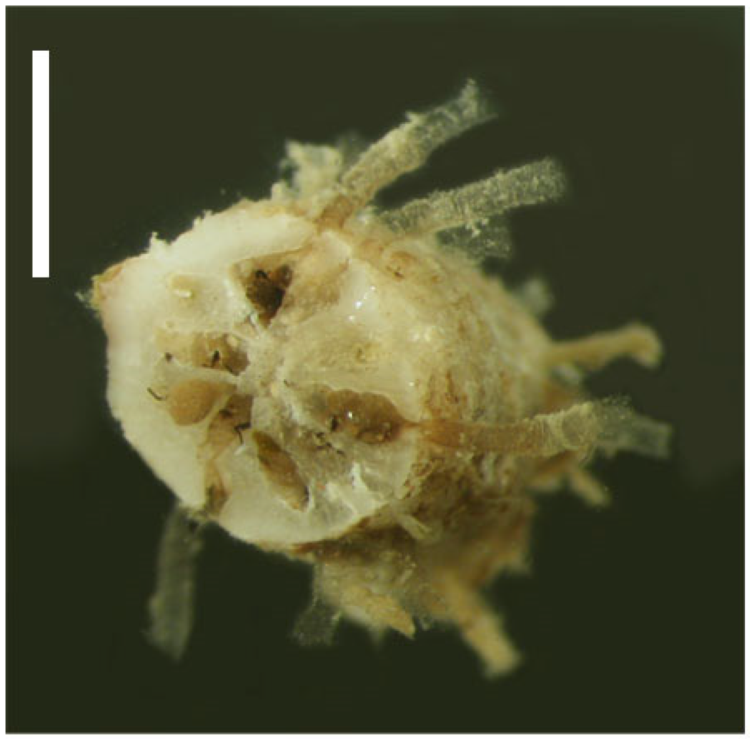
Rhabdopleura recondita Beli, Cameron, Piraino, 2018 is a benthic graptolite discovered on the southern Italian coasts, but it is possibly distributed throughout the Western Mediterranean basin [31]. It is an atypical rhabdopleurid in that the colonies reside inside of the calcareous zoarium of dead bryozoan colonies, such as Myriapora truncata (Pallas, 1766) and Schizoretepora serratimargo Hincks, 1886. The burrows and the zooecia (i.e., the skeletal body walls of chambers previously inhabited by the bryozoan zooids) in the bryozoan skeleton (or zoarium) provide habitat for the 
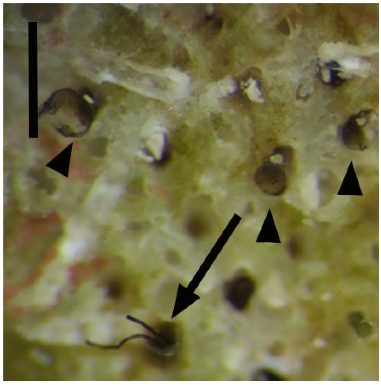
R. recondita zooids, whose interconnecting creeping tubes are smooth and lack fuselli. The erect tubes are a few millimeters in length and extend perpendicularly from the bryozoan zoarium into the water column; they are typically graptolite-like, with fuselli (Figure 1). Here we make observations and experiments on the development and tubes of R. recondita kept in captivity. We observe (i) dome secretion, larval metamorphosis, tube secretion, and tube repair by adult zooids, and (ii) we experimentally evaluate the plastic phenotypic response of the number of tubes and tube form to different water flow velocities.

2. Materials and Methods

Rhabdopleura recondita colonies are not visible to the naked eye because they are concealed inside clumps of dead bryozoan zoaria of S. serratimargo and M. truncata. These clumps were collected by hand by SCUBA diving between 10–20 m depth along the Salento coast, south of Otranto, Italy. They were transported in thermally insulated coolers to the University of Salento, where they were kept in an aquarium at 19 °C, the temperature of the collection site. Colonies were kept at a 12-h photoperiod alternating between light and dark.

2.1. Larval and Adult Tube Building

Larvae, zooids, and tube-building behavior were observed daily with a Leica MZ6 ocular stereo microscope, and pictures were taken with a Sony-RX100-I camera. The search for larvae occurred by breaking the bryozoan skeletons using steel probes and chisels. We found six larvae which were recovered with a pipette and transferred to six water bowls with 3 mL of filtered seawater at 19 °C. The bowls were covered with a nylon mesh and immersed together in a one-liter vessel of seawater with an aerator. The vessel also held fragments of colonized bryozoans. (i) Five larvae, one per bowl, were observed without a substrate to evaluate their ability to settle, metamorphose and build a colony, and (ii) one larva was provided with a deeply fragmented M. truncata to let it feel inside a host allowing external observation instead.
Rhabdopleura recondita zooids were collected in the same manner as the larvae and placed into bowls to evaluate their ability to build tubes and colonies. We prepared a total of five bowls, each covered with a nylon mesh. Zooids were fed coral food twice a week (H&O by Sicce) and kept together in a one-liter vessel with an aerator. (i) Into three separate bowls, to observe progress in tube building, we isolated one developing zooid and two adult zooids exposed naked, which means without an erect tube, but with attached stolon, creeping tube, and bryozoan calcium carbonate zoarium where they could not shelter. (ii) In the fourth bowl, we observed the behavior of two zooids exposed partially naked, attached to a common stolon, and with a reduced capacity to shelter inside the bryozoan zoarium. (iii) In a fifth bowl, to observe the ability of zooids to secrete new tubes and the timing of tube development, we cut all 23 standing tubes from a 1.5 cm bryozoan fragment containing an R. recondita colony and observed it over time.
Finally, one colony was fixed in alcohol and placed in vinegar overnight to remove the calcium carbonate matrix of the bryozoan skeleton. This treatment made it possible to observe the colony in the absence of the host skeleton.

2.2. Flow-Induced Response

To test the plastic phenotypic response of R. recondita tube secretion and development to different water velocities, a preliminary trial was performed for four colonies deprived of tubes and exposed to 4.8 cm/s.
Following the preliminary experiment, 16 colony fragments were put in lower velocity channels to form tubes. The channel velocities were established based on velocities measured at the collection sites as reported in the Istituto Superiore per la Protezione e Ricerca Ambientale (ISPRA, 2012 http://www.strategiamarina.isprambiente.it/consultazioni/consultazione-2012/files/3.1%20mar_adr%20caratteristiche%20fisiche.pdf accessed on 5 September 2016). Colony fragments with abundant 
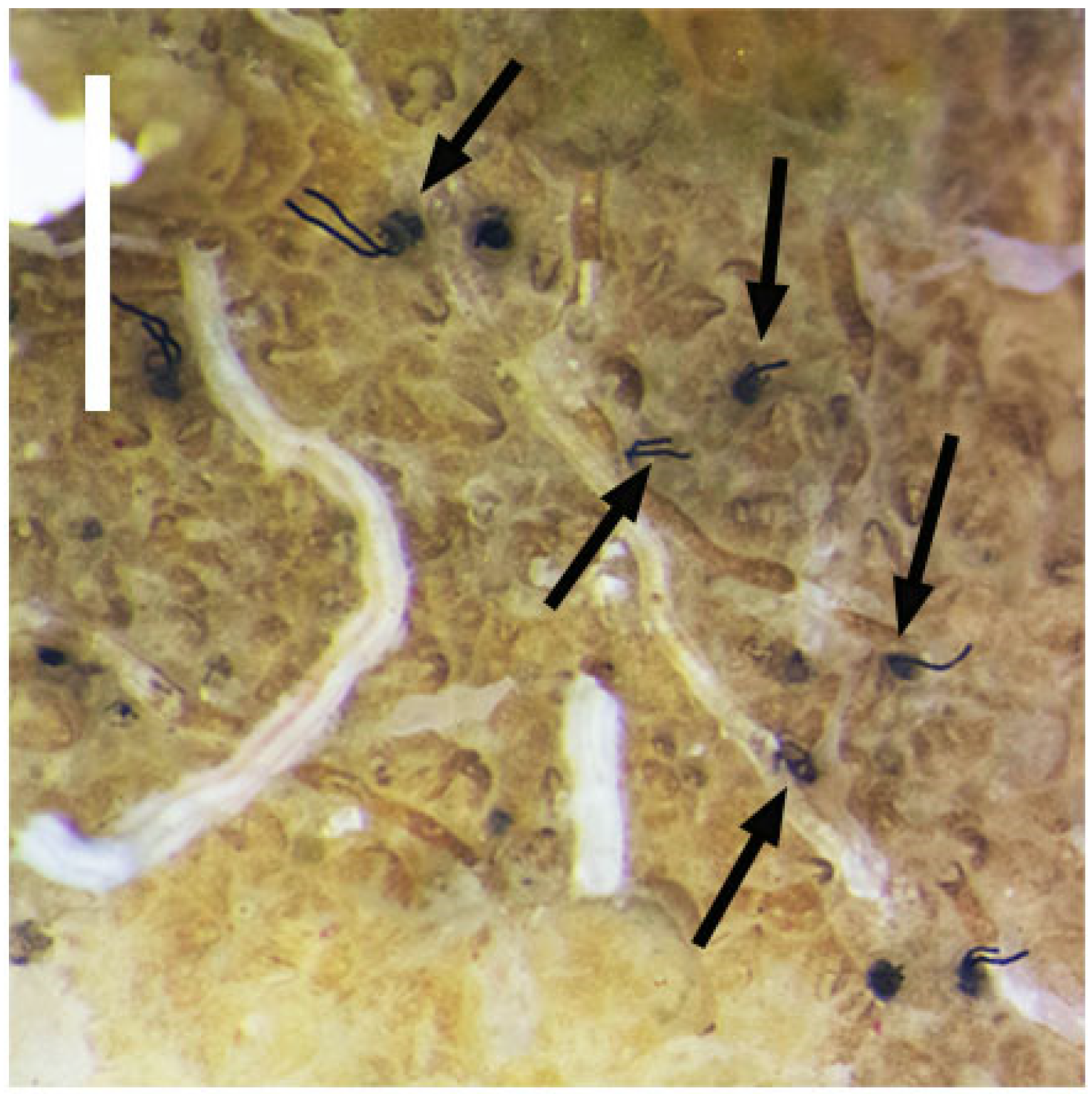
R. recondita tubes and zooids were chosen, though the number of zooids inside of any fragments was unknown because of the hidden nature of zooids. Images were captured with an SMZ 25 stereo microscope equipped with a DS-Ri2 video camera and a video-interactive image analysis system NIS–Elements BR 4.30.02 Nikon Instruments software, and with a Sony RX100 digital camera. Colonies were drawn with particular attention paid to the number and position of erect tubes. The tubes were then removed using tweezers, measured, washed in filtered seawater, and preserved in Picric Acid-Formaldehyde-Glutaraldehyde (PAFG).
These 16 colonies, denuded of erect tubes (Figure 2), were then placed into flow channels with one of four experimental flow velocities. Colonies were numbered from 1 to 16 based on the flow velocity treatment. Numbers 1–4 were maintained at 1.6 cm/s; 5–8 were maintained at 2 cm/s; 9–12 were maintained at 3.3 cm/s, and the control colonies 13–16 were maintained at 0 cm/s.
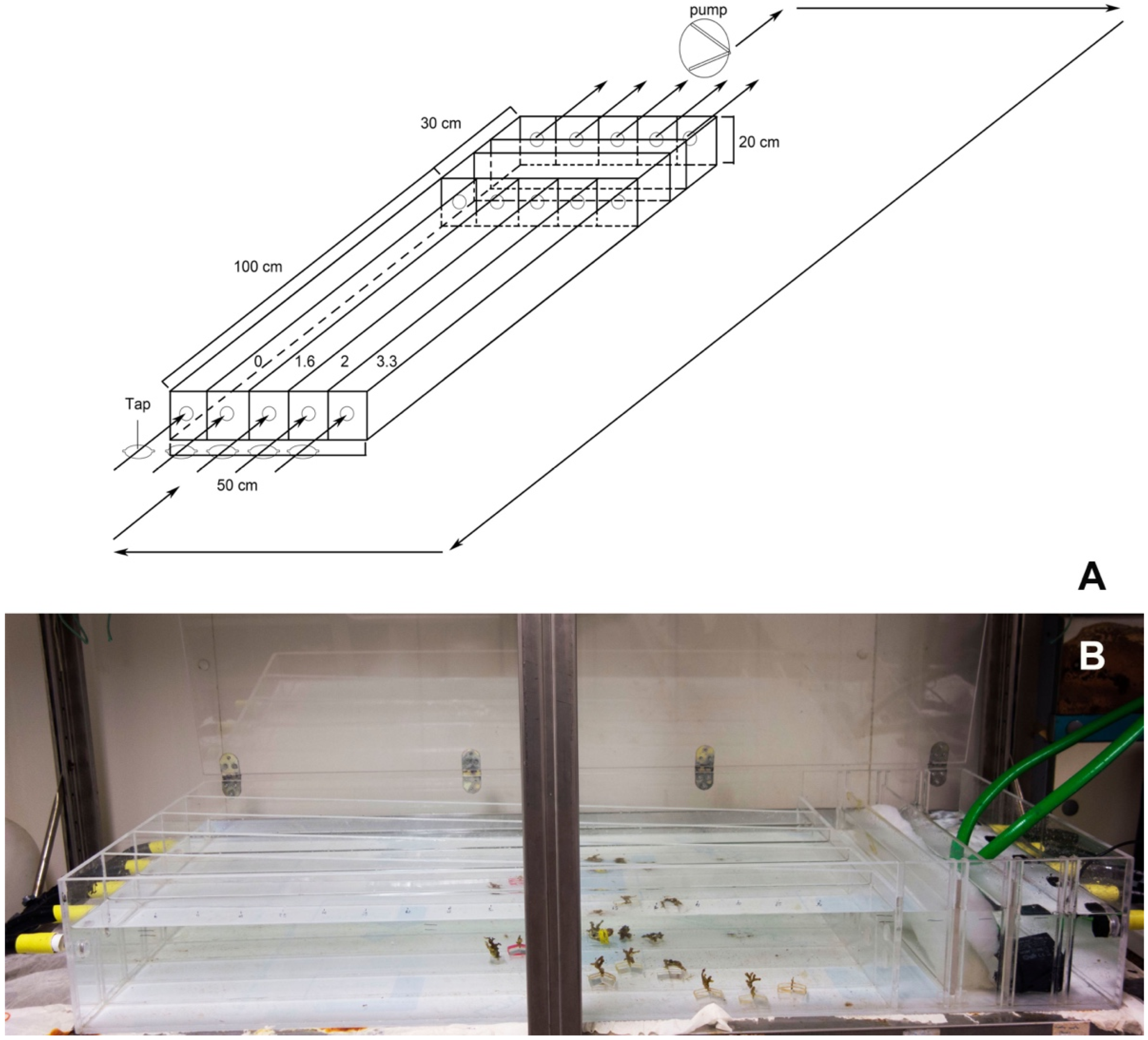
The flow channel aquarium was equipped with a 12,300 L/h pump, filtering and refrigerating system, and taps to regulate the flow velocities through four parallel channels (Figure 3A,B). Filtered seawater from the Rhabdopleura collection site was maintained at the collection site temperature of 19 °C. Salinity, dissolved oxygen, and pH were checked weekly and kept at 38‰, 7 mg/L, and 8.2, respectively. Distilled water was sometimes added to counter evaporation. Full spectrum fluorescent bulbs were set to a 12-h daylight photoperiod. The precise flow velocities, controlled with taps, were established using a fluorescein dye. The channel velocities were 1.6 cm/s, 2 cm/s, and 3.3 cm/s.
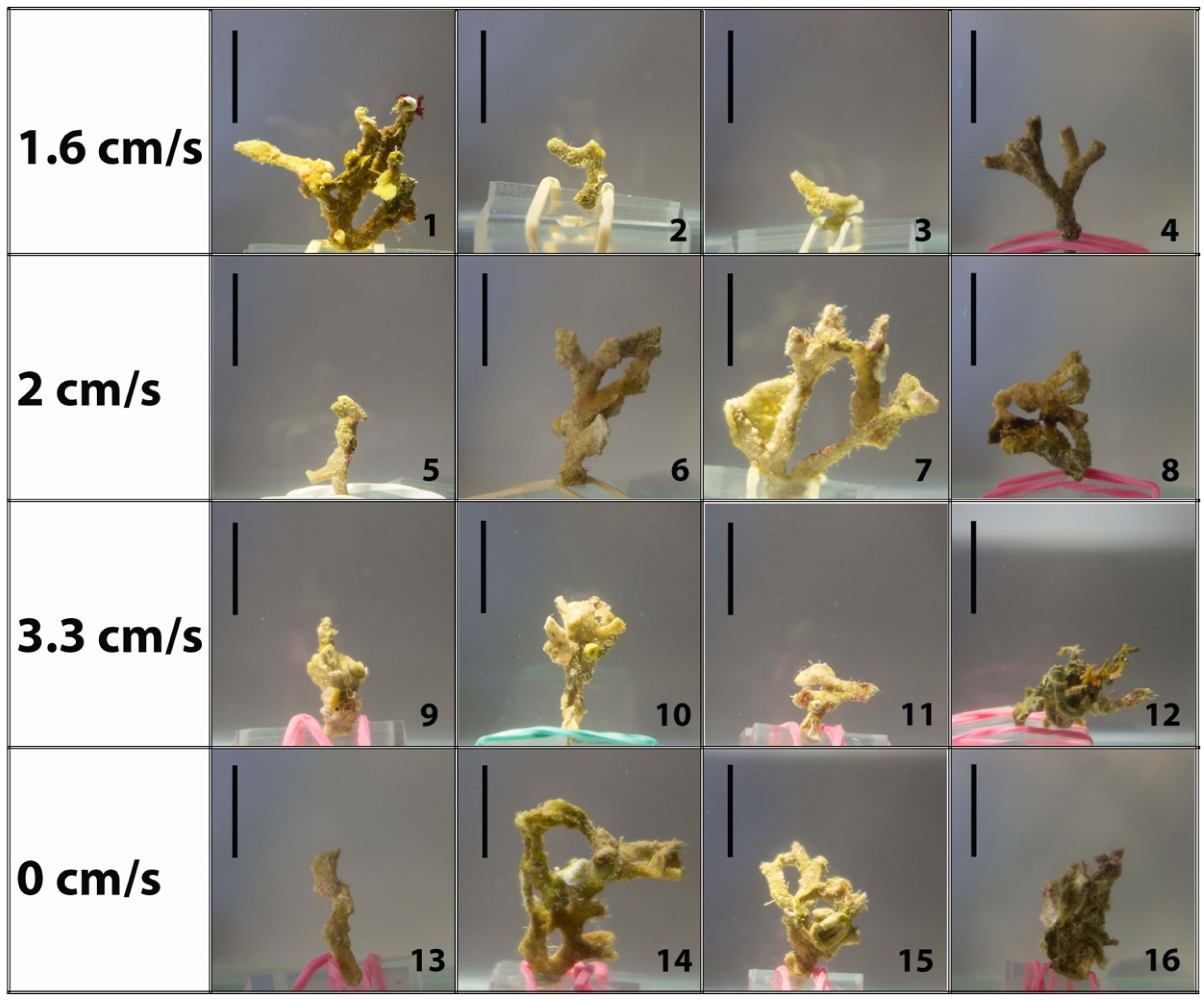
Four colonies were placed in each channel where the flow was laminar. The control channel at the velocity of 0 cm/s had a drip that prevented stagnation, though no flow could be detected. To minimize a boundary layer effect, colonies were attached to a weighted Plexiglas base that was 1cm deep and projected the fragments up into the center of the flow channel (Figure 4). Animals were fed twice a week with coral food (H&O by Sicce) and allowed to secrete new tubes for 40 days. The progress of tube construction was observed and photographed every eight days (i.e., days 0, 8, 16, 24, 32, and 40).

2.3. Statistical Analysis

Initially, the data related to the number of new tubes formed and the length of tubes (original and new tubes) were tested for homogeneity and normality using graphical analysis and the Shapiro-Wilk [32] test. No data tested fit the assumptions of normality, and non-parametric tests were performed. We used the Kruskal-Wallis [33] analysis to test if (i) the number of new tubes formed differed significantly between water velocity treatments and (ii) the new tube lengths differed significantly between velocity treatments. If the result of the Kruskal-Wallis were significant, the Dunn [34] post-hoc test was performed to highlight the significance of the differences between treatments.
Additionally, we tested if there were differences between the new tubes’ lengths and the original pre-tweezered tube lengths with respect to velocity. To test if there was a difference in tube length before and after the experiment (new length versus original length) and, if different, was it conditioned to the velocity treatment, we performed a General Linear Models (GLMs) test. First, a complete model was created using the new tube length as a response variable and the original length and speed treatment as explanatory variables. With the model created, a family selection was made to determine the best distributional family for this model. To select the family, we compared them with the Akaike criteria. With the best family selected, the model was tested to highlight the significance, if there was any. In addition, the adjustment for the model was observed using graphs (model vs. residuals). All analyses were performed using the R software [35]. The level of significance adopted in this study was 5%.

3. Results

3.1. Larval and Adult Tube Building

Discerning between an alive or dead larva (as well as a zooid) is simple. When living, these soft-bodied organisms are turgid and move or react to stimuli. Their surface is covered with beating cilia, visible with the stereo microscope. When dead, cilia do not move, and animals show signs of decomposition within a few hours. We initially observed the development of six larvae. (i) From the five larvae without a substratum, one of them settled, produced a dome, metamorphosed, and secreted a short creeping tube on the tenth day (Figure 5A), but then the development stopped, and the newly metamorphosed zooid died. Four larvae settled; two of them showed the ability to creep and swim during dome secretion (Figure 5B), and the other two were able to swim during metamorphosis (Figure 5C). However, the four larvae died within 24 h.
(ii) One larva was cultured in a bowl with a fragmented bryozoan skeleton. This larva crept among the fragments, stopped among the fragments, and extended its anterior upward. It remained in the same position at each observation until, on the 26th day, we noticed a dome in which it was able to rotate while it continued secreting the dome fabric. Twenty-nine days after, the larva ceased its rotation and began to metamorphose. After 37 days after the start of the observation, the zooid resorbed a part of the dome, and from that hole, it built a transparent creeping tube. At the time of the creeping tube secretion, the zooid was tripartite, with a distinct cephalic shield, collar, and trunk, but lacked fully developed arms, tentacles, and gut. No further development was observed (Figure 6A–D). On day 40, two additional larvae, each in a dome, were discovered, and on day 47, another larva settled and formed a dome. These three additional larvae emerged from the colonized bryozoan fragments that were kept in the larger vessel that housed the experimental treatment bowls. None formed a secreted tube from the prosicular dome.
Parallel experiments were done to determine the success of tube building by adult zooids. Here we discovered that zooids that were isolated from the colony did not secrete tube material. (i) In the three bowls with one zooid each, one immature and two adults, which were attached to a length of stolon and a piece of calcium carbonate zoarium that offered no shelter, no tubes were secreted. The immature zooid survived eight days. One adult zooid survived 24 h, and the second adult survived 18 days without secreting a tube (Figure 7A,B). (ii) A fourth bowl had two zooids that were united by a common stolon and partially exposed on top of a bryozoan zoarium fragment. These zooids survived and, at 20 days, had secreted new erect tubes. One zooid was able to move marginally deeper inside of the zoarium and built a normal tube with a smooth base and 10 fuselli (Figure 7C). The more exposed zooid built an irregularly shaped tube that was flared distally, providing some protection for its fragile body (Figure 7D). In the fifth bowl, we put an entire colony but removed from it 23 erect tubes. After 10 days, three short new tubes of the original 23, each with about five fuselli, were re-secreted. Other tubes were not remade though zooids were seen alive and perched from the bryozoan apertures (Figure 7E). The new tube material was transparent in part due to the absence of occluded foreign particles. On those new tubes, a kind of “scar” was visible at the junction of the old and the new tubes, with the margin of the old tube damaged and abandoned and the new tube anchored to a deeper fusellum (Figure 7F). After 90 days of observation, no further growth of the tubes was observed. The scarce number of tubes re-secreted in this control experiment reflects what was observed during the flow experiments below.
One last colony was treated in vinegar to remove the calcium carbonate matrix of the bryozoan skeleton. Here we confirmed our previous observations and found that R. recondita is unusual in that it lacked a creeping tube with fuselli [31]. Instead, it lined the internal burrows of the bryozoan zoarium with smooth sheets of cortical material. The creeping tubes took the form of the walls of the burrows occurring in the host zoarium, and no zigzag fuselli were found, whereas the erect tubes, that project outwards from the bryozoan zoarium, had typical fuselli. Interestingly, a brood chamber was observed, made of a spirally formed smooth creeping tube that contained, from the outside to the inside, a degenerated female zooid and four larvae, from younger to older (Figure 8A). A degenerated female zooid is characterized by reduced tentacles and reduced internal organs, and sometimes a gonad is visible (Figure 8B). It is possible these individuals do not feed themselves [14].

3.2. Flow-Induced Response

In the preliminary experiment, with colonies exposed to 4.8 cm/s, no new tubes were made, so we repeated the experiment at four lower flow velocities, as follows. Colony numbers 1–4 were maintained at 1.6 cm/s; 5–8 were maintained at 2 cm/s; 9–12 were maintained at 3.3 cm/s, and the control colonies 13–16 were maintained at 0 cm/s. Colonies were alive for the duration of the 40-day experimental period. Zooids are delicate and rapidly withdraw into their tubes within the bryozoan zoarium when disturbed by shadows, vibrations, and experimental manipulation. Other R. recondita zooids may not have emerged from the bryozoan zoarium. For this reason, it was not possible to evaluate the number of living zooids in each colony before the removal of the original tubes, but for each observation during the flow speed treatment, we noticed the presence of at least one zooid. Many zooids that we observed did not secrete new tubes. Of the sixteen experimental colonies, eleven formed new tubes, 9/11 of which were exposed to water velocities, and 2/11 from the controls exposed to 0 cm/s flow (Figure 9; Table 1). The intermediate velocities treatments seem to favour tube regrowth (Figure 10A–E and Figure 11A), with a higher number of tubes formed at 1.6 cm/s and 2 cm/s (mean 7.5 and 7.8, respectively), and fewer tubes were formed at 0 cm/s and the highest velocity of 3.3 cm/s (mean 1.5 and 2.5, respectively). These differences were not significant (p = 0.541).
Concerning the relationship between water velocity and tube length (Figure 10F; Table 2): at 0 cm/s, the mean tube length of the new tubes was 0.3 mm; at 1.6 cm/s, the mean tube length was greatest at 0.42 mm; at 2 cm/s mean tube length was 0.33 mm, and at 3.3 cm/s mean tube length was 0.31 mm. The statistical test did not show significance to the effect of water velocity on the tube length (p = 0.083). However, the differences between the new tube length (secreted during the experiment) and the original ones from the colonies collected in the field (mean length: 1.14, 1.14, 1.05, 1.08 mm from low to highest velocities) were significant (p = 7.65 × 10⁻⁵) (Figure 11B), but these differences were not due to velocity treatments (1.6 cm/s, p = 0.138; 2 cm/s, p = 0.575; 3.3 cm/s, p = 0.892).
The bulk of tube secretion occurred during the first 16–24 days of the experimental trials, then growth mostly stopped after 32 days (Figure 10A–D). Exceptions, based on the trend lines in Figure 11A, were some tube growth, especially in the medium velocity treatments (1.6 and 2 cm/s) with a higher slope. In this case, however, only the regression line of 0 cm/s treatment shows significance (p = 0.04). An unexpected observation made during these flow treatments was the presence of a closed lid on some of the shortest tubes in each colony at all flow velocity treatments, presumably secreted by the zooids (Figure 12).

4. Discussion

Rhabdopleura recondita is unusual in that it inhabits the interior empty burrows and zooecia (skeletal holes) of dead bryozoan colonies [31].
Zooids and larvae were obtained by fracturing this calcium carbonate matrix, and our best observations on the tube structure inside of the skeletal followed acid digestion of the matrix. We previously reported how the burrows and zooecia walls of the bryozoan zoaria are coated with cortical layers of tube material without fuselli [
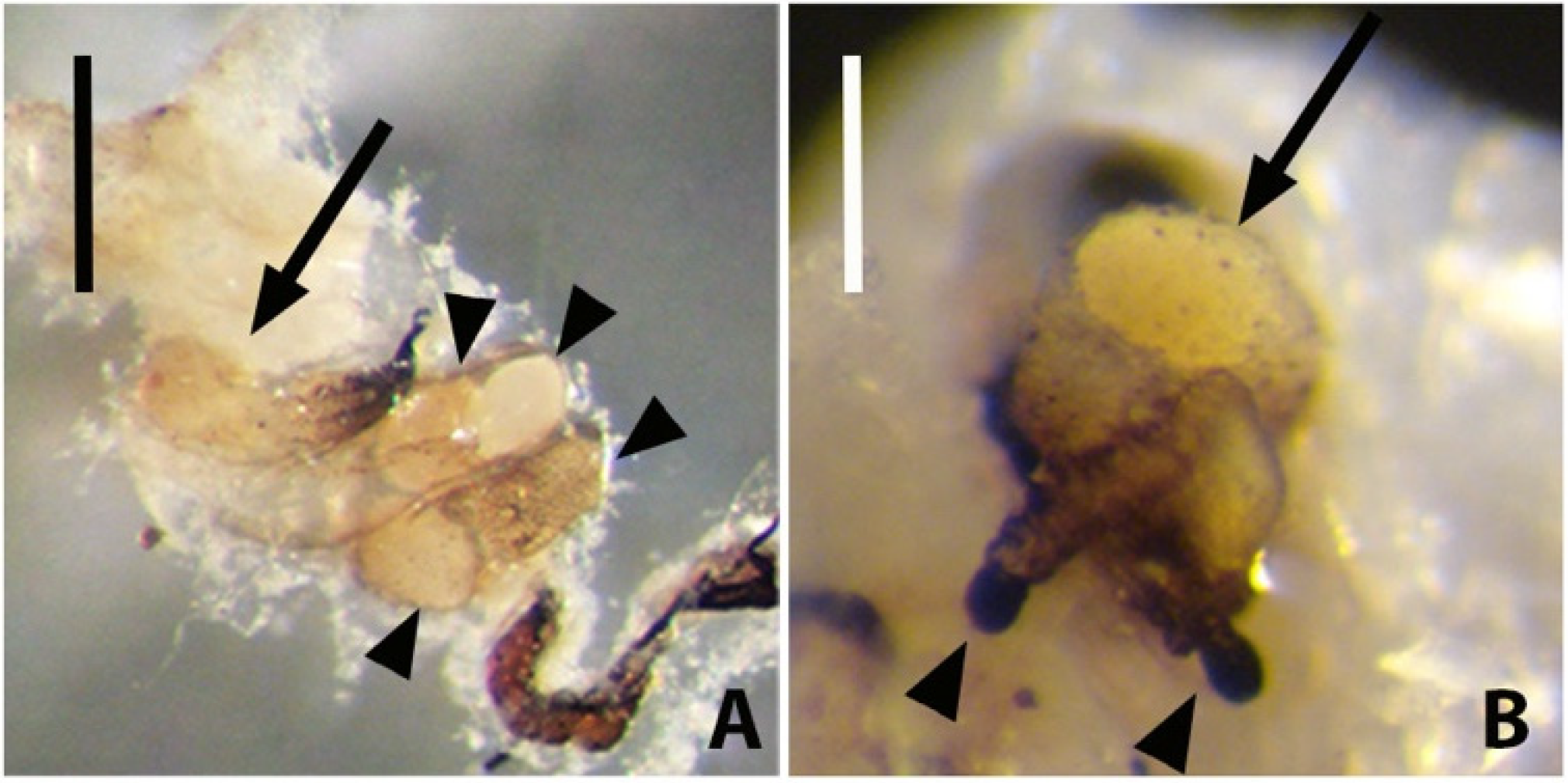
31]. Here we add to that an observation of an R. recondita brood chamber, which is known for R. normani [36], and one with a female zooid, embryos, and larvae for R. compacta [37]. The brood chamber of R. recondita had four larvae at different stages of maturity, demonstrating asynchronous brood development. The most mature larva was positioned the most distant from the tube aperture. Like R. compacta, it would need to squeeze past earlier-stage larvae and the parent zooid to exit the colony tubarium [38].
Fertilization and early cleavage of pterobranch embryos [36] and observations of larvae are exceedingly rare [2,37,38] so any new information, including rearing conditions, is valuable. Our observations and experiments with the larvae and zooids highlight the adaptations and constraints to the hidden life of R. recondita. Congeneric species typically develop on exposed surfaces, frequently of calcium carbonate [31], though occasionally they occupy internal cavities [39,40,41,42,43]. During this study, we did not observe 
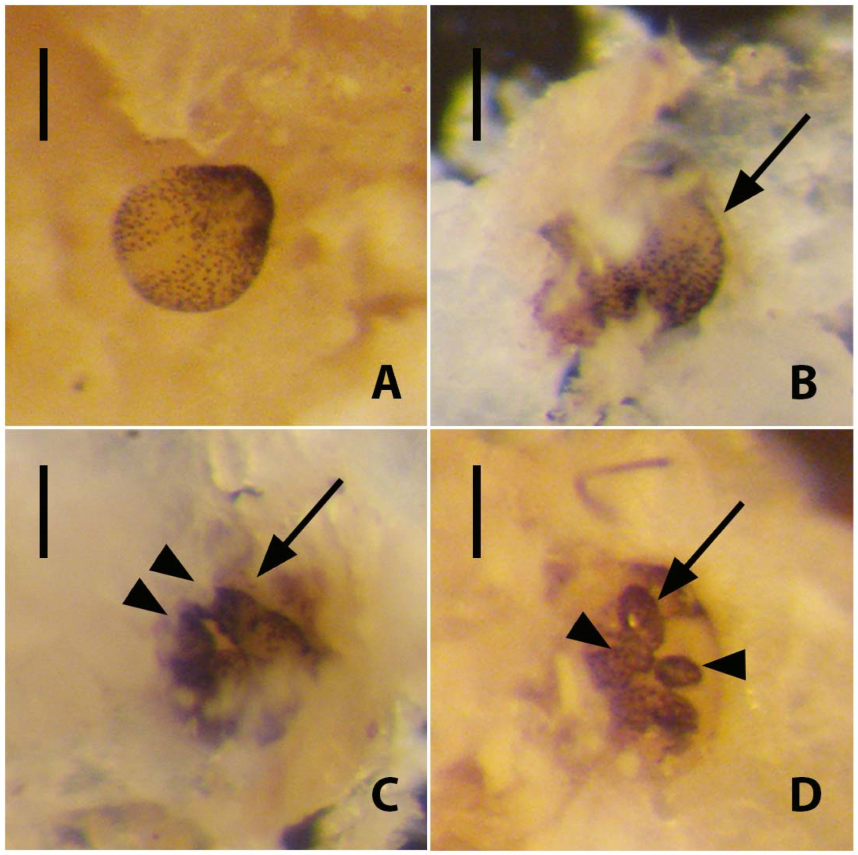
R. recondita building a new tubarium or enlarging a pre-existing one outside of the bryozoan host zoarium. Of the larvae that settled in the absence of bryozoan substrate, some began to secrete a dome (Figure 5A,B) and swam to the start of metamorphosis, like R. normani, but only one achieved metamorphosis, suggesting that the host matrix increases normal settlement and metamorphosis (Figure 5A). This truncation to development and dome secretion may have been due to disturbing the larvae or the lack of ideal developmental conditions [36], and the absence of a bryozoan zoarium seems most likely. Only one larva without the zoarium substratum settled and completed metamorphosis within 10 days (Figure 5A). By contrast, the larva in a bowl with bryozoan fragments secreted a dome and metamorphosed after 25 days (Figure 6). In this case, following its metamorphosis, additional larvae emerged from the fragments, aggregated, settled, and metamorphosed, all in the presence of CaCO3 substrate. This result supports the finding that host fragments may increase settlement and metamorphosis and that larvae hidden inside the bryozoan zoarium are best positioned for normal development.
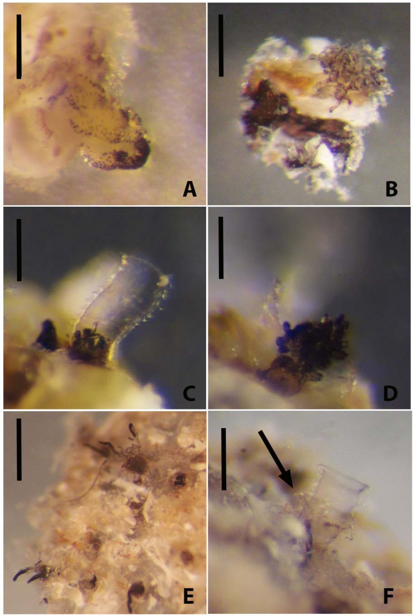
Three experiments were done to observe tube building by adult zooids. Two bowls each held an isolated adult zooid, and a third bowl held an immature zooid. These zooids did not reform a new tube, the immature zooid did not develop further, and none formed a stolon or budded new zooids (Figure 7A,B). They survived for few hours or days, supporting the idea that the absence of other connected zooids and a tubarium are incompatible with life [14]. The two zooids connected by a common stolon and cultured in the presence of host zoarium fragments survived and secreted erect tubes with fuselli (Figure 7C,D). One zooid moved marginally deeper inside the tubarium and built a normally shaped erect tube with fuselli (Figure 7C), whereas a more exposed zooid (Figure 7D) secreted a distally flared and abnormally shaped tube. This difference in tube form may suggest that normal tube secretion depends not only on the host material but access to interior burrows of the bryozoan zoarium to build a tube. These observations suggest that R. recondita tube secretion and colony formation are adapted and constrained to life inside an empty bryozoan host zoarium.
In a fifth bowl, a fragment of bryozoan zoarium contained R. recondita zooids that extended their arms for filter feeding (Figure 7E). These zooids’ erect tubes were removed, which did not appear to disturb normal emergence and feeding activities. Of these zooids, only three of the 23 re-secreted a new tube, like what we found in colonies that were subjected to experimental flow velocities. This dependence on the host zoarium for normal tube secretion and the low ratio of zooids that re-secrete tubes posed challenges to our experiments on the plastic response to tube secretion in response to water flow velocity. Also, because most of the tubarium astogeny, or overall colony form, is inside of the host zoarium, and the standing tube spacing is constrained by the availability of channels and exit pores from the zoarium, our experiments on induced plasticity were limited to measurements of standing tube number and lengths. Despite these limitations, we conducted the standing tube plasticity experiments. With five extant species of rhabdopleurid known, R. recondita is the only one with extensive colonies that can be readily collected and maintained in the lab.
This first experimental attempt of induced phenotypic plasticity in R. recondita did not find the expected outcome, that longer tubes would be re-secreted at low flow velocities and shorter tubes at higher velocities, similar to what has been reported for sponge ostia, barnacle legs, and regenerated tube feet of sea urchins [26,27,
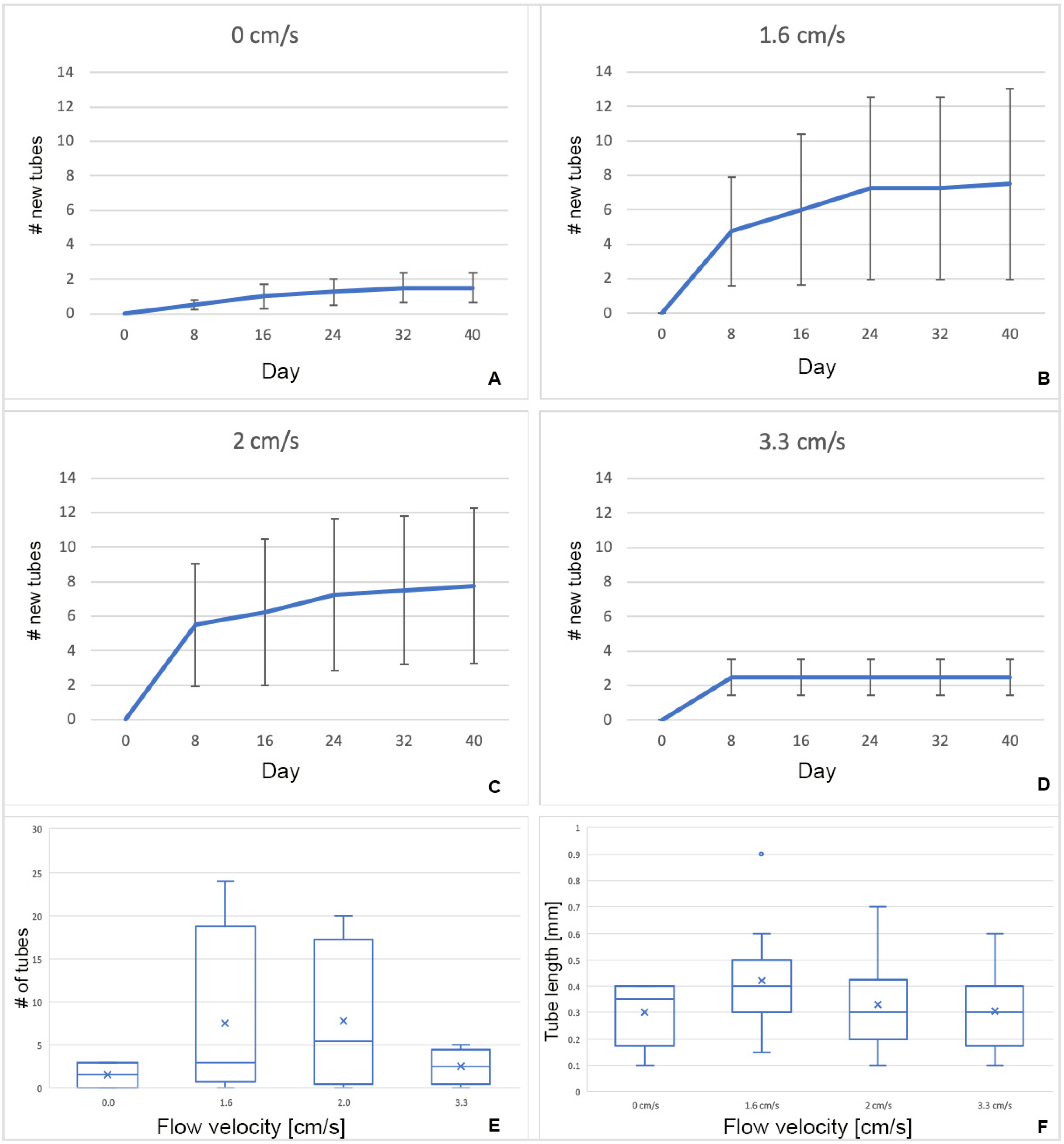
28]. Our preliminary experiment at 4.8 cm/s completely arrested the activity of zooids, and no tube building was done. Further experiments at lower velocities found a tendency for a higher number of tubes to build at intermediate flow velocities and no difference in newly secreted tube length. The number of new erect tubes formed by zooids of R. recondita exposed to different flow velocities (0, 1.6, 2, and 3.3 cm/s) showed a higher regrowth and potential for growing (Figure 10E and Figure 11) at 1.6 and 2 cm/s, though these differences were not statistically significant. More significant results may be obtained with a larger sample size, but we think it is unlikely. Our results do show that 
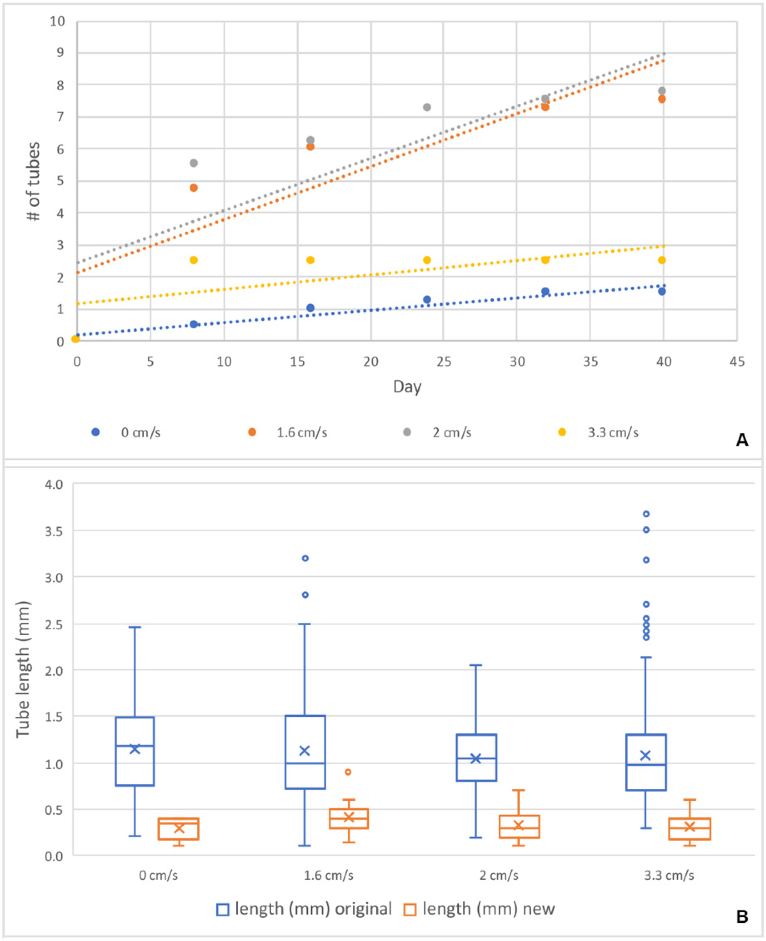
R. recondita lives in a low and narrow range of flow velocity, and within this range, it does not significantly vary the number of tubes re-secreted or tube length. Despite finding no difference in the length of re-secreted tubes among the four velocity treatments, there was a significant length difference between the originally collected colony tubes and the tubes re-secreted after dissection (Figure 11B). Standing tubes of colonies collected from the Adriatic Sea were longer and more variable than those secreted in the lab (Figure 11B). Assuming that the tubarium where tubes were removed housed a zooid, the low re-secretion ratio and short lengths may be explained by one of six possibilities. (i) The food that we used, developed for corals, was too limited or nutritionally inadequate for ideal colony growth. Nothing is known about culturing Rhabdopleura in captivity [44]. Alternatively, the coral food may have been so abundant that secreting a new tube was unnecessary. (ii) Tube secretion and colony enlargement may be seasonal or sporadic [44]. (iii) Tube formation is rare because tubes are used from generation to generation [2,45,46]. (iv) The boundary layer effect may have exposed zooids to lower velocities than those set in each flow velocity channel, which in turn may have reduced the gas exchange. (v) Colonies may require much more time, even years, to re-secrete in terms of number and length [
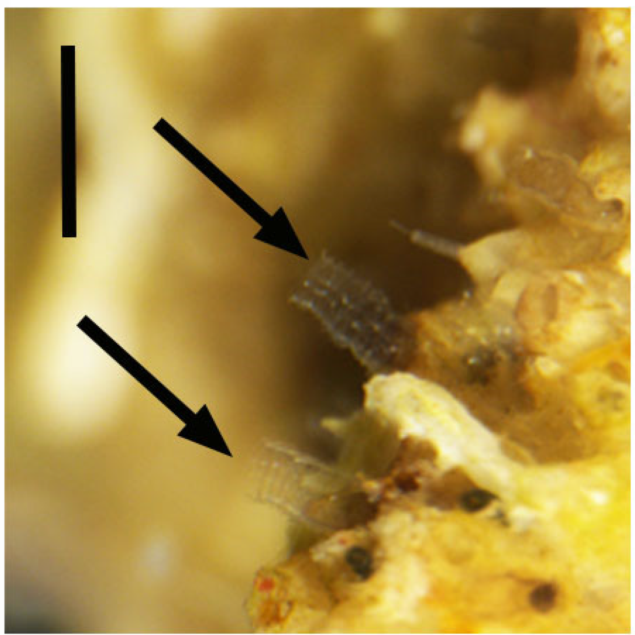
47]. (vi) Stress caused by the loss of all the tubes in each colony and the energy supply required for their reconstruction. We observed zooids that lacked erect tubes emerged from the zoarium to feed, but perhaps they did not have the energy to secrete new tubes. At the terminal end of some excised tubes, we observed a lid on the aperture. These lids are a novel finding, but as to the function, we can only speculate. Rhabdopleurids can produce dormant buds to overcome adverse periods [2,7], and these lids may be the sign of zooids’ encystment to face stressful conditions caused by tube removal and aquarium environment.
These results show that the R. recondita tube number and length exhibit no significant variation in response to water flow velocity. Together, the results of the preliminary experiment, where we found no tube secretion in flow velocities at 4.8 cm/s, suggest that R. recondita inhabits a low and narrow water flow velocity range. Tube development appears constrained. This suggests that small differences that distinguish early encrusting fossil graptolite species are real. Of course, other living rhabdopleurids, especially those with tubaria that are free of a host, may have a plastic response to flow, food, predation, or temperature. This logic extends to Palaeozoic fossil rhabdopleurids. The developmental toolkit, including plasticity, of early graptolites may have been differed from that of R. recondita. Peterson et al. [48] hypothesized that an accumulation of microRNAs over geological time may have increased the silencing and post-translational regulation of gene expression, limiting developmental experimentation. We see no reason that this hypothesis cannot be extended from secreted skeletal matrices (i.e., exoskeletons and endoskeletons) to tube matrices. Others have quantified the number and variation of graptolite forms over time and concluded that these are largely determined by extinctions rather than developmental constraints [49]. Our results suggest that the tube form of R. recondita may, in fact, be constrained, but we are cautious about extending this finding to other fossil or extant species. It is conceivable that some graptolite species and clades exhibited tremendous tubarium evolvability. With respect to Rhabdopleura, its evolvability has nothing to do with survivability. It is among the oldest graptolite lineage that has survived five major extinction events, and it is the only graptolite genus that survives to date.

Author Contributions

Conceptualization, C.B.C.; Data curation, E.B., L.M.D.C.M. and C.B.C.; Formal analysis, E.B. and L.M.D.C.M.; Funding acquisition, S.P. and C.B.C.; Investigation, E.B.; Methodology, E.B. and C.B.C.; Project administration, E.B. and C.B.C.; Resources, S.P.; Supervision, C.B.C.; Visualization, L.M.D.C.M.; Writing—original draft, E.B.; Writing—review & editing, S.P. and C.B.C. All authors have read and agreed to the published version of the manuscript.

Funding

This research was funded by CBC NSERC grant number Discovery RGPIN-2017-05058.

Acknowledgments

Many thanks are due to Mario Congedo, Mario Ciotti and Scuba Diving Otranto for their support during sampling. Also, we thank Francesco De Leo for his valuable suggestions in the phenotypic plasticity experimental settings. Precious insights were provided by Jean-François Pflieger, Sandra Binning and Charles Mitchell during the revision of Elena Beli’s doctoral thesis.

Conflicts of Interest

The authors declare no conflict of interest.

References

  1. Maletz, J.; Bates, D.E.; Brussa, E.D.; Cooper, R.A.; Lenz, A.C.; Riva, J.F.; Toro, B.A.; Zhang, Y. Part V, revised, Chapter 12: Glossary of the Hemichordata. Treatise Online 2014, 62, 1–23. [Google Scholar]
  2. Stebbing, A.R.D. Aspects of the reproduction and life cycle of Rhabdopleura compacta (Hemichordata). Mar. Biol. 1970, 5, 205–212. [Google Scholar] [CrossRef]
  3. Strano, F.; Micaroni, V.; Beli, E.; Mercurio, S.; Scarì, G.; Pennati, R.; Piraino, S. On the larva and the zooid of the pterobranch Rhabdopleura recondita Beli, Cameron and Piraino, 2018 (Hemichordata, Graptolithina). Mar. Biodivers. 2019, 49, 1657–1666. [Google Scholar] [CrossRef] [Green Version]
  4. Maletz, J. Tracing the evolutionary origins of the Hemichordata (Enteropneusta and Pterobranchia). Palaeoworld 2019, 28, 58–72. [Google Scholar] [CrossRef]
  5. Maletz, J.; Lenz, A.C.; Bates, D.E.B. Part V, Second Revision, Chapter 4: Morphology of the Pterobranch Tubarium. Treatise Online 2016, 76, 1–63. [Google Scholar] [CrossRef]
  6. Maletz, J. The classification of the Pterobranchia (Cephalodiscida and Graptolithina). Bull. Geosci. 2014, 1977, 477–540. [Google Scholar] [CrossRef] [Green Version]
  7. Maletz, J.; Beli, E. Part V, Second Revision, Chapter 15: Subclass Graptolithina and Incertae Sedis Family Rhabdopleuridae: Introduction and Systematic Descriptions. Treatise Online 2018, 101, 101–114. [Google Scholar] [CrossRef]
  8. Maletz, J. The identity of Didymograptus (Expansograptus) suecicus (Tullberg) and related species (Graptoloidea, Dichograptidae). Paläont. Z. 1996, 70, 203–212. [Google Scholar] [CrossRef]
  9. Rickards, R.; Stait, B. Psigraptus, its classification, evolution and zooid. Alcheringa 1984, 8, 101–111. [Google Scholar] [CrossRef]
  10. Bjerreskov, M. Pyrite in Silurian graptolites from Bornholm, Denmark. Lethaia 1991, 24, 351–361. [Google Scholar] [CrossRef]
  11. Durman, P.N.; Sennikov, N.V. A new rhabdopleurid hemichordate from the Middle Cambrian of Siberia. Palaeontology 1993, 36, 283–296. [Google Scholar]
  12. Chapman, A.J.; Durman, P.N.; Rickards, R.B. Rhabdopleuran hemichordates: New fossil forms and review. Proc. Geol. Assoc. 1995, 106, 293–303. [Google Scholar] [CrossRef]
  13. Loydell, D.K.; Orr, P.J.; Kearns, S. Preservation of soft tissues in Silurian graptolites from Latvia. Palaeontology 2004, 47, 503–513. [Google Scholar] [CrossRef]
  14. Maletz, J.; Cameron, C.B. Part V, Second Revision, Chapter 3: Introduction to Class Pterobranchia Lankester, 1877. Treatise Online 2016, 82, 1–15. [Google Scholar] [CrossRef]
  15. Zhang, Y.; Chen, X.; Goldman, D. Diversification patterns of Early and Mid Ordovician graptolites in south China. Geol. J. 2007, 42, 315–337. [Google Scholar]
  16. Allmon, W.D.; Yacobucci, M.M. Species and Speciation in the Fossil Record; University Chicago Press: Chicago, IL, USA, 2016. [Google Scholar]
  17. Ramírez-Guerrero, G.M.; Cameron, C.B. Systematics and evolution of pterobranchs from the Cambrian Period Burgess Shales of Canada. Bull. Geosci. 2021, 96, 1–18. [Google Scholar] [CrossRef]
  18. Erdtmann, B.D.; Maletz, J.; Marco, J.C.G. The new Early Ordovician (Hunneberg Stage) graptolite genus Paradelograptus (Kinnegraptidae), its phylogeny and biostratigraphy. Paläont. Z. 1987, 61, 109–131. [Google Scholar] [CrossRef]
  19. Erdtmann, B.D. The earliest Ordovician nematophorid graptolites: Taxonomy and correlation. Geol. Mag. 1988, 125, 327–348. [Google Scholar] [CrossRef]
  20. Štorch, P. Biotic crises and post-crisis recoveries recorded by Silurian planktonic graptolite faunas of the Barrandian area (Czech Republic). Geolines 1995, 3, 59–70. [Google Scholar]
  21. West-Eberhard, M.J. Developmental Plasticity and Evolution; Oxford University Press: Oxford, UK, 2003. [Google Scholar]
  22. Lenz, A.C.; Melchin, M. Convergent evolution of two Silurian graptolites. Acta Palaeontol. Pol. 2008, 53, 449–460. [Google Scholar] [CrossRef] [Green Version]
  23. Blackett, E.; Page, A.; Zalasiewicz, J.; Williams, M.; Rickards, B.; Davies, J. A refined graptolite biostratigraphy for the late Ordovician-early Silurian of central Wales. Lethaia 2009, 42, 83–96. [Google Scholar] [CrossRef] [Green Version]
  24. Graus, R.R.; Chamberlain, J.A., Jr.; Boker, A.M. Structural modification of corals in relation to waves and currents: Reef biota. In Reefs and Related Carbonates Ecology and Sedimentology; Frost, S.H., Weiss, M.P., Saunders, J.B., Eds.; AAPG Bulletin: Tulsa, OK, USA, 1977; pp. 135–153. [Google Scholar]
  25. Palumbi, S.R. How body plans limit acclimation: Responses of a demosponge to wave force. Ecology 1986, 67, 208–214. [Google Scholar] [CrossRef]
  26. Marchinko, K.B. Dramatic phenotypic plasticity in barnacle legs (Balanus glandula Darwin): Magnitude, age dependence, and speed of response. Evolution 2003, 57, 1281–1290. [Google Scholar] [PubMed]
  27. Vicente, V.P. An ecological evaluation of the West Indian demosponge Anthosigmella varians (Hadromerida: Spirastrellidae). Bull. Mar. Sci. 1978, 28, 771–779. [Google Scholar]
  28. Narvaez, C.A.; Moura, A.J.; Scutella, D.F.; Cucchiara, J.P.; Stark, A.Y.; Russell, M.P. Plasticity in fluctuating hydrodynamic conditions: Tube feet regeneration in sea urchins. J. Exp. Biol. 2022, 225, jeb242848. [Google Scholar] [CrossRef]
  29. Vo, M.; Mehrabian, S.; Étienne, S.; Pelletier, D.; Cameron, C.B. The hemichordate pharynx and gill pores impose functional constraints at small and large body sizes. Biol. J. Linn. Soc. 2019, 127, 75–87. [Google Scholar] [CrossRef]
  30. Mitchell, C.E.; Melchin, M.J.; Cameron, C.B.; Maletz, J. Phylogenetic analysis reveals that Rhabdopleura is an extant graptolite. Lethaia 2013, 46, 34–56. [Google Scholar] [CrossRef]
  31. Beli, E.; Aglieri, G.; Strano, F.; Maggioni, D.; Telford, M.J.; Piraino, S.; Cameron, C.B. The zoogeography of extant rhabdopleurid hemichordates (Pterobranchia: Graptolithina), with a new species from the Mediterranean Sea. Invertebr. Syst. 2018, 32, 100–110. [Google Scholar] [CrossRef]
  32. Shapiro, S.S.; Wilk, M.B. An analysis of variance test for normality (complete samples). Biometrika 1965, 52, 591–611. [Google Scholar] [CrossRef]
  33. Kruskal, W.H.; Wallis, W.A. Use of Ranks in One-Criterion Variance Analysis. J. Am. Stat. Assoc. 1952, 47, 583–621. [Google Scholar] [CrossRef]
  34. Dunn, O.J. Multiple comparisons using rank sums. Technometrics 1964, 6, 241–252. [Google Scholar] [CrossRef]
  35. R Development Core Team. R: A Language and Environment for Statistical Computing; R Foundation for Statistical Computing: Vienna, Austria, 2011; Available online: http://www.R-project.org/ (accessed on 5 September 2016).
  36. Lester, S.M. Ultrastructure of adult gonads and development and structure of the larva of Rhabdopleura normani (Hemichordata: Pterobranchia). Acta Zool. 1988, 69, 95–109. [Google Scholar] [CrossRef]
  37. Sato, A. Seasonal reproductive activity in the pterobranch hemichordate Rhabdopleura compacta. J. Mar. Biolog. Assoc. 2008, 88, 1033–1041. [Google Scholar] [CrossRef]
  38. Dilly, P.N. The larva of Rhabdopleura compacta (Hemichordata). Mar. Biol. 1973, 18, 69–86. [Google Scholar] [CrossRef]
  39. Schepotieff, A. Die Pterobranchier des Indischen Ozeans. Zool. Jahrb. Abt. Syst. 1909, 28, 429–448. [Google Scholar]
  40. Johnston, T.H. A note on the occurrence of Rhabdopleura annulata in South Australian waters. Rec. Aust. Mus. 1937, 6, 105–107. [Google Scholar]
  41. Stebbing, A.R.D. The status and ecology of Rhabdopleura compacta (Hemichordata) from Plymouth. J. Mar. Biolog. Assoc. 1970, 50, 209–221. [Google Scholar] [CrossRef]
  42. Barnes, R.D. New record of a pterobranch hemichordate from the western hemisphere. Bull. Mar. Sci. 1977, 27, 340–343. [Google Scholar]
  43. Lester, S.M. Settlement and metamorphosis of Rhabdopleura normani (Hemichordata: Pterobranchia). Acta Zool. 1988, 69, 111–120. [Google Scholar] [CrossRef]
  44. Cavers, P. Quantifying Variation in a Population of Rhabdopleura compacta. Unpublished Zoology Honours Thesis, University of Edinburgh, Edinburgh, UK, 2005. [Google Scholar]
  45. Rigby, S. Erect tube growth in Rhabdopleura compacta (Hemichordata: Pterobranchia) from off Start Point, Devon. J. Zool. 1994, 233, 449–455. [Google Scholar] [CrossRef]
  46. Briggs, D.E.; Kear, A.J.; Baas, M.; de Leeuw, J.W.; Rigby, S. Decay and composition of the hemichordate Rhabdopleura: Implications for the taphonomy of graptolites. Lethaia 1995, 28, 15–23. [Google Scholar] [CrossRef]
  47. Maletz, J. Graptolites: Fossil and living. Geol. Today 2017, 33, 233–240. [Google Scholar] [CrossRef]
  48. Peterson, K.J.; Dietrich, M.R.; McPeek, M.A. MicroRNAs and metazoan macroevolution: Insights into canalization, complexity, and the Cambrian explosion. BIOEEJ 2009, 31, 736–747. [Google Scholar] [CrossRef] [PubMed]
  49. Bapst, D.W.; Bullock, P.C.; Melchin, M.J.; Sheets, H.D.; Mitchell, C.E. Graptoloid diversity and disparity became decoupled during the Ordovician mass extinction. Proc. Natl. Acad. Sci. USA 2012, 109, 3428–3433. [Google Scholar] [CrossRef]
Figure 1. Transversal section of the bryozoan Myriapora truncata colonized by Rhabdopleura recondita. Erect semi-transparent tubes of R. recondita project from the bryozoan host zoarium. Scale bar 2 mm.
Diversity 14 01080 g001
Figure 2. Colony deprived of tubes. The paired arms of several individuals are visible (arrows). Scale bar 1 mm.
Diversity 14 01080 g002
Figure 3. The experimental flow channel aquarium. (A) Drawing and (B) photograph of the experimental aquarium with flow channels. One channel was used for control, with 0 cm/s flow, and three channels were maintained at flow velocities of 1.6 cm/s, 2 cm/s, and 3.3 cm/s. In figure (A), arrows indicate the direction of flow, and velocities in cm/s are reported on each channel. Only four of the five channels were used.
Diversity 14 01080 g003
Figure 4. Photos of the 16 experimental colonies maintained in place by Plexiglas weights. Velocities are reported for each row. Scale bar 2.5 cm.
Diversity 14 01080 g004
Figure 5. Rhabdopleura recondita metamorphosing larvae. (A) A metamorphosing larva shows the dome (arrow), arms (arrowheads), and cephalic shield (cs). (B) A larva started the secretion of the dome (arrow). (C) A furrow (arrow) between the trunk and the cephalic shield (cs) is an early sign of metamorphosis. Larvae in (B,C) were swimming. Scale bar 80 µm.
Diversity 14 01080 g005
Figure 6. Rhabdopleura recondita larval development on bryozoan zoarium fragments. (A) A larva with the anterior end up. (B) The larva started secreting a dome (arrow). (C) After 29 days from settlement, metamorphosis began. The arm buds (arrowheads) are visible beyond the dome (arrow). (D) At 37 days post-settlement, the zooid is tripartite, with arms lacking tentacles (arrowheads), and has pierced the dome (arrow) to form a creeping tube. Scale bars 50 µm.
Diversity 14 01080 g006
Figure 7. Experimental zooids and colonies. (A) A developing zooid survived eight days with no further development. (B) One of the two naked zooids attached to a creeping tube and stolon embedded in a bryozoan fragment. (C,D), Two zooids united by a common stolon and exposed following the fracturing of the bryozoan zoarium. (C) The zooid, able to descend into the host zoarium matrix, built a normal erect tube. (D) The exposed zooid built an unusual, flared tube. (E) After the removal of the original tubes, Rhabdopleura recondita zooids perched from the bryozoan zoarium apertures. (F) A new tube is re-secreted, arrow indicates the rim of the old tube. Scale bars 150 µm for (A), 250 µm for (BD), and (F), and 450 µm for (E).
Diversity 14 01080 g007
Figure 8. Experimental zooids and colonies. (A) A brooding chamber made of a smooth and spiraled creeping tube. Female zooid (arrow) and larvae (arrowheads). (B) Female zooid with ovary (arrow) and reduced tentacles (arrowheads). Scale bars 350 µm for (A), 150 µm for (B).
Diversity 14 01080 g008
Figure 9. Experimental zooids and colony. Two new tubes are indicated by arrows. Scale bar 800 µm.
Diversity 14 01080 g009
Figure 10. The number of re-secreted tubes and tube length with respect to flow velocity. (AD), a plot for each treatment. On the abscissa, the days, on the ordinate, the mean number of new tubes re-secreted with error bar. (E) The number of tubes re-secreted as a function of the flow velocity. (F) The length of tubes re-secreted in the function of flow velocity. Each box delimits the distance between the first and third quartile, the median is the horizontal bar in the box, and the “×” is the mean value. The whiskers reach the lowest and largest data points, excluding the outliers (dot).
Diversity 14 01080 g010
Figure 11. The number of tubes re-secreted and tube length with respect to flow velocity. (A) The number of new tubes built per day, grouped by flow velocity treatment, plotted together for comparison. Trend lines used the method of least squares to find the regression line. (B) Differences in tube length between the original tubes (blue) and the new tubes (orange) for each treatment. Each box delimits the distance between the first and third quartile, the median is the horizontal bar in the box, and the “×” is the mean value. The whiskers reach the lowest and largest data points, excluding the outliers (dots).
Diversity 14 01080 g011
Figure 12. Experimental zooids and colony. Some of the removed erect tubes were closed with a lid secreted by the zooids (arrowheads). The lid on the left is broken. Scale bar 80 µm.
Diversity 14 01080 g012
Table 1. Number of new tubes reformed for each colony and flux treatment.
# Colony1.6 cm/s2 cm/s3.3 cm/s0 cm/s (Controls)
# New Tubes/
# Original Tubes
# New Tubes/
# Original Tubes
# New Tubes/# Original Tubes# New Tubes/
# Original Tubes
124/152
23/102
30/32
43/115
5 2/27
6 9/68
7 20/121
8 0/13
9 2/35
10 5/63
11 3/22
12 0/27
13 3/13
14 3/38
15 0/27
16 0/44
Table 2. Mean tube length for each colony, before and after the flow-induced response.
# Colony1.6 cm/s2 cm/s3.3 cm/s0 cm/s (Controls)
Mean Length Original TubesMean Length New TubesMean Length Original TubesMean Length New TubesMean Length Original TubesMean Length New TubesMean Length Original TubesMean Length New Tubes
11.210.43
21.380.25
31.150
40.870.2
5 0.900.45
6 1.070.32
7 1.050.35
8 1.150
9 1.090.6
10 0.960.29
11 0.670.3
12 1.700
13 0.890.33
14 0.890.27
15 1.270
16 0.920
Publisher’s Note: MDPI stays neutral with regard to jurisdictional claims in published maps and institutional affiliations.

