Article

Marinobacterium sedimentorum sp. nov., Isolated from the Bottom Sediments of the Okhotsk Sea

1
G.B. Elyakov Pacific Institute of Bioorganic Chemistry, Far Eastern Branch, Russian Academy of Sciences, Prospect 100 Let Vladivostoku, 159, Vladivostok 690022, Russia
2
A.V. Zhirmunsky National Scientific Center of Marine Biology, Far Eastern Branch, Russian Academy of Sciences, Palchevskogo Street 17, Vladivostok 690041, Russia
*
Authors to whom correspondence should be addressed.
Diversity 2022, 14(11), 944; https://doi.org/10.3390/d14110944
Submission received: 23 June 2022 / Revised: 28 October 2022 / Accepted: 1 November 2022 / Published: 3 November 2022
(This article belongs to the Section Microbial Diversity and Culture Collections)

Abstract

:
A Gram-negative, aerobic, rod-shaped, motile bacterium designated KMM 9957T was isolated from a bottom sediment sample obtained from the Okhotsk Sea, Russia. Phylogenetic analyses based on the 16S rRNA gene and concatenated conserved protein-coding sequences positioned the novel strain KMM 9957T in the genus Marinobacterium as a distinct line adjacent to Marinobacterium rhizophilum CL-YJ9T, Marinobacterium profundum PAMC 27536T, and Marinobacterium aestuarii KCTC 52193T with 16S rRNA gene sequence similarities of 99%, 98.3%, and 98.2%, respectively. The average nucleotide identity and digital DNA–DNA hybridization values between strain KMM 9957T and M. aestuarii KCTC 52193T, M. profundum PAMC 27536T, and M. rhizophilum DSM 18822T were 89.4%, 87.9%, and 84.7% and 38.6%, 34.8%, and 28.4%, respectively. The genomic DNA G+C content of strain KMM 9957T was 58.4 mol%. The dominant respiratory quinone was ubiquinone Q-8, and the major fatty acids were C18:1, C10:0 3-OH, C16:0, and C16:1ω7c. The polar lipids of strain KMM 9957T consisted of phosphatidylethanolamine, diphosphatidylglycerol, phosphatidylcholine, an unidentified aminophospholipid, two unidentified aminolipids, eight unidentified phospholipids, and three unidentified lipids. Based on the combination of phylogenetic and phenotypic characteristics, strain KMM 9957T represents a novel species of the genus Marinobacterium, for which the name Marinobacterium sedimentorum sp. nov. is proposed.

1. Introduction

During a survey of the biodiversity of bacteria associated with a deep-sea environment, a Gram-negative, aerobic, motile bacterium designated KMM 9957T was isolated from a bottom sediment sample obtained from the Okhotsk Sea, Russia. Phylogenetic analysis assigned the novel bacterium to the genus Marinobacterium on the basis of 16S rRNA gene sequences where it formed an individual line adjacent to the Marinobacterium aestuarii KCTC 52193T cluster. The genus Marinobacterium was described by González et al. [1] with the description of the type species Marinobacterium georgiense. The genus Marinobacterium belongs to the family Alteromonadaceae, class Gammaproteobacteria, and currently contains 19 species with validly published names listed at https://lpsn.dsmz.de/genus/marinobacterium accessed on 28 October 2022, including the recently described Marinobacterium aestuarii [2], Marinobacterium boryeongense [3], and Marinobacterium ramblicola [4]. Bacteria of the genus Marinobacterium are widespread in marine environments such as sea water [3,5], tidal flat, estuarine, and deep-sea sediments [2,6,7,8,9,10,11], mucus of coral [12], rhizosphere of a halophytic plant [13], mangrove roots [14], hypersaline soil [4], and pulp mill waste [1]. Several Marinobacterium members have been reported to be aromatic hydrocarbon-degrading bacteria [2,10]. In the present study the taxonomic position of the novel marine bacterium KMM 9957T is defined. On the basis of combined phylogenomic analyses and phenotypic properties, a novel species, Marinobacterium sedimentorum sp. nov., is described.

2. Materials and Methods

2.1. Bacterial Strains

A sediment sample was obtained at a depth of 21.8 m from the Okhotsk Sea, Russia, during the expedition of R/V Academician Oparin, in September 2020. Strain KMM 9957T was isolated by a standard dilution plating method and incubated on marine agar 2216 (MA; BD Difco) at 25 °C. The novel bacterium was cultivated aerobically on Marine Agar 2216 (MA) or in Marine Broth (MB) 2216 (BD Difco) at 25 °C, and stored at −70 °C in MB 2216 supplemented with 20% (v/v) glycerol. The strain, KMM 9957T, was deposited in the Collection of Marine Microorganisms (KMM), G.B. Elyakov Pacific Institute of Bioorganic Chemistry, Far Eastern Branch, Russian Academy of Sciences, Vladivostok, Russia, and in the Korean Agricultural Culture Collection (KACC), Agricultural Microbiology Division, National Academy of Agricultural Science, Korea, as KACC 22805T. The type strain Marinobacterium aestuarii KCTC 52193T was purchased from the Korean Collection for Type Cultures, KCTC, Korea, to be used in the comparative phenotypic tests.

2.2. Phenotypic Characterization

Gram-staining, oxidase and catalase reactions, and motility (the hanging drop method) were determined as described by Gerhardt et al. [15]. The morphology of cells grown on MA 2216 and negatively stained with a 1% phosphotungstic acid on carbon-coated 200-mesh copper grids was examined by STEM microscopy using Sigma 300VP (ZEISS), provided by the Far Eastern Centre of electronic microscopy, A.V. Zhirmunsky National Scientific Center of Marine Biology, Far Eastern Branch, Russian Academy of Sciences. Hydrolysis of starch, casein, gelatin, Tween 80, DNA, L-tyrosine, chitin, and growth at different salinities (0–12% NaCl), temperatures (5–45 °C), and pH values (4.0–10.5) were carried out using artificial sea water (ASW) as described in a previous paper [16]. The artificial sea water (ASW) contained (per liter of distilled water): 24 g NaCl, 4.9 g MgCl2, 2.0 g MgSO4, 0.5 g CaCl2, 1.0 g KCl, and 0.01 g FeSO4. Biochemical tests were performed using API 20E, API 20NE, API ID32 GN, and API ZYM test kits (bioMérieux, Marcy-l’Étoile, France) as described by the manufacturer except the cultures were suspended in ASW.

2.3. Chemotaxonomic Characterization

Strain KMM 9957T and related type strain Marinobacterium aestuarii KCTC 52193T were grown on MA 2216 at 25 °C. Lipids were extracted using the method of Folch et al. [17]. Two-dimensional thin layer chromatography of polar lipids was carried out on Silica gel 60 F254 (10 × 10 cm, Merck, Darmstadt, Germany) using chloroform–methanol–water (65:25:4, v/v) for the first direction, and chloroform–methanol–acetic acid–water (80:12:15:4, v/v) for the second one [18], as well as spraying with specific reagents [19]. Respiratory lipoquinones were analyzed by reversed-phase high performance thin-layer chromatography as described by Mitchell and Fallon [20]. Fatty acid methyl esters (FAMEs) were prepared according to the procedure of the Microbial Identification System (MIDI) [21]. The analysis of FAMEs was performed using the GC-2010 chromatograph (Shimadzu, Kyoto, Japan) equipped with capillary columns (30 m × 0.25 mm I.D.), one coated with Supecowax-10 and the other with SPB-5. Identification of FAMEs was accomplished by equivalent chain length values and comparing the retention times of the samples to those of standards. In addition, FAMEs were analyzed using a GLC-MS Shimadzu GC-MS model QP2020 (column Shimadzu SH-Rtx-5MS, the temperature program from 160 °C to 250 °C, at a rate of 2 °C/min).

2.4. 16S rRNA Gene Sequence and Phylogenetic Analysis

Genomic DNA of strain KMM 9957T was extracted using a commercial genomic DNA extraction kit (Thermo Fisher Scientific Baltics, Vilnius, Lithuania) following the manufacturer’s instruction. The 16S rRNA gene was PCR-amplified and sequenced as described in a previous paper [22]. The 16S rRNA gene sequence of strain KMM 9957T was compared with those of the closest relatives using the BLAST (http://www.ncbi.nlm.nih.gov/blast/, accessed on 15 May 2021) and EzBioCloud services [23]. Phylogenetic analysis was conducted using Molecular Evolutionary Genetics Analysis (MEGA X) [24]. Phylogenetic trees were constructed by the neighbor-joining and maximum-likelihood methods, and the distances were calculated according to the Kimura two-parameter model [25]. The robustness of phylogenetic trees was estimated by the bootstrap analysis of 1000 replicates.

2.5. Whole-Genome Sequencing and Genome-Based Phylogenetic Analysis

The genomic DNA was obtained from the strain KMM 9957T using the High Pure PCR Template Preparation Kit (Roche, Basel, Switzerland). The quantity and quality of the genomic DNA was measured using DNA gel electrophoresis and the Qubit 4.0 Fluorometer (Thermo Fisher Scientific, Singapore, Singapore). Preparation of the DNA sequencing library was carried out using Nextera DNA Flex kits (Illumina, San Diego, CA, USA) and whole-genome sequencing was performed subsequently using paired-end runs on an Illumina MiSeq platform with a 150-bp read length. The reads were trimmed using Trimmomatic version 0.39 [26] and their quality assessed using FastQC version 0.11.8 (https://www.bioinformatics.babraham.ac.uk/projects/fastqc/ accessed on 21 August 2021). Filtered reads were assembled into contigs with SPAdes version 3.15.3 [27] and genome metrics were calculated with the help of QUAST version 5.0.2 [28]. The genome completeness and contamination were estimated by CheckM version 1.1.3 based on the taxonomic-specific workflow (family Alteromonadaceae) [29]. The draft genome assembly was annotated using NCBI Prokaryotic Genome Annotation Pipeline (PGAP), Rapid Annotation using Subsystem Technology (RAST), and the PathoSystems Resource Integration Center (PATRIC) [30,31,32]. Comparisons of the Average Nucleotide Identity (ANI) and in silico DNA-DNA hybridization (dDDH) values of strain KMM 9957T and its closest neighbors were performed with the online server ANI/AAI-Matrix [33] and TYGS platform [34], respectively. The phylogenomic analysis was performed using PhyloPhlAn 3.0 software based on a set of 400 conserved bacterial protein sequences [35]. To identify the plastics-active enzymes in the KMM 9957T, we used the PAZy Database, accessed on 12 June 2022 [36].

3. Results and Discussion

3.1. Phylogenetic and Phylogenomic Analyses

The strain KMM 9957T was isolated from a bottom sediment sample obtained from the Okhotsk Sea (53.40.0 N; 137.40.9 E) at a depth of 21.8 m in September 2020. Strain KMM 9957T was found to be close to Marinobacterium rhizophilum CL-YJ9T, Marinobacterium profundum PAMC 27536T, and Marinobacterium aestuarii KCTC 52193T based on 16S rRNA gene sequence similarities of 99%, 98.3%, and 98.2%, respectively. Phylogenetic trees based on 16S rRNA gene sequences (Figure 1) and on concatenated conserved protein-coding sequences (Figure 2) showed that strain KMM 9957T formed a distinct line within the genus Marinobacterium. The neighbor-joining topology of the 16S rRNA tree was also supported by the maximum-likelihood method. The ANI values between the genomes of strain KMM 9957T and three closely related type strains M. aestuarii KCTC 52193T, M. profundum PAMC 27536T, and M. rhizophilum DSM 18822T were 89.4%, 87.9%, and 84.7%, respectively, which were lower than the threshold value (95–96%) for species discrimination [37]. The dDDH values based on the draft genomes between strain KMM 9957T and three close relatives, M. aestuarii KCTC 52193T, M. profundum PAMC 27536T, and M. rhizophilum DSM 18822T, were 38.6%, 34.8%, and 28.4%, respectively, which were below the threshold value of 70% accepted for species delineation [37,38]. The AAI values between the genomes of strain 9957T and three related type strains were as follows: M. aestuarii ST58-10T—91.40%; M. profundum PAMC 27536T—89.62%; M. rhizophilum DSM 18822T—86.16%; these values fall into the range of values between 65 and 95% proposed by Konstantinidis et al. [39] to delineate bacterial species. These phylogenetic data suggest that strain KMM 9957T represents a novel species in the genus Marinobacterium.

3.2. Genomic Characteristics

The whole genome sequence of strain KMM 9957T was determined using Illumina MiSeq platform. The draft genome was de novo assembled into 246 contigs, with a N50 value of 124,865 bp, and a L50 value of 12. The genome size was estimated at 5,489,610 bp in length with coverage of 112 X. The quality of the genome sequence showed high completeness (99.32%) and low contamination (2.45%). The 16S rRNA sequence extracted from the genome of KMM 9957T was 100% identical to those obtained by PCR amplification, verifying the authenticity of this genome. The genome sequence was in accordance with the proposed minimal standards for the bacterial taxonomy [37]. The DNA G+C content of 58.4 mol% was calculated from the genome sequence of strain KMM 9957T which is in line with other related type strains (Table 1). The genome sequence contains a total of 4787 genes: 4684 coding sequences, 49 tRNAs, and 4 rRNA genes (one each of 5S and 16S and two genes of 23S).
Based on RASTtk annotation (RAST and PATRIC services), the genome of strain KMM 9957T exhibited the presence of genes for enzymes of the central and peripheral carbohydrate metabolism including glycolysis (24 genes), pentose phosphate (22 genes), and tricarboxylic acid (19 genes) pathways, as well as metabolisms of glyoxylate and dicarboxylate (24 genes), fructose and mannose (19 genes), pyruvate (30 genes), starch and sucrose (16 genes), propanoate (22 genes), and butanoate (21 genes). This strongly supports a heterotrophic lifestyle of strain KMM 9957T. The draft genome contained many different genes for aromatic compound degradation, transport, resistance, and nitrogen metabolism. Detailed bioinformatics analysis of genes for the aromatic compound degradation was done using PAZy database. Strain KMM 9957T has genes encoding the poly(3-hydroxybutyrate) depolymerases (4 genes), phthalate dioxygenase (23 genes), 1,2-dihydroxy-3,5-cyclohexadiene-1,4-dicarboxylate dehydrogenase (1 genes), poly(DL-lactic acid) depolymerases (3 genes), carboxylesterase (2 genes), polyethylene glycol dehydrogenase (3 genes), aryl esterase (2 genes), and phthalate dihydrodiol dehydrogenase (3 genes).
The genome of strain KMM 9957T encoded genes for the synthesis and uptake of the most important osmoprotectants (glycine betaine and ectoine). Finally, consistently with its microscopical visualization, genes encoding flagellum were found during the genomic analysis.

3.3. Morphological, Physiological, and Biochemical Characteristics

Cells of KMM 9957T were observed to be Gram-negative, aerobic, catalase- and oxidase-positive, motile rods. Colonies were yellowish shiny ones with regular edges of 2–3 mm in diameter on MA 2216. Electron microscope images of the cells depicted rod-shaped morphology and one or two polar and/or lateral flagella (Figure 3). The cell sizes were 1.5–2.5 μm long and 0.6–0.8 μm in diameter. Capsular material around cells was produced.
The temperature range for growth was 5–35 °C, with an optimum of 25 °C. NaCl was required for growth; growth occurred at concentrations of 0.5–5% NaCl (w/v) and was optimal in 2–3% NaCl. The pH range was 5.5–9.5 with an optimum of 6.5–7.5. Hydrolysis of DNA and L-tyrosine was positive (Table 1). Hydrolysis of casein, gelatin, starch, and Tween 80 was negative in conventional tests.
In the API 20E, growth was positive for citrate utilization and oxidation of L-arabinose; and negative for ONPG, arginine dihydrolase, lysine decarboxylase, ornithine decarboxylase, H2S and urease production under anaerobic conditions, tryptophane deaminase, indole production, acetoin production (Voges-Proskauer reaction), gelatin hydrolysis, and oxidation/fermentation of D-sucrose, D-glucose, D-mannitol, inositol, D-sorbitol, L-rhamnose, D-melibiose, and amygdalin.
According to the API 20NE, growth was negative for nitrate reduction, esculin and gelatin hydrolysis, PNPG test, indole production, glucose fermentation, arginine dihydrolase, urease, assimilation of D-glucose, D-mannitol, maltose, D-gluconate, L-malate assimilation of D-mannose, L-arabinose, N-acetylglucosamine, caprate, adipate, citrate, and phenylacetate.

3.4. Chemotaxonomy

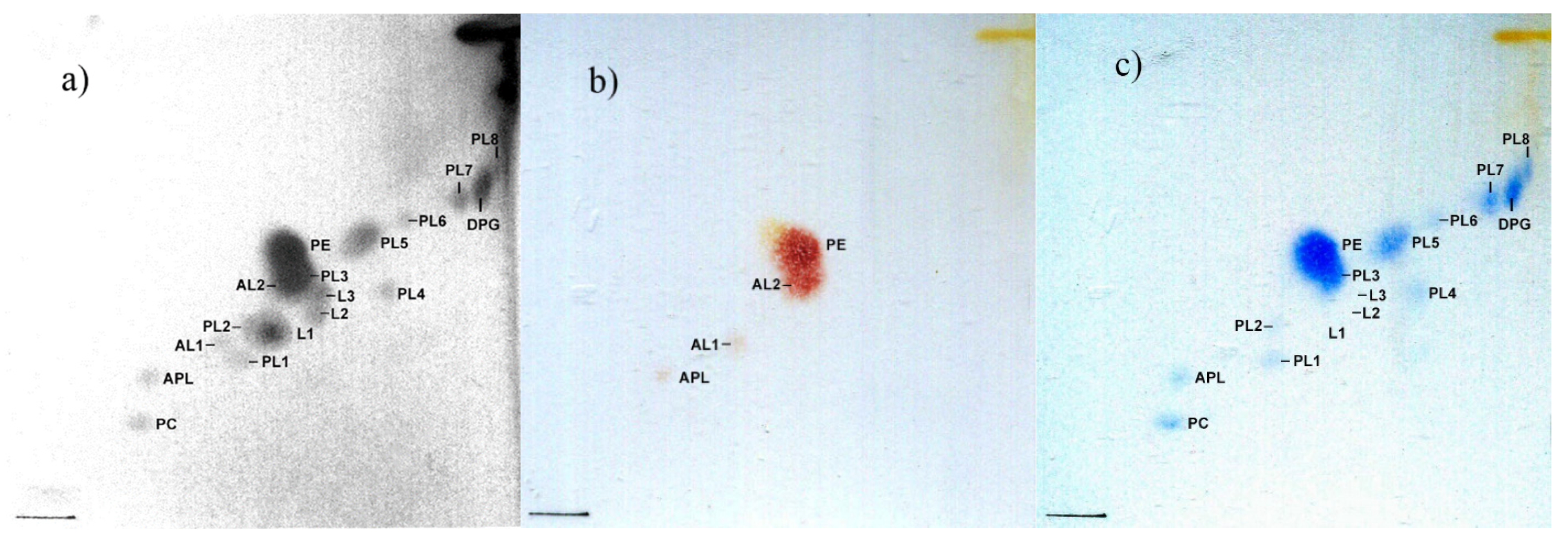
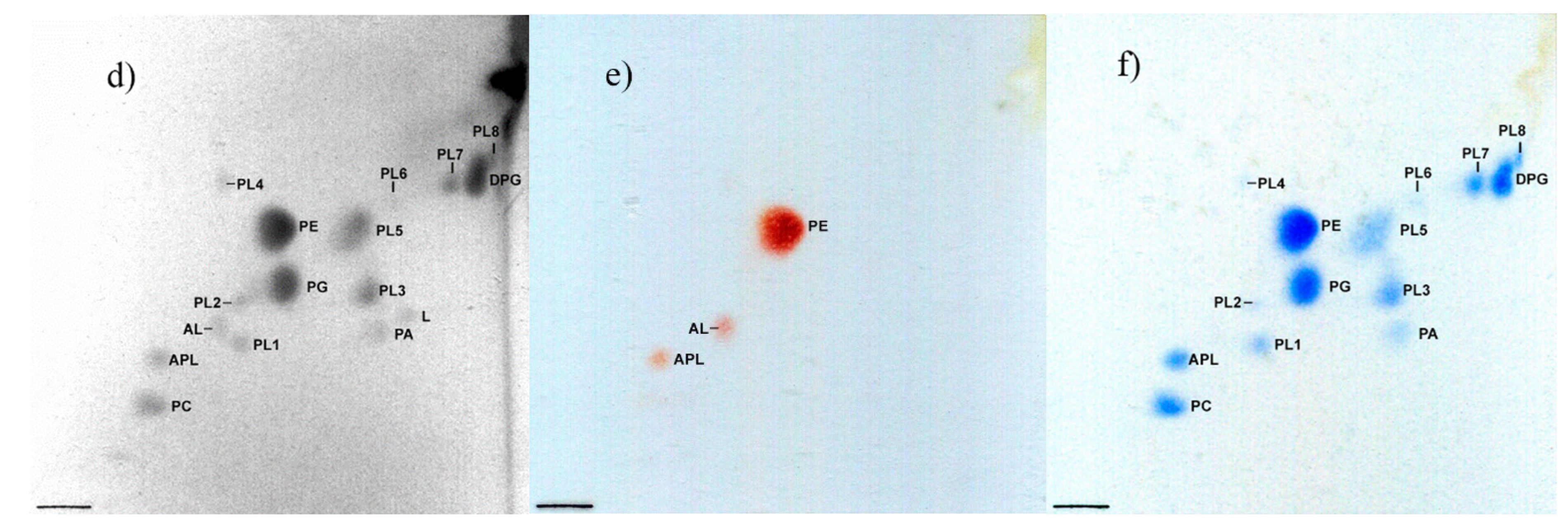
The major fatty acids (>10%) of KMM 9957T were C18:1, C10:0 3-OH, C16:0, and C16:1ω7c, which were similar to the case for M. aestuarii KCTC 52193T. Compared to M. profundum PAMC 27536T, and M. rhizophilum DSM 18822T, strain KMM 9957T and M. aestuarii KCTC 52193T each contained a higher proportion of C14:0 and C10:0 3-OH and a lower content of C16:1ω7c (Table 2). Moreover, KMM 9957T contained certain proportions of C18:1ω9c, C20:4ω6, C20:5ω3, and C20:1, which occurred in a trace amount or absent in M. aestuarii KCTC 52193T and absent in other related type strains. The detailed fatty acid composition of strain KMM 9957T and related type strains are shown in Table 2. The dominant respiratory quinone was ubiquinone Q-8. Polar lipids of strain KMM 9957T consisted of phosphatidylethanolamine, diphosphatidylglycerol, phosphatidylcholine, an unidentified aminophospholipid, two unidentified aminolipids, eight unidentified phospholipids, and three unidentified lipids (Figure 4).
It is evident from the results obtained that novel isolate KMM 9957T can be assigned to the genus Marinobacterium on the basis of its physiological, biochemical, and chemotaxonomic characteristics. Strain KMM 9957T could be distinguished from related Marinobacterium species in being to form polar and lateral flagella, whereas the most related species are characterized by having a single polar flagellum, in being able to grow at 35 °C (excepting M. aestuarii KCTC 52193T), in being able to hydrolyze DNA, and in not being able to assimilate most compounds which are included to the API 20 NE and API 32GN strips, as well as in enzymes activities in API ZYM tests. Differential phenotypic characteristics are listed in Table 1. Based on the combination of phylogenetic and phenotypic characteristics, strain KMM 9957T represents a novel species of the genus Marinobacterium, for which the name Marinobacterium sedimentorum sp. nov. is proposed.

4. Conclusions

Description of Marinobacterium sedimentorum sp. nov.
Marinobacterium sedimentorum (se.di.men.to’rum. L. gen. pl. n. sedimentorum, of sediments, pertaining to source of isolation).
Gram-negative, aerobic, catalase- and oxidase-positive. Cells are rod-shaped at 1.5–2.5 μm long and 0.6–0.8 μm in diameter. Capsular material around cells is produced. Motile by a mean of one or two polar and/or lateral flagella. On MA 2216 produces yellowish shiny colonies with the regular edges of 2–3 mm in diameter. Required NaCl for growth; growth occurs at concentrations of 0.5–5% NaCl (w/v), optimal in 2–3% NaCl. The temperature range for growth is 5–35 °C, with an optimum of 25 °C. The pH range is 5.5–9.5 with an optimum of 6.5–7.5. Positive for hydrolysis of DNA and L-tyrosine. Negative for hydrolysis of casein, gelatin, starch, and Tween 80 in conventional tests.
Positive for citrate utilization and oxidation of L-arabinose; and negative for ONPG, arginine dihydrolase, lysine decarboxylase, ornithine decarboxylase, H2S and urease production under anaerobic conditions, tryptophane deaminase, indole production, acetoin production (Voges-Proskauer reaction), gelatin hydrolysis, and oxidation/fermentation of D-sucrose, D-glucose, D-mannitol, inositol, D-sorbitol, L-rhamnose, D-melibiose, and amygdalin.
Negative for nitrate reduction, esculin and gelatin hydrolysis, PNPG test, indole production, glucose fermentation, arginine dihydrolase, urease, assimilation of D-glucose, D-mannitol, maltose, D-gluconate, L-malate assimilation of D-mannose, L-arabinose, N-acetylglucosamine, caprate, adipate, citrate, and phenylacetate.
Enzymatic activity tests were positive for alkaline phosphatase, naphthol-AS-BI-phosphohydrolase; weakly-positive for leucine arylamidase; and negative for esterase (C 4), esterase lipase (C 8), lipase (C 14), valine arylamidase, cystine arylamidase, acid phosphatase, trypsin, α-chymotrypsin, N-acetyl-β-glucosaminidase, α-galactosidase, β-galactosidase, α-glucosidase, β-glucosidase, β-glucuronidase, α-mannosidase, and α-fucosidase. The dominant respiratory quinone was ubiquinone Q-8. Major fatty acids were C18:1, C10:0 3-OH, C16:0, and C16:1ω7c. The polar lipids consisted of phosphatidylethanolamine, diphosphatidylglycerol, phosphatidylcholine, an unidentified aminophospholipid, two unidentified aminolipids, eight unidentified phospholipids, and three unidentified lipids. The DNA G+C content of 58.4% was calculated from the genome sequence. The type strain of the species is strain KMM 9957T (=KACC 22805T), isolated from a bottom sediment sample, collected from the Okhotsk Sea, Russia.
The annotated draft genome of type strain KMM 9957T comprising 5,489,610 bp is deposited in a public database (GenBank, NCBI) under accession number JALDYX010000000.1.

Author Contributions

Isolation, morphological, and biochemical characterization of strains, L.R. and V.K.; chemotaxonomic characterization, P.V.; Sanger sequencing, S.B.; genome sequencing, phylogenetic, and phylogenomic analyses, N.O. and M.I.; resources, M.I. and V.M.; writing—original draft preparation, L.R. and M.I.; manuscript editing, M.I. and V.M. All authors have read and agreed to the published version of the manuscript.

Funding

This research was funded by a grant from the Ministry of Science and Higher Education, Russian Federation 15.BRK.21.0004 (Contract No. 075-15-2021-1052).

Institutional Review Board Statement

Not applicable.

Data Availability Statement

The 16S rRNA gene sequence (1450 bp) and genome sequence of strain KMM 9957T were deposited in GenBank/EMBL/DDBJ under the accession numbers OL677195 and JALDYX010000000.1, respectively. Strain KMM 9957T was deposited in the Korean Agricultural Culture Collection (KACC) under the number of KACC 22805T.

Conflicts of Interest

The authors declare no conflict of interest.

References

  1. González, J.M.; Mayer, F.; Moran, M.A.; Hodson, R.E.; Whitman, W.B. Microbulbifer hydrolyticus gen. nov., sp. nov., and Marinobacterium georgiense gen. nov., sp. nov., two marine bacteria from a lignin-rich pulp mill waste enrichment community. Int. J. Syst. Bacteriol. 1997, 47, 369–376. [Google Scholar] [CrossRef] [Green Version]
  2. Bae, S.S.; Jung, J.; Chung, D.; Baek, K. Marinobacterium aestuarii sp. nov., a benzene-degrading marine bacterium isolated from estuary sediment. Int. J. Syst. Evol. Microbiol. 2018, 68, 651–656. [Google Scholar] [CrossRef]
  3. Kang, J.Y.; Kim, M.J.; Chun, J.; Son, K.P.; Jahng, K.Y. Marinobacterium boryeongense sp. nov., isolated from seawater. Int. J. Syst. Evol. Microbiol. 2019, 69, 493–497. [Google Scholar] [CrossRef]
  4. Durán-Viseras, A.; Castro, D.J.; Reina, J.C.; Béjar, V.; Martínez-Checa, F. Taxogenomic and metabolic insights into Marinobacterium ramblicola sp. nov., a new slightly halophilic bacterium isolated from Rambla Salada, Murcia. Microorganisms. 2021, 9, 1654. [Google Scholar] [CrossRef]
  5. Han, S.B.; Wang, R.J.; Yu, X.Y.; Su, Y.; Sun, C.; Fu, G.Y.; Zhang, C.Y.; Zhu, X.F.; Wu, M. Marinobacterium zhoushanense sp. nov., isolated from surface seawater. Int. J. Syst. Evol. Microbiol. 2016, 66, 3437–3442. [Google Scholar] [CrossRef]
  6. Chang, H.W.; Nam, Y.D.; Kwon, H.Y.; Park, J.R.; Lee, J.S.; Yoon, J.H.; An, K.G.; Bae, J.W. Marinobacterium halophilum sp. nov., a marine bacterium isolated from the Yellow Sea. Int. J. Syst. Evol. Microbiol. 2007, 57, 77–80. [Google Scholar] [CrossRef] [Green Version]
  7. Kim, S.J.; Park, S.J.; Yoon, D.N.; Park, B.J.; Choi, B.R.; Lee, D.H.; Roh, Y.; Rhee, S.K. Marinobacterium maritimum sp. nov., a marine bacterium isolated from Arctic sediment. Int. J. Syst. Evol. Microbiol. 2009, 59, 3030–3034. [Google Scholar] [CrossRef]
  8. Huo, Y.Y.; Xu, X.W.; Cao, Y.; Wang, C.S.; Zhu, X.F.; Oren, A.; Wu, M. Marinobacterium nitratireducens sp. nov. and Marinobacterium sediminicola sp. nov., isolated from marine sediment. Int. J. Syst. Evol. Microbiol. 2009, 59, 1173–1178. [Google Scholar] [CrossRef] [Green Version]
  9. Kim, J.M.; Lee, S.H.; Jung, J.Y.; Jeon, C.O. Marinobacterium lutimaris sp. nov., isolated from a tidal flat. Int. J. Syst. Evol. Microbiol. 2010, 60, 1828–1831. [Google Scholar] [CrossRef] [Green Version]
  10. Hwang, C.Y.; Yoon, S.J.; Lee, I.; Baek, K.; Lee, Y.M.; Yoo, K.C.; Yoon, H.I.; Lee, H.K. Marinobacterium profundum sp. nov., a marine bacterium from deep-sea sediment. Int. J. Syst. Evol. Microbiol. 2016, 66, 1561–1566. [Google Scholar] [CrossRef]
  11. Park, S.; Jung, Y.T.; Kim, S.; Yoon, J.H. Marinobacterium aestuariivivens sp. nov., isolated from a tidal flat. Int. J. Syst. Evol. Microbiol. 2016, 66, 1718–1723. [Google Scholar] [CrossRef]
  12. Chimetto, L.A.; Cleenwerck, I.; Brocchi, M.; Willems, A.; De Vos, P.; Thompson, F.L. Marinobacterium coralli sp. nov., isolated from mucus of coral (Mussismilia hispida). Int. J. Syst. Evol. Microbiol. 2011, 61, 60–64. [Google Scholar] [CrossRef]
  13. Kim, Y.G.; Jin, Y.A.; Hwang, C.Y.; Cho, B.C. Marinobacterium rhizophilum sp. nov., isolated the rhizosphere of the coastal tidal-flat plant Suaeda japonica. Int. J. Syst. Evol. Microbiol. 2008, 58, 164–167. [Google Scholar] [CrossRef] [Green Version]
  14. Alfaro-Espinoza, G.; Ullrich, M.S. Marinobacterium mangrovicola sp. nov., a marine nitrogen-fixing bacterium isolated from mangrove roots of Rhizophora mangle. Int. J. Syst. Evol. Microbiol. 2014, 64, 3988–3993. [Google Scholar] [CrossRef] [Green Version]
  15. Gerhardt, P.; Murray, R.G.E.; Wood, W.A.; Krieg, N.R. Methods for General and Molecular Bacteriology; American Society for Microbiology: Washington, DC, USA, 1994. [Google Scholar]
  16. Romanenko, L.A.; Tanaka, N.; Svetashev, V.I.; Falsen, E. Description of Cobetia amphilecti sp. nov., Cobetia litoralis sp. nov. and Cobetia pacifica sp. nov., classification of Halomonas halodurans as a later heterotypic synonym of Cobetia marina and emended descriptions of the genus Cobetia and Cobetia marina. Int. J. Syst. Evol. Microbiol. 2013, 63, 288–297. [Google Scholar] [CrossRef]
  17. Folch, J.; Lees, M.; Sloane Stanley, G. A simple method of isolation and purification of total lipids from animal tissues. J. Biol. Chem. 1957, 226, 497–509. [Google Scholar] [CrossRef]
  18. Collins, M.D.; Shah, H.N. Fatty acid, menaquinone and polar lipid composition of Rothia dentocariosa. Arch. Microbiol. 1984, 137, 247–249. [Google Scholar] [CrossRef]
  19. Collins, M.D.; Goodfellow, M.; Minnikin, D.E. Fatty acid, isoprenoid quinone and polar lipid composition in the classification of Curtobacterium and related taxa. J. Gen. Microbiol. 1980, 118, 29–37. [Google Scholar] [CrossRef] [Green Version]
  20. Mitchell, K.; Fallon, R.J. The determination of ubiquinone profiles by reversed-phase high-performance thin-layer chromatography as an aid to the speciation of Legionellaceae. J. Gen. Microbiol. 1990, 136, 2035–2041. [Google Scholar] [CrossRef] [Green Version]
  21. Sasser, M. Identification of Bacteria by Gas Chromatography of Cellular Fatty Acids; MIDI Technical Note 101; MIDI Inc.: Newark, DE, USA, 1990. [Google Scholar]
  22. Romanenko, L.A.; Kurilenko, V.V.; Guzev, K.V.; Svetashev, V.I. Characterization of Labrenzia polysiphoniae sp. nov. isolated from red alga Polysiphonia sp. Arch. Microbiol. 2019, 201, 705–712. [Google Scholar] [CrossRef]
  23. Yoon, S.H.; Ha, S.M.; Kwon, S.; Lim, J.; Kim, Y.; Seo, H.; Chun, J. Introducing EzBioCloud: A taxonomically united database of 16S rRNA gene sequences and whole-genome assemblies. Int. J. Syst. Evol. Microbiol. 2017, 67, 1613. [Google Scholar] [CrossRef]
  24. Kumar, S.; Stecher, G.; Li, M.; Knyaz, C.; Tamura, K. MEGA X: Molecular evolutionary genetics analysis across computing platforms. Mol. Biol. Evol. 2018, 35, 1547–1549. [Google Scholar] [CrossRef]
  25. Kimura, M. A simple method for estimating evolutionary rate of base substitutions through comparative studies of nucleotide sequences. Mol. Biol. Evol. 1980, 16, 111–120. [Google Scholar] [CrossRef]
  26. Bolger, A.M.; Lohse, M.; Usadel, B. Trimmomatic: A flexible trimmer for Illumina sequence data. Bioinformatics. 2014, 30, 2114–2120. [Google Scholar] [CrossRef] [Green Version]
  27. Bankevich, A.; Nurk, S.; Antipov, D.; Gurevich, A.A.; Dvorkin, M.; Kulikov, A.S.; Lesin, V.M.; Nikolenko, S.I.; Pham, S.; Prjibelski, A.D.; et al. SPAdes: A new genome assembly algorithm and its applications to single-cell sequencing. J. Comput. Biol. 2012, 19, 455–477. [Google Scholar] [CrossRef] [Green Version]
  28. Gurevich, A.; Saveliev, V.; Vyahhi, N.; Tesler, G. QUAST: Quality assessment tool for genome assemblies. Bioinformatics. 2013, 29, 1072–1075. [Google Scholar] [CrossRef] [Green Version]
  29. Parks, D.H.; Imelfort, M.; Skennerton, C.T.; Hugenholtz, P.; Tyson, G.W. CheckM: Assessing the quality of microbial genomes recovered from isolates, single cells, and metagenomes. Genome Res. 2015, 25, 1043–1055. [Google Scholar] [CrossRef] [Green Version]
  30. Tatusova, T.; DiCuccio, M.; Badretdin, A.; Chetvernin, V.; Nawrocki, E.P.; Zaslavsky, L.; Lomsadze, A.; Pruitt, K.D.; Borodovsky, M.; Ostell, J. NCBI prokaryotic genome annotation pipeline. Nucleic Acids Res. 2016, 44, 6614–6624. [Google Scholar] [CrossRef]
  31. Aziz, R.K.; Bartels, D.; Best, A.A.; DeJongh, M.; Disz, T.; Edwards, R.A.; Formsma, K.; Gerdes, S.; Glass, E.M.; Kubal, M.; et al. The RAST Server: Rapid annotations using subsystems technology. BMC Genom. 2008, 9, 75. [Google Scholar] [CrossRef] [Green Version]
  32. Wattam, A.R.; Abraham, D.; Dalay, O.; Disz, T.L.; Driscoll, T.; Gabbard, J.L.; Gillespie, J.J.; Gough, R.; Hix, D.; Kenyon, R.; et al. PATRIC, the bacterial bioinformatics database and analysis resource. Nucleic Acids Res. 2014, 42, D581–D591. [Google Scholar] [CrossRef]
  33. Rodriguez-R, L.M.; Konstantinidis, K.T. The enveomics collection: A toolbox for specialized analyses of microbial genomes and metagenomes. PeerJ Prepr. 2016, 4, e1900v1. [Google Scholar] [CrossRef]
  34. Meier-Kolthoff, J.P.; Göker, M. TYGS is an automated high-throughput platform for state-of-the-art genome-based taxonomy. Nat. Commun. 2019, 10, 2182. [Google Scholar] [CrossRef] [Green Version]
  35. Asnicar, F.; Thomas, A.M.; Beghini, F.; Mengoni, C.; Manara, S.; Manghi, P.; Zhu, Q.; Bolzan, M.; Cumbo, F.; May, U.; et al. Precise phylogenetic analysis of microbial isolates and genomes from meta-genomes using PhyloPhlAn 3.0. Nat. Commun. 2020, 11, 2500. [Google Scholar] [CrossRef]
  36. Buchholz, P.C.F.; Feuerriegel, G.; Zhang, H.; Perez-Garcia, P.; Nover, L.L.; Chow, J.; Streit, W.R.; Pleiss, J. Plastics degradation by hydrolytic enzymes: The plastics-active enzymes database—PAZy. Proteins: Struct. Funct. Genet. 2022, 90, 1443–1456. [Google Scholar] [CrossRef]
  37. Chun, J.; Oren, A.; Ventosa, A.; Christensen, H.; Arahal, D.R.; da Costa, M.S.; Rooney, A.P.; Yi, H.; Xu, X.W.; De Meyer, S.; et al. Proposed minimal standards for the use of genome data for the taxonomy of prokaryotes. Int. J. Syst. Evol. Microbiol. 2018, 68, 461–466. [Google Scholar] [CrossRef]
  38. Richter, M.; Rosselló-Móra, R. Shifting the genomic gold standard for the prokaryotic species definition. Proc. Natl. Acad. Sci. USA 2009, 106, 19126–19131. [Google Scholar] [CrossRef] [Green Version]
  39. Konstantinidis, K.T.; Rosselló-Móra, R.; Amann, R. Uncultivated microbes in need of their own taxonomy. ISME J. 2017, 11, 2399–2406. [Google Scholar] [CrossRef]
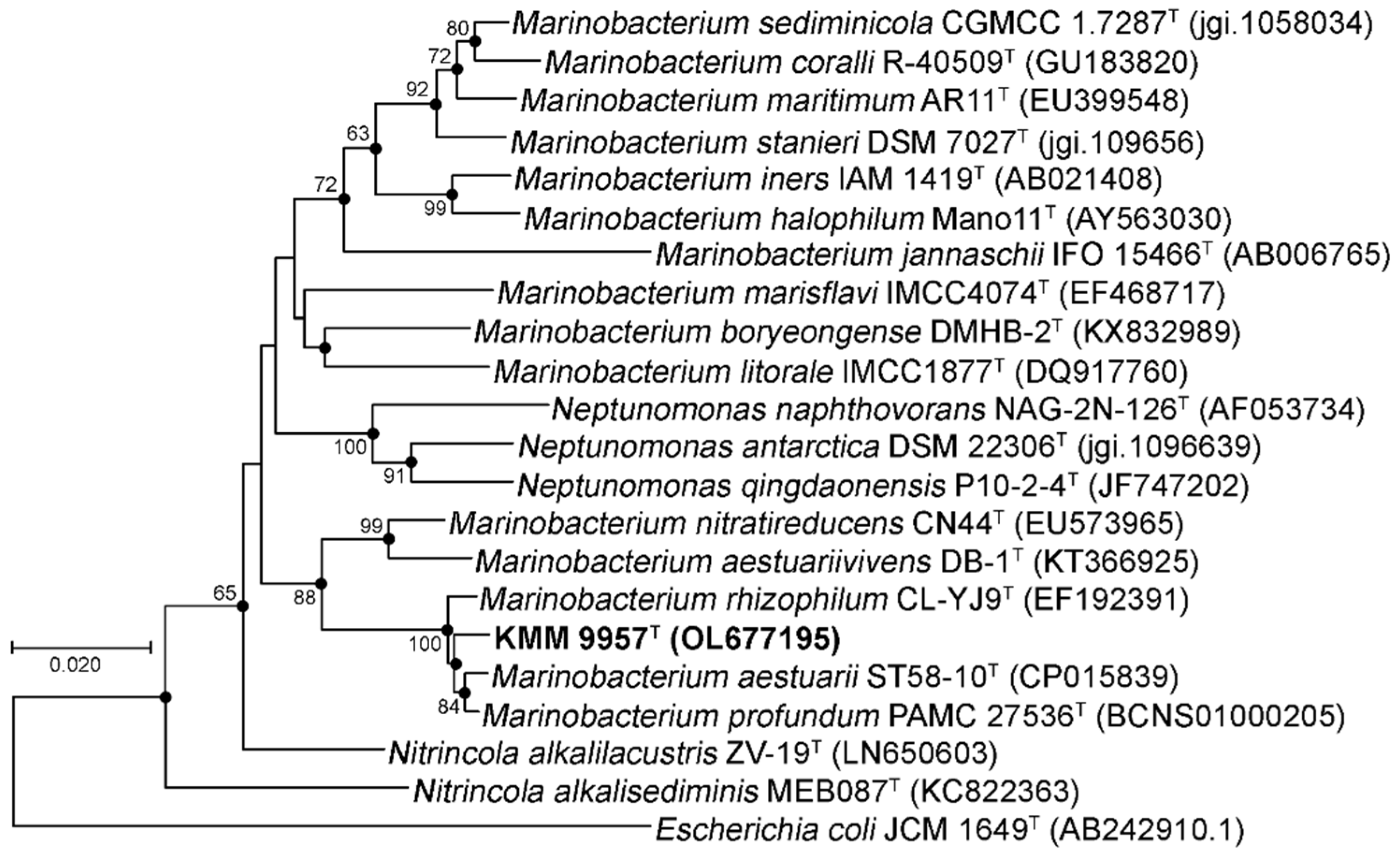
Figure 1. Neighbor-joining tree based on 16S rRNA gene sequences available from the GenBank/EMBL/DDBJ databases (accession numbers are given in parentheses) showing relationships of the novel strain KMM 9957T and Marinobacterium species. Filled circles indicate the corresponding nodes that were observed in the maximum-likelihood tree. Numbers indicate bootstrap values as percentage greater than 60. These values are based on 1000 replicates. Bar, 0.020 substitutions per nucleotide position.
Diversity 14 00944 g001
Figure 2. Maximum-likelihood tree based on concatenated 400 protein sequences from genome sequences showing phylogenetic position of the novel strain KMM 9957T among Marinobacterium species and related taxa. Bootstrap values are based on 100 replicates. Oceanospirillum linum ATCC 11336T was used as an outgroup. Bar, 0.05 substitutions per amino acid position.
Diversity 14 00944 g002
Figure 3. Scanning transmission electron microscope images of negatively stained cells of strain KMM 9957T grown on MA 2216 at 25 °C for 24 h. Bar, 1.0 µm (a) and 0.2 µm (b).
Diversity 14 00944 g003
Figure 4. Two-dimensional thin-layer chromatograms of polar lipids of strains: (ac) KMM 9957T; (df) Marinobacterium aestuarii KCTCT. (a,d), non-specific detection of lipids prepared with 10% H2SO4 in methanol; (b,e), stained with ninhydrin; (c,f), stained with molybdate reagent. Abbreviations: PC, phosphatidylcholine; PG, phosphatidylglycerol; PE, phosphatidylethanolamine; DPG, diphosphatidylglycerol; APL, an unidentified aminophospholipid; PA, phosphatidic acid; PL1-PL8, unidentified phospholipids; AL, AL1, AL2, unidentified aminolipids; L, L1, L2, L3, unidentified lipids.
Diversity 14 00944 g004aDiversity 14 00944 g004b
Table 1. Differential characteristics of strain KMM 9957T and the type strains of most closely related Marinobacterium species.
Characteristics1234
DNA G+C content (%) *58.458.857.258.5
Growth in NaCl (%)0.5–50.5–71–41–5
Growth at 35 °C++
Hydrolysis of:
DNA+NDND
Tween-80++
Enzyme activity (API ZYM):
Esterase C4 ++
Esterase lipase C8+(+)
Valine arylamidase+
Acid phosphatase++
α-glucosidase+
Strains: 1, KMM 9957T; 2, Marinobacterium aestuarii KCTC 52193T (data were obtained from the present study unless otherwise indicated); 3, Marinobacterium profundum PAMC 27536T (data from [10]); 4, Marinobacterium rhizophilum DSM 18822T (data from [13]). +, Positive; −, negative; ND, not determined; (+), weak reaction. All strains were motile and positive for oxidase, catalase, sodium ions requirement, alkaline phosphatase, and naphthol-AS-BI-phosphohydrolase, and negative for nitrate reduction, hydrolysis of gelatine, esculin, casein, starch, lipase C 14, cystine arylamidase, trypsin, α-chymotrypsin, α-galactosidase, β-galactosidase, β-glucuronidase, β-glucosidase N-acetyl-β-glucosaminidase, α-mannosidase, and α-fucosidase. * The DNA G+C contents of strain KMM 9957T, M. aestuarii KCTC 52193T, M. profundum PAMC 27536T, and M. rhizophilum DSM 18822T were derived from the QUAST and GenBank.
Table 2. Cellular fatty acid composition (%) of strain Marinobacterium sedimentorum sp. nov. KMM 9957T and related type strains of the genus Marinobacterium.
Fatty Acid1234
C10:01.43.84.83.9
C10:0 3-OH14.816.17.77.1
C12:02.02.83.94.0
C14:06.68.1Tr-
C16:1ω7c14.119.442.2 a40.3 b
C16:014.717.419.116.6
C18:1ω9c5.00.6--
C18:1ω7c12.119.920.9 c26.6
C18:02.91.1--
C20:4ω61.7---
C20:5ω35.8---
C20:12.00.4--
Strains: 1, KMM 9957T; 2, Marinobacterium aestuarii KCTC 52193T (data were obtained from present study); 3, Marinobacterium profundum PAMC 27536T (data from [10]); 4, Marinobacterium rhizophilum DSM 18822T (data from [13]). Fatty acids representing < 1% in all strains tested are not shown; -, not detected; Tr, trace amounts. a Summed feature (C16:1ω6c and/or C16:1ω7c); b Summed feature (C16:1ω7c and/or iso-C15:0 2-OH); c Summed feature (C18:1ω6c and/or C18:1ω7c).
Publisher’s Note: MDPI stays neutral with regard to jurisdictional claims in published maps and institutional affiliations.