Article

Bloom of Prorocentrum cordatum in Paracas Bay, Peru

1
Banco de Germoplasma de Organismos Acuáticos, Instituto del Mar del Perú (IMARPE), Esquina Gamarra y Gral. Valle s/n, Callao 07021, Peru
2
Departamento de Acuicultura, Facultad de Ciencias del Mar, Universidad Católica del Norte, Larrondo 1281, Coquimbo 1780000, Chile
3
Programa Cooperativo Doctorado en Acuicultura, Universidad Católica del Norte, Larrondo 1281, Coquimbo 1780000, Chile
4
Centro de Investigación y Desarrollo Tecnológico en Algas (CIDTA), Facultad de Ciencias del Mar, Universidad Católica del Norte, Larrondo 1281, Coquimbo 1780000, Chile
5
Facultad de Ciencias, Universidad Nacional Agraria La Molina, Avenida La Universidad s/n, La Molina, Lima 15024, Peru
6
Bluewater Consulting Company, Castro 5700000, Chile
7
Dirección General de Investigaciones en Hidroacústica, Sensoramiento Remoto y Artes de Pesca, Instituto del Mar del Perú (IMARPE), Esquina Gamarra y Gral. Valle s/n, Callao 07021, Peru
8
Centro de Investigacións Mariñas (Xunta de Galicia), Pedras de Corón s/n, 36620 Vilanova de Arousa, Pontevedra, Spain
*
Authors to whom correspondence should be addressed.
Diversity 2022, 14(10), 844; https://doi.org/10.3390/d14100844
Submission received: 31 August 2022 / Revised: 24 September 2022 / Accepted: 30 September 2022 / Published: 6 October 2022
(This article belongs to the Section Marine Diversity)

Abstract

:
During the austral winter of 2017, a bloom of Prorocentrum spp. occurred, reaching a cell density of 2.73 × 10⁶ cells L−1, in Paracas Bay, Peru. In order to identify which, type of species generated this event and determine its toxicity, the values of the environmental parameters (temperature, winds and salinity) that induced the rapid growth of the dinoflagellate in this bloom were identified. A clonal culture was established for taxonomic (SEM), phylogenetic (ITS) and toxicological analysis via LC-MS/MS to determine the presence of tetrodotoxin (TTX) and whether the species represents a food safety hazard. This event coincided with the coastal upwelling process, which generated high concentrations of phytoplankton biomass (>10 mg m−3 chlorophyll-a) and allowed the rapid growth of P. cordatum (IMP-BG 450) in Paracas Bay. However, toxicological analyses of the IMP-BG 450 strain culture did not show the presence of TTX quantifiable through the technique used. Due to the antecedents of the presence of TTX in mollusks from other latitudes during blooms of this species, it is recommended that analyses of this toxin be carried out both in filter-feeding mollusks and in this species during a new bloom.

1. Introduction

The increase in nutrients during recent years in oceanic, continental and estuarial waters due to human activity has favored more intense and frequent harmful algal blooms (HABs) [1]. Together with the effects of climate change and maritime transport, many species have increased their geographic dispersion, leading to a variety of harmful impacts on natural aquatic ecosystems’ populations [1,2,3,4,5,6,7,8,9]. The rise of nutrients in the coastal waters of Peru due to intense upwelling processes [10,11] has increased diatom growth. These blooms have been strongly correlated with the incorporation of NO3 [4,12,13,14,15]. Highly productive aquatic environments are beneficial for invasive species such as those of the genus Prorocentrum, with notable increases in their frequency, duration and magnitude in tropical, subtropical and temperate climates alike [16,17,18].
Prorocentrum cordatum (Ostenfeld) J. D. Dodge, 1975 (syn. Prorocentrum minimum (Pavillard) Schiller) [19,20,21]—taxonomically accepted name according to the World Register of Marine Species (WoRMS) [20,22]—presents a global distribution associated with eutrophication, and particularly with nitrogen enrichment [16,18,23], being found near wastewater areas and fish and shrimp farming tanks: all rich in nutrients [4,23,24]. This dinoflagellate has adaptive ecological strategies, such as high intrapopulation variability in the face of environmental stress (eutrophication or pollution) [25,26] and changing its feeding from autotrophy to mixotrophy (phagotrophy) under conditions of low availability of inorganic nutrients, as in the case of the 15 blooms over three decades in the coastal waters of the Baltic Sea where most were of mixotrophic P. cordatum [27]. These strategies for its development and maintenance allow this species to be a potential invader and conquer a variety of ecological niches, including coastal ecosystems saturated with species with high anthropogenic nutrient loads [22,28].
P. cordatum blooms pose a risk to aquaculture, human health and ecosystems due to their possible link with the presence of tetrodotoxin (TTX) in shellfish [29,30,31,32]. The neurotoxin TTX is highly potent, as it can block sodium channels and thereby inhibit the propagation of action potential in muscle and nerve cells [33,34]. In Greece in 2012, TTX was identified in mussels with the presence of P. minimum (syn. = P. cordatum) [29], and Rodriguez, et al. [35] found TTX analogs in certain strains of P. minimum in symbiosis with bacteria (Roseobacter and Vibrio sp).
P. cordatum has been reported on the Peruvian coast but always at a low density (<500 × 103 cells L−1) [36,37,38,39,40]. However, between 29 August and 1 September 2017, the HAB monitoring program carried out by the National Fisheries Health Organization (SANIPES) detected a P. cordatum bloom in Paracas Bay with a density of 2.73 × 10⁶ cells L1 (https://www.sanipes.gob.pe/web/index.php/es/fitoplancton, accessed on 1 July 2022).
Paracas Bay is a highly productive area in which intense cultivation activity of Argopecten purpuratus, and therefore a bloom of a potentially toxic species such as P. cordatum can have important consequences for human health and economic activity. The aim of this study was to unequivocally identify the species by morphological and molecular characterization, and to identify the possible presence of TTX, as well as to know if the species represent a food safety hazard.

2. Materials and Methods

2.1. Environmental Parameters

The data analyzed in this case were temperature (°C), salinity, dissolved oxygen (mg L−1), oxygen saturation (%) and phytoplankton density by species (cells L−1) from the database of the HAB Monitoring Program in Paracas Bay run by SANIPES from August to September 2017 (http://desarrollo.sanipes.gob.pe/monitor/grid_monitoreo_todas/, accessed on 1 July 2022).

2.1.1. Satellite Imagery of Chlorophyll-a

Satellite images of surface chlorophyll-a concentration were used with 4 km of spatial resolution and level L3 from the timespan of 27 to 29 August 2017, coming from the VIIRS sensor (Visible Infrared Imaging Radiometer Suite) on board the Suomi-NPP satellite (Suomi National Polar-Orbiting Partnership). This information was obtained from the OceanColor web portal (https://oceancolor.gsfc.nasa.gov/, accessed on 28 September 2021 ) operated by NASA (National Aeronautics and Space Administration).

2.1.2. Satellite Wind Imagery

Satellite information about surface wind and speed was used with 25 km of spatial resolution from the following dates: 25 August, 30 August and 1 September 2017. These data comes from the analyses of different satellite wind information sources, published on https://www.ncei.noaa.gov/products/blended-sea-winds, (accessed on 28 September 2021) by the National Centers for Environmental Information at NOAA (National Oceanic and Atmospheric Administration).

2.1.3. Satellite Sea Surface Temperature

For the SST analysis, daily images of High-Resolution Sea Surface Temperature (GHRSST) Level 4 were used. These data are produced daily on an operational basis by the JPL OurOcean group using a multi-scale two-dimensional variational (MS-2DVAR) blending algorithm on a global 1 km grid. We selected images of the days 25 and 30 August and 1 September 2017 because they were free of clouds.

2.2. Strain Obtention and Culture Conditions

Phytoplankton samples were gathered in August 2017 from Ica in Paracas Bay (13°49′ S, 76°17′ W) (Figure 1) via vertical dragging with a conical net (10 μm). The obtained samples were subsequently transported to the IMARPE laboratory in 250 mL polycarbonate bottles, chilled in ice (10 °C) and protected from sunlight. in order to obtain a monoclonal culture.
P. cordatum cells were isolated under an inverted phase contrast microscope (NIKON, eclipse TS100, Tokyo, Japan), with the technique of cell washing with micropipettes [41]. The isolated cells were transferred to multiwell culture plates with 2 mL of L1-Si culture medium at a salinity of 32, which were incubated at 15 °C under a photosynthetically active radiation (PAR) of 40 µmol photons m−2 s−1 and a 12:12 h (light:darkness) photoperiod. The cultures established were transferred to borosilicate Erlenmeyer flasks with 50 mL of L1-Si medium and grown at 15 °C with a photonic flow of 80 μmol of photons m−2 s−1 and a 12:12 h photoperiod (light:darkness). Additional cultures were grown in 1 L borosilicate flasks with an L1-Si medium in the aforementioned conditions with the aim of characterizing the strain.

2.3. Morphological Description

Morphometric analysis of cells (n = 50) was carried out using an inverted light microscope Zeiss Axio S. A1 with phase contrast, micrography was performed using a Zeiss Axio cam, and cells were analyzed by means of Zen lite software (Zeiss, Oberkochen, Germany).
For scanning electron microscopy (SEM), cells from culture samples in the exponential phase were taken, preserved in 3% glutaraldehyde, washed in distilled water and dehydrated in an ascendant ethyl alcohol concentration series (20, 40, 60, 80, 95 and 100%), followed by critical point drying (Tousimis Samdri-780A, Rockville, MA, USA). Dried cells were mounted on a stub and coated with a layer of gold–palladium (JEOL, Tokyo, Japan). Finally, the samples were observed with a scanning electron microscope (Hitachi SU3500, Tokyo, Japan).

2.4. Molecular and Phylogenetic Analysis

2.4.1. DNA and PCR Extraction

To extract genomic DNA, samples were taken from cultures in the exponential growth phase. Cells were concentrated through centrifugation until a pellet was obtained and stored at −80 °C for at least 1 hour before DNA extraction. The DNA was extracted via the cetyltrimethylammonium bromide method (CTAB) [42]. Genomic DNA quality and quantification were evaluated using a NanoDrop 2000 spectrophotometer (Thermo Fisher Scientific, Waltham, MA, USA).
The ITS region (ITS1-5.8S-ITS2) was amplified via PCR using ITS1/ITS4 primers [43]. The amplification profile was as follows: initial denaturing at 95 °C for 3 min, followed by 39 cycles of 95 °C for 30 s (denaturing), 56 °C for 30 s (annealing), 72 °C for 50 s (extension) and a final extension at 72 °C for 7 min. Amplified products were visualized in agarose gel (1.2%), and were sent to Macrogen Inc. (Seoul, South Korea) to be purified and sequenced for one strand.

2.4.2. Bioinformational Analysis or Phylogenetic Analysis

The sequence obtained was manually edited in BIOEDIT [44] and compared with the public GenBank database via BLASTn to prove its identity. For phylogenetic analyses, 38 sequences were extracted from the GenBank database, including 36 sequences from the genus Prorocentrum and 2 sequences from the genera Takayama and Karenia for the external group (Supplementary Material Table S1). The sequences were aligned using the MUSCLE algorithm (Edgar, 2004) in MEGA 7 v.7.0.26 [45], the alignment was visually corrected and the distances were calculated using the uncorrected p-distance model. The final alignment was independently analyzed via neighbor-joining (NJ), maximum likelihood (ML) and Bayesian inference (BI).
The NJ analysis was performed in MEGA 7 using the Kimura-2-parameters model (K2P) with 1000 bootstraps. The best evolutionary model for the phylogenetic analysis (ML and BI) was calculated in jModelTest 2 [46] using the Akaike information criterion (AIC) for ML and the Bayesian information criterion (BIC) for BI. The ML analysis was carried out in RAxML software (v.8.2.X) [47] using the raxmlGUI v.1.5 graphic user interface [48] with the GTR + G model and 1000 repetitions. The BI was conducted in MrBayes v.3.2.6 [49] with the HKY + G model and two parallel analyses, each one with four chains (three hot chains and one cold chain) of 10,000,000 Markov chain Monte Carlo simulations sampled every 1000 generations. The executions’ convergence was proven using Tracer v.1.6.0 [50]. The consensus tree was built after a 25% burn-in and subsequent probabilities were estimated.

2.5. Toxin Analysis

The P. cordatum culture pellet was obtained based on a culture volume of 500 mL by centrifuging at 3100× g for 5 minutes using an Eppendorf 810R centrifuge (Hamburg, Germany).
Sample analyses were performed with LC-MS/MS using an Exion AD system (SCIEX, Framingham, MA, USA) coupled with a QTRAP 6500+ triple quadrupole mass spectrometer equipped with an Iondrive TurboV electrospray interfase (SCIEX, Framingham, MA, USA). Positive ionization (5500 V), 650 °C of source temperature, 75 units of GS1 and GS1, 30 units of curtain gas and “medium” collision gas were used. Two MS/MS ionic transitions were monitored for each compound (Table 1).
Chromatographic separation was conducted following the method of Boundy, et al. [51]: development of a sensitive and selective method for LC-MS/MS for analyzing the high performance of paralyzing shellfish toxins using solid-phase extraction of graphite carbon.

3. Results

3.1. Environmental Parameters

The P. cordatum bloom in Paracas Bay during winter 2017 had a density of 1.11 × 10³ cells L−1 on 25 August, rising to 2.73 × 10⁶ cells L−1 on 1 September. Similarly, rises were recorded in water surface temperature (>19 °C) and dissolved oxygen saturation (119%). After 5 September, there was a decrease in the cell density of this dinoflagellate (<100 × 103 cells L−1), along with water surface temperature (16 °C) and dissolved oxygen saturation (11%) (Figure 2a,b).

3.1.1. Satellite Chlorophyll-a

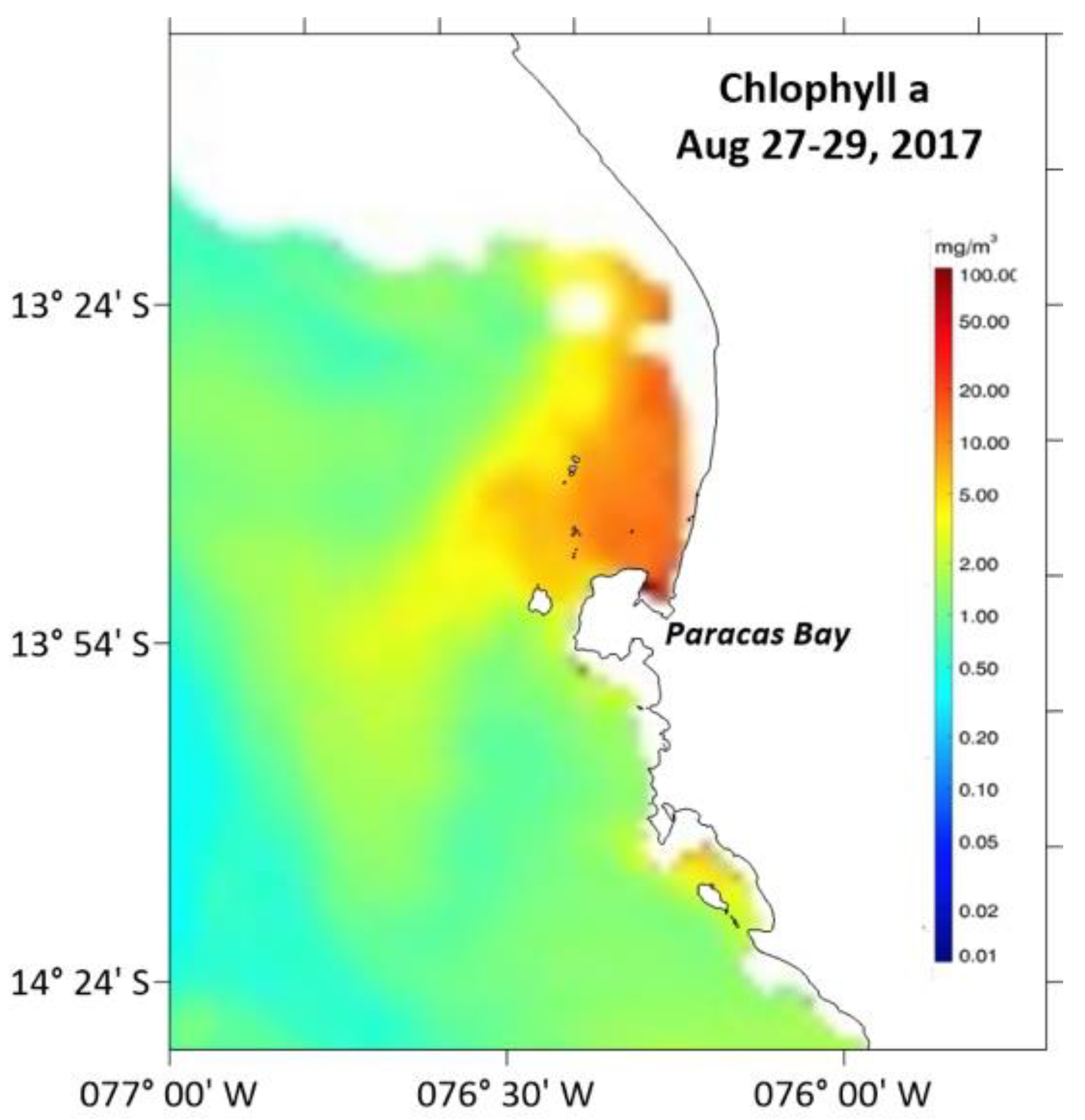
During the P. cordatum bloom, the integrated satellite image of chlorophyll-a from 27 to 29 August 2017 shows high concentrations of this photosynthetic pigment (>10 mg m−3) in Paracas Bay, extending for around 13 km northwards (Figure 3).

3.1.2. Satellite Winds

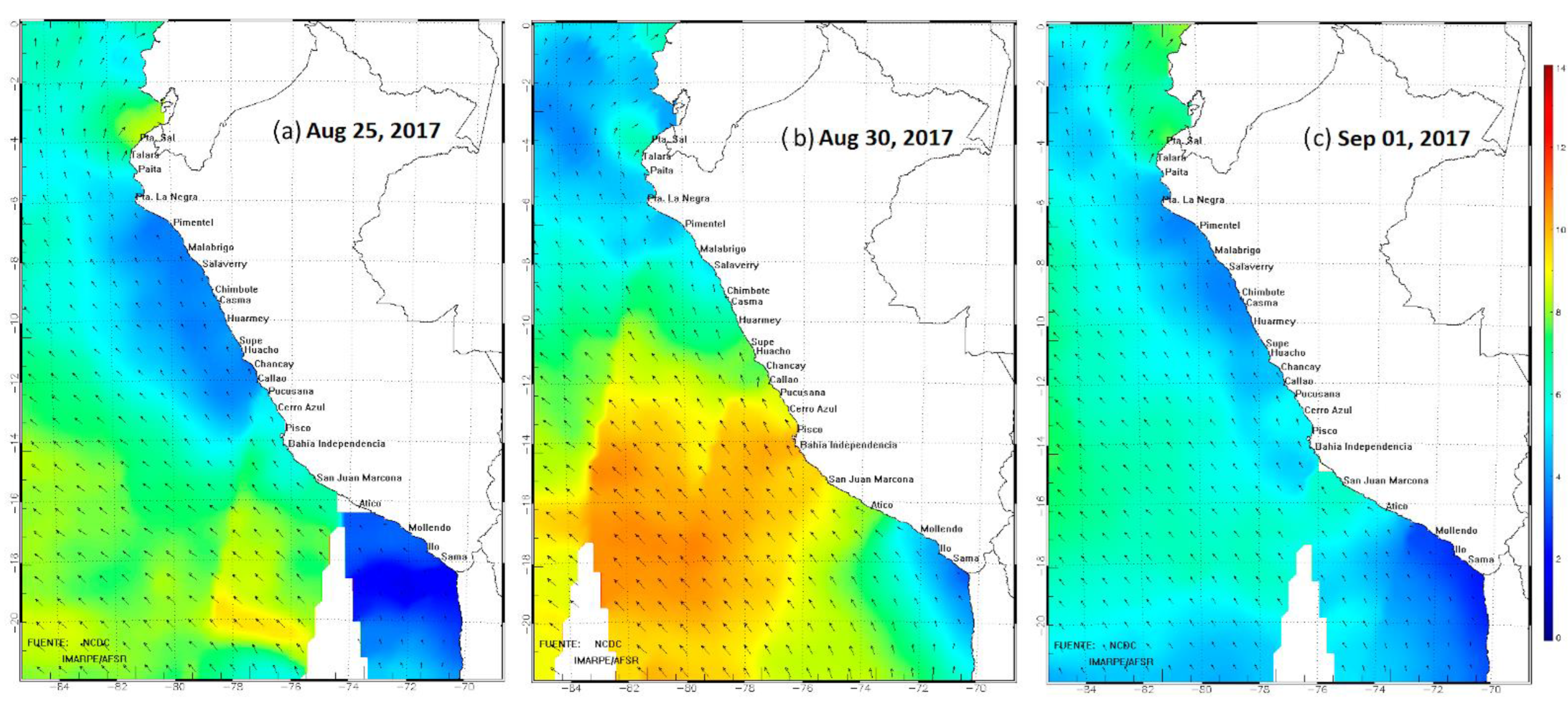
Coastal winds next to Paracas Bay came from the south and increased their intensity from 25 August (6 m s−1) to values above 10 m s−1 by 30 August, before falling to 5 m s−1 by 1 September (Figure 4a–c).

3.1.3. Sea Surface Temperature (SST)

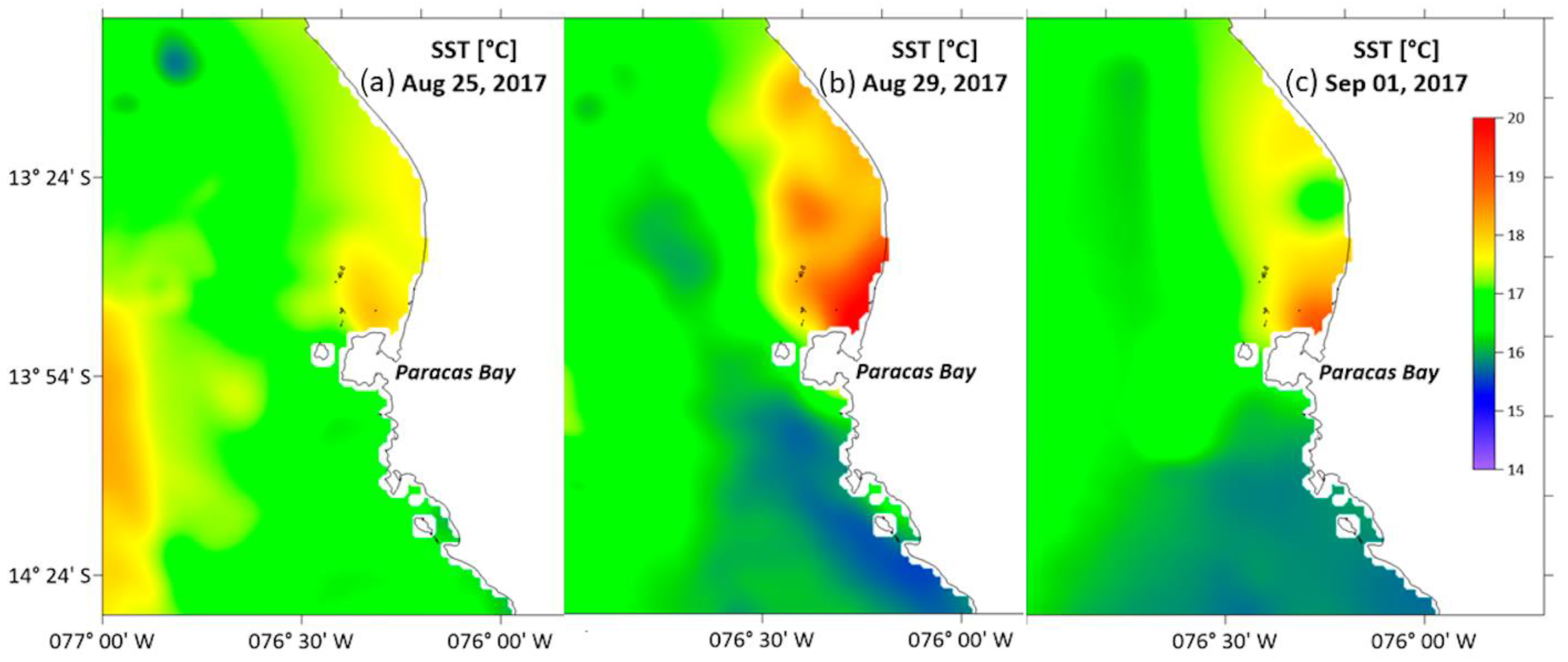
Surface temperatures in the southern exterior of Paracas Bay on 25 August were around 16.5 °C, presenting colder waters (<16 °C) until 1 September. By contrast, surface temperatures in Paracas Bay and the northern sector were 17.5 °C on 25 August, which rose to 19.5 °C on 29 August and 1 September (Figure 5a–c).

3.2. Morphology

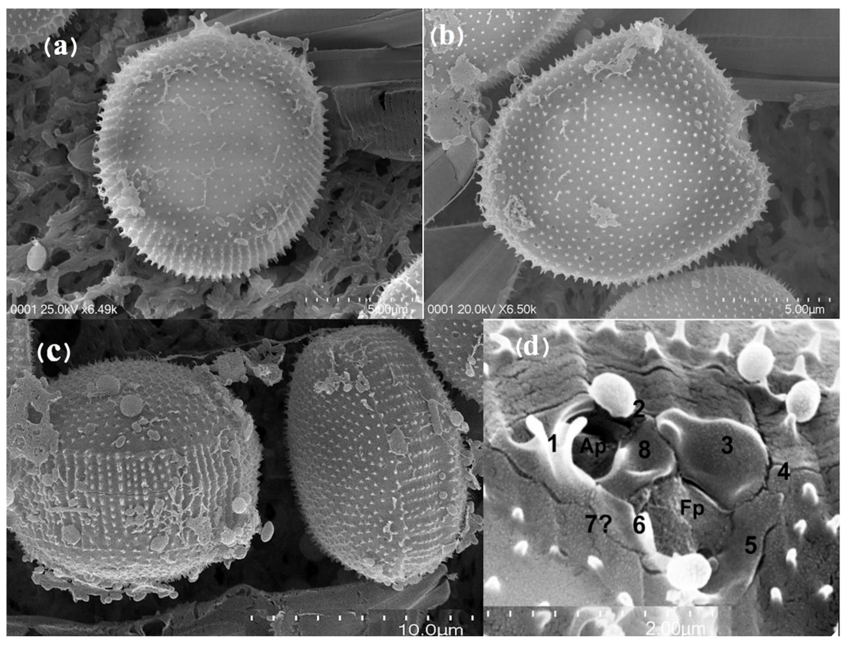
The strain isolated from Paracas Bay was coded and morphologically identified as Prorocentrum cordatum (Ostenfeld) J. D. Dodge, 1975. The cells of the strain IMP-BG 450 are between 13.8 and 17.2 µm long and 12.7–15.6 µm wide. The cells have round-oval shapes and a high frequency of pentagonal forms (Figure 6a,b), the posterior extreme of the valves is rounded to angled, and the anterior end is straight with a V-shaped central depression and an apical spine. The valves’ surfaces are covered with small, pointy spines (250–450 nm) and they also present trichocyst pores (diameter 200 nm). The intercalary band of cells in the growth phase presents spines on both valves; between four and six spines. By contrast, resting phase cells show four spines per valve (Figure 6c). The periflagellar area presents two pores, one smaller circular auxiliary pore (ap) and a longer, larger flagellar pore (fp), all of them surrounded by seven small periflagellar platelets corresponding to 1, 2, 3, 4, 5, 7? and 8 according to classification [52] (Figure 6d). An apical collar measuring 1 µm emerges from the periflagellar plates, along with a robust forked tooth (Figure 6d).

3.3. Phylogeny

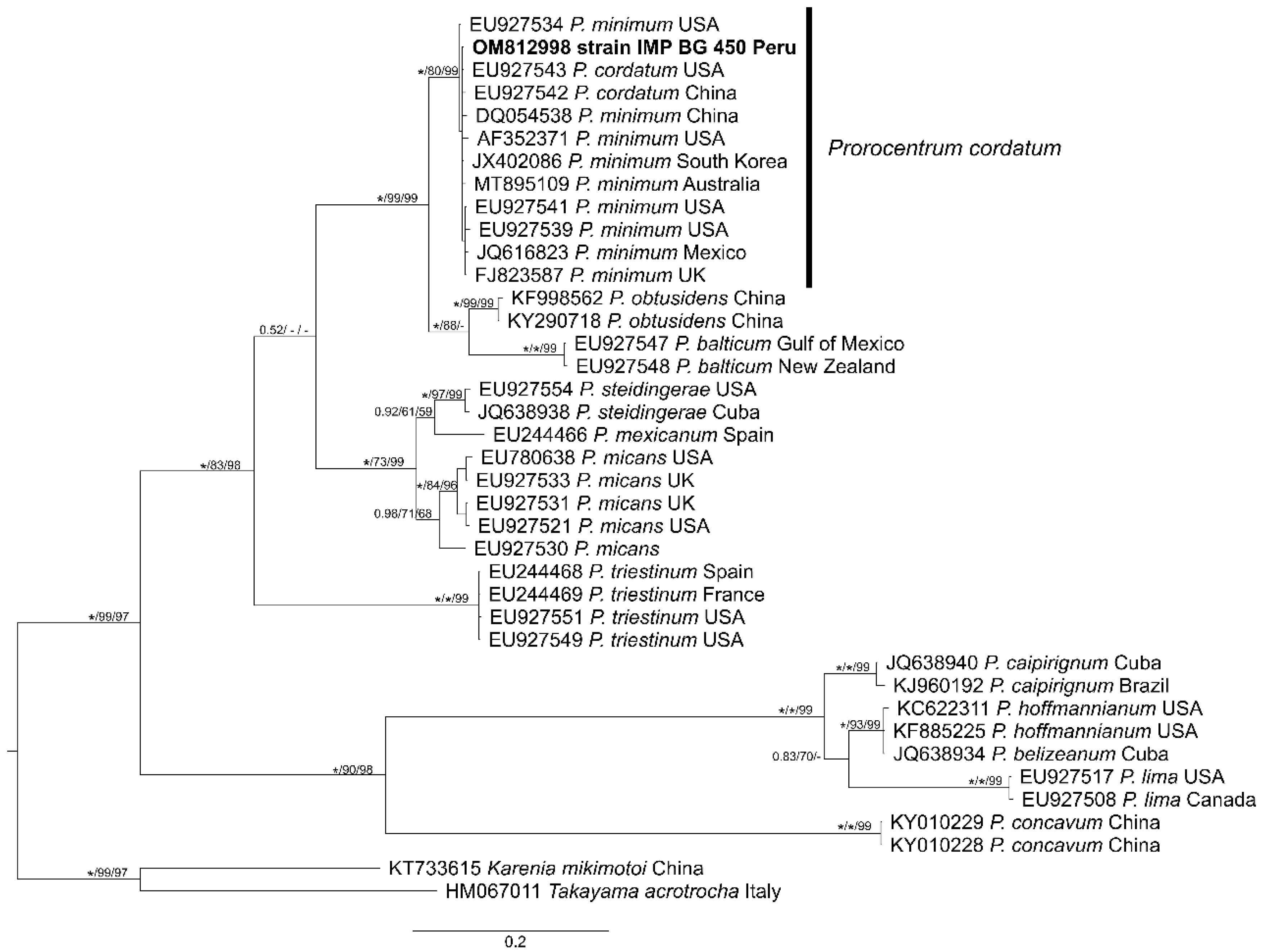
The final alignment had a length of 693 PB and includes 39 sequences from the ITS region (ITS1-5.8S-ITS2) of 507–644 pb. The bloc includes two from the genera Takayama and Karenia as an external group and 37 sequences from the genus Prorocentrum, including the sequence OM812998 (509 pb) from the studied strain IMP BG 450. Except for the difference in the location of one taxon, the phylogenetic trees arising from the Bayesian inference (BI), maximum likelihood (ML) and neighbor-joining (NJ) analyses showed topological congruencies and well-supported grouping (IB = 1.00/ML = 99/NJ = 99) of the study sequence (OM812998), with sequences identified as P. minimum (currently P. cordatum) showing divergence values (p-distance) between 0 and 0.008. The P. cordatum cluster includes North Atlantic strains from the USA (CCMP1329, New York; CCMP2233, Delaware; PmiITSC4, North Carolina; CCMP695, CCMP2811, Florida) and Scotland (CCAP1136/16, Argyll); North Pacific strains from Mexico (PIPV-1, Baja California Sur), South Korea (KMMCC 1181, Gyeongnam) and China (PMDH01, Fujian; CCMP2780, Hong Kong Island); and South Pacific strains from Australia (CAWD359, NSW) and Peru (IMP BG 450, Ica) generated in this study (Figure 7).

3.4. Toxin Analysis

The P. cordatum cultures did not contain tetrodotoxin (TTX) or its analogs in detectable amounts using LC-MS/MS.

4. Discussion

4.1. Environmental Parameters

The P. cordatum bloom (2.7 × 10⁶ cells L−1) in Paracas Bay in late winter 2017 was accompanied by species of Pseudo-nitzschia f delicatissima (351 × 103 cells L−1) and Dinophysis acuminata (11 × 103 cells L−1) (SANIPES). This event is related to increased south winds (6 m s−1) from 25 to 30 August (>10 m s−1), corresponding to the strongest coastal winds during the local winter [53,54] and linked to surface water chilling due to upwelling, with an increase in nutrients (>10 μg L−1 of N.NO3 and >1.5 μg L−1 of P.PO4;) as well as primary productivity (>2000 mg cm2 day−1) [55]. P. cordatum during the summer of 2014 on the central Peruvian coast presented a high correlation with high nitrate values [36].
This oceanographic process of upwelling, which presents more strongly away from the coast, produces a system of recirculation that provides an optimal environment for a P. cordatum bloom in the bay, where high values of surface chlorophyll-a (>10 mg m−3) (Figure 3) with warmer waters (>18.5 °C) (Figure 5b,c) extend up to 13 km north of Paracas Bay. A similar distribution structure was presented northwards from Paracas Bay in the late summer of 2015, with a moderate density of P. cordatum (403 × 103 cells L−1) and low chlorophyll-a values (<1 μg L−1) [40]. This low chlorophyll-a concentration tends to arise due to the fact that this species is mixotrophic and can feed on particulate matter and prey in a phagotrophic fashion, and thus lacks chlorophyll-a [56]. The versatility displayed by P. cordatum in obtaining nutrients—whether autotrophic or mixotrophic (phagotrophic), grazing on various species of phytoplankton, as well as being prey for other dinoflagellates and altering the phytoplanktonic community [57]—may be their key to being a successful species in Paracas Bay, increasing the frequency of and prolonging bloom events according to the reports of Glibert and Burkholder [4].
On 4 February 2011 in Paracas Bay, the reported density of P. cordatum was <200 × 103 cells L−1. Six years later, in 2017, the first bloom was reported (2.720 × 10³ cells L−1) [58]; a similar timeframe to that reported for the first Baltic Sea bloom by Olenina, et al. [59].
The oceanographical characteristics of Paracas Bay with temperatures ranging from 15–24 °C and salinities between 34 and 35, which are within the eurythermal (3–28 °C) [60,61,62] and euryhaline (3 and 38) ranges, these are conditions for the abundant growth of P. cordatum [28,60,63,64,65,66,67].
Furthermore, human intervention in Paracas Bay and nearby areas (5 to 13 km north) with strong industrial fishing and fish meal processing activity, increases the total available nutrients in this bay [68] and maintaining silt-clay sediments with strong organic loads (5–13%) [69].
There are also large anoxic reaches of the bay, with bottom water OD concentrations between 0.00 and 0.50 mL L−1 [40].
The nutrient supplies arising from frequent coastal upwelling processes and eutrophication conditions present in Paracas Bay are an optimal environment for the growth of P. cordatum and phytoplankton.
These high productivity conditions are favorable for sea bottom aquaculture of A. purpuratus, an important economic activity in the area, which could be threatened by the growth of P. cordatum. The negative effects of red tides on ocean farming, which generally lead to interruptions due to precautionary closings after these events, would prevent the exploitation of farming resources [61,70,71,72].
The first reports of P. cordatum (as Exuviaella marina) in the coastal waters of south-central Peru were reported in 1966 [37], and later became a component of the diet of larval Anchovetas (Engraulis ringens) and Sardines (Sardinops sagax) [38].
This dinoflagellate was presented in moderate density (400 × 103 cells L−1) in the Paracas Bay sector during the years 2005–2015 [39,40]. Meanwhile, in Ecuador, this species was detected on four occasions between 2003 and 2004 in the Jambelí Channel and Salado Estuary next to the Gulf of Guayaquil [73].
In the South Atlantic, this species has been reported since 1998 on the Uruguayan coast [74,75]. The highest densities reached in this area have been detected on the northern coastline of the Buenos Aires Province, Argentina, with a maximum concentration of ~15 × 106 cells L−1. This event coincided with fish die-offs (Akselman, 1999 in Carreto [76]). During this episode, plankton and bivalve samples demonstrated toxicity in bioassays with mice; however, they were not associated with the presence of P. minimum as the cause of the toxicity (Montoya 1998 in [76]).

4.2. Morphology

The present study reports the first detailed taxonomic SEM and phylogenetic study of P. cordatum on both the Pacific and Atlantic coasts of South America. The morphological examination of the IMP-BG 450 P. cordatum strain presented measurements of 13.8–17.2 μm in length and 12.7–15.6 μm in width when cultivated at 34 PSU, similar to the Chesapeake Bay strain at 14–18 μm length and 13–16 μm width, cultivated at a salinity of 12 PSU. These cell sizes were within the range described by Dodge [77]. Some authors [19,63,64] mention that the variable cell size of this species is related to environmental salinity. However, Monti-Birkenmeier, et al. [78] state that salinity is not a determining factor for size variation in P. cordatum.
Within the taxonomical characteristics of the Paracas Bay P. cordatum strain, there were no observations of triangular cells, as described by Martin [79], frequently cited for the Baltic, Caspian Sea, Black Sea and Bulgarian waters [19,77,80,81].
Meanwhile, the round- or oval-shaped P. cordatum cells described for this bay are less frequently recorded along the West Coast of the USA, Japan, the Gulf of Mexico, the Gulf of California, the English Channel, the Caspian Sea, the Black Sea and the Mediterranean [19,77,80,82,83,84].
Furthermore, the pentagonal shape of this IMP-BG450 strain has been described within the morphological variations within the Gulf of Finland [63,80,85].
The intercalary band structure of these P. cordatum cells presents four rows of spines, similar to the descriptions by Monti, et al. [86], presenting distancing during the growth phase and without an increase in the number of spine rows. By contrast, the P. cordatum in Paracas Bay presented a rise in the number of threads during the growth phase, except for the distancing between them.
The Paracas Bay P. cordatum presented eight periflagellar plates, but plate seven was not observed in SEM images. The total number coincides with the observations by Loeblich III [87] and Taylor [88] for this species. A series of spines can be found upon the periflagellum plates, with the most important one being the apical collar described by P. minimum (Pavillard) Schiller [19,82,89], and its absence in P. cordatum (Ostenfeld) Dodge.
After comparing SEM micrography images of P. cordatum from the Baltic Sea [78] and Paracas Bay, we can visualize morphological details of the apical collar presenting a height greater than 1 μm. However, its forms are slightly different for both strains. This taxonomical trait is what separated P. minimum from P. cordatum [77] as a distinct species. However, greater detail is observed for the apical structure of both strains, with a long thin forked tooth for the Baltic variety, while the one from Paracas Bay is thick and shorter. The valvular spines on the Baltic Sea strains are thick and blunt tipped, while those from Paracas Bay are thin and pointed. The morphological variability present in both strains is related to different environmental conditions as well as genetics.

4.3. Phylogeny

Molecular analyses of the strain (OM812998) IMP-BG450 were carried out based on sequences from the ITS region to confirm the presence of Prorocentrum cordatum, (Ostenfeld) J. D. Dodge, 1975, in Paracas Bay. Previously, the ITS region has been useful in recognizing dinoflagellate species [90,91] and P. cordatum (as P. minimum in McLenna [92]), demonstrating cutoff values at the species level below 0.02 (p-distance) for the ITS region in dinoflagellates [91]. The phylogeny obtained shows a well-resolved P. cordatum clade with high support and low intra-specific divergence values (<0.02), which groups the studied sequence with exemplars of the species P. cordatum, mainly coming from the Northern Hemisphere of the Atlantic and Pacific Oceans, and one recently reported in the South Pacific [92].

4.4. Toxins Analysis

P. cordatum strain IMP-BG450 from Paracas Bay did not present TTX, also no intoxications or mortalities were reported in the 2017 bloom, while Vlamis, Katikou, Rodriguez, Rey, Alfonso, Papazachariou, Zacharaki, Botana and Botana [29] detected TTX in mollusks in the presence of P. cordatum in Vistonikos Bay, Lagos, Rhodope (Greece).
Toxin production is related to the genetic variability of strains of this species, environmental conditions (temperature, salinity and irradiance) [60] and culture media [16,93], as well as symbiotic association with bacteria (Roseobacter and Vibrio sp.) [35]. Because of this, the possibility cannot be ruled out that at some moment the conditions may arise to generate toxicity from P. cordatum in Paracas Bay.
We suggest that the HAB Monitoring Program should incorporate P. cordatum as a hazardous species and carry out toxicological analyses of each event arising on the Peruvian coast, since any harmful species could cause environmental damage and economic loss, implying increased mortality among fish, shellfish and subsequent human poisoning during blooms [16,23,59,61,81,94,95,96].

5. Conclusions

The microalgal bloom of P. cordatum in the austral winter of 2017 in Paracas Bay, presented the highest historical density of this species, being stimulated by an intense upwelling process which produced a recirculation towards the interior of Paracas Bay, providing an optimal environment for the bloom.
Taxonomic and molecular analyses indicate the presence of P. cordatum in Paracas Bay. It was found that P. cordatum during this event did not generate TTX. However, more assays with different factors are needed to evaluate whether the toxins can be expressed in the laboratory. It is suggested to consider the evaluation of TTX analysis in the monitoring carried out by the control entity to avoid problems with this toxin.

Supplementary Materials

The following supporting information can be downloaded at: https://www.mdpi.com/article/10.3390/d14100844/s1, Table S1: List of Prorocentrum, Takayama and Karenia sequences used in phylogenetic analyses. Strain, collection information and GenBank accession number are presented.

Author Contributions

Conceptualization, C.T., G.Á., E.U. and J.B.; methodology, C.T., M.P.-A., G.Á., J.B., C.P., E.U. and J.L.B.; software, C.T., M.P.-A., J.L.B., C.P. and J.B.; investigation, C.T., G.Á., M.P.-A., J.B. and E.U.; formal analysis, C.T., G.Á., M.P.-A., E.U. and J.B.; writing—original draft preparation, C.T., G.Á., M.P.-A., E.U. and J.B.; writing—review and editing, G.Á., E.U. and J.B.; visualization, C.T., G.Á., M.P.-A. and J.B.; supervision, E.U., G.Á. and J.B.; funding acquisition G.Á. and C.T.; All authors have read and agreed to the published version of the manuscript.

Funding

This research was funded by the “ANID + FONDEF/PRIMER CONCURSO INVESTIGACIÓN TECNOLÓGICA TEMATICO ENSISTEMAS PESQUERO ACUICOLAS FRENTE A FLORECIMIENTOS ALGALES NOCIVOS FANS IDeA DEL FONDO DE FOMENTO AL DESARROLLO CIENTÍFICO Y TECNOLÓGICO, FONDEF/ANID 2017, IT17F10002 developed within the framework of a cooperation agreement between the Consellería do Mar, Xunta de Galicia, Spain and the Universidad Católica del Norte, Chile and the Budget National Program N°094 (PpR094): “Management and Development of Aquaculture”-PRODUCE, PERU, through the project “Technological Development in Aquaculture.

Institutional Review Board Statement

Not applicable.

Data Availability Statement

Data can be found within the paper and Supplementary Materials.

Conflicts of Interest

The authors declare no conflict of interest.

References

  1. Glibert, P.M. Harmful algae at the complex nexus of eutrophication and climate change. Harmful Algae 2020, 91, 101583. [Google Scholar] [CrossRef] [PubMed]
  2. Anderson, D.M.; Glibert, P.M.; Burkholder, J.M. Harmful algal blooms and eutrophication: Nutrient sources, composition, and consequences. Estuaries 2002, 25, 704–726. [Google Scholar] [CrossRef]
  3. Hallegraeff, G.M. A review of harmful algal blooms and their apparent global increase. Phycologia 1993, 32, 79–99. [Google Scholar] [CrossRef] [Green Version]
  4. Glibert, P.M.; Burkholder, J.M. Harmful Algal Species Fact Sheet: Prorocentrum. In Harmful Algal Blooms: A Compendium Desk Reference; Shumway, S.E., Burkholder, J.M., Morton, S.L., Eds.; Wiley Blackwell: Hoboken, NJ, USA, 2018; pp. 625–628. [Google Scholar]
  5. Heisler, J.; Glibert, P.; Burkholder, J.; Anderson, D.; Cochlan, W.; Dennison, W.; Gobler, C.; Dortch, Q.; Heil, C.; Humphries, E.; et al. Eutrophication and Harmful Algal Blooms: A Scientific Consensus. Harmful Algae 2008, 8, 3–13. [Google Scholar] [CrossRef] [PubMed] [Green Version]
  6. Anderson, D.M. Toxic algal bloom and red tides: A global perspective. In Red Tides: Biology, Environmental Science and Technology; Okaichi, T., Anderson, D.M., Nemoto, T., Eds.; Elsevier: New York, NY, USA, 1989; pp. 11–16. [Google Scholar]
  7. Hallegraeff, G.M. Ocean climate change, phytoplankton community responses, and harmful algal blooms: A formidable predictive challenge 1. J. Phycol. 2010, 46, 220–235. [Google Scholar] [CrossRef]
  8. Dale, B.; Edwards, M.; Reid, P.C. Climate change and harmful algal blooms. In Ecology of Harmful Algae; Springer: Berlin/Heidelberg, Germany, 2006; pp. 367–378. [Google Scholar]
  9. Edwards, M.; Johns, D.G.; Leterme, S.C.; Svendsen, E.; Richardson, A.J. Regional climate change and harmful algal blooms in the northeast Atlantic. Limnol. Oceanogr. 2006, 51, 820–829. [Google Scholar] [CrossRef] [Green Version]
  10. Echevin, V.; Gévaudan, M.; Espinoza-Morriberón, D.; Tam, J.; Aumont, O.; Gutierrez, D.; Colas, F. Physical and biogeochemical impacts of RCP8. 5 scenario in the Peru upwelling system. Biogeosciences 2020, 17, 3317–3341. [Google Scholar] [CrossRef]
  11. Chavez, F.P.; Bertrand, A.; Guevara-Carrasco, R.; Soler, P.; Csirke, J. The northern Humboldt Current System: Brief history, present status and a view towards the future. Prog. Oceanogr. 2008, 79, 95–105. [Google Scholar] [CrossRef]
  12. Lomas, M.W.; Glibert, P.M. Interactions between NH+ 4 and NO 3 uptake and assimilation: Comparison of diatoms and dinoflagellates at several growth temperatures. Mar. Biol. 1999, 3, 541–551. [Google Scholar] [CrossRef]
  13. Goldman, J.C. Potential role of large oceanic diatoms in new primary production. Deep Sea Res. Part I Oceanogr. Res. Pap. 1993, 40, 625–628. [Google Scholar] [CrossRef]
  14. Kudela, R.M.; Dugdale, R.C. Nutrient regulation of phytoplankton productivity in Monterey Bay, California. Deep-Sea Res. Part II 2000, 47, 1023–1053. [Google Scholar] [CrossRef]
  15. Wilkerson, F.P.; Dugdale, R.C.; Kudela, R.M.; Chavez, F.P. Biomass and productivity in Monterey Bay, California: Contribution of the large phytoplankton. Deep Sea Res. Part II Top. Stud. Oceanogr. 2000, 47, 1003–1022. [Google Scholar] [CrossRef]
  16. Heil, C.A.; Glibert, P.M.; Fan, C. Prorocentrum minimum (Pavillard) Schiller A review of a harmful algal bloom species of growing worldwide importance. Harmful Algae 2005, 4, 449–470. [Google Scholar] [CrossRef]
  17. Klanjšček, J.; Geček, S.; Klanjšček, T.; Legović, T. Nutrient quotas and carbon content variability of Prorocentrum minimum (Pavillard) Schiller, 1933. Harmful Algae 2016, 51, 16–25. [Google Scholar] [CrossRef] [PubMed]
  18. Glibert, P.M.; Mayorga, E.; Seitzinger, S. Prorocentrum minimum tracks anthropogenic nitrogen and phosphorus inputs on a global basis: Application of spatially explicit nutrient export models. Harmful Algae 2008, 8, 33–38. [Google Scholar] [CrossRef]
  19. Velikova, V.; Larsen, J. The Prorocentrum cordatum/Prorocentrum minimum taxonomic problem. Grana 1999, 38, 108–112. [Google Scholar] [CrossRef]
  20. Guiry, M.D.; Guiry, G.M. AlgaeBase. World-Wide Electronic Publication; National University of Ireland. Galway. Available online: https://www.algaebase.org/search/species/detail/?species_id=71381 (accessed on 8 July 2022).
  21. Moestrup, O.; Akselmann-Cardella, R.; Churro, C.F.S.; Hoppenrath, M.; Iwataki, M.; Larsen, J.; Lundholm, N.; Zingone, A. IOC-UNESCO Taxonomic Reference List of Harmful Micro Algae. Prorocentrum cordatum (Ostenfeld) J.D.Dodge. 1976. Available online: http://www.marinespecies.org/hab/aphia.php?p=taxdetails&id=232376 (accessed on 8 July 2022).
  22. Khanaychenko, A.N.; Telesh, I.V.; Skarlato, S.O. Bloom-forming potentially toxic dinoflagellates Prorocentrum cordatum in marine plankton food webs. Protistology 2019, 13, 95–125. [Google Scholar] [CrossRef]
  23. Glibert, P.M.; Burkholder, J.M.; Kana, T.M. Recent insights about relationships between nutrient availability, forms, and stoichiometry, and the distribution, ecophysiology, and food web effects of pelagic and benthic Prorocentrum species. Harmful Algae 2012, 14, 231–259. [Google Scholar] [CrossRef]
  24. Sierra-Beltrán, A.; Cortés-Altamirano, R.; Cortés-Lara, M.d.C. Occurrences of Prorocentrum minimum (Pavillard) in México. Harmful Algae 2005, 4, 507–517. [Google Scholar] [CrossRef]
  25. Matantseva, O.; Skarlato, S.; Vogts, A.; Pozdnyakov, I.; Liskow, I.; Schubert, H.; Voss, M. Superposition of individual activities: Urea-mediated suppression of nitrate uptake in the dinoflagellate Prorocentrum minimum revealed at the population and single-cell levels. Front. Microbiol. 2016, 7, 1310. [Google Scholar] [CrossRef]
  26. Telesh, I.V.; Schubert, H.; Skarlato, S.O. Ecological niche partitioning of the invasive dinoflagellate Prorocentrum minimum and its native congeners in the Baltic Sea. Harmful Algae 2016, 59, 100–111. [Google Scholar] [CrossRef]
  27. Telesh, I.; Schubert, H.; Skarlato, S. Abiotic stability promotes dinoflagellate blooms in marine coastal ecosystems. Estuar. Coast. Shelf Sci. 2021, 251, 107239. [Google Scholar] [CrossRef]
  28. Skarlato, S.O.; Telesh, I.V.; Matantseva, O.V.; Pozdnyakov, I.A.; Berdieva, M.A.; Schubert, H.; Filatova, N.A.; Knyazev, N.A.; Pechkovskaya, S.A. Studies of bloom-forming dinoflagellates Prorocentrum minimum in fluctuating environment: Contribution to aquatic ecology, cell biology and invasion theory. Protistology 2018, 12, 113–157. [Google Scholar] [CrossRef]
  29. Vlamis, A.; Katikou, P.; Rodriguez, I.; Rey, V.; Alfonso, A.; Papazachariou, A.; Zacharaki, T.; Botana, A.M.; Botana, L.M. First detection of tetrodotoxin in Greek shellfish by UPLC-MS/MS potentially linked to the presence of the dinoflagellate Prorocentrum minimum. Toxins 2015, 7, 1779–1807. [Google Scholar] [CrossRef] [Green Version]
  30. Bordin, P.; Dall’Ara, S.; Tartaglione, L.; Antonelli, P.; Calfapietra, A.; Varriale, F.; Guiatti, D.; Milandri, A.; Dell’Aversano, C.; Arcangeli, G.; et al. First occurrence of tetrodotoxins in bivalve mollusks from Northern Adriatic Sea (Italy). Food Control 2021, 120, 107510. [Google Scholar] [CrossRef]
  31. Turner, A.D.; Dhanji-Rapkova, M.; Coates, L.; Bickerstaff, L.; Milligan, S.; O’Neill, A.; Faulkner, D.; McEneny, H.; Baker-Austin, C.; Lees, D.N.; et al. Detection of Tetrodotoxin Shellfish Poisoning (TSP) Toxins and Causative Factors in Bivalve Molluscs from the UK. Mar. Drugs 2017, 15, 277. [Google Scholar] [CrossRef] [Green Version]
  32. Gerssen, A.; Bovee, T.; Klijnstra, M.; Poelman, M.; Portier, L.; Hoogenboom, R. First Report on the Occurrence of Tetrodotoxins in Bivalve Mollusks in The Netherlands. Toxins 2018, 10, 450. [Google Scholar] [CrossRef] [PubMed] [Green Version]
  33. Narahashi, T.; Moore, J.W.; Scott, W.R. Tetrodotoxin blockage of sodium conductance increase in lobster giant axons. J. Gen. Physiol. 1964, 47, 965–974. [Google Scholar] [CrossRef] [PubMed]
  34. Narahashi, T. Pharmacology of tetrodotoxin. J. Toxicol. Toxin Rev. 2001, 20, 67–84. [Google Scholar] [CrossRef]
  35. Rodriguez, I.; Alfonso, A.; Alonso, E.; Rubiolo, J.A.; Roel, M.; Vlamis, A.; Katikou, P.; Jackson, S.A.; Menon, M.L.; Dobson, A.; et al. The association of bacterial C9-based TTX-like compounds with Prorocentrum minimum opens new uncertainties about shellfish seafood safety. Sci. Rep. 2017, 7, 40880. [Google Scholar] [CrossRef] [PubMed]
  36. Sánchez, S.; Bernales, A.; Delgado, E.; Chang, F.; Jacobo, N.; Quispe, J. Variability and Biogeographical Distribution of Harmful Algal Blooms in Bays of High Productivity off Peruvian Coast (2012–2015). J. Environ. Anal. Toxicol. 2017, 7, 2161-0525.1000530. [Google Scholar] [CrossRef]
  37. Strickland, J.D.H.; Eppley, R.W.; de Mendiola, B.R. Poblaciones de fitoplancton, nutrientes y fotosíntesis en aguas costeras peruanas. Bol. Instit. Mar. Perú 1969, 2, 4–45. [Google Scholar]
  38. Muck, P.E.T.E.R.; Rojas de Mendiola, B.; Antonietti, E.M.I.R.A. Comparative studies on feeding in larval anchoveta (Engraulis ringens) and sardine (Sardinops sagax). In The Peruvian Upwelling Ecosystem: Dynamics and Interactions; ICLARM: Manila, Philippines, 1989; Volume 18, pp. 86–96. [Google Scholar]
  39. Solano Cornejo, D.; Mendoza Díaz, V.; CONAM, PNUMA. Informe Sobre el Estado del Ambiente: GEO Bahía Paracas-Pisco; Lima, Peru, 2007; pp. 1–163. [Google Scholar]
  40. Sánchez, S.; Jacobo, N.; Bernales, A.; Franco, A.; Quispe, J.; Flores, G. Seasonal variability in the distribution of phytoplankton in Paracas Bay/Peru, as a response to environmental conditions. J. Environ. Sci. Eng. B 2018, 371–380. [Google Scholar] [CrossRef]
  41. Andersen, R.A.; Kawachi, M. Tradiotinal Microalgae Isolation Techniques. In Algal Culturing Techniques; Andersen, R.A., Ed.; Elsevier: Amsterdam, The Netherlands, 2005; Volume 578, pp. 83–100. [Google Scholar]
  42. Doyle, J.J.; Doyle, J.L. A rapid DNA isolation procedure for small quantities of fresh leaf tissue. Phytochem. Bull. 1987, 19, 11–15. [Google Scholar]
  43. White, T.J.; Bruns, T.; Lee, S.; Taylor, J. Amplification and direct sequencing of fungal ribosomal RNA genes for phylogenetics. In PCR protocols: A Guide to Methods and Applications; Innis, M., Gelfand, D., Sninsky, J., White, T., Eds.; Academic Press: New York, NY, USA, 1990; pp. 315–322. [Google Scholar]
  44. Hall, T.A. BioEdit: A user-friendly biological sequence alignment editor and analysis program for Windows 95/98/NT. Nucleic Acids Symp. Ser. 1999, 41, 95–98. [Google Scholar]
  45. Kumar, S.; Stecher, G.; Tamura, K. MEGA7: Molecular evolutionary genetics analysis version 7.0 for bigger datasets. Mol. Biol. Evol. 2016, 33, 1870–1874. [Google Scholar] [CrossRef] [PubMed] [Green Version]
  46. Darriba, D.; Taboada, G.L.; Doallo, R.; Posada, D. jModelTest 2: More models, new heuristics and parallel computing. Nat. Methods 2012, 9, 772. [Google Scholar] [CrossRef] [PubMed] [Green Version]
  47. Stamatakis, A. RAxML version 8: A tool for phylogenetic analysis and post-analysis of large phylogenies. Bioinformatics 2014, 30, 1312–1313. [Google Scholar] [CrossRef] [Green Version]
  48. Silvestro, D.; Michalak, I. raxmlGUI: A graphical front-end for RAxML. Org. Divers. Evol. 2012, 12, 335–337. [Google Scholar] [CrossRef]
  49. Ronquist, F.; Teslenko, M.; Van Der Mark, P.; Ayres, D.L.; Darling, A.; Höhna, S.; Larget, B.; Liu, L.; Suchard, M.A.; Huelsenbeck, J.P. MrBayes 3.2: Efficient Bayesian phylogenetic inference and model choice across a large model space. Syst. Biol. 2012, 61, 539–542. [Google Scholar] [CrossRef] [Green Version]
  50. Rambaut, A.; Suchard, M.A.; Xie, D.; Drummond, A.J. Tracer v1. 6. Available online: http://tree.bio.ed.ac.uk/software/tracer (accessed on 8 July 2022).
  51. Boundy, M.J.; Selwood, A.I.; Harwood, D.T.; McNabb, P.S.; Turner, A.D. Development of a sensitive and selective liquid chromatography-mass spectrometry method for high throughput analysis of paralytic shellfish toxins using graphitised carbon solid phase extraction. J. Chromatogr. A 2015, 1387, 1–12. [Google Scholar] [CrossRef] [PubMed]
  52. Hoppenrath, M.; Chomérat, N.; Horiguchi, T.; Schweikert, M.; Nagahama, Y.; Murray, S. Taxonomy and phylogeny of the benthic Prorocentrum species (Dinophyceae)—A proposal and review. Harmful Algae 2013, 27, 1–28. [Google Scholar] [CrossRef]
  53. Bakun, A.N.D.R.E.W.; Mendelssohn, R. Alongshore wind stress, 1953–1984: Correction, reconciliation and update through 1986. In The Peruvian Upwelling Ecosystem: Dynamics and Interactions, Proceedings of the ICLARM Conference Procceedings; ICLARM: Manila, Philippines, 1989; pp. 77–81. [Google Scholar]
  54. Gutiérrez, D.; Bouloubassi, I.; Sifeddine, A.; Purca, S.; Goubanova, K.; Graco, M.; Field, D.; Méjanelle, L.; Velazco, F.; Lorre, A.; et al. Coastal cooling and increased productivity in the main upwelling zone off Peru since the mid-twentieth century. Geophys. Res. Lett. 2011, 38, 1–6. [Google Scholar] [CrossRef]
  55. Calienes, R.; Guillén, O.; Lostaunau, N. Variabilidad espacio-temporal de clorofila, producción primaria y nutrientes frente a la costa peruana. Bol. Inst. Mar. Perú 1985, 10, 1–44. [Google Scholar]
  56. Sukhanova, I.N.; Flint, M.V.; Hibaum, G.; Karamfilov, V.; Kopylov, A.I.; Matveeva, E.; Rat’kova, T.N.; Sazhin, A.F. Exuviaella cordata red tide in Bulgarian coastal waters (May to June 1986). Mar. Biol. 1988, 99, 1–8. [Google Scholar] [CrossRef]
  57. Jeong, H.J.; Du Yoo, Y.; Kim, J.S.; Seong, K.A.; Kang, N.S.; Kim, T.H. Growth, feeding and ecological roles of the mixotrophic and heterotrophic dinoflagellates in marine planktonic food webs. Ocean. Sci. J. 2010, 45, 65–91. [Google Scholar] [CrossRef] [Green Version]
  58. SANIPES. Fisheries Health Organization of Perú- Organismo Nacional de Sanidad Pesquera. Available online: https://www.sanipes.gob.pe/web/index.php/es/fitoplancton (accessed on 8 July 2022).
  59. Olenina, I.; Wasmund, N.; Hajdu, S.; Jurgensone, I.; Gromisz, S.; Kownacka, J.; Toming, K.; Vaiciūtė, D.; Olenin, S. Assessing impacts of invasive phytoplankton: The Baltic Sea case. Mar. Pollut. Bull. 2010, 60, 1691–1700. [Google Scholar] [CrossRef]
  60. Grzebyk, D.; Berland, B. Influences of temperature, salinity and irradiance on growth of Prorocentrum minimum (Dinophyceae) from the Mediterranean Sea. J. Plankton Res. 1996, 18, 1837–1849. [Google Scholar] [CrossRef] [Green Version]
  61. Tango, P.; Magnien, R.; Butler, W.; Luckett, C.; Luckenbach, M.; Lacouture, R.; Poukish, C. Impacts and potential effects due to Prorocentrum minimum blooms in Chesapeake Bay. Harmful Algae 2005, 4, 525–531. [Google Scholar] [CrossRef]
  62. Pertola, S. Diffusive and Ship-Mediated Spread of Dinoflagellates in the Baltic Sea with Prorocentrum Minimum as a Special Case; University of Helsinki: Helsinki, Finland, 2006. [Google Scholar]
  63. Hajdu, S.; Edler, L.; Olenina, I.; Witek, B. Spreading and establishment of the potentially toxic dinoflagellate Prorocentrum minimum in the Baltic Sea. Int. Rev. Hydrobiol. A J. Cover. All Asp. Limnol. Mar. Biol. 2000, 85, 561–575. [Google Scholar]
  64. Olenina, I.; Vaičiukynas, E.; Šulčius, S.; Paškauskas, R.; Verikas, A.; Gelžinis, A.; Bačauskienė, M.; Bertašiūtė, V.; Olenin, S. The dinoflagellate Prorocentrum cordatum at the edge of the salinity tolerance: The growth is slower but cells are larger. Estuar. Coast. Shelf Sci. 2016, 168, 71–79. [Google Scholar] [CrossRef]
  65. Knyazev, N.; Pechkovskaya, S.; Skarlato, S.; Telesh, I.; Filatova, N. The impact of temperature stress on DNA and RNA synthesis in potentially toxic dinoflagellates Prorocentrum minimum. J. Evol. Biochem. Physiol. 2018, 54, 383–389. [Google Scholar] [CrossRef]
  66. Pertola, S.; Kuosa, H.; Olsonen, R. Is the invasion of Prorocentrum minimum (Dinophyceae) related to the nitrogen enrichment of the Baltic Sea? Harmful Algae 2005, 4, 481–492. [Google Scholar] [CrossRef]
  67. Kimor, B.; Moigis, A.G.; Dohms, V.; Stienen, C. A case of mass occurrence of Prorocentrum minimum in the Kiel Fjord. Mar. Ecol. Prog. Ser. 1985, 27, 209–215. [Google Scholar] [CrossRef]
  68. Kahru, M.; Michell, B.G.; Diaz, A.; Miura, M. MODIS detects a devastating algal bloom in Paracas Bay, Peru. Eos Trans. Am. Geophys. Union 2004, 85, 465–472. [Google Scholar] [CrossRef]
  69. IMARPE. Instituto del Mar del Perú. Laboratorio Costero de Pisco. Bases Técnicas Para el Ordenamiento Pesquero y Acuícola de la Bahía Paracas. Available online: http://www2.produce.gob.pe/RepositorioAPS/3/jer/ACUISUBMENU4/estudios-bahia-paracas.pdf (accessed on 8 July 2022).
  70. Pitcher, G.C.; Bernales Jiménez, A.; Kudela, R.M.; Reguera, B. Harmful algal blooms in Eastern Boundary upwelling systems: A Geohab Core Research Project. Oceanography 2017, 30, 22. [Google Scholar] [CrossRef]
  71. Dyson, K.; Huppert, D.D. Regional economic impacts of razor clam beach closures due to harmful algal blooms (HABs) on the Pacific coast of Washington. Harmful Algae 2010, 9, 264–271. [Google Scholar] [CrossRef]
  72. Blanco-Pérez, J. Episodios nocivos por fitoplancton. In Los Moluscos Pectínidos de Iberoamérica: Ciencia y Acuicultura; Maeda-Martínez, A.N., Ed.; Limusa: Mexico City, Mexico, 2001; pp. 285–324. [Google Scholar]
  73. Borbor-Cordova, M.J.; Torres, G.; Mantilla-Saltos, G.; Casierra-Tomala, A.; Bermúdez, J.R.; Renteria, W.; Bayot, B. Oceanography of Harmful Algal Blooms on the Ecuadorian Coast (1997–2017): Integrating Remote Sensing and Biological Data. Front. Mar. Sci. 2019, 6, 13. [Google Scholar] [CrossRef] [Green Version]
  74. Méndez, S.; Ferrari, G. Floraciones algales nocivas en Uruguay: Antecedentes, proyectos en curso y revisión de resultados. In Floraciones Algales Nocivas en el Cono Sur Americano; Sar, E.A., Ferrario, M.E., Reguera, B., Eds.; Instituto Español de Oceanografia: Madrid, Spain, 2002; pp. 271–288. [Google Scholar]
  75. Ferrari, G.; Del Carmen Perez, M. Fitoplancton de la costa platense y Atlántica de Uruguay (1993–1994). Iheringia. Ser. Bot. 2002, 57, 263–278. [Google Scholar]
  76. Carreto, J.I.; Montoya, N.G.; Akselman, R.; Roja, P.M. Proyecto” Protección Ambiental del Río de la Plata y su Frente Marítimo. Prevención y Control de la Contaminación y Restauración de Hábitats” Proyecto PNUD/GEF RLA/99/G31. RLA 2004, 99, G31. [Google Scholar]
  77. Dodge, J. The Prorocentrales (Dinophyceae). II. Revision of the taxonomy within the genus Prorocentrum. Bot. J. Linn. Soc. 1975, 71, 103–125. [Google Scholar] [CrossRef]
  78. Monti-Birkenmeier, M.; Berden Zrimec, M.B.; Drinovec, L.; Beran, A.; Zrimec, A.; Cataletto, B.; Fonda Umani, S. Influence of salinity on growth and cell volume in three strains of Prorocentrum cordatum (Dinophyceae). Aquat. Biol. 2019, 28, 1–12. [Google Scholar] [CrossRef] [Green Version]
  79. Martin, G.W. Dinoflagellates from marine and brackish waters of New Jersey. In University of Lowa Studies in Natural History; 1929; Volume 12, pp. 3–32. [Google Scholar]
  80. Pertola, S.; Faust, M.A.; Kuosa, H.; Hallfors, G. Morphology of Prorocentrum minimum (Dinophyceae) in the Baltic Sea and in Chesapeake Bay: Comparison of Cell Shapes and Thecal Ornamentation. Bot. Mar. 2003, 46, 477–486. [Google Scholar] [CrossRef]
  81. Witek, B.; Plinski, M. The first recorded bloom of Prorocentrum minimum (Pavillard) Schiller in the coastal zone of the Gulf of Gdańsk. Oceanologia 2000, 42, 29–36. [Google Scholar]
  82. Marasović, I.; Pucher-Petković, T.; Petrova-Karadjova, V. Prorocentrum minimum (Dinophyceae) in the Adriatic and Black sea. J. Mar. Biolog. Assoc. UK 1990, 70, 473–476. [Google Scholar] [CrossRef]
  83. Muciño-Márquez, R.E.; Gárate-Lizárraga, I.; López-Cortés, D.J. Seasonal Variation of the Genus Prorocentrum (Dinophyceae) in Two Tuna Farms in the Bahía De La Paz, Mexico. Acta Biol. Colomb. 2014, 20, 195–206. [Google Scholar] [CrossRef]
  84. Petrova, D.; Gerdzhikov, D. Development of a traditional blooming phytoplankton species Prorocentrum cordatum Dodge, 1975 along the Bulgarian coast (2008–2010). In Proceedings of the Union of Scientists in Bulgaria-Varna, Series “Marine Science”, Bulgaria, Varna, 2013; pp. 12–17. [Google Scholar]
  85. Hajdu, S.; Pertola, S.; Kuosa, H. Prorocentrum minimum (Dinophyceae) in the Baltic Sea: Morphology, occurrence—A review. Harmful Algae 2005, 4, 471–480. [Google Scholar] [CrossRef]
  86. Monti, M.; Stoecker, D.K.; Cataletto, B.; Talarico, L. Morphology of the flagellar pore complex in Prorocentrum minimum (Dinophyceae) from the Adriatic and Baltic Seas. Bot. Mar. 2010, 53. [Google Scholar] [CrossRef]
  87. Loeblich, A.R., III. Dinoflagellate evolution: Speculation and evidence. J. Protozool. 1976, 23, 13–28. [Google Scholar] [CrossRef]
  88. Taylor, F. On dinoflagellate evolution. BioSystems 1980, 13, 65–108. [Google Scholar] [CrossRef]
  89. Taylor, F.; Fukuyo, Y.; Larsen, J.; Hallegraeff, G. Taxonomy of harmful dinoflagellates. In Harmful Marine Microalgae; Hallegraeff, G.M., Anderson, D.M., Cembella, A.D., Eds.; UNESCO: Paris, France, 2003; pp. 389–482. [Google Scholar]
  90. Wayne Litaker, R.; Vandersea, M.W.; Kibler, S.R.; Reece, K.S.; Stokes, N.A.; Lutzoni, F.M.; Yonish, B.A.; West, M.A.; Black, M.N.D.; Tester, P.A. Recognizing Dinoflagellate species using ITS rDNA sequences. J. Phycol. 2007, 43, 344–355. [Google Scholar] [CrossRef]
  91. Stern, R.F.; Andersen, R.A.; Jameson, I.; Küpper, F.C.; Coffroth, M.A.; Vaulot, D.; Le Gall, F.; Véron, B.; Brand, J.J.; Skelton, H.; et al. Evaluating the ribosomal internal transcribed spacer (ITS) as a candidate dinoflagellate barcode marker. PLoS ONE 2012, 7, e42780. [Google Scholar] [CrossRef] [PubMed]
  92. McLennan, K.; Ruvindy, R.; Ostrowski, M.; Murray, S. Assessing the Use of Molecular Barcoding and qPCR for Investigating the Ecology of Prorocentrum minimum (Dinophyceae), a Harmful Algal Species. Microorganisms 2021, 9, 510. [Google Scholar] [CrossRef] [PubMed]
  93. Nakazima, M. Studies on the source of shellfish poison in Lake Hamana. III. Poisonous effects of shellfish feeding on Prorocentrum sp. Bull. Jpn. Soc. Sci. Fish 1965, 31, 281–285. [Google Scholar] [CrossRef] [Green Version]
  94. Al-Hashmi, K.A.; Smith, S.L.; Claereboudt, M.; Piontkovski, S.A.; Al-Azri, A. Dynamics of potentially harmful phytoplankton in a semi-enclosed bay in the Sea of Oman. Bull. Mar. Sci. 2015, 91, 141–166. [Google Scholar] [CrossRef]
  95. Denardou Queneherve, A.; Grzebyk, D.; Pouchus, Y.F.; Sauviat, M.P.; Alliot, E.; Biard, J.F.; Berland, B.; Verbist, J.F. Toxicity of French strains of the dinoflagellate Prorocentrum minimum experimental and natural contaminations of mussels. Toxicon 1999, 37, 1711–1719. [Google Scholar] [CrossRef]
  96. Wikfors, G.H.; Smolowitz, R.M. Experimental and histological studies of four life-history stages of the eastern oyster, Crassostrea virginica, exposed to a cultured strain of the dinoflagellate Prorocentrum minimum. Biol. Bull. 1995, 188, 313–328. [Google Scholar] [CrossRef]
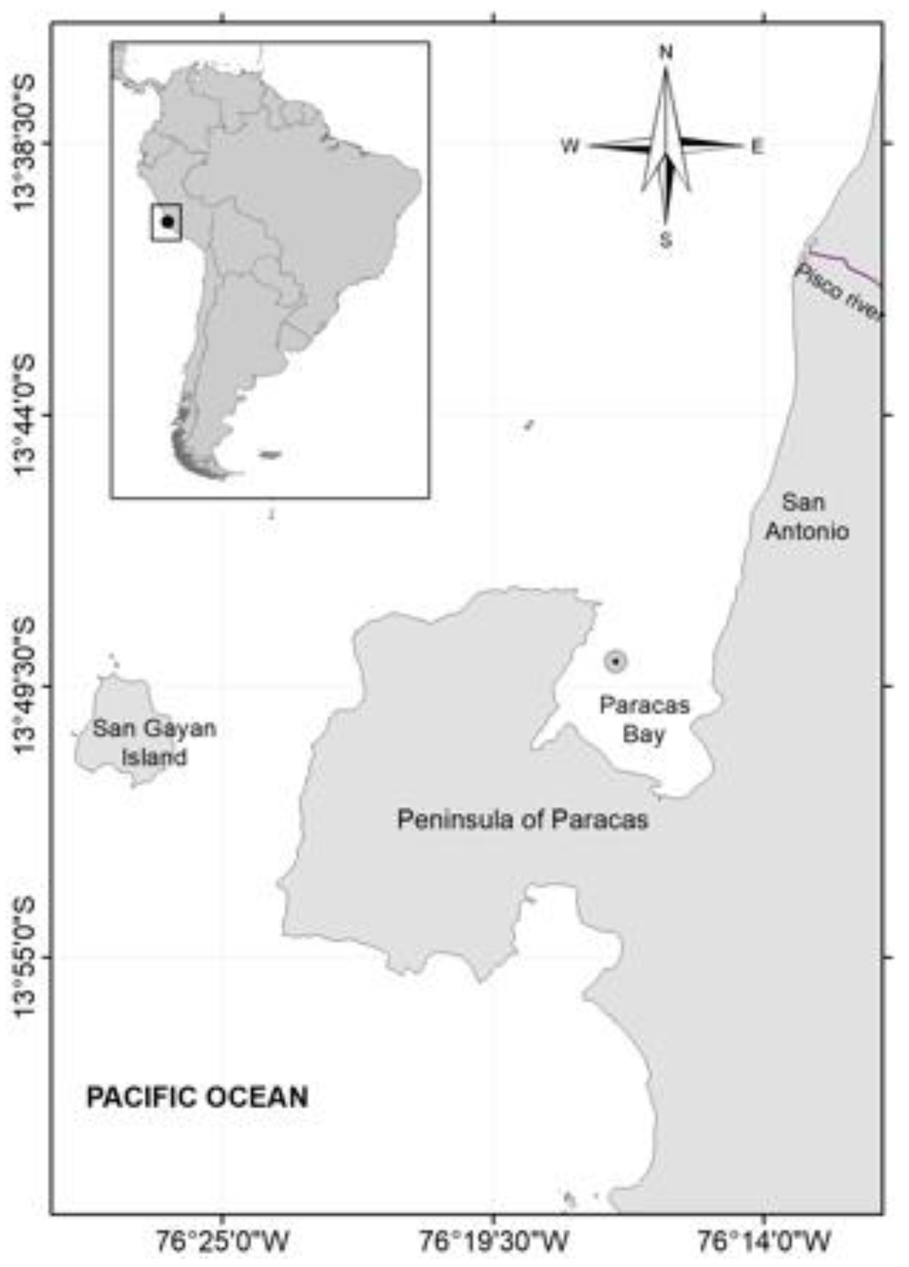
Figure 1. Map Paracas Bay, Peru (13°49′ S, 76°17′ W).
Diversity 14 00844 g001
Figure 2. (a) Cell density and temperature and (b) cell density and dissolved oxygen from Paracas Bay.
Diversity 14 00844 g002
Figure 3. Surface distribution of chlorophyll-a VIIRS-NPP (27–29 August 2017).
Diversity 14 00844 g003
Figure 4. Wind speed satellite images (m s−1) (a) 25 August 2017; (b) 30 August 2017; (c) 1 September 2017.
Diversity 14 00844 g004
Figure 5. Sea surface temperature (SST) in Paracas Bay and environs; (a) 25 August 2017, (b) 30 August 2017 and (c) 1 September 2017 (GHRSST).
Diversity 14 00844 g005
Figure 6. Images from scanning electron microscopy (SEM) of the IMP-BG 450 strain of P. cordatum; (a) view of the left side valve showing the apical collar, (b) right side valve view, (c) intercalary band and (d) periflagellar platelets (1: forked tooth; 2: small tooth; 3, 4, 5, 6, 7?, 8; Ap: auxiliary pore; Fp: flagellar pore).
Diversity 14 00844 g006
Figure 7. Prorocentrum Bayesian tree based on ITS sequences. The numbers above lines are nodal supports that indicate Bayesian posterior probabilities (PP), and bootstrap support values from ML and NJ. * Indicates total support values. “-” Indicates that phylogeny structure for ML or NJ analyses are different at the node. The sequence obtained in this study is indicated in bold.
Diversity 14 00844 g007
Table 1. Transitions used for TTX and their analogs.
CompoundTransitionCollision Energy
5,6,11-TrideoxyTTX272.1 > 162.140
5,6,11-TrideoxyTTX272.1 > 254.131
11-nor-TTX-6-ol290.1 > 162.140
11-nor-TTX-6-ol290.1 > 272.131
4,9-anhydroTTX302.1 > 162.140
4,9-anhydroTTX302.101 > 256.131
5-deoxyTTX, 11-deoxyTTX304.1 > 176.140
5-deoxyTTX, 11-deoxyTTX304.1 > 286.131
TTX, 4-epiTTX320.1 > 162.140
TTX, 4-epiTTX320.1 > 302.135
C9-265 *265.1 > 179.135
C9-265 *265.1 > 162.140
C9-308 *308.1 > 180.140
C9-308 *308.1 > 162.135
* Possible TTX analogs [35].
Publisher’s Note: MDPI stays neutral with regard to jurisdictional claims in published maps and institutional affiliations.