Article

Alternative Development Strategies of Clinostomum chabaudi (Digenea) Metacercariae in Frog Hosts (Hyperolius spp.)

1
Department of Biology, Institute of Integrated Sciences, University of Koblenz-Landau, D-56070 Koblenz, Germany
2
Laboratory of Medical Parasitology, Department XXI (Medical Microbiology), Central Military Hospital Koblenz, D-56072 Koblenz, Germany
*
Author to whom correspondence should be addressed.
Diversity 2021, 13(2), 93; https://doi.org/10.3390/d13020093
Submission received: 28 January 2021 / Revised: 13 February 2021 / Accepted: 18 February 2021 / Published: 21 February 2021

Abstract

:
Clinostomum metacercariae are common endoparasites of fish and frogs. In this study, we examine taxonomic identity and developmental strategy of Clinostomum metacercariae infesting reed frogs Hyperolius kivuensis and H. viridiflavus in Rwanda. Moreover, we evaluate the impact of infestation on demographic and morphological life-history traits of the hosts. Morphological and molecular features, particularly genital morphology and COX1 sequences, provided evidence that the metacercariae belong to C. chabaudi Vercammen-Grandjean, 1960. Depending on the host’s defensive behavior and the availability of resources, metacercariae develop either as sedentary “yellow grubs” encysted in the lymphatic sacs or mouth of the host or as initially encysted, but later free-ranging individuals invading the host´s body cavity. Nutrition on lymphatic fluid within the cyst leads to yellow-colored gut content, feeding on blood or host tissue, to brownish green gut content in free-ranging individuals. Almost all metacercariae opted for the first developmental strategy in H. kivuensis, whereas the second strategy dominated in metacercariae infesting H. viridiflavus. Hyperolius kivuensis suffered significant morphological modifications, when infested with encysted metacercariae. Both developmental modes permitted a coexistence with the host of less than one year. We hypothesize that the presence of alternative development modes is an adaptation of C. chabaudi to cope with resource limitation within host-produced cysts.

1. Introduction

Clinostomum spp. (Digenea: Clinostomidae) are digenetic trematodes with freshwater snails as first intermediate hosts, freshwater fishes and amphibians as second intermediate hosts, and fish-eating birds, reptiles and occasionally mammals as definite hosts [1]. They are distributed to all ecozones of the world. Clinostomum spp. infest a wide variety of intermediate and definite host species, indicating a remarkable variability in the developmental requirements of distinct life stages [2,3,4,5]. Phylogenetic analyses based on molecular data result in a clear separation between the ‘Old World’ and ‘New World’ species [6,7,8]. The ‘Old World’ clade includes currently ten morphologically and genetically distinct species (C. complanatum, C. cutaneum, C. phalacrocoracis, C. phillipinense, C. sinensis, C. tilapiae, C. ukolii, morphotypes 2–4) [7,9,10]. The validity of the taxon C. chabaudi Vercammen-Grandjean (1960) from DR Congo remains currently unresolved, as the description is based exclusively on morphological features of metacercariae [11].
As the host is providing resources for the growth and maturation of the parasite, it is a parasitic relationship [12]. Consequently, we hypothesize that Clinostomum metacercariae balance the quantity of resources drawn from the host between developmental growth requirements and host survival until the passage to the definite host. Most available information regarding the effects of the parasites to the host originates from C. complanatum infesting fish of economic value. Metacercaria presence affects the foraging behavior of Loricariichthys platymetopon by making infested fishes an easier prey for fish-eating birds [13]. The effects on the body condition differ among host species and infestation intensity [14,15,16,17]. Tissue damage, hemorrhage, and activation of immune response to metacercaria attachment were proven reactions in several fish species [18,19,20,21]. The hematological manifestation of the infestation led to a marked decrease in the content of hemoglobin and erythrocyte number indicating blood feeding [22,23]. Encystation of metacercaria represents a defensive reaction of the host, surrounding the parasite within two cellular layers contributed by the host [24]. Result of encystation is a motionless metacercaria with yellow-colored gut content, known as the “yellow grub”. Excystation of metacercariae may also occur within the intermediate host. It is triggered by rising ambient temperatures, probably serving as an indicator of the passage into a warm-blooded predator [2]. Unspecific water warming also activated metacercariae to penetrate the host´s body wall, and triggered mass mortality in cultivated Salmo gairdneri, Plecoglossus alivelis and Carassius auratus [2,25,26,27].
In contrast to the impressive data available on fish-Clinostomum interactions, there are scarcely data available about the interactions of Clinostomum metacercariae with amphibians as intermediate hosts [28]. Yellow grub-like cysts within the choroid layer of the eyes were detected in the toad Rhinella marina, probably affecting the visual perception of the host [29]. Low infestation intensity of metacercariae did not cause obvious clinical signs in two newt species, but a heavily infested Ambystoma tigrinum developed scoliosis [28,30]. The annual survival rates of infested crested newts remained unaffected [31].
In this study, we analyze the infestation and development of Clinostomum metacercariae in two syntopic reed frog species, Hyperolius kivuensis and H. viridiflavus, in the wetlands of Rwanda [32]. In these areas, which are used for intensive agriculture, many frogs were detected carrying cysts with yellow grubs. The yellow grubs were distributed in different parts of the body. Since reed frogs are short-living amphibians in the Rwandan wetlands, we studied whether low life expectancy was related to infestation [33]. The specific aims of our current study were:
(1) Taxonomic identification of the frog-infesting Clinostomum species. We used standard morphometrics, genital morphology and partial sequences of the nuclear ribosomal ITS1-5.8S-ITS2 locus and the mitochondrial cytochrome oxidase subunit 1 (COX1I), which have proved reliable for species delimitation within the genus Clinostomum [7,9,34].
(2) Detection of host-specific variations in the developmental strategies of metacercariae infesting H. kivuensis and H. viridiflavus. Hereby, we considered the life-history parameters prevalence, infestation intensity, location of cysts, and growth and behavior of metacercariae.
(3) Assessment of host efforts in response to the infestation by Cliniostomum spp. We focused on life-history traits such as growth, demography, and short- and long-term survival.

2. Materials and Methods

2.1. Sampling

A total of 89 adult Kivu reed frogs (Hyperolius kivuensis, HK) and 100 common reed frogs (H. viridiflavus, HV) were sampled from a population inhabiting the cultivated wetlands (‘marais’, 2.60° S, 29.76° E, 1645 m a.s.l.) near Huye (=Butare), Rwanda [32]. Collecting dates were October 2, 2015 (n = 28 HK; n = 26 HV), October 1, 2016 (n = 4 HV), October 14, 2018 (n = 27 HV), January 10, 2017 (n = 35 HK; n = 25 HV) and March 19, 2017 (n = 26 HK; n = 18 HV), covering the complete rainy period at this locality. Complementary samples of H. kivuensis were collected in wetlands near Musanze (=Ruhengeri, 1.51° S, 29.65° E, 1807 m a.s.l.; n = 26) on October 6, 2015. Complementary samples of H. viridiflavus were collected in the Rugeramigozi wetland near Muhanga (=Gitarama, 2.12° S, 29.82° E, 1650 m a.s.l.; n = 29) on October 16, 2018 [35]. Fifty-seven reed frogs (32 HK, 25 HV) collected in October 2015 were transported to the laboratory in the Koblenz university, and kept in terraria with food ad libitum for 60 d. Food consisted of cricket larvae (2–8 mm), which were dusted with vitamin powder to improve food quality. Then, the surviving frogs were euthanized by immersion into a 2% solution of tricaine methane-sulfonate (MS 222). Those frogs, dying before the deadline, were examined immediately after finding the carcass. All host specimens collected after 2015 were euthanized immediately after collection. Within one hour after euthanizing the host, its body cavity, digestive tract, lungs, kidneys, and bladder were subsequently examined macroscopically and light-microscopically for the presence of endoparasites. Carcasses of frogs were fixed in 10% buffered formalin, transferred to 70% ethanol for long-term storage, and deposited in the collection of the Zoologisches Forschungsmuseum Alexander Koenig, Bonn, Germany (ZFMK; HK, 102849-102962, HV, 102963–103062).
The detected endoparasites included several species of annelids, nematodes, trematodes, and cestodes [36,37]. In the current study, we focus exclusively on frogs infested with the clinostomid metacercariae and on parasite-free specimens as controls. The parasites were excysted manually, washed in saline and preserved either in Eppendorf tubes with 1.5 mL pure ethanol for molecular analysis or in 10% buffered formalin for morphological examination. We sequenced four specimens collected from four HK hosts, and six from six HV hosts, respectively. The remaining metacercariae were examined morphometrically, body length was recorded, and 128 voucher specimens were deposited at the Centrum für Naturkunde (CeNak)—Center of Natural History, Universität Hamburg—Zoologisches Museum, Germany (Tr 13202–13207).
All applicable international, national and/or institutional guidelines for the care and use of animals were followed, and all procedures performed were in accordance with the ethical standards of the University of Koblenz-Landau (approval # Si 2015/01, ethic committee of FB3).

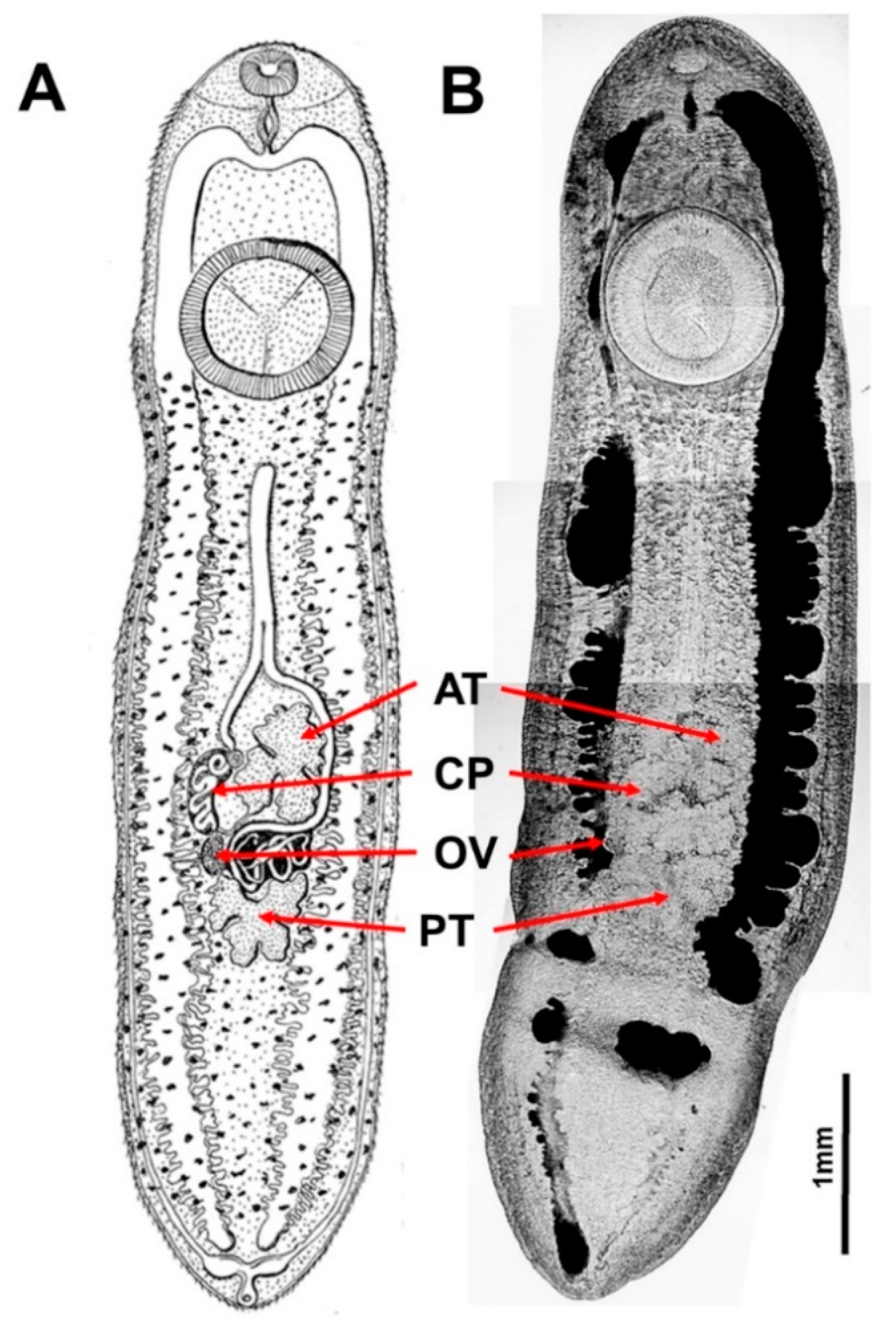
2.2. Morphological Examination of Metacercariae

Cysts were removed from the host and opened manually. Excysted metacercariae were first examined alive using a digital Keyence microscope 85 VHX-6000. The natural color of their intestine content (yellow vs. brownish green) was recorded, and digitally documented using Keyence VHX-H1M1 software. Then, each metacercaria was squeezed plane on a cover slipped microscope slide during formalin fixation. Unstained, but lactophenol-clarified specimens were examined using an Olympus BX 50 microscope equipped with the high-resolution camera Olympus DP20 for documentation with the Cell Imaging software. Complete morphometric features were taken from the digital images of 15 specimens collected from H. kivuensis and 21 obtained from H. viridiflavus. Analogous to previous studies [7,11] we measured [to the nearest µm] 16 distances: (1) Body Length (BL), (2) Body Width (BW), (3) Oral Sucker length (OSL), (4) Oral Sucker Width (OSW), (6) Ventral Sucker length (VSL), (7) Ventral Sucker width (VSW), (8) Distance between the outer rim of Suckers (DBS), (9) Anterior Testis Length (ATL), (10) Anterior Testis Width (ATW), (11) Posterior Testis Length (PTL), (12) Posterior Testis Width (PTW), (13) Ovary Length (OL), (14) Ovary Width (OW), (15) Cirrus Pouch Length (CPL), (16) Cirrus Pouch Width (CPW). From these morphometric landmarks we derived 8 ratios: (1) Body length/width (=BL/BW), (2) Oral sucker width/body width (=OSW/BW), (3) Ventral/Oral Sucker width (=VSW/OSW), (4) Ventral Sucker width/body width (=VSW/BW), (5) Anterior Testis width/length (=ATW/ATL), (6) Posterior Testis width/length (=PTW/PTL), (7) Ovary width/length (=OW/OL), (8) Cirrus Pouch length/Body length (=CPL/BL). Parasite terminology (prevalence, intensity of infections) is used in accordance with Bush et al (1997) [38].

2.3. Morphological and Demographic Traits of Frog Hosts

The carcasses of 174 reed frogs (101 HK, 73 HV) were examined for potential effects of metacercaria infestation on size, femur growth, femur histology and age. Each individual was sexed and the snout-vent length (SVL, distance between snout tip and cloaca) was measured to the nearest 0.1 mm using a caliper. Then, the right femur was removed completely and cleaned from attached muscle and cartilage tissue. The bone was dried at 70 °C for 48 h and weighed to the nearest 0.1 mg using a Sartorius precision balance type 1712004. Femur length (to the nearest 0.01 mm) was recorded digitally using a Keyence microscope 85 VHX-6000 with VHX-H1M1 software. Age and growth estimation based on the central 2–3 mm of femur diaphysis. Laboratory protocols followed the standard methods of skeletochronology [33,39]. The samples were embedded in Historesin™ (JUNG) and stained with 0.5% Cresylviolet. Diaphysis was cross-sectioned at 12 μm using a JUNG RM2055 rotation microtome. Cross sections were examined light microscopically for the presence of growth marks at magnifications of 400× using an OLYMPUS BX 50. We distinguished strongly stained lines of arrested growth (LAGs) in the periosteal bone, separated by faintly stained broad growth zones. We selected diaphysis sections in which the size of the medullar cavity was at its minimum and that of periosteal bone at its maximum. The number of LAGs was assessed independently by US and JMD to estimate age. Finally, we recorded the maximum width of periosteal and endosteal bone to the nearest µm in the cross section used for age estimation. Life-history traits recorded for each frog were: (1) overall growth as SVL, (2) jumping performance as femur length, (3) overall bone stability as quotient of femur mass and length, (4) age as number of LAGs, (5) bone growth as width of periosteal tissue, (6) bone stabilization by growth of endosteal tissue.

2.4. Statistical Procedures

Data distribution was checked first for normality. Deviations from normality were overcome by log10-transformation of original data. Descriptive statistics included arithmetic mean, corresponding standard error, minimum and maximum. We used the t-test for unequal variances to perform two group comparisons of means in morphometric and survival data, the non-parametric Mann–Whitney W-test for the two group comparisons of age. Multiple group comparison based on ANOVA. ANCOVA was used for the comparison of host traits (e.g., SVL, infestation intensity of different tissue) between host species, which were influenced by continuous covariates (e.g., collection date within the rainy season (1 October set to 1), total number of metacercariae per host specimen). Morphometrics of metacercariae obtained from distinct host species were compared using a principal component analysis on the standardized distances and ratios related to body and sucker size. We used the first three principal components to derive size and shape-related features. Significance level was set at alpha = 0.05. Statistics were calculated using the program package Statgraphics Centurion 18, version 18.1.13.

2.5. Nucleic Acid Extraction and Polymerase Chain Reactions

The ethanol-fixed trematodes were incubated in 1.5 mL reaction tubes, with open cap, at 40 °C in a Thermomixer (Eppendorf, Hamburg, Germany) until the ethanol was completely evaporated. The sample was mixed with 180 µL ATL buffer and 20 µL proteinase K, incubated at 56 °C until the specimen was completely lysed, and then further processed following the protocol for DNA purification from tissues of the QIAamp DNA mini kit (Qiagen, Hilden, Germany). The extracted DNA was used to amplify parts of nuclear ribosomal locus (TS1-5.8S-ITS2) and the mitochondrial cytochrome oxidase subunit 1 (COX1) locus (Table 1). Polymerase chain reaction (PCR) was conducted using Taq PCR core kit as recommended by the manufacturer (Qiagen). The following programs were used: 40 cycles of 1 min at 94 °C, 1 min at 56 °C and 1 min at 72 °C for ITS1-5.8S-ITS2; 40 cycles of 1 min at 94 °C, 1 min at 50 °C and 1 min at 72 °C for COX1. All PCR cycles were initiated with a denaturation step for 3 min at 94 °C and terminated with an extension step of 72 °C for 7 min. Amplicons were separated on a 1% agarose gel, stained with ethidium bromide, and visualized on a UV transilluminator. Prior to sequencing, PCR products were purified using QIAquick Purification Kit (Qiagen).

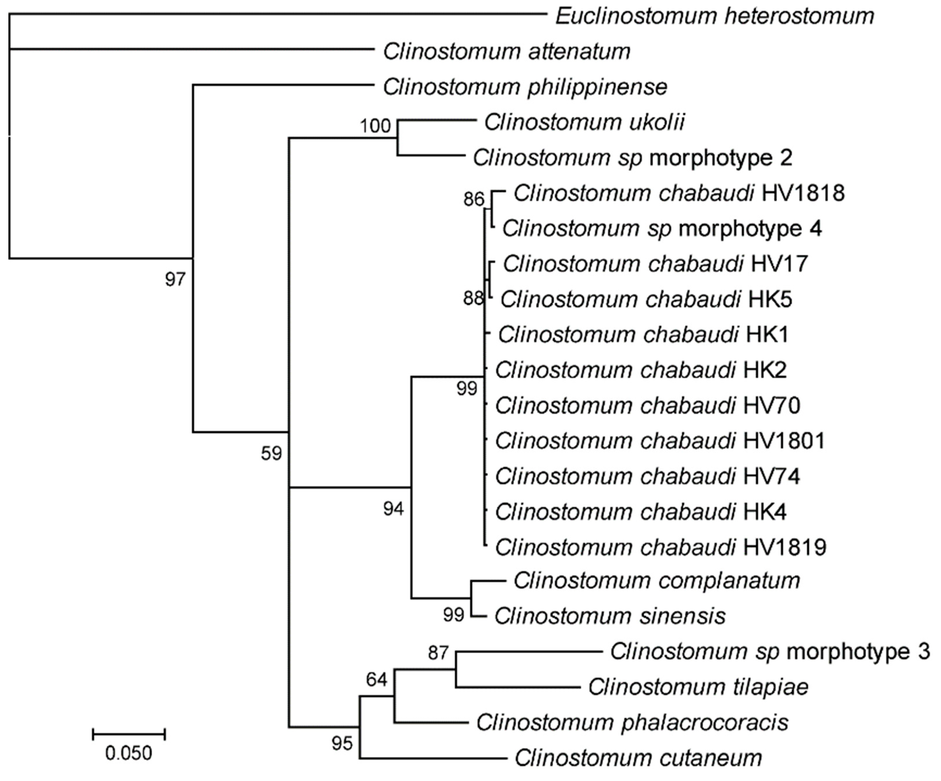
2.6. Sequencing and Phylogenetic Analysis

For bidirectional sequencing, the same primers as for the PCR were applied, except for COX1 fragments where M13 primers were used (Table 1). Assembly of DNA sequence files was conducted with DNA Baser (Heracle BioSoft, Mioveni, Romania) and primer sequences were clipped. Sequences were deposited in GenBank (Table 2). Closest matches of sequences were identified by a BLAST search against GenBank entries [40].
Phylogenetic trees were computed from the alignments using Bayesian interference in MrBayes [41]. Posterior probabilities were approximated over 1,000,000 generations. Positions containing gaps and missing data were eliminated. Trees were rooted by the sequences of outgroup species Clinostomum attenuatum and Euclinostomum heterostomum, retrieved from GenBank. The resulting trees were manually refined using Figure Tree v1.4.2.

3. Results

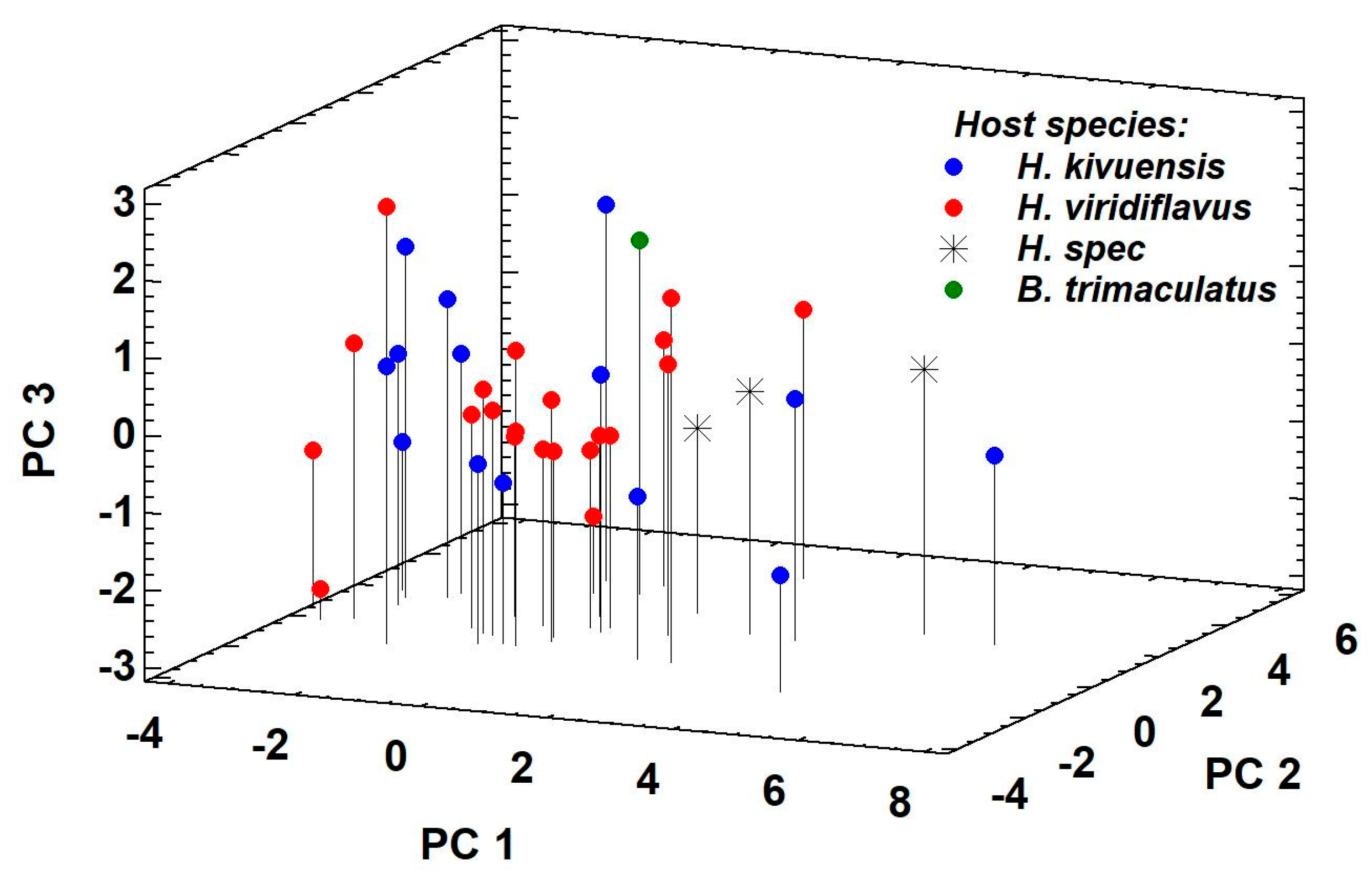
3.1. Morphological and Molecular Identification of Clinostomum Metacercaria

Forty-five reed frogs H. kivuensis and H. viridiflavus were infested by numerous Clinostomum metacercariae, which were morphologically identified as C. chabaudi Vercammmen-Grandjean, 1960 (Figure 1). Size range, sucker and genital morphology did not differ among the specimens collected from H. kivuensis and H. viridiflavus, and those collected from unidentified Hyperolius and Ptychadena frogs [11] and from the fish Barbus trimaculatus [7], respectively (Table 3).
Principal Component Analysis (PCA) based on 10 morphometric distances describing body size and sucker morphology revealed three PCs with an eigenvalue greater than 1, which accounted for 89.6% of total variance. PC1 (eigenvalue = 5.47, 54.8% of variance explained) represented the size-related aspects of the data set, PC2 (eigenvalue = 2.34, 23.4% of variance explained) the shape of the oral sucker, and PC3 (eigenvalue = 1.14, 11.4% of variance explained) the shape of the ventral sucker (Figure 2). Principal component scores of specimens originating from distinct host species overlapped completely. Genital morphology agreed with the original descriptions [7,11]. Testes were strongly digitated; anterior and posterior testes were located in the posterior part of the body. Testes were symmetrical, triangular, with two lateral and one posterior lobes trilobed. The cirrus pouch was banana-shaped, between margin of anterior testis and right caecum. The ovary was small, oval to kidney-shaped, in the intertesticular space below the cirrus pouch.
The identity of the metacercaria collected from H. kivuensis and H. viridiflavus with that described as morphotype 4 in Caffara et al. (2017) was confirmed comparing the ITS1-5.8S-ITS2 and COX1 partial sequences [7]. Whereas ITS1-5.8S-ITS2 sequences do no differentiate C. chabaudi from C. complanatum and C. sinensis (Figure S1 in supplement), the COX1 sequence is diagnostic revealing that C. chabaudi is the sister species of a clade formed by C. complanatum and C. sinensis (Figure 3). Based on morphological and molecular features we concluded the metacercariae belong to Clinostomum chabaudi Vercammen-Grandjean, 1960.

3.2. Infestation Ecology of C. chabaudi Metacercaria

Metacercaria cysts were easily detected in H. kivuensis because intestines were filled with a bright yellow substance in 80 specimens sampled, whereas in one specimen the intestine content turned out to be brownish green (Figure 4). In contrast, the intestine color distribution of metacercaria infesting H. viridiflavus was reversed with 82 specimens showing intestines colored brownish green, and only seven specimens with yellow intestine content.

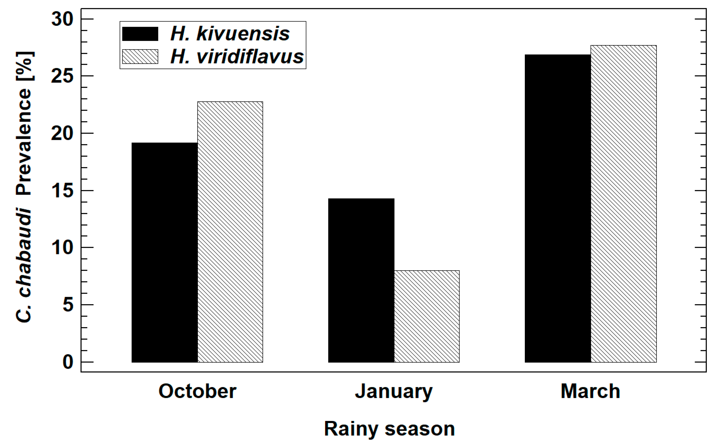
3.2.1. Prevalence and Infestation Intensity

Overall prevalence was considerably higher in Huye (HK 19.1%, HV 20.0%) compared to Musanze (HK 15.4%) and Muhanga (HV 10.3%; Table 4). In Huye, however, Clinostomum prevalence was significantly higher at the beginning and the end of the rainy season in comparison to the prevalence during the short intermediate dry period during December/January (ANOVA, F2,5 = 14.92, P = 0.0276; Figure 5). The intensity of infestation (log10-normalized data, least square mean of metacercaria per frog: 2.4) did not differ significantly between host species (ANCOVA, F1,44 = 0.02, P = 0.8822), considering the sampling dates during the rainy season as a continuous covariable (ANCOVA, F1,44 = 0.85, P = 0.3617).

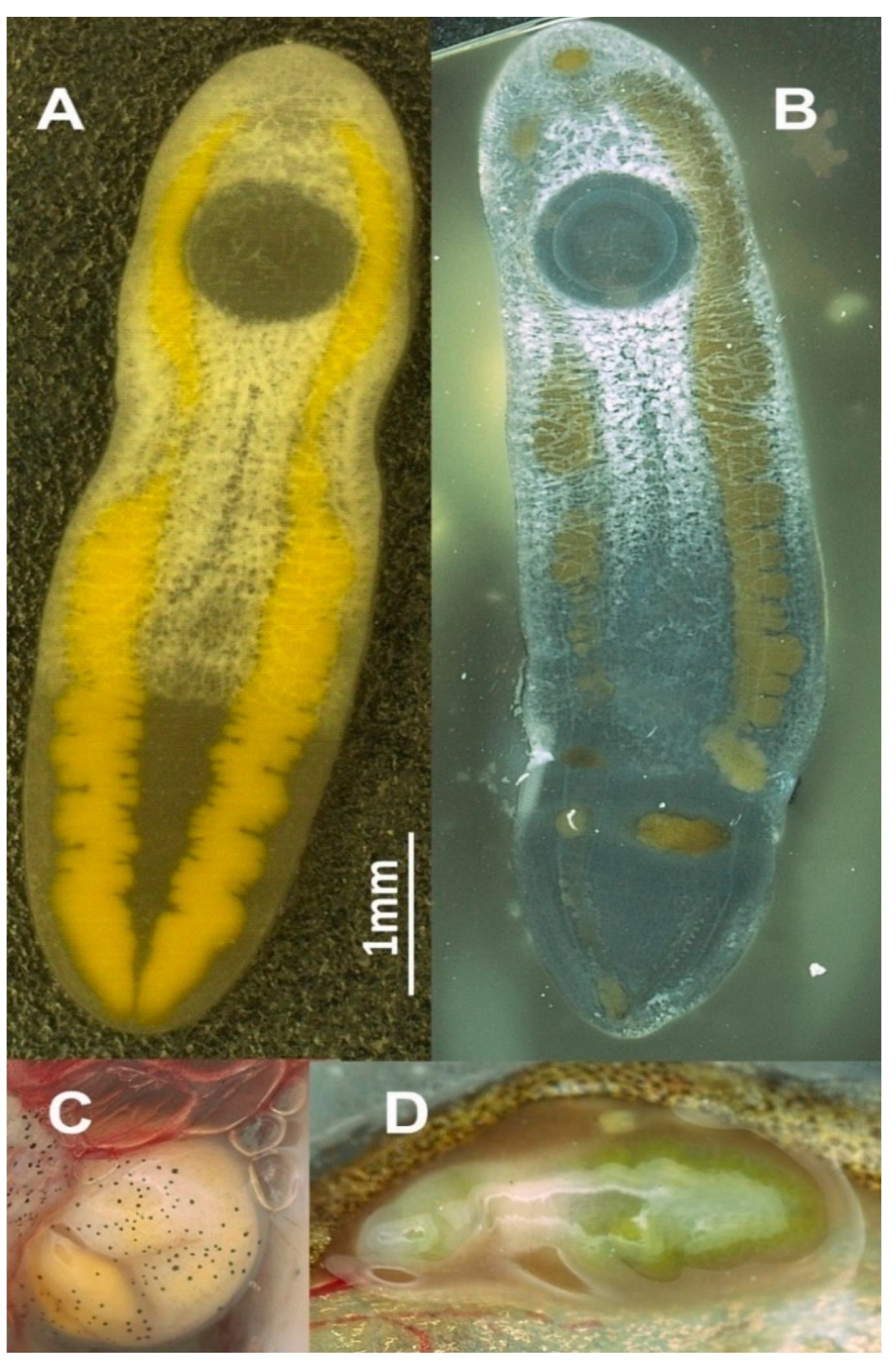
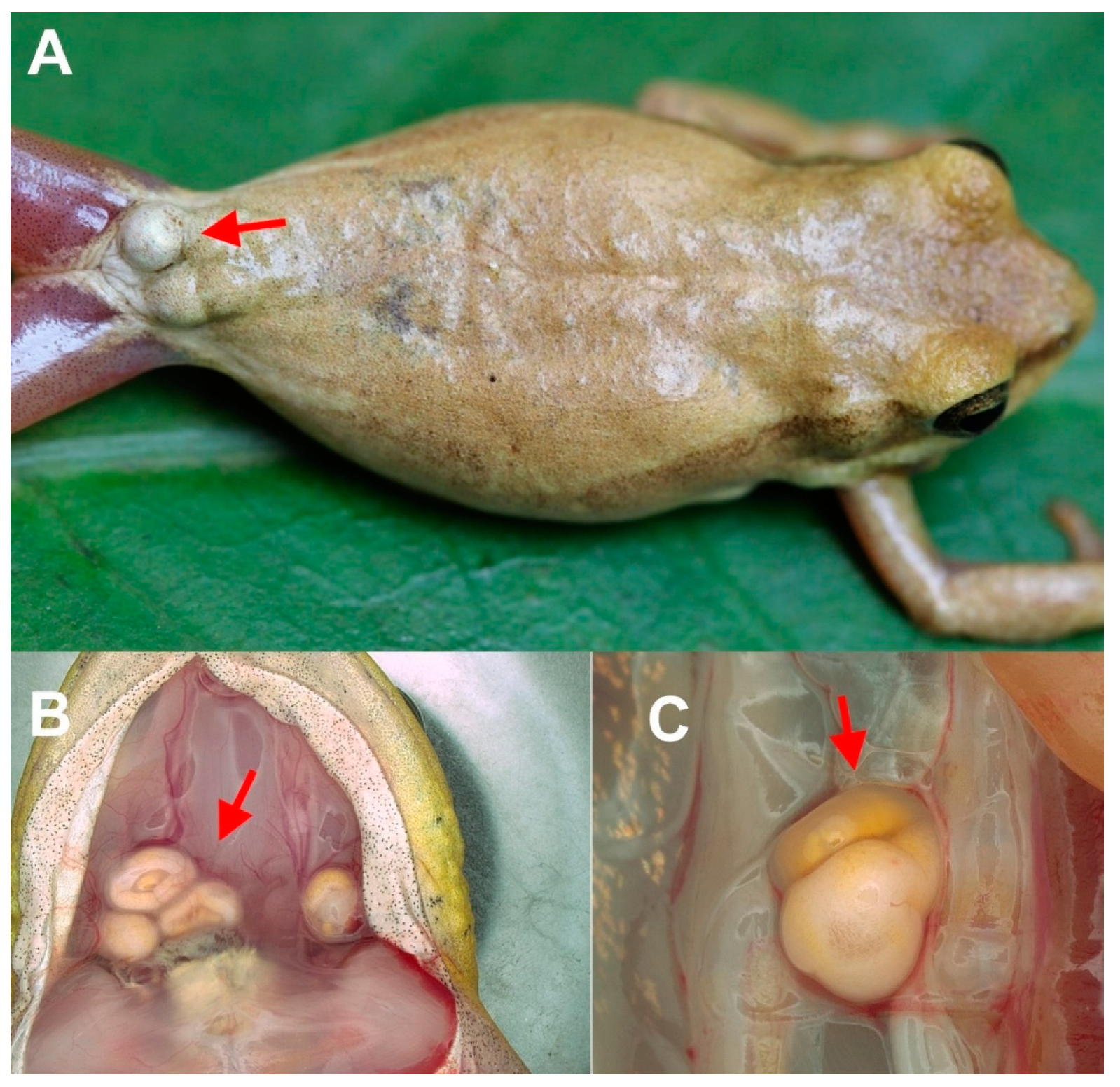
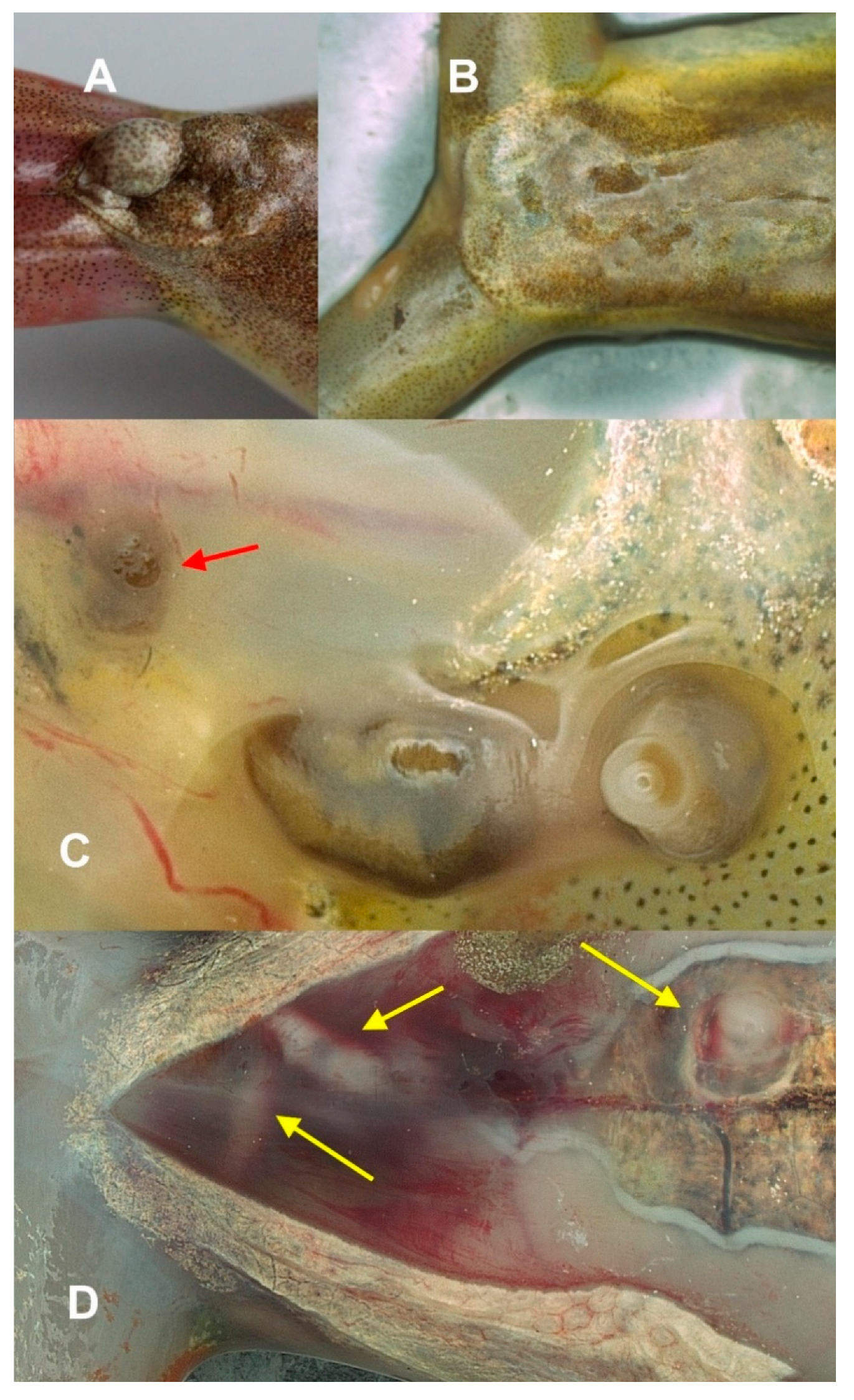
3.2.2. Metacercaria Location in the Host

The encystation sites of metacercariae were examined in 21 H. kivuensis and 24 H. viridiflavus. Most infested H. kivuensis carried cysts externally visible in skin and subdermal lymphatic sacs (81.0%; Figure 6A). Further examination of frogs yielded more cysts at the bottom of their mouth (57.1%; Figure 6B) and within the body cavity (28.6%, percentage does not add up to 100% because of simultaneous infestation at distinct organs; Figure 6C). We never found spontaneously excysted metacercariae moving freely in the body of the recently euthanized host, i.e., independent of the sampling date all metacercariae were found encysted.
External examination yielded only 25% of the infested H. viridiflavus specimens, with cysts or moving metacercariae in the subdermal lymphatic sacs near the urostyle (Figure 6A). Further cysts were located at the bottom of their mouth (50%) and within the body cavity (58.3%) of infested frogs, several with free moving metacercariae. The excystation of the metacercariae, still within the cyst pouch, began about half an hour following euthanization of the host. Unlike the metacercaria behavior in H. kivuensis, several specimens initially encysted within the subdermal lymphatic sac hatched spontaneously in the living frog host. In those frogs, held captive, spherical cysts located at the urostyle of the frog flattened after two to four weeks, metacercariae hatched, moved, and fed within subdermal lymphatic sacs (Figure 7A–C). Blood-filled wounds in the peritoneum suggested that some specimens originating from the subdermal lymphatic sacs had penetrated the body cavity (Figure 7D). Free moving metacercariae were often observed within the body cavity during examination of the euthanized frog hosts.
The quantitative distribution of metacercariae in the body differed significantly between the host species, when their number per body region (subdermal lymphatic sacs, mouth bottom, body cavity) was adjusted for the total number of cysts per specimen and the collection date in the rainy season in an ANCOVA. The skin and subdermal lymphatic sacs contained significantly more cysts in H. kivuensis than in H. viridiflavus (ANCOVA, F1,44 = 12.78, P = 0.0009; least square means ± SE: 2.18 ± 0.29 vs. 0.72 ± 0.27). The number of metacercariae within the body cavity of H. viridiflavus was significantly higher than in that of H. kivuensis (ANCOVA, F1,44 = 4.69, P = 0.0362; 1.50 ± 0.31 vs. 0.48 ± 0.34). In contrast, the number of cysts, located at the mouth bottom, was identical among the host species (mouth: ANCOVA, F1,44 = 0.97, P = 0.3301).

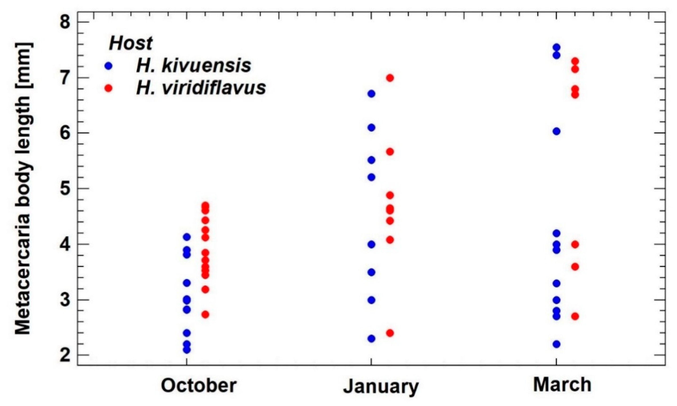
3.2.3. Metacercaria Growth

All metacercariae of both host species collected during the beginning of the rainy season in October showed a body length of 2.1–4.7 mm (Figure 8). Metacercariae within the October size range were also found in the January and March samples, but additionally increasingly larger specimens. The largest specimens collected in March were similar-sized to those collected by Vercammen-Grandjean (1960) in March 25 and April 3, 1950 [11].

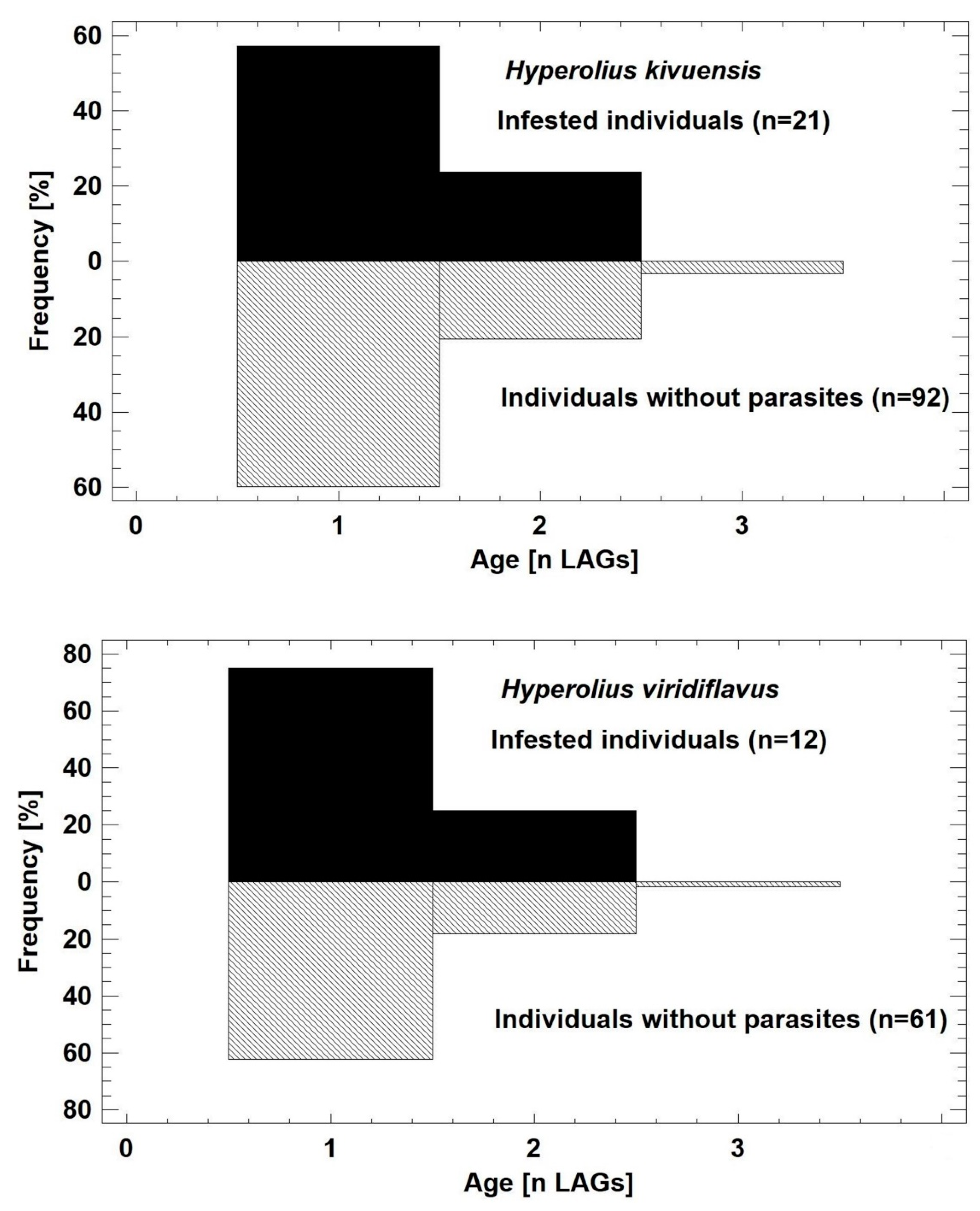
3.3. Morphological and Demographic Traits of Frog Hosts

Infestation of frog hosts started at the onset of the reproduction period in September/October when adult male frogs search for females in or near water bodies and pairs in amplexus deposit eggs masses. Age distribution of infested and metacercaria-free individuals did not differ significantly in H. kivuensis (Mann-Whitney W-test: W = 994.5, P = 0.8146) or in H. viridiflavus (W = 298.5, P = 0.2408; Figure 9). In H. kivuensis, infected individuals were significantly smaller than those without metacercariae, if SVL was adjusted for age and season (ANCOVA, F1,112 = 5.22, P = 0.0242; Table 5).
Frogs, collected at the beginning of rainy season, i.e., recently infested, and held captive for 60 d, did not differ with respect to average survival period (±SE) from metacercaria-free conspecifics. Infested H. kivuensis survived 47.6 ± 6.6d, whereas parasite-free individuals survived 57.6 ± 1.2d (t-test for unequal variances, t = 1.48, P = 0.1734). The corresponding data on H. viridiflavus were 49.8 ± 6.2d in infested and 52.1 ± 2.9d in parasite-free individuals (t-test for unequal variances, t = 0.34, P = 0.7450). Still, free-ranging, metacercariae-carrying frogs did not survive the dry season following initial infestation because adult frogs with fully grown metacercariae (larger than 5 mm body length) were never found in October.
Significant sublethal effects of infestation on bone growth were detected in the femora of H. kivuensis, but not in those of H. viridiflavus (Table 5). Femur length and femur mass did not differ between infested and parasite-free frogs, but the histological fine structure did. In the transverse femur sections of infested individuals, both periosteal and endosteal bone tissues were significantly thinner, i.e., metabolic investment in bone growth was less than in parasite-free H. kivuensis.

4. Discussion

In this study, we revealed that an infestation of Hyperolius frogs by Clinostomum chabaudi metacercariae may follow two alternative pathways, with either permanently sedentary encysted parasites (in H. kivuensis) or initially encysted and later free-ranging ones (in H. viridiflavus). This pattern seems to correspond to that observed in C. complanatum infesting fish hosts, with the common syndrome of encysted yellow grubs in muscle tissue or with excysted free-ranging worms in the body cavity [4,19,44,45]. Therefore, we propose that the chosen developmental strategy is related to the nutritional resources available to metacercaria in the host. In response to the strategy of the parasite, life-history traits of the frog host are affected in different ways. In addition to the details of host–parasite interactions, we succeeded in providing evidence for the taxonomic identity of the Clinostomum metacercaria.

4.1. Taxonomic Identification of Clinostomum Metacercariae Infesting Reed Frogs in Rwanda

Our morphological and molecular data profoundly indicate, that the metacercariae infesting the two reed frog species belong to the same species, despite of considerable and widely host-specific differences in coloration within the intestines and distinct development strategy within the frogs. General morphometry, genital morphology and COX1 gene sequence provide evidence for species identity. Agreement in morphology [11] and in partial COX1 sequences [7] show that this Clinostomum species has been described already in frog and fish hosts, and is identified as C. chabaudi Vercammen-Grandjean, 1960. The total number of 10 palearctic Clinostomum species remains the same because morphotype 4 is an unrecognized C. chabaudi [7,9,10]. Phylogenetically, C. chabaudi is the sister taxon of the clade formed by C. complanatum and C. sinensis [10].
Unfortunately, our knowledge on the complex life cycle of C. chabaudi is limited to the metacercaria stage, which infests at least four intermediate hosts, Hyperolius kivuensis, H. viridiflavus, Ptychadena spec. and Barbus trimaculatus [7,11]. The disjunct geographical distribution (DR Congo, Rwanda, South Africa) is most probably a result of “undersampling” and human-mediated dispersal due to fish aquaculture because infested frog populations inhabit wetlands in close vicinity to Oreochromis niloticus fish farms [46]. In Rwanda, the freshwater snails Pila ovata and Bulinus spec. are often found in these fishponds and are possible first intermediate hosts [47,48]. Fish- and frog-eating birds, potential definite hosts, are quite common in the wetlands and fish farms of Rwanda, but no infections with flukes in birds have been reported so far.

4.2. Alternative Metacercaria Life-Histories in Reed Frogs

In syntopic populations, prevalence and infestation intensity were similar in the two host frog species, as evidenced by parallel seasonal variation at the same locality (Table 4, Figure 5). The environmental cues guiding Clinostomum cercariae to potential second intermediate hosts are unknown, but if they are of chemical nature, the indiscriminate infection of distinct syntopic frog species (here Hyperolius spp, Ptychadena) may indicate that the environmental cue is not host-specific [11]. Prevalence seems to depend on local environmental factors, either on the abundance of the first intermediate host or its infestation rate. Following skin penetration of the cercaria and encystment, metacercaria development of C. chabaudi showed two alternative variations, the sedentary “yellow grub” lifestyle in H. kivuensis and the free-ranging “tissue damaging” one in H. viridiflavus.
In H. kivuensis, most infesting cercariae encysted within the lymphatic sacs or the mouth tissues, indicating that the host immune system limits effectively further penetration into the body cavity. Metacercariae remained motionless within the cysts, while growing to a maximum size of about 8 mm at the end of the rainy season. They most probably feed on dissolved nutrients gained from the host´s body fluid, as observed in the loach Misgurnus anguillicaudatus infested by C. complanatum [18]. In agreement with the observations in the loach, we never found cysts with damaged tissue layers or spontaneously excysted metacercariae indicating that host nutrients were absorbed directly through the metacercaria tegument and/or lymphatic fluid was ingested through the digestive tract [18]. The intensive yellow color of intestine fluid seems to be associated with the described type of nutrition [18,49]. The “yellow grub” developmental strategy seems to prevent activation of metacercaria in the live frog host, as cysts were always intact in recently euthanized frogs. We conclude that a signal may result from the death of the host to trigger excystation, i.e., usually the consumption by a potential definite host.
In H. viridiflavus, primary encystation sites of cercariae seem to be like those in H. kivuensis, but only 2–4 weeks following encystation in the lymphatic sacs, several metacercariae started moving first within the cyst (externally visible) and then excysted spontaneously. The reason for the diverging metacercaria behavior is probably poor nutrition because the typical yellow grub aspect was limited to few individuals quiescent within the cysts, whereas all individuals with movement activity had brownish green gut content indicating a distinct source of nutrition. Before hatching, the inner cyst layer was often damaged and bleeding (see Figure 7D), and free moving metacercariae excreted brown feces (see Figure 7C). Since blood and tissue feeding was observed in closely related C. complanatum metacercariae [22,23], color of gut content and of feces may indicate the use of blood cells for nutrition in C. chabaudi as well. Movements of metacercariae were always directed to the body cavity, which they reached by perforation of the peritoneum, resulting in heavy bleeding. In contrast, spontaneously excysted C. complanatum penetrate the body wall of the fish host to reach the outside water body [2,25,26,27]. The adaptive value of intra-host migrations is probably active foraging, because development and final body size did not differ from that of the sedentary yellow grubs.

4.3. Metacercaria Impact on the Life-History of Reed Frogs

Our study provides evidence that alternative metacercaria behavior (sedentary/free-ranging) and nutrition (lymphatic fluid/blood and tissue) impose distinct effects on the frog host. Regarding H. kivuensis exposed to sedentary metacercariae feeding on lymphatic fluid, the length growth (SVL) is reduced in time, and bones remain thinner than in parasite-free individuals, indicating a substantial allocation of host resources to the parasite. The distinct morphological manifestations of resource deficiency in the frog suggests a parasitic relationship with a prolonged co-development of host and parasite. In fact, the short-term host survival (<2 months) in captivity did not differ between infested and parasite-free hosts, but that does not rule out the possibility that infested specimens are more vulnerable to predation in field, as observed in fish hosts [4,13]. The absence of frogs infested by large (=fully grown) metacercariae at the beginning of the rainy season indicates that long-term host survival (>1 year) is rare, if possible, at all. Since it is hardly conceivable that all infested frogs die due to predation, the continuous loss of resources to the parasite may starve the hosts to death during the dry season, as observed in hibernating newts infested by Strigea robusta [50]. Thus, the association between H. kivuensis and C. chabaudi is limited to a single host reproduction period in which breeding behavior increases visibility of infested specimens to predators (=potential definitive hosts of the metacercaria).
To our surprise, growth traits of H. viridiflavus did not show any adverse impact from metacercariae, despite of their ferocious lifestyle including blood feeding, tissue damaging and intra-host migrations. Moreover, unaffected short-term host survival in captivity and the detection of fully-grown metacercariae within the body cavity of these frogs in field at the end of the rainy season emphasize that a prolonged co-development of host and parasite has occurred, i.e., the absence of morphological manifestations in host traits is probably due a lower transfer of resources to the parasite. As in H. kivuensis, host survival to the next rainy season does not seem to occur. The unknown defense mechanism of H. viridiflavus forces obviously most metacercariae to change the common yellow grub development strategy to the invasive tissue-damaging one.

5. Conclusions

Metacercaria development in fish hosts (C. complanatum) and in frogs (C. chabaudi) follows the same alternative pathways. Depending on the host´s defensive behavior and the availability of resources, metacercariae choose between the sedentary yellow grub mode of life or the actively foraging one. Interestingly, this is an individual “decision” of the parasite because in H. viridiflavus the two modes co-occurred sometimes in the same host individual. Anecdotal data on other Clinostomum species suggest that the sedentary yellow grub pathway is the most common one [21,49,51,52,53,54]. Yet, the alternative free-ranging developmental mode has also been observed in C. giganticum and in C. piscidium [52,55]. We hypothesize that the presence of alternative development modes is a common feature of all Clinostomum metacercariae and represents an adaptation to cope with resource limitation within host-produced cysts.

Supplementary Materials

The following are available online at https://www.mdpi.com/1424-2818/13/2/93/s1, Figure S1: Bayesian tree of ITS1-5.8rDNA-ITS2 sequence from African Clinostomum species. The South American Euclinostomum heterostomum is used as outgroup. Posterior probability values are indicated. Scale bar gives substitutions per site.

Author Contributions

Conceptualization, U.S., J.M.D., P.S. and C.B.; methodology, U.S.; software, U.S. and C.B.; validation, U.S.; formal analysis, U.S.; investigation, U.S., J.M.D., P.S. and C.B.; resources, U.S. and C.B.; data curation, U.S. and C.B.; writing-original draft preparation, U.S., J.M.D., P.S. and C.B.; writing-review and editing, U.S., J.M.D., P.S. and C.B.; visualization, U.S. and C.B.; supervision, U.S.; and project administration, U.S. All authors have read and agreed to the published version of the manuscript.

Funding

This research received no external funding.

Institutional Review Board Statement

The study was conducted according to the guidelines of the Declaration of Helsinki, and approved by the ethic committee of FB3 at the University of Koblenz-Landau (approval # Si 2015/01 at 24 June 2015).

Data Availability Statement

Data is contained within the article, host and parasite specimens analyzed in this study are deposited in the natural history Museums of Bonn and Hamburg, Germany (collections numbers within the article), and are available on request to the collection managers.

Acknowledgments

We are indebted to Bonny Dumbo for assistance during the collection of Hyperolius in the field. Collecting and export permits (Nos. 2/ORTPN/V.U/09, 08/RDB-T&C/V.U/12, 18/RDB-T&C/V.U/12 and 10/RDB-T&C/V.U/15) of Hyperolius for parasitological screening were issued by the Rwandan Development Board. B. Nilow-Lange, L. Schober, and L. Schürmann provided help in the skeletochronogical laboratory procedures. The manuscript benefited from the comments of two anonymous reviewers.

Conflicts of Interest

The authors declare no conflict of interest.

References

  1. Kanev, I.; Radev, V.; Fried, B. Family Clinostomidae Lühe, 1901. In Keys to the Trematoda; Gibson, D.I., Jones, A., Bray, R.A., Eds.; CAB International and the Natural History Museum: Wallingford, UK, 2002; Volume 1, pp. 113–120. [Google Scholar]
  2. Lo, C.F.; Huber, F.; Kou, G.H.; Lo, C.J. Studies of Clinostomum complanatum (RUD., 1819). Fish Pathol. 1981, 15, 219–227. [Google Scholar] [CrossRef]
  3. Aohagi, Y.; Shibahara, T.; Machida, N.; Yamaga, Y.; Kagota, K.; Hayashi, T. Natural infections of Clinostomum complanatum (Trematoda, Clinostomatidae) in wild herons and egrets, Tottori prefecture, Japan. J. Wildl. Dis. 1992, 28, 470–471. [Google Scholar] [CrossRef] [Green Version]
  4. Dias, M.L.; Eiras, J.C.; Machado, M.H.; Souza, G.T.; Pavanelli, G.C. The life cycle of Clinostomum complanatum Rudolphi, 1814 (Digenea, Clinostomidae) on the floodplain of the high Parana river, Brazil. Parasitol. Res. 2003, 89, 506–508. [Google Scholar] [CrossRef]
  5. Caffara, M.; Davidovich, N.; Falk, R.; Smirnov, M.; Ofek, T.; Cummings, D.; Gustinelli, A.; Fioravanti, M.L. Redescription of Clinostomum phalacrocoracis metacercariae (Digenea: Clinostomidae) in cichlids from Lake Kinneret, Israel. Parasite. 2014, 21. [Google Scholar] [CrossRef] [Green Version]
  6. Caffara, M.; Locke, S.A.; Gustinelli, A.; Marcogliese, D.J.; Fioravanti, M.L. Morphological and molecular differentiation of Clinostomum complanatum and Clinostomum marginatum (Digenea: Clinostomidae) metacercariae and adults. J. Parasitol. 2011, 97, 884–891. [Google Scholar] [CrossRef]
  7. Caffara, M.; Locke, S.A.; Echi, P.C.; Halajian, A.; Benini, D.; Luus-Powell, W.J.; Tavakol, S.; Fioravanti, M.L. A morphological and molecular study of Clinostomid metacercariae from African fish with a redescription of Clinostomum tilapiae. Parasitol. 2017, 144, 1519–1529. [Google Scholar] [CrossRef] [PubMed]
  8. Locke, S.A.; Caffara, M.; Marcogliese, D.J.; Fioravanti, M.L. A large-scale molecular survey of Clinostomum (Digenea, Clinostomidae). Zool. Scr. 2015, 44, 203–217. [Google Scholar] [CrossRef]
  9. Caffara, M.; Locke, S.A.; Echi, P.C.; Halajian, A.; Luus-Powell, W.J.; Benini, D.; Tedesco, P.; Fioravanti, M.L. A new species of Clinostomum Leidy, 1856 based on molecular and morphological analysis of metacercariae from African siluriform fishes. Parasitol. Res. 2020, 119, 885–892. [Google Scholar] [CrossRef] [PubMed]
  10. Locke, S.A.; Caffara, M.; Barčák, D.; Sonko, P.; Tedesco, P.; Fioravanti, M.L.; Li, W. A new species of Clinostomum Leidy, 1856 in East Asia based on genomic and morphological data. Parasitol. Res. 2019, 118, 3253–3265. [Google Scholar] [CrossRef] [PubMed]
  11. Vercammen-Grandjean, P.H. Les Trématodes du Lac Kivu sud (Vermes). Annales Musée Royale de l’Afrique Centrale 1960, 5, 1–171. [Google Scholar]
  12. Bush, A.O.; Fernández, J.C.; Esch, G.W.; Seed, J.R.; Ndez, J.C.F. Parasitism: The Diversity and Ecology Animal Parasites; University Press: Cambridge, UK, 2001. [Google Scholar]
  13. Nicola, D.N.; Affonso, I.P.; Takemoto, R.M. Behavioral changes in host foraging: Experiments with Clinostomum (Trematoda, Digenea) parasitizing Loricariichthys platymetopon (Loricariidae). Exp. Parasitol. 2020, 216. [Google Scholar] [CrossRef] [PubMed]
  14. Hazen, T.C.; Esch, G.W. Observations on ecology of Clinostomum marginatum in largemouth bass (Micropterus salmoides). J. Fish Biol. 1978, 12, 411–420. [Google Scholar] [CrossRef]
  15. Silva-Souza, A.T.; Ludwig, G. Parasitism of Cichlasoma paranaense Kullander, 1983 and Gymnotus carapo Linnaeus, 1814 by Clinostomum complanatum (Rudolphi, 1814) Metacercariae in the Taquari river. Braz. J. Biol. 2005, 65, 513–519. [Google Scholar] [CrossRef] [Green Version]
  16. Li, B.F.; Liu, X.-H.; Ge, H.-L.; Xie, C.-Y.; Cai, R.-Y.; Hu, Z.-C.; Zhang, Y.-G.; Wang, Z.-J. The discovery of Clinostomum complanatum metacercariae in farmed Chinese sucker, Myxocyprinus Asiaticus. Aquac. 2018, 495, 273–280. [Google Scholar] [CrossRef]
  17. Fedorčák, J.; Šmiga, Ľ.; Kutsokon, I.; Kolarčik, V.; Koščová, L.; Oros, M.; Koščo, J. Parasitic infection of Cobitis elongatoides Băcescu & Mayer, 1969 by zoonotic metacercariae Clinostomum complanatum (Rudolphi, 1814). J. Fish Dis. 2019, 42, 1677–1685. [Google Scholar] [CrossRef]
  18. Lo, C.-F.; Chen, S.-C.; Wang, C.-H. The study of Clinostomum complanatum (Rud., 1814) V. The influences of metacercaria of Clinostomum complanatum on fish. Fish Pathol. 1985, 20, 305–312. [Google Scholar] [CrossRef]
  19. Shareef, P.; Abidi, S. Incidence and histopathology of encysted progenetic metacercaria of Clinostomum complanatum (Digenea: Clinostomidae) in Channa punctatus and its development in experimental host. Asian Pac. J. Trop. Biomed. 2012, 2, 421–426. [Google Scholar] [CrossRef] [Green Version]
  20. de Lima, H.L.; Stefani, L.M.; Pedron, F.D.A.; Baldissera, M.D.; da Silva, A.S. Pro-Inflammatory Cytokines in the Serum of Silver Catfish (Rhamdia quelen) Naturally Infected by Clinostomum complanatum: A Preliminary Study. J. Parasitol. 2014, 100, 142–143. [Google Scholar] [CrossRef]
  21. Montes, M.M.; Plaul, S.E.; Croci, Y.; Waldbillig, M.; Ferrari, W.; Topa, E.; Martorelli, S.R. Pathology associated with three new Clinostomum metacercariae from Argentina with morphological and DNA barcode identification. J. Helminthol 2020, 94, e148. [Google Scholar] [CrossRef] [PubMed]
  22. Mendoza, J.M.; Sarabia, D.O.; Lopez, R.C.; Lopez, J.F. Helmintos del pez Dormitator maculatus (Osteichthyes: Eleotridae) de Alvarado, Veracruz, México. Rev. De Biol. Trop. 2004, 52, 393–396. [Google Scholar] [CrossRef] [Green Version]
  23. Kaur, P.; Qureshi, T.; Shrivastav, R.; Manohar, S.; Bilal, A. Histopathological and haematological investigations on Nandus nandus (Ham.) parasitized by metacercariae of Clinostomum complanatum (Rudolphi, 1819). Int. J. Environ. Sci. 2012, 2, 1324–1330. [Google Scholar]
  24. Kalantan, A.M.N.; Arfin, M.; Nizami, W.A. Histochemical nature and origin of the metacercarial cyst of Clinostomum complanatum (Trematoda: Digenea). Trans. the Am. Microsc. Soc. 1986, 105, 290–295. [Google Scholar] [CrossRef]
  25. Lo, C.F.; Wang, C.H.; Ogawa, K.; Wakabayashi, H. The mechanism of the metacercarial excystment of Clinostomum complanatum (Rud 1814). Fish Pathol. 1987, 22, 227–235. [Google Scholar] [CrossRef]
  26. Szalai, A.J.; Dick, T.A. Helminths of stocked rainbow-trout (Salmo gairdneri) with special reference to Clinostomum complanatum. J. Wildl. Dis. 1988, 24, 456–460. [Google Scholar] [CrossRef] [PubMed]
  27. Yasumoto, S.; Kabayama, T.; Kondo, M.; Takahashi, Y. Mass Mortalities of Goldfish Carassius auratus Infected with Clinostomum Metacercariae, Associated with Elevated Water Temperature. Fish Pathol. 2018, 53, 44–47. [Google Scholar] [CrossRef] [Green Version]
  28. Caffara, M.; Bruni, G.; Paoletti, C.; Gustinelli, A.; Fioravanti, M.L. Metacercariae of Clinostomum complanatum (Trematoda: Digenea) in European newts Triturus carnifex and Lissotriton vulgaris (Caudata: Salamandridae). J. Helminthol 2014, 88, 278–285. [Google Scholar] [CrossRef]
  29. Etges, F.J. Clinostomum attenuatum (Digenea) from the Eye of Bufo marinus. J. Parasitol. 1991, 77, 634–635. [Google Scholar] [CrossRef]
  30. Perpiñán, D.; Garner, M.M.; Trupkiewicz, J.G.; Malarchik, J.; Armstrong, D.L.; Lucio-Forster, A.; Bowman, D.D. Scoliosis in a tiger salamander (Ambystoma tigrinum) associated with encysted digenetic trematodes of the genus Clinostomum. J. Wildl. Dis. 2010, 46, 579–584. [Google Scholar] [CrossRef]
  31. Bruni, G.; Angelini, C. No short term effect of Clinostomum complanatum (Trematoda: Digenea: Clinostomatidae) on survival of Triturus carnifex (Amphibia: Urodela: Salamandridae). Acta Herpetol 2016, 11, 75–79. [Google Scholar] [CrossRef]
  32. Sinsch, U.; Lümkemann, K.; Rosar, K.; Schwarz, C.; Dehling, J.M. Acoustic niche partitioning in an anuran community inhabiting an Afromontane wetland (Butare, Rwanda). Afr. Zool. 2012, 47, 60–73. [Google Scholar] [CrossRef]
  33. Sinsch, U.; Dehling, J.M. Tropical anurans mature early and die young: Evidence from eight Afromontane Hyperolius species and a meta-analysis. PLOS ONE 2017, 12, e0171666. [Google Scholar] [CrossRef] [PubMed] [Green Version]
  34. De Leon, G.P.P.; Garcia-Varela, M.; Pinacho-Pinacho, C.D.; Sereno-Uribe, A.L.; Poulin, R. Species delimitation in trematodes using DNA sequences: Middle-American Clinostomum as a case study. Parasitol. 2016, 143, 1773–1789. [Google Scholar] [CrossRef] [Green Version]
  35. Tumushimire, L.; Mindje, M.; Sinsch, U.; Dehling, M.J. Anuran diversity of cultivated wetlands in Rwanda: Melting pot of generalists? Salamandra 2020, 56, 99–112. [Google Scholar]
  36. Sinsch, U.; Dehling, J.M.; Scheid, P.; Balczun, C. A new African species of parasitic Dero (Annelida, Clitellata, Naididae) in the urinary tract of reed frogs. Parasitol. Res. 2019, 118, 3359–3370. [Google Scholar] [CrossRef] [PubMed]
  37. Sinsch, U.; Dehling, J.M.; Scheid, P.; Balczun, C. Molecular Diversity of Nematode Parasites in Afrotropical Reed Frogs (Hyperolius spp.). Diversity 2020, 12, 265. [Google Scholar] [CrossRef]
  38. Bush, A.; Lafferty, K.; Lotz, J.; Shostak, A. Parasitology meets ecology on its own terms: Margolis et al. revisited. J. Parasitol. 1997, 83, 575–583. [Google Scholar] [CrossRef] [PubMed]
  39. Sinsch, U. Review: Skeletochronological assessment of demographic life-history traits in amphibians. Herpetol. J 2015, 25, 5–13. [Google Scholar]
  40. Altschul, S.F.; Gish, W.; Miller, W.; Myers, E.W.; Lipman, D.J. Basic local alignment search tool. J. Mol. Biol. 1990, 215, 403–410. [Google Scholar] [CrossRef]
  41. Huelsenbeck, J.P.; Ronquist, F. MRBAYES: Bayesian inference of phylogenetic trees. Bioinform 2001, 17, 754–755. [Google Scholar] [CrossRef] [Green Version]
  42. White, T.J.; Bruns, T.; Lee, S.; Taylor, J. Amplification and direct sequencing of fungal ribosomal RNA genes for phylogenetics. In PCR Protocols: A Guide to Methods and Applications; Innis, M.A., Gelfand, D.H., Sninsky, J.J., White, T.J., Eds.; Academic Press: New York, NY, USA, 1990; Volume 18, pp. 315–322. [Google Scholar] [CrossRef]
  43. Moszczynska, A.; Locke, S.A.; McLaughlin, J.D.; Marcogliese, D.J.; Crease, T.J. Development of primers for the mitochondrial cytochrome c oxidase I gene in digenetic trematodes (Platyhelminthes) illustrates the challenge of barcoding parasitic helminths. Mol. Ecol. Resour. 2009, 9, 75–82. [Google Scholar] [CrossRef]
  44. Siddiqui, A.; Nizami, W. Seasonal population dynamics of the metacercariae of Clinostomum complanatum (Trematoda: Digenea) in relation to sex of the host. Riv. di Parassitol. 1982, 43, 275–279. [Google Scholar]
  45. Kalantan, A.; Arfin, M.; Nizami, W.A. Seasonal incidence and pathogenicity of the metacercariae of Clinostomum complanatum in Aphanius dispar. Jpn. J. Parasitol. 1987, 36, 17–23. [Google Scholar]
  46. Tabaro, S.R.; Mutanga, O.; Rugege, D.; Micha, J.C. Optimum rabbit density over fish ponds to optimise Nile tilapia production in an integrated rabbit–fish system in Rwanda. Afr. J. Aquat. Sci. 2012, 37, 165–174. [Google Scholar] [CrossRef]
  47. Brown, D.S. Freshwater Snails of Africa and Their Medical Importance; Taylor and Francis Ltd.: London, UK, 1980. [Google Scholar]
  48. Isabwe, A.; Ruberanziza, E.; Mupfasoni, D.; Ruxin, J.; Clerinx, J.; White, P. Potential for transmission of schistosomiasis in Kayonza district. Rwanda Med J. 2012, 69, 14–19. [Google Scholar]
  49. Osborn, H.L. On the structure of Clinostomum marginatum, a trematode parasite of the frog, bass and heron. J. Morphol. 1912, 23, 189–229. [Google Scholar] [CrossRef] [Green Version]
  50. Sinsch, U.; Kaschek, J.; Wiebe, J. Heavy metacercariae infestation (Parastrigea robusta) promotes the decline of a smooth newt population (Lissotriton vulgaris). Salamandra 2018, 54, 210–221. [Google Scholar]
  51. Acosta, A.A.; Caffara, M.; Fioravanti, M.L.; Utsunomia, R.; Zago, A.C.; Franceschini, L.; da Silva, R.J. Morphological and Molecular Characterization of Clinostomum detruncatum (Trematoda: Clinostomidae) Metacercariae Infecting Synbranchus marmoratus. J. Parasitol. 2016, 102, 151–156. [Google Scholar] [CrossRef]
  52. Agarwal, S.M. Studies on the morphology, systematics and life history of Clinostomum giganticum n.sp. (Trematoda: Clinostomatidae). Indian J. Helminthol 1960, 11, 75–115. [Google Scholar]
  53. Ukoli, F.M.A. On Clinostomum tilapiae n. sp., and C. phalacrocoracis Dubois, 1931 from Ghana, and a Discussion of the Systematics of the Genus Clinostomum Leidy, 1856. J. Helminthol 1966, 40, 187–214. [Google Scholar] [CrossRef]
  54. Ahmad, F.; Sofi, O.; Sofi, T.A.; Fazili, K.M.; Sheikh, B.A. Morphological, molecular and karyological analysis of Clinostomum schizothoraxi kaw, 1950 (Digenea) from fishes of Kashmir Valley, India. Int. J. Pure Appl. Zool. 2017, 5, 33–44. [Google Scholar]
  55. Tansatit, T.; Sobhon, P.; Sahaphong, S.; Sangsuriya, P.; Klinsrithong, S. Prevalence and Histopathology of Trichogaster pectoralis Harbouring Metacercaria of Clinostomum piscidium (Southwell and Prashad, 1918) in Central Thailand. Thai J. Vet. Med. 2014, 44, 223–230. [Google Scholar]
Figure 1. Ventral view on metacercariae of Clinostomum chabaudi. (A) Modified schematic drawing from Vercammen-Grandjean (1960) [11]. (B) Photograph of a fully grown, live metacercaria infesting the dorsal lymphatic sac of a H. viridiflavus male in March 2017. Scale bar = 1 mm. Genital complex, abbreviations: AT = anterior testis, CP = cirrus pouch, OV = ovary, PT = posterior testis.
Diversity 13 00093 g001
Figure 2. Morphometric features of Clinostomum chabaudi metacercariae collected from four host species. Specimen data are given as principal component scores derived from variables describing body size and sucker morphology (for details see text). Each dot represents a single specimen, the asterisks the type specimens in Vercammen-Grandjean (1960) [11].
Diversity 13 00093 g002
Figure 3. Molecular identification of Clinostomum chabaudi based on COX1 sequences and phylogenetic relationships among the palearctic Clinostomum species by Bayesian inference. The South American Clinostomum attenuatum and Euclinostomum heterostomum are used as outgroups (Table 2). Posterior probability values are indicated. Scale bar gives substitutions per site.
Diversity 13 00093 g003
Figure 4. Color variation of the gut content in fully grown metacercaria C. chabaudii (body length 7–8 mm). (A,C) Specimen with yellow content of intestines collected from H. kivuensis. (B,D) Brownish green counterpart collected from H. viridiflavus. Excysted (A,B) and encysted specimens (C,D) are shown in life. Note that the specimen shown in (B) was illuminated by transmitted light in contrast to the other specimens, which were illuminated by incident light.
Diversity 13 00093 g004
Figure 5. Seasonal variation of metacercaria prevalence in the reed frogs at Huye.
Diversity 13 00093 g005
Figure 6. Location of Clinostomum cysts in reed frogs. (A) Dorsal view on a male H. viridiflavus with five cysts (red arrows) near the urostyle. (B) View on the mouth bottom of a male H. kivuensis with four cysts (red arrows). (C) Intact cyst attached to the vertebral column within the body cavity of a male H. kivuensis.
Diversity 13 00093 g006
Figure 7. Intra-host migration of C. chabaudi metacercaria in a male H. viridiflavus, starting from cysts in the subdermal lymphatic sacs to the abdominal body cavity. (A) Metacercaria cysts at the urostyle (October 26, 2015). (B) Hatched metacercariae moving within the lymphatic sac (November 30, 2015). (C) Free moving metacercaria and its brown feces (red arrow) within the dorsal lymphatic sac. (D) Ventral view on abdominal peritoneum. Two metacercariae (left yellow arrows) move within the bleeding body cavity. A third metacercaria is hatching from a cyst within the peritoneum.
Diversity 13 00093 g007
Figure 8. Seasonal body length variation of metacercaria size collected from reed frogs at Huye. Each dot represents one specimen. At each sampling date, specimens originating from 3–5 host frogs per species were measured.
Diversity 13 00093 g008
Figure 9. Age distribution of infested and metacercaria-free reed frogs H. kivuensis and H. viridiflavus. Details on statistical comparison are given in the text.
Diversity 13 00093 g009
Table 1. Primers used for amplification and sequencing. The M13 sequences [M13(-21)F and M13(-27)R] at the 5’ ends of the degenerated COX1 primers are underlined.
GenePrimerSequenceReference
ITS1-5.8S-ITS2ITS15′-TCCGTAGGTGAACCTGCGG-3′[42]
ITS45′-TCCTCCGCTTATTGATATGC-3′[42]
COIMplatCOX1dF5′-TGTAAAACGACGGCCAGTTTWCITTRGATCATAAG-3′[43]
MplatCOX1dR5′-CAGGAAACAGCTATGACTGAAAYAAYAIIGGATCICCACC-3′[43]
Table 2. Accession numbers of nuclear ribosomal sequences and mitochondrial COX1 sequences used for phylogenetic reconstruction. Reed frog hosts and their individual collection number: HK = Hyperolius kivuensis; HV = H. viridiflavus. Original references to sequences from palearctic Clinostomum retrieved from GenBank and used for comparison are [7,10].
Trematode Species/Host SpecimenLocus
ITS1-5.8S-ITS2COX1
Clinostomum chabaudi
    ex HK1MW528862MW525123
    ex HK2MW528861MW525125
    ex HK4MW528859MW525129
    ex HK 5MW528855MW525124
    ex HV17MW528854MW525122
    ex HV 70MW528856MW525126
    ex HV 74MW528857MW525128
    ex HV 1801MW528858MW525127
    ex HV 1818MW528860MW525121
    ex HV 1819MW528863MW525130
Morphotype 4 ex B. trimaculatusKY865644KY865661
Palearctic Species:
Morphotype 2KY865645KY865662
Morphotype 3KY865648KY865667
Clinostomum complanatumKM518259KM518253
Clinostomum cutaneumKP110564KP110515
Clinostomum phalacrocoracisKJ786975KJ786967
Clinostomum phillipinese-KP110523
Clinostomum sinensisMK796826MK801711
Clinostomum tilapiaeKY649349KY649357
Clinostomum ukoliiKY865623KY865641
Outgroup Species:
Clinostomum attenuatum-JF718587
Euclinostomum heterostomumKP721439KP721421
Table 3. Morphometric features of Clinostomum chabaudi metacercariae collected from different host species. Data are given as the minimum-maximum range, arithmetic mean and corresponding standard deviation. Abbreviations: BL = Body Length, BW = Body Width, OSL = Oral Sucker length, OSW = Oral Sucker Width, VSL = Ventral Sucker length, VSW = Ventral Sucker width, DBS = Distance between Sucker, ATL = Anterior Testis Length, ATW = Anterior Testis Width, PTL = Posterior Testis Length, PTW = Posterior Testis Width, OL = Ovary Length, OW = Ovary Width, CPL = Cirrus Pouch Length, CPW = Cirrus Pouch Width. * measurements given as diameter; n.a. = not available.
HostH. kivuensisH. viridiflavusHyperolius sp.Barbus trimaculatus
Metacercaria N152131
SourceThis studyThis study[11][7]
Size features:
BL [µm]2205–7544 (4496 ± 1778)2735–7159 (4331 ± 928)4778–7380 (6262 ± 1332)3666
BW [µm]916–2473 (1460 ± 508)982–1838 (1373 ± 204)1496–1826 (1677 ± 167)1176
BL/BW2.4–3.9 (3.1 ± 0.4)2.7–4.0 (3.1 ± 0.4)3.2–4.0 (3.7 ± 0.4)3.1
Sucker features:
OSL [µm]229–696 (343 ± 123)202–390 (295 ± 55)328–428 (363 ± 57) *256
OSW [µm]144–441 (235 ± 94)131–335 (224 ± 54)328–428 (363 ± 57) *287
OSW/BW0.09–0.23 (0.16 ± 0.03)0.11–0.28 (0.16 ± 0.04)0.19–0.23 (0.22 ± 0.02)0.24
VSL [µm]501–1073 (759 ± 196)410–986 (740 ± 134)785–1000 (880 ± 110) *773
VSW [µm]479–1040 (738 ± 200)378–942 (682 ± 125)785–1000 (880 ± 110) *811
VSW/BW0.42–0.63 (0.52 ± 0.05)0.33–0.59 (0.50 ± 0.05)0.50–0.55 (0.52 ± 0.02)0.68
VSW/OSW2.34–4.94 (3.29 ± 0.63)2.14–4.98 (3.14 ± 0.64)2.34–2.58 (2.44 ± 0.13)2.82
DBS [µm]105–549 (375 ± 125)168–733 (381 ± 118)n.a.719
Genital features:
ATL [µm]226–755 (530 ± 168)341–497 (387 ± 48)414–612 (546 ± 114)447
ATW [µm]208–408 (302 ± 66)248–426 (306 ± 56)342–456 (394 ± 58)489
ATW/ATL0.39–0.92 (0.61 ± 0.18)0.65–0.97 (0.79 ± 0.10)0.63–0.83 (0.73 ± 0.10)1.09
PTL [µm]209–385 (347 ± 63)224–377 (281 ± 51)314–528 (437 ± 110)305
PTW [µm]318–708 (549 ± 127)360–534 (461 ± 50)485–600 (538 ± 58)534
PTW/PTL1.25–1.85 (1.58 ± 0.19)1.34–2.12 (1.68 ± 0.31)1.00–1.54 (1.27 ± 0.27)1.75
OL [µm]97–199 (136 ± 27)77–194 (112 ± 67)172–214 (195 ± 21)n.a.
OW [µm]106–160 (135 ± 55)74–161 (108 ± 55)86–160 (121 ± 37)n.a.
OW/OL0.69–1.24 (0.98 ± 0.28)0.65–1.09 (0.96 ± 0.38)0.40–0.80 (0.63 ± 0.20)n.a.
CPL [µm]251–619 (480 ± 186)232–529 (445 ± 156)280–428 (378 ± 85)374
CPW [µm]88–178 (132 ± 42) 89–166 (127 ± 48) 128–300 (200 ± 90) 193
CPL/BL0.083–0.095 (0.089 ± 0.006)0.063–0.099 (0.091 ± 0.008)0.058–0.065 (0.060 ± 0.004)0.10
Table 4. Clinostomum chabaudi metacercariae infesting lymphatic sacs, mouth, and body cavity of Hyperolius reed frogs in Rwanda: number of metacercariae (Ntot), prevalence (presented as the number of parasitized frog hosts by the total number examined), mean intensity of infection ± SD (as mean number of parasites per individual frog), and range of metacercariae recorded.
Host Species
N Specimens
Locality
H. kivuensis
89
Huye
H. kivuensis
26
Musanze
H. viridiflavus
100
Huye
H. viridiflavus
29
Muhanga
Number of metacercariaeNtot = 63 Ntot = 18 Ntot = 85 Ntot = 4
Prevalence [%]19.115.420.010.3
Intensity of infection3.7 ± 5.84.5 ± 3.06.5 ± 4.41.3 ± 0.6
Range1–222–81–151–2
Table 5. Sublethal impact of Clinostomum chabaudi metacercariae on size and femur features of reed frogs in Rwanda. Data are given as least square mean ± SE, calculated in an ANCOVA with infestation state as fixed factor, and age and day within the rainy season as continuous covariables. Significance of pairwise comparisons: ns = P > 0.05, * = P < 0.05, ** = P < 0.01, *** = P < 0.001.
H. kivuensisH. viridiflavus
Infested
N = 20
Not Infested
N = 81
Infested
N = 12
Not Infested
N = 61
Log10 (SVL)1.441 ± 0.0081.463 ± 0.004 *1.443 ± 0.0121.456 ± 0.005 ns
Femur length/SVL0.429 ± 0.0070.416 ± 0.003 ns0.479 ± 0.0130.467 ± 0.006 ns
Femur mass/Femur length0.404 ± 0.0190.427 ± 0.010 ns0.390 ± 0.0220.376 ± 0.010 ns
Log10 (Periosteal bone width)1.889 ± 0.0291.968 ± 0.014 *1.943 ± 0.0291.950 ± 0.013 ns
Log10 (Endosteal bone width)0.867 ± 0.0581.083 ± 0.028 ***0.802 ± 0.1410.825 ± 0.069 ns
Publisher’s Note: MDPI stays neutral with regard to jurisdictional claims in published maps and institutional affiliations.

