Article

A New Shrimp Genus (Crustacea: Decapoda) from the Deep Atlantic and an Unusual Cleaning Mechanism of Pelagic Decapods

by
Alexander Vereshchaka
*,
Dmitry Kulagin
and
Anastasiia Lunina
Shirshov Institute of Oceanology, Russian Academy of Sciences, Nakhimovski Prospekt 36, 117997 Moscow, Russia
*
Author to whom correspondence should be addressed.
Diversity 2021, 13(11), 536; https://doi.org/10.3390/d13110536
Submission received: 7 October 2021 / Revised: 22 October 2021 / Accepted: 22 October 2021 / Published: 26 October 2021
(This article belongs to the Special Issue 2021 Feature Papers by Diversity’s Editorial Board Members)

Abstract

:
The deep sea is the largest biome on Earth and hosts the majority of as yet undescribed species; description of these may trigger a new mindset about evolution and function of characters. We describe and diagnose a new genus and species Sclerodora crosnieri sp. nov. belonging to the superfamily Oplophoroidea. We examined and coded 81 characters for morphological analyses and used four gene markers for molecular analyses involving the new taxon and representatives of all other genera of Oplophoroidea. Retrieved morphological and molecular trees were similar and suggested that the new genus is a sister group to Hymenodora and both form a clade sister to the rest of Acanthephyridae. We provide an amended key to all genera of Oplophoroidea. We found an unusual chelate structure on the dactyl of the fifth pereopod, tested and confirmed a hypothesis that this structure is common for the whole family Acanthephyridae. We suggest that this derived structure is linked to an active cleaning of branchia—a function associated with chelipeds in some other carid shrimps. Convergent chelate structures are likely efficient for cleaning branchia, whichever appendage is adapted for these functions. In Oplophoridae (sister to Acanthephyridae), cleaning function is carried out by well-developed epipods.

1. Introduction

The deep sea (i.e., below 200 m in depth) is the largest biome on Earth; the deep-pelagic domain accounts for nearly 94% of the habitable volume of the World Ocean [1], whereas only 16% of all named species on Earth are marine [2]. The deep-sea is suggested to host the majority of as yet undescribed species, which results in continuous discovery of new taxa from this environment. This process, which is usually a routine in zoology, occasionally yields taxa triggering a new mindset about evolution of characters and their functions.
In fact, while examining the deep pelagic fauna of the Central Atlantic, we found an unusual shrimp of the superfamily Oplophoroidea [3], which could not be attributed to any of the oplophoroid genera. Further sequencing of gene markers confirmed results of morphological analyses and the generic status of the new taxon. Here we examine and code 81 characters for morphological analyses and use four gene markers for molecular analyses to map the taxon on the phylogenetic tree.
Morphological examinations of the new genus resulted in a reanalysis of cleaning mechanism of the whole superfamily Oplophoroidea. Cleaning and grooming of branchia is an important function and a significant challenge for decapods and involves various mechanisms [4]. In Caridea, one of mechanism (a passive one) is linked to setobranchs and a hooked epipod unique to this group. The epipod hook of one appendage fits around the bases of the setobranch setae on the appendage posterior to it. During limb movements, when the coxae of these two limbs move apart, the setobranch setae are drawn down over the gill lamellae. When the coxae move toward each other, the setobranch setae are guided back to the gills through the epipod hook. When the epipod hook is displaced from the setobranch, the setae of the latter lose their location with respect to the gills [4]. An alternative mechanism (an active one) is linked to grooming chelipeds: one pair is generally used in body grooming and cleaning the gills when epipod-setobranch complexes have been lost [5,6]. Generally, each of these mechanisms is conservative at the genus and family level in the Caridea and the active and passive cleaning do not occur together [4].
In our specimen, neither of the described mechanisms was possible: epipods on the last two pairs of the pereopods (fourth and fifth) were absent and no gill-cleaning structures on the chelipeds were observed. Instead, the specimen has a very specialized dactyl of the fifth pereopods: short and forming a very characteristic chelate structure. We hypothesized that this character may mirror an alternative active cleaning mechanism involving the fifth pereopod, not the chelipeds as in other carids. In order to test this hypothesis, we checked structure of epipods and fifth pereopods in all other species of the superfamily Oplophoroidea, ran phylogenetic analyses, and mapped these characters on the resulting trees.
Oplophoroidea hitherto included 70 valid species within the two families, Oplophoroidea and Acanthephyridae; Oplophoridae encompass three genera (Janicella Chace, 1986, Oplophorus H. Milne Edwards, 1837, and Systellaspis Spence Bate, 1888) and are considered as a sister clade to Acanthephyridae [7], which includes Acanthephyra A. Milne-Edwards, 1881, Ephyrina Smith, 1885, Heterogenys Chace, 1986, Hymenodora G.O. Sars, 1877, Kemphyra Chace, 1986, Meningodora Smith, 1882, and Notostomus A. Milne-Edwards, 1881. Most genera are widely distributed and have been explored in numerous publications of the 19th and 20th centuries (e.g., [8]). Oplophoroidea was recently revised on the basis of both morphological and molecular analyses ([7,9,10,11]) and the finding of an undescribed genus and species belonging to this superfamily is surprising.

2. Methods

2.1. Morphological Analysis

We chose outgroups from Pasiphaeoidea and Bresilioidea, both representing the sister clade to Oplophoroidea ([12], Figure 1). In analysis 1 we used Pasiphaea sivado (Risso, 1816), the type species of Pasiphaea, as the outgroup. In Analysis 2, we used Alvinocaris longirostris Kikuchi and Ohta, 1995 as the outgroup. In addition to a new species, we included as the ingroups representatives of all valid species of Hymenodora (four species), and representatives of all other genera of Oplophoroidea: three genera of Oplophoridae and seven genera of Acanthephyridae (Table 1).
For each included taxon we identified and encoded 81 morphological characters (not weighted, Supporting Information, File S1. The dataset (File S2) was handled and analyzed using a combination of programs using maximum parsimony settings: WINCLADA/NONA and TNT [13,14]. Trees were generated in TNT with 30,000 trees in memory, under the ‘implicit enumeration’ algorithm. Relative stability of clades was assessed by standard bootstrapping (sample with replacement) with 10,000 pseudoreplicates and by Bremer support (algorithm TBR, saving up to 10,000 trees up to 8 steps longer). In all analyses, clades were considered robust if they had synchronously Bremer support ≥3 and bootstrap support ≥70.

2.2. Molecular Analyses

In order to resolve the phylogenetic position of the new species within the superfamily Oplophoroidea, we selected two mitochondrial (COI, 16S) and two nuclear genes (18S, H3) owing to their phylogenetic utility and different inheritance patterns. Outgroups and ingroups were the same as in the morphological analysis. NCBI GenBank accession numbers of sequences taken for phylogenetic analysis are listed in Table 2.
Total genomic DNA was extracted from the fifth pleopod using the Qiagen DNeasy® Blood and Tissue Kit in accordance with the manufacturer’s protocol. Polymerase chain reaction (PCR) amplification of the COI gene was performed with the primers COI-acant-for2a (5′-GGDGTNGGNACDGGNTGRAC-3′) and/COH6 [15]. We have designed a new internal primer within the barcoding region, since all attempts at amplification with LCO 1490 [16] or COL6 [17] primers were unsuccessful. The length of the resulting fragment was 397 bp. The mitochondrial large subunit 16S rRNA was amplified by 16L2/16H3 primers (~550 bps, [18,19]), the nuclear small subunit 18S rRNA was amplified by A/L, C/Y, O/B primers (~1800 bps, [20]), and H3 gene fragment was amplified by H3A/H3B primers (~330 bps, [21]). A pre-made PCR mix (ScreenMix-HS) from Evrogene™ (1 × ScreenMix-HS, 0.4 μM of each primer, 1–1.5 μL of DNA template, and completed with milliQ H2O to make up a total volume of 20 μL) was used for the amplification. The thermal profile used an initial denaturation for 3 min at 95 °C followed by 35–40 cycles of 20 s at 94 °C, 30 s at 47–56 °C depending on primer pair, 1 min at 72 °C and a final extension of 7 min at 72 °C. PCR products were purified by ethanol precipitation and sequenced in both directions using BigDye Terminator v3.1 (Applied Biosystems, Foster City, CA, USA). Each sequencing reaction mixture, including 0.5 μL of BigDye Terminator v3.1, 0.8 μL of 1 μM primer, and 1–2 μL of purified PCR template, was run for 30 cycles of 96 °C (10 s), 50 °C (5 s), and 60 °C (4 min). Sequences were purified by ethanol precipitation to remove unincorporated primers and dyes. Products were re-suspended in 14 μL formamide and electrophoresed in ABI Prism-3500 sequencer (Applied Biosystems) at the joint usage center ‘Methods of molecular diagnostics’ of the IEE RAS. The nucleotide sequences were cleaned and assembled using CodonCode Aligner version 7.1.1. Protein-coding sequences (COI, H3) were checked for indels and stop codons to prevent the inclusion of pseudogenes. All sequences were then compared to genes reported in GenBank using BLAST (National Center for Biotechnology Information, NCBI) to check for potential contamination.
For each gene-fragment, the sequences were aligned using MUSCLE [22] implemented in MEGA version X [23], and the alignment accuracy was adjusted by eye. Missing data were designated with a ‘‘?’’ for any incomplete sequences. All obtained sequences were submitted to the NCBI GenBank database (Table 2).
In order to assess phylogenetic relationships between species, we run Bayesian Inference (BI) and Maximum likelihood (ML) analyses. We ran ML analysis in RAxML GUI v2.0 [24,25] applying the GTR + G model. Bootstrap resampling with 1000 replicates was made using the thorough bootstrap procedure to assign support to branches in the ML tree. Trees were generated for each individual gene dataset and examined for conflicting topologies. Final ML tree was generated using the partitioned by gene and codone dataset of all concatenated genes.
The BI analysis was conducted in MrBayes v3.2.6 [26] for the concatenated dataset of all genes. The combined dataset was partitioned and analyzed using models selected by PartitionFinder2 [27]. AICc metric implemented in PartitionFinder2 was used to obtain the optimal partitioning scheme. Two independent runs, each consisting of four chains, were executed for this analysis. A total of 10,000,000 generations were performed for the combined dataset, with sampling every 1000 generations, and the first 25% trees (i.e., 2500 trees for combined dataset) were discarded as “burn-in”. A 1% average standard deviation of split frequencies was reached after about 0.75 million generations.
We considered the clades statistically supported if they had a synchronous support of posterior probabilities ≥0.9 on the BI tree and bootstrap value ≥70% on the ML tree.
To quantify COI genetic distances between species/genera of Oplophoroidea, we used the Kimura 2-parameter model [28] implemented in MEGA X.

3. Results

3.1. Morphological Analyses and Supporting Synapomorphies

Examination of epipods and fifth pereopods in all species of Oplophoroidea revealed a great conformity between both characters:
Family Acanthephyridae, all species: epipods on the fourth pereopods absent; dactyli of the fifth pereopods short, greatly modified in a chelate structure (Figure 1A–H).
Family Oplophoridae, all species: epipods of the fourth pereopod well-developed with a prominent hook serving for cleaning of posterior branchia; dactyli of the fifth pereopods long and not greatly specialized (Figure 1I–K).
Phylogenetic Analysis 1 with Pasiphaea sivado as outgroup retrieved a single most parsimonious (MP) tree (Figure 2A, Files S3 and S4) with a score of 101 (Ci = 80, Ri = 84). The tree showed three major clades: Oplophoridae, Hymenodora + Sclerodora, and the rest of Acanthephyridae. Oplophoridae was sister group to Acanthephyridae, and Hymenodora + Sclerodora was sister group to the rest of Acanthephyridae. Hymenodora was sister group to Sclerodora.
Analysis 2 with Alvinocaris longirostris as outgroup also retrieved a single MP tree (Figure 2A, File S3) with a score of 100 (Ci = 81, Ri = 85). Tree topology was the same as in Analysis 1.
The clade Hymenodora + Sclerodora was supported by five synapomorphies (Figure 2B, File S5): the presence of dorsal subuliform teeth (1) and the loss of subtriangular teeth (4) on the rostrum; a left mandible with the molar process compressed and sub-bilinear (46), a second maxilla with the proximal endite elongate, without submarginal papilla and lamina (56), and a first maxilliped with the endopod two-segmented, greatly overreaching endites (58). Within this clade, Sclerodora was supported by the presence of dorsal subuliform teeth both on the rostrum and carapace extending from the dorsal ridge (2), a second maxilliped with the terminal segment subtriangular and attached transversely (60) and bearing robust terminal setae (61). Hymenodora was supported by the presence of dorsal subuliform teeth only on the rostrum (3), a reticulum of carinae on membranous carapace (6), and a second maxilliped with the subovoid terminal segment attached diagonally (64).

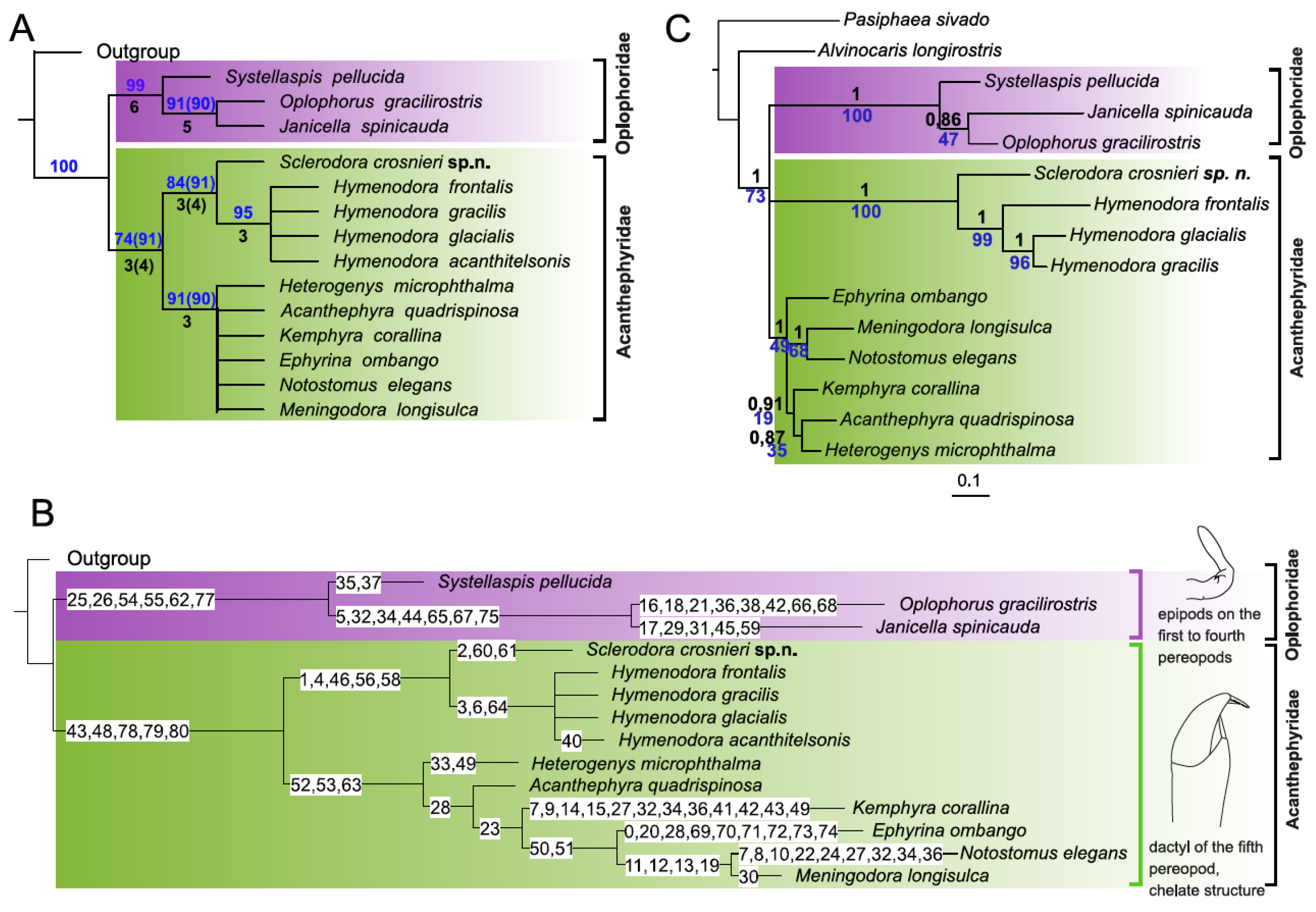
3.2. Molecular Analyses

A total of 15 species representing all genera of the superfamily Oplophoroidea and two outgroup species were put in the data matrix. In addition, all species of Hymenodora deposited in GenBank (three out of four) were also added to the data matrix. Prior to the analyses, all sequences from GenBank were checked for contamination or possible misidentification using BLAST search and preliminary phylogenetic reconstruction with each gene separately. ML trees generated for each individual gene dataset revealed no conflicting topologies between genes, at least in branching with bootstrap values ≥60% (File S6). The concatenated four-marker dataset comprised 3321 bp. Results from PartitionFinder2 recommended a 7-partition scheme by gene and codon (H3, COI), which was used in the final analyses (File S7).
Molecular analyses (Figure 2C, File S8) showed that the new species was a sister group to Hymenodora, and both formed a common robust clade. The rest of Acanthephyridae and Oplophoridae also formed robust clades; deeper nodes within Oplophoroidea remained unresolved.
Genetic K2P distances between the new species and three Hymenodora species ranged from 31.9% to 32.9% in COI gene (File S9). These values significantly exceeded K2P distances between all Hymenodora species (9.4–27.0%) as well as K2P distances between representatives of six genera of Acanthephyridae (17.9–28.4%) and three genera of Oplophoridae (23.3–28.9%).

4. Discussion

4.1. Taxonomic Implication

Results of morphological and molecular analyses were very similar and suggested the same position of Sclerodora on the phylogenetic tree. This taxon was sister to Hymenodora, and, along with Hymenodora, formed a robust clade sister to the rest of the Acanthephyridae. Calculations of genetic K2P distances suggested a generic status of the new taxon: Sclerodora was more distant from the sister Hymenodora than any pair of genera within Acanthephyridae or Oplophoridae from each other. In addition to a significant genetic distance, Sclerodora was supported by remarkable synapomorphies linked to the carapace (the presence of dorsal subuliform teeth extending from the dorsal ridge) and mouthparts (shape and articulation of the terminal segment of the second maxilliped, unique in the superfamily Oplophoroidea). Both molecular and morphological evidences suggest the generic status of Sclerodora and its position within the clade Hymenodora + Sclerodora and within the major clade Acanthephyridae.
In order to encapsulate results of morphological and molecular analyses in the phylogenetic classification, we here erect and diagnose the new genus and provide an amended key to all genera of Oplophoroidea.

4.1.1. Sclerodora gen. nov.

Emended diagnosis: Integument robust; rostrum overreaching eye cornea, armed with subuliform dorsal teeth, no ventral teeth; carapace with dorsal ridge armed with subuliform teeth in anterior part; antennal angle rounded, branchiostegal spine rudimentary, no hepatic spine, no uninterrupted lateral carina extending from near orbit to near posterior margin, hepatic and branchiostegal carinae weak; abdomen with all somites dorsally rounded and lacking teeth; 6th somite longer than 5th. Eyes with cornea narrower than eyestalk; antennal scale without lateral teeth; mandibles dissimilar, molar process with transverse distal surface triangular on right member of pair and compressed and sub-bilinear on left member, incisor process toothed along entire opposable margin; 1st maxilla with endopod bearing distal prominence with a single robust seta; 2nd maxilla with proximal endite elongate, lacking papilla and submarginal lamina; 1st maxilliped with two-segmented endopod greatly overreaching endites; 2nd maxilliped with distal segment subtriangular, attached transversely to preceding segment and bearing terminal robust setae; 3rd maxilliped and 1st pereopod with exopods not unusually broad or rigid; pereopods with neither ischium nor merus broadly compressed, fourth pair without epipod.
Species included: Sclerodora crosnieri sp. nov.
Type species: Sclerodora crosnieri sp. nov. (type by monotypy).
Etymology: From Greek ‘σκληρόσ’, firm, hard, and ‘δορα’, integument; a reference to the integument of the new species, which is firmer than that in the sister genus Hymenodora.
Remarks: Sclerodora is similar to Hymenodora and both differ from other Oplophoroidea in the replacement of usual subtriangular teeth on the rostrum with subuliform structures spaced from each other; in having an unusual molar process (compressed and sub-bilinear) on the left mandible; in a unique elongate proximal endite of the second maxilla lacking submarginal papilla and lamina; in a two-segmented endopod greatly overreaching endites of the first maxilliped. At the same time, Sclerodora differs from Hymenodora in a presence of the dorsal subuliform teeth extending from the common carina both on the rostrum and the carapace. Such a character of Sclerodora as a subtriangular terminal segment of the second maxilliped, attached transversely and bearing robust terminal setae, is unique and not found in other Oplophoroidea.

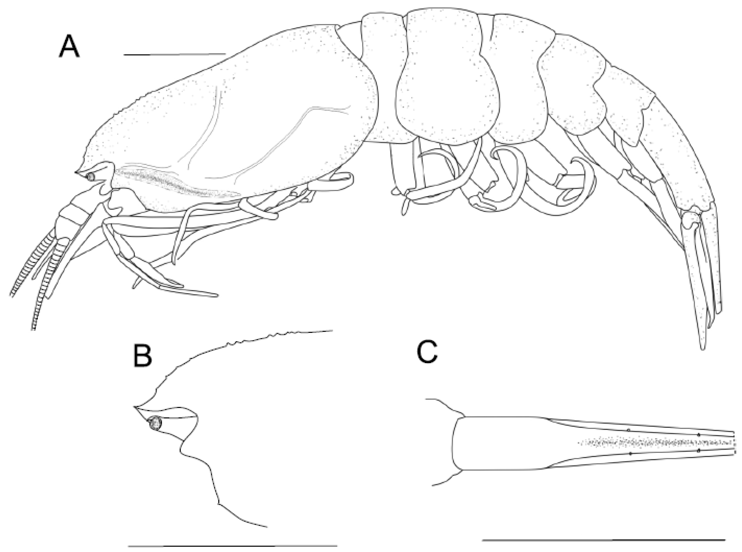
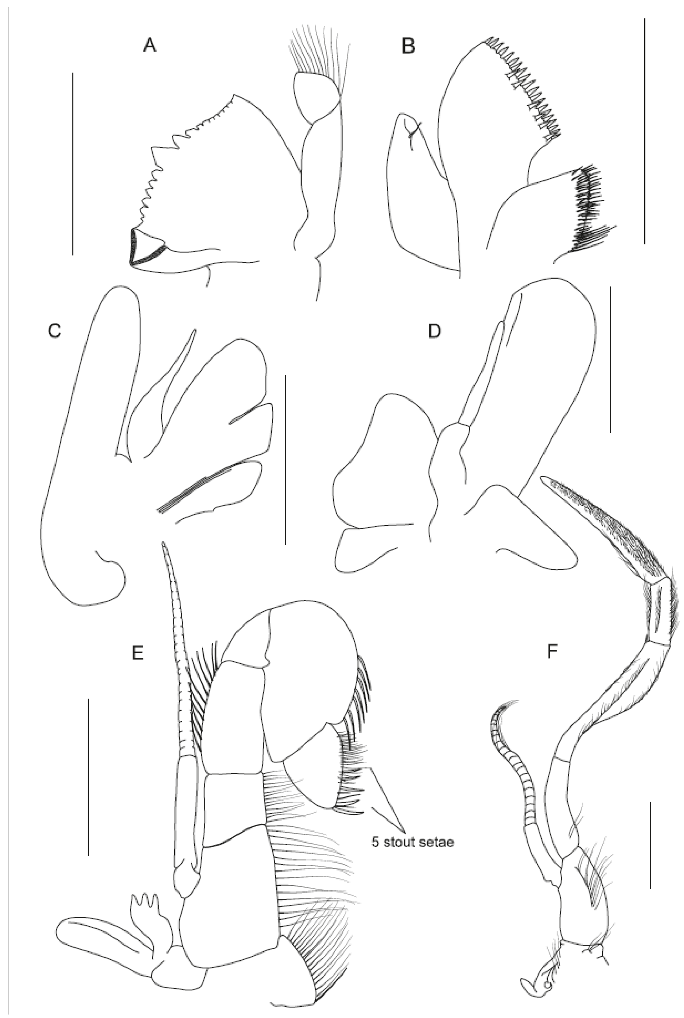
4.1.2. Sclerodora crosnieri sp. nov.

Material: Holotype, female, 26 mm carapace length, 80 mm total length (telson broken); 39th Cruise of R/V “Professor Logachev”; 2018, March 2; 15° N, 45° W; Isaacs-Kidd midwater trawl, oblique tow 0–2500 m; kept in the Natural History Museum, Copenhagen University, Denmark.
Description: Carapace smooth, 1.73 times as long as high, suprabranchial and hepatic ridges prominent (Figure 3A); dorsal carina with eight small irregular teeth in ¼ anterior part; rostrum with four dorsal teeth (Figure 3B). Abdomen with sixth somite twice as long as fifth; telson (broken) with dorsolateral spines (Figure 3C).
Mandible (Figure 4A) with 2-segmented palp; first maxilla with distal endite bearing two rows of robust setae (Figure 4B); second maxilla with two distal endites subequal (Figure 4C); first maxilliped with distal segment of endopod nearly twice as long as basal segment (Figure 4D); second maxilliped with distal segment bearing five terminal stout setae (Figure 4E); third maxilliped with well-developed hook-bearing epipod, distal segment densely covered with setae over entire margin (Figure 4F). First pereopod with carpus bearing distal tooth, propodus densely covered with setae over flexor margin, bearing large terminal and tiny subterminal spines, inner margins of chela rifled (Figure 5A–C); second pereopod with propodus bearing large terminal and tiny subterminal spines, inner margins of chela rifled (Figure 5D–F); third pereopod with ischium armed with three spines and dactyl bearing seven robust setae on flexor margin (Figure 5G); fourth pereopod with ischium armed with a single spine and dactyl bearing seven robust setae on flexor margin (Figure 5H); fifth pereopod with propodus covered with rifled setae over flexor margin and a single terminal robust seta in the chelate structure, dactyl curved and bearing two terminal robust setae (Figure 5I–J).
Etymology: named after the late Alain Crosnier, prominent carcinologist greatly contributed to taxonomy of decapods and, in particular, oplophoroid shrimps.

4.1.3. Key to Genera of Oplophoroidea

1. Sixth abdominal somite with distinct dorsal carina………………………………………….2
- Sixth abdominal somite dorsally smooth……………………………………………………….6
2. Hepatic spine present…………………………………………………....Kemphyra Chace, 1986
- Hepatic spine absent……………………………………………………………………………....3
3. Third abdominal somite with long dorsal tooth overreaching fourth somite……………...
……………………………………………………………………………...Heterogenys Chace, 1986
- Tooth on third abdominal somite, if present, not overreaching fourth somite…………….4
4. Carapace dorsally denticulate over nearly entire length; first abdominal somite dorsally
carinate……………………………………………………...Notostomus A. Milne-Edwards, 1881
- Carapace dorsally not denticulate on posterior half; first abdominal somite smooth…….5
5. A single continuous lateral carina on carapace (extending from near orbit to near
posterior margin on carapace…………………………………………..Meningodora Smith, 1882
- None or two continuous lateral carinae on carapace (one extending from near orbit,
another extending from near branchiostegal spine) ……………………………………………..
……………………………………………………………...Acanthephyra A. Milne-Edwards, 1881
6. Rostrum unarmed. Meri and ischia of pereopods greatly wide and compressed ………...
………………………………………………………………………………...Ephyrina Smith, 1885
- Rostrum denticulate. Meri and ischia of pereopods not greatly wide and compressed…..7
7. Rostral teeth subuliform, spaced from each other. Cornea subequal or narrower than
eyestalk……………………………………………………………………………………………….8
- Rostral teeth subtriangular, extending from a common crest. Cornea wider than eyestalk
………………………………………………………………………………………………………....9
8. Dorsal subuliform teeth only on rostrum……………………………………………………....
………………………………………………………………………....Hymenodora G.O. Sars, 1877
- Dorsal subuliform teeth both on rostrum and anterior part of carapace …………………...
……………………………………………………………………………………Sclerodora gen.nov.
9. Carapace strongly chitinized, subtriangular in cross-section. Abdomen with sixth somite
not longer than fifth, third to fourth somites with strong dorsomedial spines (at least ½ of
segment length)……………………………………………………………………………………10
- Carapace moderately chitinized, suboval in cross-section. Abdomen with sixth somite
nearly twice as long as fifth, third to fourth somites without strong dorsomedial spines
………………………………………………………………………..Systellaspis Spence Bate, 1888
10. Second abdominal somite with strong dorsomedial spine.…………Janicella Chace, 1986
- Second abdominal somite without strong dorsomedial spine …………………………….....
………………………………………………………………….......Oplophorus H. Milne Edwards

4.2. A New Suggested Cleaning and Grooming Mechanism

Examination of epipods and fifth pereopods in all species of Oplophoroidea reveals a remarkable co-evolution between both characters. In this superfamily, reduction of the fourth epipod is associated with development of a chelate structure on the fifth pereopod. This structure is morphologically similar in all genera (Figure 1) and remarkably resembles grooming chelae in other carids as illustrated in [4]. When we map these synapomorphies on a phylogenetic tree (Figure 2B), we can see that a chelate structure on the fifth pereopod (linked to a lost epipod on the fourth pereopod and indicating active grooming) is a derived structure, whereas a long dactyl not forming chelate structure (linked to a full set of the epipods and passive grooming) occurs basally on the morphological tree and likely plesiomorphic as suggested by Bauer [4].
We suggest that Acanthephyridae evolved an active cleaning mechanism, which is a derived one and alternative to that described by Bauer [4,5]: posterior branchiae are groomed and cleaned by the fifth pereopods instead of the chelipeds. Convergent chelate structures suggest that the chela is especially efficient for cleaning and grooming branchiae, whichever appendage is adapted for these functions. In Oplophoridae, which are basal on the phylogenetic morphological tree, the cleaning function is carried out passively by well-developed epipods.
Our results confirm Bauer’s [4,5] statement that the major type of gill-cleaning method is generally a characteristic at the family level and that the active cleaning is more derived than the passive one. Interestingly, in Oplophoridae the last three pereopods likely take another function and act as a holding structure during mating [10], which may favor copulation in the turbulent water column [9].

Supplementary Materials

The following are available online at https://www.mdpi.com/article/10.3390/d13110536/s1, File S1: Character list, File S2: Character state, File S3: Retrieved morphological trees. Bremer support, File S4: Retrieved morphological trees. Bootstrap support, File S5: Synapomorphies, File S6: Maximum likelihood (RAxML) phylograms for each individual gene dataset, File S7: Partitioning scheme and best models selected by PartitionFinder2, File S8: Molecular BI and ML trees, File S9: Estimates of evolutionary divergence between species.

Author Contributions

A.L. analyzed the specimens morphologically; D.K. ran the genetic analyses; A.V., A.L. and D.K. wrote the paper and participated in the revisions of it. All authors have read and agreed to the published version of the manuscript.

Funding

This research was supported by RSF Project No. 18-14-00231.

Institutional Review Board Statement

Not applicable.

Data Availability Statement

Data is contained within the article and supplementary material.

Acknowledgments

We thank S.G. Kobylyansky for material from the 39th Cruise of the R/V “Professor Logatchev”; H. Braken-Grissom, Laura Corbari, J. Olesen and T. Sutton for possibility to examine specimens of Oplophoroidea.

Conflicts of Interest

None of the authors have any financial competing interests.

References

  1. Dawson, M.N. Species richness, habitable volume, and species densities in freshwater, the sea, and on land. Front. Biogeogr. 2012, 4. [Google Scholar] [CrossRef] [Green Version]
  2. Costello, M.J.; Chaudhary, C. Marine biodiversity, biogeography, deep-sea gradients, and conservation. Curr. Biol. 2017, 27, 511–527. [Google Scholar] [CrossRef] [PubMed]
  3. WoRMS Editorial Board. World Register of Marine Species. 2021. Available online: http://www.marinespecies.org (accessed on 4 October 2021). [CrossRef]
  4. Bauer, R.T. Decapod crustacean grooming: Functional morphology, adaptive value, and phylogenetic significance. In Functional Morphology of Feeding and Grooming in Crustacea; CRC Press: Boca Raton, FL, USA, 2020. [Google Scholar]
  5. Bauer, R.T. Antifouling adaptations of caridcan shrimp (Decapoda: Caridea): Gill cleaning mechanisms and grooming of brooded embryos. Zool. J. Linn. Soc. 1979, 65, 281–303. [Google Scholar] [CrossRef]
  6. Bauer, R.T. Grooming behavior and morphology in the decapod Crustacea. J. Crust. Biol. 1981, 1, 153–173. [Google Scholar] [CrossRef]
  7. Wong, J.M.; Pérez-Moreno, J.L.; Chan, T.Y.; Frank, T.M.; Bracken-Grissom, H.D. Phylogenetic and transcriptomic analyses reveal the evolution of bioluminescence and light detection in marine deep-sea shrimps of the family Oplophoridae (Crustacea: Decapoda). Mol. Phylogenet. Evol. 2015, 83, 278–292. [Google Scholar] [CrossRef]
  8. Chace, F.A. The Caridean Shrimps (Crustacea: Decapoda) of the Albatross Philippine Expedition, 1907–1910, Part 4: Families Oplophoridae and Nematocarcinidae; Smithsonian Institution Press: Washington, DC, USA, 1986; p. 81. [Google Scholar]
  9. Lunina, A.; Vereshchaka, A. The role of the male copulatory organs in the colonization of the pelagic by shrimp-like eucarids. Deep Sea Res. Part II Top. Stud. Oceanogr. 2017, 137, 327–334. [Google Scholar] [CrossRef]
  10. Lunina, A.A.; Kulagin, D.N.; Vereshchaka, A.L. Oplophoridae (Decapoda: Crustacea): Phylogeny, taxonomy and evolution studied by a combination of morphological and molecular methods. Zool. J. Linn. Soc. 2019, 186, 213–232. [Google Scholar] [CrossRef]
  11. Lunina, A.A.; Kulagin, D.N.; Vereshchaka, A.L. Phylogenetic revision of the shrimp genera Ephyrina, Meningodora and Notostomus (Acanthephyridae: Caridea). Zool. J. Linn. Soc. 2020, zlaa161. [Google Scholar] [CrossRef]
  12. Li, C.P.; De Grave, S.; Chan, T.Y.; Lei, H.C.; Chu, K.H. Molecular systematics of caridean shrimps based on five nuclear genes: Implications for superfamily classification. Zool. Anz. 2011, 250, 270–279. [Google Scholar] [CrossRef]
  13. Nixon, K. The parsimony ratchet, a new method for rapid parsimony analysis. Cladistics 1999, 15, 407–414. [Google Scholar] [CrossRef]
  14. Goloboff, P.; Farris, S.; Nixon, K. TNT: Tree Analysis Using New Technology. 2000. Available online: http://www.lillo.org.ar/phylogeny/tnt (accessed on 24 October 2021).
  15. Schubart, C.D.; Huber, M.G.J. Genetic comparisons of German populations of the stone crayfish, Austropotamobius torrentium (Crustacea: Astacidae). Bull. Français Pêche Piscic. 2006, 380–381, 1019–1028. [Google Scholar] [CrossRef] [Green Version]
  16. Folmer, O.; Black, M.; Hoeh, W.; Lutz, R.; Vrijenhoek, R. DNA primers for amplification of mitochondrial cytochrome c oxidase subunit I from diverse metazoan invertebrates. Mol. Mar. Biol. Biotechnol. 1994, 3, 294–299. [Google Scholar] [PubMed]
  17. Schubart, C.D. Mitochondrial DNA and decapod phylogenies: The importance of pseudogenes and primer optimization. Crustacean 2009, 18, 47–65. [Google Scholar]
  18. Schubart, C.D.; Cuesta, J.A.; Felder, D.L. Glyptograpsidae, a new brachyuran family from Central America: Larval and adult morphology, and a molecular phylogeny of the Grapsoidea. J. Crust. Biol. 2002, 22, 28–44. [Google Scholar] [CrossRef] [Green Version]
  19. Reuschel, S.; Schubart, C.D. Phylogeny and geographic differentiation of Atlanto–Mediterranean species of the genus Xantho (Crustacea: Brachyura: Xanthidae) based on genetic and morphometric analyses. Mar. Biol. 2006, 148, 853–866. [Google Scholar] [CrossRef]
  20. Apakupakul, K.; Siddall, M.E.; Burreson, E.M. Higher level relationships of leeches (Annelida: Clitellata: Euhirudinea) based on morphology and gene sequences. Mol. Phylogenetics Evol. 1999, 12, 350–359. [Google Scholar] [CrossRef] [Green Version]
  21. Colgan, D.J.; McLauchlan, A.; Wilson, G.D.F.; Livingston, S.P.; Edgecombe, G.D.; Macaranas, J.; Gray, M.R. Histone H3 and U2 snRNA DNA sequences and arthropod molecular evolution. Aust. J. Zool. 1998, 46, 419–437. [Google Scholar] [CrossRef]
  22. Edgar, R.C. MUSCLE: Multiple sequence alignment with high accuracy and high throughput. Nucleic Acids Res. 2004, 32, 1792–1797. [Google Scholar] [CrossRef] [PubMed] [Green Version]
  23. Kumar, S.; Stecher, G.; Li, M.; Knyaz, C.; Tamura, K. MEGA X: Molecular Evolutionary Genetics Analysis across computing platforms. Mol. Biol. Evol. 2018, 35, 1547–1549. [Google Scholar] [CrossRef] [PubMed]
  24. Stamatakis, A. RAxML version 8: A tool for phylogenetic analysis and post-analysis of large phylogenies. Bioinformatics 2014, 30, 1312–1313. [Google Scholar] [CrossRef]
  25. Edler, D.; Klein, J.; Antonelli, A.; Silvestro, D. raxmlGUI 2.0 beta: A graphical interface and toolkit for phylogenetic analyses using RAxML. Methods Ecol. Evol. 2021, 12, 373–377. [Google Scholar] [CrossRef]
  26. Ronquist, F.; Teslenko, M.; Van Der Mark, P.; Ayres, D.L.; Darling, A.; Höhna, S.; Huelsenbeck, J.P. MrBayes 3.2: Efficient Bayesian phylogenetic inference and model choice across a large model space. Syst. Biol. 2012, 61, 539–542. [Google Scholar] [CrossRef] [PubMed] [Green Version]
  27. Lanfear, R.; Frandsen, P.B.; Wright, A.M.; Senfeld, T.; Calcott, B. PartitionFinder 2: New methods for selecting partitioned models of evolution for molecular and morphological phylogenetic analyses. Mol. Biol. Evol. 2016, 34, 772–773. [Google Scholar] [CrossRef] [PubMed] [Green Version]
  28. Kimura, M. A simple method for estimating evolutionary rate of base substitutions through comparative studies of nucleotide sequences. J. Mol. Evol. 1980, 16, 111–120. [Google Scholar] [CrossRef] [PubMed]
Figure 1. Terminal part of the fifth pereopod in Oplophoroidea (schematic, most non-robust setae removed): (A)—Sclerodora crosnieri, sp. nov., (B)—Hymenodora frontalis, side view, (C)—Hymenodora frontalis, inner view, (D)—Acanthephyra acutifrons, (E)—Notostomus elegans, (F)—Kemphyra corallina, (G)—Meningodora longisulca, (H)—Heterogenys microphthalma, (I)—Systellaspis debilis, (J)—Oplophorus gracilirostris, (K)—Janicella spinicauda.
Diversity 13 00536 g001
Figure 2. Phylogenetic trees with Pasiphaea sivado and Alvinocaris longirostris as outgroups. (A)—morphological MP tree; only clades supported by both Bremer values (black, below branches) and bootstrap values (blue, above branches) are shown; if support values in analyses differed, values retrieved in Analysis 2 are given in parentheses. (B)—synapomorphies, above branches, see coding in File S1. (C)—molecular BI and ML tree, only supported clades are shown. The horizontal scale bar marks the number of expected substitutions per site. Statistical support indicated as Bayesian posterior probabilities (black, above branches) and ML bootstrap with 1000 replicates (blue, below branches).
Diversity 13 00536 g002
Figure 3. Body of Sclerodora crosnieri sp. nov., holotype: (A)—general view. (B)—anterior part of carapace, lateral view. (C)—telson, dorsal view. All scales: 10 mm.
Diversity 13 00536 g003
Figure 4. Mouthparts of Sclerodora crosnieri sp. nov., holotype, right parts: (A)—mandible. (B)—first maxilla, inner view. (C)—second maxilla, setae removed. (D)—first maxilliped, setae removed. (E)—second maxilliped. (F)—third maxilliped. All scales: 3 mm.
Diversity 13 00536 g004
Figure 5. Pereopods of Sclerodora crosnieri sp. nov., holotype: (A)—right first pereopod. (B)—right first chela, inner view. (C)—left first chela. (D)—right second pereopod. (E)—right second chela. (F—left second chela. (G)—right third pereopod. (H)—left fourth pereopod. (I)—right fifth pereopod. (J)—terminal part of right fifth pereopod. All scales for entire pereopods: 10 mm, all scales for their tips: 1 mm.
Diversity 13 00536 g005
Table 1. Individuals used in morphological analyses. MNHN - National Museum of Natural History (Paris, France); ZMUK—National History Museum, Copenhagen, Denmark; IO RAN—Institute of Oceanology, Russian Academy of Scienses, USNM -National Museum of Natural History, Smithsonian Institution.
SpeciesCoordinatesOther InformationMuseum, Number
Acanthephyra quadrispinosa29°39′ S, 44°16′ EExpedition ATIMO VATAE. SUD MADAGASCAR, Sud Pointe Barrow. Chaultier “Nosy Be 11”, Stn. CP 3596, 986–911 m. 12.05.2010.MNHN-IU-2010-4285
Acanthephyra acutifrons14°43′ N, 45° 02′ W“Professor Logatchev” 39 cruise St 215 RT, RTAKIO RAN 39L 215 RT №1
Ephyrina ombango10°23,17′ N, 46°45,34′ WDEMERABY, CP07, chalutage 4850 m. 20.09.80MNHN-IU-2018-1579
Ephyrina ombango9°18′ S, 11°10′ E“Ombango”, C14, St.325,midwater traul, 0–725 m, 02.03.1961, 23h00–23h15MNHN-IU-2014-11098
Heterogenis microphtalmaNo dataCollection de S.A.S.le Prince de Monaco, Station 7/3. №12h, 16–19.8.96. Chal 4360 mMNHN-IU-2018-1578
Hymenodora acanthitelsonis45°18′ N, 125°43′ W–45°17′ N, 125°49′ WPacific Ocean, Unated States, Oregon, W of Pacific City. Yaqina BMT.189, 18.03.1970.USNM 137500
Hymenodora glacialis02°03′ S, 118°45′ EIndonesie, CORINDON -Makassar. St CH286, 1710–1730 mNa 10655
Hymenodora glacialis73°28′ N, 10°07′ WMer de Norvege, Campagne NORBI, N.O. “Jean Charcot”, Stn CP16, 2937 m, 07.08.1975MNHN-IU-2008-16833
Hymenodora gracilis37°39′ S, 77°26′ EIle Amsterdam, Campagne Jasus (MD 50), N.O. “Marion Dufresne”, Stn CP193, 2800–3075 m. 27.06.1986MNHN-IU-2008-16839
Hymenodora frontalis15° N, 45° WROV “Vityaz”, 59 th cruise, st. 7497, № 271. 18.06.1976, 1500–2500 m
Janicella spinicauda1°28′ S, 48°06′ EROV “Vityaz”, 17th cruise, St. 2604,13.11.88, 670–690 mZMUK
Janicella spinicauda8°44′ S, 43°54′ EDana Expedition, St. 3939-1, 23.12.1929, 500 meter wireZMUK
Kemphyra corallina37°54′ S, 77°22′ EIles St Paul et Amsterdam, “Marion Dufresne” Cne MD Jasus Stn CP 56. 2280–2310 m. 14.07.1986. 20h02–22h31MNHN-IU-2018-1581
Kemphyra corallina33°59′ S, 43°55′ EIndian Ocean: Walters shoal, Plaine Sud. N.O. “Marion Dufresne”, Campagne MD208(Walters Shoal). Stn CP49156 1865–2058 m, 12.05.2017MNHN-IU-2016-9402
Meningodora longiscula9°55′ N, 142°00′ ENouvelle-Caledonie, Campagne Caride V. Stn 15, 1000 m. 12.09.1969MNHN-IU-2011-5635
Notostomus elegans 37 cruise RV Logatchev, St 156 TSIO RAN
Oplophorus gracilirostris25°11′ N, 122°35′ EDana Expedition, St. 3722-3, 300 m wireZMUK
Oplophorus gracilirostris20°08′ N, 82°59′ WDana Expedition, St. 1218, 800 m wireZMUK
Oplophorus gracilirostris12°30′ S, 48°16′ EROV “Vityaz”, 17th cruise, St. 2597, 12.11.88, 360–555 m wire.ZMUK
Oplophorus gracilirostris22°06′ N, 84°58′ WDana Expedition, St. 1223, 500 m wire ZMUK
Pasiphaea sivado35°47′ N, 05°17′ WDetroit de Gibraltar, N.O. “Cryos”, BALGIM, St. CP150, 280–300 m, 18.06.1984MNHN-IU-2018-1611
Sclerodora crosnierisp.nov.16° N, 46° W39th Cruise of R/V “Professor Logatchev”, March 2018ZMUK
Systellaspis pellucida12°30′ S, 48°16′ EIndian Ocean. North end of Madagascar. ROV “Vityaz”, 17th cruise, St. 2597, 360–555 mIO RAN
Systellaspis pellucida25°11′ N, 122°35′ ENorth Western Pacific Ocean. S.E. and E. of Formosa. Dana Expedition 3722(2) 29.05.1929, 600 mwZMUK
Systellaspis pellucida25°11′ N, 122°35′ ENorth Western Pacific Ocean. S.E. and E. of Formosa. Dana Expedition 3722(1) 29.05.1929, 1000 mwZMUK
Table 2. Details of the analyzed species and sequences used in the study. Newly retrieved sequences are highlighted in bold; ‘N’—missing data.
TaxonGenBank Accession NumbersSource
COI16S18SH3
Outgroup taxa
Pasiphaeoidea Dana, 1852
Pasiphaeidae Dana, 1852
Pasiphaea sivado (Risso, 1816)KP759487KP725629KP725826MF279416Aznar-Cormano et al., 2015; Liao et al., 2017
Bresilioidea Calman, 1896
Alvinocarididae Christoffersen, 1986
Alvinocaris longirostris Kikuchi & Ohta, 1995KP215329KP215285KP215300KP215342Aznar-Cormano et al., 2015
Ingroup taxa
Oplophoroidea Dana, 1852
Oplophoridae Dana, 1852
Janicella spinicauda (A. Milne-Edwards, 1883)MH572546KP075932MH100869MH107256Wilkins and Bracken-Grissom, 2020 (GenBank); Wong et al., 2015; Lunina et al., 2019
Oplophorus gracilirostris A. Milne-Edwards, 1881KP076150KP075920KP075847KP076072Wong et al., 2015
Systellaspis pellucida (Filhol, 1884)JQ306184KP075925JF346250KP076077Matzen da Silva et al., 2011; Wong et al., 2015; Li et al., 2011
Acanthephyridae Spence Bate, 1888
Acanthephyra quadrispinosa Kemp, 1939KP759363KP725479KP725677KP726051Aznar-Cormano et al., 2015
Ephyrina ombango Crosnier & Forest, 1973MW043004MW043448MW043463MW052289Lunina et al., 2020
Heterogenys microphthalma (Smith, 1885)KP076183KP075898KP075787KP076124Wong et al., 2015
Kemphyra corallina (A.Milne-Edwards, 1883)MW043006MW043450MW043465MW052291Lunina et al., 2020
Meningodora longisulca Kikuchi, 1985MW043007MW043451MW043466MW052292Lunina et al., 2020
Notostomus elegans A. Milne-Edwards, 1881MW043011MW043455MW043470MW052296Lunina et al., 2020
Hymenodora frontalis Rathbun, 1902DQ882080NNNCosta et al., 2007
Hymenodora glacialis (Buchholz, 1874)FJ602519GQ131896GQ131915NBucklin et al., 2010; Chan et al., 2010
Hymenodora gracilis Smith, 1886MH572613MH542891KP075827KP076134Wilkins and Bracken-Grissom, 2020 (GenBank); Wong et al., 2015
Sclerodora crosnieri gen. nov., sp. nov.OK382996OK382953OK382952OK424597This study
Publisher’s Note: MDPI stays neutral with regard to jurisdictional claims in published maps and institutional affiliations.

