Article

Spatial Diversity in Bacterial Communities across Barren and Vegetated, Native and Invasive, Coastal Dune Microhabitats

by
Brianna L. Boss
1,
Bianca R. Charbonneau
2,* and
Javier A. Izquierdo
1,*
1
Department of Biology, Hofstra University, Hempstead, NY 11549, USA
2
Department of Biology, University of Pennsylvania, Philadelphia, PA 19104, USA
*
Authors to whom correspondence should be addressed.
Diversity 2021, 13(11), 525; https://doi.org/10.3390/d13110525
Submission received: 1 October 2021 / Revised: 19 October 2021 / Accepted: 20 October 2021 / Published: 23 October 2021
(This article belongs to the Special Issue Biological Invasions and Conservation in Coastal Dune Ecosystems)

Abstract

:
The microbial community composition of coastal dunes can vary across environmental gradients, with the potential to impact erosion and deposition processes. In coastal foredunes, invasive plant species establishment can create and alter environmental gradients, thereby altering microbial communities and other ecogeomorphic processes with implications for storm response and management and conservation efforts. However, the mechanisms of these processes are poorly understood. To understand how changing microbial communities can alter these ecogeomorphic dynamics, one must first understand how soil microbial communities vary as a result of invasion. Towards this goal, bacterial communities were assessed spatially along foredune microhabitats, specifically in barren foredune toe and blowout microhabitats and in surrounding vegetated monocultures of native Ammophila breviligulata and invasive Carex kobomugi. Across dune microhabitats, microbial composition was more dissimilar in barren dune toe and blowout microhabitats than among the two plant species, but it did not appear that it would favor the establishment of one plant species over the other. However, the subtle differences between the microbial community composition of two species could ultimately aid in the success of the invasive species by reducing the proportions of bacterial genera associated exclusively with A. breviligulata. These results suggest that arrival time may be crucial in fostering microbiomes that would further the continued establishment and spread of either plant species.

1. Introduction

Coastal dunes are invaluable habitats that are growing increasingly vulnerable to the impact of sea level rise and climate change [1,2]. These highly dynamic habitats buffer developed and natural inland areas from storm surge and high tides [3]. Plants act as ecosystem engineers to build, stabilize, and recover dune habitats [4,5,6]. Specifically, belowground biomass stabilizes grains (reducing erosion) and aboveground biomass provides surface cover and promotes sediment accretion and retention [7,8,9]. Microbes may also directly and indirectly contribute to belowground sediment cohesion, but this has received minimal research attention [10]. Variations in plant conditions (i.e., biomass, density, composition, and distribution) can directly impact dune stability and storm response, although the mechanisms remain poorly understood [6,11,12]. Given this, variations in microbial communities supporting these vegetation communities have the potential to directly and indirectly underpin storm response. However, few studies have delved into the biology of microbial dune communities [13,14,15,16].
Variations in microbial community compositions can impact their functional role in system-level processes. Bacteria that fix nitrogen or solubilize phosphorus create more favorable environments for plant growth and survival while also contributing to nutrient cycling, organic matter decomposition, and soil structure, all of which contribute to plant community composition and ecosystem function [17,18,19,20]. Plants naturally vary in nutrient needs and root exudate production, influencing microbial community composition and activities [21,22,23]; this includes the production of polysaccharides (which can impact sand grain adhesion and aggregation [24,25], with variations among different taxa) [26]. Microbes may contribute to dune plant survival in such harsh environments through plant growth-promoting properties such as nutrient acquisition and drought tolerance [27]. Bacterial functional roles and niches change over time and space, with alterations in microbial community composition between microhabitats [28] having the potential to cause a cascade of effects in associated vegetation.
Despite a handful of dune-specific microbial studies, much remains unknown regarding the understanding of spatiotemporal variations in and the implications of varying microbial compositions. Actinobacteria, Firmicutes, Proteobacteria, Bacteroidetes, and Cloroflexi have been observed as the dominant bacterial phyla in inland desert dunes [15]. Conversely, in coastal dunes, the dominant phyla observed included only Actinobacteria and Proteobacteria [13,16]. Furthermore, coastal dunes in different developmental stages may maintain their own distinct microbial communities [16]. Shet et al. found that Bacillus (Firmicutes) is predominant on dunes in the early stage of formation, whereas Streptomyces (Actinobacteria) and Kouleothrix (Chloroflexi) are the dominant genera in later stages of formation. Bacterial abundance increased with dune age and comprised the major phyla Acidobacteria, Bacteroidetes, Verrucomicrobia, Actinobacteria, Proteobacteria, and Firmicutes [16]. Many bacterial genera belonging to these phyla have been observed to produce compounds for plant growth promotion [16]. Some microbial communities may not impact plant succession and establishment [29,30], but generally, microbial communities and dune vegetation appear inextricably intertwined.
Abiotic gradients can affect plant and microbial community compositions [17,25,31]. Stressors associated with vicinity to the ocean (i.e., low nutrients and water retention, high salt concentrations, sediment mobility, etc.) decrease moving inland, and vegetation cover, diversity, and abundance increase in turn [3,27,32,33]. Concurrently, microbial biomass, composition, and diversity also change, typically increasing moving inland and varying with plant cover [16,25,34,35,36]. In particular, salinity is considered a major driver of dune plant community compositions [37] and has also been found to impact microbial communities in other habitats [38,39]. For example, microbial community composition has been linked to soil salinity, where areas with higher salinity closer to the water had higher levels of Gram-negative bacteria [31]. More stressful habitats in general support less plant diversity, and similarly, lower levels of microbial activity have been detected in barren dune areas [31,40].
Coastal dunes are vulnerable to invasive plant species, which can become highly prevalent [41,42,43] and affect ecosystem processes by altering microbial biomass and diversity [44,45,46,47]. Invasive and native species may differ in plant morphology, nutrient acquisition, and root exudates, which can alter soil properties influencing microbial community composition and soil structure [45,48]. The presence of nitrogen-fixing bacteria may increase invasive success [49] and invasive-induced changes in microbial communities can create positive feedbacks favoring further invasive dominance [20,50]. Invasive species reduce and replace native species cover across a dune system [41,43,46], and this can have short- and long-term effects even after eradication regarding legacy impacts to microbial communities and soil structure [51,52,53].
Understanding if and how an invasive species alters dune microbial communities is an essential part of understanding the depth of impact to current and future dune development and storm response as compared to their native counterparts. Understanding how microbial communities might vary across dune habitat gradients is key to better understand the overall heterogeneity of the system that underscores storm response. Towards this goal, bacterial community compositions were categorized within and among four foredune microhabitats: (1) in barren blowouts and (2) the foredune toe and among abutting vegetated monocultures of (3) native American Beachgrass (Ammophila breviligulata) and (4) invasive Asiatic Sand Sedge (Carex kobomugi). Variations in microbial community composition and diversity were examined spatially relative to stress gradients in the broader foredune habitat and among barren and vegetated microhabitats. Among these microhabitats, emphasis was placed on examining if and how invasive C. kobomugi alters community compositions with regards to potential legacy effects impacting the success of future restoration efforts post-eradication [52,54]. There are many unknowns regarding relationships between microbes and higher plants and animals in these and other landscapes [19]. Understanding how microbial communities and invasive species alter microbial processes is a first step towards understanding the cascading impacts this may have on biological and geomorphic processes related to their management and conservation.

2. Materials and Methods

2.1. Field Site

This work was conducted at Island Beach State Park (IBSP), Ocean County, NJ, USA. It is a micro-tidal ≈17 km high barrier island [55] with a sandy shoreline transitioning from littoral to foredune, secondary, and tertiary dune, maritime forest, and tidal marsh. Salt spray is considered a major driver of dune species distributions that characteristically decreases with distance from the ocean at IBSP, as measured in Charbonneau et al., 2020 [41]. The foredunes are dominated by native A. breviligulata or invasive C. kobomugi with few exceptions [6]. Invasive C. kobomugi arrived in the US in 1929 at IBSP such that stands here are the oldest and possibly largest in the US [41,56]. The invasive species has since spread from Massachusetts to North Carolina [57] and may invest in relationships with different microbial communities as a native in Asia than as an invasive in the mid-Atlantic United States [58].
This research was conducted at three sites, labeled A17, A19, and A21 (denoting the beach access area they are found), each associated with a bowl blowout within a 3-km foredune span (Supplementary S1) [59]. This area of IBSP was not over washed or inundated in Hurricane Sandy (October 2012) [6,60]. The blowouts were all greater than 550 m apart, and of similar diameter, depth, and area (506 m2, 485 m2, and 402 m2). Each blowout had monospecific stands of both C. kobomugi and A. breviligulata, respectively, abutting its edges. The extent of the blowouts was mapped October 2018 using ArcPad on a Trimble GeoXT GeoExplorer (submeter accuracy: 0.8 m ± 95%), where blowout edges were defined as where plants became ≤1-m apart outside of the depression. Carex kobomugi stands were also mapped to define where they abutted A. breviligulata stands, considering ramets < 1 m apart part of the same stand [6].

2.2. Soil Sample Collection

At each of the three sites, soil samples were collected on 5 April 2018, from four microhabitats: the blowout itself, abutting monoculture vegetation stands (C. kobomugi and A. breviligulata, respectively), and the foredune toe. Prior to the field effort, the collection location of each sample (randomly assigned within microhabitats) was determined using the mapped data and the ArcGIS Random Point Generator tool with >1.5-m distance between points. Sample sizes include 8 samples from each vegetation stand, 16 from within the blowout, and 6 from the closest foredune toe, for a total of 38 samples at each of the three sites and 114 total samples (Supplementary S1).
Each sample consisted of three homogenized 810 cm3 cores collected 15 cm apart [61]. The top 10 cm of soil was removed with a trowel to reach the edaphic zone so each core encompassed 10–30 cm depth, which for vegetated samples contained both edaphic and rhizospheric soil and roots. Cores were collected by driving a 2.8 × 18 cm PVC pipe into the sand, after sterilizing both the trowel and corer with ethanol between samples. Complimentary edaphic data was not collected in consort with these cores, as preliminary analyses of a subset of cores collected in December 2016 revealed low exchangeable cations and base saturation across all microhabitats (Supplementary S2). All samples were stored at 4 °C for 1 month until DNA extraction.

2.3. DNA Extraction & Sequencing

Of the 114 samples, DNA was successfully extracted from 88 samples using a DNeasy PowerSoil Extraction Kit (Qiagen, Carlsbad, CA, USA) with triplicate extractions per sample. Samples with low DNA concentrations (less than 5 ng/μL) were not used for sequencing. Quality control of the extracted DNA samples consisted of DNA quantification using a Take3 Multi-Volume Plate reader (Biotek, Winooski, VT, USA) using wavelengths of 260 and 280 nm. Triplicate extractions were pooled per sample together for sequencing. The 16S rRNA gene V4 variable region PCR primers 515/806 with a barcode on the forward primer were used in a 30-cycle PCR using the HotStarTaq Plus Master Mix Kit (Qiagen, Carlsbad, CA, USA) under the following conditions: 94 °C for three minutes, followed by 30 cycles of 94 °C for 30 s, 53 °C for 40 s, and 72 °C for 1 min, with a final elongation step performed at 72 °C for 5 minutes. After amplification, PCR products were verified in 2% agarose gel to determine the success of amplification and the relative intensity of bands. Multiple samples were pooled together in equal proportions based on their DNA concentrations. Pooled samples were purified using calibrated Ampure XP beads. Then, the pooled and purified PCR product was used to prepare an Illumina DNA library. Sequencing was performed at MR DNA (Shallowater, TX, USA) on a MiSeq following the manufacturer’s guidelines. Sequence data were processed using MR DNA analysis pipeline (MR DNA, Shallowater, TX, USA). In summary, sequences were joined, depleted of barcodes then sequences <150 bp removed, sequences with ambiguous base calls removed. Sequences were denoised, OTUs were generated, and chimeras were removed. Operational taxonomic units (OTUs) were defined by clustering at 3% divergence (97% similarity). Final OTUs were taxonomically classified using BLASTn against a curated database derived from RDPII and NCBI (www.ncbi.nlm.nih.gov, http://rdp.cme.msu.edu), consolidated in October 2019.
Extracted and sequenced samples are even for each of the three blowouts and three foredune toe sections, with nine and six samples at each, respectively. The samples are relatively evenly distributed across the vegetated stands as only three A. breviligulata and two C. kobomugi samples lacked DNA after extraction. OTU data distribution for each sample was used for the calculation of Shannon’s diversity indexes using the vegan package in R version 4.0.3 [62]. OTU taxonomic assignment was used to determine percentages of phylum and genus composition at each microhabitat to be used in all statistical analyses.

2.4. GIS & Statistical Analyses

Sample locations were referenced to dune system features using ArcGIS 10.3.1. The Near tool was used to determine the distance of each blowout sample to the closest edge and each C. kobomugi sample to its closest stand edge abutting an A. breviligulata stand. This tool was also used to calculate the distance of each sample to the foredune crest, defined as where the plateaued foredune habitat drops to the seaward foredune, mapped as a polyline in January 2016, and did not shift between the core collection and its mapping [58].
All statistical analyses were performed in JMP Pro 14.0 and R version 4.0.3 [62]. All statistical tests were two-tailed using α = 0.05 and all means are reported ± S.E. We performed ANOSIM analyses using the “vegan” package in R [62] to compare community composition across sites for each microhabitat and confirmed that community composition did not vary significantly between the three sites (p > 0.05); given this, we pooled the data across sites in statistical analyses. Additionally, we also used ANOSIM in pairwise comparisons of community composition between microhabitats. All of our ANOSIM analyses were performed with Bray–Curtis similarities after 9999 permutations.
Data from the blowouts and foredune toe were treated as one barren category compared to the two vegetated microhabitats, which were kept separate. ANOVA was used to examine if there are differences in the percent composition of the most prevalent microbial genera, and to examine differences in diversity, calculated as the Shannon Index using OTU data from each sample. Principal components analysis (PCA) was computed from the resulting distance matrices using phylum and genus percent community composition data using R version 4.0.3 [62] and reducing dimensionality to two dimensions (PC1 vs. PC2). PCA analysis of phylum data was performed with all samples combining all sites, as well as for each of the individual sampling sites A17, A19, and A21.
Linear regression was used to test whether there are spatial patterns in composition and diversity regarding the position of each sample point relative to its linear distance from the crest. For the invasive and blowout microhabitat samples, which maintain distinct habitat edges, regression was used to test if composition and diversity vary within the habitat relative to distance to the stand center. The proportion of samples containing the most prevalent genera and phylum were determined and compared between the four microhabitats, and among barren vs. vegetated microhabitats using Fisher’s exact test.

3. Results

3.1. Bacterial Community Composition & Diversity

A total of 3,415,118 raw Illumina reads were obtained from 88 samples. On average, 27,903 ± 594 reads were obtained per sample after quality control, denoising, and filtering of chimeric reads. A total of 8146 bacterial OTUs were identified, with an average of 1667 OTUs per sample, ranging from 670 to 3445 OTUs. All sequencing data were deposited in the GenBank database accession numbers SRR15979670-SRR15979757 and the BioProject number PRJNA764730.
Across all 88 samples, Proteobacteria was consistently the most abundant phylum, ranging from 41.21% to 80.54% of community composition (Figure 1A). Other abundant phyla included Actinobacteria (4.21% to 22.78%) and Bacteroidetes (5.99% to 25.11%). Less abundant phyla that represented at least 1% of the community composition in samples included Firmicutes (2.16% to 7.32%), Verrucomicrobia (1.18% to 6.51%), Acidobacteria (0.41% to 5.02%), Chloroflexi (0.35% to 4.92%), Fibrobacteres (0.08% to 4.31%), Cyanobacteria (0.49% to 3.19%), Gemmatimonadetes (0.87% to 2.86%), and Planctomycetes (0.66% to 2.65%). Other phyla that contributed less than 1% to the community composition were pooled together and accounted for 0.43% to 2.38% of the community composition.
In vegetated microhabitats, Proteobacteria represented 49.74 ± 2.59% and 44.36 ± 0.86% of community composition in A. breviligulata and C. kobomugi samples, respectively (Figure 1A). Proteobacteria had higher representation in barren microhabitats with 59.41 ± 2.01% and 55.77 ± 2.44% in dune toe and blowout microhabitats, respectively. Conversely, Bacteroidetes were more abundant in vegetated microhabitats with 13.74 ± 1.68% and 17.45 ± 1.25% of community composition in A. breviligulata and C. kobomugi microhabitats, respectively, than in the dune toe and blowouts which contained 9.87 ± 1.38% and 8.28 ± 1.24%, respectively. Fibrobacteres were also found to have a larger representation in vegetated microhabitats, contributing 1.75 ± 0.37% to the community composition in A. breviligulata microhabitats and 3.09 ± 0.711% in C. kobomugi microhabitats, while dune toe and blowout microhabitats contained 0.19 ± 0.02% and 0.15 ± 0.03%, respectively.
Shannon diversity also varied across microhabitats (Figure 1B). Vegetated microhabitats had the highest Shannon indexes and lowest variability with values of 5.61 ± 0.76 in A. breviligulata stands and 5.88 ± 0.37 in C. kobomugi stands. Barren microhabitats had Shannon indexes of 4.41 ± 1.57 at the dune toe and 5.31 ± 0.82 in blowouts. The two vegetated microhabitats do not vary significantly in Shannon diversity, but both maintain greater diversity than the barren microhabitats (F2,85 = 10.12, p < 0.0001).

3.2. Phylum-Level Variability in Microbiome Composition

Prevalence varied across phylum (F11,96 = 52.69, p < 0.0001) and microhabitat type (F3,96 = 8.96, p < 0.0001) with a significant interaction between the two (F33,96 = 1.93, p < 0.01). ANOSIM analysis revealed that community composition varied significantly between both vegetated microhabitats and barren microhabitats (Table 1). Pairwise comparisons between A. breviligulata and C. kobomugi microhabitats (R = 0.08866, p < 0.05) and between A. breviligulata and barren microhabitats (R = 0.06821, p < 0.05) revealed significant differences, but the most significant differences were observed between C. kobomugi and the barren microhabitats (R = 2294, p < 0.001).
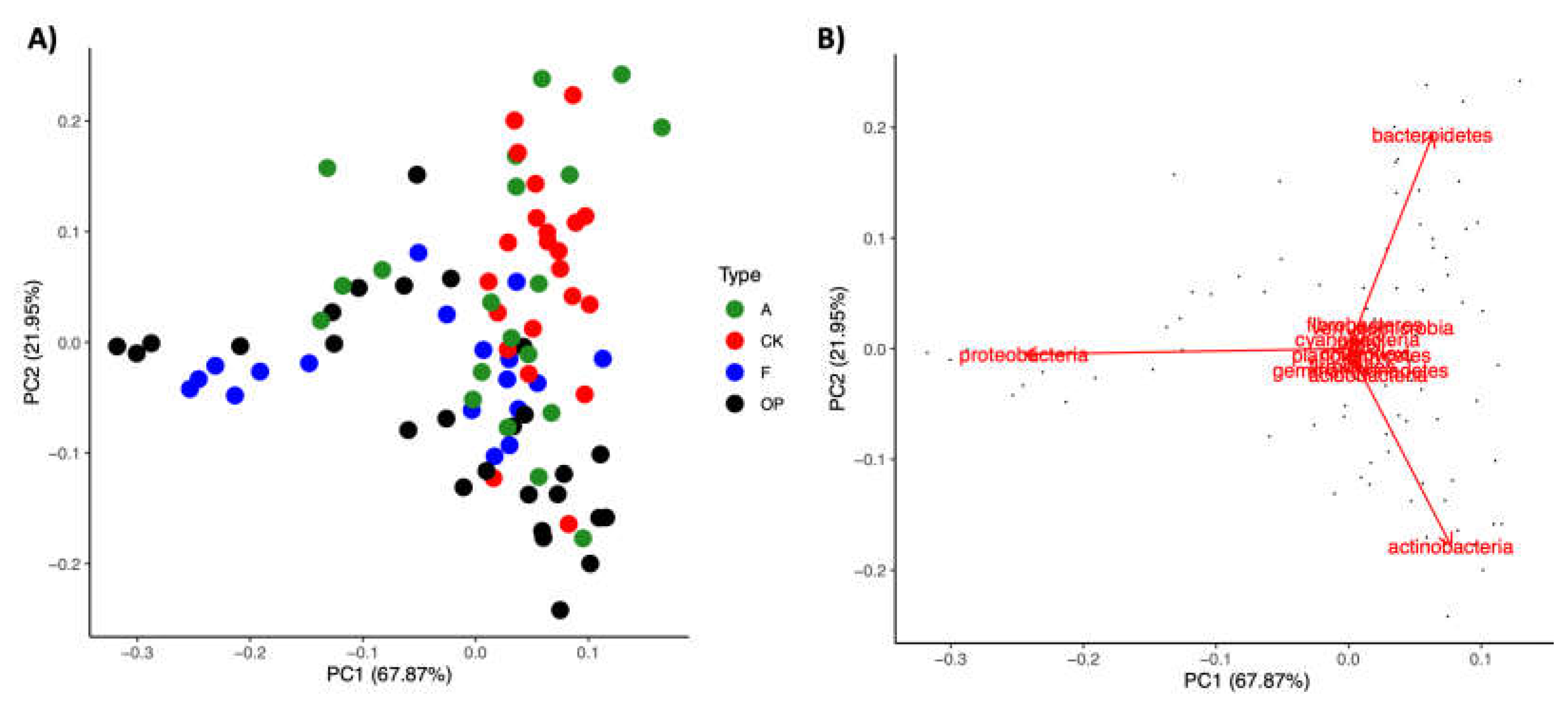
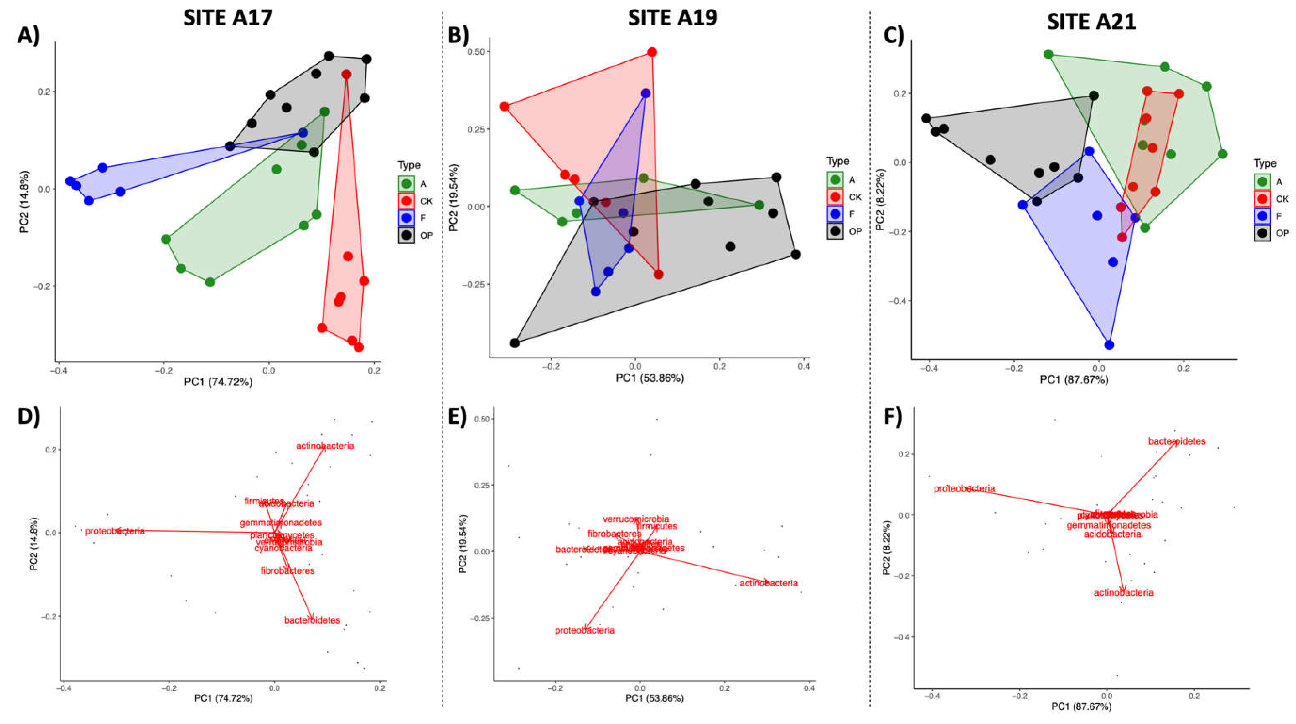
Principal components analysis revealed distinctive clustering of vegetated microhabitats with some overlap with barren microhabitats when using phylum composition data of all samples from all three sites (Figure 2A). When looking at the data from all three the sampling sites together, the biggest drivers of these community shifts seem to be Bacteroidetes for the vegetated samples, while Proteobacteria and Actinobacteria seem to be the largest drivers for the barren samples (Figure 2B). When the same phylum-level PCA analysis was performed for each individual sampling site, separate clustering between vegetated and barren samples was also observed in sites A17 and A21 (Figure 3). At these sites, it was possible to distinguish clustering of the foredune toe and blowout. However, Actinobacteria and Proteobacteria seem to be key drivers for blowout samples in A17 and in A21, respectively, while their role was reversed in foredune toe samples. On both sites, Bacteroidetes is a key driver for the vegetated stands. Fibrobacteria seem to also be a driver in vegetated stands in A17. At site A19, clustering was less distinct between the four microhabitats, although Proteobacteria and Actinobacteria were observed once again to be key drivers, particularly for the blowout samples.

3.3. Genus-Level Variability in Microbiome Composition

Differences in genus composition are presented in Table 2. Of the 58 genera that represented more than 0.1% of the community composition, 14 were found to have significant differences between microhabitat categories. The genera Devosia (F2,85 = 35.88, p < 0.0001), Fibrobacter (F2,85 = 42.64, p < 0.0001), Mesorhizobium (F2,85 = 14.01, p < 0.0001), Mucilaginibacter (F2,85 = 24.13, p < 0.0001), Ohtaekwangia (F2,85 = 19.87, p < 0.0001) and Sphingobacterium (F2,85 = 13.10, p < 0.0001) were significantly more abundant in both vegetated microhabitats compared to barren microhabitats. In Ammophila stands, Bacteriovorax (F2,85 = 4.80, p = 0.01) and Flavisolibacter (F2,85 = 10.23, p < 0.0001) were significantly more abundant than in barren microhabitats. In invasive stands, Cytophaga (F2,85 = 74.78, p < 0.0001) and Streptomyces (F2,85 = 24.04, p < 0.0001) were significantly more prevalent than in A. breviligulata stands, and they are significantly more prevalent in vegetated microhabitats than in barren habitats. In addition, the genera Ktedonobacter (F2,85 = 6.65, p = 0.002) and Opitutus (F2,85 = 19.28, p < 0.0001) had significantly higher numbers in invasive stands than in both native A. breviligulata stands and barren microhabitats. Barren microhabitats also had significantly higher numbers of Acidobacterium (F2,85 = 4.40, p = 0.02) and Bacillus (F2,85 = 6.88, p = 0.002) than in invasive stands.
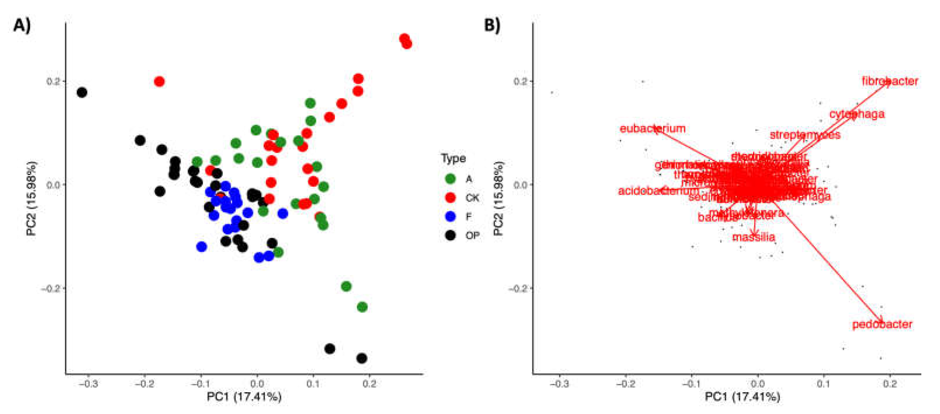
The genus percentage composition of the 58 genera that represented more than 0.1% of the community composition was also used in principal component analysis to investigate their role in community composition of all dune microhabitats (Figure 4). Similar to the results observed in phylum-level PCA analyses, the most distinct clustering was observed in the vegetated microhabitats with much clearer separation from the barren microhabitats. The major drivers for the vegetated microhabitats were found to be OTUs of the genus Fibrobacter, Cytophaga and Streptomyces, whereas the barren microhabitats seem to be largely driven by OTUs from the genus Pedobacter, Eubacterium, Massilia, Acidobacterium, and Bacillus.
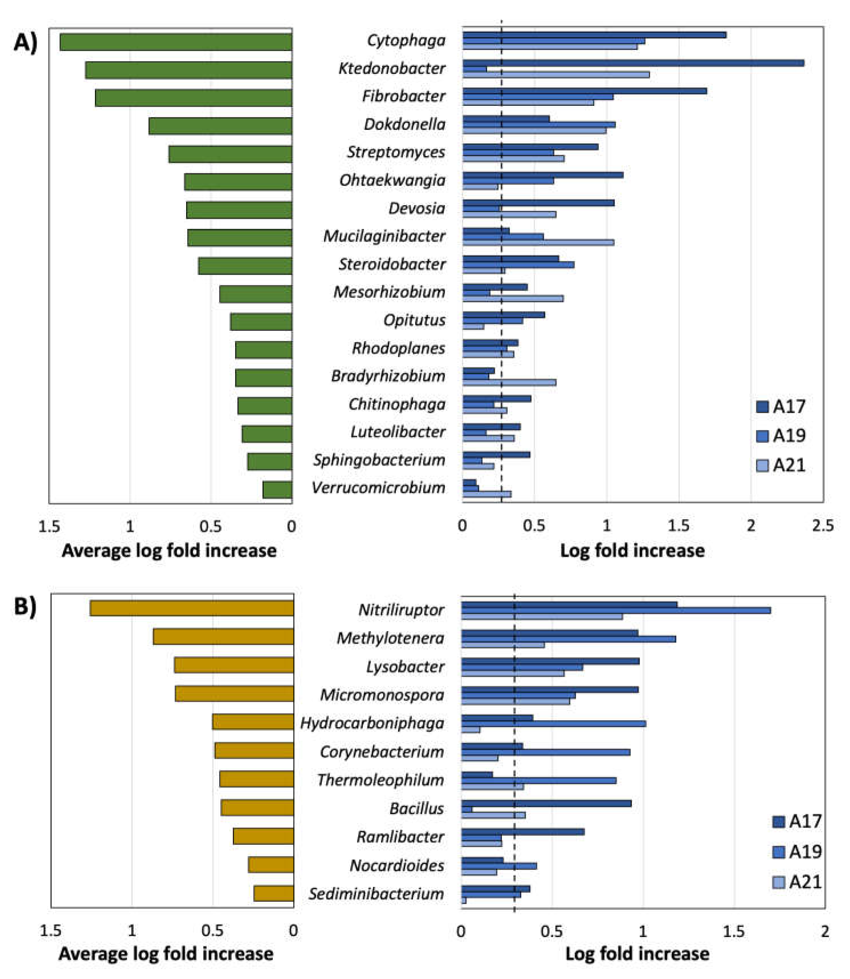
Genus composition data from all sites were used to determine which genera had the highest log-fold differences in composition, although it should be noted that these might not translate into changes in absolute abundances. A total of 17 genera in invasive stands and 11 genera in barren microhabitats were found to have two-fold higher proportions relative to each other in at least one of the three sampling sites (A17, A19 and A21 (Figure 5)). The top three genera with the highest average log-fold changes more prevalent in C. kobomugi microhabitats compared to barren sites were Cytophaga (1.43 log fold, 26.9 fold change), Ktedonobacter (1.27 log fold, 18.6 fold change), and Fibrobacter (1.21 log fold, 16.2 fold change). In addition, seven genera that were consistently found to have a log-fold difference higher than 0.301 (2-fold) between invasive C. kobomugi stands and barren microhabitats across all three sampling sites consisted of Cytophaga, Fibobacter, Dokdonella, Streptomyces, Mucilaginibacter, Steroidobacter, and Rhodoplanes. When looking at samples collected from A. breviligulata stands compared to barren microhabitats, a total of 17 genera from A. breviligulata and 13 genera in barren microhabitats were found to have a two-fold higher proportions relative to each other in at least one of the three sampling sites (Figure 6). The three genera with the highest log-fold changes with higher proportions in A. breviligulata stands were Fibrobacter (1.12 log fold, 13.2 fold change), Cytophaga (1.03 log fold, 10.7 fold change), and Leptospira (1.10 log fold, 12.6 fold change). In addition, five genera that were consistently found to have a log-fold difference higher than 0.301 (2-fold) between native A. breviligulata stands and barren microhabitats across all three sampling sites: Fibrobacter, Cytophaga, Leptospira, Devosia, and Streptomyces.
The two plant species shared 12 distinct genera among the genera more prevalent in vegetated than barren samples (Figure 5 and Figure 6): Fibrobacter, Chitinophaga, Mucilaginibacter, Ohtaekwangia, Sphingobacterium, Devosia, Dokdonella, Mesorhizobium, Rhodoplanes, Steroidobacter, and Streptomyces. There were five genera unique to C. kobomugi stands (Bradyrhizobium, Ktedonobacter, Luteolibacter, Opitutus, and Verrucomicrobium) and five genera unique to from A. breviligulata stands (Flavisoilibacter, Geobacter, Bacteriovorax, Pelobacter, and Leptospira).

3.4. Spatial Patterns

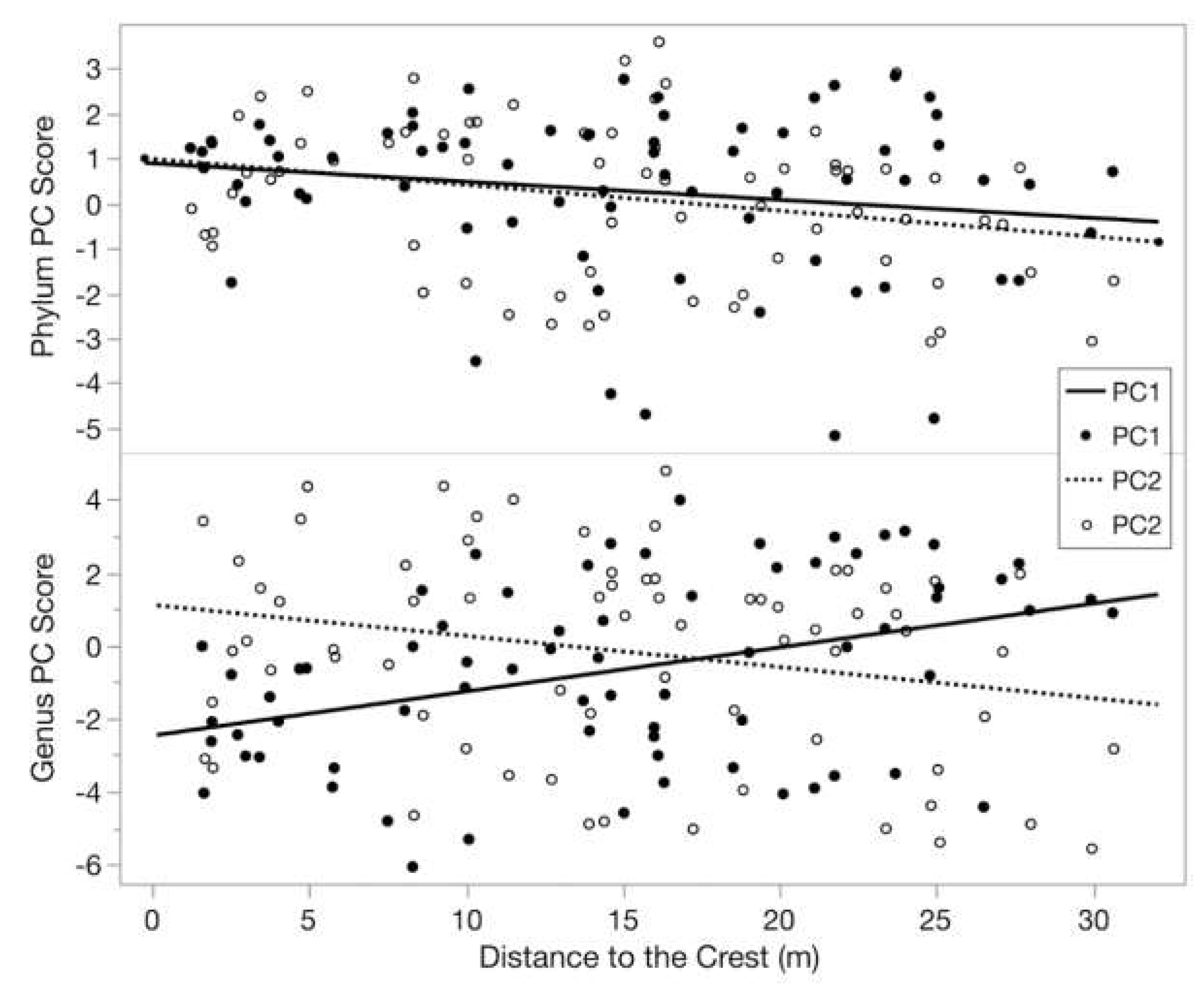
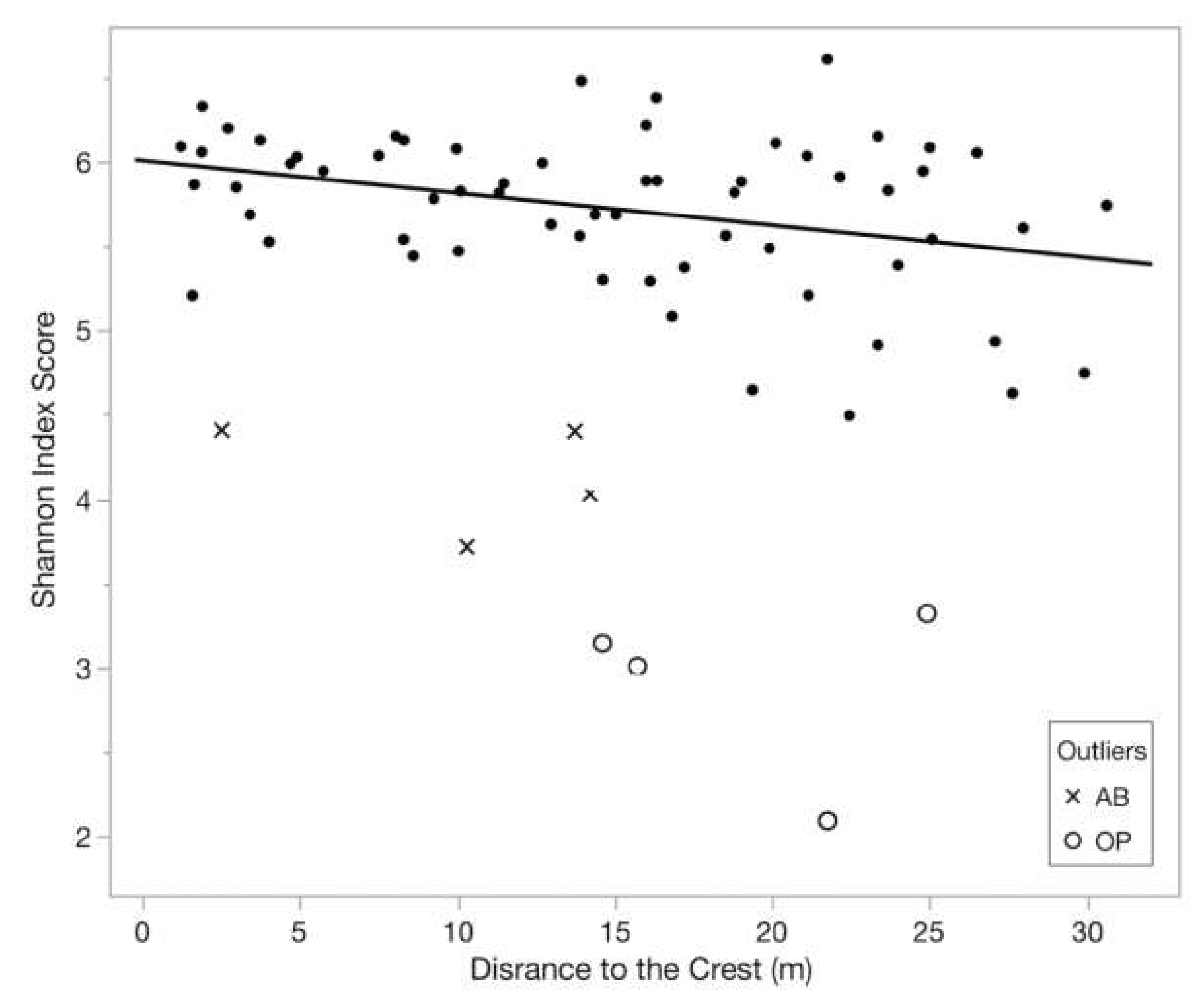
Principal component ordination using genus composition for PC1 (Genus PC1) increases moving inland from the crest (R2 = 0.15, F1,68 = 12.14, p = 0.0009). Conversely, genus PC2 (R2 = 0.06, F1,68 = 4.39, p = 0.04), phylum PC2 (R2 = 0.08, F1,68 = 5.70, p = 0.02), and Shannon diversity (R2 = 0.12, F1,60 = 8.30, p = 0.005) decrease moving inland from the crest (Figure 7). Phylum PC1 is also higher at the crest and decreases inland, but this pattern is not significant (R2 = 0.03, F1,68 = 2.19, p = 0.14; Figure 7). Shannon diversity also shows greatest diversity at the crest, decreasing moving inland. This relationship is consistent as a trend with the eight outliers included, which had Shannon diversity scores lower than 4.5 (R2 = 0.05, F1,68 = 3.34, p = 0.07), and was strengthened when they were removed (R2 = 0.12, F1,60 = 8.30, p = 0.005; Figure 8).
In invasive and blowout microhabitats, genus PC1 decreases moving towards habitat edges (R2 = 0.14, F1,47 = 7.392, p = 0.007), whereas phylum PC1 is greatest closer to the edges and decreases moving towards the microhabitat center (R2 = 0.09, F1,47 = 4.30, p = 0.04). The same patterns emerge examining PC2, but they are not statistically significant (p > 0.05). Shannon diversity does not vary with position in these microhabitats.

4. Discussion

Bacterial OTU diversity (Shannon index) was higher in vegetated native A. breviligulata and invasive C. kobomugi stands than in barren blowout and dune toe microhabitats. Additionally, phylum and genus PCAs revealed significant spatial relationships in PC score relative to sample distance to the crest. Plant diversity and soil salinity have been reported to affect microbial community composition in coastal dunes [31,40], but the analyses between sites here suggest that there may be other factors at play.
Phylum-level community composition distinctions between microhabitats largely stem from differences among vegetated and barren microhabitats. Although minor changes in community compositions might have occurred between sampling and DNA extraction, consistent with previous studies of dune and desert environments [13,14,16,63], Proteobacteria, Actinobacteria, and Bacteroidetes were the dominant phyla across all habitats. The observed prevalence of Bacteroidetes in vegetated versus barren microhabitats has not been previously reported. However, Bacteroidetes contain well-known organic matter degraders [64,65], and endophytic bacteria associated with plants inhabiting similarly challenging environments (such as the Chilean desert and Arctic [66,67]) make them likely candidates to be found in these environments. Our finding that Proteobacteria and Actinobacteria are conversely more prevalent in barren microhabitats supports previous studies on coastal dunes [13,16]. The community compositions in vegetated microhabitats are more similar to each other than to barren microhabitats, with similar percent compositions of Firmicutes, Bacteroidetes, and Actinobacteria. These phyla have all been found in other coastal and desert dune systems and include species that have been identified as plant associated microorganisms [13,14,68]. Specifically, higher levels of Verrucomicrobia and Fibrobacteres were found in invasive C. kobomugi stands, two ubiquitous soil phyla with few culture representatives and cellulose degrading capabilities, respectively [69,70,71].
Plant presence appears to drive increased prevalence of genera that have been found to benefit plants in other habitats in ways that would increase dune plant fitness. Specifically, Fibrobacter, Cytophaga, Streptomyces, Sphingobacterium, Devosia, Mesorhizobium, and Mucilaginibacter were all more prevalent in vegetated stands than barren microhabitats and can assist in plant growth promotion, such as through phosphorus solubilization and the production of plant growth promoting compounds [72,73,74,75,76,77]. Mucilaginibacter specifically had the largest log-fold changes between barren and vegetated microhabitats and can be common in soils and plant tissue of coastal environments [78,79,80]. Similarly, Devosia and Mesorhizobium species have been demonstrated to form root nodules in aquatic and coastal plants [76,81]. Fibrobacter and Cytophaga species can degrade cellulose and complex soil carbohydrates aiding in organic matter decomposition [71,82,83]. Streptomyces and Sphingobacterium species can both reduce plant soil salinity stress [84,85,86,87] and Streptomyces and Sphingobacterium can positively impact soil aggregation [24,25,88,89] and suppress harmful fungal growth [74,90], respectively.
The dominant genera we found in barren microhabitats are known to dwell in desert soils promoting plant growth. Barren microhabitats had larger proportions of OTUs corresponding to the genera Bacillus, Eubacterium, Pedobacter, Acidobacterium, and Massilia. Bacillus is a very diverse genus of well-known soil-dwelling organisms, with some members being able to produce many important secondary metabolites [91,92]. Bacillus species have previously been isolated from extreme sandy environments, including Bacillus sonorensis (which was isolated from the Sonoran Desert [93]) and Bacillus vallismortis, isolated from Death Valley [94]. Among the genera more prominent in barren environments, some species are known to have plant associations, such as those from soil Pedobacter [95,96,97]. Pedobacter species have been found in biological soil crusts in the Gurnbantunggut Desert [98]. Similarly, the genus Massilia can be an important microbial community member in desert soils and includes rhizosphere colonizers [99,100,101]. These genera include species with adaptations that would serve them well in both barren and vegetated environments, but they do not appear to establish associations with either the native or invasive plant.
The plant species largely share similar community compositions. However, we have identified differences in composition in this study that may contribute to the success of each respective plant on coastal dunes. Invasive species can alter community composition of the surrounding soil, potentially impacting the ability of native species to recruit specific microbial communities [45,46,53]. These changes in microbial community composition may have negative consequences for the establishment of native species within the ecosystem [20,46,102]; altering plant establishment and succession in this ecosystem [4,5] can have cascading impacts on dune stability [6,11,12]. Despite belonging to different taxonomic families (Poaceae and Cyperaceae), the two plant species share twelve dominant genera, and five unique genera each.
The unique genera found in invasive C. kobomugi stands (Bradyrhizobium, Ktedonobacter, Luteolibacter, Opitutus, and Verrucomicrobium) may impact its success as an invader. Nitrogen-fixing bacteria, like the genus Bradyrhizobium, have the ability to drive the establishment of invasive plant species [49]. Other genera seem to be associated with reduced nutrient availability. This is the case of Ktedonobacter, a genus that has been linked to plant disease as it competes for nitrogen as a soil nutrient [103]. Two genera belonging to the phylum Verrucomicrobia, Opitutus and Verrucomicrobium, also play important roles in nitrogen and nutrient availability. Opitutus species have an important role in denitrification and are prevalent in nitrate-rich environments [104,105] and the genus Verrucomicrobium has been associated with soils with low nutrient availability [106,107]. The extensive root system and density of C. kobomugi further reduce nutrient availability in these already nutrient poor habitats [6,41,57,58], favoring the higher prevalence of plant-associated organisms capable of inhabiting low-nutrient environments.
The unique genera found in native A. breviligulata stands (Flavisolibacter, Geobacter, Bacteriovorax, Pelobacter, and Leptospira) may aid in its ability to establish itself as a pioneer through stress reduction. These unique genera may be more prevalent given variations in root exudates from vegetation [23,108]. Nitrogen as a limiting nutrient could come into play again, since Geobacter and Pelobacter are capable of nitrogen fixation [109,110]. A. breviligulata may also actively recruit organisms involved in disease suppression. Flavisolibacter has been positively correlated with disease suppression against the pathogens Rhizoctonia solani [111] and Plasmodiophora brassicae in infected cabbage plants [112]. Similarly, Bacteriovorax is a genus of known predatory bacteria that can serve as a biocontrol of gram-negative plant pathogens [113]. Genera unique to A. breviligulata may also play an important role in soil aggregation in the dunes, since Geobacter, Bacteriovorax, and Leptospira species are known to produce biofilms through extracellular polymeric substances production [114,115,116].

5. Conclusions

While vegetated invasive C. kobomugi and native A. breviligulata stands together were more different than barren dune toe and blowout habitats, and the subtle differences between the microbial community composition of two species could ultimately aid in the invader’s success. The changes in microbial community composition associated with C. kobomugi appear to reduce the proportions of bacterial genera associated exclusively with A. breviligulata which have been documented as affecting plant disease suppression and soil aggregation. The displacement of microbes protecting plant health and dune development have the potential to alter dune geomorphology and succession. However, these changes represent a small fraction of the community compared to the proportion of plant-associated genera shared by both vegetated microhabitats. To fully understand the specific roles these genera play in vegetated microbial communities and the surrounding environment they create, further investigation into specific bacterial species present at these sites and their functional abilities is needed. Similarly, removing C. kobomugi and planting A. breviligulata or vice versa, with microbial sample collection over time, would reveal the timescale at which the microbial communities change after conservation efforts or invasion, respectively. Regardless, barren microhabitat microbial composition does not appear to disproportionately alter the success or failure of one plant species or the other in establishing itself to recolonize these open niche areas. Given this, plant arrival time may be crucial followed by the subsequent recruitment and fostering of favorable microbiomes that would further increase the likelihood of continued survival and spread of a native or invasive plant.

Supplementary Materials

The following are available online at https://www.mdpi.com/article/10.3390/d13110525/s1, Supplementary S1: Field Collection Details. Supplementary S2: Soil Characteristic Analyses.

Author Contributions

Conceptualization, Resources, Visualization, Supervision, and funding—B.R.C. and J.A.I.; Methodology, formal analysis, investigation, and writing—original Draft Preparation and Review & Editing—all authors; Data Curation and Project Administration—J.A.I. All authors have read and agreed to the published version of the manuscript.

Funding

This research was conducted in part with United States Government support by the National Science Foundation grant IOS-1645909 and by the Department of Defense, Air Force Office of Scientific Research, National Defense Science and Engineering Graduate (NDSEG) Fellowship, 32 CRF 168a.

Institutional Review Board Statement

Not applicable.

Informed Consent Statement

Not applicable.

Data Availability Statement

All data are available upon request. Additionally, all microbiome sequence files are publicly available under GenBank accession numbers SRR15979670-SRR15979757 and the BioProject number PRJNA764730.

Acknowledgments

We thank Brenda Casper for her support. We thank the NJ Fish and Wildlife service and Island Beach State Park for their support conducting this research.

Conflicts of Interest

The authors declare no conflict of interest.

References

  1. Meyer, L.; Brinkman, S.; van Kesteren, L.; Leprince-Ringuet, N.; van Boxmeer, F. Climate Change 2014: Synthesis Report. Contribution of Working Groups I, II and III to the Fifth Assessment Report of the Intergovernmental Panel on Climate Change Technical Support. Unit for the Synthesis Report; IPCC: Geneva, Switzerland, 2014; p. 169. [Google Scholar]
  2. Portner, H.O.; Roberts, D.C.; Masson-Delmotte, V.; Zhai, P.; Tignor, M.; Poloczanska, E.; Mintenbeck, K.; Alegría, A.; Nicolai, M.; Okem, A.; et al. Summary for Policymakers. In IPCC Special Report on the Ocean and Cryosphere in a Changing Climate; IPCC: Geneva, Switzerland, 2019. [Google Scholar]
  3. Isaac, P.G. Essentials of Nucleic Acid Analysis: A Robust Approach. Ann. Bot. 2009, 104, 5–6. [Google Scholar] [CrossRef] [Green Version]
  4. Jones, C.; Lawton, J.H.; Shachak, M. Organisms as Ecosystem Engineers. Okios 1994, 69, 373–386. [Google Scholar] [CrossRef]
  5. Cheplick, G.P. Changes in Plant Abundance on a Coastal Beach Following Two Major Storm Surges. J. Torrey Bot. Soc. 2016, 143, 180–191. [Google Scholar] [CrossRef]
  6. Charbonneau, B.R.; Wootton, L.S.; Wnek, J.P.; Langley, J.A.; Posner, M.A. A Species Effect on Storm Erosion: Invasive Sedge Stabilized Dunes More than Native Grass during Hurricane Sandy. J. Appl. Ecol. 2017, 54, 1385–1394. [Google Scholar] [CrossRef] [Green Version]
  7. Tanaka, N.; Nandasena, N.A.K.; Jinadasa, K.B.S.N.; Sasaki, Y.; Tanimoto, K.; Mowjood, M.I.M. Developing Effective Vegetation Bioshield for Tsunami Protection. Civ. Eng. Environ. Syst. 2009, 26, 163–180. [Google Scholar] [CrossRef]
  8. Zarnetske, P.L.; Hacker, S.D.; Seabloom, E.W.; Ruggiero, P.; Killian, J.R.; Maddux, T.B.; Cox, D. Biophysical Feedback Mediates Effects of Invasive Grasses on Coastal Dune Shape. Ecology 2012, 93, 1439–1450. [Google Scholar] [CrossRef] [PubMed]
  9. Sigren, J.M.; Figlus, J.; Armitage, A.R. Coastal Sand Dunes and Dune Vegetation: Restoration, Erosion, and Storm Protection. Shore Beach 2014, 82, 5–12. [Google Scholar]
  10. Forster, S.M.; Nicolson, T.H. Aggregation of Sand from a Maritime Embryo Sand Dune by Microorganisms and Higher Plants. Soil Biol. Biochem. 1981, 13, 199–203. [Google Scholar] [CrossRef]
  11. Hesp, P.A. A Review of Biological and Geomorphological Processes Involved in the Initiation and Development of Incipient Foredunes. Proc. R. Soc. Edinb. 1989, 96. [Google Scholar] [CrossRef]
  12. Hacker, S.D.; Jay, K.R.; Cohn, N.; Goldstein, E.B.; Hovenga, P.A.; Itzkin, M.; Moore, L.J.; Mostow, R.S.; Mullins, E.V.; Ruggiero, P. Species-Specific Functional Morphology of Four US Atlantic Coast Dune Grasses: Biogeographic Implications for Dune Shape and Coastal Protection. Diversity 2019, 11, 82. [Google Scholar] [CrossRef] [Green Version]
  13. Lin, Y.T.; Whitman, W.B.; Coleman, D.C.; Chen, T.H.; Chiu, C.Y. Composition of Bacterial Communities in Sand Dunes of Subtropical Coastal Forests. Biol. Fertil. Soils 2014, 50, 809–814. [Google Scholar] [CrossRef]
  14. Liu, R.; Li, K.; Zhang, H.; Zhu, J.; Joshi, D. Spatial Distribution of Microbial Communities Associated with Dune Landform in the Gurbantunggut Desert, China. J. Microbiol. 2014, 52, 898–907. [Google Scholar] [CrossRef]
  15. Abdul Majid, S.; Graw, M.F.; Chatziefthimiou, A.D.; Nguyen, H.; Richer, R.; Louge, M.; Sultan, A.A.; Schloss, P.; Hay, A.G. Microbial Characterization of Qatari Barchan Sand Dunes. PLoS ONE 2016, 11, e0161836. [Google Scholar] [CrossRef] [Green Version]
  16. Shet, S.A.; Garg, S. Prokaryotic Diversity of Tropical Coastal Sand Dunes Ecosystem Using Metagenomics. 3 Biotech. 2021, 11, 252. [Google Scholar] [CrossRef]
  17. Howes, A. Environmental Factors, Not. Plants, Contribute to Functional Diversity of Soil Bacteria in the Dunes of Lake Michigan; University of Louisville: Louisville, KY, USA, 2017. [Google Scholar]
  18. Nayak, S.; Behera, S.; Dash, P.K. Potential of Microbial Diversity of Coastal Sand Dunes: Need for Exploration in Odisha Coast of India. Sci. World J. 2019, 2019, 2758501. [Google Scholar] [CrossRef] [Green Version]
  19. Viles, H.A. Microbial Geomorphology: A Neglected Link between Life and Landscape. Geomorphology 2012, 157–158, 6–16. [Google Scholar] [CrossRef]
  20. Batten, K.M.; Scow, K.M.; Espeland, E.K. Soil Microbial Community Associated with an Invasive Grass Differentially Impacts Native Plant Performance. Microb. Ecol. 2008, 55, 220–228. [Google Scholar] [CrossRef]
  21. Chang, E.H.; Chen, C.T.; Chen, T.H.; Chiu, C.Y. Soil Microbial Communities and Activities in Sand Dunes of Subtropical Coastal Forests. Appl. Soil Ecol. 2011, 49, 256–262. [Google Scholar] [CrossRef]
  22. Bertin, C.; Yang, X.; Weston, L.A. The Role of Root Exudates and Allelochemicals in the Rhizosphere. Plant. Soil 2003, 256, 67–83. [Google Scholar] [CrossRef]
  23. Huang, X.-F.; Chaparro, J.M.; Reardon, K.F.; Zhang, R.; Shen, Q.; Vivanco, J.M. Rhizosphere Interactions: Root Exudates, Microbes, and Microbial Communities. Botany 2014, 92, 267–275. [Google Scholar] [CrossRef]
  24. Godinho, A.L.; Bhosle, S. Sand Aggregation by Exopolysaccharide-Producing Microbacterium arborescens––AGSB. Curr. Microbiol. 2009, 58, 616–621. [Google Scholar] [CrossRef]
  25. Wasserstrom, H.; Kublik, S.; Wasserstrom, R.; Schulz, S.; Schloter, M.; Steinberger, Y. Bacterial Community Composition in Costal Dunes of the Mediterranean along a Gradient from the Sea Shore to the Inland. Sci. Rep. 2017, 7, 40266. [Google Scholar] [CrossRef] [Green Version]
  26. Soka, G.; Ritchie, M. Arbuscular Mycorrhizal Symbiosis and Ecosystem Processes: Prospects for Future Research in Tropical Soils. Open J. Ecol. 2014, 4, 11–22. [Google Scholar] [CrossRef] [Green Version]
  27. Yang, J.; Kloepper, J.W.; Ryu, C.-M. Rhizosphere Bacteria Help Plants Tolerate Abiotic Stress. Trends Plant. Sci. 2009, 14, 1–4. [Google Scholar] [CrossRef]
  28. Li, W.; Sheng, H.; Ekawati, D.; Jiang, Y.; Yang, H. Variations in the Compositions of Soil Bacterial and Fungal Communities Due to Microhabitat Effects Induced by Simulated Nitrogen Deposition of a Bamboo Forest in Wetland. Forests 2019, 10, 1098. [Google Scholar] [CrossRef] [Green Version]
  29. David, A.S.; Seabloom, E.W.; May, G. Plant Host Species and Geographic Distance Affect the Structure of Aboveground Fungal Symbiont Communities, and Environmental Filtering Affects Belowground Communities in a Coastal Dune Ecosystem. Microb. Ecol. 2016, 71, 912–926. [Google Scholar] [CrossRef] [PubMed]
  30. Titus, J.H.; del Moral, R. The Role of Mycorrhizal Fungi and Microsites in Primary Succession on Mount St. Helens. Am. J. Bot. 1998, 85, 370–375. [Google Scholar] [CrossRef]
  31. Rajaniemi, T.K.; Allison, V.J. Abiotic Conditions and Plant Cover Differentially Affect Microbial Biomass and Community Composition on Dune Gradients. Soil Biol. Biochem. 2009, 41, 102–109. [Google Scholar] [CrossRef]
  32. Muñoz Vallés, S.; Cambrollé, J.; Gallego-Fernández, J.B. Effect of Soil Characteristics on Plant Distribution in Coastal Ecosystems of SW Iberian Peninsula Sand Spits. Plant. Ecol. 2015, 216, 1551–1570. [Google Scholar] [CrossRef]
  33. Gallego-Fernández, J.B.; Martínez, M.L. Environmental Filtering and Plant Functional Types on Mexican Foredunes along the Gulf of Mexico. Écoscience 2011, 18, 52–62. [Google Scholar] [CrossRef]
  34. Ley, R.E.; Lipson, D.A.; Schmidt, S.K. Microbial Biomass Levels in Barren and Vegetated High Altitude Talus Soils. Soil Sci. Soc. Am. J. 2001, 65, 111–117. [Google Scholar] [CrossRef]
  35. Li, Z.; Zu, C.; Wang, C.; Yang, J.; Yu, H.; Wu, H. Different Responses of Rhizosphere and Non-Rhizosphere Soil Microbial Communities to Consecutive Piper nigrum L. Monoculture. Sci. Rep. 2016, 6, 35825. [Google Scholar] [CrossRef]
  36. Poosakkannu, A.; Nissinen, R.; Männistö, M.; Kytöviita, M.-M. Microbial Community Composition but Not Diversity Changes along Succession in Arctic Sand Dunes: Deschampsia flexuosa Associated Microbiomes. Environ. Microbiol. 2017, 19, 698–709. [Google Scholar] [CrossRef] [PubMed] [Green Version]
  37. Du, J.; Hesp, P.A. Salt Spray Distribution and Its Impact on Vegetation Zonation on Coastal Dunes: A Review. Estuaries Coasts 2020, 43, 1885–1907. [Google Scholar] [CrossRef]
  38. Wichern, J.; Wichern, F.; Joergensen, R.G. Impact of Salinity on Soil Microbial Communities and the Decomposition of Maize in Acidic Soils. Geoderma 2006, 137, 100–108. [Google Scholar] [CrossRef]
  39. Yang, J.; Ma, L.; Jiang, H.; Wu, G.; Dong, H. Salinity Shapes Microbial Diversity and Community Structure in Surface Sediments of the Qinghai-Tibetan Lakes. Sci. Rep. 2016, 6, 25078. [Google Scholar] [CrossRef] [PubMed] [Green Version]
  40. Elbl, J.; Zahora, J. The Comparison of Microbial Activity in Rhizosphere and Non-Rhizosphere Soil Stressed by Drought. Mendel. Net. 2014, 7, 234–240. [Google Scholar]
  41. Charbonneau, B.R.; Nicoletta, R.; Wootton, L.S. A Decade of Expansion of the Invasive Plant Carex kobomugi in a Coastal Foredune System. Biol. Invasions 2020, 22, 2099–2112. [Google Scholar] [CrossRef]
  42. Fei, S.; Phillips, J.; Shouse, M. Biogeomorphic Impacts of Invasive Species. Annu. Rev. Ecol. Evol. Syst. 2014, 45, 69–87. [Google Scholar] [CrossRef] [Green Version]
  43. Gallego-Fernández, J.B.; Martínez, M.L.; García-Franco, J.G.; Zunzunegui, M. Multiple Seed Dispersal Modes of an Invasive Plant Species on Coastal Dunes. Biol. Invasions 2021, 23, 111–127. [Google Scholar] [CrossRef]
  44. Kourtev, P.S.; Ehrenfeld, J.G.; Haggblom, M. Exotic Plant Species Alter the Microbial Community Structure and Function in the Soil. Ecology 2002, 83, 3152–3166. [Google Scholar] [CrossRef]
  45. Batten, K.M.; Scow, K.M.; Davies, K.F.; Harrison, S.P. Two Invasive Plants Alter Soil Microbial Community Composition in Serpentine Grasslands. Biol. Invasions 2006, 8, 217–230. [Google Scholar] [CrossRef]
  46. Leege, L.M.; Murphy, P.G. Ecological Effects of the Non-Native Pinus nigra on Sand Dune Communities. Can. J. Bot. 2001, 79, 429–437. [Google Scholar] [CrossRef]
  47. Stefanowicz, A.M.; Zubek, S.; Stanek, M.; Grześ, I.M.; Rożej-Pabijan, E.; Błaszkowski, J.; Woch, M.W. Invasion of Rosa rugosa Induced Changes in Soil Nutrients and Microbial Communities of Coastal Sand Dunes. Sci. Total Environ. 2019, 677, 340–349. [Google Scholar] [CrossRef] [PubMed]
  48. Jurkšienė, G.; Janušauskaitė, D.; Baliuckas, V. Microbial Community Analysis of Native Pinus sylvestris L. and Alien Pinus mugo L. on Dune Sands as Determined by Ecoplates. Forests 2020, 11, 1202. [Google Scholar] [CrossRef]
  49. Wu, B.; Wang, S.; Wei, M.; Zhou, J.; Jiang, K.; Du, D.; Wang, C. The Invasive Tree Staghorn sumac Affects Soil N2-fixing Bacterial Communities in North China. Plant. Biol. 2019, 21, 951–960. [Google Scholar] [CrossRef] [PubMed]
  50. Day, N.J.; Antunes, P.M.; Dunfield, K.E. Changes in Arbuscular Mycorrhizal Fungal Communities during Invasion by an Exotic Invasive Plant. Acta Oecologica 2015, 67, 66–74. [Google Scholar] [CrossRef]
  51. Elgersma, K.J.; Ehrenfeld, J.G.; Yu, S.; Vor, T. Legacy Effects Overwhelm the Short-Term Effects of Exotic Plant Invasion and Restoration on Soil Microbial Community Structure, Enzyme Activities, and Nitrogen Cycling. Oecologia 2011, 167, 733–745. [Google Scholar] [CrossRef] [PubMed]
  52. Tanner, R.A.; Gange, A.C. The Impact of Two Non-Native Plant Species on Native Flora Performance: Potential Implications for Habitat Restoration. Plant. Ecol. 2013, 214, 423–432. [Google Scholar] [CrossRef]
  53. Malacrinò, A.; Sadowski, V.A.; Martin, T.K.; Cavichiolli de Oliveira, N.; Brackett, I.J.; Feller, J.D.; Harris, K.J.; Combita Heredia, O.; Vescio, R.; Bennett, A.E. Biological Invasions Alter Environmental Microbiomes: A Meta-Analysis. PLoS ONE 2020, 15, e0240996. [Google Scholar] [CrossRef]
  54. Corbin, J.D.; D’Antonio, C.M. Gone but Not Forgotten? Invasive Plants’ Legacies on Community and Ecosystem Properties. Invasive Plant Sci. Manag. 2012, 5, 117–124. [Google Scholar] [CrossRef]
  55. Durán Vinent, O.; Moore, L.J. Barrier Island Bistability Induced by Biophysical Interactions. Nat. Clim. Chang. 2015, 5, 158–162. [Google Scholar] [CrossRef]
  56. Small, J.A. Carex kobomugi at Island Beach, New Jersey. Ecology 1954, 35, 289–291. [Google Scholar] [CrossRef]
  57. Wootton, L.S.; Halsey, S.D.; Bevaart, K.; McGough, A.; Ondreicka, J.; Patel, P. When Invasive Species Have Benefits as Well as Costs: Managing Carex kobomugi (Asiatic Sand Sedge) in New Jersey’s Coastal Dunes. Biol. Invasions 2005, 7, 1017–1027. [Google Scholar] [CrossRef]
  58. Charbonneau, B.R. From The Sand They Rise: Post-Storm Foredune Plant Recolonization And Its Biogeomorphic Implications; University of Pennsylvania: Philadelphia, PA, USA, 2019; pp. 2–19. [Google Scholar]
  59. Hesp, P. Foredunes and Blowouts: Initiation, Geomorphology and Dynamics. Geomorphology 2002, 48, 245–268. [Google Scholar] [CrossRef]
  60. Sallenger, A.H.J. Storm Impact Scale for Barrier Islands. J. Coast. Res. 2000, 16, 890–895. [Google Scholar]
  61. Friese, C.F.; Koske, R.E. The Spatial Dispersion of Spores of Vesicular-Arbuscular Mycorrhizal Fungi in a Sand Dune: Microscale Patterns Associated with the Root Architecture of American Beachgrass. Mycol. Res. 1991, 95, 952–957. [Google Scholar] [CrossRef]
  62. R Core Team. R: A Language and Environment for Statistical Computing. Vienna, Austria, 2016. Available online: Https://Www.R-Project.Org/ (accessed on 1 August 2021).
  63. Janssen, P.H. Identifying the Dominant Soil Bacterial Taxa in Libraries of 16S RRNA and 16S RRNA Genes. Appl. Environ. Microbiol. 2006, 72, 1719–1728. [Google Scholar] [CrossRef] [Green Version]
  64. Thomas, F.; Hehemann, J.-H.; Rebuffet, E.; Czjzek, M.; Michel, G. Environmental and Gut Bacteroidetes: The Food Connection. Front. Microbiol. 2011, 2, 93. [Google Scholar] [CrossRef] [Green Version]
  65. Larsbrink, J.; McKee, L.S. Bacteroidetes bacteria in the soil: Glycan acquisition, enzyme secretion, and gliding motility. In Advances in Applied Microbiology; Elsevier: Amsterdam, The Netherlands, 2020; Volume 110, pp. 63–98. ISBN 978-0-12-820703-1. [Google Scholar]
  66. Zhang, B.; Hong, J.; Zhang, Q.; Jin, D.; Gao, C. Contrast in Soil Microbial Metabolic Functional Diversity to Fertilization and Crop Rotation under Rhizosphere and Non-Rhizosphere in the Coal Gangue Landfill Reclamation Area of Loess Hills. PLoS ONE 2020, 15, e0229341. [Google Scholar] [CrossRef] [PubMed]
  67. Zhang, Q.; Acuña, J.J.; Inostroza, N.G.; Mora, M.L.; Radic, S.; Sadowsky, M.J.; Jorquera, M.A. Endophytic Bacterial Communities Associated with Roots and Leaves of Plants Growing in Chilean Extreme Environments. Sci. Rep. 2019, 9, 4950. [Google Scholar] [CrossRef] [Green Version]
  68. Zhang, Q.; Acuña, J.J.; Inostroza, N.G.; Duran, P.; Mora, M.L.; Sadowsky, M.J.; Jorquera, M.A. Niche Differentiation in the Composition, Predicted Function, and Co-Occurrence Networks in Bacterial Communities Associated With Antarctic Vascular Plants. Front. Microbiol. 2020, 11, 1036. [Google Scholar] [CrossRef] [PubMed]
  69. Bergmann, G.T.; Bates, S.T.; Eilers, K.G.; Lauber, C.L.; Caporaso, J.G.; Walters, W.A.; Knight, R.; Fierer, N. The Under-Recognized Dominance of Verrucomicrobia in Soil Bacterial Communities. Soil Biol. Biochem. 2011, 43, 1450–1455. [Google Scholar] [CrossRef] [Green Version]
  70. Abdul Rahman, N.; Parks, D.H.; Vanwonterghem, I.; Morrison, M.; Tyson, G.W.; Hugenholtz, P. A Phylogenomic Analysis of the Bacterial Phylum Fibrobacteres. Front. Microbiol. 2016, 6, 1469. [Google Scholar] [CrossRef] [Green Version]
  71. Ransom-Jones, E.; Jones, D.L.; McCarthy, A.J.; McDonald, J.E. The Fibrobacteres: An Important Phylum of Cellulose-Degrading Bacteria. Microb. Ecol. 2012, 63, 267–281. [Google Scholar] [CrossRef] [PubMed]
  72. Dias, M.P.; Bastos, M.S.; Xavier, V.B.; Cassel, E.; Astarita, L.V.; Santarém, E.R. Plant Growth and Resistance Promoted by Streptomyces spp. in Tomato. Plant. Physiol. Biochem. 2017, 118, 479–493. [Google Scholar] [CrossRef]
  73. Mello, L.D.P.S.; dos Santos, A.C.; dos Santos, R.M.; Kandasamy, S.; Lazarovits, G.; Rigobelo, E.C. Application of the Bacterial Strains Ruminobacter amylophilus, Fibrobacter succinogenes and Enterococcus faecium for Growth Promotion in Maize and Soybean Plants. Aust. J. Crop. Sci. 2020, 14, 2020–2027. [Google Scholar] [CrossRef]
  74. Xu, L.; Zhang, H.; Xing, Y.-T.; Li, N.; Wang, S.; Sun, J.-Q. Complete Genome Sequence of Sphingobacterium psychroaquaticum Strain SJ-25, an Aerobic Bacterium Capable of Suppressing Fungal Pathogens. Curr. Microbiol. 2020, 77, 115–122. [Google Scholar] [CrossRef] [PubMed]
  75. Wani, P.A.; Khan, M.S.; Zaidi, A. Chromium-Reducing and Plant Growth-Promoting Mesorhizobium Improves Chickpea Growth in Chromium-Amended Soil. Biotechnol. Lett. 2007, 30, 159–163. [Google Scholar] [CrossRef]
  76. Rivas, R.; Velázquez, E.; Willems, A.; Vizcaíno, N.; Subba-Rao, N.S.; Mateos, P.F.; Gillis, M.; Dazzo, F.B.; Martínez-Molina, E. A New Species of Devosia That Forms a Unique Nitrogen-Fixing Root-Nodule Symbiosis with the Aquatic Legume Neptunia natans (L.f.) Druce. Appl. Environ. Microbiol. 2002, 68, 5217–5222. [Google Scholar] [CrossRef] [Green Version]
  77. Madhaiyan, M.; Poonguzhali, S.; Lee, J.-S.; Senthilkumar, M.; Lee, K.C.; Sundaram, S. Mucilaginibacter gossypii sp. nov. and Mucilaginibacter gossypiicola sp. nov., Plant-Growth-Promoting Bacteria Isolated from Cotton Rhizosphere Soils. Int. J. Syst. Evol. Microbiol. 2010, 60, 2451–2457. [Google Scholar] [CrossRef]
  78. Kang, S.J.; Jung, Y.T.; Oh, K.H.; Oh, T.K.; Yoon, J.H. Mucilaginibacter boryungensis sp. nov., Isolated from Soil. Int. J. Syst. Evol. Microbiol. 2011, 61, 1549–1553. [Google Scholar] [CrossRef] [PubMed]
  79. Yoon, J.-H.; Kang, S.-J.; Park, S.; Oh, T.-K. Mucilaginibacter litoreus sp. nov., Isolated from Marine Sand. Int. J. Syst. Evol. Microbiol. 2012, 62, 2822–2827. [Google Scholar] [CrossRef]
  80. Kämpfer, P.; Busse, H.J.; McInroy, J.A.; Glaeser, S.P. Mucilaginibacter auburnensis sp. nov., Isolated from a Plant Stem. Int. J. Syst. Evol. Microbiol. 2014, 64, 1736–1742. [Google Scholar] [CrossRef] [PubMed]
  81. Bautista, V.V.; Monsalud, R.G.; Yokota, A. Devosia yakushimensis sp. nov., Isolated from Root Nodules of Pueraria Lobata (Willd.) Ohwi. Int. J. Syst. Evol. Microbiol. 2020, 60, 627–632. [Google Scholar] [CrossRef]
  82. Mayrberger, J.M. Studies of Genera Cytophaga-Flavobacterium in Context of the Soil Carbon Cycle; Michigan State University: East Lansing, MI, USA, 2011. [Google Scholar]
  83. Arntzen, M.Ø.; Várnai, A.; Mackie, R.I.; Eijsink, V.G.H.; Pope, P.B. Outer Membrane Vesicles from Fibrobacter succinogenes S85 Contain an Array of Carbohydrate-Active Enzymes with Versatile Polysaccharide-Degrading Capacity: F. succinogenes Outer Membrane Vesicles. Environ. Microbiol. 2017, 19, 2701–2714. [Google Scholar] [CrossRef] [Green Version]
  84. Tolba, S.T.M.; Ibrahim, M.; Amer, E.A.M.; Ahmed, D.A.M. First Insights into Salt Tolerance Improvement of Stevia by Plant Growth-Promoting Streptomyces Species. Arch. Microbiol. 2019, 201, 1295–1306. [Google Scholar] [CrossRef]
  85. Xu, L.; Sun, J.-Q.; Wang, L.-J.; Gao, Z.-W.; Sun, L.-Z.; Wu, X.-L. Sphingobacterium alkalisoli Sp. Nov., Isolated from a Saline-Alkaline Soil. Int. J. Syst. Evol. Microbiol. 2017, 67, 1943–1948. [Google Scholar] [CrossRef] [PubMed]
  86. Vaishnav, A.; Singh, J.; Singh, P.; Rajput, R.S.; Singh, H.B.; Sarma, B.K. Sphingobacterium sp. BHU-AV3 Induces Salt Tolerance in Tomato by Enhancing Antioxidant Activities and Energy Metabolism. Front. Microbiol. 2020, 11, 433. [Google Scholar] [CrossRef] [PubMed]
  87. Nozari, R.M.; Ortolan, F.; Astarita, L.V.; Santarém, E.R. Streptomyces spp. Enhance Vegetative Growth of Maize Plants under Saline Stress. Braz. J. Microbiol. 2021, 52, 1371–1383. [Google Scholar] [CrossRef] [PubMed]
  88. Costa, O.Y.A.; Raaijmakers, J.M.; Kuramae, E.E. Microbial Extracellular Polymeric Substances: Ecological Function and Impact on Soil Aggregation. Front. Microbiol. 2018, 9, 1636. [Google Scholar] [CrossRef] [PubMed] [Green Version]
  89. Sedlacek, D.J. Soil Aggregation by Streptomyces Bacteria. Ohio J. Sci. 2018, 118, A45. [Google Scholar]
  90. Matsuda, Y.; Ida, Y.; Shinogi, T.; Kakutani, K.; Nonomura, T.; Toyoda, H. In Vitro Supression of Mycelial Growth of Fusarium oxysporum by Extracellular Chitosanase of Sphingobacterium multivorum and Cloning of the Chitosanase Gene csnSM1. J. Gen. Plant. Pathol. 2001, 67, 318–324. [Google Scholar] [CrossRef]
  91. Martin, P.A.W.; Travers, R.S. Worldwide Abundance and Distribution of Bacillus thuringiensis Isolates. Appl. Environ. Microbiol. 1989, 55, 2437–2442. [Google Scholar] [CrossRef] [Green Version]
  92. González, A.; Díaz, R.; Díaz, M.; Borrero, Y.; Bruzón, Y.R.; Carreras, B.; Gato, R. Characterization of Bacillus thuringiensis Soil Isolates from Cuba, with Insecticidal Activity against Mosquitoes. Rev. Biol. Trop. 2011, 59, 1007–1016. [Google Scholar] [CrossRef] [PubMed] [Green Version]
  93. Palmisano, M.M.; Nakamura, L.K.; Duncan, K.E.; Istock, C.A.; Cohan, F.M. Bacillus sonorensis sp. nov., a Close Relative of Bacillus licheniformis, Isolated from Soil in the Sonoran Desert, Arizona. Int. J. Syst. Evol. Microbiol. 2001, 51, 1671–1679. [Google Scholar] [CrossRef]
  94. Roberts, M.S.; Nakamura, L.K.; Cohan, F.M. Bacillus vallismortis sp. nov., a Close Relative of Bacillus subtilis, Isolated from Soil in Death Valley, California. Int. J. Syst. Bacteriol. 1996, 46, 470–475. [Google Scholar] [CrossRef] [Green Version]
  95. Yoon, J.-H.; Kang, S.-J.; Oh, T.-K. Pedobacter terrae sp. nov., Isolated from Soil. Int. J. Syst. Evol. Microbiol. 2007, 57, 2462–2466. [Google Scholar] [CrossRef]
  96. Dahal, R.H.; Kim, J. Pedobacter humicola sp. nov., a Member of the Genus Pedobacter Isolated from Soil. Int. J. Syst. Evol. Microbiol. 2016, 66, 2205–2211. [Google Scholar] [CrossRef]
  97. Roh, S.W.; Quan, Z.-X.; Nam, Y.-D.; Chang, H.-W.; Kim, K.-H.; Kim, M.-K.; Im, W.-T.; Jin, L.; Kim, S.-H.; Lee, S.-T.; et al. Pedobacter agri sp. nov., from Soil. Int. J. Syst. Evol. Microbiol. 2008, 58, 1640–1643. [Google Scholar] [CrossRef] [Green Version]
  98. Zhang, B.; Kong, W.; Wu, N.; Zhang, Y. Bacterial Diversity and Community along the Succession of Biological Soil Crusts in the Gurbantunggut Desert, Northern China: Variation of Bacteria along the Succession of BSCs. J. Basic Microbiol. 2016, 56, 670–679. [Google Scholar] [CrossRef]
  99. Ofek, M.; Hadar, Y.; Minz, D. Ecology of Root Colonizing Massilia (Oxalobacteraceae). PLoS ONE 2012, 7, e40117. [Google Scholar] [CrossRef]
  100. Nagy, M.L.; Pérez, A.; Garcia-Pichel, F. The Prokaryotic Diversity of Biological Soil Crusts in the Sonoran Desert (Organ Pipe Cactus National Monument, AZ). FEMS Microbiol. Ecol. 2005, 54, 233–245. [Google Scholar] [CrossRef] [Green Version]
  101. Ren, M.; Li, X.; Zhang, Y.; Jin, Y.; Li, S.; Huang, H. Massilia armeniaca sp. nov., Isolated from Desert Soil. Int. J. Syst. Evol. Microbiol. 2018, 68, 2319–2324. [Google Scholar] [CrossRef]
  102. Marchante, E.; Kjøller, A.; Struwe, S.; Freitas, H. Invasive Acacia longifolia Induce Changes in the Microbial Catabolic Diversity of Sand Dunes. Soil Biol. Biochem. 2008, 40, 2563–2568. [Google Scholar] [CrossRef]
  103. Niu, J.; Rang, Z.; Zhang, C.; Chen, W.; Tian, F.; Yin, H.; Dai, L. The Succession Pattern of Soil Microbial Communities and Its Relationship with Tobacco Bacterial Wilt. BMC Microbiol. 2016, 16, 233. [Google Scholar] [CrossRef] [PubMed] [Green Version]
  104. Tanikawa, D.; Nakamura, Y.; Tokuzawa, H.; Hirakata, Y.; Hatamoto, M.; Yamaguchi, T. Effluent Treatment in an Aquaponics-Based Closed Aquaculture System with Single-Stage Nitrification–Denitrification Using a down-Flow Hanging Sponge Reactor. Int. Biodeterior. Biodegrad. 2018, 132, 268–273. [Google Scholar] [CrossRef]
  105. Yi, X.; Yi, K.; Fang, K.; Gao, H.; Dai, W.; Cao, L. Microbial Community Structures and Important Associations Between Soil Nutrients and the Responses of Specific Taxa to Rice-Frog Cultivation. Front. Microbiol. 2019, 10, 1752. [Google Scholar] [CrossRef] [Green Version]
  106. Navarrete, A.A.; Soares, T.; Rossetto, R.; van Veen, J.A.; Tsai, S.M.; Kuramae, E.E. Verrucomicrobial Community Structure and Abundance as Indicators for Changes in Chemical Factors Linked to Soil Fertility. Antonie Van Leeuwenhoek 2015, 108, 741–752. [Google Scholar] [CrossRef] [PubMed] [Green Version]
  107. Aguirre-von-Wobeser, E.; Rocha-Estrada, J.; Shapiro, L.R.; de la Torre, M. Enrichment of Verrucomicrobia, Actinobacteria and Burkholderiales Drives Selection of Bacterial Community from Soil by Maize Roots in a Traditional Milpa Agroecosystem. PLoS ONE 2018, 13. [Google Scholar] [CrossRef]
  108. Tian, Y.; Gao, L. Bacterial Diversity in the Rhizosphere of Cucumbers Grown in Soils Covering a Wide Range of Cucumber Cropping Histories and Environmental Conditions. Microb. Ecol. 2014, 68, 794–806. [Google Scholar] [CrossRef] [PubMed]
  109. Calderoli, P.A.; Collavino, M.M.; Behrends Kraemer, F.; Morrás, H.J.M.; Aguilar, O.M. Analysis of Nif H-RNA Reveals Phylotypes Related to Geobacter and Cyanobacteria as Important Functional Components of the N2-Fixing Community Depending on Depth and Agricultural Use of Soil. MicrobiologyOpen 2017, 6, e00502. [Google Scholar] [CrossRef] [PubMed]
  110. Masuda, Y.; Yamanaka, H.; Xu, Z.-X.; Shiratori, Y.; Aono, T.; Amachi, S.; Senoo, K.; Itoh, H. Diazotrophic Anaeromyxobacter Isolates from Soils. Appl. Environ. Microbiol. 2020, 86, e00956-20. [Google Scholar] [CrossRef]
  111. Bonanomi, G.; Cesarano, G.; Antignani, V.; Di Maio, C.; De Filippis, F.; Scala, F. Conventional Farming Impairs Rhizoctonia solani Disease Suppression by Disrupting Soil Food Web. J. Phytopathol. 2018, 166, 663–673. [Google Scholar] [CrossRef]
  112. Lebreton, L.; Guillerm-Erckelboudt, A.-Y.; Gazengel, K.; Linglin, J.; Ourry, M.; Glory, P.; Sarniguet, A.; Daval, S.; Manzanares-Dauleux, M.J.; Mougel, C. Temporal Dynamics of Bacterial and Fungal Communities during the Infection of Brassica rapa Roots by the Protist Plasmodiophora brassicae. PLoS ONE 2019, 14, e0204195. [Google Scholar] [CrossRef] [Green Version]
  113. Olanya, O.M.; Lakshman, D.K. Potential of Predatory Bacteria as Biocontrol Agents for Foodborne and Plant Pathogens. J. Plant. Pathol. 2015, 97, 405–417. [Google Scholar]
  114. Yang, G.; Lin, J.; Zeng, E.Y.; Zhuang, L. Extraction and Characterization of Stratified Extracellular Polymeric Substances in Geobacter Biofilms. Bioresour. Technol. 2019, 276, 119–126. [Google Scholar] [CrossRef]
  115. Yin, W.; Wang, Y.; Liu, L.; He, J. Biofilms: The Microbial “Protective Clothing” in Extreme Environments. Int. J. Mol. Sci. 2019, 20, 3423. [Google Scholar] [CrossRef] [Green Version]
  116. Gomes, D.O.; da Chagas, L.G.S.; Ramos, G.B.; Ciuffa, A.Z.; Rezende, L.M.; Cuccato, L.P.; Dos Reis, T.F.M.; Pires, B.C.; Lima, A.M.C. Biofilm Production of Leptospira spp. Strains. Acta Sci. Vet. 2018, 46, 5. [Google Scholar] [CrossRef]
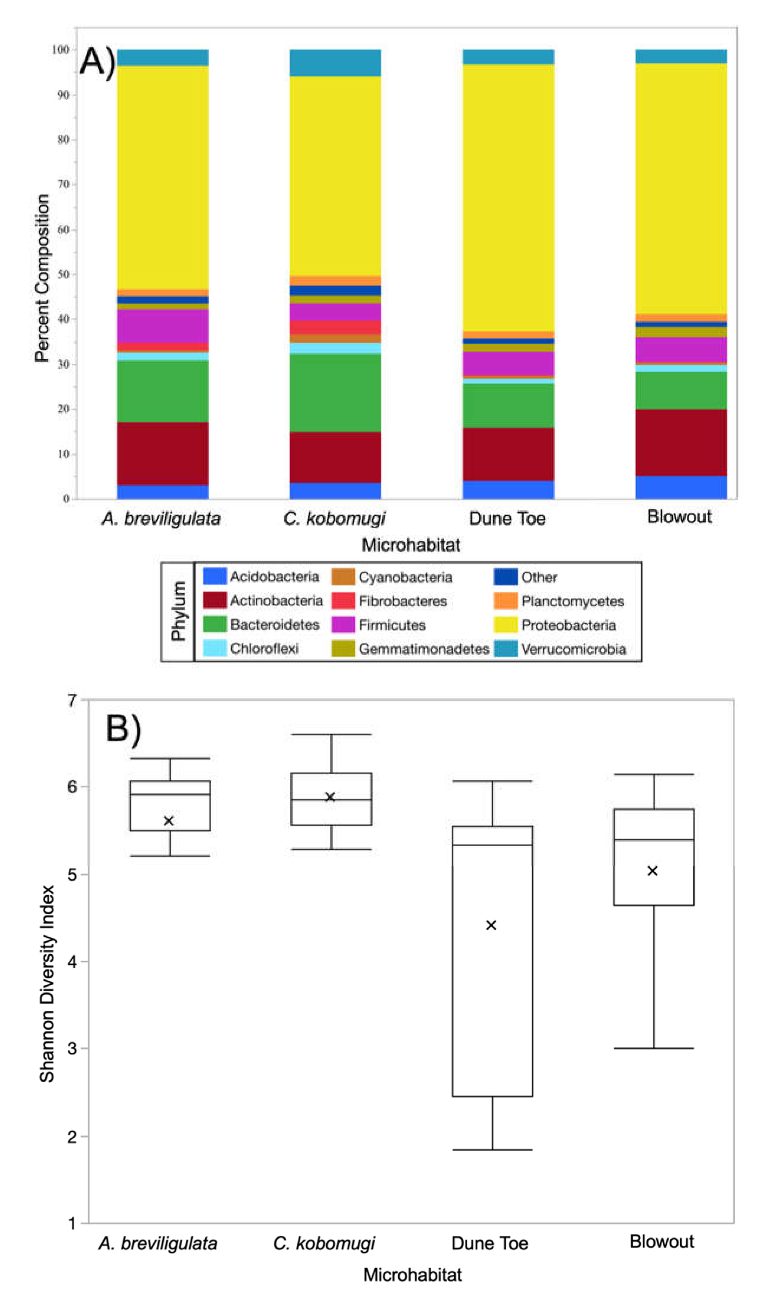
Figure 1. Phylum composition and diversity across microhabitats. (A) Phylum composition across microhabitats varied, (B) as did Shannon diversity, whereby the two vegetated habitats are more similar to each other than to either barren microhabitat.
Diversity 13 00525 g001
Figure 2. Principal components analysis of dune microhabitats (blowout (OP), A. breviligulata (A), C. kobomugi (CK), and foredune toe (F)) microbiomes based on percentage community composition at the phylum level. All the samples collected in this study are shown based on their respective microhabitat (A) and with arrows representing the vectors of factor loadings (B). The percent variation explained by the PCs of the principal components analysis is indicated on the axes.
Diversity 13 00525 g002
Figure 3. Principal components analysis of the four dune microhabitat (blowout (OP), A. breviligulata (A), C. kobomugi (CK), and foredune toe (F)) microbiomes based on percentage community composition at the phylum level. Analysis was performed separately for sites A17 (A), A19 (B), and A21 (C), with arrows representing the vectors of factor loadings of key genera (DF, respectively). The percent variation explained by the PCs of each principal components analysis is indicated on the axes.
Diversity 13 00525 g003
Figure 4. Principal components analysis of dune microhabitat (blowout (OP), A. breviligulata (A), C. kobomugi (CK), and foredune toe (F)) microbiomes based on percentage community composition at the genus level. All the samples collected in this study are shown based on their respective microhabitat (A) and with arrows representing the vectors of factor loadings (B). The percent variation explained by the PCs of the principal components analysis is indicated on the axes.
Diversity 13 00525 g004
Figure 5. Genera with the largest average fold changes in invasive C. kobomugi stands (A) and barren microhabitats (B) when compared to each other. Left panel represents average log fold increases across all sites, while the right panel represents log fold increase for sites A17, A19 and A21. A log fold increase of 0.301 (2-fold increase) is shown in a segmented line for reference.
Diversity 13 00525 g005
Figure 6. Genera with the largest average fold changes in native A. breviligulata stands (A) and barren microhabitats (B) when compared to each other. The left panel represents average log fold increases across all sites, while the right panel represents log fold increase for sites A17, A19, and A21. A log fold increase of 0.301 (2-fold increase) is shown in a segmented line for reference.
Diversity 13 00525 g006
Figure 7. Genus and phylum spatial patterns using PC1 and PC2 scores from PCA analyses. Relationships are significant except phylum PC1.
Diversity 13 00525 g007
Figure 8. OTU diversity decreases moving inland from the crest. This relationship is a trend including eight highlighted outliers and significant with them removed. The eight outliers are explicitly from A. breviligulata stands (AB) and blowout (OP) microhabitats.
Diversity 13 00525 g008
Table 1. Results of pairwise ANOSIM of community composition. R values were derived from Bray–Curtis similarity matrices using 9999 permutations.
Pairwise ComparisonR Valuep Value
A. breviligulata versus C. kobomugi0.088660.0466
A. breviligulata versus Barren0.068210.0231
C. kobomugi versus Barren0.22940.0005
Table 2. Percent composition of the topmost prevalent genera varied across the two plant species, native A. breviligulata (AB) and invasive C. kobomugi (CK) and the microhabitats lacking vegetation (Barren). Means are ± standard error. Asterisk (*) indicate P-values below 0.05.
Microhabitat % CompositionStatistical Result
GenusABCKBarrenANOVATukey HSD
Devosia0.86 ± 0.140.90 ± 0.090.24 ± 0.05F2,85 = 35.88, p < 0.0001 *(AB = CK) > Barren
Fibrobacter2.24 ± 0.423.09 ± 0.710.17 ± 0.03F2,85 = 42.64, p < 0.0001 *
Mesorhizobium0.76 ± 0.110.91 ± 0.090.41 ± 0.07F2,85 = 14.01, p < 0.0001 *
Mucilaginibacter0.72 ± 0.170.79 ± 0.190.13 ± 0.01F2,85 = 24.13, p < 0.0001 *
Ohtaekwangia0.86 ± 0.260.60 ± 0.150.10 ± 0.04F2,85 = 19.87 p < 0.0001 *
Sphingobacterium0.90 ± 0.100.86 ± 0.070.50 ± 0.05F2,85 = 13.10, p < 0.0001 *
Bacteriovorax0.44 ± 0.110.24 ± 0.070.16 ± 0.03F2,85 = 4.80, p = 0.01 *AB > Barren
Flavisolibacter0.75 ± 0.180.42 ± 0.080.22 ± 0.03F2,85 = 10.23, p < 0.0001 *
Cytophaga1.15 ± 0.242.86 ± 0.540.09 ± 0.01F2,85 = 74.78, p < 0.0001 *CK >AB > Barren
Streptomyces1.10 ± 0.322.12 ± 0.340.35 ± 0.04F2,85 = 24.04, p < 0.0001 *
Ktedonobacter0.01 ± 0.000.46 ± 0.260.00 ± 0.00F2,85 = 6.65, p = 0.002 *CK > (AB = Barren)
Opitutus0.66 ± 0.071.15 ± 0.130.50 ± 0.05F2,85 = 19.28, p < 0.0001 *
Acidobacterium2.63 ± 0.312.50 ± 0.233.75 ± 0.30F2,85 = 4.40, p = 0.02 *Barren > CK
Bacillus1.34 ± 0.200.79 ± 0.192.14 ± 0.31F2,85 = 6.88, p = 0.002 *
Publisher’s Note: MDPI stays neutral with regard to jurisdictional claims in published maps and institutional affiliations.